Chapter 9Peptide Design
This chapter presents methods focusing on peptide design. The three areas of focus are peptide backbone reconstruction from a subset of atom coordinates [1], design of peptides to target transmembrane -helices [6], and foldamers [7].
Peptides
Peptides are “short” sequences of amino acids that are linked by peptide bonds. They are important for many biological processes and experiments. One important use of peptides is to create antibodies against full proteins. Peptides can be synthesized and then inserted into an organism to make antibodies against that peptide, resulting in antibodies against proteins containing that peptide sequence. Peptides also act as protein ligands and can be used to probe protein: protein interactions (PPIs). Finally, there are peptides that have antimicrobial properties and have been shown to kill bacteria and other target cells.
It will be useful to clarify the distinction between (a) inhibitors of enzymatic activity versus inhibitors of PPIs, and (b) peptide inhibitors versus small organic molecule inhibitors. While numerous inhibitors of enzymatic activity have been developed, the development of inhibitors of protein:protein interactions has only recently come to the forefront as a viable approach. For example, an approximation to the algorithm (chapter 12) was used by Gorczynski et al. [3] to predict the free energy of protein:ligand interactions to develop novel allosteric small-molecule inhibitors of the binding of two leukemia-associated proteins. Allosteric inhibition of such PPIs presents a number of advantages, including not having to compete for binding with the partner protein. However, there are very few previous examples of such inhibitors. The inhibitors of Gorczynski et al. [3] block the binding of RUNX1 to , two proteins whose translocations play a critical role in the development of acute myeloid leukemia and acute lymphocytic leukemia. Treatment of human leukemia cell lines with these inhibitors resulted in changes in morphology indicative of increased differentiation versus untreated cells, providing support for this as a potential therapeutic approach. See chapter 8 for a discussion of .
While the study of [3] designed small organic molecules to inhibit PPIs, in this chapter we focus on the design of peptides, which also show considerable promise for inhibition of PPIs.
Furthermore, the design of small peptides builds on the algorithms for the design of proteins, which form a major computational theme in this book (see chapters 10–12).
There is much to gain from understanding peptide interactions and peptide structure. By understanding peptide structure, general rules could be abstracted out that could be applied to protein folding. Pharmaceuticals could be rationally designed not only to take advantage of known peptide properties, but also exhibit new physicochemical characteristics. Also, designed peptides could be used to create novel protein binders and inhibitors.
Peptide Backbone Reconstruction
Problem Statement
The goal of peptide backbone reconstruction (PBR) is to take as input the coordinates of one type of protein backbone atoms ( or ) and output the all-atom representation of the protein backbone. Figure 9.1 is a schematic cartoon where the atoms are shown on the left and the all-atom backbone representation of the protein is on the right (see plate 3).
Motivation
There are several current uses for PBR. PBR can be used to enhance low-resolution protein structures. In crystallography, certain atoms scatter better and have a better-known position. By using only those atoms, PBR could potentially be used to enhance the resolution of those structures. PBR could also be used to convert coarse-grained structures into all-atom models. Programs for ab initio folding and homology modeling often only output the coordinates for a protein, making PBR applicable. Normal mode analysis (chapters 21, 23, and 48) also generally coarse grains its model to incorporate only atoms, so PBR could be applied when analyzing mode motions.
There are several methods for solving PBR [1]. Some methods use collected fragment libraries and construct the backbone by ranking structures based on energy, homology, or geometry. Other methods perform de novo construction based on statistical position data, molecular dynamics, or
Figure 9.1 (plate 3)
Schematic of going from only atoms to a full backbone model (PDB id: 1DUR). Credit: Kyle Roberts.
Monte Carlo simulations. Unfortunately, none of these current methods can rebuild a structure from the atoms, but only from the atoms.
Algorithm
The algorithm uses dead-end elimination (DEE) and a library of peptide sequences to find the all-atom backbone global energy minimum conformation (GMEC) using a database-derived empirical force field [1]. The algorithm takes as input a library of amino acid peptide sequences and a set of or coordinates. It then overlays all of the peptides in the library onto every input coordinate. Then DEE is used to prune backbone peptides until a GMEC is obtained.
Backbone Fragments In order to create the peptide backbone fragment library, three-residue backbone fragments were taken from 1,336 random nonredundant PDB structures. All the fragments were clustered by RMSD and duplicates were discarded.
Fragment Overlap Fragments were overlapped with the input coordinates by minimizing the sum of squared distances. Kearsley’s method [4] was adopted, using quaternion algebra to turn this problem into an eigenvalue problem. The advantages to this approach are that the method is not iterative, improper rotations are not produced, and no special cases need to be handled.
Dead-End Elimination Dead-end elimination [5] was used to prune the backbone conformations until one minimum energy conformation was left. The energy function minimized was
where is residue with the backbone conformation . The original and simple DEE criteria were used in both the singles and pair criteria in order to prune conformations. DEE is discussed further in chapters 11, 12.
Database Force Field The pairwise energy function used was developed by noting that there is a relationship between the energy between two atom types at a given distance and their probability of occurring at that distance. Thus, there is a relationship between the energy and the radial distribution function. The radial distribution function for all pairwise atom type combinations was approximated by analyzing many structures in the PDB.
Structure Generation Structures were generated by pruning the aligned peptide backbone fragments using DEE until the GMEC was obtained.
Results
The algorithm was able to obtain structures with RMSDs from the crystal structure ranging from 0.2 to 0.6 Å. The - angle correlation was on average 0.95 and 0.88, respectively. Also, the computation could be completed in minutes for a protein. When using coordinates as input, the RMSD was always worse than using coordinates. The author attributed this to less constraint at the position. He found that increasing the fragment length could increase the accuracy of the structures.
Peptides That Target Transmembrane Helices
The idea of Yin et al. [6] was to develop a peptide -helix that could insert into the membrane and bind to a target membrane -helix [6]. Some methods exist to design proteins that bind water-soluble proteins, but this problem is much harder since less is known about membrane proteins.
Algorithm
- Choose the target alpha helix sequence. In Yin et al. [6] the integrin IIb helix was chosen.
- Find templates in the PDB that have similar motifs to the target helix. The IIb helix was known to have a small- -small motif and a right-handed crossing angle. Helix-helix dimers in the PDB were found with this motif and crossing angle to be used as design templates.
- Thread the target sequence onto one of the template helices. All the amino acid identities of one of the template helices (the “target” helix) were changed to the target sequence (the sequence of IIb). These amino acids are shown in green in the middle of figure 9.2 (plate 4).
- Choose positions that will be mutated on “anti” helix. Find positions on the other template helix that are close to the helix-helix interface to be considered for redesign. These amino acid positions to be mutated are shown in pink in the middle of figure 9.2 (plate 4).
- Mutate those positions to all rotamers and repack. The “anti” peptide was designed with Monte Carlo simulated annealing. At each step, one residue identity is changed and then the rotamers are optimized with DEE. The energy is computed using a Lennard-Jones potential and membrane depth-dependent knowledge-based potential.
Figure 9.2 (plate 4)
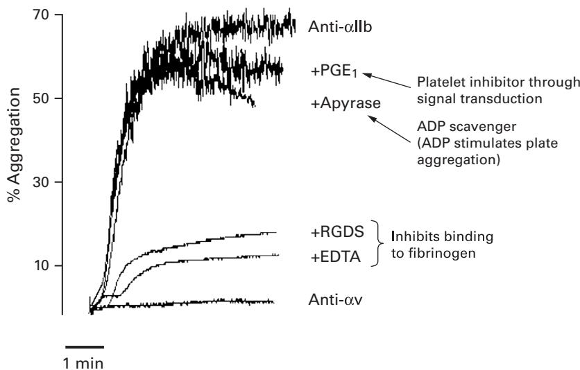
Figure 9.3 Percent platelet aggregation monitored over time with exposure to either anti- IIb (peptide designed for the target helix) or anti- (peptide designed for a homologous target helix) [6].
Results
The authors did many experiments to validate that their designed peptide helix was binding to the target helix. Fluorescence resonance energy transfer was used to confirm that the helices were binding in micelles. TOXCAT was used to analyze the specificity and affinity of the designed peptide for the target peptide. Very good specificity was obtained, and the binding interface seemed to be what they predicted it would be.
One experiment that summarizes these good results is presented in figure 9.3. The role of the target integrin used in this study is to promote platelet aggregation through binding of fibrinogen. This process is naturally induced by ADP. Thus, since the binding of the designed peptide to the target integrin helix would cause the integrin to activate, this binding should promote platelet aggregation as well. As can be seen in figure 9.3, when exposed to anti- IIb, platelets aggregate confirming that the designed peptide promotes aggregation. The PGE and Apyrase controls show that it is indeed the designed helix causing the aggregation. The RGDS and EDTA controls show that the designed peptide is working as hypothesized. Also, note that the design process is very specific because another designed peptide for a similar target helix () causes no aggregation at all.
Foldamers
Proteins and RNA are special polymers, in that they adopt a specific compact stable conformation. But biology has been fairly constrained to utilizing only certain monomers to create these structures. In theory, new compounds with stable compact conformations could be created using a variety of monomers. This idea of finding new molecules with compact structure is the study of foldamers. A foldamer is “any polymer with a strong tendency to adopt a specific compact conformation” [2].
To create these new polymers with compact conformations, there are three goals that must be achieved. First, new backbone units with suitable folding propensities must be found to be used as monomers. Second, these new backbone units must be given interesting chemical functions. Without interesting function they are of little use biologically. Finally, in order to take full advantage of these foldamers and analyze their properties they need to be produced efficiently.
There are two main uses for foldamers [7]. First, foldamers can be used to test our understanding of protein function. It is possible that our knowledge of proteins has been overfit since we only observe -amino acids. Creating foldamers makes it possible to test whether or not our knowledge can be generally applied to other systems. Second, foldamers use different building blocks so they could potentially be used for the design of new pharmaceuticals, diagnostic agents, and catalysts.
Types of Monomer Frameworks
At a high level, monomers for the foldamer framework can be classified into two categories: aromatic and aliphatic (see figure 9.4).
Foldamer Structure
Much analysis has been done on foldamer secondary structure, especially for foldamers incorporating -amino acids. Foldamers incorporating -amino acids will form a helix and have hydrogen-bonding patterns similar to -helices. The composition of the foldamer (incorporation of or or both types of amino acids) determines the number of atoms per H-bond ring and the number of residues in the hydrogen-bonding helix spacing as well as the helix polarity [7] (figure 9.5, plate 5).
It turns out that most of our developed intuition for -amino acids can be transferred to structures with different monomers. For example, adding salt bridges spaced one turn apart will stabilize -peptides. Also, charged side-chains at the helix ends will stabilize the helix corresponding to the helix’s polarity as observed in -helices.
There has been very limited success in designing foldamer tertiary structure. In one case, a zinc fingerlike motif was built consisting of -peptides with a hairpin and 14-helix that could both bind zinc. Also, an octomer was created consisting of -peptide helices, which maintained the tertiary structure using only noncovalent interactions.
Foldamer Function
There have been many promising designs of foldamers with biological functions. Foldamers were created that could penetrate bacterial cells in a passive process. By varying the length and amphilicity of these helices, much insight can be gained about the function of antimicrobial peptides. Many peptides were successfully designed to interrupt protein-protein interactions with very good , even to the nanomolar range. Finally, a foldamer peptide using a combined -amino acid sequence was found to disrupt the Bak-Bcl- interaction with tenfold higher affinity than the native peptide. This is remarkable because previous efforts using only -amino acid ligands failed.
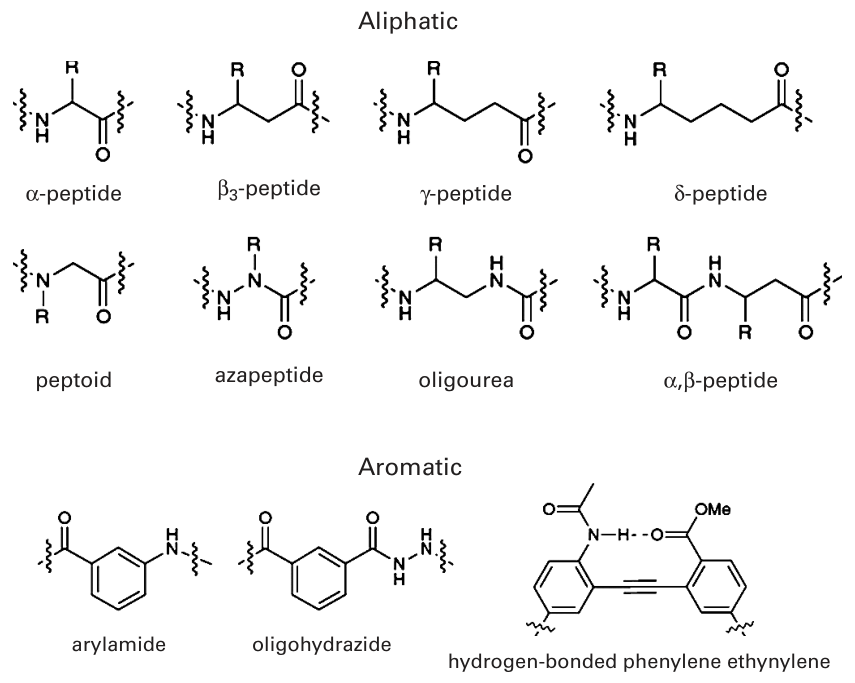
Figure 9.4 Monomers for foldamer framework are classified into two categories: aliphatic and aromatic [7]. Reprinted by permission from Macmillan publishers Ltd.
Foldamer Benefits
Foldamers are more resistant to enzymatic attack than peptides, because the monomers used are non-natural. Hence, enzymes such as proteases are not designed to efficiently break them down. Also, fewer monomeric units for foldamers are needed than for -amino acid compounds to adopt a well-defined secondary structure. This means that synthetically making these structures could be simpler, but they still should have the needed biological function. Finally, since many backbone monomers exist for foldamers, these backbone monomers can be used as an efficient way to downsize peptides into small molecules, which generally make better drugs.
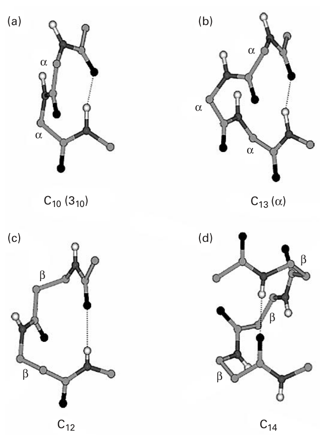
Figure 9.5 (plate 5) Secondary structure of and amino acid helices [8]. Reprinted with permission. Copyright 2005 American Chemical Society.
References
- [1] Stewart A. Adcock. Peptide backbone reconstruction using dead-end elimination and a knowledge-based forcefield. Journal of Computational Chemistry 25;1 (2004): 16–27.
- [2] S. H. Gellman. Foldamers: A manifesto. Acc. Chem. Res. 31(1998): 173–180.
- [3] M. J. Gorczynski, J. Grembecka, Y. Zhou, Y. Kong, L. Roudaiya, M. G. Douvas, et al. Allosteric inhibition of the protein-protein interaction between the leukemia-associated proteins RUNX1 and CBF . Chemistry & Biology 14; 10 (2007).
- [4] Simon K. Kearsley On the orthogonal transformation used for structural comparisons. Acta Crystallographica A45: (1989) 208–210.
[5] J. Desmet, M. De Maeyer, B. Hazes, and I. Lasters, The Dead End Elimination Theorem and its use in protein side-chain positioning. Nature 356(1992): 539–542.
[6] H. Yin, J. S. Slusky, B. W. Berger, R. S. Walters, G. Vilaire, R. I. Litvinov, et al. Computational design of peptides that target transmembrane helices. Science 315;5820 (2007 Mar 30): 1817–1822.
[7] C. M. Goodman, S. Choi, S. Shandler, and W. F. DeGrado. Foldamers as versatile frameworks for the design and evolution of function. Nature Chemical Biology 3;5 (2007 May): 252–262.
[8] K. Ananda, P. G. Vasudev, A. Sengupta, K. M. Raja, N. Shamala, and P. Balaram. Polypeptide helices in hybrid peptide sequences. Journal of the American Chemical Society, 127;47 (2005 Nov 30): 16668–16674.