Chapter 28Kinetic Studies of the Initial Module PheATE of Gramicidin S Synthetase
In this chapter, we study the kinetics of the initiation module PheATE (GrsA) of gramicidin S synthetase mainly based on studies by Walsh and co-workers [3, 7, 8]. These enzymes are used repeatedly as examples in this book, and this chapter describes their kinetics, which is provided to give a more complete understanding of their function. It also provides a primer on some of the experimental methods used to validate computational enzyme designs.
Background
Peptidic-type products, including ribosomally synthesized peptides and nonribosomally synthesized peptides, constitute a large family of natural products that are widely used for developing therapeutic drugs with novel activities. The basic structure of nonribosomally synthesized peptides is constituted by joining several small molecule building blocks. Not only the 20 standard amino acids, but also a vast number of different molecules can be incorporated into the building blocks, which provides a huge amount of diversity. The synthesis of nonribosomal peptides is carried out by the nonribosomal peptide synthetases (NRPSs). These enzymes are modularly organized multidomain complexes, from which different domains catalyze the incorporation of each building block into the elongated peptide product. The antibiotic gramicidin S is synthesized by the gramicidin S synthetase, which contains two multimodular subunits in which the GrsA subunit recognizes and initiates the incorporation of the first amino acid Phe, while the GrsB subunit elongates the rest of 4 amino acids to form the peptide chain (see figure 12.1). The initial module of the gramicidin S synthetase (GrsA) contains three domains: the N-terminal adenylation (A) domain followed by a small thiolation (T) domain and a C-terminal epimerase (E) domain. The A domain recognizes and activates L-phenylalanine (L-Phe) by adenylation to form a covalently bound L-phenylalanyl-adenosine-5’-monophosphate diester (L-Phe-AMP). The T domain uses an HS-phosphopantetheine arm (Ppant) to carry the L-Phe-AMP to the E domain, where the L-Phe-S-4’-Ppant-acyl enzyme complex is epimerized to D-Phe-S-4’-Ppant-acyl enzyme, which can be recognized by the GrsB subunit. The ATE domain module provides a well-established model for the amino acid incorporation by the nonribosomal peptide synthetases. Nevertheless, how they specifically select Phe as the substrate remains unclear. Computational structure-based protein redesign provides an incisive tool to dissect this nonribosomal code (chapter 12).
In this lecture, we use the following terms to distinguish the apo from the holo form of the multifunctional enzyme: apoPheATE (without Ppant link to T domain), and holoPheATE (with Ppant link to T domain).
Binding of the Amino Acid Substrate to the A Domain of GrsA
The catalysis of adenylation by the A domain requires binding of magnesium, ATP, and amino acid substrate into the binding pocket. Magnesium coordinates the binding of the ATP, which may induce a conformational change to facilitate association of the amino acid substrate. To determine the equilibrium binding constants, a fluorescence titration method was used to monitor the change of the fluorescence signal, which is proportional to the concentration of the substrate-enzyme complex. Percentage of fluorescence difference () was measured as a function of substrate concentration [ ]. The dissociation constant () can be calculated from the slope by plotting against or by directly fitting into the equation:
The fluorescence signal decreases as more substrate binds to the enzyme and the graph shows a hyperbolic curve. The results show similar values with L-Phe and D-Phe, which suggest that the binding pocket can accommodate both stereoisomers equally well. The results also indicate that the binding pocket is large enough to accommodate most other amino acids, and is not very sensitive to the size and charge of the side-chains.
Aminoacyl-AMP Formation Catalyzed by the A Domain
Both steady-state and pre-steady-state kinetics were used by Luo and Walsh [8] to monitor the rate of adenylation. Michaelis-Menten constants , and were obtained by the steady-state methods while the pre-steady-state gave microscopic rate constant. The reaction follows a reversible kinetics model (reprinted from [3]):
The Steady-State Assays
ATP-PPi Exchange Assay The reactions are initiated by addition of -pyrophosphate. Radioactive phosphate is then incorporated into ATP by the reverse reaction. The formation of radioactive ATP is separated and counted for radioactivity and converted into initial velocity. Because of the reverse measurement of ATP, both the forward and reverse rates of reaction are included.
Continuous PPi Release Assay During the reaction process, inorganic pyrophosphate (PPi) is released into solution. The PPi release can be detected by employing a coupled enzyme, inorganic pyrophosphatase (PPi-ase), which converts PPi into Pi. The Pi product, together with 2-amino-6-mercapto-7-methylpurine ribonucleoside (MesG) are then catalyzed by purine nucleoside phosphorylase (PNP) to form 2-amino-6-mercapto-7-methylpurine, which has an absorbance at 360 nm. The initial velocity is calculated by monitoring the absorbance at 360 nm during the reaction.
Both methods show similar catalytic efficiency toward L-Phe and D-Phe but at least 10-fold less efficiency toward L-Trp, L-Tyr, and L-Leu. The results also show that apoPheATE and holoPheATE enzyme have similar catalytic efficiency of adenylation. The ATP-PPi exchange assay measures both forward and reverse reaction, which doesn’t represent the true rate of the reaction we want to measure. However, it is still the traditional way to compare the adenylation activity. The continuous assay is limited by the slower off-rates when the a.a.-AMP intermediate occupies the active site of the enzyme during multiple turnover.
The Pre-Steady-State Assay
Single turnover methods were used to determine the rate of transient kinetics. In order to approach the single turnover condition, excess enzyme is used to saturate the substrate so that each substrate can undergo only one turnover during catalysis. Because of the fast rate, a rapid-quench flow apparatus is used to rapidly quench the reaction with millisecond timescale. Radiolabeled amino acid substrate is used and converted to radioactive aminoacyl-AMP (a.a.-AMP) product. A cellulose thin-layer chromatography (TLC) plate is used to separate the product from the substrate and the amount of radioactivity was counted and converted to real concentration of each species. The microscopic kinetic constants ( and ) are thus obtained.
The observed rate reflects the rate of the catalytic step and is rate limiting. The rate constants for a.a.-AMP formation are reduced 0.7-fold for D-Phe-AMP, 2.5-fold for L-Trp-AMP, 910-fold for L-Tyr-AMP, and 20-fold for L-Leu-AMP compared to that of L-Phe-AMP.
Loading of the Amino Acid Substrate to the T Domain
After formation of the aminoacyl-AMP, the intermediate can be recognized and loaded to the T domain by attaching to the cofactor 4’-phosphopantetheine (Ppant). Because the amino acid substrate now is covalently bound to the T domain, radioactivity can be detected by trichloroacetic acid (TCA) precipitation to separate substrate, a.a.-AMP and a.a.-PheATE complex. The results showed that 88% of the L-Phe was attached to the T domain during thiolation. Among the 17 amino acids tested, L-Phe, D-Phe, L-Trp, L-Tyr and L-Leu showed significant loading on the T domain. The schematic diagram shows the reversible reaction model for loading of the aminoacyl-AMP to the T domain with the release of AMP (Reprinted from [3]):
Epimerization of the L-Form Substrate-Enzyme Complex to D-Form by the E Domain
The final step is to convert the T domain-attached L-form amino acid to a D-form amino acid. This is done by the epimerase domain (E domain). The 4’-phosphopantetheine cofactor carries the L-form amino acid from the A domain to the E domain for its epimerization. After the reaction, D-form amino acid is attached to the 4’-phosphopantetheine cofactor on the T domain. The reaction assay was done in the same manner as the single turnover assay for adenylation but the holoPheATE was used to ensure the whole process was complete. The reaction was rapidly quenched at varying times. Protein with both L-form and D-form amino acid attached to it was TCA precipitated to be separated from substrate and a.a-AMP intermediate. Chiral TLC was used to distinguish L-and D-form amino acid from the protein pellet while TLC was used to separate substrate from a.a-AMP in supernatant. The schematic diagram shows the model of the reversible reaction for the whole reaction pathway (Reprinted from [3]):
Free Energy Profiles for HoloPheATE Catalysis
The microscopic rate constants and the thermodynamic equilibrium constants can be used to calculate the energy for the three catalysis steps. Free energy is calculated using the following equation:
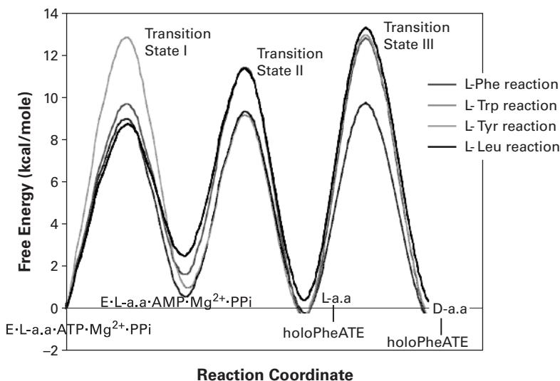
Figure 28.1 (plate 32) Reaction free energy profiles for holoPheATE-catalyzed adenylation, thiolation, and epimerization reactions using L-Phe, L-Trp, L-Tyr, and L-Leu as the starting amino acid substrate [3]. Reprinted with permission. Copyright 2001 American Chemical Society.
Here, is Planck’s constant. The equilibrium constants () are used to calculate the ground-state free energy and the rate constants are used to calculate the transition-state free energy. The constructed energy profiles show an energy barrier selective for the noncognate amino acids in the adenylation and the epimerization steps. When compared to the native substrate L-Phe, the first two steps show a net selectivity of 1/1200 for L-Tyr, 1/608 for L-Trp, and 1/245 for L-Leu. The E domain contributes another major selectivity with 1/300 for L-Tyr-S-enzyme, 1/545 for L-Trp-S-enzyme, and 1/245 for L-Leu-S-enzyme (figure 28.1, plate 32).
References
- [1] J. Grunewald and M. A. Marahiel. Chemoenzymatic and template-directed synthesis of bioactive macrocyclic peptides, Microbiol Mol Biol Rev 70(2006):121–146.
- [2] T. Stachelhaus and M. A. Marahiel. Modular structure of peptide synthetases revealed by dissection of the multifunctional enzyme GrsA, Journal of Biological Chemistry 270(1995):6163–6169.
- [3] L. Luo, M. D. Burkart, T. Stachelhaus, and C. T. Walsh. Substrate recognition and selection by the initiation module PheATE of gramicidin S synthetase. Journal of the American Chemical Society 123(2001):11208–11218.
[4] R. H. Lilien, B. W. Stevens, A. C. Anderson, and B. R. Donald. A novel ensemble-based scoring and search algorithm for protein redesign and its application to modify the substrate specificity of the gramicidin synthetase a phenylalanine adenylation enzyme, Journal of Computational Biology 12(2005):740–761.
[5] E. Conti, T. Stachelhaus, M. A. Marahiel, and P. Brick. Structural basis for the activation of phenylalanine in the non-ribosomal biosynthesis of gramicidin S. EMBO Journal 16(1997):4174–4183.
[6] T. Stachelhaus, H. D. Mootz, and M. A. Marahiel. The specificity-conferring code of adenylation domains in nonribosomal peptide synthetases, Chemistry & Biology 6(1999):493–505.
[7] J. R. Lai, A. Koglin, and C. T. Walsh. Carrier protein structure and recognition in polyketide and nonribosomal peptide biosynthesis. Biochemistry 45(2006):14869–14879.
[8] L. Luo and C. T. Walsh. Kinetic analysis of three activated phenylalanyl intermediates generated by the initiation module PheATE of gramicidin S synthetase. Biochemistry 40(2001):5329–5337.