Chapter 8Energy, Enzymes, and Metabolism
PART THREE Cells and Energy
Key Concepts
- 8.1 Chemical Transformations Involve Energy and Energy Transfers
- 8.2 ATP Plays a Key Role in Biochemical Energetics
- 8.3 Enzymes Speed Up Biochemical Transformations
- 8.4 Enzymes Bring Substrates Together so Reactions Readily Occur
- 8.5 Enzyme Activities Can Be Regulated
Edward Stone’s letter to the Royal Society, 1763 This report ultimately led to the mass production of aspirin. More than 100 billion tablets are taken every year.
E. Stone, 1763, Philos Trans R Soc Lond 53: 195-200

Investigating Life: How Aspirin Works
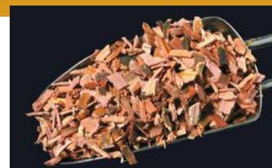
Despite suffering from the “ague,” the Reverend Edward Stone went walking in 1758 in the English countryside. Feverish, tired, with aching muscles and joints, he came across a willow tree. Although apparently unaware that many ancient healers such as the Greek physician Hippocrates in 400 BCE had used willow bark extracts to reduce fever, the clergyman knew of the tradition of natural remedies for various diseases. The willow reminded him of the bitter extracts from the bark of South American trees then being sold (at high prices) to treat fevers. Removing some willow bark, Stone sucked on it and found it did indeed taste bitter—and that it relieved his symptoms.
Later he gathered a pound of willow bark and ground it into a powder, which he gave to about 50 people who complained of fever and joint pain; all said they felt better. Stone reported the results of this “clinical test” in a letter to the Royal Society, England’s most respected scientific body. Stone had discovered salicylic acid, the basis of the most widely used drug in the world. The date of his letter (which still exists) was April 25, 1763.
The chemical structure of salicylic acid (named for Salix, the willow genus) was worked out about 70 years later, and soon chemists could synthesize it in the laboratory. Although the compound alleviated pain, its acidity irritated the digestive system. In the late 1890s, the German chemical company Bayer synthesized a milder yet equally effective form, acetylsalicylic acid, which it marketed as aspirin. The new medicine’s success launched Bayer to world prominence as a pharmaceutical company, a position it maintains today. Uses for this drug have expanded to include prevention of blood clotting in people after a heart attack. For several years it’s been popular for people without symptoms of heart abnormalities to take aspirin in low doses to prevent clots in arteries, but this use was recently proven to be unsupported by clinical trials.
Fever, joint pain, headache, blood clots: what do these symptoms have in common? They all are mediated by fatty acid products called prostaglandins and molecules derived from them. Salicylic acid blocks the synthesis of the primary prostaglandin. The biochemical mechanism by which aspirin works was described in 1971. As we will see, an understanding of this mechanism requires an understanding of protein and enzyme function—two subjects of this chapter.
QA How do anti-inflammatory drugs work as enzyme inhibitors? (Find the answer on page 171.)
© TH Foto/Alanny Stock Photo
Chemical Transformations Involve Energy and Energy Transfers
Learning Objectives
- 8.1.1 Apply the second law of thermodynamics to biological systems.
- 8.1.2 Differentiate between exergonic and endergonic reactions.
A chemical reaction occurs when atoms have sufficient energy to combine or change their bonding partners. Consider the hydrolysis of the disaccharide sucrose to its component monomers, glucose and fructose (see Figure 3.18):
In this equation, sucrose and water are the reactants, and glucose and fructose are the products. During the reaction, some of the bonds in sucrose and water are broken and new bonds are formed, resulting in products with chemical properties that are very different from those of the reactants. The sum total of all the chemical reactions occurring in a biological system at a given time is called metabolism. Metabolic reactions involve energy changes; for example, the energy contained in the chemical bonds of sucrose (reactants) is greater than the energy in the bonds of the two products, glucose and fructose.
Physicists define energy as the capacity to do work, which occurs when a force operates on an object over a distance. In biochemistry, it is more useful to think of energy as the capacity for change. In biochemical reactions, energy changes are usually associated with changes in the chemical compositions and properties of molecules.
There are two basic types of energy
Energy comes in many forms: chemical, electrical, heat, light, and mechanical (Table 8.1). But all forms of energy can be considered as one of two basic types (Figure 8.1):
Table 8.1 Energy in Biology
| Form of energy | Example in biology |
|---|---|
| Chemical-bond: Stored in bonds | Chemical energy stored in covalent bonds is released during the hydrolysis of polymers |
| Electrical: Separation of charges | Electrical gradients across cell membranes help drive the movement of ions through channels |
| Heat: Transfer due to temperature difference | Heat can be released by chemical reactions, and this can alter the internal temperature of an organism |
| Light: Electromagnetic radiation stored as photons | Light energy is captured by pigments in the eye and by plant pigments in photosynthesis |
| Mechanical: Energy of motion | Mechanical energy is used in muscle movements and movements within cells |
- 1. Potential energy is the energy of state or position—that is, stored energy. It can be stored in many forms: in covalent bonds, as a concentration gradient, or even as an electric charge imbalance.
- 2. Kinetic energy is the energy of movement—that is, the type of energy that does work, that makes things change. For example, heat causes molecular motions and can even break chemical bonds.
To produce a change, energy must be transformed, either by conversion from one form to another or from one place to another. Potential energy can be converted into kinetic energy and vice versa, and the form that the energy takes can also be converted. Think of reading this book: light energy is converted to chemical energy in your eyes, and then is converted to electrical energy in the nerve cells that carry messages to your brain. When you decide to turn a page, the electrical and chemical energy of nerves and muscles are converted to kinetic energy for movement of your hand and arm.
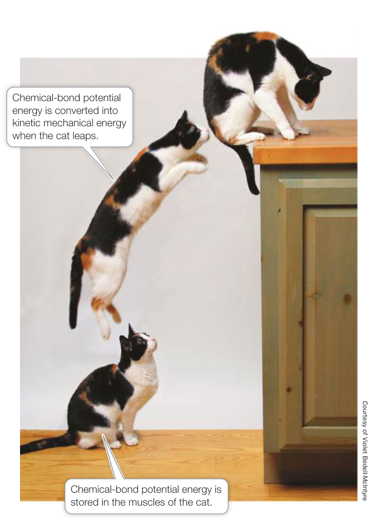
Figure 8.1 Energy Conversions and Work A leaping cat illustrates both the conversion between potential and kinetic energy and the conversion of energy from one form (chemical-bond) to another (mechanical). Q: What are the energy conversions that occur when a woman dives from a board into a pool, splashes, and begins swimming?
There are two basic types of metabolism
Energy changes in living systems usually occur mostly as chemical changes, in which energy is stored in, or released from, chemical bonds.
Anabolic reactions (collectively anabolism) typically link smaller molecules to form larger, more complex molecules. Anabolic reactions require an input of energy. Energy is captured in the chemical bonds that are formed (for example, the glycosidic bond between the two monosaccharides forming sucrose). This captured energy is stored in the chemical bonds as potential energy:
Catabolic reactions (collectively catabolism) break down larger, more complex molecules into smaller ones and often release the energy stored in the chemical bonds. For example, when sucrose is hydrolyzed, energy is released. In a biological system the released energy may be recaptured in new chemical bonds, or it may be used as kinetic energy—moving atoms, molecules, cells, or the whole organism:
Catabolic and anabolic reactions are often linked. The energy released in catabolic reactions is often used to drive anabolic reactions—that is, to do biological work. For example, the energy released by the breakdown of glucose (catabolism) is used to drive anabolic reactions such as the synthesis of triglycerides. This is why you can accumulate fat if you eat food in excess of your energy requirements.
The laws of thermodynamics (thermo, “energy,” + dynamics, “change”) were derived from studies of the fundamental physical properties of energy and the ways it interacts with matter. The laws apply to all matter and all energy transformations in the universe. Their application to living systems helps us understand how organisms and cells harvest and transform energy to sustain life.
The first law of thermodynamics: Energy is neither created nor destroyed
The first law of thermodynamics states that in any energy conversion, energy is neither created nor destroyed. In other words, during any conversion of energy—whether from one form to another or from one location to another—the total energy in the system under consideration before and after the conversion is the same (Figure 8.2A). As you will see in the next two chapters, the potential energy present in the chemical bonds of carbohydrates and lipids can be converted to potential energy in the form of adenosine triphosphate (ATP). This can then be converted into kinetic energy to do mechanical work (such as muscle contractions) or biochemical work (such as anabolism).
The second law of thermodynamics: Disorder tends to increase
Although energy cannot be created or destroyed, the second law of thermodynamics states that when energy is converted from one form to another, some of that energy becomes unavailable for doing work (Figure 8.2B). In other words, no physical process or
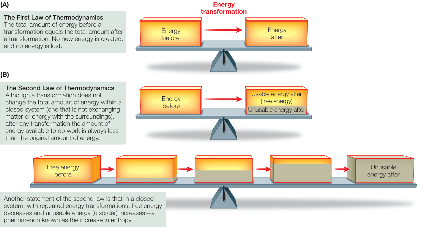
Figure 8.2 The Laws of Thermodynamics (A) The first law states that energy cannot be created or destroyed. (B) The second law states
that after energy transformations, some energy becomes unavailable to do work.
chemical reaction is 100% efficient; some of the released energy is lost to a form associated with disorder. Think of disorder as a kind of randomness that is due to the thermal motion of particles; this energy is of such a low value and so spread out that it is unusable.
Entropy is a measure of the disorder in a system.
It takes energy to impose order on a system. Unless energy is applied to a system, it will be randomly arranged or disordered. The second law applies to all energy transformations, but we will focus here on transformations in living systems. The second law applies on the micro scale, in chemical reactions and pathways in the cell. But it also applies on the macro scale, in communities of organisms interacting in the environment.
Connect the Concepts See Key Concept 56.3, which discusses the flow of energy in food chains, where there is a decline in usable energy in each step.
NOT ALL ENERGY CAN BE USED In any system, the total energy includes the usable energy that can do work and the unusable energy that is lost to disorder:
In biological systems, the total energy is called enthalpy (). The usable energy that can do work is called free energy (). Free energy is what cells require for all the chemical reactions involved in growth, cell division, and maintenance. The unusable energy is represented by entropy () multiplied by the absolute temperature (). Thus we can rewrite the word equation above more precisely as:
Because we are interested in usable energy, we rearrange Equation 8.1:
Although we cannot measure , , or absolutely, we can determine the change in each at a constant temperature. Such energy changes are measured in calories (cal) or joules (J).1 A change is represented by the Greek letter delta (). The change in free energy () of any chemical reaction is equal to the difference in free energy between the products and the reactants:
A change in free energy can be either positive or negative; that is, the free energy of the products can be more or less than the free energy of the reactants. If the products have more free energy than the reactants ( is positive), then there must have been some input of energy into the reaction. (Remember that energy cannot be created, so some energy must have been added from an external source.)
At a constant temperature is defined in terms of the change in total energy () and the change in entropy ():
Equation 8.4 tells us whether free energy is released or required by a chemical reaction:
- If is negative (), free energy is released.
- If is positive (), free energy is required.
If the necessary free energy is not available, the reaction does not occur. The sign and magnitude of depend on the two factors on the right side of the equation:
- : In a chemical reaction, is the total amount of energy added to the system () or released ().
- : Depending on the sign and magnitude of , the entire term, , may be negative or positive, large or small. In the chemistry lab, temperature can affect . But living systems don’t vary their temperature that much, so the magnitude and sign of can depend on changes in entropy.
If a reaction increases entropy, its products are more disordered or random than its reactants. For example, when a protein is hydrolyzed to its amino acids, the products have considerable freedom to move around. The disorder in a solution of amino acids will be large compared with that in the protein, in which peptide bonds and other forces prevent free movement. So in hydrolysis, the change in entropy () will be positive. Conversely, if there are fewer products and they are more restrained in their movements than the reactants (as for amino acids being joined in a protein), will be negative. This means that according to Equation 8.4, is positive and energy input is required.
DISORDER TENDS TO INCREASE Consider the human body, with its highly organized tissues and organs composed of large, complex molecules. You might think that this order and complexity are in conflict with the second law—after all, entropy, or disorder, should be maximized—but they are not, for two reasons:
- Getting ordered is coupled to the generation of disorder. Making 1 kg of a human body (soft tissues, not bones) requires the catabolism of about 10 kg of highly ordered biological materials (our food), which are converted into , , and other simple molecules. So this process creates far more disorder (more energy is lost to entropy in the small molecules) than the amount of order (total energy; enthalpy) stored in large molecules in 1 kg of a person.
- Life requires a constant input of energy to maintain order. Without this energy, the complex structures of living systems would break down. Because energy is used to generate and maintain order, there is no conflict with the second law of thermodynamics.
Having seen that the laws of thermodynamics apply to organisms, let’s see how these laws apply to biochemical reactions inside the cell.
Chemical reactions release or consume energy
As you saw earlier, anabolic reactions link simple molecules to form more complex molecules, so they tend to increase complexity (order) in the cell. By contrast, catabolic reactions break down
1A calorie is the amount of heat energy needed to raise the temperature of 1 gram of pure water from 14.5°C to 15.5°C. In the SI system, energy is measured in joules. 1 J = 0.239 cal; conversely, 1 cal = 4.184 J. Thus, for example, 486 cal = 2,033 J, or 2.033 kJ. Although they are defined here in terms of heat, the calorie and the joule are measures of mechanical, electrical, or chemical energy. When you compare data on energy, always compare joules with joules and calories with calories.
complex molecules into simpler ones, so they tend to decrease complexity (generate disorder).
- Catabolic reactions may break down an ordered reactant into smaller, more randomly distributed products. Reactions that release free energy () are called exergonic reactions (Figure 8.3A). For example:
Complex molecules free energy + small molecules
- Anabolic reactions may make a single product (a highly ordered substance) out of many smaller reactants (less ordered). Reactions that require or consume free energy () are called endergonic reactions (Figure 8.3B). For example:
Free energy + small molecules complex molecules
In principle, chemical reactions are reversible and can run both forward and backward. For example, if compound A can be converted into compound B (), then B, in principle, can be converted into A (), although the concentrations of A and B
determine which of these directions will be favored. You can think of the overall reaction as resulting from competition between the forward and reverse reactions (). According to the law of mass action, increasing the concentration of A makes the forward reaction () happen more often than the reverse reaction, just as increasing the concentration of B favors the reverse reaction ().
There are concentrations of A and B at which the forward and reverse reactions take place at the same rate. At these concentrations, no further net change in the system is observable, although individual molecules are still forming and breaking apart. This balance between forward and reverse reactions is known as chemical equilibrium. Chemical equilibrium is a state of no net change, and a state in which .
Chemical equilibrium and free energy are related
Chemical reactions do not necessarily proceed to completion, with all reactants converted into products. Each reaction has a specific equilibrium point, which is related to the relative free energy contents of the reactants and products. To understand the principle of equilibrium, consider the interconversion of glucose 1-phosphate and glucose 6-phosphate, a rearrangement of a phosphate group from one position on the ring of carbon atoms to another:
Unraveling what goes on chemically in the cell is made possible by that fact that in many cases, after a cell’s membrane has been ruptured to release its contents, the cell-free system with the cell contents (cytoplasm) in a test tube, usually in a buffered solution, has the same chemical properties as the intact cell. So the reaction can be studied in a test tube. Suppose we start out with an aqueous solution of glucose 1-phosphate that has a concentration of 0.02 M. (M stands for molar concentration; see Key Concept 2.4.) The solution is maintained under constant environmental conditions (25°C and pH 7). As the reaction proceeds to equilibrium, the concentration of the product, glucose 6-phosphate, rises from 0 M to 0.019 M, while the concentration of the reactant, glucose 1-phosphate, falls from 0.02 M to 0.001 M, at which point equilibrium is reached (Figure 8.4). At equilibrium, the reverse reaction, from glucose 6-phosphate to glucose 1-phosphate, progresses at the same rate as the forward reaction.
At equilibrium, then, this reaction has a product-to-reactant ratio of 19:1 (0.019/0.001), so the forward reaction has gone 95% of the way to completion (“to the right,” as written above). This result is obtained every time the experiment is run under the same conditions.
The change in free energy () for any reaction is related directly to its point of equilibrium. The further toward completion the point of equilibrium lies, the more free energy is released. In an exergonic reaction, is a negative number. The total value of also depends on the beginning concentrations of the reactants and products and other conditions such as temperature, pressure, and pH of the solution. Biochemists often calculate using standard laboratory conditions: 25°C, 1 atmosphere pressure, 1 molar (1 M) concentrations of the solutes, and pH 7. The standard free energy change calculated using these conditions is designated . In our example of the conversion of glucose 1-phosphate to glucose 6-phosphate, kcal/mol, or kJ/mol.
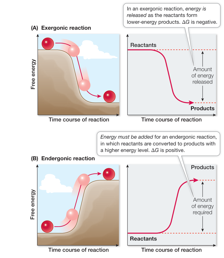
Figure 8.3 Exergonic and Endergonic Reactions (A) In an exergonic reaction, the reactants behave like a ball rolling down a hill, and energy is released. (B) A ball will not roll uphill by itself. Driving an endergonic reaction, like moving a ball uphill, requires the addition of free energy.
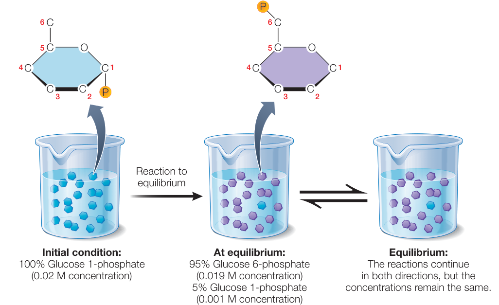
Figure 8.4 Chemical Reactions Run to Equilibrium No matter what quantities of glucose 1-phosphate and glucose 6-phosphate are dissolved in water, when equilibrium is attained, there will always be 95% glucose 6-phosphate and 5% glucose 1-phosphate. Q: What would happen in this system if, after formation, the glucose 6-phosphate was removed by a subsequent reaction? Figure 8.4 Chemical Reactions Run to Equilibrium No matter what quantities of glucose 1-phosphate and glucose 6-phosphate are dissolved in water, when equilibrium is attained, there will always be 95% glucose 6-phosphate and 5% glucose 1-phosphate. Q: What would happen in this system if, after formation, the glucose 6-phosphate was removed by a subsequent reaction?
A large, positive for a reaction means that it does not proceed to the right (A B). If the concentration of B is initially high relative to that of A, such a reaction runs “to the left” (A B), and at equilibrium nearly all of B is converted into A. A value near zero is characteristic of a readily reversible reaction: reactants and products have almost the same free energies.
In Chapters 9 and 10 you will learn the biochemical conversions that extract energy from food and light. In turn, this energy is used to synthesize carbohydrates, lipids, and proteins. All of the chemical reactions carried out by living organisms are governed by the principles of thermodynamics and equilibrium.
Recap and Assess
Two laws of thermodynamics govern energy transformations in biological systems. A biochemical reaction can release or consume energy, and it may not run to completion, but instead end up at a point of equilibrium.
- What is the difference between endergonic and exergonic reactions, and what is the importance of positive and negative ?
- What makes it possible for endergonic reactions to proceed in organisms?
The principles of thermodynamics that we have been discussing apply to all energy transformations in the universe, so they are very powerful and useful. Now let’s apply them to reactions in cells that involve the currency of biological energy, ATP.
ATP Plays a Key Role in Biochemical Energetics
Learning Objectives
- 8.2.1 Explain the characteristics of ATP that account for the high free energy released during its hydrolysis to form ADP and .
- 8.2.2 Analyze evidence to identify reactions as exergonic or endergonic.
- 8.2.3 Predict biochemical transformations that require coupling to ATP hydrolysis.
Cells use adenosine triphosphate (ATP) for the capture and transfer of the free energy they require to do chemical work. ATP operates as a kind of “energy currency.” Just as it is more efficient and convenient for you to trade money for a lunch in a café than to trade your actual labor, it’s useful for cells to have a single currency for transferring energy between different reactions and cell processes. So some of the free energy that is released by exergonic reactions is captured in the formation of ATP from adenosine diphosphate (ADP) and inorganic phosphate ( , which is commonly abbreviated to ). The ATP can then be hydrolyzed in the cell to release free energy to drive endergonic reactions.
ATP has another important role in the cell beyond its use as an energy currency: it can be converted into a building block for nucleic acids (see Chapter 4). The structure of ATP is similar to that of other nucleoside triphosphates, but two things about ATP make it especially useful to cells:
- ATP releases a relatively large amount of energy when hydrolyzed to ADP and .
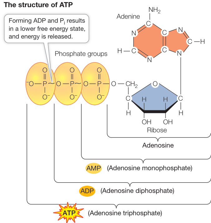
Figure 8.5 ATP ATP is richer in energy than ADP and AMP. The conversion of ATP to ADP releases energy.
- ATP can phosphorylate (donate a phosphate group to) many different molecules, which gain some of the energy that was stored in the ATP.
ATP (Figure 8.5) is a nucleoside triphosphate (see Figure 4.1). The hydrolysis of a molecule of ATP yields free energy, as well as ADP and an inorganic phosphate ion (). Thus:
The important property of this reaction is that it is exergonic, releasing free energy. Under standard laboratory conditions, the change in free energy () for this reaction is about kcal/mol ( kJ/mol). This change in free energy is negative because the chemical bonds in ATP and are weaker than the chemical bonds in ADP and . A molecule of ATP can be hydrolyzed either to ADP and or to adenosine monophosphate (AMP) and a pyrophosphate ion ( , commonly abbreviated as ). Two characteristics of ATP account for the free energy released by the loss of one or two of its phosphate groups:
- Because phosphate groups are negatively charged and so repel each other, it takes energy to get two phosphates near enough to each other to make the covalent bond that links them together. Some of this energy is stored as potential energy in the bonds between the phosphates in ATP (the wavy line indicates a high-energy bond).
- The free energy of this bond (called a phosphoanhydride bond) is much higher than the energy of the O—H bond that forms as a result of hydrolysis. So it is the lower free energy state of the system that releases energy.
Cells use the energy released by ATP hydrolysis to fuel endergonic reactions (such as the biosynthesis of complex molecules), for active transport, for movement, and even for the production of light (bioluminescence). Another interesting example of the use of ATP involves converting its chemical energy into light energy.
ATP couples exergonic and endergonic reactions
As you have just seen, the hydrolysis of ATP is exergonic and yields ADP, , and more free energy (or AMP, , and more free energy). The reverse reaction, the formation of ATP from ADP and , is endergonic and consumes as much free energy as is released by the hydrolysis of ATP:
Many exergonic reactions in the cell can provide the energy to convert ADP into ATP. For eukaryotes and many prokaryotes, the most important of these reactions is cellular respiration, in which some of the energy released from fuel molecules is captured in ATP. The formation and hydrolysis of ATP constitute what might be called an “energy-coupling cycle,” in which ADP picks up energy from exergonic reactions to become ATP, which then donates energy to endergonic reactions. ATP is the common component of these reactions and is the agent of coupling, as illustrated in Figure 8.6.
Coupling of exergonic and endergonic reactions is very common in metabolism. Free energy is captured and retained in the bonds of ATP, which is hydrolyzed in conjunction with another reaction. The free energy released drives an endergonic reaction. For example, the formation of glucose 6-phosphate from glucose (Figure 8.7), which has a positive (is endergonic), will not proceed without the input of free energy from ATP hydrolysis, which has a negative (is exergonic). The overall for the coupled reactions (when the two s are added together) is negative. Hence the reactions proceed exergonically when they are coupled by occurring at the same time and place, and glucose 6-phosphate is synthesized. As you will see in Chapter 9, this is the initial reaction in the catabolism of glucose.
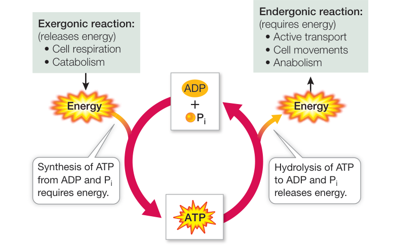
Figure 8.6 Coupling of Reactions Exergonic reactions in the cell release the energy needed to convert ADP to ATP. The energy released from the conversion of ATP back to ADP can be used to fuel endergonic reactions.
An active cell requires the production of millions of molecules of ATP per second to drive its biochemical machinery. From previous chapters, you may already be familiar with some of the activities in the cell that require energy from the hydrolysis of ATP:
- Active transport across a membrane (see Figure 6.14)
- Condensation reactions that use enzymes to form polymers (see Figure 3.4A)
- Modifications of cell signaling proteins by protein kinases (see Figure 7.15)
- Motor proteins that move vesicles along microtubules (see Figure 5.21)
An ATP molecule is typically consumed within a second of its formation. At rest, an average person produces and hydrolyzes about 40 kg of ATP per day—as much as some people weigh. This means that each ATP molecule undergoes about 10,000 cycles of synthesis and hydrolysis every day!
Recap and Assess
ATP is the “energy currency” of cells. Potential energy is stored in the P-O bonds of ATP. Some of the free energy released by exergonic reactions can be captured in the form of ATP. This energy can then be released by ATP hydrolysis and used to drive endergonic reactions.
- What chemical properties allow ATP to store energy?
- The following is a biochemical reaction that involves ATP:
Phosphoenolpyruvate
pyruvate
(
kcal/mol)
- Is the reaction exergonic or endergonic?
- Is ATP hydrolyzed or formed during the reaction?
ATP is synthesized and used up very rapidly. But these biochemical reactions—and nearly all the others that take place inside a cell—could not proceed so rapidly without the help of enzymes.
Enzymes Speed Up Biochemical Transformations
Learning Objectives
- 8.3.1 Describe a catalyst.
- 8.3.2 Describe how an enzyme–substrate complex forms.
- 8.3.3 Explain the relationship between an enzyme and the equilibrium point of the reaction it catalyzes.
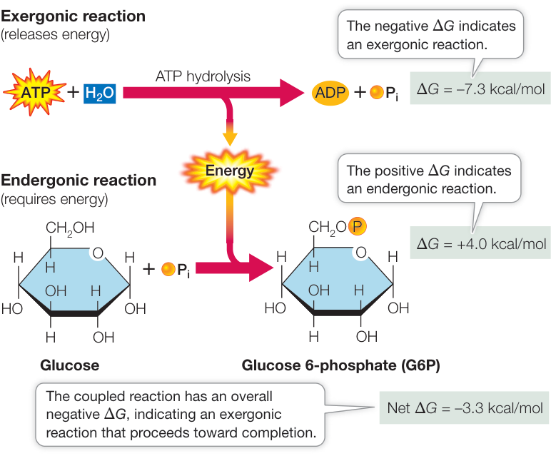
Figure 8.7 Coupling of ATP Hydrolysis to an Endergonic Reaction The addition of phosphate derived from the hydrolysis of ATP to glucose forms the molecule glucose 6-phosphate (in a reaction catalyzed by hexokinase). ATP hydrolysis is exergonic, and the energy released drives the second reaction, which is endergonic. Q: What would be the magnitude of the of a different conversion that could drive the synthesis of ATP?
When we know the change in free energy () of a reaction, we know where the equilibrium point of the reaction lies: the more negative the value is, the further the reaction proceeds to products and toward completion. We call such a reaction spontaneous. However, tells us nothing about the rate of a reaction—how fast it moves toward equilibrium. Most reaction rates in chemical transformations in living systems are slow—indeed, too slow to result in the rapid changes needed for an organism to respond to environmental changes and perform its cellular functions. In chemistry, catalysts are substances that speed up reactions without themselves being permanently altered. Often, a catalyst provides a surface on which to line up reactants so they will be more likely to collide and react than they would in solution. A catalyst does not cause a reaction to occur that would not proceed without it, but merely increases the rate of the reaction, allowing equilibrium to be approached more rapidly. This is an important point: No catalyst makes a reaction occur that cannot otherwise occur.
Most biological catalysts are proteins called enzymes. A biological catalyst is a molecular framework or scaffold within which chemical catalysis takes place. The enzyme binds the reactants and sometimes participates in the reaction itself; however, such participation does not permanently change the enzyme. That’s the hallmark of any catalyst: it ends up in exactly the same chemical condition after a reaction as before it.
In this key concept we will discuss the energy barrier that controls the rate of a chemical reaction. Then we will focus on the roles of enzymes: how they interact with specific reactants, how they lower the energy barrier, and how they permit reactions to proceed more quickly.
To speed up a reaction, an energy barrier must be overcome
An exergonic reaction may release free energy, but without a catalyst it will take place very slowly because there is an energy barrier between reactants and products. Think about the hydrolysis of sucrose, which we described in Key Concept 8.1:
In your body, this reaction is part of digestion. Even if water is abundant, the sucrose molecule will only rarely bind the H atom and the —OH group of water at the appropriate locations to break the covalent bond between glucose and fructose unless there is an input of energy to initiate the reaction. This input of energy will place the sucrose into a reactive mode called the transition state. The energy input required for sucrose to reach this state is called the activation energy ():
Once the transition state is formed, the reaction proceeds rapidly:
In this case, the overall reaction is exergonic, releasing energy (Figure 8.8). But note that activation energy is needed to start endogenic reactions as well.
Where does activation energy come from? If reactants are at the human body temperature (37°C), they are moving around. A few are moving fast enough that their kinetic energy can supply activation energy, and these reactant molecules enter the transition state and react. If the system is heated, all the reactant molecules move faster and have more kinetic energy, and the reaction speeds up. You have probably used this method in the chemistry laboratory or the kitchen.
However, adding heat to increase the average kinetic energy of the molecules so they can react would not work in living systems. Such a nonspecific approach would accelerate all chemical reactions, including destructive ones such as the denaturation of proteins (see Chapter 3). A more effective way to speed up a reaction in a living system is to lower the energy barrier by bringing the reactants close together. In living cells, enzymes accomplish this task.
Enzymes bind specific reactants at their active sites
As we have already noted, some chemical catalysts get reactants into the transition state by providing a surface where the reactants line up beside one another in the proper orientation. The molecular structure of proteins, with their myriad three-dimensional shapes (see Figure 3.8), makes them ideally suited to act as biological catalysts. Enzymes are highly specific. An enzyme usually recognizes and binds to only one or a few closely related reactants, yielding specific products.
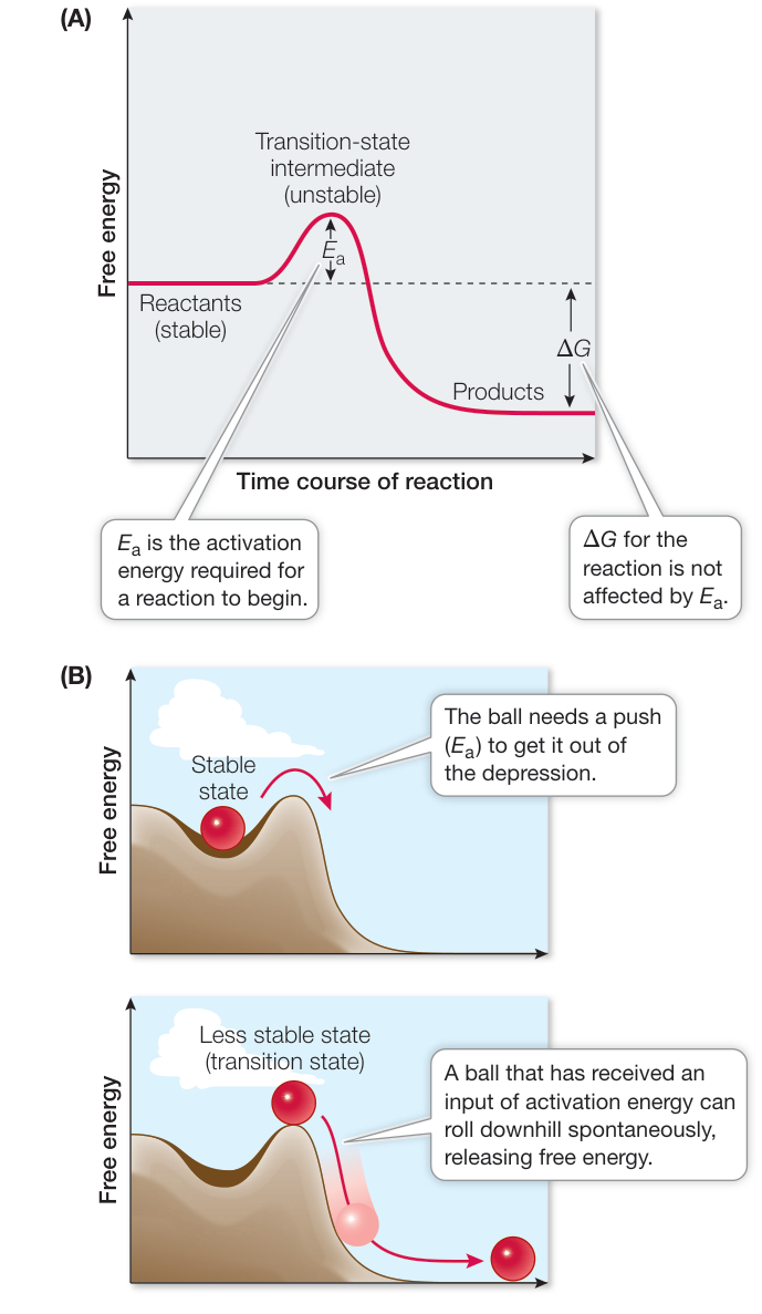
Figure 8.8 Activation Energy Initiates Reactions (A) In any chemical reaction, an initial stable state must become less stable before change is possible. (B) A ball on a hillside provides a physical analogy for the biochemical principle graphed in (A).
Connect the Concepts To understand enzymes and their activity, you should review the structures and chemical properties of proteins. See Key Concept 3.2.
In an enzyme-catalyzed reaction, the reactants are called substrates. Substrate molecules bind to a specific three-dimensional location on the enzyme protein called the active site, where catalysis takes place (Figure 8.9). This specificity is comparable to the specific binding of a membrane transport protein or receptor protein to its specific ligand, as described in Chapters 6 and 7. The names of enzymes often reflect their functions and end with the suffix “ase.” For example, the enzyme sucrase catalyzes the hydrolysis of sucrose, which we have referred to earlier:
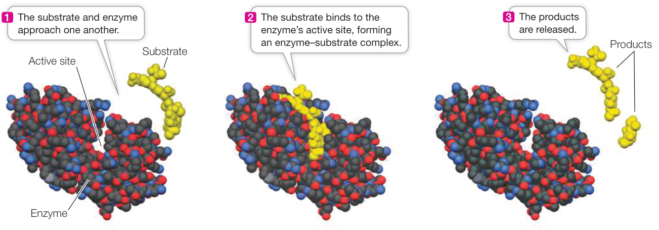
Figure 8.9 Enzyme and Substrate A reaction involving an enzyme is illustrated by lysozyme. Lysozyme catalyzes breakage of bonds in the peptidoglycans of bacterial cell walls. (See Key Concept 5.2 for a description of peptidoglycans.) (Data from PDB 148L. R. Kuroki et al. 1993. Science 262: 2030–2033.)
Q: Is “fitting in the lock” the only necessary function for enzyme–substrate interactions?
Thousands of enzymes have been identified, each catalyzing the transformation of its particular substrate(s). In general, enzymes fall into six categories:
- Oxidoreductases transfer electrons between molecules, especially in energy metabolism. An example of an enzyme that catalyzes this reaction is oxidoreductase:
You will see other examples in Chapters 9 and 10.
- Transferases transfer groups of atoms (functional groups) (see Figure 3.1) between molecules. The basic reaction can be shown as:
where A = the donor, B = the acceptor, and X = the functional group. For example, aminotransferases transfer groups between molecules, linking carbohydrates and amino acids.
- Hydrolases add water to covalent bonds to break down molecules. An example (shown earlier) is sucrase, which hydrolyzes sucrose to its constituent monosaccharides.
- Lyases are a diverse set of enzyme that catalyze non-hydrolytic bond breakage, often forming a new bond in the process. For example, a lyase catalyzes the cleavage of the phosphorus–oxygen bond of ATP (see Figure 7.11):
- Isomerases move functional groups from one place to another within a molecule, forming an isomer with the same atoms, differently bonded. For example, glucose and fructose are isomers of (see Figure 3.17).
- Ligases join two molecules together. You will see examples of ligases when you learn about DNA replication in Chapter 13.
A special note on language: When biologists refer to an enzyme, they usually use a verb for its action. For example, as noted for ligases (#6), we used the words “ligases join….” It is important for you to realize that this wording is shorthand for what really happens: ligases catalyze the joining. Enzymes don’t “do” the reaction; they catalyze it.
When a substrate binds to the active site of an enzyme, the result is called an enzyme–substrate complex (ES) (see Figure 8.9). The chemical forces that hold the complex together may include hydrogen bonding, electrical attraction, or temporary covalent bonding. The enzyme–substrate complex gives rise to product and free enzyme:
where E is the enzyme, S is the substrate, P is the product, and ES is the enzyme–substrate complex. Using the example of sucrose hydrolysis, we can write out the steps of the reaction:
The free enzyme (E, sucrase) is in the same chemical form at the end of the reaction as at the beginning. While bound to the substrate, it may change chemically, but by the end of the reaction it has been restored to its initial form and is ready to bind more substrate.
Connect the Concepts Binding of a substrate to an enzyme involves not just a “fit” but also noncovalent interactions between the enzyme and substrate. See Key Concept 3.2 for an outline of noncovalent interactions.
The relationship between enzyme and substrate may seem familiar; it is analogous to the binding of a receptor and ligand that we described in Chapter 7. In that instance, we described the affinity of a receptor for its ligand with the dissociation constant (). The lower the , the tighter the binding. For enzymes and their substrates, values are often in the range to M, which favors the formation of the ES. In practical terms, the values of the ES are such that the binding between enzyme and substrate is somewhat reversible; the substrate can be released before the reaction. However, many enzyme-catalyzed reactions will proceed because the ES is short-lived and the product(s) form quickly.
Enzymes lower the energy barrier but do not affect equilibrium
Reactants that are bound to the enzyme, forming an enzyme-substrate complex, require less activation energy than do the transition-state intermediates in the corresponding uncatalyzed reaction (Figure 8.10). Thus the enzyme lowers the activation energy needed for the reaction, speeding it up. Note that both the forward and the reverse reactions speed up, so the enzyme-catalyzed reaction proceeds toward equilibrium more rapidly than the uncatalyzed reaction. The final equilibrium is the same with or without the enzyme. Similarly, adding an enzyme to a reaction does not change the difference in free energy () between the reactants and the products (see Figure 8.10).
What do we mean by “speeding it up”? Consider a protein that has arginine as its terminal amino acid. This is an organized situation, and a hydrolysis reaction—releasing the arginine—is spontaneous (entropy increases). But spontaneous does not mean fast. In solution, it takes about 7 years for half of the protein molecules to undergo the hydrolysis reaction. Enter the enzyme carboxypeptidase A. When it catalyzes the reaction, half the arginines are released in less than a second! Rate enhancement by enzymes varies from 1 million times to an amazing by sulfate monoesterases. The consequence of catalysis for living cells is not difficult to imagine. Such increased reaction rates make new realities possible.
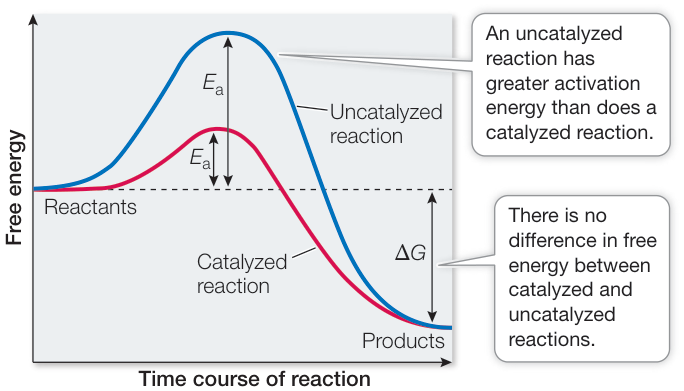
Figure 8.10 Enzymes Lower the Energy Barrier Although the activation energy is lower in an enzyme-catalyzed reaction than in an uncatalyzed reaction, the energy released is the same with or without catalysis. In other words, is lower but is unchanged. Lower activation energy means the reaction will take place at a faster rate. Activity 8.2 Free Energy Changes

Recap and Assess
A chemical reaction requires a “push” over the energy barrier to get started. An enzyme reduces the activation energy needed to start a reaction by binding the reactants (substrates), which speeds up the reaction. Biological catalysts are highly specific; only a narrow range of substrates can fit into the enzyme’s active site, so generally only one specific reaction can be catalyzed by a given enzyme. Enzymes affect only the rates at which reactions proceed—they do not affect the equilibrium or free energy associated with a given reaction.
- Explain how the structure of an enzyme makes that enzyme specific.
- What is the relationship between an enzyme and the equilibrium point of a reaction?
- When potatoes are peeled, the enzyme polyphenol oxidase causes
discoloration by catalyzing the oxidation of certain molecules, using
as a substrate. Explain these observations:
- Browning is reduced in potatoes that are peeled under water and kept there.
- Potatoes that have been boiled at 100°C and then cooled do not brown when they are sliced.
Now that you have a general understanding of the structures, functions, and specificities of enzymes, let’s look more closely at how they work.
Enzymes Bring Substrates Together so Reactions Readily Occur
Learning Objectives
- 8.4.1 Give examples of mechanisms for enzyme catalysis.
- 8.4.2 Describe coenzymes and cofactors and how they function.
- 8.4.3 Predict changes in the rate of an enzyme-catalyzed reaction as factors in the reaction are varied.
During and after the formation of the enzyme-substrate complex, chemical interactions occur. These interactions contribute directly to the breaking of old bonds and the formation of new ones.
Enzymes lower activation energy in several ways
In catalyzing a reaction, an enzyme may use one or more strategies.
SUBSTRATE ORIENTATION When free in solution, substrates are moving from place to place randomly while at the same time vibrating, rotating, and tumbling. They only rarely have the proper orientation to interact when they collide. When bound to an enzyme, however, substrates are held in the correct orientation to bring the appropriate chemical groups of substrates together, facilitating bond breakage and formation (Figure 8.11A).
INDUCING STRAIN Once a substrate has bound to its active site, an enzyme can cause bonds in the substrate to stretch, putting it in an unstable transition state (Figure 8.11B). This causes the strained bonds to have higher potential energy (they are weaker), making them prone to break.
ADDING CHEMICAL GROUPS The side chains (R groups) of an enzyme’s amino acids may be direct participants in making the enzyme’s substrates more chemically reactive (Figure 8.11C).
- In acid–base catalysis, the acidic or basic side chains of the amino acids in the active site transfer to or from the substrate, destabilizing a covalent bond in the substrate, and permitting it to break.
- In covalent catalysis, a functional group in a side chain forms a temporary covalent bond with a portion of the substrate.
- In metal ion catalysis, metal ions such as copper, iron, and manganese, which are often firmly bound to side chains of enzymes, can lose or gain electrons without detaching from the enzymes. This ability makes them important participants in oxidation–reduction reactions, which involve the loss or gain of electrons.
Molecular structure determines enzyme function
Many enzymes are much larger than their substrates. An enzyme is typically a protein containing hundreds of amino acids. It may consist of a single folded polypeptide chain or of several subunits (see Key Concept 3.2). Its substrate is generally a small molecule or a small part of a large molecule. The active site of the enzyme is usually quite small, not more than 6 to 12 amino acids. You may be asking yourself:
- What features of the enzyme’s active site allow it to recognize and bind the substrate?
- What is the role of the rest of the protein?
THE ACTIVE SITE IS SPECIFIC TO THE SUBSTRATE(S) The remarkable ability of an enzyme to select exactly the right substrate(s) depends on a precise interlocking of molecular shapes and interactions of chemical groups at the active site. The binding of a substrate to the active site depends on the same kinds of forces that maintain the tertiary structure of the enzyme: hydrogen bonds, the attraction and repulsion of electrically charged groups, and hydrophobic interactions. The specific fit of the substrate in the active site of lysozyme is illustrated in Figures 8.9 and 8.11B.
AN ENZYME CHANGES SHAPE WHEN IT BINDS A SUBSTRATE Just as a membrane receptor protein may undergo precise changes in conformation upon binding its ligand (see Chapter 7), some enzymes change their shapes when they bind their substrate(s). These shape changes, which are called induced fit, alter the shape of the active site(s) of the enzyme.
An example of induced fit can be seen in the enzyme hexokinase, which catalyzes the reaction
Induced fit brings reactive side chains from the hexokinase active site into alignment with the substrates (Figure 8.12), facilitating its catalytic mechanisms. Equally important, the folding of hexokinase to fit around the substrates (glucose and ATP) excludes water from the active site. This is essential, because if water were present, the
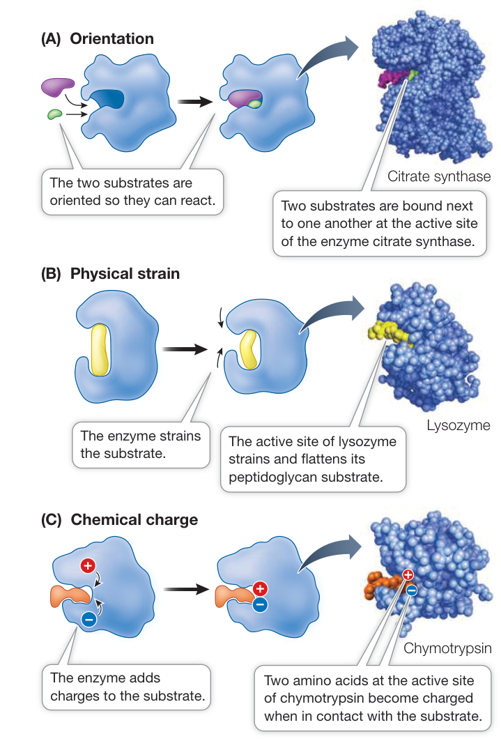
Figure 8.11 Life at the Active Site Enzymes use three basic mechanisms to cause their substrates to enter the transition state: (A) orientation, (B) physical strain, and (C) chemical charge. (A data from PDB 1AL6. B. Schwartz et al. 1997; B data from PDB 1BB6. V. B. Vollan et al. 1999. Acta Crystallogr D Biol Crystallogr 55: 60–66; C data from PDB 1AB9. N. H. Yennawar et al. 1994. Biochemistry 33: 7326–7366.) Q: The enzyme chymotrypsin cleaves the peptide bonds of proteins after the amino acids tyrosine, phenylalanine, or tryptophan. Locations on the polypeptide chains with other amino acids are not cleaved by the enzyme. Looking at Table 3.2 (amino acids), can you explain the substrate specificity?
ATP could be hydrolyzed to ADP and . But since water is absent, the transfer of a phosphate from ATP to glucose is favored.
Induced fit at least partly indicates what the role(s) of the rest of the protein may be:
- It provides a framework so that the amino acids of the active site are properly positioned in relation to the substrate(s).
- It participates in significant changes in protein shape and structure that result in induced fit.
- It provides binding sites for regulatory molecules (see Key Concept 8.5).
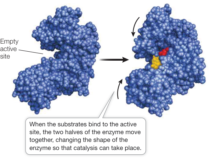
Figure 8.12 Some Enzymes Change Shape When Substrate Binds to Them Shape changes result in an induced fit between enzyme and substrate, improving the catalytic ability of the enzyme. Induced fit can be observed in the enzyme hexokinase, seen here with and without its substrates, glucose (red) and ATP (yellow). (Data from PDB 1G8. P. R. Kuser et al. 2000. J Biol Chem 275: 20814–20821 and PDB 1BDG. A. M. Mulichak et al. 1998. Nat Struct Biol 5: 555–560.)
Not all enzymes are proteins
Some RNA molecules can also act as biological catalysts; in these cases they are called ribozymes. As you saw in Figure 4.4, RNAs can fold into specific three-dimensional shapes that can bind substrates. Indeed, it takes only 25 bases in an RNA polymer to generate specific binding. The bases in RNA contain polar groups and hydrogen bonding possibilities with substrates. Some ribozymes act on themselves as self-cleaving RNAs; examples include RNAs in mitochondria and plastids. Other ribozymes act on chemically different substrates; the most notable among these is RNA in the ribosome that acts as a catalyst for peptide bond formation in protein synthesis. The catalytic ability of RNA is important evidence for the “RNA world” hypothesis, relating to the origin of life (see Key Concept 4.3).
Some enzymes require other molecules in order to function
Many enzymes require the presence of nonprotein chemical “partners” in order to function (Table 8.2):
- Prosthetic groups are distinct, non-amino acid atoms or molecular groupings that are permanently bound to their enzymes. An example is flavin adenine dinucleotide (FAD), which is bound to succinate dehydrogenase, an important enzyme in cellular respiration (see Key Concept 9.3).
- Inorganic cofactors include ions such as copper, zinc, and iron that are permanently bound to certain enzymes. For example, the enzyme alcohol dehydrogenase contains the cofactor zinc.
- A coenzyme is a nonprotein carbon-containing molecule that is required for the action of one or more enzymes. It is usually relatively small compared with the enzyme to which it binds.
A coenzyme moves from enzyme to enzyme, adding or removing chemical groups from the substrate. A coenzyme is like a substrate in that it does not permanently bind to the enzyme: it binds to the active site, changes chemically during the reaction, and then separates from the enzyme to participate in other reactions. There is actually no clear distinction between the functions of coenzymes and some substrates. For example, ATP and ADP have been described as coenzymes, even though they are really substrates that gain or lose phosphate groups during chemical reactions. The term “coenzyme” was coined before the functions of these molecules were fully understood. Biochemists continue to use the term, and for consistency with the field, we use it in this book.
Substrate concentration affects the reaction rate
For a chemical reaction of the type , the rate of the uncatalyzed reaction is directly proportional to the concentration of the substrate, . The higher the concentration of substrate, the faster the rate of the reaction. The appropriate enzyme not only speeds up the reaction; it also changes the shape of a plot of rate versus substrate concentration (Figure 8.13). For a given concentration of enzyme, the rate of the enzyme-catalyzed reaction initially increases as the substrate concentration increases from zero, but then it levels off. At some point, further increases in the substrate concentration do not significantly increase the reaction rate—the maximum rate has been reached.
The concentration of an enzyme is usually much lower than that of its substrate (e.g., in cells expressing the enzyme sucrase, there are far more molecules of sucrose than of the enzyme). As more and more substrate molecules bind to the available enzyme molecules, there is a saturation phenomenon like the one that occurs in facilitated diffusion (see Figure 6.12). When all the enzyme molecules are bound with substrate molecules, the enzyme is working as fast as it can—at its maximum rate. Nothing is gained by adding more substrate, because no free enzyme molecules are left to act as catalysts. Under these conditions the active sites are said to be saturated.
Table 8.2 Some Examples of Nonprotein “Partners” of Enzymes
| Type of molecule | Role in catalyzed reactions |
|---|---|
| PROSTHETIC GROUPS | |
| Heme | Binds ions, , and electrons |
| FAD | Carries electrons/protons |
| Retinal | Converts light energy |
| INORGANIC COFACTORS | |
| Iron ( or ) | Oxidation/reduction |
| Copper ( or ) | Oxidation/reduction |
| Zinc () | Stabilizes DNA binding structure |
| COENZYMES | |
| Biotin | Carries —COO— |
| Coenzyme A | Carries —CO—CH 3 |
| NAD | Carries electrons/protons |
| ATP | Provides/extracts energy |
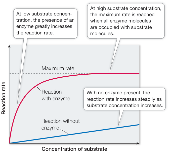
Figure 8.13 Catalyzed Reactions Reach a Maximum Rate Because there is usually less enzyme than substrate present, the reaction rate levels off when the enzyme becomes saturated.
The maximum rate of a catalyzed reaction can be used to measure how efficient the enzyme is. The turnover number is the maximum number of substrate molecules that one enzyme molecule can convert to product per unit of time. This number ranges from 1 molecule every 2 seconds for lysozyme to an amazing 40 million molecules per second for the liver enzyme catalase.
Recap and Assess
Enzymes orient their substrates to bring together specific atoms so that bonds can form. An enzyme can participate in the reaction it catalyzes by temporarily changing shape or destabilizing the enzyme–substrate complex. Some enzymes require prosthetic groups, inorganic cofactors, or coenzymes in order to function.
- The chemical composition of an enzyme before and after a catalyzed reaction is the same, but during the reaction an enzyme can change. How and why does this happen?
- Compare enzymes and coenzymes with regard to chemical structure and function.
- Plot reaction rate versus substrate concentration, and explain the shape of the curve.
You’ve seen in this key concept how individual enzymes work on their substrates. However, enzymes inside organisms don’t operate in isolation—there may be thousands of different enzymes within a given cell. Let’s see how all these different enzymes work together in a complex organism.
Enzyme Activities Can Be Regulated
Learning Objectives
- 8.5.1 Explain how metabolic pathways can shift in different directions.
- 8.5.2 Distinguish between different types of inhibitors.
- 8.5.3 Apply the concept of allosteric regulation to explain experimental results.
The chemical reactions in cells operate within metabolic pathways in which the product of one reaction is a reactant for the next. These pathways do not exist in isolation, but interact extensively, and each reaction in each pathway is catalyzed by a specific enzyme. Within a cell or organism, the presence and activity of enzymes determine the “flow” of chemicals through different metabolic pathways. The amount of enzyme activity, in turn, can be controlled in two ways:
- Regulation of gene expression. The gene for an enzyme protein may be expressed more or less, resulting in more or fewer enzyme molecules in the cell. You learned in Chapter 7 that some signal transduction pathways result in changes in gene expression, and that often the genes that are switched on or off encode enzymes.
- Regulation of enzyme activity. The enzyme may change its shape to hide its active site from the substrate, or a regulator molecule may prevent the substrate from reaching the active site. Blocking the activity of a single enzyme can affect an entire metabolic pathway.
The flow of molecules through interacting metabolic pathways can be studied, but this process quickly becomes complicated, because each pathway influences the others. Computer algorithms are used to model these pathways and show how they mesh in an interdependent system (Figure 8.14). Such models can help predict what will happen if the concentration of one molecule or another is altered. This field of biology is called systems biology and it has many applications.
In this key concept we will investigate the roles of enzymes in organizing and regulating metabolic pathways. We will also examine how the environment—particularly temperature and pH—affects enzyme activity.
Enzymes can be regulated by inhibitors
Chemical inhibitors can bind to enzymes, slowing down the rates of the reactions they catalyze. Some inhibitors occur naturally in cells; others are natural in the diet; still others are artificial. Naturally occurring inhibitors regulate metabolism; artificial ones can be used to treat disease, to kill pests, or to study how enzymes work. In some cases the inhibitor binds the enzyme irreversibly, and the enzyme becomes permanently inactivated. In other cases the inhibitor has reversible effects; it can separate from the enzyme, allowing the enzyme to function fully as before. The removal of a natural reversible inhibitor increases an enzyme’s rate of catalysis.
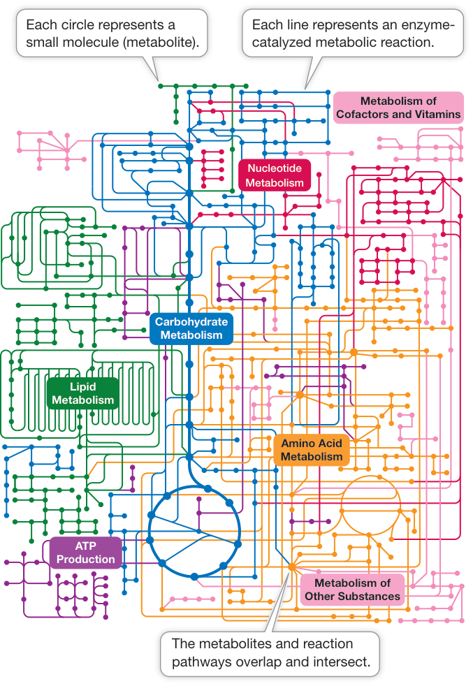
Figure 8.14 Metabolic Pathways The complex interactions of metabolic pathways can be modeled by the tools of systems biology. In cells, the main elements controlling these pathways are enzymes. (After KEGG: Kyoto Encyclopedia of Genes and Genomes. Map 01100 © 2004 Kanehisa Laboratories; M. Kanehisa et al. 2017. Nucleic Acids Res 45: D353–D361.)
IRREVERSIBLE INHIBITION If an inhibitor covalently binds to certain side chains at the active site of an enzyme, it will permanently inactivate the enzyme by destroying its capacity to interact with its normal substrate. An example of an irreversible inhibitor is aspirin, which we described in the opening of this chapter. Aspirin (acetylsalicylic acid) binds to a target enzyme called cyclooxygenase (COX) near its active site. When it binds, aspirin releases its acetyl group () and this group of atoms is transferred to bond covalently to serine at its polar hydroxyl group (Figure 8.15). The group of atoms now “coating” serine sticks out into a channel in the protein and prevents the substrate, arachidonic acid, from reaching the active site. So prostaglandin is no longer produced and its stimulation of inflammation and pain is blocked. The elucidation of the mechanism of aspirin’s action as an inhibitor of a pathway involved in inflammation was a landmark in modern pharmacology, the study of drugs (Investigating Life: How Do Anti-Inflammatory Drugs Work as Enzyme Inhibitors?).
REVERSIBLE INHIBITION In some cases an inhibitor is similar enough to a particular enzyme’s natural substrate to bind non-covalently to its active site, yet different enough that the enzyme catalyzes no chemical reaction. While such a molecule is bound to the enzyme, the natural substrate cannot enter the active site and the enzyme is unable to function. Such a molecule is called a competitive inhibitor because it competes with the natural substrate for the active site (Figure 8.16A,B). In this case, the degree of inhibition depends on the relative concentrations of the substrate and the inhibitor: if the inhibitor concentration is higher, it is more likely to bind the active site of the enzyme than the substrate, and vice versa. The inhibition is reversible because if the concentration of substrate is increased or if the concentration
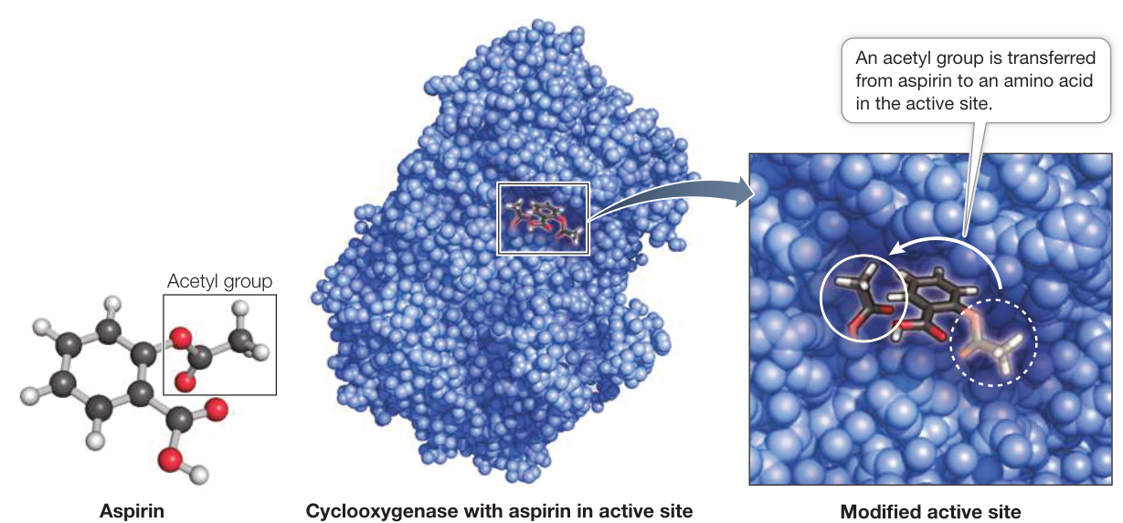
Figure 8.15 Irreversible Enzyme Inhibition Aspirin transfers an acetyl group, which covalently bonds to a serine amino acid near the active site of its target enzyme. This permanent change prevents the substrate from binding. (Data from PDB 1PTH. P. J. Loll et al. 1995. Nat Struct Biol 2: 637–643.) Q: What would be the chemical effects of adding an acetyl group ( ) to the active site serine?
Investigating Life: How Do Anti-Inflammatory Drugs Work as Enzyme Inhibitors?
Work with the Data
Experiment
Original Paper: J. R. Vane. 1971. Inhibition of prostaglandin synthesis as a mechanism of action of aspirin-like drugs. Nature 231: 232–235.
J. B. Smith and A. L. Willis. 1971. Aspirin selectively inhibits prostaglandin production in human platelets. Nature 231: 235–238.
The opening story in this chapter describes how willow bark, the source of what became aspirin, is a centuries-old remedy for pain and inflammation. Until the late twentieth century, it was assumed that aspirin acted directly on the nervous system. Working at England’s Royal College of Surgeons, a group led by John Vane showed that aspirin acts instead as an inhibitor of the enzymatic reaction that produces prostaglandin (PG), a fatty acid derivative important in inflammation.
HYPOTHESIS Aspirin acts as an anti-inflammatory drug by inhibiting an enzyme that catalyzes prostaglandin synthesis.
METHOD
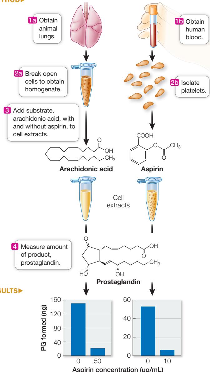
CONCLUSION In both animal and human cells, aspirin blocks the synthesis of the pro-inflammatory molecule, prostaglandin, in test tube experiments.
For his discovery of the mechanism by which aspirin relieves pain, John Vane was awarded the Nobel Prize and knighted by Queen Elizabeth II. Key to the experiments was the assumption that enzyme activity and mechanism are the same outside the organism as they are inside. In the lab, if given its substrate and the same set of environmental conditions as in the cytoplasm, an enzyme will catalyze production of its product.
QUESTIONS
- In the first set of experiments, lung tissue from guinea pigs was broken up to form a cell-free extract (homogenate). The substrate, arachidonic acid, was added to the extract, and after 30 minutes the amount of prostaglandin (PG) was measured. The results are shown in Table A. Plot PG synthesis versus aspirin concentration. What can you conclude?
- A similar set of experiments was performed on human platelets (membrane-bound cell fragments derived from white blood cells). These cells are known to produce PG by the same enzyme mechanism as lung tissue under certain environmental conditions. The results are shown in Table B. What can you conclude from these results and about the generalizability of your conclusion from Question 1?
- In a third set of experiments, platelets were isolated from human volunteers and their ability to make PG was measured (no aspirin present). Then these people were given a clinically effective dose of aspirin, and after a short period their platelets were isolated and PG synthesis measured (again, no aspirin in the test tube experiments). The results from three people are shown in Table C. How do these data either reinforce or refute your conclusion from Questions 1 and 2?
Table A
| Aspirin concentration () | PG synthesis (ng) |
|---|---|
| 0 | 220 |
| 1 | 172 |
| 2 | 136 |
| 10 | 99 |
| 50 | 33 |
| 80 | 0 |
Table B
| Aspirin concentration () | PG synthesis (ng) |
|---|---|
| 0 | 53 |
| 0.01 | 48 |
| 0.1 | 35 |
| 1 | 18 |
| 10 | 7 |
Table C
| Prostaglandin synthesis (ng) | ||
|---|---|---|
| Individual | Before aspirin | After aspirin |
| 1 | 160 | 16 |
| 2 | 168 | 5 |
| 3 | 103 | 20 |
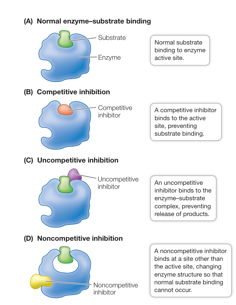
Figure 8.16 Reversible Enzyme Inhibition There are several mechanisms of enzyme inhibition.
of inhibitor is reduced, the substrate is more likely to bind, and the enzyme is active again.
An example of a competitive inhibitor is the drug methotrexate (Figure 8.17). An important co-enzyme in the formation of purines (components of nucleic acids) is tetrahydrofolate, which is formed from dihydrofolate in a reaction catalyzed by dihydrofolate reductase (DHFR):
When cancer cells reproduce, they need to replicate their DNA, and so they need to produce purines. This makes DHFR an ideal target for an anticancer drug. A team led by Sidney Farber at Harvard Medical School first showed that an analog of dihydrofolate could treat leukemia. The drug used by Farber (aminopterin) has since been replaced by a similar analog, methotrexate. This successful drug is used to treat inflammatory diseases such as psoriasis and rheumatoid arthritis, as well as cancer.
An uncompetitive inhibitor (Figure 8.16C) binds to the enzyme–substrate complex, preventing the complex from releasing products. Unlike competitive inhibition, uncompetitive inhibition cannot be overcome by adding more substrate.
A noncompetitive inhibitor binds to an unbound enzyme at a site distinct from the active site. Binding causes a change in the shape of the enzyme that alters its activity (Figure 8.16D). The active site may no longer bind the substrate, or if it does, the rate of product formation may be reduced. Like competitive inhibitors, noncompetitive inhibitors can become unbound, so their effects are reversible.
Allosteric enzymes are controlled via changes in shape
The change in enzyme shape that is due to noncompetitive inhibitor binding is an example of allosteric (allo, “different,” + stereo, “shape”). Allosteric regulation occurs when a non-substrate molecule binds to a site other than the active site of an enzyme, inducing the enzyme to change its shape. The change in shape alters the affinity of the active site for the substrate, and so the rate of the reaction is changed.
Often, an enzyme will exist in the cell in more than one possible shape (Figure 8.18):
- The active form of the enzyme has the proper shape for substrate binding.
- The inactive form of the enzyme has a shape that cannot bind the substrate.
Other non-substrate molecules can influence which form the enzyme takes:
- Binding of an inhibitor to a site other than the active site can stabilize the inactive form of the enzyme, making it less likely to convert to the active form.
- The active form can be stabilized by the binding of an activator to another site on the enzyme.
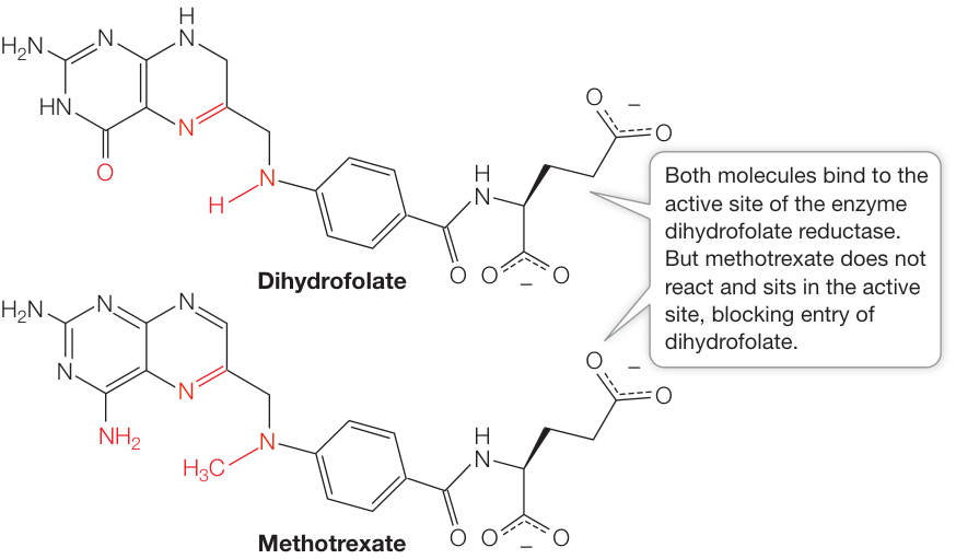
Figure 8.17 A Competitive Inhibitor to Treat Cancer Methotrexate has a structure similar to that of the substrate dihydrofolate, an enzyme important to cancer cell reproduction.
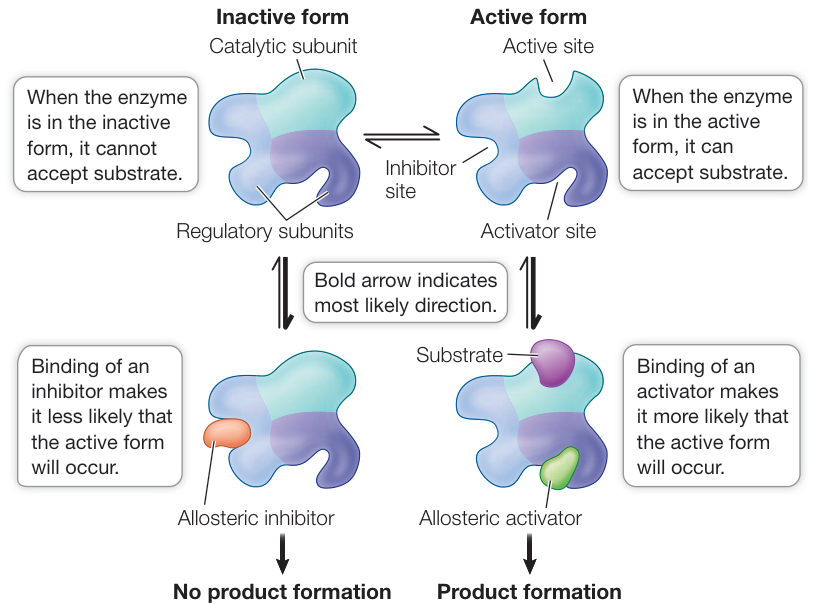
Figure 8.18 Allosteric Regulation of Enzymes Active and inactive forms of an enzyme can be interconverted, depending on the binding of non-substrate molecules at sites other than the active site. Binding an inhibitor stabilizes the inactive form, and binding an activator stabilizes the active form. Q: Many enzymes with allosteric regulation have several polypeptide chains (quaternary structure). In some cases, the subunit with the binding site for the regulatory molecule is similar in several different enzymes. How would this be important in regulating biochemical pathways (see Figure 8.14)?
Like substrate binding, the binding of inhibitors and activators to their regulatory sites (also called allosteric sites) is highly specific. Most (but not all) enzymes that are allosterically regulated are proteins with quaternary structure; that is, they are made up of multiple polypeptide subunits. The polypeptide that has the active site is called the catalytic subunit. The allosteric sites are often located on different polypeptides, called the regulatory subunits (see Figure 8.18).
Some enzymes have multiple subunits containing active sites, and the binding of substrate to one of the active sites causes allosteric effects. When substrate binds to one subunit, there is a slight change in protein structure that influences the adjacent subunit. The slight change to the second subunit makes its active site more likely to bind to the substrate. So the reaction speeds up as the sites become sequentially activated.
As a result, an allosteric enzyme with multiple active sites and a non-allosteric enzyme with a single active site differ greatly in their reaction rates when the substrate concentration is low. Graphs of reaction rates plotted against substrate concentrations show this relationship (Figure 8.19):
For a non-allosteric enzyme, the reaction rate first increases sharply with increasing substrate concentration, then tapers off to a constant maximum rate as the supply of enzyme becomes saturated.
For a multisubunit allosteric enzyme, the reaction rate at first increases only gradually as substrate concentration increases. After the substrate binds to the first active site of the enzyme (the slowly increasing part of the curve), there is a change in the enzyme’s quaternary structure such that the other sites become more likely to bind substrate, so the reaction speeds up (the rapidly increasing part of the curve). Once all sites are saturated with substrate, the reaction rate reaches a plateau (the upper, flat part of the curve). Within a certain range, the reaction rate is extremely sensitive to relatively small changes in substrate concentration. In addition, allosteric enzymes are very sensitive to low concentrations of inhibitors. Because of this sensitivity, allosteric enzymes are important in regulating entire metabolic pathways.
Allosteric effects regulate metabolic pathways
Metabolic pathways typically involve a starting material, various intermediate products, and an end product that is used for some purpose by the cell. In each pathway there are a number of reactions, each forming an intermediate product and each catalyzed by a different enzyme. The first step in a pathway is called the commitment step, meaning that once this enzyme-catalyzed reaction occurs, the “ball is rolling,” and the other reactions happen in sequence, leading to the end product. But what if the cell has no requirement for that product—for example, if that product is available from the cell’s environment in adequate amounts? It would be energetically wasteful for the cell to continue making something it does not need.
Connect the Concepts The number of molecules of a particular enzyme present in a cell at a given time is controlled in large part by the synthesis of that protein. See Key Concept 16.1 for a discussion of the regulation of protein synthesis.
One way to avoid this problem is to shut down the metabolic pathway by having the final product inhibit the enzyme that catalyzes the commitment step (Figure 8.20). Often this inhibition occurs allosterically. When the end product is present at a high concentration, some of it binds to an allosteric site on the commitment step enzyme, thereby causing it to become inactive. Thus the final product acts as a noncompetitive inhibitor (described earlier in this key concept) of the first enzyme in the pathway. This mechanism is known as feedback inhibition or end-product inhibition. We will describe many other examples of such inhibition in later chapters.
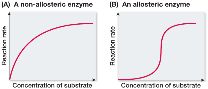
Figure 8.19 Reactions of Non-Allosteric and Allosteric Enzymes Differ
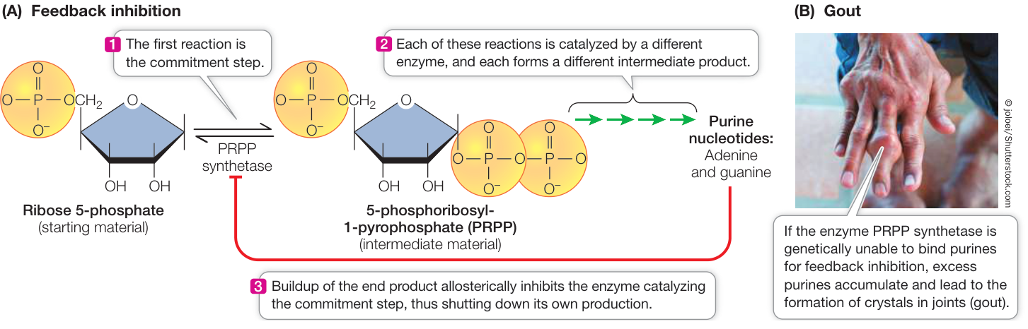
Figure 8.20 Feedback Inhibition of Metabolic Pathways (A) The first reaction in a metabolic pathway is referred to as the commitment step. It is often catalyzed by an enzyme that can be allosterically inhibited by the end product of the pathway. The specific pathway shown here is the synthesis of purine nucleotides (ATP, GTP) from ribose 5-phosphate in humans. (B) Gout, an accumulation of crystals in joints caused by an excess of purines, can occur if there is a failure of feedback inhibition. Q: The synthesis of cholesterol involves a biochemical pathway that begins with the enzyme HMG-CoA reductase. Statins are drugs that are widely used to inhibit cholesterol synthesis. How do you suppose statins work?
Many enzymes are regulated through reversible phosphorylation
Many enzymes involved in signal transduction are regulated via reversible phosphorylation (see Figure 7.14A). An enzyme can be activated by a protein kinase, which adds a phosphate from ATP to one or more specific amino acids. This results in a change in the shape of the enzyme, making it active. Such activation is reversible because another enzyme called a protein phosphatase can catalyze the hydrolysis and removal of phosphate groups, so that the enzyme becomes inactive again. In addition to the enzymes involved in signal transduction, many other enzymes and proteins in the cell (such as ion channels) are regulated via reversible phosphorylation. Reflecting the important role of protein phosphorylation in cell functions, the human genome contains more than 500 protein kinase genes: about 2% of all the protein-coding genes we have.
Enzymes are affected by their environment
Enzymes enable cells to perform chemical reactions and carry out complex processes rapidly without using the extremes of temperature and pH employed by chemists in the laboratory. However, because of their three-dimensional structures and the chemistry of the side chains in their active sites, enzymes (and their substrates) are highly sensitive to changes in temperature and pH. In Key Concept 3.2 we described the general effects of these environmental factors on proteins. Here we examine their effects on enzyme function (which, of course, depends on enzyme structure and chemistry).
pH AFFECTS ENZYME ACTIVITY The rates of most enzyme-catalyzed reactions depend on the pH of the solution in which they occur. While the water inside cells is generally at a neutral pH of 7, the presence of acids, bases, and buffers can alter this. Each enzyme is most active at a particular pH; its activity decreases as the solution is made more acidic or more basic than the ideal (optimal) pH (Figure 8.21). As an example, consider the human digestive system (see Key Concept 49.3). The pH inside the human stomach is highly acidic, around pH 1.5. However, many enzymes that hydrolyze macromolecules in the intestines, such as proteases, have pH optima in the neutral range. So when food enters the small intestine, a buffer (bicarbonate) is secreted into the intestine to raise the pH to 6.5. This allows the hydrolytic enzymes to be active and digest the food.
An important factor in the effect of pH on enzyme function is ionization of the carboxyl, amino, and other groups on either the substrate or the enzyme. In neutral or basic solutions, carboxyl groups () release to become negatively charged
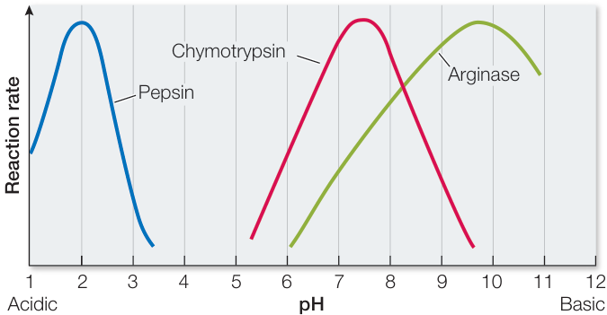
Figure 8.21 pH Affects Enzyme Activity An enzyme catalyzes its reaction at a maximum rate. The activity curve for each enzyme peaks at its optimal pH. For example, pepsin is active in the acidic environment of the stomach, whereas chymotrypsin is active in the small intestine. Q: Inside the cell, the pH of the cytosol is typically 7.2, but inside the lysosome the pH is 4.8. A protease, which catalyzes the hydrolysis of proteins, is active within the lysosome but is inactive in the cytosol. How can this occur? Figure 8.21 pH Affects Enzyme Activity An enzyme catalyzes its reaction at a maximum rate. The activity curve for each enzyme peaks at its optimal pH. For example, pepsin is active in the acidic environment of the stomach, whereas chymotrypsin is active in the small intestine. Q: Inside the cell, the pH of the cytosol is typically 7.2, but inside the lysosome the pH is 4.8. A protease, which catalyzes the hydrolysis of proteins, is active within the lysosome but is inactive in the cytosol. How can this occur?
carboxylate groups (). However, in neutral or acidic solutions, amino groups () accept to become positively charged groups (see the discussion of acids and bases in Key Concept 2.4). Thus in a neutral solution, an amino group is electrically attracted to a carboxyl group on another molecule or another part of the same molecule, because both groups are ionized and have opposite charges. If the pH changes, however, the ionization of these groups may change. For example, at a low pH (high concentration, such as the stomach contents where the enzyme pepsin is active), the excess may react with to form . If this happens, the group is no longer negatively charged and can no longer interact with positively charged groups in the protein, so the folding of the protein may be altered. If such a change occurs at the active site of an enzyme, the enzyme may no longer be able to bind to its substrate.
TEMPERATURE AFFECTS ENZYME ACTIVITY In general, warming increases the rate of a chemical reaction because a greater proportion of the reactant molecules have enough kinetic energy to provide the activation energy for the reaction. Enzyme-catalyzed reactions are no different (Figure 8.22). However, temperatures that are too high inactivate enzymes, because at high temperatures enzyme molecules vibrate and twist so rapidly that some of their noncovalent interactions such as hydrogen bonds break. When an enzyme’s tertiary structure is changed by heat it loses its function. Some enzymes denature at temperatures only slightly above that of the human body, but a few are stable even at the boiling point (or freezing point) of water. All enzymes, however, have an optimal temperature for activity.
In general, enzymes adapted to warm temperatures do not denature at those temperatures, because their tertiary structures are held together largely by covalent bonds and disulfide bridges, instead of the more heat-sensitive weak chemical interactions. Most enzymes in humans are more stable at high temperatures than are those of the bacteria that infect us, so that a moderate fever tends to denature bacterial enzymes, but not our own.
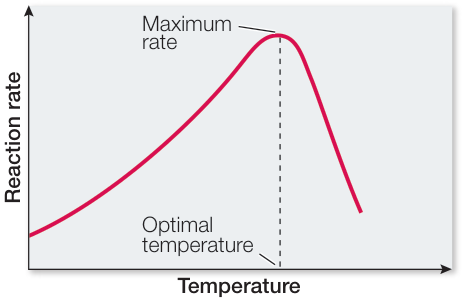
Figure 8.22 Temperature Affects Enzyme Activity Each enzyme is most active at a particular optimal temperature. At higher temperatures the enzyme becomes denatured and inactive; this explains why the activity curve falls off abruptly at temperatures above the optimum.
Recap and Assess
The rates of most enzyme-catalyzed reactions are affected by interacting molecules (such as inhibitors and activators) and by environmental factors (such as temperature and pH). Reversible phosphorylation is another important mechanism for regulating enzyme activity.
- What is feedback inhibition? How might the reactions shown in Figure 8.20A fit into a systems diagram such as the one shown in Figure 8.14?
- Consider an enzyme that is subject to allosteric regulation. If a competitive inhibitor (not an allosteric inhibitor) is added to a solution containing such an enzyme, the ratio of enzyme molecules in the active form to those in the inactive form increases. Explain this observation.
- In humans, hydrogen peroxide () is a dangerous toxin produced as a by-product of several metabolic pathways. The accumulation of is prevented by its conversion to harmless , a reaction catalyzed by the appropriately named enzyme catalase. Air pollutants can inhibit this enzyme and leave individuals susceptible to tissue damage by . How would you investigate whether catalase has an allosteric or a non-allosteric mechanism and whether the pollutants are acting as competitive or noncompetitive inhibitors?
Investigating Life: QA How do anti-inflammatory drugs work as enzyme inhibitors?
Go into a drugstore or watch ads on TV, and it becomes obvious that there are many drugs that alleviate pain. Like aspirin, many of them target the prostaglandin pathway for inflammation. John Vane’s research and later work showed that aspirin irreversibly inhibits the enzyme COX. But it turns out that there are two forms of COX, called COX-1 and COX-2, and there are several kinds of prostaglandins. COX-1 catalyzes the production of protective prostaglandins that are involved in blood clotting (which is why some people are advised to take aspirin to prevent blood clots after a heart attack) and the integrity of the lining of the stomach. COX-2 catalyzes production of prostaglandins involved with inflammation and associated pain. Aspirin inhibits both COX-1 and COX-2. So it is not surprising that people are advised to go easy on aspirin: it may block pain, but its use can lead to stomach upsets and an inability to clot blood after a wound. Knowledge of aspirin’s limitations has led to a search for inhibitors of COX-2 only. Most of them are competitive inhibitors. How would this affect how often you need to take the drug? Look up one of these drugs and read the description of how it works.
Future directions
As details at the atomic level of the binding of enzymes to substrates are worked out, biologists use the data to try to predict which substrates might bind to which enzymes. An important consideration in binding is ; the less positive this parameter, the more likely binding will occur. Factors such as ionic bonds and van der Waals forces contribute to the of binding. These factors are also important in the binding of proteins to non-substrate molecules, including other proteins and RNA. Indeed, it is becoming apparent that in almost all cases a protein in the cell does not exist in isolation, but is bound to something(s) else. If the protein is an enzyme, its substrate binding must be its most preferred partner. Understanding all the possible molecular interactions will result in a deeper understanding of what really goes on at the chemical level inside the cell.
E. Stone. 1763. Philos Trans R Soc Lond 53: 195–200
You should be able to relate each summary to the adjacent figures. If you go to this visual summary in Achieve, you can follow links to figures, animations, activities, and simulations that will help you consolidate the material.
Visual Summary
Chemical Transformations Involve Energy and Energy Transfers
- The laws of thermodynamics apply to living organisms. The first law states that energy cannot be created or destroyed. The second law states that energy transformations decrease the amount of energy available to do work (free energy, or G) and increase disorder.
- An exergonic reaction releases free energy and has a negative . An endergonic reaction consumes or requires free energy and has a positive .
Questions
- The disaccharide sucrose is hydrolyzed into its component monosaccharides glucose and fructose: How does this conversion relate to the laws of thermodynamics?
- Is the reaction in Question 1 exergonic or endergonic? Explain.
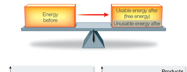
Figure 8.2
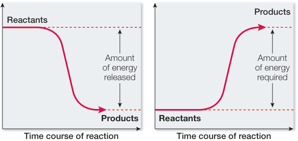
Figure 8.3
ATP Plays a Key Role in Biochemical Energetics
- Adenosine triphosphate (ATP) serves as an energy currency in cells. Hydrolysis of ATP releases a relatively large amount of free energy.
- The ATP cycle couples exergonic and endergonic reactions, harvesting free energy from exergonic reactions, and providing free energy for endergonic reactions.
Questions
- Are all exergonic reactions in the cell coupled to the formation of ATP from ADP? Explain.
- Adenine, as part of adenosine monophosphate (AMP), is a nucleotide base in DNA. If DNA is hydrolyzed to nucleotides, can the AMP in DNA be used as a source of energy in coupled reactions?
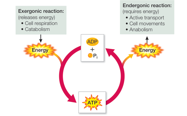
Figure 8.6
Enzymes Speed Up Biochemical Transformations
- The rate of a chemical reaction is independent of but is determined by the need for energy for the reactant(s) to reach a transition state.
- Enzymes are protein catalysts that affect the rates of biological reactions by supplying the activation energy () needed to initiate reactions.
Questions
- Do the primary, secondary, or tertiary structures of an enzyme change during and after its catalyzed reaction?
- In terms of energy, do enzymes catalyze exergonic but not endergonic reactions?
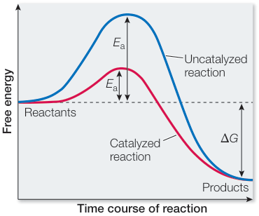
Figure 8.10
Enzymes Bring Substrates Together so Reactions Readily Occur
- ■ At the active site, a substrate can be oriented correctly, chemically modified, or strained.
- ■ Binding substrate causes many enzymes to change shape, exposing their active site(s) and allowing catalysis. The change in enzyme shape caused by substrate binding is known as induced fit.
- ■ Substrate concentration affects the rate of an enzyme-catalyzed reaction.
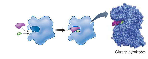
Figure 8.11
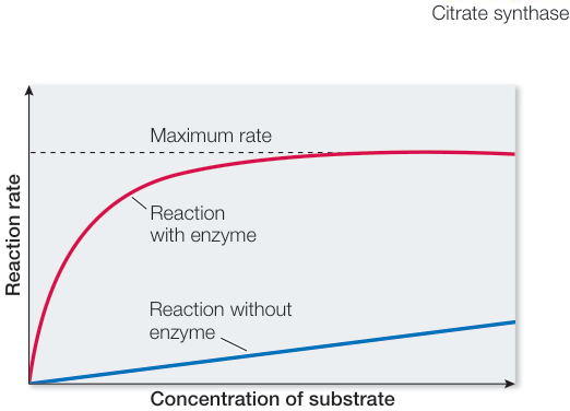
Figure 8.13
Questions
- Does a substrate need to fit exactly in the active site on an enzyme?
- How do reaction rates for an enzyme-catalyzed reaction compare in the presence of the enzyme versus in the presence of an enzyme that has been placed in boiling water for 10 minutes prior to the reaction? See Figure 8.13.
Enzyme Activities Can Be Regulated
- ■ Metabolism is organized into pathways in which the product of one enzyme-catalyzed reaction is a reactant for the next reaction.
- ■ Enzyme activity is subject to regulation. Some inhibitors bind irreversibly to enzymes. Others bind reversibly.
- ■ The end product of a metabolic pathway may inhibit an enzyme that catalyzes the commitment step of that pathway.
- ■ Enzymes are sensitive to their environments. Both pH and temperature affect enzyme activity.
Questions
- What is the difference between a competitive enzyme inhibitor and a noncompetitive inhibitor?
- A cell can make the amino acid glutamine through an eight-step biochemical pathway, starting with glucose. The pathway is active when there is no glutamine in the environment, but shuts down when there is abundant glutamine in the environment. How might the shutdown occur?
Go to Animations 8.1 and 8.2 and Simulation 8.1
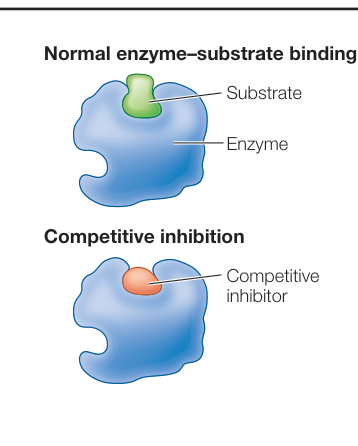
Figure 8.16
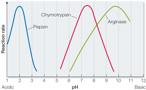
Figure 8.21