Chapter 18Recombinant DNA and Biotechnology
© Erik Isakson/Getty Images
Key Concepts
- 18.1 DNA from Different Sources Forms Recombinant DNA
- 18.2 There Are Several Ways to Insert DNA into Cells
- 18.3 DNA for Study Is Obtained in a Variety of Ways
- 18.4 Several Tools Are Used to Study the Function of DNA
- 18.5 DNA Can Be Manipulated for Commercial Use
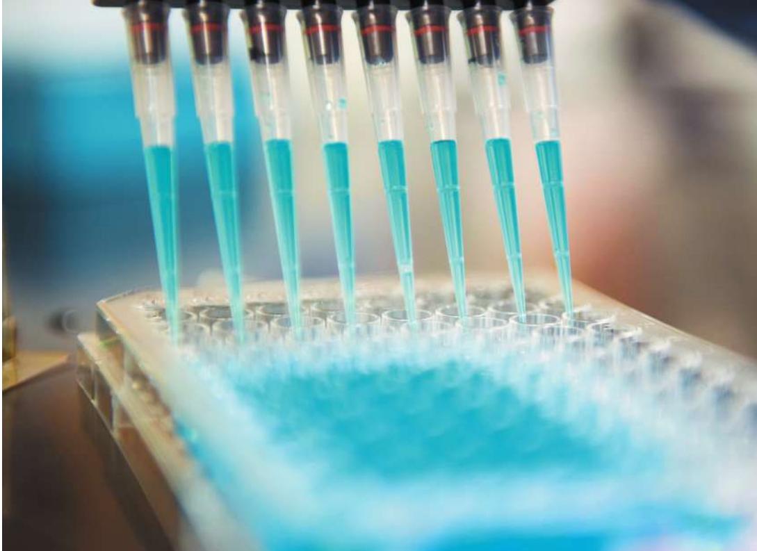
Nucleic acids and proteins can be analyzed and changed in small volumes, in this case far less than 1 milliliter.
Investigating Life: DNA Technology Meets Medicine
Janet was arriving at her office when she first felt numb on the left side of her face. She became confused and had difficulty finding her office. After a passerby called 911, an emergency response team transported Janet to a nearby hospital. She had had a stroke, but amazingly, she was back at work and fully functional two days later.
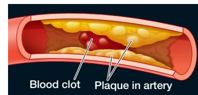
A stroke can occur when a large blood vessel that supplies the brain with oxygen and nutrients is blocked. Blockage is often associated with the formation of a blood clot on a plaque – a fat deposit on the inner walls of a blood vessel. Within minutes of blockage, cellular respiration in affected brain cells is reduced, and within minutes to hours, the cells die. Nerves controlling muscles in Janet’s face were affected first, followed by brain regions involved in cognitive processes.
Janet was treated with tissue plasminogen activator, or TPA, a protein involved in dissolving blood clots. Your body produces TPA when you have a cut; its action is seen when the clot “goes away” a few days later. Janet’s cells would have gradually made TPA to slowly dissolve the clot blocking the vessel to her brain, but in the meantime her brain cells would have died. To speed up the process, Janet was injected with TPA right around the clot.
Where did the TPA used in Janet’s treatment come from? TPA is a protein made in tiny amounts by blood vessel cells. Extracting enough TPA from tissues is impractical; recombinant
DNA technology is required to get enough of it for therapeutic injection into a bloodstream. As you will learn in Investigating Life: Producing TPA, the process involves inserting the gene that encodes TPA into living bacterial cells in the lab. These cells are then biochemically coaxed into expressing TPA protein in quantity. The production of TPA is but one example of how knowledge of molecular biology and advances in biotechnology have transformed biological research and launched new industries that employ microbes to produce proteins and other useful chemicals.
QA How is biotechnology changing medicine? (Find the answer on page 418.)
DNA from Different Sources Forms Recombinant DNA
Learning Objectives
- 18.1.1 Describe how staggered cuts made in DNA by restriction enzymes create sticky ends.
- 18.1.2 Explain why DNA ligase is used in addition to restriction enzymes to make recombinant DNA.
- 18.1.3 Summarize the Cohen and Boyer experiments on creating recombinant DNA.
Recombinant DNA is a DNA molecule that has been made in the laboratory using at least two different sources of DNA. The DNAs used for recombination can be from the same organism (e.g., from different chromosomes), from different members of the same species, or from entirely different organisms.
In Chapter 15 you learned that restriction enzymes are able to cleave double-stranded DNA at particular palindromic recognition sequences (restriction sites). When restriction enzymes cut at their restriction sites, they can create either blunt ends or complementary overhangs (short sequences of single-stranded DNA) termed sticky ends (Figure 18.1). Hundreds of restriction enzymes are known, many of which create their own unique sticky ends.
Any two DNA molecules with sticky ends that are complementary can form hydrogen bonds with one another. The sticky ends of the original molecule may bond (anneal), or two different fragments may bond. Indeed, a fragment from one source, such as a human, can bond to a fragment from any other source, such as a bacterium, as long as the ends are complementary. The fragments are initially held together by only a few weak hydrogen bonds, since there is a missing phosphodiester bond (a “nick”) in each strand of the double helix. However, the nicks can be sealed by adding the enzyme DNA ligase to catalyze the formation of the strong, covalent, phosphodiester bonds between adjacent nucleotides in each of the strands, joining them to form a single, larger molecule (Figure 18.2).
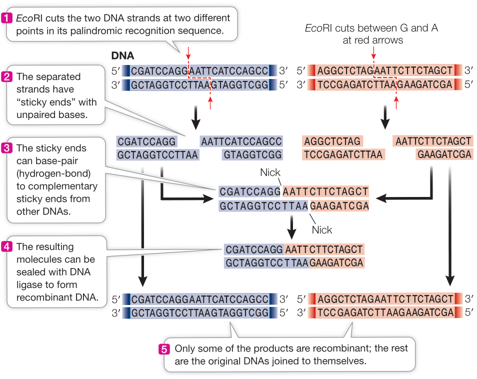
Figure 18.2 Cutting, Splicing, and Joining DNA EcoRI can be used to cut two different DNA molecules (blue and orange). The resulting sticky ends can hydrogen-bond with complementary exposed bases on other DNA fragments, which can then be covalently joined using DNA ligase to form the missing phosphodiester bonds in each strand of the DNA backbone, creating a recombinant molecule. Q: If two DNA molecules were digested with EcoRI and KpnI restriction enzymes, respectively, and then mixed together with DNA ligase, would you expect recombinant molecules between them to be produced?
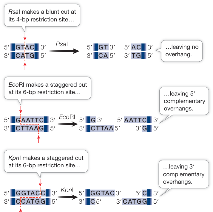
Figure 18.1 Restriction Enzymes Cut at Restriction Sites Some restriction enzymes cut the two strands of DNA at sites that are directly opposite one another, resulting in blunt-ended fragments. Other enzymes make cuts that are offset from one another, which results in staggered ends that have a 5’ or 3’ overhang. These are referred to as “sticky ends” because they are able to form hydrogen bonds with (that is, “stick to”) complementary sequences on other DNA molecules. The names of the enzymes reflect their organism of origin. EcoRI, for example, was found in E. coli.
With these tools—restriction enzymes and DNA ligase—scientists are able to create recombinant DNA and have been doing so since 1973. In that year, Stanley Cohen at Stanford University, Herbert Boyer at the University of California, and their colleagues created recombinant bacterial plasmids. As illustrated in Figure 18.3, they isolated two different plasmids (small circular DNA molecules that replicate independently of the main chromosome; see Figure 12.25) from Escherichia coli. Each of the two plasmids contained a different antibiotic resistance gene. The scientists cut the two plasmids with restriction enzymes, mixed the fragments together, and then used DNA ligase to rejoin DNA molecules, creating recombinant plasmids. The recombinant plasmids of this ligation reaction were then inserted into new E. coli cells, and the cells were grown in the presence of both antibiotics. A few of the transformed bacteria could grow on the medium, and the researchers were able to show that a single recombinant plasmid containing the genes for resistance to both antibiotics explained this result. With this experiment, recombinant DNA technology was born.
Recently, new tools have been developed for making recombinant DNA. Methods based on the polymerase chain reaction (PCR) allow the joining of any two DNA molecules without the need for conveniently placed restriction enzyme sites. Despite these advances, restriction enzymes and DNA ligase are still used routinely for recombinant DNA construction in biology labs.
Recap and Assess
DNA fragments from different sources can be joined together to make recombinant DNA. DNA can be cut with a restriction enzyme and then rejoined with DNA ligase. There are many different restriction enzymes, each with its own recognition sequence. Several restriction enzymes produce sticky ends, which facilitate the production of recombinant DNA.
- The following is the sequence of one strand of the restriction site for the HindIII restriction enzyme: 5’ … AAGCTT … 3’. What is the sequence of the other strand? If this enzyme results in a four-nucleotide 5’ overhang, where are the cut sites?
- How did Cohen and Boyer make the first recombinant DNA molecule and confirm that they had done so?
Recombinant DNA has no biological significance until it is inserted inside a living cell, which can replicate and transcribe the transplanted genetic information. How can recombinant DNA made in the laboratory be inserted and expressed in living cells?
There Are Several Ways to Insert DNA into Cells
Learning Objectives
- 18.2.1 List the three requirements of a bacterial plasmid cloning vector and describe the reason each is required.
- 18.2.2 Explain how a reporter gene such as GFP allows for easy screening for recombinant plasmids in bacterial cloning.
- 18.2.3 Outline the steps involved in transforming a multicellular organism using zygotic injection of foreign DNA.
- 18.2.4 Outline the steps involved in transforming a multicellular organism using injection of transformed, cultured cells during early development.
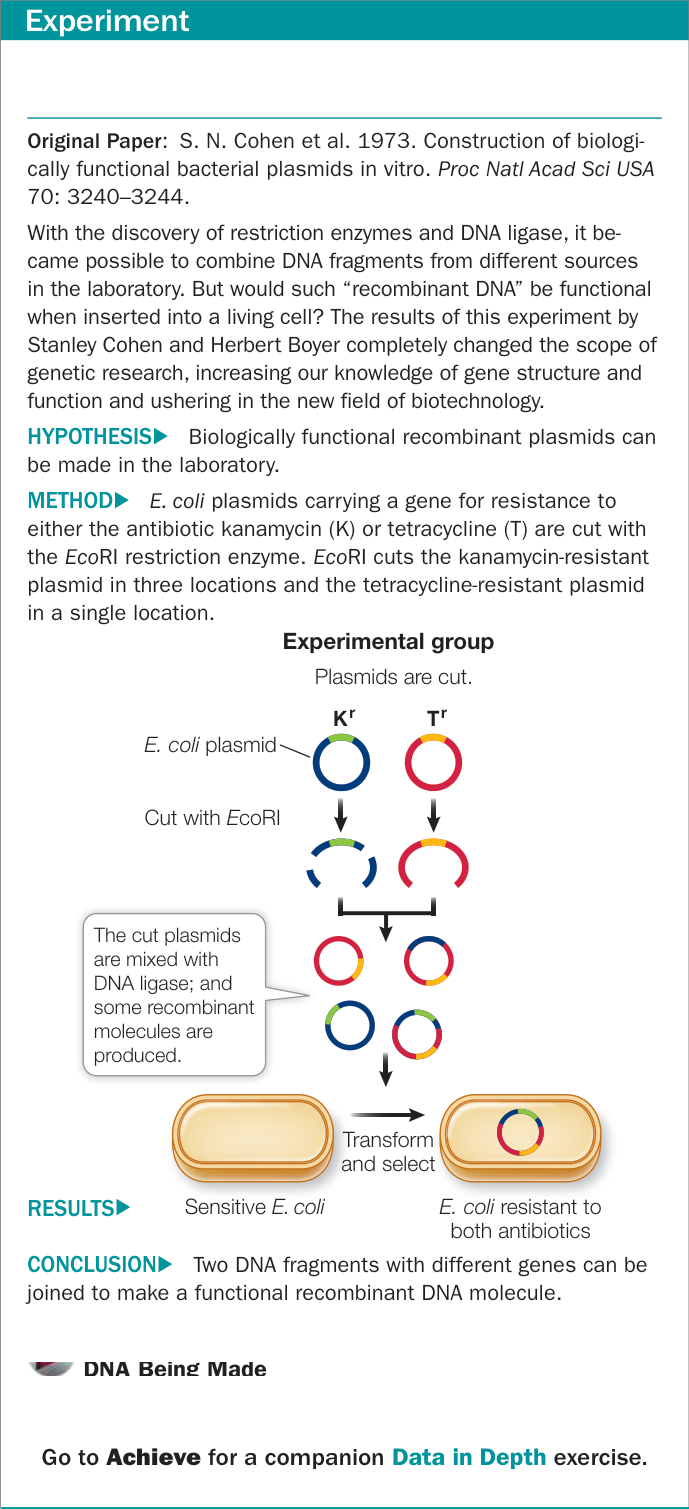
Figure 18.3 Can DNA from Two Different Sources Be Recombined into a Single Functional DNA Molecule? Figure 18.3 Can DNA from Two Different Sources Be Recombined into a Single Functional DNA Molecule?
One reason for making recombinant DNA is to clone—that is, to produce many identical copies of—a particular gene or other DNA sequence. We have seen the term “clone” used in the context of whole cells or organisms (see Chapters 11 and 12) that are genetically identical to one another. The easiest way to clone a piece of DNA is to use an organism that can make clones of itself. For example, a gene can be cloned by inserting it into a bacterial cell such as E. coli. If a new piece of DNA is inserted and the bacterium is allowed to reproduce, it will produce millions of identical cells, all carrying identical copies of the piece of DNA. Cloning might be done to get enough DNA for sequencing and subsequent analysis, to produce a protein product in quantity, or as a step toward creating an organism with a new phenotype.
In single-celled organisms such as E. coli that are able to make identical copies of themselves, the primary challenge is getting the foreign DNA into the cell. Recombinant DNA is cloned by inserting it into host cells in a process known as transformation—or transfection if the host cells are derived from an animal. A host cell or organism that contains recombinant DNA is referred to as a transgenic cell or organism, and the non-native DNA is called a transgene. Later in this chapter we will encounter many examples of transgenic organisms, including yeast, rice plants, and even cattle.
Selectable genetic markers are used to identify host cells containing recombinant DNA
Various methods are used to create transgenic cells. Generally these methods are inefficient in that only a few of the cells that are exposed to the recombinant DNA actually become transformed with it. In order to isolate and reproduce only transgenic cells, selectable marker genes, such as genes that confer resistance to antibiotics, are often included as part of the recombinant DNA molecule. When an antibiotic resistance gene is used as the selectable marker, the cells from the transformation experiment are grown in the presence of the antibiotic; the antibiotic kills all nontransgenic cells, leaving only the transgenic cells. Antibiotic resistance genes were the selectable markers used in Cohen and Boyer’s experiment (see Figure 18.3).
Genes can be inserted into prokaryotic or eukaryotic cells
In theory, any cell or organism can act as a host for the introduction of recombinant DNA. For single-celled organisms such as bacteria and yeasts, creating a single transgenic cell results in a transgenic organism. Both bacteria and yeasts have been heavily used as research organisms:
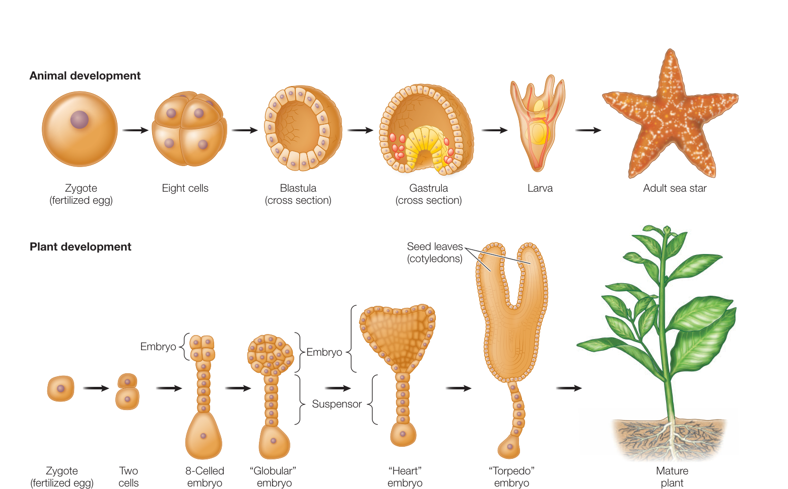
Figure 18.4 From Fertilized Egg to Adult The stages of development from a single-celled zygote to a mature organism containing millions or trillions of cells are shown for an animal and for a plant.
- Bacteria are easily grown and manipulated in the laboratory. Much of their molecular biology is known, especially for well-studied bacteria such as E. coli. Furthermore, bacteria contain plasmids, which are easily manipulated to carry recombinant DNA into the cell. While bacteria have been extremely useful for cloning pieces of DNA, they are not always well suited as hosts for expressing eukaryotic genes because they differ in the details of transcription, RNA processing, translation, and posttranslational modification (see Chapter 14).
- Yeasts such as Saccharomyces cerevisiae are commonly used as eukaryotic hosts for recombinant DNA studies. The advantages of using yeasts include their rapid cell division (less than 2 hours to proceed through the cell cycle), ease of growth in the laboratory, and relatively small genome size (see Table 17.2). In addition, yeast cells have most of the characteristics of other eukaryotic cells, with a notable exception being those associated with multicellularity.
For multicellular organisms, it is also possible to produce a transgenic organism by transforming a single cell. Multicellular sexual organisms pass through a single-celled zygote stage (see Figure 11.14) that develops into an adult organism containing many thousands to many trillions of cells (Figure 18.4). One method for creating transgenic individuals involves creating transgenic zygotes by injecting fertilized eggs with a transgene. The transgenic zygotes then develop into transgenic organisms, with every cell transgenic (Figure 18.5).
An alternative method for obtaining a transgenic multicellular organism is to use cell culture. The cultured cells must be totipotent stem cells—cells that have the ability to develop into any other cell type. Transformation is performed in culture, and transgenic cells are selected and allowed to produce large numbers of daughter cells. In plants, once cells are transformed in cell culture, they can be coaxed to develop into a mature plant (see Figure 35.4). This is not possible in an animal. Instead, transgenic cells are injected into a developing animal, usually at a very early stage of development such as the blastula (see Figure 18.4). The result of injecting transgenic cells is an embryo that is a mixture of original cells and transgenic cells (Figure 18.6). If transgenic cells give rise to gametes in the adult animal, then the resulting zygotes will have the transgene and will develop into offspring that have the transgene in all of their cells.
Inserted DNA is usually integrated into the host chromosome
Some prokaryotic cells can take up intact DNA molecules from the environment, which makes transformation quite easy; recall the bacterium Streptococcus pneumonia in which the R strain can be transformed by DNA of the S strain present in the environment (see Figure 13.2). However, most cells cannot do this, and so various laboratory methods have been devised to get DNA into cells. Cells can be chemically treated to make their outer membranes more permeable, and then mixed with the DNA so that it can diffuse into the cells. Another approach is called electroporation: a short electric shock is used to create temporary holes in the membranes through which the DNA can diffuse into the cell. Viruses can be altered so that they carry recombinant DNA into cells. A common method for transforming plants involves a specific bacterium that inserts DNA into plant cells. Transgenic animals can be produced by injecting recombinant DNA into the nuclei of fertilized eggs (see Figure 18.5). There are even “gene guns” that “shoot” the host cells with tiny metal particles coated with the DNA.
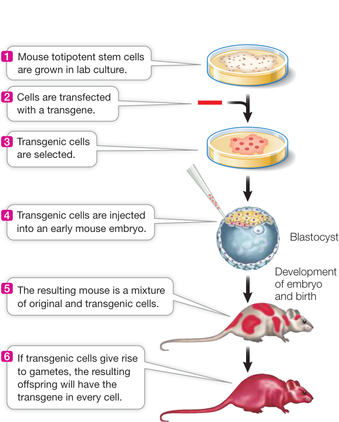
Figure 18.6 Injecting Transgenic Cells to Make a Transgenic Organism One method for obtaining a transgenic multicellular organism is to transform cells in culture and inject them into an early developmental stage, thereby creating an embryo that is a mixture of original cells and transgenic cells. If any of the transgenic cells give rise to gametes in the adult animal, the resulting zygotes will have the transgene and will develop into offspring that have the transgene in all of their cells. Figure 18.6 Injecting Transgenic Cells to Make a Transgenic Organism One method for obtaining a transgenic multicellular organism is to transform cells in culture and inject them into an early developmental stage, thereby creating an embryo that is a mixture of original cells and transgenic cells. If any of the transgenic cells give rise to gametes in the adult animal, the resulting zygotes will have the transgene and will develop into offspring that have the transgene in all of their cells.
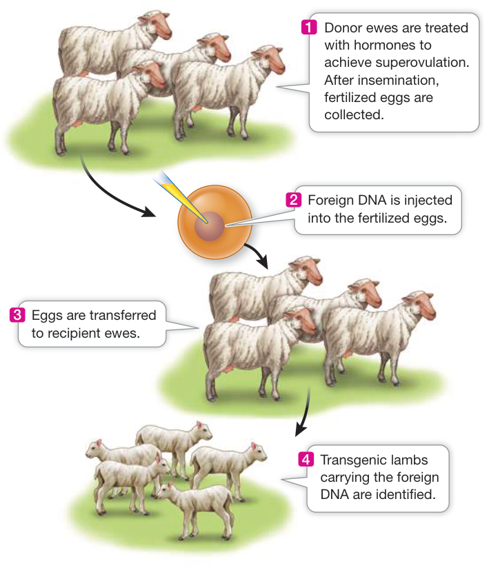
Figure 18.5 Transforming a Zygote to Make a Transgenic Organism One method for obtaining a transgenic multicellular organism is to transform a zygote (fertilized egg) by injecting it with foreign DNA and then screening for transgenic offspring that carry the foreign DNA incorporated into their genome.
The challenge of inserting new DNA into a cell lies not only in getting it into the host cell, but also in getting it to replicate as the host cell divides. As you saw in Key Concept 13.3, DNA polymerase does not bind to and copy just any sequence. If the new DNA is to be replicated, it must become part of a segment of DNA that contains an origin of replication. Such a DNA molecule is called a replicon, or replication unit.
There are two general ways in which the newly introduced DNA can become part of a replicon within the host cell:
- It can be spliced into a host chromosome directly.
- It can enter the host cell as part of a carrier DNA sequence, called a vector, and can then either integrate into a host chromosome or be replicated from its own origin of replication.
Several types of vectors are used to get DNA into cells, some of which we now describe in more detail.
PLASMIDS AS VECTORS As we described in Chapter 12, plasmids are generally small, circular DNA molecules that replicate autonomously in many prokaryotic cells. Several characteristics make plasmids useful as transformation vectors:
- Plasmids are relatively small (an E. coli plasmid is usually 2,000–6,000 bp) and are therefore easy to manipulate in the laboratory.
- A plasmid often has one or more restriction enzyme recognition sequences that each occur only once in the plasmid sequence. This makes it easy to insert new DNA into the prokaryotic plasmid before it is used to transform host cells.
- Many plasmids contain genes that confer resistance to antibiotics, which can serve as selectable markers.
- Plasmids have a bacterial origin of replication (ori) and can replicate independently of the host chromosome. It is not uncommon for a bacterial cell to contain hundreds of copies of a recombinant plasmid. For this reason, the power of bacterial transformation to amplify a gene is extraordinary. A 1-liter culture of bacteria harboring the human -globin gene in a typical plasmid has as many copies of that gene as there are cells in a typical adult human ().
A plasmid used as a vector in the laboratory has been extensively altered to include convenient features (Figure 18.7A):
- Multiple unique restriction sites for cloning purposes (see EcoRV, BamHI, SalI, and PstI in Figure 18.7A)
- An origin of replication that will function in a particular type of host cell (see ori in Figure 18.7A)
- One or more selectable markers (see ampicillin resistance and tetracycline resistance in Figure 18.7A)
PLASMID VECTORS FOR PLANTS An important vector for carrying new DNA into many types of plants is a plasmid found in the bacterium Agrobacterium tumefaciens. This bacterium lives in the soil, infects plants, and causes a disease called crown gall, which is characterized by the presence of growths (or tumors) on the plant. Agrobacterium contains a large plasmid called Ti (for tumor-inducing) (Figure 18.7B). When the bacterium infects a plant cell, a region of the Ti plasmid called the T DNA is inserted into the host plant cell, where it becomes incorporated into one of the plant’s chromosomes. The Ti plasmid carries the genes needed for this transfer and incorporation of the T DNA. The T DNA carries genes that are then expressed by the host cell, causing the growth of tumors and the production of specific sugars that the bacterium uses as sources of energy. Scientists have exploited this remarkable natural “genetic engineer” to insert foreign DNA into the genomes of plants.
When the Agrobacterium Ti plasmid is used as a vector for plant transformation, the tumor-inducing and sugar-producing genes on the T DNA are removed and replaced with foreign DNA.
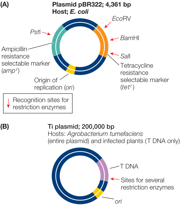
Figure 18.7 Plasmid Vectors (A) E. coli cloning plasmids contain one or more selectable markers to identify transgenic cells, an origin of replication, and one or more restriction sites for inserting foreign DNA. (B) Agrobacterium injects the T DNA region of its Ti plasmid during infection. When the Ti plasmid is used as a vector for plant transformation, genes on the T DNA are removed and replaced with foreign DNA. The creation of recombinant Ti plasmids allows for the transformation of many different host plant species.
The recombinant Ti plasmids are first used to transform Agrobacterium cells from which the original Ti plasmids have been removed. Then the Agrobacterium cells are used to infect plant cells and transfer the T DNA, now containing genes placed there by the researcher, to the host cell chromosomes.
VIRUSES AS VECTORS As a DNA molecule becomes larger than about 10,000 base pairs, it becomes challenging to get it across a cell membrane using chemical or electrical methods. However, in some cases the goal is to clone or transform with a larger DNA molecule. Although many prokaryotic genes are smaller than 10,000 base pairs, most eukaryotic genes—with their introns and extensive flanking sequences—are bigger. A vector that accommodates larger DNA inserts is needed for these genes.
Both prokaryotic and eukaryotic viruses are used as vectors for eukaryotic DNA. Bacteriophage , which infects E. coli, has a DNA genome of about 45,000 base pairs, all of which are injected into a host cell during infection. About 20,000 base pairs are not necessary for the bacteriophage to complete the lytic part of its life cycle (see Figure 16.15). These 20,000 base pairs can be deleted and replaced with DNA from another organism, which then gets replicated along with the virus DNA. Alternatively, essentially the entire phage genome can be replaced with DNA from another organism and packaged into phage heads. These phage heads are able to inject the DNA into the host bacterial cell exactly as they would inject their own genome. Once inside the cell, the DNA either circularizes to form a large plasmid or becomes incorporated into the genome.
Reporter genes help select or identify host cells containing recombinant DNA
Transformation is inefficient; only a very small percentage of a population of cells will be successfully transformed. Furthermore, the process of making recombinant DNA is far from perfect. During a ligation reaction, DNA molecules can combine in various ways, many of which do not produce the desired recombinant molecule (see Figures 18.2 and 18.3). Methods have been developed to improve the chance that a desired combination will occur. A simple approach is to cut the vector with two different restriction enzymes that have sites near each other. This produces a molecule with incompatible sticky ends, which is much less likely to simply recircularize during the ligation reaction. The desired insert molecule is cut with the same two enzymes, so that theoretically a functional circular plasmid can be produced only if the vector and insert ligate with one another. Even so, it is often the case that only a small proportion of the ligation products have the desired recombinant sequence.
How can we identify or select the host cells that contain the desired sequence? One way is to use selectable markers, such as those for antibiotic resistance used in early experiments with recombinant DNA (see Figure 18.3). Selectable markers are one type of reporter gene, which is any gene whose expression is easily observed. Here are several types of reporter genes:
As described earlier, an antibiotic resistance gene in a plasmid or other vector allows the detection of transformed host cells grown in the presence of the selected antibiotic. If the host cells are normally sensitive to the antibiotic, they will grow on medium containing the antibiotic only if they have been transformed by the vector. This approach is used in the selection of transgenic prokaryotic and eukaryotic cells, including those of plants and animals. In some cases a plasmid contains two antibiotic resistance genes and one of them is used for cloning. For example, suppose the plasmid in Figure 18.7A was used to clone a piece of foreign DNA at the BamHI site. Transformation could be selected for using ampicillin. The resulting bacteria could then be tested (screened) for resistance to tetracycline. Bacteria that were resistant to ampicillin and sensitive to tetracycline would have the plasmid containing a piece of foreign DNA.
The -galactosidase (lacZ) gene in the E. coli lac operon (see Figure 16.4) codes for an enzyme that can convert the artificial substrate X-Gal into a bright blue product. Many plasmids include the lacZ gene with multiple unique restriction sites where target DNA sequences can be inserted within its sequence. These plasmids also carry genes for antibiotic resistance. Bacterial colonies containing the plasmid are selected on a solid medium containing the antibiotic. X-Gal is also included in the medium. If a bacterial colony contains a recombinant plasmid that carries the foreign DNA inserted into the lacZ gene, it will not make -galactosidase and the colony will be white. Clones that contain the original plasmid with no insert express the lacZ gene and make blue colonies (Figure 18.8). The lacZ gene can thus be used to screen transformed colonies to see if their plasmid contains an inserted fragment of foreign DNA.
Green fluorescent protein (GFP), which normally occurs in the jellyfish Aequorea victoria, emits visible green light when exposed to ultraviolet light. The gene for this protein has been isolated and incorporated into vectors and can be used similarly to lacZ to screen for recombinant plasmids containing foreign DNA (see Figure 18.8). GFP has also been modified to emit other colors when exposed to ultraviolet light, allowing multiple reporters to be used at the same time.
Both lacZ and GFP are widely used as reporter genes in other situations besides cloning. The protein-coding sequence of one of these reporter genes can be attached to promoters or other regulatory sequences from other genes to study how these sequences regulate gene expression under different conditions or in different tissues of a transgenic multicellular organism (Figure 18.9A). The coding sequence of a reporter gene can also be added to the amino or carboxyl end of another protein-coding gene to “tag” the protein produced by that gene. For example, GFP can be used to study how and where a protein is localized in eukaryotic cells (Figure 18.9B).
How are numerous transgenic individuals produced from one?
When a researcher goes to the effort of making a transgenic organism, the goal is to produce numerous transgenic individuals, not just one. With single-celled organisms, one transgenic individual can easily be cloned by normal, asexual cell division (binary fission in prokaryotes or mitosis in eukaryotes) into numerous identical
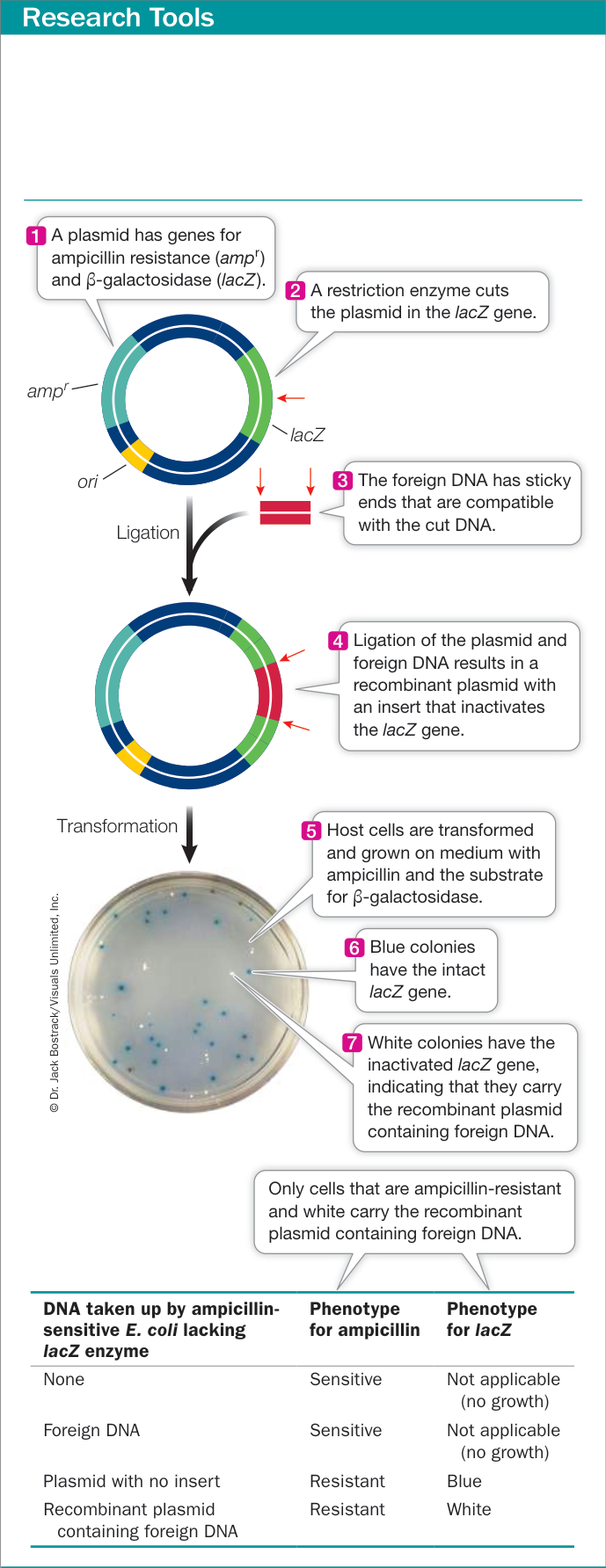
Figure 18.8 Selection for Recombinant Plasmids Selectable marker (reporter) genes are used by biologists to select for bacteria that have taken up a plasmid and check for foreign DNA. In a typical experiment, most of the bacteria will not take up any DNA. Of those that do, only a small fraction will take up a recombinant plasmid containing foreign DNA.
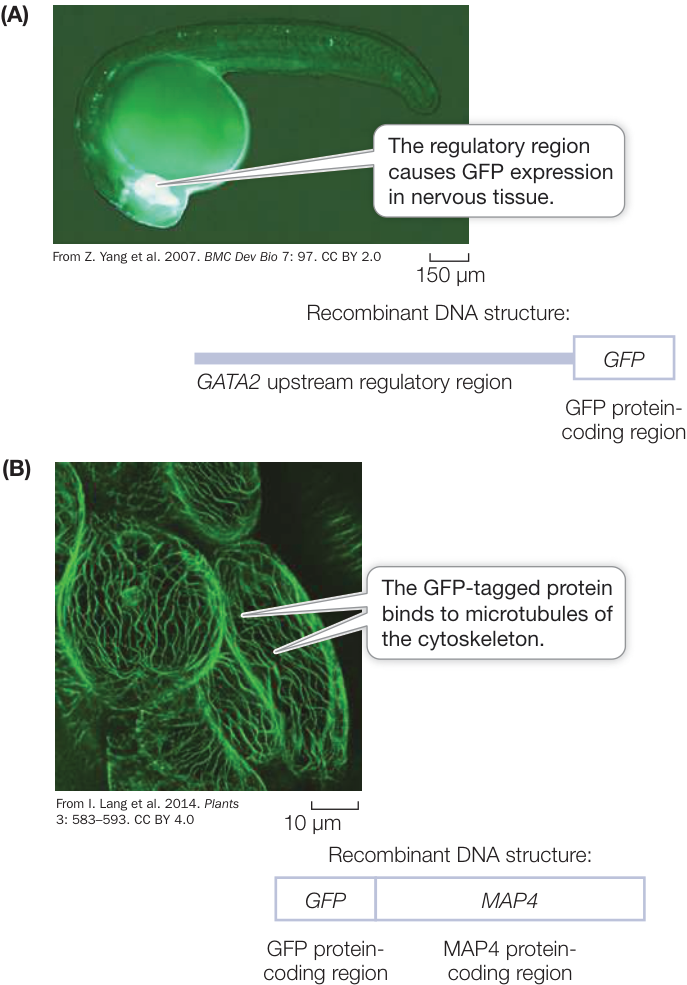
Figure 18.9 Green Fluorescent Protein as a Reporter Gene (A) Expression of GFP protein in a transgenic zebrafish (Danio rerio) 24 hours after fertilization. The recombinant DNA consists of 7.3 kilobase pairs (kbp) of the upstream region of a transcription factor gene (GATA2) joined to the coding sequence of GFP. The GFP fluorescence is strongest in neuronal cells (in the brain) of the embryo, indicating that this is where the GATA2 transcription factor is normally expressed. (B) GFP-tagged MAP4 protein expressed in Arabidopsis thaliana shoot tissue. Because MAP4 binds to microtubules, the GFP fluorescence shows the structure of the cytoskeleton in the cells. Q: Are cells killed during GFP visualization? Why is this important?
transgenic individuals (see Key Concept 11.4). In sexual eukaryotes, cloning a single individual is not as easy. The first cloned mammal, a sheep, was produced in 1996 by Ian Wilmut and his colleagues at the Roslin Institute in Edinburgh. The method involved the fusion of a somatic (nonreproductive) cell from an adult animal, containing the donor nucleus, with an enucleated egg. After the fused cell underwent several cell divisions, the resulting early embryo was transplanted into the womb of a surrogate mother. Eventually a lamb named Dolly was born (Figure 18.10). Dolly carried the same genetic material as the nuclear donor and thus was a clone of that donor. Although this process has been repeated with more than 20 mammal species, including cats, deer, dogs, horses, pigs, rabbits, and mice, it is expensive and not practical for producing large numbers of clones. How, then, to produce numerous, genetically identical transgenic individuals?
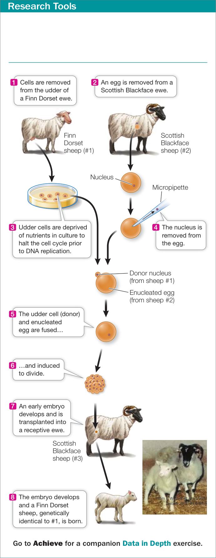
Figure 18.10 Cloning a Mammal The experimental procedure described here produced the first cloned mammal, a sheep named Dolly (shown on the left in the photo). As an adult, Dolly mated and subsequently gave birth to a normal offspring, thus showing that a cloned mammal is fully functional.
The technique that is used most often is to transform a genetically uniform (true-breeding) strain that has been obtained from many generations of inbreeding (see Key Concept 12.3). Such inbred strains are homozygous at most genes. Once a transgenic individual is obtained using one of the methods described earlier (see Figures 18.5 and 18.6), it is bred to a nontransgenic individual to produce offspring, some of which are heterozygous for the transgene. Heterozygotes are then interbred to produce individuals homozygous for the transgene. Although they are not technically clones, the resulting homozygotes are genetically uniform and can be interbred to make additional transgenic individuals of essentially identical genotype.
Recap and Assess
Recombinant DNA can be cloned by using a vector to insert it into a suitable host cell. Plasmid vectors contain a selectable marker; an origin of replication; and a cloning site, which may reside in a reporter gene. In multicellular eukaryotes, transformation involves zygotic transformation or transformation in cell culture. Once a transgenic individual is produced, asexual reproduction can be used to produce multiple clones. Although cloning is possible, copying a transgenic animal most often involves crossing genetically identical individuals that are homozygous for the transgene. Reporter genes can be used to check for a DNA insertion in cloning vectors or to show when a protein is being expressed or where it is localized.
- Assume you are using a plasmid vector that contains genes encoding
ampicillin resistance and GFP with a restriction site in the latter gene
only. Arrange a-e below to reflect the sequence of steps for inserting a
piece of foreign DNA into this plasmid, introducing the recombinant
plasmid into bacteria, and verifying that the plasmid and the foreign
gene are both present in the bacteria.
- Transform host cells.
- Put cells on medium containing antibiotic and grow resistant colonies.
- Select colonies that do not fluoresce under ultraviolet light.
- Digest vector and foreign DNA with a restriction enzyme.
- Ligate the digested plasmid together with the foreign DNA.
- Compare the use of a plasmid whose coding site is in a GFP gene versus a plasmid whose coding site is in a streptomycin resistance gene in terms of detecting cells containing a recombinant plasmid carrying foreign DNA.
- When transforming mice, transgenic cells originally derived from a brown mouse are often injected into an early developmental stage of a white mouse. Assuming white is recessive to brown coat color, how might mice carrying transgenic cells be identified following the injections and how might mice containing the transgene in every cell be produced?
We have described how DNA can be cut with restriction enzymes, inserted into a vector, and introduced into host cells. We have also seen how host cells carrying recombinant DNA can be identified. Now let’s consider where the genes or DNA fragments used in these procedures come from.
DNA for Study Is Obtained in a Variety of Ways
Learning Objectives
- 18.3.1 Describe the differences between genomic and cDNA libraries.
- 18.3.2 Explain why genomic libraries do not differ across tissues but cDNA libraries do.
- 18.3.3 Explain how RT-PCR is used to amplify a specific gene sequence.
- 18.3.4 Describe what is meant by synthetic DNA and give examples of its use.
A major goal of molecular biology is to learn the functions of particular DNA sequences and the proteins they encode. DNA sequences for study are obtained from several sources that we describe in this key concept: random fragments of chromosomes that are maintained as gene libraries, complementary DNA obtained by reverse transcription from mRNA, products of the polymerase chain reaction (PCR), and artificially synthesized or mutated DNA.
DNA for study can come from a library
DNA is contained in chromosomes, of which there are usually many in a cell. And chromosomes can be enormous, containing millions to billions of base pairs and hundreds to thousands of genes. Intact genomic DNA is too large to be introduced into host cells. How, then, are single genes, containing only thousands of base pairs, isolated from such large molecules? One method is to first break chromosomes into smaller DNA molecules, clone each molecule, and then find the gene of interest among the clones.
A genomic library is a collection of DNA fragments that make up the entire genome of an organism. Restriction enzyme digestion or other means, such as mechanical shearing, can be used to break chromosomes into small pieces. Each fragment is then cloned by inserting it into a vector, which is then taken up by a host cell. Proliferation of a single such transformed cell produces a colony of cells, each of which harbors many copies of the same fragment of DNA (Figure 18.11A). Many clones are required to make a genomic library for an entire genome.
If a plasmid vector is used, about 700,000 separate clones are required to make a library of the human genome. By using bacteriophage , which can carry about four times as much DNA as a typical plasmid, the number of clones in the library can be reduced to about 160,000. Although this still seems like a large number, a single petri plate can hold thousands of phage colonies, or plaques, which are easily screened for the presence of a particular DNA sequence by hybridization to an appropriate nucleic acid probe.
Connect the Concepts The technique of nucleic acid hybridization, illustrated in Figure 14.7, involves using a single-stranded nucleic acid probe with a complementary sequence to the target DNA. If the probe forms a double-stranded hybrid molecule, the target DNA fragment has been identified.
cDNA is made from mRNA transcripts
In many cases scientists are interested in protein-coding regions of DNA, which represent a very small fraction of some eukaryotic genomes (see Key Concept 17.3). To find these genes, a much smaller DNA library—one that includes only the genes transcribed in a particular tissue at a particular point in time—can be made from complementary DNA, or cDNA (Figure 18.11B). Making cDNA involves isolating mRNA from cells, then making cDNA copies of that mRNA by complementary base pairing in a process called reverse transcription. Reverse transcription is catalyzed by the enzyme reverse transcriptase.
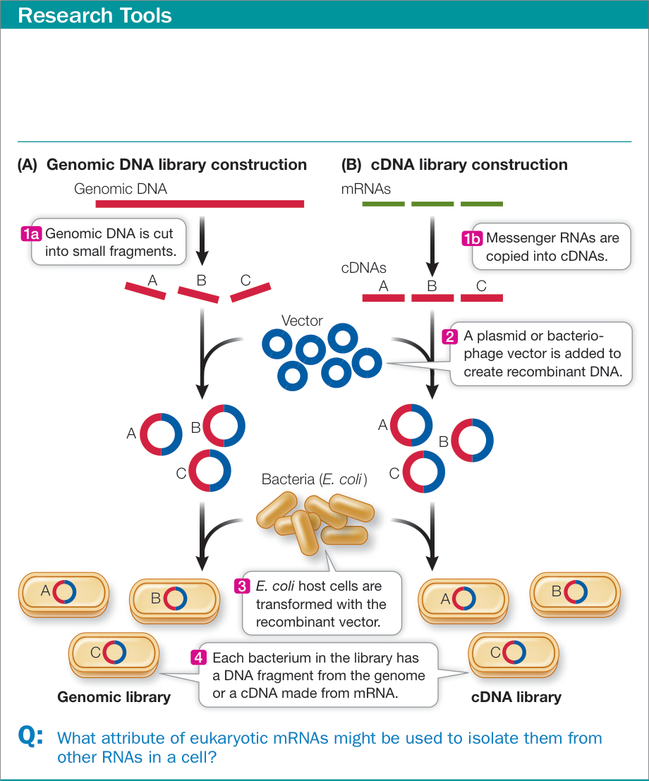
Figure 18.11 Constructing Libraries (A) A genomic library can be made by breaking the DNA into small fragments, incorporating the fragments into a vector, and then transforming host cells with the recombinant vectors. Each colony of cells contains many copies of a small part of the genome. (B) The many mRNAs in a cell can be copied into cDNAs and a library made from them.
A collection of cDNAs from a particular tissue at a particular time in the life cycle of an organism is called a cDNA library. Because mRNAs do not last long in the cytoplasm, the types and amounts of mRNAs present in a cell are a good representation of the transcription rates of all the genes in that cell. So a cDNA library is a “snapshot” that captures the transcription pattern of a set of cells at a given point in time. cDNA libraries have been invaluable for comparing gene expression in different tissues at different stages of development. For example, researchers have found that up to one-third of all the genes of an animal are expressed only during development. cDNA is also a good starting point for cloning eukaryotic genes, because the clones contain only the coding sequences of the genes (the introns having been spliced out; see Figure 14.8). Also, if a eukaryotic gene is highly expressed in a particular tissue, a cDNA library made from that tissue will be enriched for the gene, making it easier to identify and clone the gene.
DNA can be amplified using PCR or can be synthesized artificially
When the DNA sequence surrounding a region of interest is known, a DNA library is not needed. Instead, PCR can be used to amplify the target sequence. PCR can begin with as little as g of DNA (a picogram). Any fragment of DNA can be amplified by PCR as long as appropriate primers are available, which requires knowing the DNA sequence surrounding the target. If the appropriate primers (two are needed—one for each strand of DNA) are added to template DNA in a PCR reaction, millions of copies of the DNA sequence between the primers can be produced in just a few hours. This amplified DNA can then be inserted into a vector and cloned in host cells.
Connect the Concepts As illustrated in Figure 13.20, the polymerase chain reaction (PCR) automates replication of a DNA fragment in a test tube. Also called DNA amplification, the process involves repeatedly synthesizing DNA complementary to a target sequence to make enough DNA for chemical analysis and genetic manipulations.
PCR can also be used to amplify a specific cDNA sequence. In this case, mRNA is isolated from an organism or tissue, and reverse transcriptase is used to make cDNA from the mRNA. Then PCR is used to amplify a specific sequence directly from the sample of cDNA. This procedure, called RT-PCR, has become an invaluable tool for studying the expression of particular genes in cells and organisms.
Artificial DNA can be synthesized using organic chemistry to link nucleotides together in a specified sequence. This process is now fully automated, and a laboratory can make large numbers of short-to medium-length sequences overnight. PCR primers, for example, are made in this way. The synthesis of artificial DNA does not require a template, so DNA with any sequence can be made. This flexibility is useful for creating DNA fragments with desirable characteristics, such as convenient restriction sites or specific mutations. Longer synthetic sequences can be pieced together to construct completely artificial genes that have been designed for specific purposes.
For example, a gene might be designed to be highly expressed in a particular cell type, or to encode a highly active enzyme.
Connect the Concepts Plasmids containing synthetic transposon DNA have been used to transfect host cells to inactivate genes one by one and give insight into the minimal genome. See Key Concept 17.2.
Recap and Assess
DNA for study can be obtained from genomic libraries, cDNA libraries, and PCR, or it can be artificially synthesized.
- What type of approach would you use for analyzing each of the
following, and why?
- The expression of a gene encoding amylase, a starch hydrolyzing enzyme, in seeds as they germinate
- The sequence of the amylase gene in two different genetic strains of corn
- How could RT-PCR be used to compare the expression of a particular gene in muscle versus liver tissues?
- Describe some uses for synthetic DNA.
We’ve explored the various sources of DNA that can be used to make recombinant DNA molecules, and how organisms are transformed with recombinant DNA. We now turn to some of the ways that recombinant DNA and transformation methods can be used to study the functions of genes and proteins.
Several Tools Are Used to Study the Function of DNA
Learning Objectives
- 18.4.1 Give examples of ways in which particular genes are chosen for study.
- 18.4.2 Describe how DNA microarrays are used to examine patterns of gene expression.
- 18.4.3 Outline how RNA interference can be used to reduce expression of a particular gene.
- 18.4.4 Explain how CRISPR can be used to knock down, knock out, or replace a specific gene.
So far in this chapter we have seen how recombinant DNA is made and how organisms are transformed with recombinant DNA. In this key concept we examine some of the ways in which genes of interest are identified and describe several techniques for studying the functions of genes.
Genes of interest can be identified by genetic mapping or by studying gene expression
There are many thousands of genes in a genome (see Chapter 17). Many of these genes function in multiple tissues or at different times of development. How does a scientist decide which among these genes to study? In some cases a gene can be identified by genetic mapping of a Mendelian disorder in a pedigree (see Figure 15.17). Many genes have been identified in this way. For example, the gene that causes cystic fibrosis was identified in the late 1980s using this method and has been under intense study ever since. In other cases gene expression can be used to find a gene to study. A researcher might be interested in a gene that is expressed in a particular tissue or stage of development, or that is misexpressed in diseased individuals. For example, embryonic cells that will eventually give rise to skin cells, normal adult skin cells, and skin cancer cells all express a unique set of genes. A developmental biologist might be interested in the genes uniquely expressed in the embryonic cells, while an oncologist might be interested in those uniquely expressed in the cancer cells.
To identify distinct patterns of gene expression, scientists could isolate mRNA from cells and measure the amount of each gene’s mRNA one gene at a time by hybridization or RT-PCR. But that would be expensive and time-consuming. It is far simpler to do these hybridizations for every gene in a single step, and that is made possible with DNA microarray technology.
DNA microarrays (“gene chips”) contain a series of DNA sequences attached to a glass slide in a precise order. In Chapter 17 we discussed the use of DNA microarrays for single nucleotide polymorphism (SNP) genotyping (see Figure 17.13). For RNA expression, each microscopic spot contains thousands of copies of a particular oligonucleotide of 20 or more bases that is the sequence of an exon from a protein-coding gene in the genome. Each oligonucleotide can hybridize with only one RNA sequence and thus is a unique identifier of expression of that gene. If a gene is highly expressed, there will be many RNA molecules in the sample to hybridize to the corresponding spot on the microarray. Alternatively, spots representing genes that are not expressed will show no hybridization. Gene microarrays are usually used to examine differential expression; gene expression in one sample is often compared with that in another sample. In the field of oncology, for example, patterns of gene expression provide invaluable information to clinicians trying to characterize patients’ tumors. To do this, RNAs are extracted from tumor and normal tissues, labeled with different fluorescent colors (e.g., red and green), and then RNAs from both tissues are hybridized to the microarray. After hybridization, spots of a single color (red or green) represent genes expressed more highly in one tissue, while spots with both colors (yellow) represent genes with similar expression in both tissues (Figure 18.12).
The function of a gene can be determined by modifying its expression post-transcriptionally
Once a gene has been identified, there are many questions that can be addressed to determine its function. Where and when in the organism is the gene expressed? Where is the protein produced by the gene localized? What happens when the gene is knocked down (has its expression reduced) or is knocked out (eliminated), such that a functional protein is not made?
Where and when a gene is expressed and the localization of the protein product can be examined using reporter genes; the regulatory region for a gene can be placed upstream of a reporter to determine expression pattern, or a coding sequence can be added to a gene to determine where the protein localizes (see Figure 18.9). But how do we knock down or knock out a gene?
One way to reduce expression is by blocking the translation of a gene’s mRNA. The process developed for blocking translation is yet another example of scientists imitating nature. As described in Key Concept 16.5, gene expression is sometimes regulated by the production of double-stranded RNA molecules that are processed to produce short, single-stranded microRNAs (miRNAs) and small interfering RNAs (siRNAs). These miRNAs and siRNAs are complementary to specific mRNA sequences and combine with proteins to form an RNA-induced silencing complex (RISC). When miRNAs and siRNAs are hybridized (bound) to their target mRNAs, the RISC inhibits translation (miRNAs) or causes degradation (siRNAs) of the mRNA (see Figure 16.22). Scientists use RNA-induced silencing for blocking the expression of particular genes post-transcriptionally. An organism can be transformed to produce miRNAs or siRNAs that are complementary to and thus bind to specific endogenous mRNAs (Figure 18.13A,B). A related method uses synthetic single-stranded RNA sequences complementary to target mRNAs that prevent translation by hybridizing to the mRNA. The synthetic RNA is called antisense RNA because it binds by base pairing to the “sense” bases on the mRNA (Figure 18.13C). Both methods of knocking down expression of a gene have been widely used to test cause-and-effect relationships in biological research.
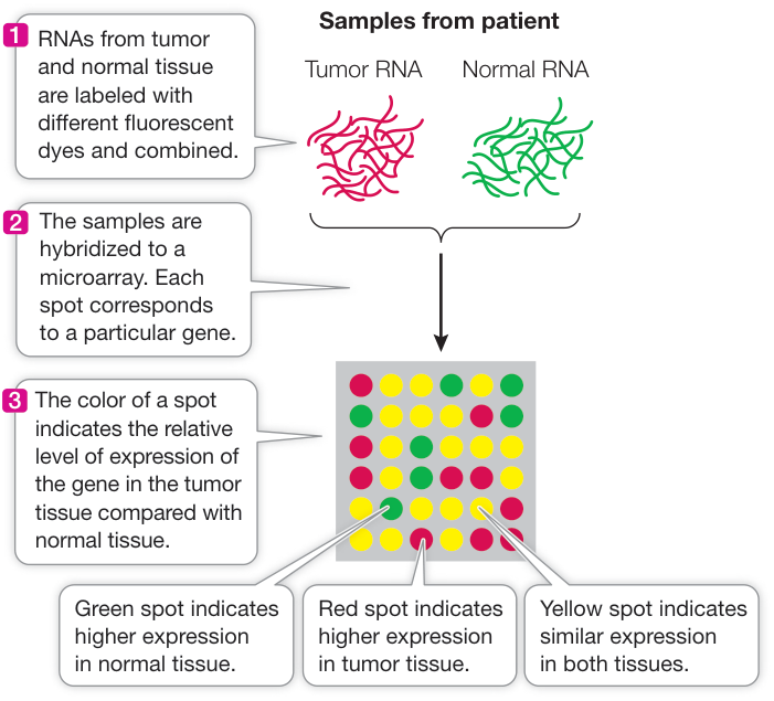
Figure 18.12 Using DNA Microarrays for Clinical Decision-Making The pattern of expression of a group of genes in tumor tissues (the pattern of colored spots) helps clinicians decide on the optimal treatment for a patient. Actual arrays have more spots than shown here.
It may be possible to make drugs that treat diseases by reducing gene expression post-transcriptionally. For example, people with the genetic disease familial hypercholesterolemia are unable to break down cholesterol in their blood and thus accumulate fat deposits in their blood vessels (as was the case with Janet, whose story opened this chapter). This excess cholesterol is transported in blood as part of a lipoprotein. Mipomersen is an antisense RNA drug directed against the translation of the mRNA for the “protein” part of this lipoprotein. When people who have hypercholesterolemia take mipomersen, the level of the lipoprotein is reduced and so is the level of cholesterol in their bloodstream. Fat accumulation in their blood vessels is lowered. An siRNA-based therapy has been developed to treat macular degeneration, an eye disease that results in near-blindness when blood vessels proliferate in the eye. The signaling molecule that stimulates vessel proliferation is a growth factor. An siRNA that targets this growth factor’s mRNA shows promise in stopping and even reversing the progress of the disease.
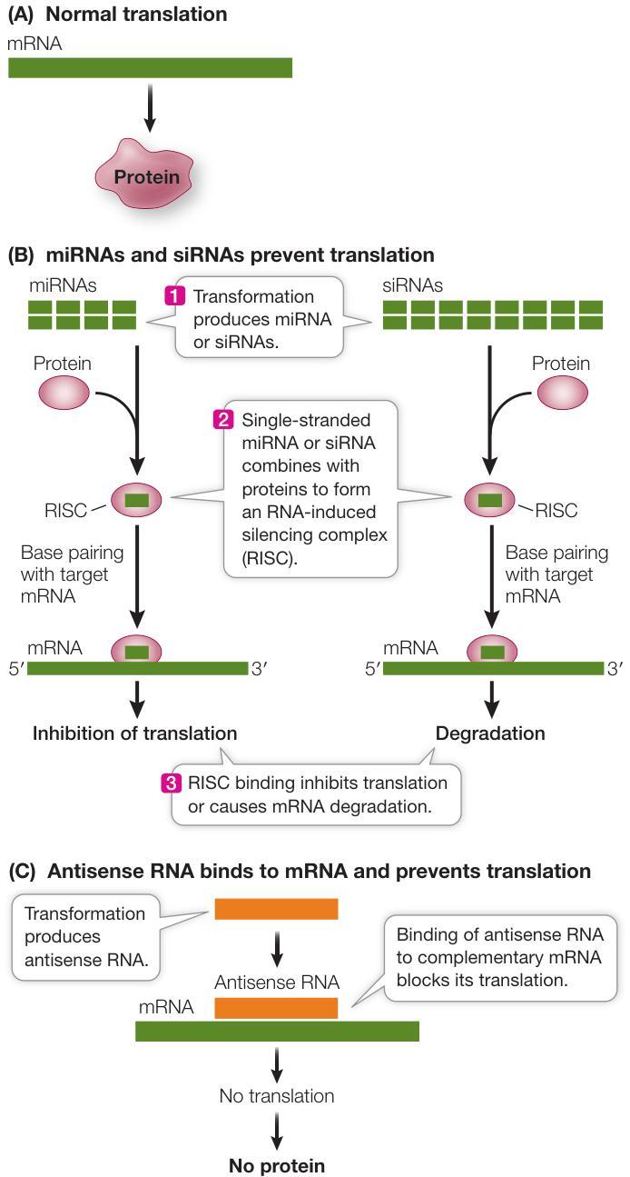
Figure 18.13 Using RNA-Induced Silencing and Antisense RNA to Block Translation of mRNA (A) Normally an mRNA is translated to produce a protein. Translation of a target mRNA can be prevented with (B) RNA-induced silencing using miRNA or siRNA, or with (C) antisense RNA that is complementary to the target mRNA.
Genes can be inactivated and altered using CRISPR technology
Mutations that occur in nature have been important in demonstrating cause-and-effect relationships for specific genes. However, mutations in nature are rare events, and they occur at random locations in genes. DNA technology allows us to inactivate a particular gene in a specific manner to gain insight into its normal function. In addition, DNA technology allows us to explore the consequences of various specific mutations by creating them artificially. Synthetic DNA can be made with any desired sequence and used to replace the wild-type sequence, and its effect on the organism can be observed when the mutant DNA is expressed in host cells. Several methods can be used to knock out specific genes and replace wild-type sequences with particular mutant sequences (or vice versa). One method that has become widespread in recent years involves CRISPR.
CRISPR technology offers a powerful approach for “gene editing” (adding, disrupting, or changing the sequence of specific genes) and for altering gene regulation (Figure 18.14). As with many other tools of biotechnology, the method emulates a mechanism found in nature, one that some bacteria and many archaea use to defend themselves against viral infections. The genomes of these species have many short (24–48 bp) palindromic sequences of DNA (see Key Concept 18.1), with unique 24-base-pair spacer DNA in between, which are known by the acronym CRISPR (clustered regularly interspaced short palindromic repeat sequences). While the CRISPR sequence is the same for each repeating unit, the spacers are different. Each spacer is a fragment of DNA from a virus that previously infected the cell but did not end up killing its host. The spacer sequences thus provide “genetic mug shots” that inform the cell carrying them what viruses to look out for. When a virus carrying a sequence similar to the spacer DNA injects its DNA into the cell, the unit—CRISPR and spacer—is transcribed into RNA, to which a second short RNA attaches. This RNA complex then performs the following two functions:
- Because the spacer sequence is complementary to part of the viral genome, it attaches to it by base pairing.
- The complex binds a protein called CAS9, which is a nuclease that creates a double-strand break of the invading virus DNA at the recognized sequence, destroying its ability to produce new viruses and thereby protecting the host cell.
Therefore any cell that was once infected by a virus has the “memory” of that infection encoded in its genome, and those of all cells that are produced from it.
Once this natural immune system was described, it occurred to Emmanuelle Charpentier at Umeå University in Sweden and Jennifer Doudna at the University of California, Berkeley that it might be adapted to cut any DNA at a specific location: all that was needed was the RNA complementary to the target (a “guide” RNA) and the CAS9 enzyme. It is possible to synthesize a guide RNA that is complementary to any sequence in a genome. This means that transformation of a cell with the CAS9 gene and the appropriate guide RNA can be used to cut host DNA at any site of one’s choosing.
What happens once the DNA has been cut? In a living cell, a double-strand break causes repair pathways to be activated to fix the break. One pathway, called non-homologous end joining (NHEJ), simply ligates the two broken ends back together. NHEJ is error-prone, meaning that repair is imperfect and so mutations are often present after ligation (see Figure 18.14B). If the cut was in a protein-coding gene, then repair by NHEJ can lead to missense, nonsense, or frameshift mutations, potentially rendering the protein nonfunctional.
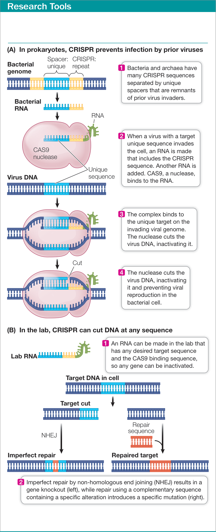
Figure 18.14 Inactivating or Mutating a Gene by CRISPR A molecular system used by bacteria to combat invading viruses (A) has been adapted to inactivate or mutate any gene (B).
The simplicity of CRISPR technology to inactivate genes holds great promise. To give just one example, a fungal disease called powdery mildew can devastate a wheat crop. Many wheat varieties have a gene whose expression inhibits wheat’s natural defenses against this disease, so the only way to control the disease is through heavy application of fungicides. Scientists in China used CRISPR technology to inactivate this gene, so that natural resistance is favored.
CRISPR technology can also be used to create specific mutations. Biologists can specify the mutation by synthesizing a short DNA sequence complementary to the region at the break. Then, when CAS9 cuts the DNA molecule, a repair pathway that uses complementary DNA may use the introduced DNA to make the repair so that the new sequence will have a specific alteration (see Figure 18.14B).
Perhaps more dramatic than the ability to induce mutations is the possibility of correcting mutations, particularly those that cause disease. In these cases CAS9 cuts the mutated gene in the host chromosome and then repair uses a complementary DNA with the wild-type sequence. For example, the disease cystic fibrosis is caused by mutation of a membrane transport protein. Scientists in the Netherlands have used CRISPR to correct the mutation in cells from patients with this disease. While this is not a cure (the corrected gene would have to function in many tissues), the possibility arises that the mutated gene could be corrected in germ line cells, such as those of an early embryo. The ease with which the human germ line genome can be altered using CRISPR technology has elicited numerous ethical and legal concerns.
CRISPR can also be used to knock down expression of a gene. This approach uses a CAS9 that has been modified so that it can no longer cut DNA. The inactivated CAS9 binds both the guide RNA and DNA and can thus be directed to a gene. Once bound, the inactivated CAS9 physically prevents transcription of the gene, even though it does not cut the DNA.
Recap and Assess
Researchers identify genes to study by genetic mapping or by analyzing gene expression, often using DNA microarrays. Gene function can be studied by examining the phenotype following reduction in expression or inactivation of the gene. miRNAs, siRNAs, and antisense RNAs can be used to reduce gene expression by selectively blocking mRNA translation. CRISPR technology allows selective inactivation, alteration, or reduced expression of particular genes.
- In some bladder cancers, the gene for the cell signaling protein, RAS, is mutated, resulting in an altered protein that stimulates cell division. How might RNA-induced silencing be used to develop an RNA drug to treat this disease? Would the drug be specific for the cancer?
- As pea seeds germinate, the embryo changes which genes are expressed. How would you use microarrays to analyze this?
- Both antisense RNA and CRISPR technology can be used to block the expression of specific genes. What additional advantage does CRISPR have?
Now that you’ve seen how DNA can be fragmented, recombined, manipulated, and put back into living organisms, let’s see some examples of how these techniques are used to make useful products.
DNA Can Be Manipulated for Commercial Use
Learning Objectives
- 18.5.1 Describe the essential features of bacterial expression vectors.
- 18.5.2 Summarize the advantages of modern recombinant DNA technology over selective breeding in agriculture.
- 18.5.3 Evaluate the ethical and environmental concerns posed by specific uses of biotechnology, taking into account the risks and rewards that would likely come from their use.
Biotechnology is the use of cells or whole living organisms to produce materials useful to people, such as foods, medicines, and chemicals. People have been using various forms of biotechnology for a long time. For example, the use of yeasts to brew beer and wine dates back at least 8,000 years, and the use of bacterial cultures to make cheese and yogurt is a technique many centuries old. For a long time people exploited these biochemical transformations without being aware of the organisms and genes involved. Today, with recombinant DNA techniques, almost any gene can be inserted into bacteria, yeasts, mammalian cells, plant cells, or even whole organisms, inducing them to make a gene product in large amounts.
Cells can be turned into factories for a desired protein
If a eukaryotic gene is inserted into a typical bacterial plasmid and used to transform E. coli, none of the gene product will be made unless other key prokaryotic DNA sequences are included with the gene so that it is expressed. Chapter 14 described the characteristics of a bacterial gene, all of which are required for expression of a transgene. A bacterial promoter, a transcription termination signal, and a sequence for binding of bacterial ribosomes to the mRNA (the Shine–Dalgarno sequence) must all be included in the transformation vector if the transgene is to be transcribed and translated in a bacterial cell.
To ensure expression of foreign genes by host cells, scientists make expression vectors having not only the characteristics of cloning vectors, but the extra sequences needed for the transgene to be expressed in the host cell. The insertion of an expression vector for a bacterial host is illustrated in Figure 18.15. An expression vector for a eukaryotic host would require the poly A–addition sequence for mRNA stability and a promoter that contains all the elements needed for expression in a eukaryotic cell.
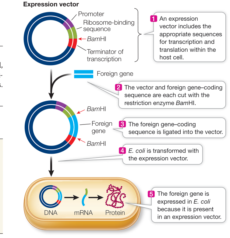
Figure 18.15 Expression of a Transgene in a Host Cell Produces Large Amounts of Its Protein Product To be expressed in E. coli, a gene derived from a eukaryotic requires bacterial sequences for transcription initiation (promoter), transcription termination, and ribosome binding. Expression vectors contain these additional sequences, enabling the eukaryotic protein to be synthesized in the prokaryotic cell. Q: Would a eukaryotic gene obtained from a genomic library produce a functional protein if expressed in a bacterial expression plasmid?
An expression vector can be designed to deliver transgenes to any class of prokaryotic or eukaryotic host and may include additional features:
- An inducible promoter, which responds to a specific signal, can be included. For example, a promoter that responds to hormonal stimulation can be used so that the transgene will be expressed at high levels only when the hormone is added.
- A tissue-specific promoter, which is expressed only in a certain tissue at a certain time, can be used if localized expression is desired. For example, many seed proteins are expressed only in the plant embryo. Coupling a transgene to a seed-specific promoter will allow the gene to be expressed only in seeds.
- Signal sequences can be added so that the gene product is directed to an appropriate destination. For example, when a protein is made by yeast or bacterial cells in a liquid medium, it is economical to include a signal directing the protein to be secreted into the extracellular medium for easier recovery.
Table 18.1 Some Medically Useful Products of Biotechnology
| Product | Use |
|---|---|
| Colony-stimulating factor | Stimulates production of white blood cells in patients with cancer and AIDS |
| Erythropoietin | Prevents anemia in patients undergoing kidney dialysis and cancer therapy |
| Factor VIII | Replaces clotting factor missing in patients with hemophilia A |
| Growth hormone | Replaces missing hormone in people of short stature |
| Insulin | Stimulates glucose uptake from blood in people with insulin-dependent (type I) diabetes |
| Platelet-derived growth factor | Stimulates wound healing |
| Tissue plasminogen activator | Dissolves blood clots after heart attacks and strokes |
| Vaccine proteins: hepatitis B, herpes, influenza, Lyme disease, meningitis, pertussis, etc. | Prevent and treat infectious diseases |
Medically useful proteins can be made using biotechnology
Biotechnology produces many medically useful products (Table 18.1), and more are in various stages of development. A good illustration of a medical application of biotechnology is the manufacture of tissue plasminogen activator (TPA), discussed in the opening of this chapter. As noted there, TPA is a human protein involved in hydrolysis of blood clots and can be used to remove clots that block blood flow in a stroke.
To make TPA in amounts needed for clinical use, scientists first isolated the TPA gene by taking the mRNA for TPA and making a cDNA copy in the lab. The cDNA was then inserted into an expression vector and used to transform mammalian cells (Investigating Life: Producing TPA). The transgenic cells made the protein in quantity, and it soon became available commercially. As you saw at the start of the chapter, the drug has been a success in treating strokes.
Another way of making medically useful products in large amounts is pharming: the production of pharmaceuticals in farm animals or plants. For example, a gene encoding a useful protein might be placed next to the promoter of the gene that encodes lactoglobulin, an abundant milk protein. Transgenic animals carrying this recombinant DNA will secrete large amounts of the foreign protein into their milk. These natural “bioreactors” can produce abundant supplies of the protein, which can be separated easily from the other components of the milk (Figure 18.16).
DNA manipulation is changing agriculture
The cultivation of plants and the husbandry of animals provide the world’s oldest examples of manipulating genetics for human use, dating back more than 10,000 years. Over the centuries, people have adapted crops and farm animals to their needs, producing organisms with desirable characteristics such as large seeds, high fat content in milk, or resistance to disease.
Until recently, the most common way to improve crop plants and farm animals was to identify individuals with desirable phenotypes that existed as a result of natural variation. Through many deliberate crosses—a process called selective breeding—the genes responsible for the desirable trait could be introduced into a widely used variety or breed of that organism.
Connect the Concepts In genetics experiments, typically only a few genes and alleles are involved. But crop plants have phenotypes determined by many genes and alleles. This makes selecting genetically stable, desirable offspring from crosses between two varieties challenging. See Key Concepts 12.2 and 12.3.
Despite some spectacular successes—among them the breeding of high-yielding varieties of wheat, rice, and hybrid corn—such deliberate crossing can be a hit-or-miss affair. Many desirable traits are controlled by multiple genes, and it is hard to predict the results of a cross or to maintain a prized combination as a true-breeding variety year after year, especially because in sexual reproduction combinations of desirable genes are quickly separated by meiosis. Furthermore, traditional breeding takes a long time: many plants and animals take years to reach maturity and then can reproduce only once or twice a year—a far cry from the rapid reproduction of bacteria.
Here are just some of the advantages that modern recombinant DNA technology offers over traditional methods of breeding:
- The ability to identify specific genes. The development of genetic markers allows breeders to select for specific desirable genes, making the breeding process more precise and rapid.
- The ability to introduce any gene from any organism into a plant or animal species. This ability, combined with mutagenesis techniques, vastly expands the range of possible new traits.
- The ability to generate new organisms quickly. Manipulating cells in the laboratory and regenerating a whole plant or animal by cloning is much faster than traditional breeding.
Consequently, recombinant DNA technology has found many applications in agriculture (Table 18.2). Here we describe a few examples to demonstrate the approaches that plant scientists have used to improve crop plants.
PLANTS THAT MAKE THEIR OWN INSECTICIDES Plants are subject to infections by viruses, bacteria, and fungi, but probably the most important crop pests are herbivorous insects. From the locusts
Table 18.2 Agricultural Applications of Biotechnology under Development
| Goal | Technology/genes |
|---|---|
| Improving the environmental adaptations of plants | Genes for drought tolerance, salt tolerance |
| Improving nutritional traits | High-lysine seeds; -carotene in rice |
| Improving crops after harvest | Delay of fruit ripening; sweeter vegetables |
| Using plants as bioreactors | Plastics, oils, and drugs produced in plants |
Investigating Life: Producing TPA
Experiment
Original Paper: D. Collen et al. 1984. Biological properties of human tissue-type plasminogen activator obtained by expression of recombinant DNA in mammalian cells. J Pharmacol Exp Ther 231: 146–152.
For a protein such as TPA to become useful in the clinic, the expression of recombinant DNA (in this case, the TPA gene coupled to an active promoter) in cells must result in the production of a large amount of the protein. The protein is then purified and used to treat patients with a stroke, by catalyzing the dissolution of a blood clot in an artery leading to the brain.
HYPOTHESIS A clinically useful TPA can be made by recombinant DNA technology.
METHOD
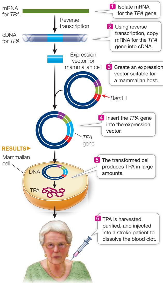
CONCLUSION TPA made by biotechnology can be used to treat patients.
Work with the Data
It is essential that a purified molecule made by biotechnology have the desired biological properties. In the case of TPA, the goal was to mimic the properties of the natural clot-dissolving molecule. Désiré Collen, a physician-scientist in Belgium, led an international team that compared laboratory-made TPA with the natural molecule made by human cells. They performed two types of experiments, one on clots suspended in human blood in the lab, and the other on clots that had developed in rabbits. The team also investigated the time course of clot dissolution and the dose of TPA needed for the effect.
QUESTIONS
- Human blood clots were prepared in the lab using a radioactively labeled clotting molecule. Dissolution of the clot resulted in the radioactive molecules becoming hydrolysis products (monomers), so comparing soluble to insoluble radioactivity was a measure of clot dissolution. Table A shows the results of clot dissolution over time after injection with natural TPA, TPA made by biotechnology, and a control injection without TPA. Plot the data in a graph of percent clot dissolution over time. What can you conclude about the experiment?
- To determine whether lab-made TPA could dissolve clots in the mammalian blood system, clots were induced in a large vein in rabbits. After 20 hours, either natural or lab-made TPA was injected at the clot region, and the dissolution of the clot was measured 4 hours later. The results are shown in Table B. What can you conclude about this experiment? The results show the SEM (standard error of the mean) for the data. How would you use a statistical test to examine the difference, if any, between the two treatment groups and compare them with the control?
Table A
| Percent clot dissolution | |||
|---|---|---|---|
| Time (hr) | No TPA (control) | Natural TPA | Lab-made TPA |
| 1 | 0 | 4 | 5 |
| 2 | 0 | 10 | 20 |
| 3 | 1 | 20 | 35 |
| 4 | 1 | 35 | 55 |
| 5 | 2 | 50 | 65 |
Table B
| Percent clot dissolution (SEM) | ||
|---|---|---|
| Dose of TPA (units) | Natural TPA | Lab-made TPA |
| 0 (control) | 14.3 (1.4) | 14.3 (1.4) |
| 12,000 | 19.8 (5.4) | 22.3 (6.0) |
| 24,000 | 24.5 (7.9) | 30.6 (0.8) |
| 48,000 | 38.9 (4.8) | 49.3 (9.7) |
| 96,000 | 66.0 (6.3) | 75.4 (3.9) |
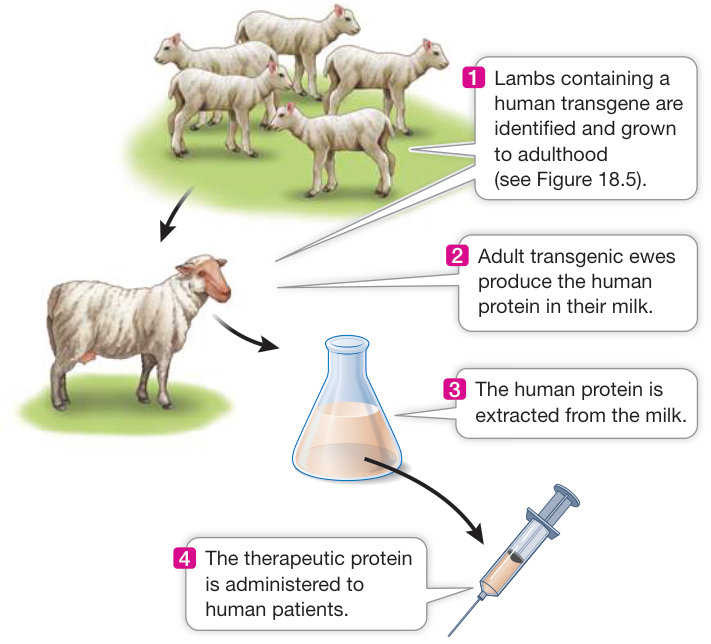
Figure 18.16 Pharming A eukaryotic expression vector carrying a human gene is used to produce transgenic offspring. As adults, these transgenic offspring express the human protein in their milk. The milk is easily harvested and the protein isolated, purified, and made clinically available to patients.
of biblical (and modern) times to the cotton boll weevil (Anthonomus grandis), insects have continually eaten the crops people grow.
The development of insecticides has improved the situation, but insecticides have their own problems. Many, including the organophosphates, are relatively nonspecific and kill beneficial insects in the broader ecosystem as well as crop pests. Some pesticides even have toxic effects on other groups of organisms, including people. What’s more, many insecticides persist in the environment for a long time.
Some bacteria produce proteins that can kill insects. For example, the bacterium Bacillus thuringiensis produces a protein that is toxic to the larvae of several species of insects, but not to mammals or birds. The toxicity of this protein to insects is 80,000 times greater than that of a typical commercial insecticide. When an unlucky larva eats the bacteria, the toxin becomes activated and binds specifically to the insect’s gut, producing holes and killing the insect. Dried preparations of B. thuringiensis have been sold for decades as safe insecticides that break down rapidly in the environment. But the biodegradation of these preparations is their limitation, because it means that the dried bacteria must be applied repeatedly during the growing season.
A longer-acting approach is to have the crop plants themselves make the toxin, and this is exactly what plant scientists have done. The toxin gene from B. thuringiensis has been isolated, cloned, and extensively modified by the addition of a plant promoter and other regulatory sequences. Transgenic corn, cotton, soybeans, tomatoes, and other crops are now being grown successfully with this added gene. Farmers growing these transgenic crops use less of other pesticides.
CROPS THAT ARE RESISTANT TO HERBICIDES Herbivorous insects are not the only threat to agriculture. Weeds may grow in fields and compete with crop plants for water and soil nutrients. Glyphosate is a widely used and effective herbicide, or weed killer, that works only on plants. It inhibits an enzyme system in the chloroplast that is involved in the synthesis of amino acids. Glyphosate is a broad-spectrum herbicide that kills most weeds, but unfortunately it also kills crop plants. One solution to this problem is to use it to rid a field of weeds before the crop plants start to grow. But as any gardener knows, when the crop begins to grow, the weeds reappear. If the crop were not affected by the herbicide, the herbicide could be applied to the field at any time without harming the crop.
Scientists have used expression vectors to make plants that synthesize a different form of the target enzyme for glyphosate that is unaffected by the herbicide. The gene for this enzyme has been inserted into corn, cotton, and soybean plants, making them resistant to glyphosate. The herbicide can then be sprayed on fields in which the resistant crop plants are actively growing, killing only the weeds. This technology has expanded rapidly, and a large proportion of cotton and soybean plants now carry this gene.
GRAINS WITH IMPROVED NUTRITIONAL CHARACTERISTICS To remain healthy, humans must consume adequate amounts of -carotene, which the body converts into vitamin A. About 400 million people worldwide suffer from vitamin A deficiency, which makes them susceptible to infections and blindness. One reason is that rice grains, which do not contain -carotene, make up a large part of their diets. Rice grains lack the two-enzyme biochemical pathway that synthesizes -carotene.
Plant biologists Ingo Potrykus and Peter Beyer isolated one of the genes for the -carotene pathway from the bacterium Erwinia uredovora and the other from daffodil plants. They added a promoter and other signals for expression in the developing rice grain and then transformed rice plants with the two genes. The resulting rice plants produce grains that look yellow because of their high -carotene content. A newer variety with a corn gene replacing the one from daffodils makes even more -carotene and is golden in color (Figure 18.17). A daily intake of about 150 grams of this cooked rice can supply all the -carotene a person needs. This new transgenic strain has been crossed with strains adapted for various local environments, in the hope of improving the diets of millions of people.
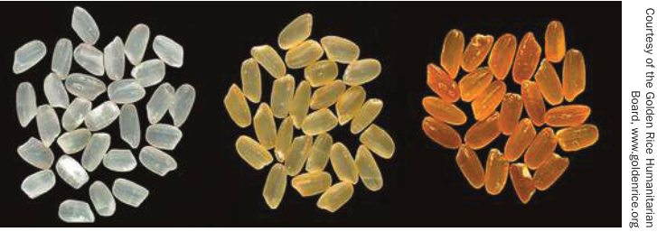
Figure 18.17 Transgenic Rice Rich in -Carotene Middle and right: The grains from these transgenic rice strains are colored because they make the pigment -carotene, which is converted to vitamin A in the human body. Left: Wild-type rice grains do not contain -carotene.
CROPS THAT ADAPT TO THE ENVIRONMENT Agriculture depends on ecological management—tailoring the environment to the needs of crop plants and animals. A farm field is an unnatural, human-designed system that must be carefully managed to maintain optimal conditions for crop growth. For example, excessive irrigation can cause increases in soil salinity. The Fertile Crescent, the region between the Tigris and Euphrates rivers in the Middle East where agriculture probably originated 10,000 years ago, is no longer fertile. It is now a desert, largely because the soil has a high salt concentration. Few plants can grow on salty soils, partly because of osmotic effects that result in wilting, and partly because excess salt ions are toxic to plant cells.
Connect the Concepts Learn about the effects of salt on plants in Key Concept 37.1.
Some plants can tolerate salty soils because they have a protein that transports ions out of the cytoplasm and into the vacuole, where the ions can accumulate without harming plant growth (see Key Concept 5.3 for a description of the plant vacuole). Scientists developed a highly active version of the gene for this transporter protein and used it to transform crop plants that are less tolerant to salt, including rapeseed, wheat, and tomatoes. When this gene was added to tomato plants, they grew in water that was four times as salty as the typical lethal level (Figure 18.18). This finding raises the prospect of growing crops on what were previously unproductive soils.
Perhaps the most spectacular transgenic crop developed so far is a strain of rice that is resistant to both salt stress (using the gene just mentioned) and drought stress (using a gene from a bacterium) and has the ability to grow on nitrogen-deficient soils (using a gene from barley). This triply-tolerant rice is undergoing field tests.
These examples illustrate what could become a fundamental shift in the relationship between crop plants and the environment. Instead of manipulating the environment to suit the plant,
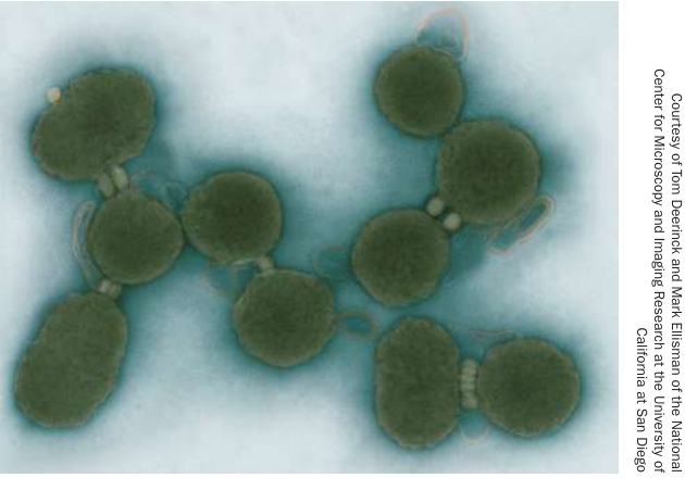
Figure 18.19 Synthetic Cells These cells of Mycoplasma mycoides contain a synthetic genome. Q: What are some potential uses of synthetic cells?
biotechnology may allow us to adapt the plant to the environment. As a result, some of the negative effects of agriculture, such as water pollution, could be lessened.
Synthetic biology can create living factories for new products
In Key Concept 17.2 you learned how inactivating genes one at a time can help define which genes are necessary for life in a particular organism and which ones confer special properties to the organism. One goal of this research is to design new life forms with specific purposes. That is, new genes could be added to the minimal genome. An important step toward this goal has been the laboratory synthesis of an artificial genome and its insertion into bacterial cells. This was first done using the bacterium Mycoplasma mycoides. The artificial genome was then inserted into empty cells of a related species, Mycoplasma capricolum, whose own DNA had been hydrolyzed. The new DNA directed the cell to perform the biochemical functions of life, including reproduction (Figure 18.19).
The aims of synthetic biology are bold: to create bacteria or other organisms with new functions, such as producing hydrocarbons for fuel, new drugs for medicine, bacteria that make or break down plastics and other polymers, and so on. But before these functions can be added to the genome, the genes involved must be clearly described, including regulatory genes and sequences such as promoters that function in host cells. Thousands of such genes and chemicals that regulate their expression have now been associated with many known gene functions. For example, a single function—bacteria making ammonium sulfate that could be used for fertilizer in farming—requires more than 70 genes.
There is public concern about biotechnology
Concerns have been raised about the wisdom and safety of genetically modifying crops and other organisms. These concerns are centered on three claims:
- Genetic manipulation is an unnatural interference with nature.
- Genetically altered foods are unsafe to eat.
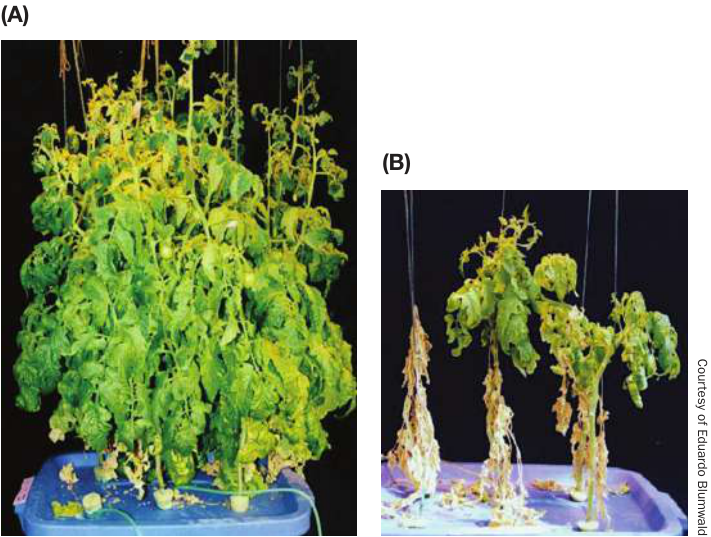
Figure 18.18 Salt-Tolerant Tomato Plants Transgenic plants containing a gene for salt tolerance thrive in salty water (A), whereas plants without the transgene die (B). This technology may allow crops to be grown on salty soils.
3. Genetically altered crop plants are dangerous to the environment.
Advocates of biotechnology tend to agree with the first claim. However, they point out that all crops are unnatural in the sense that they come from artificially bred plants growing in a manipulated environment (a farmer’s field). Recombinant DNA technology just adds another level of sophistication to these technologies. Synthetic biology, however, takes genetic manipulation dramatically further.
To counter concern about whether genetically engineered crops are safe for human consumption, biotechnology advocates point out that only single genes are added and that these genes are specific for plant function. For example, the B. thuringiensis toxin produced by transgenic plants has no effect on people. However, as plant biotechnology moves from adding genes that improve plant growth to adding genes that affect human biology, such concerns will become more pressing.
Various negative environmental impacts have been envisaged. The concern has been raised, for example, about the possible “escape” of transgenes from crops to other species. If the gene for herbicide resistance, for example, were inadvertently transferred from a crop plant to a closely related weed, that weed could thrive in herbicide-treated areas. Increased use of herbicides has already resulted in the selection of weeds with naturally occurring mutations that make them resistant to that herbicide. Widespread use of glyphosate on fields of glyphosate-resistant crops, for example, has resulted in the selection of rare mutations in weeds that make them resistant to glyphosate. Field-evolved resistance now occurs in half the major weed species examined. In addition, herbicides may cause harm either when consumed or to other non-target organisms present in the environment. For example, glyphosate use has been implicated in changes in the gut microbiome (see Key Concept 24.3) of honey bees, and is listed by the World Health Organization as a probable human carcinogen.
Because of the potential benefits of biotechnology, scientists believe that it is wise to proceed, albeit with caution.
Recap and Assess
Expression vectors allow the expression of transgenes in host cells. Biotechnology has been used to produce medicines and to develop transgenic plants with improved agricultural and nutritional characteristics.
- What are the similarities and differences between a cloning vector and an expression vector?
- Compared with traditional methods, what are the advantages of using biotechnology for plant breeding?
- After Cohen and Boyer did their experiment (see Key Concept 18.1), a group of scientists, public health officials, and bioethicists held a conference and agreed to a temporary moratorium on most recombinant DNA research. What do you think were the concerns that led to the moratorium? How was safety assured so that the moratorium was lifted?
Investigating Life: QA How is biotechnology changing medicine?
What are the limitations of treating patients with the clot-busting drug TPA, made by biotechnology? We opened this chapter with the story of Janet’s stroke, pointing out how important it is to treat stroke patients quickly. An analysis published in 2014 of many studies done in the 20 years since TPA was first clinically used showed that the probability of having no significant disability is 75% higher in people who get TPA within 3 hours of stoke onset compared with people who don’t get TPA. This benefit is reduced if a patient gets the drug later and is almost nonexistent at 6 hours poststroke.
One of the problems that arose when TPA treatments were initially administered was that the drug tended to break down rapidly in the bloodstream after injection. Biotechnologists solved this problem by altering the TPA gene slightly so that the protein gets glycosylated (adding sugars), which leads to a longer lifetime in the bloodstream. As other drugs are developed by biotechnology, the TPA story is being repeated: initial success, problems with the chemistry, and then modification.
While the commercial production of drugs by DNA biotechnology has had some successes, the examples are relatively limited and certainly not as notable as those in traditional biotechnology, where bacteria and other microbes are manipulated by their environment to make large amounts of molecules, such as antibiotics. However, as we described, DNA manipulation technologies, especially CRISPR, hold great promise to profoundly change medical diagnosis and treatment.
Future directions
A striking example of “old” biotechnology (in which a microorganism is coaxed into making a valuable product) merging with “new” biotechnology (which uses recombinant DNA) is the emerging field of cyanobacteria-produced plant secondary metabolites. Plants make these small-molecule metabolites to protect themselves against infections, injuries, and environmental factors. Many of the metabolites are strong antioxidants that react with and eliminate harmful oxidants such as oxygen atoms with unpaired electrons (superoxides) in cells under stress. Because human cells in disease states produce such oxidants that harm tissues, there is intense interest in using certain secondary metabolites that plants produce as drugs. Unfortunately, it takes a huge quantity of plants to obtain enough of a given antioxidant for clinical use.
This is where cyanobacteria come in. Cyanobacteria (sometimes incorrectly called blue-green algae) are photosynthetic single-celled organisms that grow well under light conditions in ponds or vats in the lab. Although they are not plants, cyanobacteria have genes encoding some of the enzymes involved with secondary metabolite production. Using recombinant DNA technology, genes from plants that encode other needed compounds in the pathway of certain antioxidants (specifically, phenylpropanoids) have been inserted into cyanobacteria, turning these organisms into factories that produce plant metabolites for clinical use.
© Erik Isakson/Getty Images
You should be able to relate each summary to the adjacent figures. If you go to this visual summary in Achieve, you can follow links to figures, animations, activities, and simulations that will help you consolidate the material.
Visual Summary
DNA from Different Sources Forms Recombinant DNA
- Restriction enzymes make blunt or staggered cuts in DNA at specific palindromic sequences. Staggered cuts generate fragments with sticky ends.
- DNA ligase is used to join DNA from different sources.
- The first recombinant DNA molecules combined parts of two different bacterial plasmids.
Questions
- The restriction enzyme Rsal has a four-base-pair recognition site. Assuming all four base pairs are equally frequent, how many Rsal cut sites would you expect there to be in a 5,000-base-pair plasmid?
- Why are restriction enzymes that make staggered cuts, rather than those that make blunt cuts, preferred for making recombinant DNA?
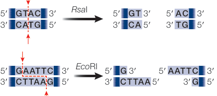
Figure 18.1
There Are Several Ways to Insert DNA into Cells
- Single-celled organisms are often used for transformation because a transgenic individual is easily made and cloned.
- Transforming multicellular organisms involves transforming zygotes or cells in culture.
- Cloning vectors contain a cloning site (a restriction enzyme recognition sequence), an origin of replication, and a selectable marker.
- Reporter genes such as lacZ and GFP can be used to screen for an insert in a cloning vector, measure expression, or determine protein location.
- Producing numerous clones of multicellular, sexual organisms involves producing inbred, genetically uniform individuals that are homozygous for the transgene.
Questions
- What are the functions of each of the required attributes of a cloning vector?
- How can a multicellular transgenic animal be produced by transforming a single cell?
- What is the difference between a genetically uniform transgenic strain of yeast and a transgenic strain of mouse?
DNA for Study Is Obtained in a Variety of Ways
- DNA libraries consist of thousands to millions of hosts (often bacterial or viral) carrying foreign DNA in cloning vectors.
- Genomic libraries are created by inserting DNA fragments from a genome into host cells; cDNA libraries are created by reverse transcription of mRNA from a particular tissue and developmental stage.
- DNA or cDNA sequences can be amplified by PCR for cloning or for measuring gene expression.
- Synthetic DNA can be used as PCR primers, to make probes for hybridization to identify genotypes, and to make custom genes with particular sequences.
Questions
- A bacterial plasmid can comfortably handle a foreign DNA insert of up to 5,000 base pairs. Approximately how many bacterial clones (each containing a single plasmid) would be needed to make a genomic library for yeast, with a haploid genome of 12 million base pairs?
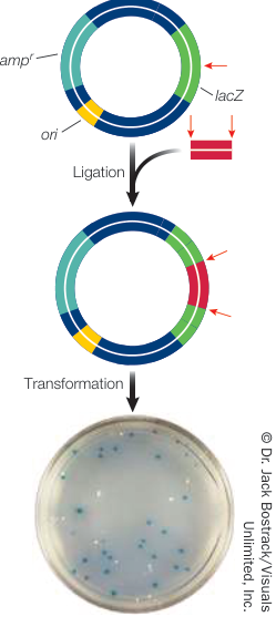
Figure 18.8
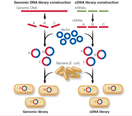
Figure 18.11
- Explain how a cDNA library constructed from human brain tissue is likely to be similar to and different from a cDNA library constructed from human pancreatic tissue.
- Explain how a genomic DNA library constructed from human brain tissue is likely to be similar to and different from a genomic library constructed from human pancreatic tissue.
Several Tools Are Used to Study the Function of DNA
and Simulation 18.1
- ■ Scientists often study genes that are responsible for genetic diseases or for desirable traits in crop species, or that are expressed at particular times or locations.
- ■ DNA microarrays are used to examine expression of all the genes in the entire genome in a particular tissue, or tissues.
- ■ The function a gene can study by using posttranscriptional RNA interference to reduce or eliminate its expression.
- ■ CRISPR allows a gene to be knocked out, knocked down, or altered.
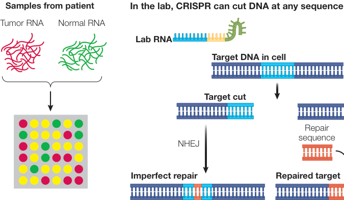
Figure 18.12
Questions
- A scientist wishes to study genes involved in early eye development in squid. How might she go about finding such genes?
- After isolating a candidate gene involved in eye development, a researcher decides to try reducing its expression
- using RNA interference and scoring the effect on eye development. Outline the general steps to do this.
- Suppose miRNA targeting of the candidate squid gene causes a subtle change in eye phenotype. The researcher now wishes to knock out the gene. How might she accomplish this using CRISPR?
DNA Can Be Manipulated for Commercial Use
- ■ A bacterial expression vector has all three characteristics of a cloning vector (cloning site, selectable marker, and origin of replication) as well as a promoter, transcriptional termination sequence, ribosome binding site (for prokaryotic hosts), and poly A sequence (for eukaryotic hosts).
- ■ Modern recombinant technology allows the introduction of specific foreign genes into a species.
- ■ Transgenic plants often have a much higher yield because of reduced levels of pathogen attack (using pesticide genes), less competition (using herbicide-resistant genes), and increased ability to grow well in poor conditions.
Questions
- Inserting a eukaryotic gene into a bacterial expression vector and obtaining a functional protein can be challenging. Even when a gene is inserted into the vector, the resulting protein may be substantially different from the normal protein. Why might this be the case?
- Sometimes a human protein-coding gene is placed under the control of a milk protein promoter and then a strain of sheep carrying the transgene is produced. What is the advantage of such a transgene?
- Transgenic plants carrying the Bacillus thuringiensis toxin gene show high yields. Why is this the case?
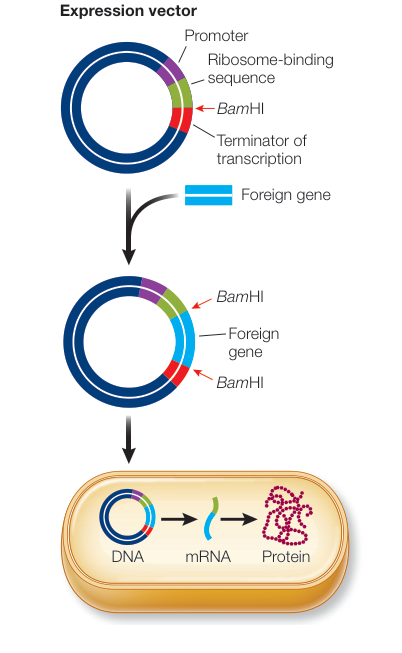
Figure 18.5
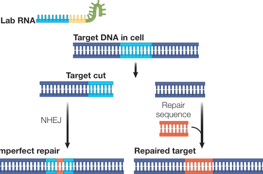
Figure 18.14