Chapter 28The Evolution and Diversity of Fungi
© Scott Science Services September 27/8/2019
Key Concepts
28.1 Fungi Digest Food Outside Their Bodies
28.2 Fungi Are Decomposers, Parasites, Predators, or Mutualists
28.3 Sex in Fungi Involves Multiple Mating Types
28.4 Fungi Have Many Practical Uses
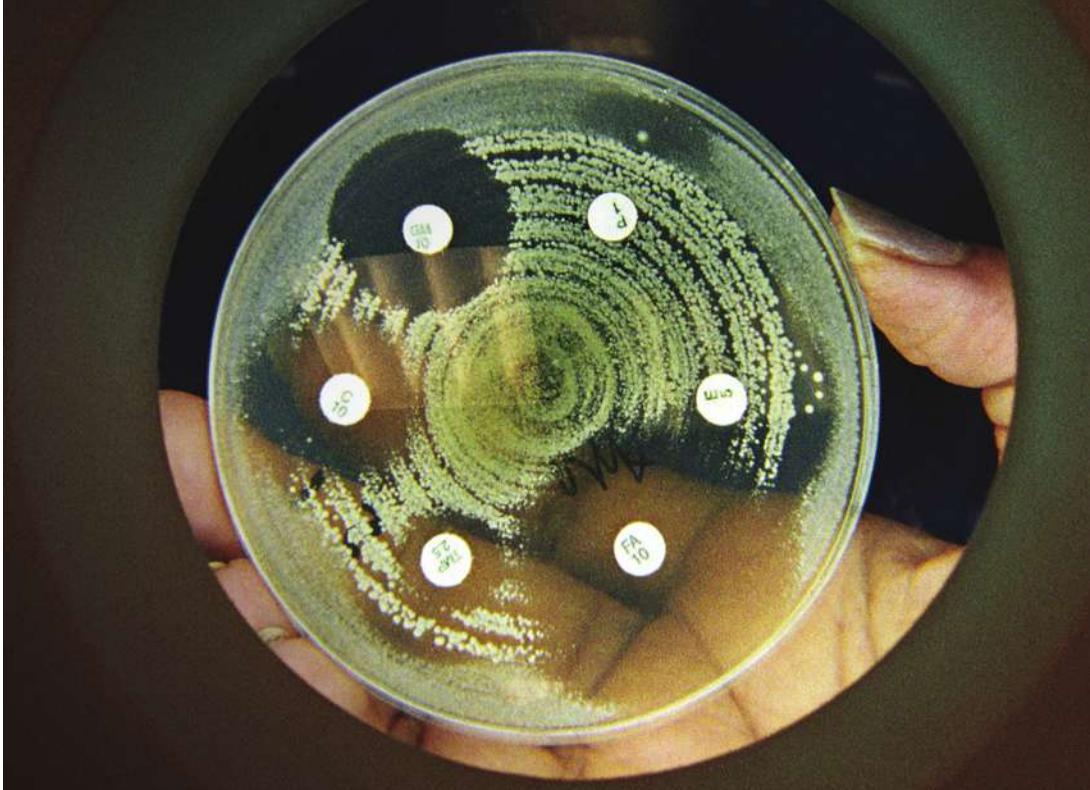
Six antibiotics (contained on white disks) are tested for their effectiveness against a bacterium (background of yellow-green colonies). The larger zones of growth inhibition (clear areas around white disks) indicate higher antibiotic effectiveness.
Investigating Life: The Accidental Discovery of Antibiotics
Alexander Fleming was already a famous scientist in 1928, but his laboratory was often a mess. That year he was studying the properties of Staphylococcus bacteria, the agents of dangerous staph infections. In August he took a long vacation with his family. When he returned in early September, he found that some of his petri dishes of Staphylococcus had become infested with a fungus that killed many of the bacteria.
Many scientists would have sighed at the loss, thrown out the petri dishes, and started new cultures of bacteria. But when Fleming looked at the dishes, he saw something exciting. Around each colony of fungi was a ring within which all the bacteria were dead.
Fleming hypothesized that the bacteria-free rings around the fungal colonies were produced by a substance excreted from the fungi, which he initially called “mould juice.” He identified the fungi as members of the genus Penicillium and eventually named the antibacterial substance they produced “penicillin.” Fleming published his discovery in 1929, but initially it received very little attention.
Over the next decade, Fleming produced small quantities of penicillin for testing as an antibacterial agent. Some of the tests showed promise, but many were inconclusive, and eventually Fleming gave up on the research. But his tests had shown enough promise to attract the attention of several chemists, who worked out the practical problems of producing a stable form of the substance. Clinical trials of this stable form of penicillin were extremely successful, and by 1945 it was being produced and distributed as an antibiotic on a large scale. That same year, Fleming and two of the chemists, Howard Florey and Ernst Chain, received the Nobel Prize in Physiology or Medicine for their work on penicillin.
The development of penicillin was one of the most important achievements in modern medicine. Until the introduction of modern antibiotics, the most widespread agents of human death included bacterial infections such as gangrene, tuberculosis, and syphilis. Penicillin proved to be highly effective in curing such infections, and its success led to the creation of the modern pharmaceutical industry. Soon many additional antibiotic compounds were isolated from other fungi or synthesized in the laboratory, leading to a “golden age” of human health.
QA Why are the regions of inhibited bacterial growth around the various antibiotics so different in size, and how is that relevant to human health care? (Find the answer on page 642.)
Fungi Digest Food Outside Their Bodies
Learning Objectives
28.1.1 Explain how the structure of fungi facilitates their ability to decompose and consume other organisms.
28.1.2 Assess the ability of fungi to exist in different environments.
Fungi are distinctive in the way they digest their food. They secrete digestive enzymes to break down large food molecules in the environment, then absorb the breakdown products through their cell membranes in a process known as absorptive heterotrophy. This mode of nutrition allows them to be successful in a wide variety of environments. Many fungi are saprobes, meaning they absorb nutrients from dead organic matter. Others are parasites or predators, feeding on or consuming other living organisms. Still others are mutualists, living in intimate associations with other organisms that benefit both partners.
Modern fungi evolved from a unicellular protist ancestor that had a flagellum, although most fungi are not motile and have now lost this structure. The probable common ancestor of the animals was also a flagellated protist much like the living choanoflagellates (see Figure 29.2). Current evidence, including the sequences of many genes, suggests that the fungi, choanoflagellates, and animals share a common ancestor not shared by other eukaryotes. These three lineages form a group known as the opisthokonts (Figure 28.1). A synapomorphy (shared derived trait) of the opisthokonts is a flagellum that, if present, is posterior, as in animal sperm. The flagella of all other eukaryotes attach at the front or sides of the cell, rather than at the rear.
Synapomorphies that distinguish the fungi as a group among the opisthokonts include absorptive heterotrophy and the presence of chitin, a nitrogen-containing structural polysaccharide, in their cell walls. The fungi represent one of the four independent evolutionary origins of large multicellular organisms (plants, brown algae, and animals are the other three).
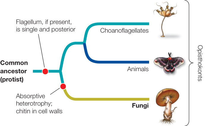
Figure 28.1 Fungi in Evolutionary Context Absorptive heterotrophy and the presence of chitin in their cell walls distinguish the fungi from other opisthokonts.
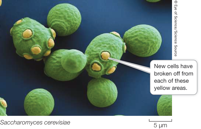
Figure 28.2 Yeasts Unicellular, free-living fungi are known as yeasts. Many yeasts reproduce by budding—mitosis followed by asymmetrical cell division—as illustrated here.
Yeasts are unicellular, free-living fungi
Most fungi are multicellular, but single-celled species are found in most fungal groups. Unicellular, free-living fungi are referred to as yeasts (Figure 28.2). Some fungi have both a yeast life stage and a multicellular life stage. Thus the term “yeast” does not refer to a single taxonomic group, but rather to a lifestyle that has evolved multiple times. Yeasts live in liquid or moist environments and absorb nutrients directly across their cell surfaces.
The ease with which many yeasts can be cultured, combined with their rapid growth rates, has made them ideal model organisms for study in the laboratory. They present many of the same advantages to laboratory investigators as do many bacteria, but because they are eukaryotes, their genome structures and cells are much more like those of humans and other eukaryotes than are those of bacteria.
Connect the Concepts Why we select and study a variety of model organisms is discussed in Key Concept 17.3. Each model organism has distinctive advantages and disadvantages. The features of yeasts make them ideal for some investigations but impractical for others.
Multicellular fungi use hyphae to absorb nutrients
The body of a multicellular fungus is called a mycelium (plural mycelia). A mycelium is composed of a branching mass of individual tubular filaments called hyphae (singular hypha; Figure 28.3A), in which absorption of nutrients takes place primarily at each tip. The cell walls of the hyphae are greatly strengthened by microscopic fibrils of chitin. In some species of fungi, the hyphae are subdivided into cell-like compartments by incomplete cross-walls called septa (singular septum). These subdivided hyphae are referred to as septate. Septa do not completely close off compartments in the hyphae. Pores at the centers of the septa allow cytoplasm and organelles—sometimes even nuclei—to move in a controlled way between compartments (Figure 28.3B). In other species of fungi, the hyphae lack septa but may contain hundreds of nuclei. These multinucleate, undivided hyphae are referred to as coenocytic. The coenocytic condition results from repeated nuclear divisions without cytokinesis.
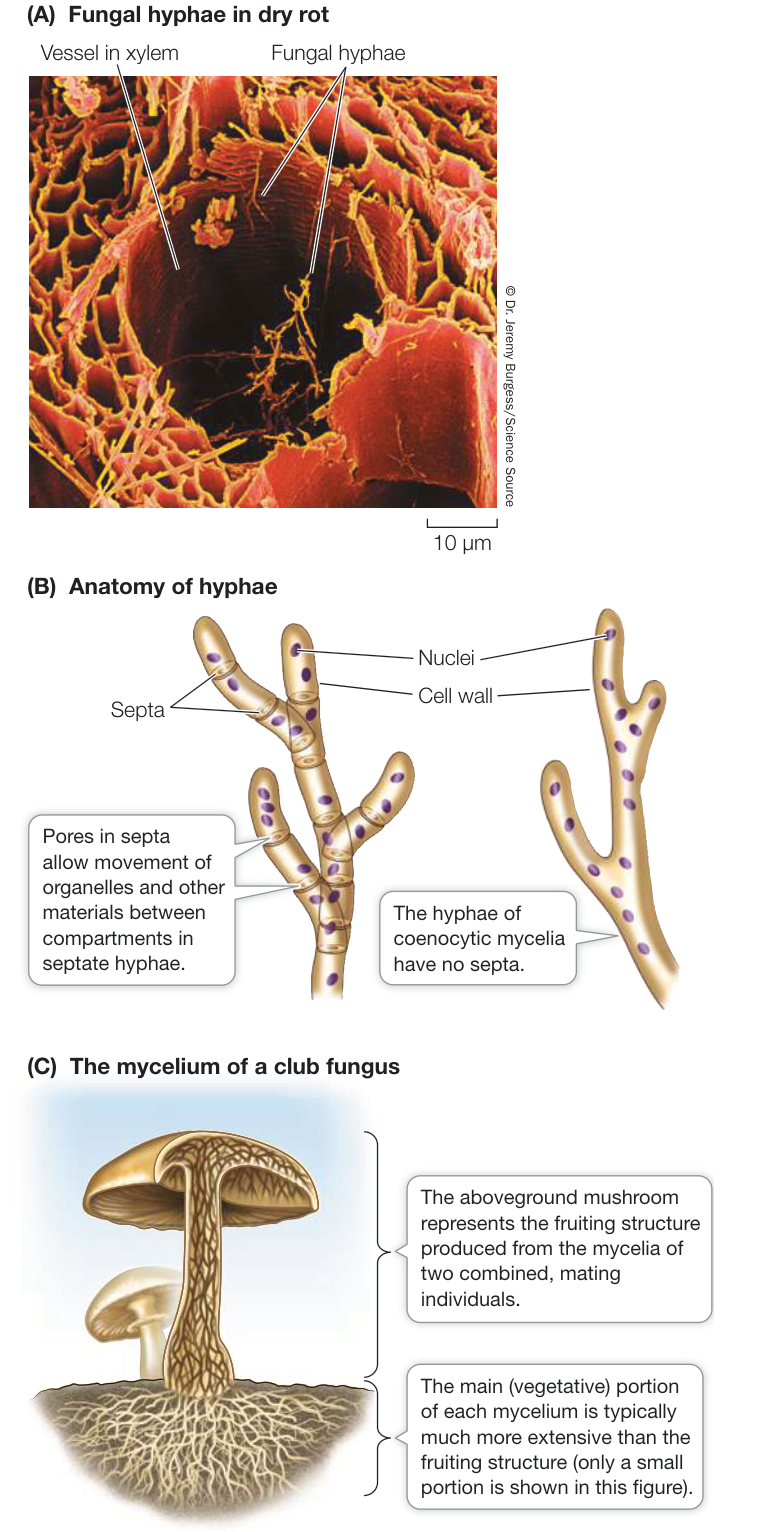
Figure 28.3 Mycelia Are Made Up of Hyphae (A) The minute individual hyphae of fungal mycelia can penetrate small spaces. In this artificially colored micrograph, hyphae (yellow structures) of a dry-rot fungus are penetrating the xylem tissues of a log. (B) The hyphae of septate fungal species are divided into organelle-containing compartments by porous septa. The hyphae of coenocytic fungal species have no septa. (C) The fruiting structure of a club fungus is short-lived, but the filamentous, nutrient-absorbing mycelium can be long-lived and cover large areas. 1. 1. How does fungal structure facilitate absorptive heterotrophy? 2. 2. What are the advantages and disadvantages to multicellular fungi of the large surface area-to-volume ratio of the mycelium? How does the mycelium restrict the types of environments in which fungi are generally found?
Certain modified hyphae, called rhizoids, anchor some fungi to their substrate (i.e., the dead organism or other matter on which they grow). These rhizoids are not homologous to the rhizoids of plants, and they are not specialized to absorb nutrients and water.
Fungi can grow very rapidly when conditions are favorable. In some species, the total hyphal growth of a fungal mycelium (not the growth of an individual hypha) may exceed 1 kilometer a day! The hyphae may be widely dispersed to forage for nutrients over a large area, or they may clump together in a cottony mass to exploit a rich nutrient source. The familiar mushrooms you may notice growing in moist areas are spore-producing fruiting structures (Figure 28.3C). In the fungal species that produce these structures, the mycelial mass is often far larger than the visible mushroom. The mycelium of one individual fungus discovered in Oregon covers almost 900 hectares underground and weighs considerably more than a blue whale (the largest animal). Aboveground, this individual is evident only as isolated clumps of mushrooms.
Fungi are in intimate contact with their environment
The filamentous hyphae of a fungus give it a unique relationship with its physical environment. The fungal mycelium has an enormous surface area-to-volume ratio compared with that of most large multicellular organisms. This large ratio allows the fungus to take in a great deal of water and mineral nutrients when soil moisture is high, which allows rapid growth in moist environments. The downside of the large surface area-to-volume ratio of the mycelium is its tendency to lose water rapidly in a dry environment. Thus fungi are most common in moist environments. You have probably observed the tendency of molds, toadstools, and other fungi to appear in damp places.
Another characteristic of some fungi is a tolerance for highly hypertonic environments (those with a solute concentration higher than their own; see Key Concept 6.3). Many fungi are more resilient than bacteria in hypertonic surroundings. Jelly, for example, does not support bacterial growth because its high sugar content makes it too hypertonic to those organisms, but it may eventually harbor mold colonies. Mold in the refrigerator illustrates yet another trait of many fungi: tolerance of temperature extremes. Many fungi grow in temperatures as low as , and some can tolerate temperatures higher than .
Recap and Assess
Fungi, like animals, are opisthokonts. Fungi are distinguished from other opisthokonts by absorptive heterotrophy and by the presence of chitin in their cell walls. Unicellular fungi called yeasts absorb nutrients directly across their cell surfaces. The body form of multicellular fungi—a mycelium made up of rapidly growing hyphae—allows them to practice absorptive heterotrophy efficiently in a variety of moist environments.
Fungi are important components of healthy ecosystems. They interact with other organisms in many ways, some of which are harmful and some beneficial to those other organisms.
Fungi Are Decomposers, Parasites, Predators, or Mutualists
Learning Objectives
- 28.2.1 Explain how saprobic fungi are critical to Earth’s carbon cycle.
- 28.2.2 Describe the relationship and summarize the benefits to each partner in a lichen association.
- 28.2.3 Describe the specific benefits that mycorrhizal interactions provide for fungi and their associated partner organisms.
Without the fungi, our planet would be very different. Picture Earth with only a few stunted plants and watery environments choked with the remains of dead organisms. Fungi do much of Earth’s garbage disposal. Fungi not only help clean up the landscape and form soil, but also play a key role in recycling mineral nutrients. Furthermore, the colonization of the terrestrial environment was made possible in large part by associations that fungi formed with land plants and other organisms.
Saprobic fungi are critical to the planetary carbon cycle
Saprobic fungi, along with bacteria, are the major decomposers on Earth, contributing to the decay of nonliving organic matter and thus to recycling of the elements used by living things. In forests, for example, the mycelia of fungi secrete extracellular enzymes that degrade plant materials into simpler compounds that are then absorbed. Fungi are the principal decomposers of cellulose and lignin, the main components of plant cell walls (most bacteria cannot break down these materials). Other fungi produce enzymes that decompose keratin and thus break down animal structures such as hair and nails.
Were it not for the fungal decomposers, Earth’s carbon cycle would fail. Great quantities of carbon atoms would remain trapped forever on forest floors and elsewhere. Instead, those carbon atoms are returned to the atmosphere in the form of CO2 by fungal respiration, where they are again available for photosynthesis by plants.
Connect the Concepts Earth’s carbon cycle is discussed in detail in Key Concept 56.4. Fungi play a critical role in this cycle by decomposing other organisms and releasing carbon dioxide.
There was a time in Earth’s history when populations of saprobic fungi declined dramatically. Vast tropical swamps existed during the Carboniferous and much of the Permian, as you saw in Chapter 23. When plants in these swamps died, they began to form peat. Peat formation led to acidification of the swamps. That acidity, in turn, drastically reduced the fungal population. The result? With the decomposers largely absent, large quantities of peat remained on the swamp floor and over time were converted into coal.
In contrast to their decline during the Carboniferous and much of the Permian, fungi did very well at the end of the Permian, a quarter of a billion years ago, when the aggregation of continents produced volcanic eruptions that triggered a global mass extinction. The fossil record shows that even as the majority of all multicellular species became extinct, fungi flourished—demonstrating both their hardness and their role in recycling the elements in dead plants and animals.
Simple sugars and the breakdown products of complex polysaccharides are the favored sources of carbon for saprobic fungi. Most fungi obtain nitrogen from proteins or the products of protein breakdown. Many fungi can use nitrate (NO3-) or ammonium (NH4+) ions as their sole source of nitrogen. No known fungus can get its nitrogen directly from inorganic nitrogen gas, however, as can some free-living and plant-associated bacteria (that is, fungi cannot fix nitrogen; see Key Concept 24.3).
What happens when a fungus faces a dwindling food supply? A common strategy is to reproduce rapidly and abundantly. When conditions are good, fungi produce great quantities of spores, but the rate of spore production is commonly even higher when nutrient supplies go down. The spores may then remain dormant until conditions improve, or they may be dispersed to areas where nutrient supplies are higher.
Not only are fungal spores abundant in number, but they are extremely tiny and easily spread by wind or water (Figure 28.4). These attributes virtually ensure that the spores will be scattered over great distances and that at least some of them will find conditions suitable for growth. The air we breathe contains as many as 10,000 fungal spores per cubic meter. No wonder we find fungi just about everywhere.
Media Clip 28.1 Fungal Decomposers Life12e.com/mc28.1
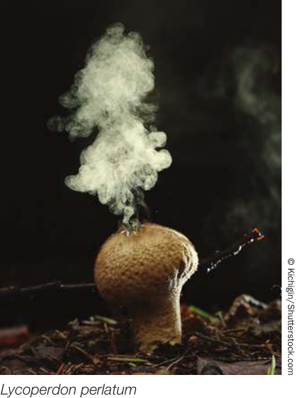
Figure 28.4 Spores Galore Puffballs (a type of club fungus) disperse trillions of spores in great bursts. Few of the spores travel very far, however; some 99% of them fall within 100 meters of the parent puffball.
Some fungi engage in parasitic or predatory interactions
Whereas saprobic fungi obtain their energy, carbon, and nitrogen directly from dead organic matter, other species of fungi obtain their nutrition from parasitic—and even predatory—interactions.
PARASITIC FUNGI Mycologists (biologists who study fungi) distinguish between two classes of parasitic fungi based on their degree of dependence on their host. Facultative parasites can grow on living organisms but can also grow independently (including on artificial media). Obligate parasites can grow only on a specific, living host. The fact that their growth depends on a living host shows that obligate parasites have specialized nutritional requirements.
Plants and insects are the most common hosts of parasitic fungi. The filamentous structure of fungal hyphae is especially well suited to a life of absorbing nutrients from living plants. The slender hyphae of a parasitic fungus can invade a plant through stomata, through wounds, or in some cases, by direct penetration of epidermal cell walls (Figure 28.5A). Once inside the plant, the hyphae branch out to expand the mycelium. Some hyphae produce haustoria, branching projections that push through cell walls into living plant cells, absorbing the nutrients within those cells. The haustoria do not break through the cell membranes inside the cell walls, but instead invaginate into the membranes, so that the cell membrane fits them like a glove (Figure 28.5B). Fruiting structures may form, either within the plant body or on its surface.
Some parasitic fungi live in a close physical (symbiotic) relationship with a plant host that is usually not lethal to the plant.
Others are pathogenic, weakening or even killing the host from which they derive nutrition.
PATHOGENIC FUNGI Although most human diseases are caused by bacteria or viruses, fungal pathogens are a major cause of death among people with compromised immune systems. Many people with AIDS die of fungal diseases, such as the pneumonia caused by Pneumocystis jirovecii. Even Candida albicans and certain other yeasts that are normally part of a healthy microbiome can cause severe diseases, such as esophagitis (which impairs swallowing), in individuals with AIDS and in individuals taking immunosuppressive drugs. Various fungi cause other, less threatening human diseases, such as ringworm and athlete’s foot. The relatively close relationship between fungi and animals makes it difficult to find drugs that target unique features of fungal metabolism without harming human patients. As a result, fungal diseases are a growing international health problem.
The worldwide decline of amphibian species has been linked to the spread of a chytrid fungus, Batrachochytrium dendrobatidis (or Bd for simplicity). In some areas of the world where this fungus has been present for millennia, amphibian populations appear to be tolerant of Bd and it does not cause widespread die-offs. But a pathogenic strain known as the global pandemic lineage of Bd appears to have been introduced around the world in recent decades, likely through commercial trade of amphibians. These introductions of the global pandemic lineage of Bd are implicated in the loss or decline of many frog and salamander populations, and even the extinction of entire species.
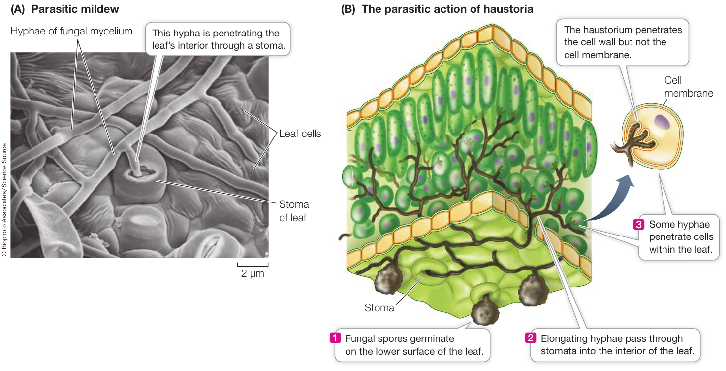
Figure 28.5 Invading a Leaf (A) Hyphae of the mildew Phyllactinia guttata growing on the surface of a hazel leaf. (B) Haustoria are fungal hyphae that push into the living cells of plants, from which they absorb nutrients.
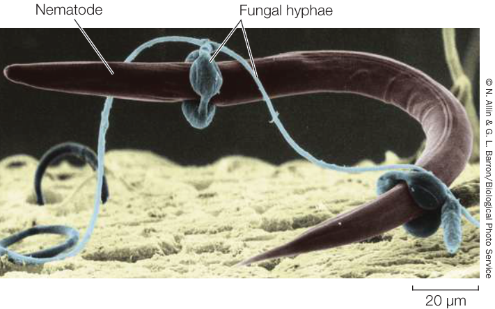
Figure 28.6 Fungus as Predator A nematode is trapped by hyphal rings of the soil-dwelling fungus Arthrobotrys dactyloides.
Fungi are by far the most important plant pathogens, causing annual crop losses amounting to billions of dollars. Major fungal diseases of crop plants include black stem rust of wheat and other diseases of wheat, corn, and oats. The agent of black stem rust is Puccinia graminis, which has a complicated life cycle that involves two plant hosts (wheat and barberry). In an epidemic in 1935, P. graminis was responsible for the loss of about one-fourth of the wheat crop in Canada and the United States.
PREDATORY FUNGI Some fungi have adaptations that enable them to function as active predators, trapping nearby microscopic protists or animals. This feeding strategy is especially important in environments with lots of available carbon but little nitrogen, such as wood. The most common predatory strategy seen in fungi is to secrete sticky substances from the hyphae so that passing organisms stick to them. The hyphae then quickly invade the trapped prey, growing and branching within it, spreading through its body, absorbing nutrients, and eventually killing it.
A more dramatic adaptation for predation is the constricting ring formed by some species of soil fungi (Figure 28.6). When nematodes (tiny roundworms) are present in the soil, these fungi form three-celled rings with a diameter that just fits a nematode. A nematode crawling through one of these rings stimulates the fungus, causing the cells of the ring to swell and trap the worm. Fungal hyphae quickly invade and digest the unlucky victim.
Mutualistic fungi engage in relationships that benefit both partners
Certain relationships between fungi and other organisms have nutritional consequences for both partners. Two relationships of this type are symbiotic (the partners live in close, permanent contact with each other) as well as mutualistic (the relationship benefits both partners).
LICHENS A lichen is not a single organism, but rather a meshwork of at least two radically different species: a fungus and a photosynthetic alga or cyanobacterium. Recent studies suggest that an additional fungal species (a unicellular yeast) is also usually involved in this mutualism. Together the organisms that constitute a lichen can survive some of the harshest environments on Earth (although they are sensitive to poor air quality; see Key Concept 28.4). The biota of Antarctica, for example, features more than 100 times as many kinds of lichens as of plants. Relatively little experimental work has focused on lichens, perhaps because they grow so slowly—typically less than 1 centimeter in a year.
There are nearly 30,000 described “species” of lichens, each of which is assigned the name of its multicellular fungal component. These fungal components may constitute as many as 20% of all fungal species. Most of them are sac fungi (Ascomycota). Some of them are able to grow independently without a photosynthetic partner, but most have never been observed in nature other than in a lichen association. The photosynthetic component of a lichen is most often a unicellular green alga, but it can be a cyanobacterium, or may even include both.
Lichens are found in all sorts of exposed habitats: on tree bark, on open soil, and on bare rock. Reindeer moss (not a moss at all, but the lichen Cladonia subtenuis) covers vast areas in Arctic, sub-Arctic, and boreal regions, where it is an important part of the diets of caribou and other large mammals.
The body forms of lichens fall into three principal categories. Crustose (crustlike) lichens adhere tightly to their substrate (Figure 28.7A). Fruticose lichens are highly branched and can grow upward like shrubs, or hang in long strands from tree branches or rocks (Figure 28.7B). Foliose (leafy) lichens are loosely attached and grow parallel to their substrate (Figure 28.7C).
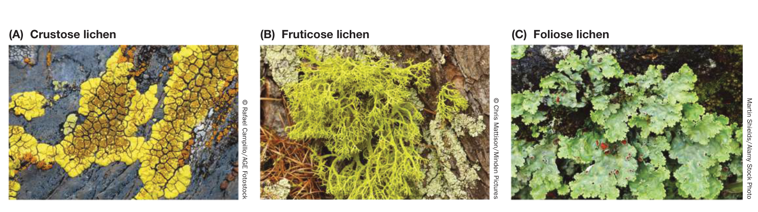
Figure 28.7 Lichen Body Forms The body forms of lichens fall into three principal categories. (A) Two crustose lichen species are growing together on this exposed rock surface. (B) The light green “shrubby” growth is a fruticose lichen. (C) Foliose lichens have a leafy appearance.
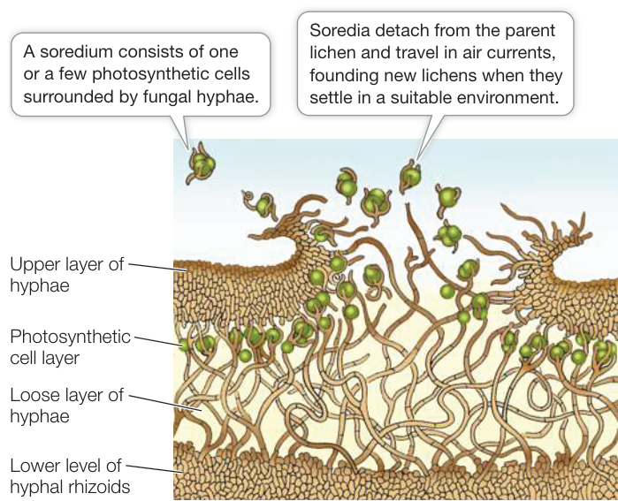
Figure 28.8 Lichen Anatomy Cross section showing the layers of a foliose lichen and the release of soredia.
A cross section of a typical foliose lichen reveals a tight upper region of fungal hyphae, a layer of photosynthetic cyanobacteria or algae, a looser hyphal layer, and finally hyphal rhizoids that attach the entire structure to its substrate (Figure 28.8). The meshwork of fungal hyphae takes up mineral nutrients needed by the photosynthetic cells and also holds water tenaciously, providing a suitably moist environment. The fungus obtains fixed carbon from the photosynthetic products of the algal or cyanobacterial cells.
Within the lichen, fungal hyphae are tightly pressed against the photosynthetic cells and sometimes invade them without breaching the cell membrane (similar to the haustoria in parasitic fungi; see Figure 28.5). The photosynthetic cells not only survive these intrusions but continue to grow. Algal cells in a lichen “leak” photosynthetic products at a greater rate than do similar cells growing on their own, and photosynthetic cells taken from lichens grow more rapidly on their own than when associated with a fungus. On the basis of these observations, we could consider lichen fungi to be parasitic on their photosynthetic partners. In many places where lichens grow, however, the photosynthetic cells could not grow at all on their own.
Lichens can reproduce simply by fragmentation of the vegetative body (the thallus) or by means of specialized structures called soredia (singular soredium). Soredia consist of one or a few photosynthetic cells bound by fungal hyphae. They become detached from the lichen, are dispersed by air currents, and upon arriving at a favorable location, develop into a new lichen thallus (see Figure 28.8). Alternatively, the fungal partner may go through its sexual reproductive cycle, producing haploid spores. When these spores are discharged, however, they disperse alone, unaccompanied by the photosynthetic partner.
Lichens are often the first colonists on new areas of bare rock. They get most of the mineral nutrients they need from the air and rainwater, augmented by minerals absorbed from dust. A lichen begins to grow shortly after a rain, as it begins to dry. As it grows, the lichen acidifies its environment slightly, and this acidity contributes to the slow breakdown of rocks, an early step in soil formation. With further drying, the lichen’s photosynthesis ceases. The water content of the lichen may drop to less than 10% of its dry weight, at which point the lichen becomes highly insensitive to extremes of temperature.
MYCORRHIZAE Many vascular plants depend on a symbiotic association with fungi. This ancient association between plants and fungi was critical to the successful exploitation of the terrestrial environment by plants. Unassisted, the root hairs of many plants often do not take up enough water or minerals to sustain growth. However, the roots of such plants usually do become infected with fungi, forming an association called a mycorrhiza (plural mycorrhizae). There are two types of mycorrhizae, distinguished by whether or not the fungal hyphae penetrate the plant cell walls.
In ectomycorrhizae, the fungus wraps around the root tip, and its mass is often as great as that of the root itself (Figure 28.9A). The fungal hyphae penetrate the root and wrap around individual root cells but do not penetrate the cell walls. An extensive web of hyphae penetrates the soil in the area around the root, so that up to 25% of the volume near the root may be fungal hyphae. The hyphae attached to the root increase the surface area for the absorption of water and minerals, and the mass of hyphae in the soil acts like a sponge to hold water in the neighborhood of the root. Infected roots are short, swollen, and club-shaped, and they lack root hairs.
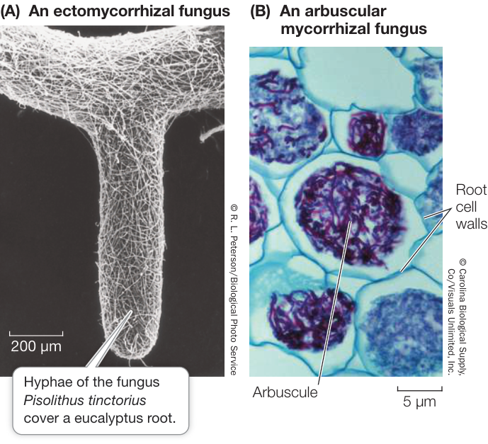
Figure 28.9 Mycorrhizal Associations (A) Ectomycorrhizal fungi wrap themselves around a plant root, increasing the area available for absorption of water and minerals. (B) Hyphae of arbuscular mycorrhizal fungi infect the root internally and penetrate the root cell walls, branching within the cells and forming a treelike structure, the arbuscule. (The cell cytoplasm has been removed to better visualize the arbuscule.)
The fungal hyphae of arbuscular mycorrhizae enter the root and penetrate the cell walls of the root cells, forming arbuscular (treelike) structures inside the cell wall but outside the cell membrane. These structures, like the haustoria of parasitic fungi and the contact regions of fungal hyphae and photosynthetic cells in lichens, become the primary site of exchange between plant and fungus (Figure 28.9B). As in the ectomycorrhizae, the fungus forms a vast web of hyphae leading from the root surface into the surrounding soil.
Connect the Concepts Mycorrhizae can vastly increase the root surface area in contact with essential nutrients in the soil. The importance of arbuscular mycorrhizae in plant nutrition is described in detail in Key Concept 34.3.
The mycorrhizal association is important to both partners. The fungus obtains needed organic compounds, such as sugars and amino acids, from the plant. In return, the fungus, because of its very high surface area-to-volume ratio and its ability to penetrate the fine structure of the soil, greatly increases the plant’s ability to absorb water and minerals (especially phosphorus). The fungus may also provide the plant with certain growth hormones and may protect it against attack by disease-causing microorganisms.
Plants that have active arbuscular mycorrhizae typically are a deeper green and may resist drought and temperature extremes better than plants of the same species that have little mycorrhizal development. Attempts to introduce some plant species to new areas have failed until some of the soil from the native area (presumably containing the fungus necessary to establish mycorrhizae) was provided. Trees without ectomycorrhizae do not grow well in the absence of abundant nutrients and water, so the health of our forests depends on the presence of ectomycorrhizal fungi. Many agricultural crops require inoculation of seeds with appropriate mycorrhizal fungi prior to planting. Without these fungi, the plants are unlikely to grow well, or in some cases at all. Certain plants that live in nitrogen-poor habitats, such as cranberry bushes and orchids, invariably have mycorrhizae. Orchid seeds will not germinate in nature unless they are already infected by the fungus that will form their mycorrhizae. Plants that lack chlorophyll always have mycorrhizae, which they often share with the roots of green, photosynthetic plants. In effect, these plants without chlorophyll are feeding on nearby green plants, using the fungus as a bridge.
Endophytic fungi protect some plants from pathogens, herbivores, and stress
In a tropical rainforest, 10,000 or more fungal spores may land on a single leaf each day. Some are plant pathogens, some do not affect the plant at all, and some invade the plant in a beneficial way. Fungi that live within aboveground parts of plants without causing obvious deleterious symptoms are called endophytic fungi. Recent research has shown that endophytic fungi are abundant in plants in all terrestrial environments.
Among the grasses, individual plants with endophytic fungi are more resistant to pathogens and to insect and mammalian herbivores than are plants lacking endophytes. The fungi produce alkaloids (nitrogen-containing compounds) that are toxic to animals. The alkaloids do not harm the host plant. In fact, some plants produce alkaloids (such as nicotine) themselves. The fungal alkaloids also increase the ability of grasses to resist stress of various types, including drought (water shortage) and salty soils. Such resistance is beneficial for some crops, although it can produce toxicity for grazing animals.
The role, if any, of endophytic fungi in most broad-leaved plants is unclear. They may convey protection against pathogens, or they may simply occupy space within leaves without conferring any benefit, but also without doing harm. The benefit, in fact, might be all for the fungus.
Recap and Assess
Fungi interact with other organisms in many ways, both harmful and beneficial. Saprobic fungi play critical roles in the recycling of elements required by living organisms. Lichens are mutualistic associations of fungi with algae or cyanobacteria. Mycorrhizae are associations of fungi and the roots of plants; they are essential for the survival of most plant species.
- What is the role of fungi in Earth’s carbon cycle?
- Describe the nature and benefits of the lichen association.
- How does the mycorrhizal association benefit both the plant and the fungus?
Before molecular techniques were widely used to study the phylogenetic relationships of fungi, one criterion used for assigning fungi to taxonomic groups was the nature of their life cycles—including the types of fruiting structures they produce. Next we take a closer look at life cycles in the major groups of fungi.
Sex in Fungi Involves Multiple Mating Types
Learning Objectives
- 28.3.1 Compare and contrast sexual reproduction in fungi with that in plants and animals.
- 28.3.2 Describe the dikaryotic stage of the fungal life cycle, distinguish which major fungal groups include a dikaryotic stage, and explain how the dikaryotic stage differs from the haploid and diploid stages.
- 28.3.3 Compare and contrast the life cycles of zygospore fungi, sac fungi, and club fungi.
Although fungal life cycles are even more diverse than was once realized, specific types of life cycles generally distinguish five major groups of fungi: chytrids (Chytridiomycota and Blastocladiomycota), zygospore fungi (Zoopagomycota), arbuscular mycorrhizal fungi (Glomeromycotina), sac fungi (Ascomycota), and club fungi (Basidiomycota). In addition, a reduced parasitic group, microsporidia, is usually considered to be related to fungi, although the exact relationships of microsporidia within eukaryotes are debated. Figure 28.10 diagrams the evolutionary relationships of these groups as they are understood today.
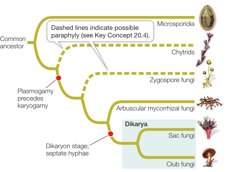
Figure 28.10 A Phylogeny of the Fungi Microsporidia are reduced parasites that are thought to be related to fungi, although their relationships are still debated. The dashed lines indicate that chytrids and zygospore fungi are possibly paraphyletic (see Table 28.1). The sac fungi and club fungi together form the clade Dikarya.
Chytrids and the zygospore fungi may not represent monophyletic groups, as they each consist of several distantly related lineages that retain some ancestral features. The clades that are thought to be monophyletic within these two informal groupings are listed in Table 28.1. Most recent evidence from DNA analyses has supported the relationship of the microsporidia to fungi, the possible paraphyly of the chytrids and the zygospore fungi, and the monophyly of sac fungi and club fungi.
Fungi reproduce both sexually and asexually
Both asexual and sexual reproduction occur among the fungi (Figure 28.11). Asexual reproduction takes several forms:
- The production of (usually) haploid spores within sporangia
- The production of haploid spores (not enclosed in sporangia) at the tips of hyphae; in sac fungi, such spores are called conidia (Greek konis, “dust”)
- Cell division by unicellular fungi—either a relatively equal division of one cell into two (fission) or an asymmetrical division in which a smaller daughter cell is produced (budding)
- Mitotic growth and fragmentation of the mycelium
Sexual reproduction is rare (or even unknown) in some groups of fungi but common in others. Sexual reproduction may not occur in some species, or it may occur so rarely that biologists have never observed it. Species in which no sexual stage has been observed were once placed in a separate taxonomic group because knowledge of the sexual life cycle was considered necessary for classifying fungi. Now, however, these species can be related to other species of fungi through analysis of their DNA sequences.
Fungi mate in the most interesting of ways. They don’t limit themselves to the confines of “males” and “females,” but rather have many different mating types, any two of which can reproduce. And when they mate, they don’t merely pass a few gamete cells between individuals like plants or animals do; instead, they fuse their bodies to become one and then exchange their nuclei. After fusing, many mating pairs remain fused for some time (a mushroom is basically two fused fungi getting it on), and the sexual act can thus last for weeks or months at a time. Eventually the nuclei fuse and recombine, and “genetic sex” finally takes place. After making millions of offspring, the exhausted fungi disintegrate their combined bodies and never text each other again.
Table 28.1 Classification of the Fungi
| Group | Common name | Features |
|---|---|---|
| Microsporidia | Microsporidia | Intracellular parasites of animals; greatly reduced, among smallest eukaryotes known; polar tube used to infect hosts; placement among fungi is debated |
| Chytridiomycota and Blastocladiomycota | Chytrids a | Mostly aquatic and microscopic; in some species, zoospores and gametes have flagella |
| Zoopagomycota | Zygospore fungi b | Reproductive structure is a unicellular zygospore with many diploid nuclei; hyphae coenocytic; no fleshy fruiting body |
| Glomeromycotina | Arbuscular mycorrhizal fungi | Form arbuscular mycorrhizae in plant roots; often reproduce asexually, but recent studies show sexual reproduction as well |
| Ascomycota | Sac fungi | Sexual reproductive saclike structure known as an ascus, which contains haploid ascospores; hyphae septate; dikaryon |
| Basidiomycota | Club fungi | Sexual reproductive structure is a basidium, a swollen cell at the tip of a specialized hypha that supports haploid basidiospores; hyphae septate; dikaryon |
aThe relationships between Chytridiomycota and Blastocladiomycota, both treated as chytrids here, are not yet well resolved.
bTwo small groups traditionally considered zygospore fungi now appear to be more closely related to arbuscular mycorrhizal fungi.
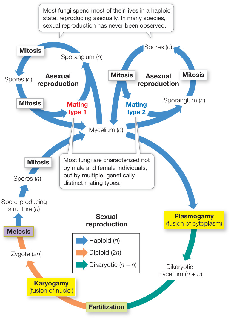
Figure 28.11 A Generalized Fungal Life Cycle Environmental conditions may determine which mode of reproduction—sexual or asexual—takes place at a given time.
Major groups of fungi differ in the details of when and how the nuclei from two individuals fuse and recombine, and in whether they produce a fleshy fruiting structure, as we discuss later in this key concept.
Microsporidia may be highly reduced, parasitic fungi
Microsporidia are highly reduced, unicellular parasites. They are among the smallest eukaryotes known, with infective spores that are only 1–40 micrometers () in diameter. About 1,500 species have been described, but many more species are thought to exist. Their relationships among the eukaryotes have puzzled biologists for many decades, although the sequences of several microsporidian genes suggest a close relationship to fungi.
Microsporidia lack true mitochondria, although they have reduced structures, known as mitosomes, that are derived from mitochondria. Unlike mitochondria, however, mitosomes contain no DNA—the mitochondrial genome has been completely transferred to the nucleus. Because microsporidia lack mitochondria, biologists initially suspected that they represented an early lineage of eukaryotes that diverged before the endosymbiotic event from which mitochondria evolved. The presence of mitosomes, however, indicates that this hypothesis is incorrect. DNA sequence analysis, along with the fact that their cell walls contain chitin, has suggested that microsporidia may be highly reduced, parasitic fungi, although their exact relationship to fungi is still being investigated.
Microsporidia are obligate intracellular parasites of animals, especially of insects, crustaceans, and fishes. Some species are known to infect mammals, including humans. Most infections by microsporidia cause chronic disease in the host, with effects that include weight loss, reduced fertility, and a shortened life span. The host cell is penetrated by a polar tube that grows from the microsporidian spore. The function of the polar tube is to inject the contents of the spore, the sporoplasm, into the host (Figure 28.12). The sporoplasm then replicates within the host cell and produces new infective spores. The life cycle of some species is complex and involves multiple hosts, whereas other species infect a single host. In some insects, parasitic microsporidia are transmitted vertically (i.e., from parent to offspring). Only asexual reproduction is known for some microsporidia, but other species appear to have both asexual and sexual cycles.
Most chytrids are aquatic
The chytrids (Figure 28.13) include several distinct lineages of mostly aquatic microorganisms once classified with the protists. However, morphological evidence (cell walls that consist primarily of chitin) and molecular evidence support their classification as early-diverging fungi. In this book we use the term “chytrid” to refer to both of the formally named clades listed as chytrids in Table 28.1, but some mycologists use this term to refer to only the Chytridiomycota. There are fewer than 1,000 described species among these two groups of chytrids.
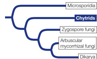
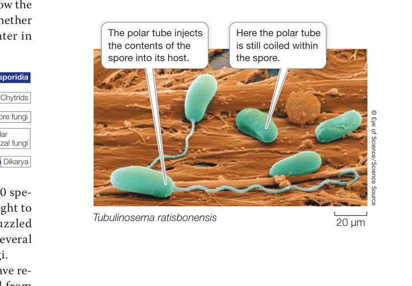
Figure 28.12 Invasion of the Microsporidia The spores of microsporidia grow polar tubes that transfer the contents of the spores into the host’s cells. The species shown here infects many animals, including humans.
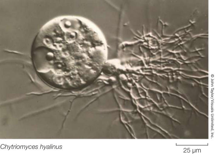
Figure 28.13 A Chytrid Branched rhizoids emerge from the sporangium of a mature chytrid.
Chytrids reproduce both sexually and asexually. Unlike other fungi, some chytrids that reproduce sexually possess flagellated gametes (similar to sperm in animals, except that both male and female gametes are flagellated in chytrids). The spores (called zoospores) are also flagellated (Figure 28.14A). Chytrids are the only fungi that include species with flagella at any life cycle stage. The retention of flagella reflects the aquatic environment in which fungi first evolved. As one type of gamete is larger than the other, the gametes can be distinguished as male (small gametes) and female (large gametes; see Figure 28.14A). Except for size, however, the two gametes are very similar.
The chytrids are diverse in form. Some are unicellular, others have rhizoids, and still others have coenocytic hyphae. They may be parasitic (on organisms such as algae, mosquito larvae, nematodes, and amphibians) or saprobic. Some have complex mutualistic relationships with foregut-fermenting animals such as cattle and deer. Many chytrids live in freshwater habitats or in moist soil, but some are marine.
Most fungal life cycles feature separate fusion of cytoplasms and nuclei
Most members of the remaining four groups of fungi are terrestrial. Although the terrestrial fungi grow in moist places, they do not have motile gametes, so liquid water is not required for fertilization. Instead, the cytoplasms of two individuals of different mating types fuse (a process called plasmogamy) before their nuclei fuse (a process called karyogamy; see Figure 28.11). Terrestrial zygospore fungi, arbuscular mycorrhizal fungi, sac fungi, and club fungi all include some sexual species.
Zygospore fungi reproduce sexually when adjacent hyphae of two different mating types release chemical signals that cause them to grow toward each other. These hyphae produce gametangia, which are specialized cells for reproduction that are retained as part of the hyphae. In the gametangia, nuclei replicate without cell division, resulting in multiple haploid nuclei in both gametangia. The two gametangia then fuse to form a unicellular zygosporangium that contains many haploid nuclei of each mating type (Figure 28.14B). These haploid nuclei quickly pair up to form multiple diploid nuclei within the zygosporangium. A thick, multilayered cell wall develops around the zygosporangium to form a well-protected resting stage that can remain dormant for months. In harsh environmental conditions, this resting stage may be the only cell that survives as the surrounding cells of the hyphae die. At this stage the single surviving cell is known as a zygospore, which is the basis of the name of the zygospore fungi. When environmental conditions improve, the nuclei within the zygospore undergo meiosis and one or more stalked sporangiophores sprout, each bearing a sporangium. Each sporangium contains the products of meiosis: haploid nuclei that are incorporated into spores. These spores disperse and germinate to form a new generation of haploid hyphae.
The zygospore fungi have traditionally included five major lineages of terrestrial fungi that live on soil as saprobes, as parasites of insects and spiders, or as mutualists of other fungi and invertebrate animals. However, recent genome analyses suggest that two of these lineages may be more closely related to the arbuscular mycorrhizal fungi. Zygospore fungi produce no cells with flagella, and only one diploid cell—the zygospore—appears in the entire life cycle. Their hyphae are coenocytic. Most species do not form a fleshy fruiting structure. Instead, the hyphae spread in a radial pattern from the spore, with occasional stalked sporangiophores reaching up into the air (Figure 28.15).
More than 1,000 species of zygospore fungi have been described. One species you may have seen is Rhizopus stolonifer, the black bread mold. Rhizopus produces many stalked sporangiophores, each bearing a single sporangium containing hundreds of minute spores (see Figure 28.14B).
Arbuscular mycorrhizal fungi form symbioses with plants
Arbuscular mycorrhizal fungi (Glomeromycotina) are terrestrial fungi that associate with plant roots in a symbiotic, mutualistic relationship (see Figure 28.9B). Only a few hundred species have been formally described, but 80% to 90% of all plants have associations with arbuscular mycorrhizal fungi, and most of the diversity of the group remains to be discovered.
The hyphae of arbuscular mycorrhizal fungi are coenocytic. These fungi use glucose from their plant partners as their primary energy source, converting it into other, fungus-specific sugars that cannot return to the plant. Until recently, arbuscular mycorrhizal fungi were known to reproduce only asexually, but recent studies demonstrate that they can undergo meiosis and genetic recombination.
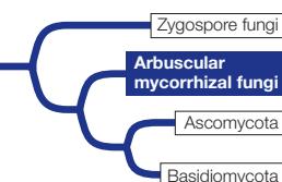
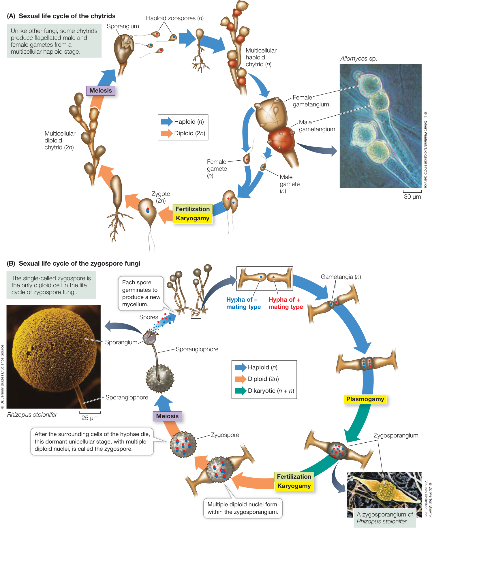
Figure 28.14 Sexual Life Cycles of Chytrids and Zygospore Fungi (A) Chytrids are the only fungi that possess flagella at any stage of the life cycle. (B) The unicellular zygospore, which contains multiple diploid nuclei, is a resting structure unique to the zygospore fungi. CHAPTER 28 The Evolution and Diversity of Fungi Q: How is one chytrid gamete identified as male and the other as female?
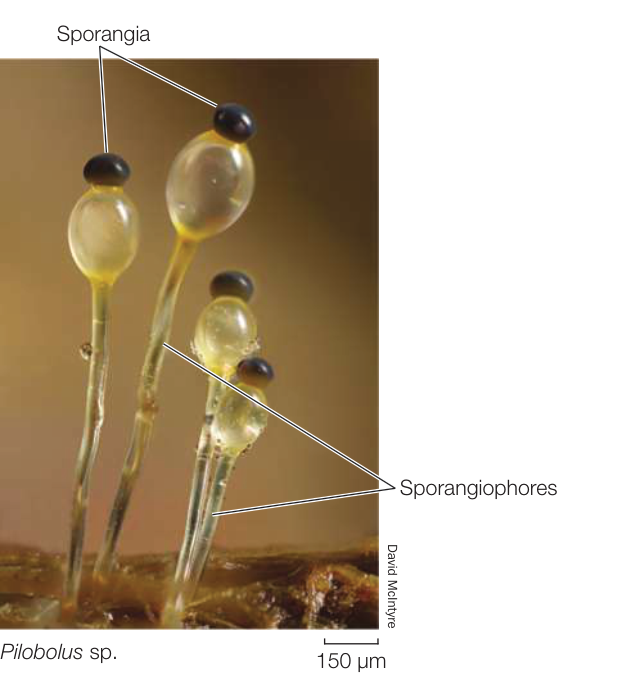
Figure 28.15 Zygospore Fungi Produce Sporangiophores These transparent structures are sporangiophores produced by a zygospore fungus growing on decomposing animal dung. The sporangiophores grow toward the light and end in tiny sporangia, which the stalked sporangiophores can eject as far as 2 meters. Animals ingest sporangia and then disseminate the spores in their feces. Animation 28.1 Life Cycle of a Zygospore Fungus
The dikaryotic condition is a synapomorphy of sac fungi and club fungi
In the two remaining groups of fungi—the sac fungi and the club fungi—some stages have a nuclear configuration other than the familiar haploid or diploid states (Figure 28.16). In these fungi, karyogamy (fusion of nuclei) occurs long after plasmogamy (fusion of cytoplasm), so that two genetically different haploid nuclei coexist and divide within each cell of the multicellular fruiting body. This stage of the life cycle is called a dikaryon (“two nuclei”), and its ploidy is indicated as . The dikaryon is a synapomorphy of the sac fungi and club fungi, which are placed together in a clade called Dikarya.
Eventually, specialized cells form on the fruiting body, within which pairs of genetically dissimilar nuclei—one from each parent—fuse, giving rise to zygotes long after the original “mating.” The diploid zygote nucleus then undergoes meiosis, producing four haploid nuclei. The mitotic descendants of those nuclei become spores, which germinate to give rise to the next haploid generation.
A life cycle with a dikaryotic stage has several unusual features. First, there are no gamete cells, only gamete nuclei. Second, the only true diploid structure is the zygote, although for a long period the genes of both parents are present in the dikaryon and can be expressed. In effect, the dikaryon is neither diploid () nor haploid (). Rather, it is dikaryotic (). Therefore a harmful recessive mutation in one nucleus may be compensated for by a normal allele on the same chromosome in the other nucleus, and dikaryotic hyphae often have characteristics that are different from their or products. The dikaryotic condition is perhaps the most distinctive of the genetic peculiarities of the fungi.
The sexual reproductive structure of sac fungi is the ascus
The sac fungi (Ascomycota) are a large and diverse group of fungi found in marine, freshwater, and terrestrial habitats. There are approximately 64,000 known species, nearly half of which are the fungal partners in lichens. The hyphae of sac fungi are segmented by more or less regularly spaced septa. A pore in each septum permits extensive movement of cytoplasm and organelles (including nuclei) from one segment to the next.
Sac fungi are distinguished by the production of sacs called asci (singular ascus), which at maturity contain sexually produced haploid ascospores (see Figure 28.16A). The ascus is the characteristic sexual reproductive structure of the sac fungi. In the past, the sac fungi were classified on the basis of whether or not the asci are contained within a specialized fruiting structure known as an ascoma (plural ascomata) and on differences in the morphology of that fruiting structure. DNA sequence analyses have resulted in a revision of these traditional groupings, however.
SAC FUNGUS YEASTS Some species of sac fungi are unicellular yeasts. The 1,000 or so species in this group are among the most important domesticated fungi. Perhaps the best known is baker’s, or brewer’s, yeast (Saccharomyces cerevisiae; see Figure 28.2 and Key Concept 28.4), which catabolizes glucose obtained from its environment into ethanol and carbon dioxide by fermentation. Other sac fungus yeasts live on fruits such as figs and grapes and play an important role in the making of wine. Many others are associated with insects. In the guts of some insects, they provide enzymes that break down materials that are otherwise difficult for the insects to digest, especially cellulose.
Sac fungus yeasts reproduce asexually by budding. Sexual reproduction takes place when two adjacent haploid cells of dissimilar mating types fuse. In some species, the resulting zygote buds to form a diploid cell population. In others, the zygote nucleus undergoes meiosis immediately. When this happens, the entire cell becomes an ascus. Depending on whether the products of meiosis then undergo mitosis, a yeast ascus contains either eight or four ascospores, which germinate to become haploid cells. The sac fungus yeasts have lost the dikaryotic stage.
FILAMENTOUS SAC FUNGI Most sac fungi are filamentous species, such as the cup fungi (Figure 28.17A), in which the ascomata are cup-shaped and can be as large as several centimeters across (although most are much smaller). The inner surfaces of the ascomata, which are covered with a mixture of specialized hyphae and asci, produce huge numbers of spores. The edible ascomata of some species, including morels (Figure 28.17B) and truffles, are regarded by humans as gourmet delicacies (and can sell at prices higher than gold). The underground ascomata of truffles have a strong odor that attracts mammals such as pigs, which then eat the fungi and disperse the spores. Humans sometimes take advantage of pigs’ attraction to truffles and use pigs to locate truffles for human consumption.
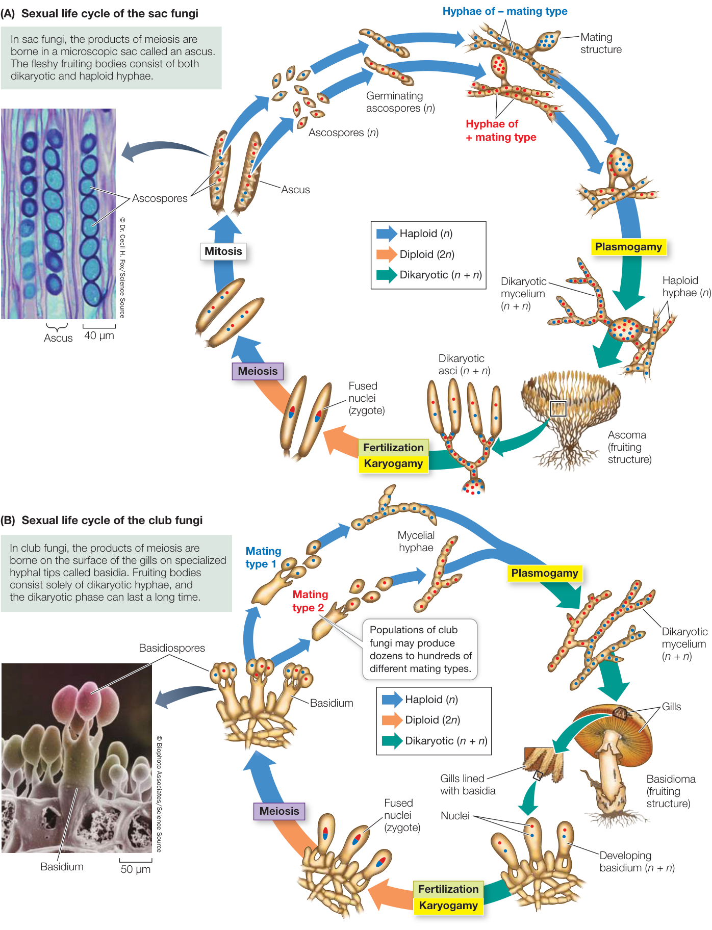
Figure 28.16 Sexual Life Cycles among the Dikarya (A) In sac fungi, the products of meiosis are borne in a microscopic sac called an ascus. The fleshy fruiting structure, the ascoma, consists of both dikaryotic and haploid hyphae. (B) The basidium is the characteristic sexual reproductive structure of the club fungi. The fruiting structures, called basidiomata, consist solely of dikaryotic hyphae, and the dikaryotic phase can last a long time. Q: How does the dikaryotic condition differ from diploidy? Activity 28.2 Life Cycle of a Dikaryotic Fungus
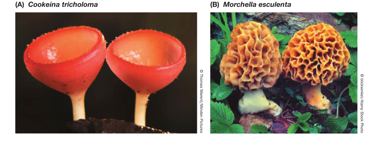
Figure 28.17 Sac Fungi (A) These brilliant red cups are the ascomata of a cup fungus. (B) Morels, which have a spongelike ascoma and a subtle flavor, are considered a culinary delicacy by humans.
The sexual reproductive cycle of filamentous sac fungi includes the formation of a dikaryon, although this stage is relatively brief compared with that in club fungi. Many filamentous sac fungi form multinucleate mating structures (see Figure 28.16A). Mating structures of two different mating types fuse and produce a dikaryotic mycelium, containing nuclei from both mating types. The dikaryotic mycelium often forms a cup-shaped ascoma, which bears the asci. Only after the formation of asci do the nuclei from the two mating types finally fuse. Both nuclear fusion and the subsequent meiosis that produces haploid ascospores take place within individual asci. The ascospores are ultimately released (sometimes shot off forcefully) by the ascus to begin the new haploid generation.
The sac fungi also include many of the filamentous fungi known as molds. Molds consist of filamentous hyphae that do not form large ascomata, although they can still produce asci and ascospores. Many molds are parasites of flowering plants. Chestnut blight and Dutch elm disease are both caused by molds. The chestnut blight fungus, which was introduced to the United States in the 1890s, had destroyed the American chestnut as a commercial species by 1940. Before the blight, this species accounted for more than half the trees in the eastern North American forests. Another familiar story is that of the American elm. Sometime before 1930, the Dutch elm disease fungus (first discovered in the Netherlands but native to Asia) was introduced into the United States on infected elm logs from Europe. Spreading rapidly—especially via bark beetles—the fungus destroyed great numbers of American elm trees.
Other plant pathogens among the sac fungi include the powdery mildews that infect cereal crops, lilacs, and roses, among many other plants. Mildews can be a serious problem for farmers and gardeners, and a great deal of research has focused on ways to control these agricultural pests.
The filamentous sac fungi can also reproduce asexually by means of conidia that form at the tips of specialized hyphae (Figure 28.18). Small chains of conidia are produced by the millions and can survive for weeks in nature. The conidia are often what give molds their characteristic colors.
The basidium is the sexual reproductive structure of club fungi
Club fungi (Basidiomycota) produce some of the most spectacular fruiting structures found among the fungi. These fruiting structures, called basidiomata (singular basidioma), include mushrooms of all kinds, puffballs (see Figure 28.4), and the bracket fungi often encountered on trees and fallen logs in a damp forest. About 30,000 species of club fungi have been described. They include about 4,000 species of mushrooms, including both poisonous (Figure 28.19A) and edible species. Bracket fungi (Figure 28.19B) play an important role in the carbon cycle by breaking down wood. They also do great damage to both cut lumber and timber stands. Some of the most economically damaging plant pathogens are club fungi, including the rust fungi and smut fungi that parasitize cereal grains. In contrast, other club fungi contribute to the survival of plants as fungal partners in ectomycorrhizae.
The hyphae of club fungi characteristically have septa with small, distinctive pores. As they grow, haploid hyphae of different mating types meet and fuse, forming dikaryotic hyphae, each cell of which contains two nuclei, one from each parent hypha. The dikaryotic mycelium grows and eventually, when triggered by rain or another environmental cue, produces a basidioma. The dikaryotic stage may persist for years, or even centuries. This pattern contrasts with the life cycle of the sac fungi, in which the dikaryon is found only in the stages leading up to formation of the asci.
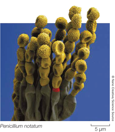
Figure 28.18 Conidia Chains of conidia (yellow) are developing at the tips of specialized hyphae arising from a Penicillium notatum mold. This species is used to produce the antibiotic penicillin.
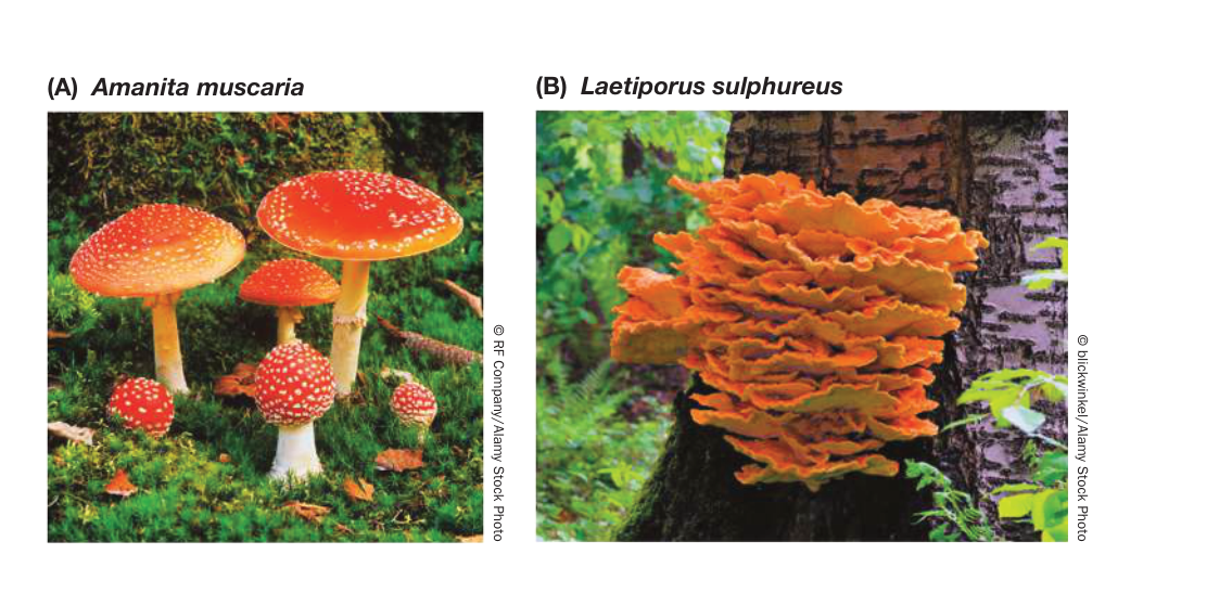
Figure 28.19 Club Fungus Basidiomata (A) The fly agaric is a poisonous mushroom that contains hallucinogenic compounds. (B) A bracket fungus growing parasitically on a tree. Although this particular species is occasionally eaten when young, many similar-appearing bracket fungi are poisonous.
The basidium (plural basidia), a swollen cell at the tip of a specialized hypha, is the characteristic sexual reproductive structure of the club fungi (see Figure 28.16B). In mushroom-forming club fungi, the basidia typically form on specialized structures of the basidiomata known as gills. The basidium is the site of nuclear fusion and meiosis and thus plays the same role in the club fungi as the ascus does in the sac fungi and the zygosporangium does in the zygospore fungi.
After nuclei fuse in the basidium, the resulting diploid nucleus undergoes meiosis, and the four resulting haploid nuclei are incorporated into haploid basidiospores, which form on tiny stalks on the outside of the basidium. A single basidioma of the common bracket fungus Ganoderma applanatum can produce as many as 4.5 trillion basidiospores in one growing season. Basidiospores typically are forcibly discharged from their basidia and then germinate, and give rise to hyphae with haploid nuclei.
Recap and Assess
Sexual reproduction is common in some groups of fungi but has never been observed in others. Many fungal species have two or more genetically distinct but visually indistinguishable mating types. To mate, individuals of most sac fungi and club fungi fuse to produce dikaryotic fruiting bodies, in which two genetically different haploid nuclei coexist in each cell of the multicellular structure.
- Explain the concept of mating types. How are they different from male and female sexes?
- Describe how microsporidia infect the cells of their animal hosts.
- What feature of the chytrids suggests that the fungi had an aquatic ancestor?
- What is the role of the zygospore in the life cycle of zygospore fungi?
- What distinguishes the fruiting bodies of sac fungi from those of club fungi?
Fungi are of special interest to biologists because of the roles they play in interactions with other organisms, including humans. But they are also useful as tools for studying many kinds of biological problems and for finding solutions to those problems.
28.4
Fungi Have Many Practical Uses
Learning Objectives
- 28.4.1 Describe four ways that fungi are used to produce food and drink for humans.
- 28.4.2 Provide an example of how fungi can be used for medical purposes.
- 28.4.3 Explain two ways that fungi can be used as bioindicators of environmental health.
We’ve briefly noted the important part that fungi play in the production of human foods and beverages. We have also described the diverse roles that fungi play in natural ecosystems, from decomposers to pathogens to mutualistic partners. These diverse ecological roles have led to the use of fungi in studies of environmental change and in remediation of environmental pollution. Many fungi are also important model organisms for laboratory investigations of basic biological process. Others, as you saw at the opening of this chapter, have given us treatments for human diseases.
Fungi are important in producing food and drink
Grains from grasses provide most of the world’s food supply for humans. But in most cases, we do not eat these grains directly as they are produced by the plants. Instead, we use them as a source of starch. To make the starch more pleasing and digestible for human consumption, we usually convert it to more complex and tasty forms of food and drink, often with the help of fungi.
Baker’s (or brewer’s) yeast (Saccharomyces cerevisiae) converts the starch from grain into ethanol. This process also forms carbon dioxide bubbles in bread dough, causing it to rise, which gives baked bread its light texture. The ethanol and carbon dioxide are baked away in bread making (which produces the pleasant aroma of baking bread). In contrast, the ethanol and
Fungi Have Many Practical Uses
carbon dioxide are retained when yeast is used to ferment grain into beer. The carbon dioxide gives beer its fizz, and the alcohol and yeast contribute to the taste and appeal of beer to those who enjoy it. Sugars, especially from fruit such as grapes, are also converted into alcohol and carbon dioxide by yeasts in the production of wine (although the carbon dioxide is not retained in most finished wine, as it is in beer). Many different strains of S. cerevisiae are used in wine production, which contributes to the distinctive nature of wine from different regions and wineries. Many other species of local, native yeasts are also used in producing distinctive local wines and beers. For example, fission yeast (Schizosaccharomyces pombe) was first isolated from African millet beer. Fission yeast takes its specific name (pombe) from the Swahili word for beer.
Brown molds of the genus Aspergillus are important in some human diets. Aspergillus tamarii acts on soybeans in the production of soy sauce, and A. oryzae is used in brewing the Japanese alcoholic beverage sake from rice. Aspergillus niger is the source of most commercial citric acid production. Citric acid gives food and soft drinks a tart taste and is also used as a food preservative. But some species of Aspergillus that grow on grains and on nuts such as peanuts and pecans produce extremely carcinogenic (cancer-inducing) compounds called aflatoxins. Aflatoxins can occur in high concentrations in foods such as peanut butter. In the United States and most other industrialized countries, moldy grain infected with Aspergillus is typically thrown out. In Africa, where food is scarcer, the grain is often eaten, moldy or not, and causes severe health problems, including high levels of certain cancers.
Penicillium is a genus of green molds, of which some species produce the antibiotic penicillin, as described in the beginning of this chapter. But several species of Penicillium are important for food production as well. For example, P. camemberti and P. roqueforti are the organisms responsible for the characteristic strong flavors of Camembert and Roquefort cheeses, respectively.
Many fungi serve directly as a human food source. Mushroom enthusiasts seek out the delicious fruiting structures of a wide variety of edible sac and club fungi. In the United States, relatively few species of mushrooms are grown commercially, and wild mushrooms are collected mostly for personal consumption. But in many parts of the world, a wide variety of wild mushrooms are collected for sale and consumption. Fungi used for food are not limited to fruiting bodies such as mushrooms, however. Various species of lichens are eaten in Arctic regions as well as in parts of North America and Asia. In southwestern China, for example, several species of lichens are used as a primary ingredient in cooking (Figure 28.20).
Fungi provide important weapons against diseases and pests
We started this chapter with the story of the discovery of penicillin. The discovery of antibiotics produced by fungi revolutionized medical treatment of bacterial diseases in humans and their domestic animals. Live strains of fungi are also used to combat various pest species of plants and animals.
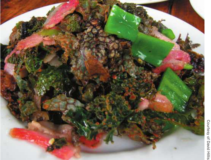
Figure 28.20 Some Lichens Are Edible In southwestern China, several species of lichens that grow on tree bark serve as a primary ingredient in the local cuisine.
In Africa, the parasitic plant witchweed (Striga) causes crop losses of about U.S.$7 billion every year. A group of Canadian biologists discovered that a strain of the mold Fusarium oxysporum could be applied to crops to control witchweed without harming the crop plants (Figure 28.21). Other strains of Fusarium that preferentially attack coca plants, the source of cocaine, have been proposed to combat illicit drug production. Still other fungi are used to attack various animal pests, such as termites and aphids, and even malaria-carrying mosquitoes.
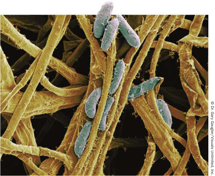
Figure 28.21 A Pathogenic Fungus Attacks a Parasitic Plant The fungus Fusarium oxysporum is a potent pathogen of witchweed (Striga), a parasitic plant that attacks crops. The fungal spores are shown in blue; fungal filaments are in tan. Both colors were added to enhance this scanning electron micrograph.
Lichen diversity and abundance are indicators of air quality
Lichens can live in many harsh environments where few other species can survive, as you saw in Key Concept 28.2. In spite of their hardiness, however, lichens are highly sensitive to air pollution because they are unable to excrete any toxic substances they absorb. This sensitivity means that lichens are good biological indicators of air pollution levels. It also explains why they are not commonly found in heavily industrialized regions or in large cities.
Monitoring the diversity and abundance of lichens growing on trees is a practical and inexpensive system for gauging air quality around cities. Maps of lichen diversity provide environmental biologists with a tool for tracking the distribution of air pollutants and their effects. Sensitive biological indicators of pollution, such as lichen growth, allow biologists to monitor air quality without the use of specialized equipment. Lichens are naturally distributed across the environment, and they can also provide a long-term measure of the effects of air pollution across many seasons and years.
Fungi record and help remediate environmental pollution
Each year, biologists deposit samples of many groups of organisms in the collections of natural history museums. These museum collections serve many purposes, one of which is to document changes in the biota of our planet over time.
Collections of fungi made over many decades or centuries provide a record of the environmental pollutants that were present when the fungi were growing. Biologists can analyze these historical samples to see how different sources of pollutants were affecting our environment before anyone thought to take direct measurements (Investigating Life: Using Fungi to Study Environmental Contamination). These long-term records are also useful for analyzing the effectiveness of cleanup efforts and regulatory programs for controlling environmental pollutants.
You have already seen that fungi are critical to the planetary carbon cycle because of their role in breaking down dead organic matter. Fungi are also used in remediation efforts to help clean up sites that have been polluted by oil spills or contaminated with toxic petroleum-derived hydrocarbons. Many herbicides, pesticides, and other synthetic hydrocarbons are broken down primarily through the action of fungi.
Reforestation may depend on mycorrhizal fungi
When a forest is cut down, it is not just the trees that are lost. A forest is an ecosystem that depends on the interaction of many species. As we have discussed, many plants depend on close relationships with mycorrhizal fungal partners. When trees are removed
▶ Investigating LIFE Using Fungi to Study Environmental Contamination
Experiment
Original Paper: A. R. Flegal et al. 2010. Declining—but persistent—atmospheric contamination in central California from the resuspension of historic leaded gasoline emissions as recorded in the lace lichen (Ramalina menziesii Taylor) from 1892 to 2006. Environ Sci Technol 44: 5613–5618.
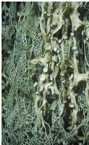
A. Russell Flegal and his colleagues analyzed museum samples of lace lichens (Ramalina menziesii) collected between 1892 and 2006 near San Francisco, California. Their goals were to determine the sources of lead contamination and to evaluate the effectiveness of efforts to reduce this contamination.
QUESTION Is environmental lead contamination through time consistent with known sources of pollution, and have efforts to reduce lead pollution been effective?
METHOD
- Obtain museum samples of lace lichen (Ramalina menziesii) collected near San Francisco.
© Sharen Jaynowitz/JPON/Contract Unit University of Stanford from a site, the populations of mycorrhizal fungi there decline rapidly. If we wish to restore the forest on the site, we cannot simply replant it with trees and other plants and expect them to survive. The mycorrhizal fungal community must be reestablished as well. For large forest restoration projects, a planned succession of plant growth and soil improvement is often necessary before forest trees can be replanted. As the community of soil fungi gradually recovers, trees that have been inoculated with appropriate mycorrhizal fungi in tree nurseries can be planted to reintroduce greater diversity to the soil fungal community.
- Measure concentrations of lead (Pb) as well as the ratios of two lead isotopes, 206Pb and 207Pb.
- Compare the ratio of the two isotopes in the lichen samples with that of the possible sources:
- Lead smelter, in operation from 1885 to 1971: 206Pb/207Pb ratio of 1.15–1.17
- Leaded gasoline, in use from 1930s to early 1980s: 206Pb/207Pb ratio of 1.18–1.23
- Resuspension of historic lead contamination as atmospheric aerosols in recent decades: 206Pb/207Pb ratio of 1.16–1.19
RESULTS Peak environmental lead contamination occurred in the 1960s to 1980s, with a rapid drop in the early 1980s after the closure of the smelter and the elimination of leaded gasoline. Average 206Pb/207Pb ratios of lead were below 1.17 before the 1930s, between 1.18 and 1.19 in the 1940s and 1950s, and >1.22 in the early 1980s. The ratios have declined since then.
CONCLUSION
- The levels of environmental lead contamination, and ratios of lead isotopes, are consistent with the identified sources.
- The closure of the lead smelter and elimination of leaded gasoline have significantly reduced environmental lead contamination.
Fungi are used as model organisms in laboratory studies
Much of what we know about many basic aspects of cell and molecular biology comes from the study of model organisms. Among the eukaryotes, some fungi have numerous advantages over model plant and animal systems for laboratory investigations.
Of particular importance as model organisms are several species of sac fungi: Aspergillus nidulans (a brown mold), Neurospora crassa (a red bread mold), Saccharomyces cerevisiae (baker’s, or brewer’s, yeast), and Schizosaccharomyces pombe (fission yeast). These species can be cultured in large numbers in small spaces, and they have short generation times, so that genetic investigations can be conducted in days rather than years. Furthermore, their genomes are relatively small and encode relatively few genes compared with those of most plants and animals, so it is easier to elucidate the functions of the fungal genes responsible for basic biological functions.
Recap and Assess
Work with the Data
The study by Flegal and his colleagues resulted in the following data:
| Sample | Year collected | Lead concentration ( of Pb/g lichen) | ratio |
|---|---|---|---|
| 1 | 1892 | 11.9 | 1.165 |
| 2 | 1894 | 4.0 | 1.155 |
| 3 | 1906 | 13.7 | 1.154 |
| 4 | 1907 | 22.9 | 1.157 |
| 5 | 1945 | 49.9 | 1.187 |
| 6 | 1957 | 34.2 | 1.185 |
| 7 | 1978 | 50.9 | 1.221 |
| 8 | 1982 | 10.0 | 1.215 |
| 9 | 1983 | 4.6 | 1.224 |
| 10 | 1987 | 1.0 | 1.198 |
| 11 | 1988 | 1.3 | 1.199 |
| 12 | 1995 | 1.9 | 1.202 |
| 13 | 2000 | 0.4 | 1.184 |
| 14 | 2006 | 1.8 | 1.184 |
Data from A. R. Flegal et al. 2010. Environ Sci Technol 44: 5613–5618.
QUESTIONS►
- Using the data in the table, plot lead concentration in the lichen samples against year of sample collection. Make a second plot, this one of ratio against year of sample collection. Calculate trend lines for your plots by plotting the average for each adjacent pair of observations, from 1892 to 2006. (In other words, plot the average for samples 1 and 2, then the average for samples 2 and 3, and so on, to produce a trend line for the data.)
- Do your analyses support the conclusions of the study? How do you think you could improve the analysis?
Fungi are important to humans in many ways. Some species are consumed directly as food, while others are important in food production. Fungi serve as important indicators of ecosystem health and are critical in reforestation and in pollution remediation efforts. Several species are important model organisms for studies of eukaryotic cell and molecular biology. Fungi are widely used to combat diseases and pests.
- What are some of the ways in which fungi or fungal products contribute to the human food supply?
- What are some advantages of using surveys of lichen diversity and museum collections of lichens to measure long-term changes in air quality, compared with direct measurements of atmospheric pollutants?
- Can you develop a strategy for tree harvest that would ease the difficulty and expense of reforestation projects by retaining viable communities of mycorrhizal fungi?
- Consider the following data or lichens at five survey sites. Predict the relative order of the sites with respect to their distance from the center of a large city. Other factors (besides distance to city center, such as prevailing wind direction) might affect your prediction. Can you think of two other major factors that might influence these results?
| Site number | Number of lichen species | Tree branches covered in lichens (%) |
|---|---|---|
| 1 | 5 | 38 |
| 2 | 1 | 2 |
| 3 | 3 | 15 |
| 4 | 8 | 75 |
| 5 | 13 | 100 |
Investigating Life
QA Why are the regions of inhibited bacterial growth around the various antibiotics so different in size, and how is that relevant to human health care?
The variable regions of inhibited growth that surround the various antibiotic samples reflect the differential effectiveness of antibiotics to a particular bacterium. Physicians who treat a bacterial infection need to know which antibiotic will be most effective for clearing that infection. An antibiotic that might work well to fight one infection might work poorly to fight another. Experiments like the one shown identify which antibiotics are best at inhibiting growth of a tested bacterium. Bigger zones of inhibited growth indicate more effective antibiotics.
Beginning in the 1940s, antibiotics derived from fungi ushered in a “golden age” of freedom from bacterial infections. Today, however, many of these antibiotics are losing their effectiveness as pathogenic bacteria evolve resistance to them (Figure 28.22).
Most medical antibiotics are chemically modified forms of substances that are found naturally in fungi and other organisms. Fungi naturally produce antibiotic compounds to defend themselves against bacterial growth and to reduce competition from bacteria for nutritional resources. These naturally occurring compounds are usually chemically modified to increase their stability, improve their effectiveness, and facilitate synthetic production. From the late 1950s to the late 1990s, no new major classes of antibiotics were discovered. In recent years, however, three new classes of antibiotics have been synthesized based on information learned from naturally occurring, fungally derived antibiotics, leading to improved treatment of some formerly resistant strains of bacteria.
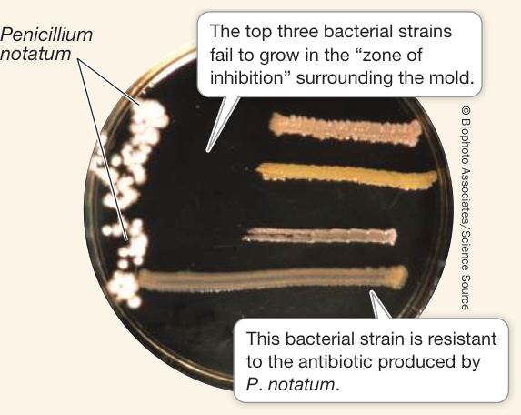
Figure 28.22 Penicillin Resistance In a petri dish similar to those in Alexander Fleming’s lab, four strains of a pathogenic bacterium have been cultured along with Penicillium mold. One strain is resistant to the mold’s antibiotic substance, as is evidenced by its growth up to the mold.
Future directions
Biologists are developing applications to use fungi to combat non-bacterial diseases. One of the more unusual applications of fungi is in the war against malaria. Biologists have discovered that two species of fungi, Beauveria bassiana and Metarhizium anisopliae, can kill malaria-causing mosquitoes when applied to mosquito netting. Mosquitoes have not yet shown evidence of developing resistance to these biological pathogens.

Visual Summary

© Sotris Zafeiris/Science Source
You should be able to relate each summary to the adjacent figures. If you go to this visual summary in Achieve, you can follow links to figures, animations, activities, and simulations that will help you consolidate the material.
Fungi Digest Food Outside Their Bodies
- ■ Fungi are distinguished from other opisthokonts by absorptive heterotrophy and by the presence of chitin in their cell walls.
- ■ Some fungi are saprobes, others are parasites or predators, and some are mutualists.
- ■ Yeasts are unicellular, free-living fungi.
- ■ The body of a multicellular fungus is a mycelium—a meshwork of filaments called hyphae. Hyphae may be septate (having septa) or coenocytic (multinucleate).
Questions
- What is a fundamental difference in the way that fungi obtain their nutrition, as compared with plants and animals?
- How do some fungi blur the distinctions between unicellularity and multicellularity?
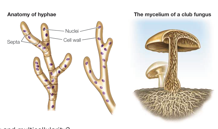
Figure 28.3
Fungi Are Decomposers, Parasites, Predators, or Mutualists
- ■ Saprobic fungi, which act as decomposers, make crucial contributions to the recycling of elements, especially carbon.
- ■ Many fungi are parasites, harvesting nutrients from host cells by means of haustoria.
- ■ Some fungi associate with unicellular green algae, cyanobacteria, or both to form lichens, which live on exposed surfaces of rocks, trees, and soil.
- ■ Mycorrhizae are mutualistic associations of fungi with plant roots. They improve a plant’s ability to take up nutrients and water. Endophytic fungi live within plants and may provide their hosts with protection against herbivores and pathogens.
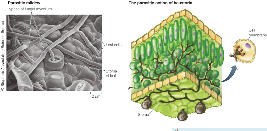
Figure 28.5
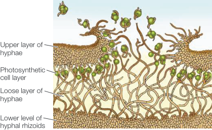
Figure 28.8
Questions
- How would life on Earth be fundamentally different without fungi?
- What are two distinct kinds of mycorrhizal mutualisms that form with plants?
Sex in Fungi Involves Multiple Mating Types
- ■ The microsporidia and chytrids diversified early in fungal evolution. The zygospore fungi, arbuscular mycorrhizal fungi, sac fungi, and club fungi form a monophyletic group, and the latter two groups form the clade Dikarya.
- ■ Many species of fungi reproduce both sexually and asexually. In many fungi, sexual reproduction occurs between individuals of different mating types.
- ■ In the sexual reproduction of terrestrial fungi, plasmogamy (fusion of cytoplasm) precedes karyogamy (fusion of nuclei).
- ■ Zygospore fungi have a resting stage known as a zygospore, which contains many diploid nuclei.
- ■ Arbuscular mycorrhizal fungi form symbiotic, mutualistic associations with plant roots; 80%–90% of all plants have associations with arbuscular mycorrhizal fungi.
- ■ In sac fungi and club fungi, a mycelium containing two genetically different haploid nuclei, called a dikaryon, is formed. The dikaryotic () condition is unique to the fungi.
Questions
- How is the dikaryotic condition different from the haploid and diploid states?
- Why are mating types in most fungi not identified as male and female?
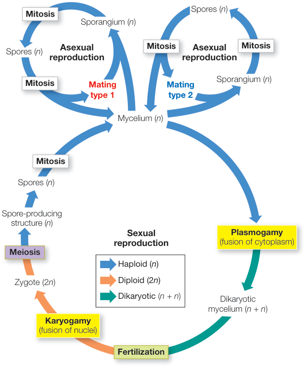
Figure 28.11
Fungi Have Many Practical Uses
- ■ Some fungi are consumed as food by humans; other fungi are critical in baking, fermentation, and flavoring food.
- ■ Fungi play important roles in cleaning up environmental pollutants such as synthetic petroleum-derived hydrocarbons. The diversity and abundance of lichen growth on trees are sensitive indicators of air quality.
- ■ Reforestation projects require restoration of the mycorrhizal fungal community.
- ■ Fungi provide important weapons against diseases and pests.
Questions
- What critical function do fungi play in baking and brewing?
- Why are lichens particularly useful as indicators of environmental pollution?
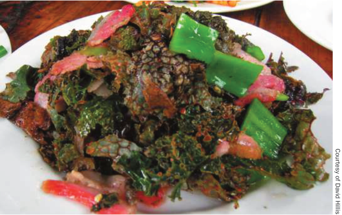
Figure 28.20
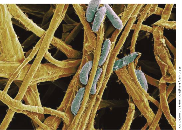
Figure 28.21