Chapter 38Physiology, Homeostasis, and Temperature Regulation
PART EIGHT
Animals: Form and Function
© dea picture ar alliances/Alamy Stock Photo
Key Concepts
38.1 Animals Are Composed of Organs Built from Four Types of Tissues
38.2 Physiological Systems Maintain Homeostasis of the Internal Environment
38.3 Biological Processes Are Temperature-Sensitive
38.4 Body Temperature Depends on the Balance between Heat In and Heat Out of the Body
38.5 Body Temperature Is Regulated through Adaptations for Heat Production and Heat Loss
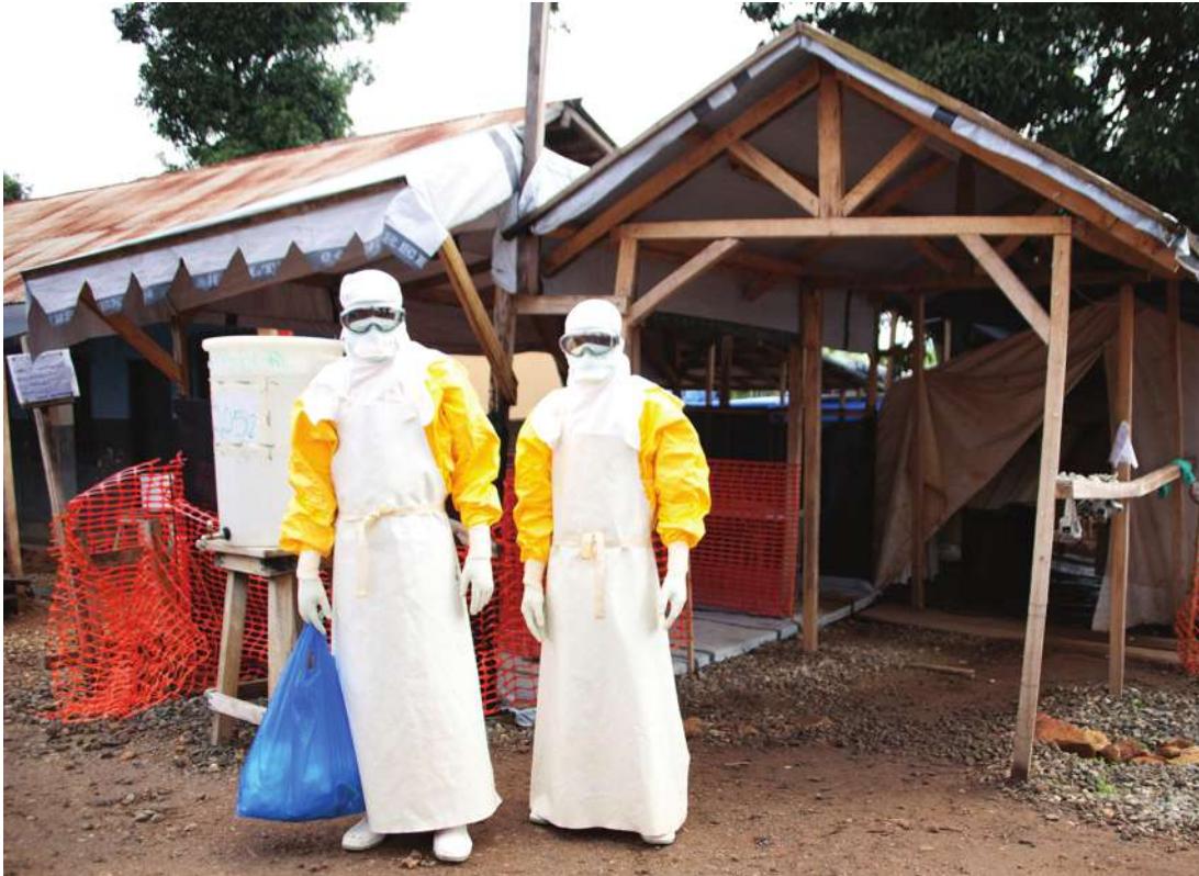
Human epidemics of Ebola have occurred numerous times in sub-Saharan Africa, and usually in hot humid climates. Ebola is highly contagious, so patients must be quarantined, and health care workers must be protected by complete encapsulation in personal protective equipment (PPE). PPE prevents heat loss from the body, so these workers can take care of patients for only a short time before they have to leave the quarantined ward and shed their PPE to avoid hyperthermia.
Investigating Life: Heat Limits Physical Performance and Can Kill
When a person’s internal body temperature rises above 40°C, major organs begin to fail, a condition known as heat stroke. Every year some athletes suffer heat stroke, which frequently leads to death if whole body cooling is not done expeditiously. Other individuals such as soldiers, firefighters, and construction workers work in hot environments while being dressed in heavy protective equipment. They must take precautions to avoid heat stroke. A special case is health care workers in Ebola outbreak areas in hot West Africa. Frequently they cannot work for more than 20 or 30 minutes in the isolation wards with infected patients.
Activity increases the danger of heat stroke because working muscles generate heat. Circulating blood carries heat out of working muscles and distributes it throughout the body, raising the temperature of the body’s internal organs. Some of the heated blood flows to the skin and is lost to the environment. Humans are subject to the heat-loss problems faced by all mammals. First, mammals have high rates of metabolic heat production. Second, their normal internal temperatures are not far from those that cause heat stroke, so they don’t have much of a safety zone. And third, most mammals have fur—great for conserving body heat in cold environments, but an impediment to heat loss in warm ones. Humans don’t have fur, but our skin is still a good insulator.
The evolution of heat-loss adaptations has been important for mammals. You know about sweating and panting, but another important mechanism was discovered recently. To increase the heat-loss potential of non-hairy skin areas, mammals have evolved special, gated blood vessels that can open to bring lots of blood, and therefore heat, to these areas. In furred mammals, these areas may include the nose, tongue, ears, face, and footpads. Humans are not furred, but our ancestors were, and we retain these mammalian blood vessel adaptations in our hands, feet, and face (which is why we can tell a person’s thermal state by shaking hands).
QA How can we increase heat loss from the body to protect against heat stress? (Find the answer on page 860.)
Animals Are Composed of Organs Built from Four Types of Tissues
Learning Objectives
- 38.1.1 Identify factors that limit cell size.
- 38.1.2 Explain the advantages of multicellularity.
- 38.1.3 Describe the characteristic features of the four tissue types.
Animals are amazing in all they do to survive, thrive, and propagate. They live in environments that could not support single cells, they extract energy from the environment, they build complex molecules and structures, they alter their environments, and they reproduce. All of these feats and more depend on complex body components and physiological systems that have evolved since the first appearance of multicellular organisms on this planet. What does it take to be an animal?
How would you build a multicellular animal?
This might seem like a silly question; even bioengineers don’t build animals. But as you begin to study animal form and function, or animal physiology, it is instructive to think of all of the problems animals must solve to survive. For example they must:
- extract energy and nutrients from the environment.
- build all of the internal structures they need.
- eliminate toxins and metabolic waste products.
- sense the environment and respond to it in various ways, including movement.
- maintain constant conditions in their internal environments.
- reproduce.
Single-and multicellular organisms have evolved to conquer these challenges. In fact, single-celled organisms are remarkably abundant and thrive in widely diverse environments. Multicellular organisms have evolved complex body systems to conquer the same challenges. Given the success of single-celled organisms, why have multicellular animals evolved? How is it advantageous to be multicellular? Multicellularity confers two advantages—large size and the opportunity for cells to specialize.
There are advantages and challenges to getting bigger
Advantages to being bigger include being able to prey on other organisms and also being able to resist or work against forces in the environment, such as wave forces in reefs. There is a problem, however, that single-celled organisms must overcome as they get bigger. Exchanges with the environment, such as uptake of oxygen and nutrients as well as elimination of wastes, occur across the cell membrane, so the extent of these exchanges is limited by the surface area of the cell membrane. For simple globular-shaped cells, their cell surface area increases with the square of the linear dimension of the cell, but the needs of the cell increase as a function of the volume of the cell, which is a function of the cube of the linear dimension.
Of course there are adaptations to get around this problem, including being flat and thin or having lots of projections and indentations in the surface, but in general the surface area-to-volume relationship is a challenge for growing bigger.
Connect the Concepts Figure 5.2 illustrates how cell volume grows faster than cell surface area as cells get bigger.
Cell specialization is essential for multicellular animals to grow bigger. If a multicellular animal consists of only a few layers of cells, those cells can carry out their own exchanges directly with the environment. A good example is a sponge (Figure 38.1). Bigger organisms have cells that are not in contact with the external environment. The needs of those cells must be supplied by an internal environment—extracellular fluid that bathes all the cells of the animal’s body. Different cells and groups of cells must be specialized to contribute to the maintenance of that internal environment. Thus each cell contributes to the internal environment, and each cell is supported by that internal environment.
Cell specializations have tremendous adaptive value for even the simplest multicellular animals. Some cells can be specialized for movement, some for intake of food, and some for reproduction. But evolving a specialized function for a cell type usually comes at the expense of other cellular functions, which may be lost or at least compromised. The specialized cells of an organism thus engage in a kind of collaborative enterprise by providing a particular service for all of the cells of the organism while benefiting from different services provided by those other cells. Thus the cells specialized for movement do not have to also capture and process food, and the reproductive cells do not have to move the whole animal to find a mate.
There are four tissue types
Specialized cells rarely function alone, but rather as groups of similar cells called tissues. There are many different kinds of cells, but they are all classified as one of four tissue types: epithelial, muscle, connective, or nervous (Figure 38.2):
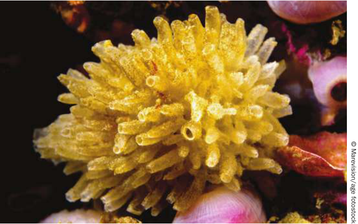
Figure 38.1 No Cell Too Far The sponge Leucosolenia consists of tubes about 2 centimeters long with an outer and an inner layer of cells and a jellylike matrix. There are pores between the cells of the tube. The inner cells have flagella that create a current of water that flows in through the pores and out through the opening at the top. The individual cells of this animal can obtain nutrients and oxygen directly from the seawater.
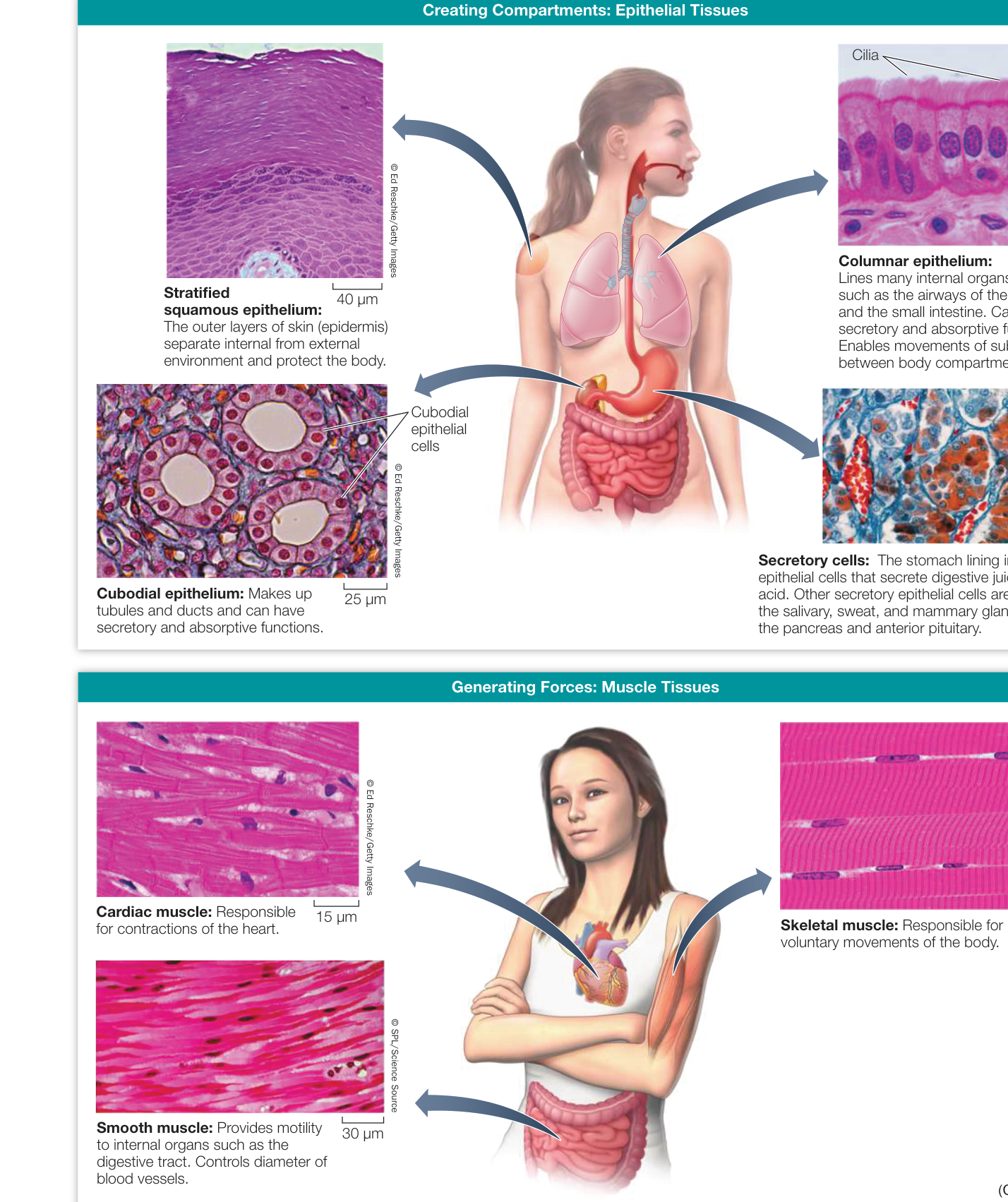
Figure 38.2 How to Build an Animal Tissues are the building blocks.
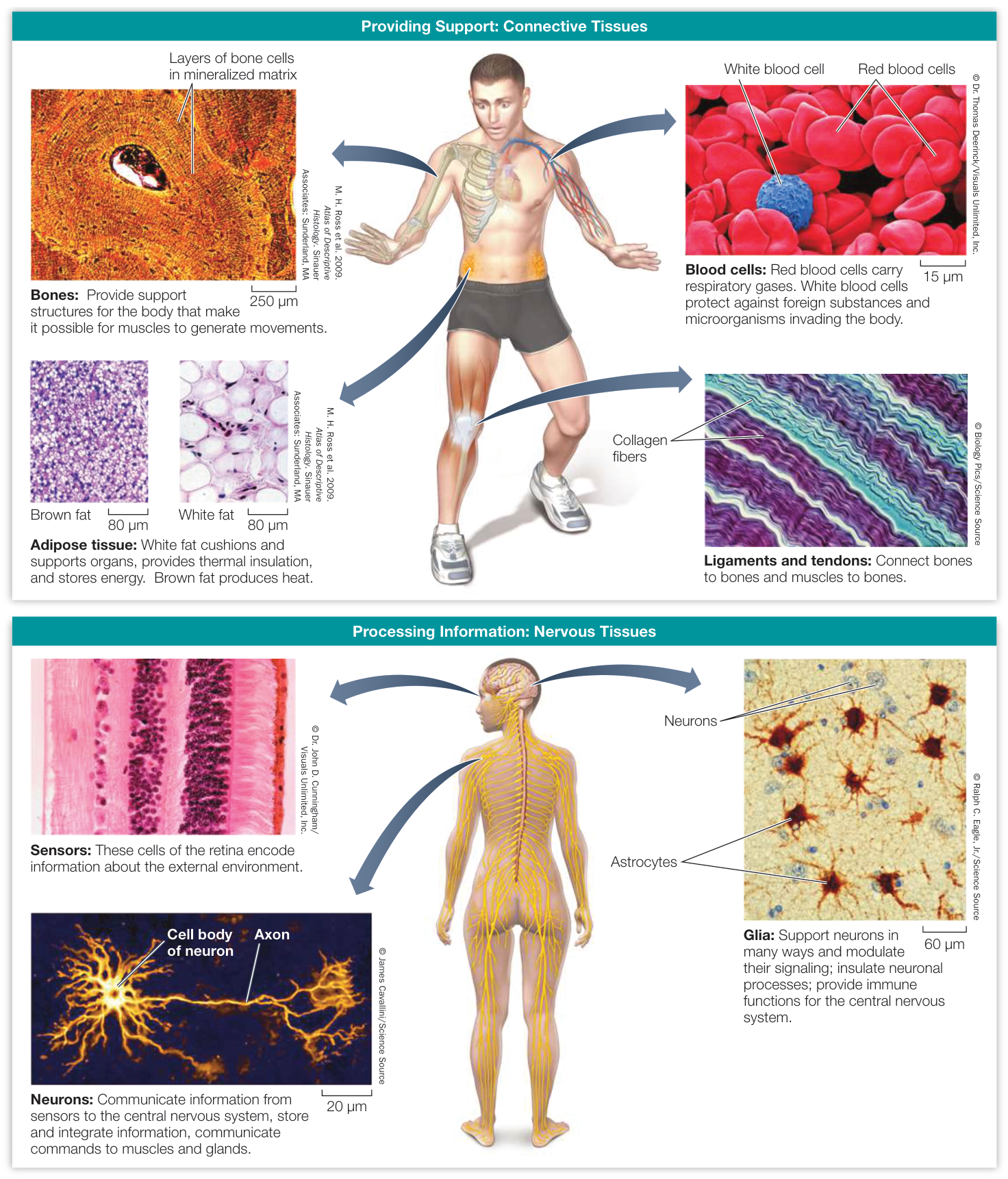
Figure 38.2 (continued)
- Epithelial tissues are sheets of cells that create barriers between different compartments and frequently have secretory functions.
- Muscle tissues contract to generate forces and movement.
- Connective tissues provide structure and support.
- Nervous tissues convey and process information.
These are very general groupings that reflect developmental origins, but within each group there are various specializations that accommodate different functions. For example, connective tissue includes bones, cartilage, fat, and blood. Epithelial tissue includes the outer layers of skin (epidermis) and many types of secretory cells that produce and release substances internally, such as hormones, and externally, such as sweat and digestive juices. You might think of digestive juices as being secreted internally, but the lumen of the digestive tract is continuous with the external environment. You will learn a lot about the diversity of functions accomplished through specializations of these four tissue types in the chapters that follow.
Organs are built from multiple tissues
Organs are internal structures that carry out specific functions. An organ is composed of an outer covering, or epithelium, and one or more other kinds of tissues. Indeed, most organs include all four tissue types. The gut (digestive tract) is a good example (Figure 38.3). Its inner surface is lined with a sheet of columnar epithelial cells. Different types of epithelial cells in this lining secrete hormones or digestive juices or absorb nutrients from the gut. Beneath the epithelial lining is a layer of connective tissue called the mucosa. Within this connective tissue are blood vessels, neurons, and glands (clusters of secretory epithelial cells). Concentric layers of smooth muscle tissue enable the gut to contract to mix food with digestive juices. A network of neurons between the muscle layers controls these movements.
An individual organ is usually part of an organ system, a group of organs that work together to carry out certain functions. The stomach, small intestine, liver, and pancreas, for example, are parts of the digestive system. The structure of an animal involves an organizational hierarchy. Cells form tissues, tissues form organs, organs work together as organ systems, and all work together as a multicellular organism.
Recap and Assess
There are limits to how large cells can be because metabolic needs are related to cell volume, and abilities to satisfy those needs are determined by cell surface area. Cell volume grows faster than cell surface area. Multicellularity and cell specialization enabled organisms to grow larger. Groups of similar cells form tissues, and multicellular animals are composed of four tissue types: epithelial, connective, nervous, and muscle. Individual organs include multiple tissue types and are part of organ systems.
- For single-celled organisms, why does increased size create a problem with obtaining oxygen, and what adaptations could reduce this problem?
- What are the advantages of being a multicellular organism rather than a single-celled organism?
- Provide an example of how multiple tissue types are involved in the construction of an organ. What does each tissue type contribute to the function of that organ?
Subsequent chapters will describe each of the organ systems in much greater detail. The remainder of this chapter focuses on the mechanisms of control and regulation of the internal environment, using temperature as the example.
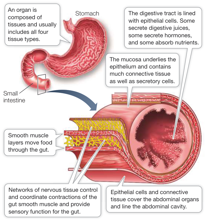
Figure 38.3 Tissues Form Organs The organs of the human digestive system, such as the stomach and small intestine, are made up of all four tissue types.
Physiological Systems Maintain Homeostasis of the Internal Environment
Learning Objectives
- 38.2.1 Explain why homeostasis of the internal environment in a multicellular animal is critical to the animal’s survival.
- 38.2.2 Discuss the relationships between the fluid compartments in the human body.
- 38.2.3 Differentiate among negative feedback, positive feedback, and feedforward control mechanisms, and give examples.
The evolution of multicellularity made it possible to enclose and therefore create an internal fluid environment that bathes all cells of the organism. Since that internal environment provided for all of the needs of the individual cells, individual cell lines could become specialized as to what they contributed to the internal environment. Specialization in one function usually goes hand in hand with losses in other functions. But those functional losses in each cell line were compensated for by the constancy of the internal environment. The evolution of organs composed of multiple tissues and the evolution of informational systems to control organ activities improved the abilities of animals to maintain a constant internal environment. In essence, the evolution of physiological systems to maintain various aspects of the internal environment made it possible for multicellular animals to become larger, and more complex, and allowed them to occupy many different environments.
The internal environment is the extracellular fluid that serves all the needs of the cells of the body
Individual cells get their nutrients from the surrounding extracellular fluid (ECF) and dump their waste products into it. A human is about 60% water. Two-thirds of that water is intracellular fluid and one-third is the ECF. About 20% of the ECF (about 3 liters) is the blood plasma that circulates in our blood vessels. The remaining 80% (about 11 liters) is the interstitial fluid that bathes every cell of the body (Figure 38.4). Water and small molecules freely exchange between the interstitial fluid and the blood plasma. Thus nutrients move into the interstitial fluid from the blood plasma, and wastes move into the blood plasma from the interstitial fluid. As long as the composition and physical conditions of the interstitial fluid are held within certain limits, cells are protected from the changes and harsh conditions of the external environment. A stable internal environment makes it possible for an animal to occupy habitats that would kill its cells if they were directly exposed to the external conditions. How is the internal environment kept constant?
The constant composition of the internal environment is always being challenged by the external environment and by the metabolic activities of the cells of the body. Organisms must maintain their internal environment in a state of homeostasis—a narrow range of stable and optimal physical and biochemical conditions. If a physiological system fails to function properly, homeostasis is compromised, and cells are damaged and can die. To avoid the loss of homeostasis, organ systems are controlled and regulated in response to changes in both the external and internal environments. The maintenance of homeostasis is a central theme of physiology.
Physiological systems are regulated to maintain homeostasis
The activities of physiological systems are controlled—speeded up or slowed down—by actions of the nervous and endocrine systems. But to regulate these systems and maintain homeostasis, information is required. As an analogy, think of the thermostat that controls the furnace and air conditioner to regulate the temperature of a house (Figure 38.5). The desired temperature is a set point, or reference point on the thermostat. The thermostat acts as a comparator by sensing the current temperature in the house and comparing that value to the set point. Thus the sensing of the air temperature is feedback information. Any difference between the set point and feedback information results in an error signal. The error signal is converted into commands to the furnace or air conditioner, turning them on or off.
Regulatory systems such as a building thermostat or a physiological regulatory system obtain, process, and integrate information. Regulatory systems use that information to issue commands to effectors such as muscles or glands that effect changes in the internal environment. Effectors are also called controlled systems because their activities are controlled by the neural or hormonal signals from their respective regulatory systems. Important components of any regulatory system are the sensors such as light-, temperature-, and pressure-sensitive cells that provide feedback information to be compared with internal set points.
Negative feedback is information used to counteract the influence that created an error signal. Whatever force is pushing the system away from its set point must be “negated.” In our thermostat analogy, an air temperature below the set point causes the furnace to be turned on, which then reverses the direction of change in the air temperature.
Although not as common as negative feedback, positive feedback also exists in physiological systems. Rather than returning a system to a set point, positive feedback amplifies a response (i.e., it increases the deviation from the set point). An example is sexual behavior, in which a little stimulation causes more behavior, which causes more stimulation, and so on. Positive feedback responses tend to reach a limit and terminate rapidly. The birth process is a good example. Contractions of the uterus push the baby into the birth canal, and stretching of the birth canal stimulates more and stronger contractions until the baby is delivered, at which time contractions cease.
Feedforward information is a feature of regulatory systems that changes the set point in anticipation of a change in conditions. The timer on a thermostat provides feedforward information by changing the system’s temperature set point, usually lowering it in the evening and raising it in the morning. Hearing the words “on your mark” before a race is feedforward information that raises your heart rate in anticipation of running. Feedforward information anticipates a change in the internal environment before that change occurs.
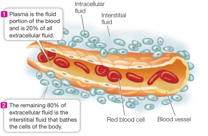
Figure 38.4 The Internal Environment The extracellular fluid (ECF) is the “internal environment,” and it is about one-third of total body water. The ECF is about 20% blood plasma and 80% interstitial fluid. The composition and physiological state of the ECF must remain stable within narrow limits, and maintaining that stability is the job of the body’s organ systems.
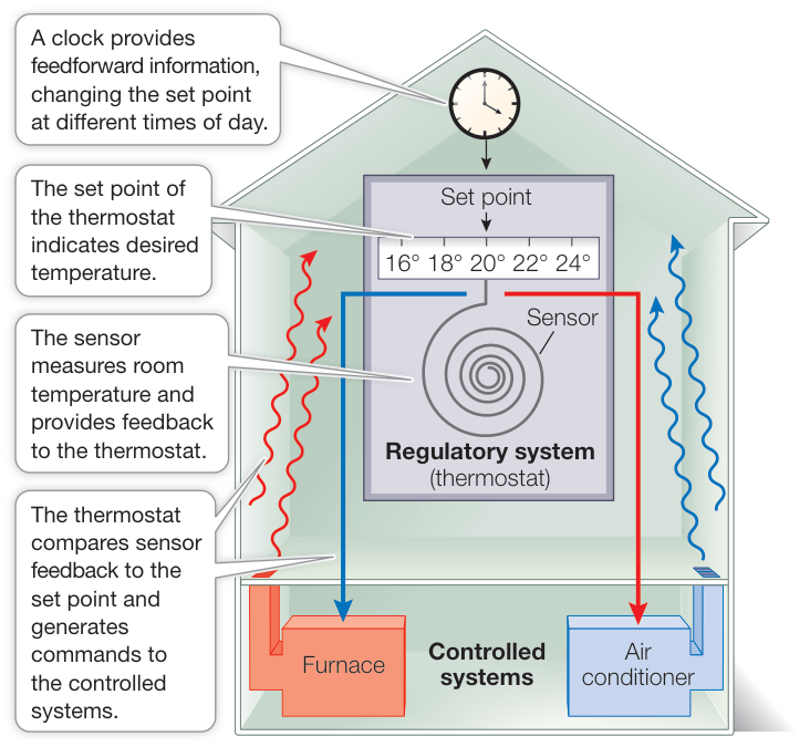
Figure 38.5 A Thermostat Regulates Temperature A thermostat regulates the temperature of a room by turning the furnace or air conditioner on or off in response to the difference between feedback information (room temperature) and set points that are programmed into the thermostat.
Recap and Assess
The internal environment provides for the needs of all the cells that make up a complex multicellular animal. The internal environment is the extracellular fluid compartments of the body. Organs and organ systems control the chemical and physical composition of the extracellular fluids to maintain homeostasis. The activities of organs and organ systems are regulated. Regulation requires information about optimal conditions (set points) and existing conditions (feedback) to create commands to effector cells and organs.
- What are the relationships between the extracellular fluid of the body, the organ systems, and homeostasis?
- Based on your body mass, estimate how much water is in your body. What is your intracellular fluid volume, your extracellular fluid volume, your blood plasma volume, and your interstitial fluid volume?
- Explain the differences between negative and positive feedback and feedforward control mechanisms.
Principles of control and regulation help organize our thinking about physiological systems. Once we understand how a system works, we can then ask how it is regulated. Part Eight of this book describes various physiological systems, how they function, and how they are regulated. We’ll begin by extending our example of a house thermoregulatory system to that of the mammalian body.
Biological Processes Are Temperature-Sensitive
Learning Objectives
- 38.3.1 Discuss the effects of temperature on living systems.
- 38.3.2 Describe the concept.
- 38.3.3 Explain how isozymes are involved in seasonal temperature acclimatization.
Temperatures of environments where organisms live vary enormously, from the boiling hot springs of Yellowstone National Park to the interior of Antarctica, where the temperature can fall below . But in general, cells can function over only a narrow range of temperatures. The evolution of thermoregulatory adaptations enables maintenance of suitable thermal conditions in the internal environment.
Temperature affects living systems
If cells cool below their freezing point, ice crystals form and damage cell structures. Some animals have adaptations, such as antifreeze molecules in their blood that help them resist freezing; others can survive freezing (Figure 38.6). Generally, however, cells must remain above freezing to stay alive. The upper temperature limit for survival in most cells is below because of protein denaturation. Some specialized algae, however, can grow in hot springs at , and some archaea live at near .
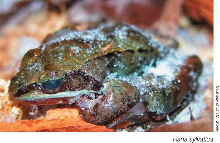
Figure 38.6 Frozen Frogs Wood frogs live in the northeastern United States, across most of Canada, and up into northern Alaska. They freeze solid in the winter and survive. During the fall and early winter, urea and glucose accumulate in the frogs’ cells and act as intracellular cryoprotectants (like antifreeze in your car radiator). Also, much intracellular water is redistributed to the extracellular fluid, which can freeze without damaging cells.
Connect the Concepts Protein denaturation by heat is due to effects on secondary and tertiary structure of the protein, as described in Key Concept 3.2.
One adaptation cells have to survive episodes of exposure to high temperatures is heat shock proteins (HSPs). HSPs are chaperone proteins that protect other proteins from denaturation, and they are found in organisms ranging from bacteria to humans (see Key Concept 3.2 and Figure 3.15). Sudden heating induces the expression of HSPs, which act to stabilize proteins and prevent or reverse errors in protein folding.
Although the survival limits for most cells fall in the range of – , most species have much narrower thermal limits for optimal function. To stay within those limits in spite of environmental conditions, animals have thermoregulatory adaptations that enable them to tolerate extreme conditions or to control their body temperatures in spite of environmental conditions. These adaptations may determine the thermal tolerances of species and thereby determine their geographic ranges. When environments change rapidly, as is happening globally at present, animals may find themselves in situations that exceed their thermal tolerances. A good example is the reef-building corals discussed in the opening of Chapter 1.
is a measure of temperature sensitivity
Even between and , changes in body temperature can create problems for animals. Most biochemical reactions and the physiological processes they are involved in are temperature-sensitive, going faster at higher temperatures. The temperature sensitivity of a reaction or process can be described in terms of , a factor calculated by dividing the rate of a process or reaction at a certain temperature, , by the rate of that same process or reaction at a temperature 10 degrees Celsius lower, :
Connect the Concepts As discussed in Key Concept 8.5, increases in temperature increase reaction rates because reactant molecules have more kinetic energy. Enzymes that control rates of biochemical reactions in metabolic pathways are influenced by temperature as well.
can be measured for a simple biochemical reaction or for a complex physiological process, such as rate of oxygen consumption. If a reaction or process is not temperature-sensitive, it has a of 1. Most biological values are between 2 and 3. A of 2 means that the reaction rate doubles as temperature increases by 10 degrees Celsius, and a of 3 indicates a tripling of the rate as temperature increases by 10 degrees Celsius (Figure 38.7).
Notice that the values (except for ) plotted in Figure 38.7 produce curves rather than straight lines. This is because temperature increases in additive intervals (10, 20, 30, etc.) but reaction rates increase in a multiplicative fashion (2, 4, 8, 16, 32, etc.). Such curvilinear plots are common in biological data.
Changes in body temperature can disrupt an animal’s physiology because not all of the biochemical reactions that constitute the metabolism of an animal have the same . These biochemical reactions are linked together in complex networks: the products of one reaction are the reactants for other reactions. Because different reactions have different ’s, changes in tissue temperature will shift the rates of some reactions more than others, disrupting the overall network. To maintain homeostasis, organisms compensate for or prevent changes in body temperature.
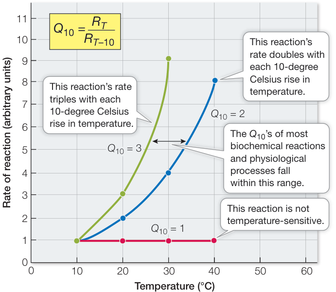
Figure 38.7 and Reaction Rate The larger the of a reaction or process, the faster its rate rises in response to an increase in temperature.
Animals acclimatize to seasonal temperatures
The body temperatures of some animals (especially aquatic animals) are coupled to environmental temperature. The body temperature of a fish in a pond, for example, is the same as the water temperature, which might range from in winter to in summer. If we bring that fish into the laboratory in the summer and measure the rates of any of its physiological or biochemical processes such as oxygen consumption at different water temperatures, we will demonstrate a relationship. On the basis of the relationship, we would predict that the fish’s oxygen consumption—a measure of its metabolic rate—would be much lower in the winter pond because the water temperature is lower. However, if we bring that fish back into the laboratory in the winter and measure its metabolic rate at winter pond temperature, we find that rate to be higher than we predicted. The fish’s biochemistry and physiology will have acclimatized to the seasonal change in water temperature so that it can remain active at winter temperatures. What could be the mechanism of this acclimatization? Many organisms express multiple forms of enzymes (isozymes) that may have different temperature optima. In winter, organisms express the forms that show maximum substrate affinity at low temperatures, and in summer they express the forms that have maximum substrate affinity at high temperature. Another mechanism of temperature acclimatization is a change in the composition of cell membranes designed to maintain optimum fluidity despite changes in temperature (see
Key Concept 6.1). Acclimatization in animals essentially means that their metabolic functions are less sensitive to long-term changes in their environment than to short-term changes.
Small changes in temperature can have large physiological effects
The story opening this chapter described how heavy exercise, exposure to hot environments, or being clad in insulative protective gear can rapidly elevate a person’s body temperature to a dangerously high level. Our ability to function physically or mentally is rapidly compromised by hyperthermia, and sustained hyperthermia of only a few degrees can lead to death. To avoid the serious consequences of hyperthermia, it is important to reduce heat-generating muscular work and to increase heat loss from the body as soon as possible. The body has protective fail-safe mechanisms, one of which is heat-induced cramps. Ask any athlete whether or not a cramp makes them stop exercising. Another fail-safe mechanism is to deprive the muscle of energy, causing muscle failure. Pyruvate kinase is an enzyme critical for production of ATP in muscles; it catalyzes the last step in glycolysis, producing pyruvate molecules that can enter the mitochondria and be metabolized to produce ATP (see Figure 9.17). Without ATP, muscles fatigue and can’t do work. Muscle pyruvate kinase inactivates at high temperatures, which shuts off muscle function and thereby prevents thermal damage to the muscle. Pyruvate kinase is reactivated when the muscle cools, and the muscle can resume work. You may wonder, if we could protect athletes and emergency workers from hyperthermia by enhancing extraction of heat from their muscles, could we increase their performances and capacity to work in the heat? We consider this question in Investigating Life: Can the Work Capacity of Workers in PPE be Extended by Extracting Heat from the Palms of the Hands?
▶ Investigating LIFE Can the Work Capacity of Workers in PPE Be Extended by Extracting Heat from the Palms of the Hands?
Experiment
Original Paper: D. A. Grahn et al. 2018. A method to reduce heat strain while clad in encapsulating outerwear. J Occup Environ Hyg 15: 573–579.
In the opening story of this chapter you learned that mammals have evolved special heat-dissipation adaptations underlying their non-hairy skin. These skin areas are small, but their adaptation is high-volume blood vessels that can rapidly transfer heat from the body core to the skin. Humans don’t have fur, but they do have these same blood vessel adaptations. In this experiment, individuals clad in personal protective equipment (PPE) were exposed to a hot ambient temperature (45°C) for 60 minutes, with or without a cooling system worn under their PPE that cooled the palms of their hands. The outcome measure was esophageal temperature measured at the level of the heart so as to obtain the best average body core temperature.
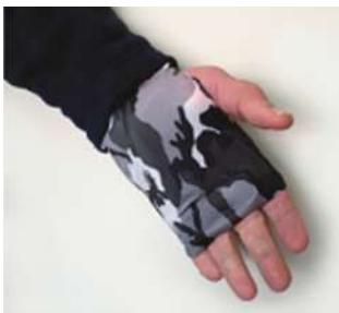
D. A. Grahn et al. 2018. J Occup Environ Hyg 15: 573–579
HYPOTHESIS Extracting heat from the palms of the hands will extend dwell time in the heat for a worker in PPE.
METHOD
Recruit 10 healthy individuals between the ages of 19 and 23 years old.
Fully encapsulate the subjects in Tyvek® PPE (DuPont Corporation) and have each subject self-insert a thermocouple (Mallinckrodt Medical Inc.) down his or her esophagus to the level of the heart.
In randomized fashion, have each subject rest in an environmental chamber at 45°C with or without a wearable palmar cooling system under their PPE.
- Terminate exposures at 60 minutes or when esophageal temperature reaches 39°C.
RESULTS For all 10 subjects, their esophageal temperatures () measured at 5-minute intervals were averaged and the standard deviations (SD) calculated.
| Time (min) | , mean (SD) | |
|---|---|---|
| Control, no cooling | Palm Cooling | |
| 0 | 36.85 (0.18) | 36.65 (0.30) |
| 5 | 36.86 (0.21) | 36.67 (0.33) |
| 10 | 36.93 (0.20) | 36.72 (0.28) |
| 15 | 37.05 (0.19) | 36.76 (0.27) |
| 20 | 37.15 (0.29) | 36.79 (0.26) |
| 25 | 37.34 (0.26) | 36.86 (0.28) |
| 30 | 37.55 (0.26) | 36.95 (0.29) |
| 35 | 37.76 (0.23) | 37.05 (0.29) |
| 40 | 37.99 (0.22) | 37.14 (0.30) |
| 45 | 38.20 (0.20) | 37.24 (0.30) |
| 50 | 38.40 (0.18) | 37.32 (0.29) |
| 55 | 38.60 (0.17) | 37.40 (0.32) |
| 60 | 38.80 (0.16) | 37.49 (0.32) |
CONCLUSION By analyzing these data, you will be able to conclude whether the hypothesis is supported or not supported by this experiment.
Investigating LIFE Work with the Data follows on the next page.
Work with the Data
This experiment involves a simple outcome measure: esophageal temperature at 5-minute intervals during exposure to 45°C while fully encapsulated in PPE, as an Ebola health care worker would experience. The experimental manipulation is heat extraction from the palms by a portable cooling system worn under the PPE.
QUESTIONS►
Plot the data with time on the x axis and on the y axis. Using the t-test formula in Figure B11 in Appendix B, test which of the pairs of data at each temperature are statistically different. That formula will give you a t-value, and knowing that your sample size is 10, you can refer to a t-statistic table to find the probability that the values are significantly different. You can also use an online statistics program to perform paired t-tests on the data.
- Using the linear parts of the curves (after 20 minutes), what are the rates of increase with and without cooling?
- Is the hypothesis supported or disproven? Explain.
- Assuming that a core temperature of 39°C is the point at which a worker must leave the “hot zone,” how much longer can the work session be with palmar cooling?
- The subjects in this experiment rested in the heat. How would you expect the data to differ if subjects sustained a modest workload?
Recap and Assess
Cells can survive only within a narrow range of temperatures, but even changes within that range can be disruptive because different physiological processes have different temperature sensitivities.
- How can fluctuations in body temperature disrupt complex physiological processes?
- Plot a curve for a physiological process using arbitrary units as measures of that process.
- What characteristic of isozymes might explain their role in metabolic seasonal temperature acclimatization?
Now that you know how animals are affected by temperature, we’ll look next at the adaptations that allow animals to survive wide ranges of thermal environments and wide ranges of metabolic activities.
38.4
Body Temperature Depends on the Balance between Heat In and Heat Out of the Body
Learning Objectives
38.4.1 Give examples of an ectotherm, an endotherm, and a heterotherm, and explain their responses to changes in environmental temperature.
38.4.2 Describe the paths of heat exchange between an animal and its environment.
38.4.3 Explain why “hot fishes” such as great white sharks can maintain a core body temperature above that of the sea while most other fishes cannot.
Body temperature is a measure of the heat content of the body. Animals produce heat internally through their metabolism, and they continuously exchange heat with their environment. The balance between heat gain and heat loss determines the animal’s body temperature. Thermoregulatory adaptations enable animals to modulate the various paths of heat exchange between themselves and their environment.
Animals can be classified by their thermoregulatory characteristics
One way of classifying animals is according to the constancy of their body temperatures. Animals that maintain a constant body temperature are called homeotherms, and animals that experience a fluctuating body temperature are called poikilotherms. Another classification is based on the source of heat that predominantly determines body temperature. Endotherms such as birds and mammals have the ability to vary their metabolic heat production to compensate for the loss of heat to the environment. Ectotherms such as invertebrates, fishes, amphibians, and non-avian reptiles are largely dependent on environmental sources of heat. We use the terminology of ectotherm and endotherm in this book, but no classification scheme is perfect. Many ectotherms can generate considerable internal heat to raise their body temperature, and endotherms that hibernate can periodically appear to give up the ability to generate internal heat. To accommodate these departures from a strict dichotomy, we add the term heterotherms—organisms that act like ectotherms some of the time and like endotherms at other times.
Endotherms produce substantial amounts of metabolic heat
Transfers of energy in biological systems are inefficient. In both ectotherms and endotherms, in every transfer of energy—from food molecules to ATP, from ATP to biological work—some of the energy is lost as heat. Working muscles produce heat, as do all of the metabolic activities of all tissues.
Connect the Concepts As discussed in Key Concept 8.1, transfers of energy are governed by the first and second laws of thermodynamics, which explain why heat is always a by-product of metabolism.
Why do endotherms produce more heat than ectotherms? The surprising answer is that the cells of endotherms are less efficient at using energy than are the cells of ectotherms. The cells of endotherms are more “leaky” to ions than are the cells of ectotherms. Therefore ions are constantly diffusing into the cells, and ions
Body Temperature Depends on the Balance between Heat In and Heat Out of the Body
are constantly diffusing out. Even an endotherm at rest must spend considerable amounts of energy to transport out of the cells and transport back in. Because of their constant need to actively transport ions, endotherms expend more energy than do ectotherms just to maintain the ion concentration gradients across their cell membranes. This situation is analogous to a leaky boat: the faster water comes in (i.e., the faster ions diffuse down their concentration gradients), the more metabolic energy has to be expended to bail the water out (i.e., pump ions back up their concentration gradients). Since endotherms expend more energy than ectotherms do to maintain ion concentration gradients, they produce more internal heat.
We can speculate that a mutation resulting in seemingly faulty or leaky ion channels might underlie the evolution of endothermy. Such a mutation in a small ectotherm would have increased its energy expenditure and therefore its heat production. Increased heat production would have enabled the animal to be active earlier in the morning or for a longer time after sunset. Being active in twilight, and eventually at night, would open up a new world of ecological opportunities—a world in which there was less competition from similar-sized ectotherms and less danger of predation.
Two major differences between endotherms and ectotherms are (1) their resting metabolic rates—the sum total of all energy expenditures in their bodies when at rest—and (2) their responses to changes in environmental temperature.
Ectotherms and endotherms respond differently to changes in environmental temperature
How do similar-sized ectotherms and endotherms respond to changes in environmental temperature? Let’s compare a lizard and a mouse. We put each animal in a closed chamber and measure its body temperature and its resting metabolic rate as we set the temperature of the chamber at different levels between and . The body temperature of the lizard equilibrates with that of the chamber, whereas the body temperature of the mouse remains stable over most of this range (Figure 38.8A). The metabolic rate of the lizard (already much lower than that of the mouse when both are at the same body temperature) decreases as the temperature drops (Figure 38.8B). In contrast, the mouse’s metabolic rate increases as the chamber temperature falls below . This increase in metabolism produces enough heat to prevent the mouse’s body temperature from falling. In other words, the mouse can regulate its body temperature by increasing its metabolic rate and thereby maintaining its physiological functions at an optimal level; the lizard cannot.
This experiment might lead us to conclude that the ectotherm cannot regulate its body temperature, but observations of the lizard in nature do not support this conclusion. In nature, the lizard can regulate its body temperature quite well. It does so by behavioral mechanisms rather than by altering its internal metabolic heat production. Air temperature in the desert can fluctuate by 40 degrees Celsius or more in a few hours; the lizard, however, can limit its variation in body temperature and even maintain a fairly stable body temperature by using behavior to alter its heat exchange with the environment. Its behavioral strategies include spending time in a burrow, basking in the sun, seeking shade, climbing vegetation, and changing its orientation with respect to the Sun (Figure 38.9).
Endotherms also use behavior as a first line of defense against thermal stress. They select the most comfortable thermal environment possible. They change posture or their orientation to the Sun, move between sunshine and shade, and move between still air and moving air. Examples of more complex thermoregulatory
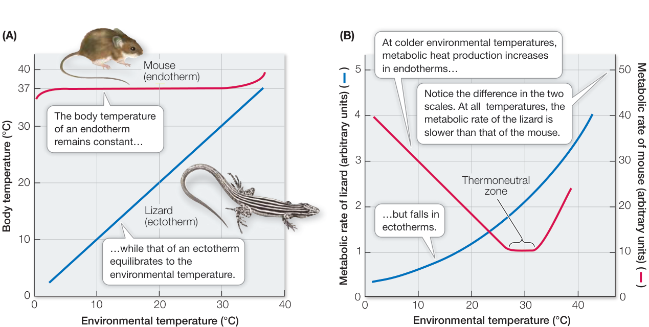
Figure 38.8 Ectotherms and Endotherms React Differently to Environmental Temperatures (A) At the same environmental temperature, an ectotherm and an endotherm of approximately the same body size (here, a lizard and a mouse) have different body temperatures. (B) The metabolic rates of the lizard and mouse react in opposite ways to cooler temperatures.
Q: In the plot of body temperature versus environmental temperature, why does the mouse curve fall on the far left side and rise on the far right side? In the plot of metabolic rate versus environmental temperature, why does the mouse’s metabolic rate rise at higher temperatures?
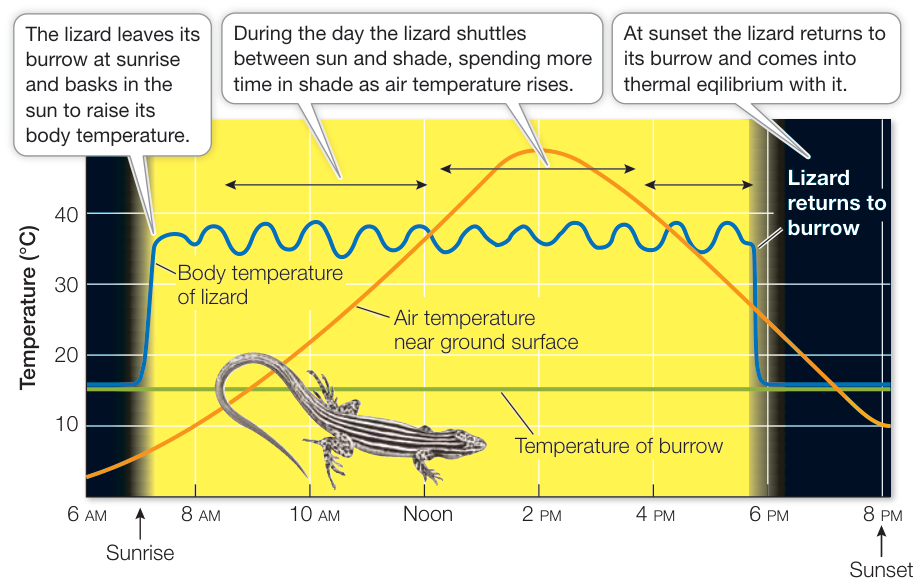
Figure 38.9 Using Behavior to Regulate Body Temperature The body temperature of an ectotherm such as a lizard depends on environmental heat, but the lizard can regulate its temperature by moving from place to place.
behaviors include nest construction and social behaviors such as huddling. Humans put on or remove clothing and burn fossil fuels to generate the energy to heat or cool buildings. Large mammals such as elephants and water buffalo (Bubalus bubalis) may wallow in water when the environment is hot.
Energy budgets reflect adaptations for regulating body temperature
Both ectotherms and endotherms influence their body temperatures by altering four avenues of heat exchange between their bodies and the environment (Figure 38.10):
- Radiation Heat moves from warmer objects to cooler ones via the exchange of infrared radiation (what you feel when you stand in front of a fire).
- Convection Heat exchanges with a surrounding medium such as air or water that flows over a surface (the wind-chill factor).
- Conduction Heat flows directly between two objects at different temperatures when they come into contact (e.g., an icepack on a sprained ankle).
- Evaporation Heat is transferred away from a surface when water evaporates on that surface (the effect of sweating).
The total balance of heat production and heat exchange can be expressed as an energy budget, based on the simple fact that if the body temperature of an animal is to remain constant, the heat entering the animal must equal the heat leaving it. The heat coming in is usually from metabolism and radiation ( , for radiation absorbed). Heat leaves the body via the four mechanisms listed above—radiation emitted (), convection, conduction, and evaporation. The energy budget takes the mathematical form
The energy budget is a useful concept because any adaptation that influences the ability of an animal to control its temperature must affect one or more components of the energy budget. The energy budget gives us the ability to quantify and compare the thermal adaptations of animals. One interesting observation is that all of the components on the right side of the energy budget equation—that is, the heat-loss side—depend on the surface temperature of the animal. In fact, if the environmental temperature is above skin surface temperature, convection and conduction are avenues of heat gain rather than loss. One way surface temperature can be controlled by altering the flow of blood to the skin.
Both ectotherms and endotherms control blood flow to the skin
Heat is mostly moved around the internal environment by blood flow. Heat produced in muscles during exercise is transported out of the muscle and to the heart in the blood. That heat is then distributed around the body by the blood, raising body temperature. Blood flow to the skin enables internal heat to be lost to the environment through radiation, convection, conduction, and evaporation, thus bringing the body temperature back toward normal. When body temperature is too low or the environment is too cold, the blood vessels supplying the skin constrict, reducing heat loss to the environment.
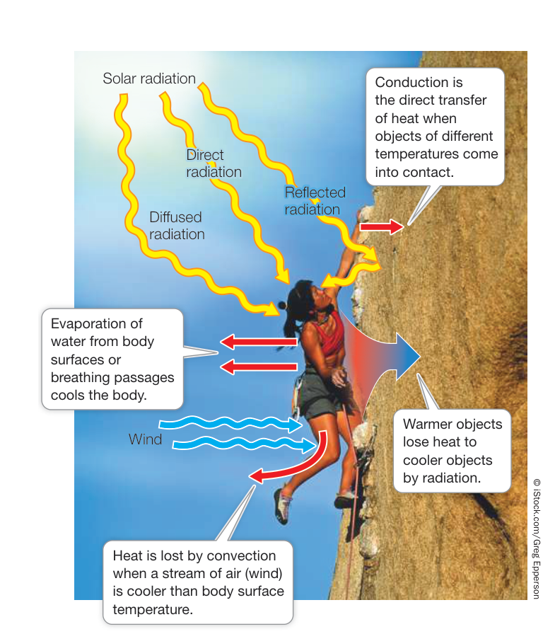
Figure 38.10 Avenues of Heat Exchange between an Animal and Its Environment An animal’s body temperature is determined by the balance between internal heat production and four avenues of heat exchange with the environment: radiation, convection, conduction, and evaporation.
The ability to control blood flow to the skin can be an important adaptation for an ectotherm such as the marine iguana (a reptile) of the Galápagos archipelago (Figure 38.11). The Galápagos are volcanic islands that lie on the equator but are bathed by cold ocean currents. Iguanas bask in the sunshine on hot black lava rocks on the shore, and periodically enter the cold ocean water to feed on seaweed. While the iguanas are feeding, they lose heat to the cold ocean water. This cooling lowers their metabolism, making them slower, more vulnerable to predators, and incapable of efficient digestion. The iguanas therefore alternate between feeding in the cold seawater and basking on the hot rocks. It is advantageous for iguanas to retain body heat as long as possible while swimming and to warm up as fast as possible when basking. They accomplish these results by changing their heart rate and the rate of blood flow to their skin and around their bodies.
What about furred mammals? Fur acts as insulation to keep body heat in, making it possible for mammals to function in cold environments. When they are active, however, mammals must get rid of excess heat, and it does little good to transport that heat to the skin under the fur. Thus as mentioned at the opening of this chapter, mammals have specialized blood vessels for transporting heat to their hairless skin surfaces. Heat loss from these areas is tightly controlled by the opening and closing of these blood vessels. When you are cold, the blood flow to your hands and feet decreases and they feel cold, but when you exercise, the blood vessels serving these skin areas dilate, increasing the blood flow and therefore the heat dissipation.
Media Clip 38.1 Thermoregulation in Animals Life12e.com/mc38.1
Some fishes conserve metabolic heat
The muscles of active fishes produce substantial amounts of metabolic heat, but most fishes cannot retain that heat. Blood pumped from the fish heart goes directly to the gills, where it comes very close to the surrounding water to exchange oxygen and carbon dioxide. Any heat the blood picks up from metabolically active muscles is lost to the water flowing across its gills. It is therefore surprising that some large, rapidly swimming fishes, such as giant bluefin tuna (Thunnus thynnus) (see Figure 1.16A) and great white sharks (Carcharodon carcharias), can maintain swimming muscle temperatures
10–15 degrees Celsius higher than the surrounding water. The heat comes from their powerful swimming muscles, and the ability of these “hot” fishes to conserve that heat is based on the remarkable arrangement of their blood vessels.
In the typical (“cold”) fish circulatory system, oxygenated blood from the gills collects in a large blood vessel, the aorta, that runs through the center of the fish, distributing blood to all organs and muscles (Figure 38.12A). “Hot” fishes have a small aorta running down their body, and most of their oxygenated blood is transported in large side vessels just under the skin (Figure 38.12B). The cold blood leaving the gills is therefore kept close to the surface of the “hot” fishes as it flows posteriorly to the swimming muscles. The cold blood flows into the muscle mass through small vessels branching off the large side vessels. These many small vessels run parallel to the vessels bringing oxygen-depleted but warm blood out of the swimming muscles and back toward the heart. Because the vessels carrying the cold blood into the muscles are in close contact with the vessels carrying warm blood out of the muscles, heat flows from the warm to the cold blood by conduction. Therefore the heat produced by the active muscles is retained in the muscle mass.
Because heat is exchanged between blood vessels carrying blood in opposite directions, this adaptation is called
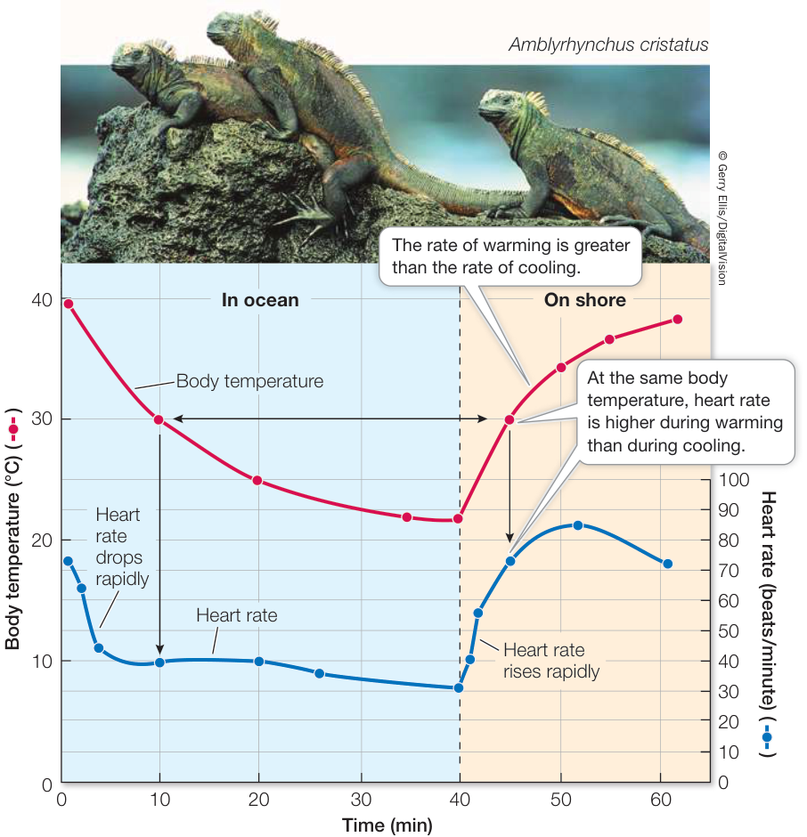
Figure 38.11 Some Ectotherms Regulate Blood Flow to the Skin Galápagos marine iguanas control blood flow to the skin to alter their heating and cooling rates. (Graph after G. A. Bartholomew and R. C. Lasiewski. 1965. Comp Biochem Physiol 16: 573–582.)
(A) “Cold” fishes
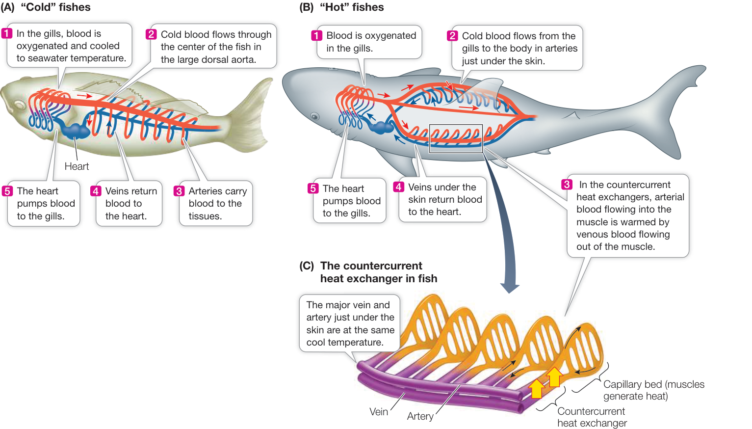
Figure 38.12 “Cold” and “Hot” Fishes (A) Blood flowing through fish gills comes into temperature equilibrium with water temperature. In most fish species, this oxygenated but cool blood flows through a large dorsal aorta to the rest of the body. Thus it is an internal cooling source. (B) The anatomy of “hot” fish species circulates most of the cool, oxygenated blood to the body through large peripheral arteries rather than through a central aorta. As this cool arterial blood flows into the swimming muscle, it is warmed by heat being carried out of the swimming muscle by the veins. Thus the heat generated by the swimming muscles is conserved in the muscle mass. (C) The close, parallel arrangement of the arteries carrying blood into the muscles and the veins carrying blood out of the muscle makes efficient countercurrent heat exchange possible.
(B) “Hot” fishes
countercurrent heat exchange (Figure 38.12C). By keeping heat within the muscles, these fishes can maintain an internal body temperature considerably higher than the water temperature. Each 10-degree Celsius rise in muscle temperature increases a “hot” fish’s power output almost threefold, giving it a faster and more powerful swimming capability.
Some ectotherms regulate metabolic heat production
By definition, ectotherms are largely dependent on environmental sources of heat for thermoregulation, but some ectotherms raise their body temperature by producing excess metabolic heat. For example, the powerful flight muscles of many insects must reach 35°C–40°C before the insects can fly, and they must maintain these high temperatures during flight. Such insects warm up to fly by contracting their flight muscles isometrically in a manner similar to shivering in mammals. The heat-producing ability of insect muscle can be quite remarkable. An impressive case is a species of scarab beetles that live mostly underground in mountains north of Los Angeles, California. The males come aboveground to mate and fly in search of females. They undertake this mating ritual at night, in winter, during snowstorms.
Honey bees regulate temperature as a group. They live in large colonies consisting mostly of female worker bees that maintain the hive and rear the larval offspring of the single queen bee. During winter, worker bees cluster around the brood (eggs and larvae). They adjust their individual metabolic heat production and density of clustering so that the brood temperature remains remarkably constant, at about 34°C, even as the outside air temperature drops below freezing.
Recap and Assess
- Endotherms use metabolic heat production to maintain constant high body temperatures. Body temperatures of ectotherms are more dependent on environmental heat sources. Both ectotherms and endotherms use behavior to control their body temperature. Heat exchange between an animal and its environment occurs via radiation, convection, conduction, and evaporation, all of which depend on body surface temperature. Some ectotherms use metabolic heat production episodically to raise body temperature.
- Why is the body temperature of an ectotherm that is exposed to variations in environmental temperature more constant in nature than in the laboratory?
- For each path of heat exchange, what determines whether heat is gained or lost by the animal?
- Explain the circulatory system adaptations that allow “hot” fishes, unlike most other (“cold”) fishes, to raise the temperature of their swimming muscles.
Endotherms respond to changes in environmental temperature by changing their rates of metabolic heat production. They also have other adaptations for controlling their rates of heat exchange with their environments. How do they regulate these various avenues of heat exchange to achieve a constant internal body temperature?
Body Temperature Is Regulated through Adaptations for Heat Production and Heat Loss
Learning Objectives
- 38.5.1 Describe and interpret a plot of metabolic rate versus environmental temperature for an endotherm.
- 38.5.2 Discuss how differences in physical features of endotherms of the same or similar species relate to the selective pressures of the climates in which they live.
- 38.5.3 Explain how the mammalian brain thermostat uses feedback and feedforward information to regulate body temperature.
- 38.5.4 Describe the unique thermoregulatory abilities of hibernators.
As explained in Key Concept 38.2, there is a difference between control and regulation. Think of driving your car. You control the speed of the car with the accelerator and the brake, but you regulate the speed of your car by comparing the speed limit (set point) with your speedometer (feedback) to generate command signals to your foot that is operating the accelerator and brake. For understanding how body temperature is regulated, you must understand the adaptations for heat production and heat loss, how they are controlled, and what information is used to modulate the control signals to achieve a regulated body temperature.
Basal heat production rates of endotherms correlate with body size
Physiologists can determine an animal’s metabolic rate and therefore its rate of heat production by measuring its consumption of or production of . Within a narrow range of environmental temperatures, called the thermoneutral zone (see Figure 38.8B), the metabolic rates of endotherms (birds and mammals) are at low levels and independent of environmental temperature. The metabolic rate of a resting endotherm at a temperature within its thermoneutral zone is its basal metabolic rate (BMR). BMR is usually measured in animals that are quiet but awake and not using energy for digestion, reproduction, or growth. Thus BMR is the rate at which a resting animal is consuming just enough energy to carry out its minimal body functions.
As you might expect, the BMR of an elephant is greater than that of a mouse. After all, the elephant is more than 100,000 times larger than the mouse. However, the BMR of the elephant is only about 7,000 times greater than that of the mouse. That means that a gram of mouse tissue uses energy at a rate 15 times greater than a gram of elephant tissue (Figure 38.13). Across all of the endotherms, BMR per gram of tissue increases as animals get smaller.
Why should this disproportionate difference exist? There are several possible reasons. As animals get bigger, they have a smaller surface area-to-volume ratio (see Figure 5.2). Since heat production is related to the volume (i.e., mass) of the animal, but its capacity to dissipate heat is related to its surface area, it has been reasoned that larger animals evolved lower metabolic rates to avoid overheating. However, this explanation alone is insufficient because the relationship between body mass and metabolic rate holds for even very small organisms and for ectotherms, in which overheating is not a problem. Other hypotheses have also been proposed. For example, a larger animal has a greater proportion of support tissues (e.g., skin and bone), which are not as metabolically active as other tissue types. The real explanation is probably a mixture of different factors, but the relationship holds over a very broad range of species.
For an endotherm, a metabolic rate versus environmental temperature curve represents the integrated response of all the animal’s thermoregulatory adaptations (Figure 38.14). The thermoneutral zone is bounded by a lower critical temperature and an upper critical temperature. When the environmental temperature is within the thermoneutral zone, an endotherm’s thermoregulatory responses do not require much energy and could be considered passive; such responses include changing posture, fluffing fur or feathers, and altering blood flow to the skin. When an endotherm is outside its thermoneutral zone, however, its thermoregulatory responses are active and require metabolic energy.
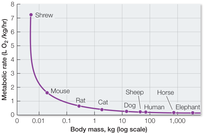
Figure 38.13 The Mouse-to-Elephant Curve On a weight-specific basis, the basal metabolic rate of small endotherms is much greater than that of larger endotherms. This classic illustration, originally published in the 1930s, plots consumption per kilogram of body mass (a measure of metabolic rate) against a logarithmic plot of body mass. Q: Only nine species are shown on this curve, but hundreds of species from the tropics to the Arctic fit on this curve as well. What does that fact allow you to conclude about whether or not basal metabolic rate is an adaptation to climate?
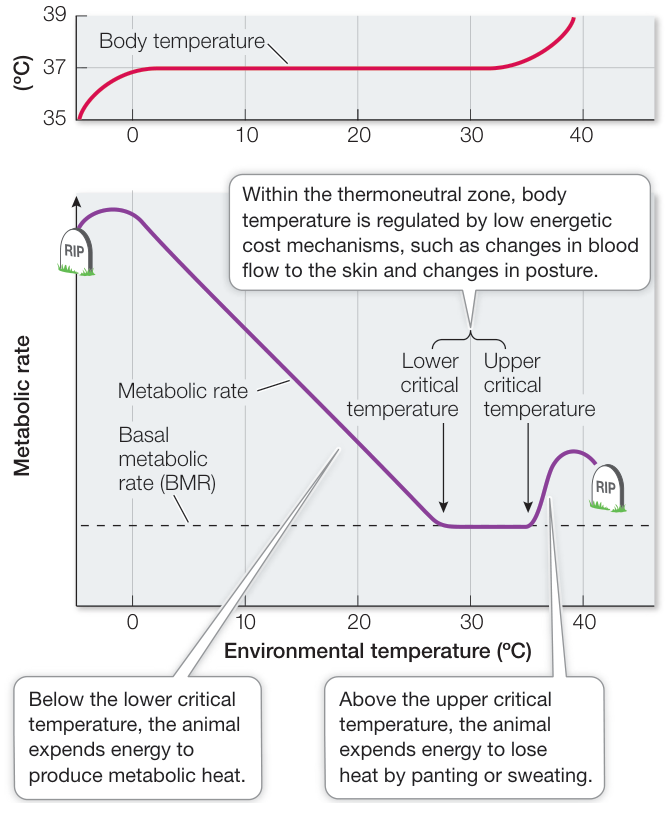
Figure 38.14 Environmental Temperature and Mammalian Metabolic Rates A plot of an endotherm’s metabolic rate versus environmental temperature represents the integrated response of all of its thermoregulatory mechanisms. Outside the thermoneutral zone, maintaining a constant body temperature requires expending energy. Outside extreme limits (0°C and 40°C in this instance), the animal cannot maintain its body temperature and dies. Q: The segment of the curve below the lower critical temperature can be described by the equation . In this equation, what does stand for? Why does this equation predict that the curve it describes will meet the x axis at a temperature that equals body temperature?
Endotherms respond to cold by producing heat and adapt to cold by reducing heat loss
When environmental temperatures fall below the lower critical temperature, endotherms increase metabolic heat production to compensate for heat loss. Mammals can accomplish this by shivering and/or nonshivering heat production. Birds use only shivering heat production. Shivering uses the contractions of skeletal muscles to convert ATP to ADP, with the energy from this process released as heat. Shivering muscles pull against each other so that little movement other than a tremor results. “Shivering heat production” is perhaps too narrow a term, however; increased muscle tone and increased body movements also contribute to increased heat production in cold environments.
Most nonshivering heat production occurs in specialized adipose tissue called brown fat. This tissue looks brown because of its abundant mitochondria and rich blood supply (see Figure
38.2). In brown fat cells, a protein called thermogenin uncouples proton movement from ATP production, allowing protons to leak across the inner mitochondrial membrane rather than having to pass through the ATP synthase and generate ATP (review the discussion of brown fat and the chemiosmotic mechanism in Key Concept 9.3). In nonshivering thermogenesis, metabolic fuels are consumed without producing ATP, but heat is still released.
Pads of brown fat are found in newborns of many mammalian species. Brown fat is also commonly found in adult mammals that are small and acclimatized to cold, and in mammals that hibernate. Recently it has been discovered that adult humans have small amounts of brown fat distributed around the body and that its metabolic activity is stimulated by cold exposure. One study found less brown fat activity in obese than in lean individuals, leading to the hypothesis that individual differences in propensity for weight gain may be related to the amount of brown fat in an individual, as described in the opening stories of Chapters 9 and 39.
In addition to their ability to produce heat, endotherms that live in cold climates have evolved adaptations to reduce their heat loss. Heat is lost from the body surface, and cold-climate species have anatomical adaptations that give them smaller surface area-to-volume ratios than their warm-climate relatives (Figure 38.15). These adaptations include rounder body shapes and shorter appendages.
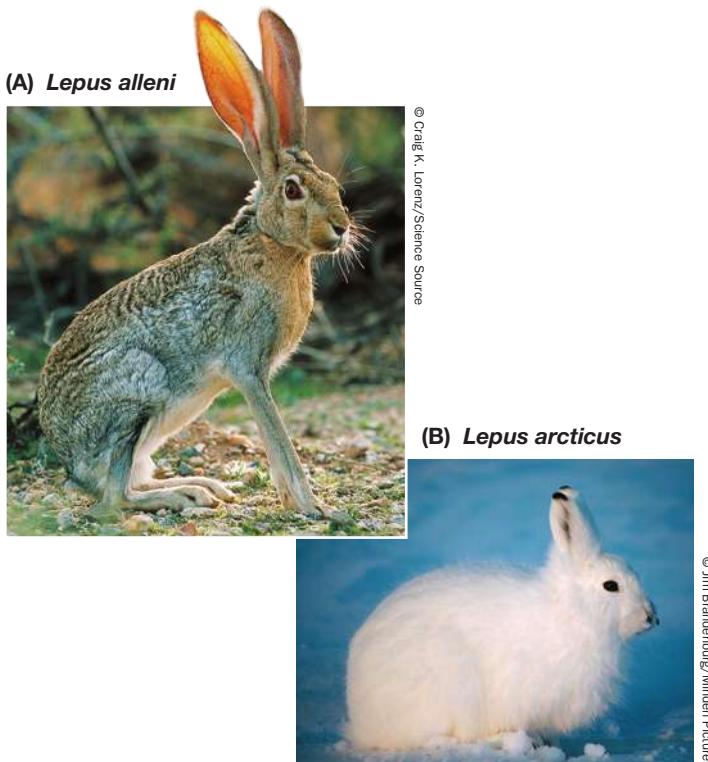
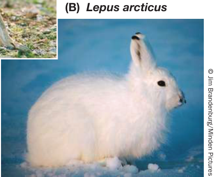
Figure 38.15 Adaptations to Cold and Hot Climates (A) The antelope jackrabbit is found in the Sonoran Desert of Arizona. Its large ears serve as heat exchangers, passing heat from the animal’s blood to the surrounding air. (B) The thick fur of the Arctic hare provides insulation, and its rounded body shape lowers its surface area-to-volume ratio. The ears and extremities are smaller than those of its warm-climate relatives, so less heat is lost to the environment.
The most common and important means of decreasing heat loss is to increase thermal insulation. Animals adapted to cold climates have much thicker layers of fur, feathers, and/or fat than do their warm-climate relatives. Fur and feathers are good insulators because they trap a layer of still, warm air close to the skin surface. If that air is displaced by water, insulation is drastically reduced. In many species, oil secretions spread through fur or feathers by grooming are critical for resisting wetting and maintaining a high level of insulation. In terms of the energy budget equation presented in Key Concept 38.4, remember that heat exchange between the animal and its environment is a function of the animal’s surface temperature. For a body area covered by fur or feathers, surface temperature is that of the outer surface of the fur or feathers.
The ability to decrease blood flow to the noninsulated skin is an important thermoregulatory adaptation for cold-climate endotherms. Constriction of blood vessels in the skin, and especially in the appendages, greatly improves an animal’s ability to conserve heat. Countercurrent heat exchange like that you saw in “hot” fishes is also an important adaptation in the appendages of endotherms. Warm blood flowing out to the paw of a wolf, the hoof of a caribou, or the foot of a bird parallels the flow of the cooler blood returning to the body core. Heat is transferred from the outgoing to the returning blood, thus retaining heat in the animal’s core.
Evaporation of water dissipates heat, but at a cost
At the upper end of the thermoneutral zone, an endotherm dissipates heat through adaptations that don’t require much energy: increasing blood flow to the skin, seeking shade and cool breezes, and decreasing activity. But when the environmental temperature exceeds the upper critical temperature, these adaptations are not sufficient and overheating becomes a problem. For exercising animals (including athletes), overheating can occur even at low environmental temperatures. Large mammals, especially those in hot habitats such as elephants, rhinoceroses, and water buffaloes, have little or no insulating fur and seek out water to wallow in when the air temperature is high. Having water in contact with the skin greatly increases heat loss because the heat-absorbing capacity of water is much greater than that of air.
Evaporation from external or internal body surfaces through sweating or panting can also cool an endotherm. A gram of water absorbs about 580 calories of heat when it evaporates. If this evaporation occurs on the skin, most of the heat comes from the skin. However, sweat or saliva that falls off the body provides no cooling. Thus when the need for heat loss is greatest, water from the internal environment can be squandered with no cooling benefit. Water is heavy, so animals do not carry an excess supply of it, and many hot environments are also arid. In habitats that are both hot and dry, sweating and panting are cooling adaptations of last resort.
Sweating and panting are active processes that require expending metabolic energy. That is why the metabolic rate increases when the upper critical temperature is exceeded (see Figure 38.14). A sweating or panting animal is generating heat in the process of dissipating heat, which can be a losing battle.
The mammalian thermostat uses feedback information
The thermoregulatory mechanisms and adaptations of endotherms are controlled by neural regulatory systems that integrate information from environmental and internal sources and then issue commands to the effectors that alter the heat content of the body. These regulatory systems are similar in principle in birds and mammals but differ in many details. Here we focus on the nervous system thermostats of mammals.
The major thermoregulatory integrative center of mammals is at the base of the brain in a structure called the hypothalamus (Figure 38.16). The hypothalamus is a key player in many regulatory systems of vertebrates. Experiments demonstrating its role have shown that slight cooling of the hypothalamus stimulates constriction of skin blood vessels and that stronger cooling increases metabolic heat production. As a result, cooling of the hypothalamus in an unchanging, thermoneutral environment will cause body temperature to rise. Conversely, hypothalamic heating causes the overall body temperature to fall (Figure 38.17).
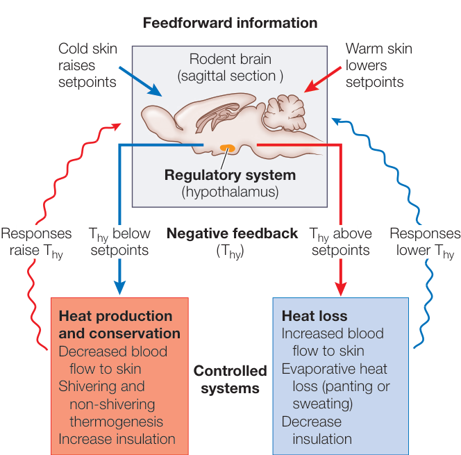
Figure 38.16 The Mammalian Thermostat Like the home thermostat in Figure 38.5, the mammalian hypothalamus is the regulatory system that controls the body’s heating and cooling responses by integrating thermosensory and other information and generating command signals to those mechanisms. The hypothalamus generates set points for activating these responses and the hypothalamic temperature ( ) is the negative feedback information. Set points are raised or lowered by feedforward information such as skin temperature, time of day, sleep, and exercise.
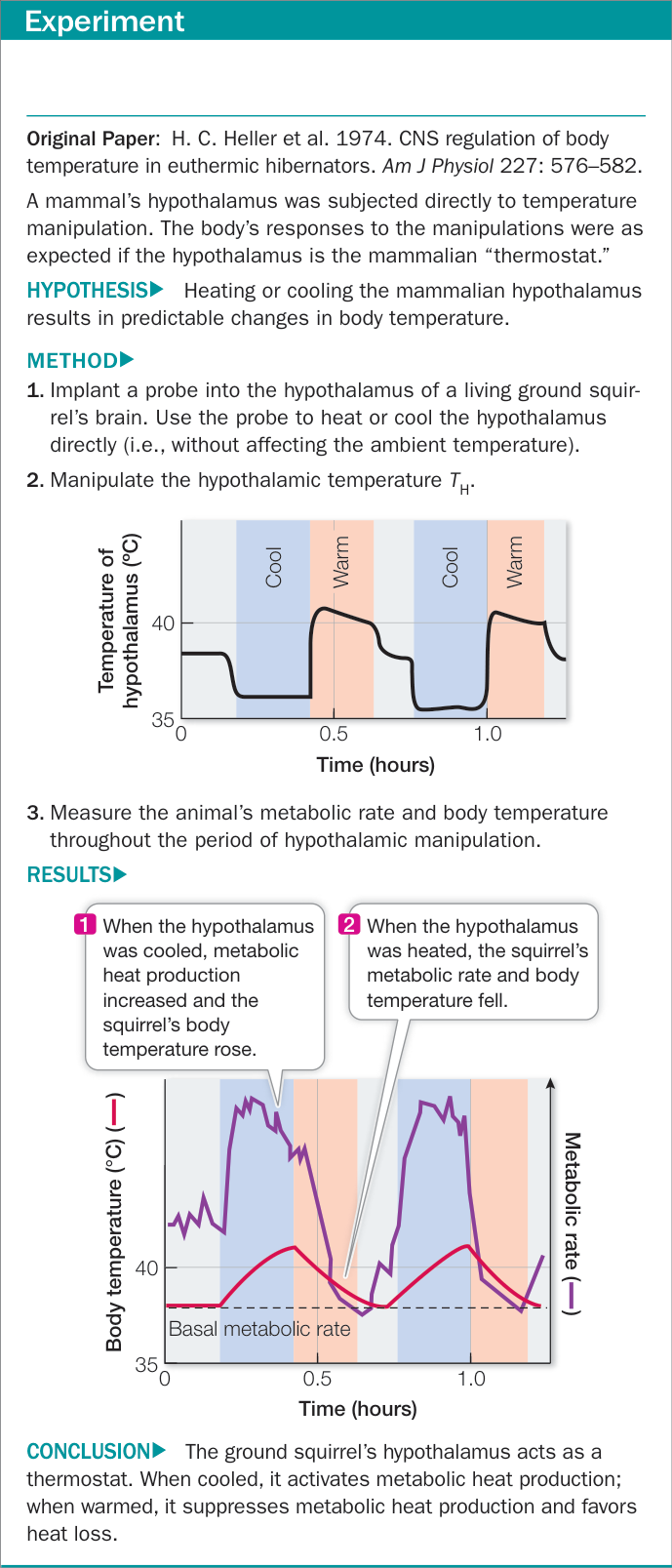
Figure 38.17 The Hypothalamus Regulates Body Temperature
In mammals, the temperature of the hypothalamus itself is the major feedback signal. The hypothalamus generates set points for various thermoregulatory responses. When the temperature of the hypothalamus exceeds or drops below those set points, thermoregulatory responses are controlled to reverse the direction of temperature change (see Figure 38.16). The system integrates other sources of information in addition to hypothalamic temperature. For example, temperature sensors in the skin register environmental temperature. A change in skin temperature is feedforward information that shifts hypothalamic set points; the set point for metabolic heat production is higher when the skin is cold and lower when the skin is warm.
Hypothalamic set points are higher during wakefulness than during sleep, and they are higher during the active part of the daily cycle than the inactive part, even if the animal is awake at both times. Even when an endotherm is kept under constant environmental conditions, its body temperature displays a daily cycle of changes in set point. This kind of cycle, a circadian rhythm, is controlled by an internal biological clock.
Connect the Concepts Key Concept 51.5 explains how a region of the hypothalamus called the suprachiasmatic nucleus generates endogenous daily cycles called circadian rhythms that influence many physiological processes, including temperature regulation.
Some animals conserve energy by turning down the thermostat
Hypothermia is a below-normal body temperature. It can result from starvation (lack of metabolic fuel), exposure to extreme cold, serious illness, or anesthesia. In each of these cases, the drop in body temperature is unregulated. However, many birds and mammals undergo regulated hypothermia to survive periods of cold and food scarcity.
Hummingbirds, for example, are very small endotherms with a high metabolic rate, and going even a single day without food could exhaust their metabolic reserves. Hummingbirds and other small endotherms extend the period over which they can survive without food by dropping their body temperature by 10–20 degrees Celsius during the portion of the day when they are normally inactive, thus lowering their metabolic rate and conserving energy. Daily bouts of regulated hypothermia are called daily torpor.
Regulated hypothermia that lasts for days or even weeks, during which the body temperature falls close to environmental temperature, is called hibernation (Figure 38.18). Many species of mammals, including bats, bears, marmots, and ground squirrels, hibernate, but only one species of bird (the Common Poorwill, Phalaenoptilus nuttallii) has been shown to hibernate. The metabolic rate needed to sustain a hibernating animal may be only one-fiftieth its basal metabolic rate, and many hibernating animals maintain body temperatures close to freezing. Arousal from hibernation occurs when the hypothalamic set point returns to the normal level. The ability of animals to enter daily torpor or deep hibernation to reduce their thermoregulatory set point so dramatically probably evolved as an extension of the set point decrease that accompanies sleep in all mammals and birds.
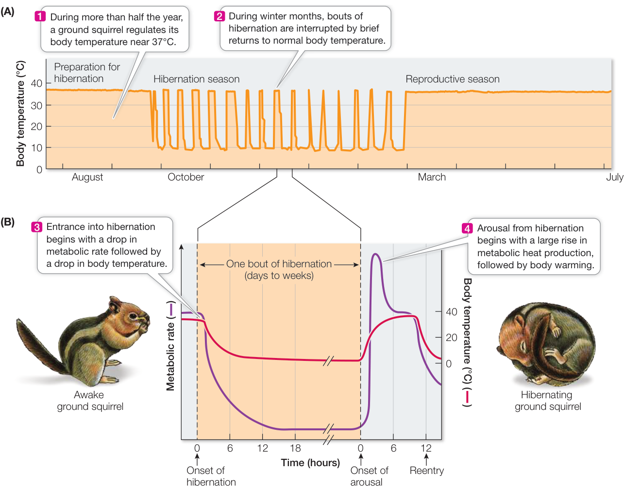
Figure 38.18 Hibernation Patterns in a Ground Squirrel (A) During most of the year, the ground squirrel regulates its body temperature around 37°C. During winter months, however, these animals hibernate in underground burrows, living off stored fuel (in the form of either fat or cached food). (B) The metabolic demands for these stored fuels are decreased by bouts of torpor, during which body temperature drops close to that of the environment for long periods of time.
Recap and Assess
Within the thermoneutral zone, an endotherm controls its body temperature by passive means. Above or below the thermoneutral zone, an endotherm must expend considerable metabolic energy to control body temperature. Thermoregulatory responses in mammals are regulated by the hypothalamus.
- What determines the upper and lower boundaries of the thermoneutral zone?
- Explain why arctic species of mammals tend to be bigger and stockier than closely related species in warmer environments.
- What is the role of feedforward information in the mammalian thermoregulatory system?
- Explain why the mammalian thermoregulatory system has been described as a proportional thermostat with an adjustable set point.
Investigating Life
Q&A How can we increase heat loss from the body to protect against heat stress?
In the opening story to this chapter you saw that intense physical work or high ambient temperatures can rapidly cause a human to become hyperthermic. Even within a safe zone, body temperature is a critical factor in limiting physical performance. A working body has to dissipate heat to the environment, and that requires a difference in temperature between the body surface and the environment. This is why the fastest marathon times occur when ambient temperatures are between 10°C and 16°C. The relevant body surface temperature for furred mammals is mostly the surface of the fur. For us it is mostly the surface of our clothing. We can take off our clothes, but furred mammals cannot take off their fur. Mammals have evolved special blood vessel adaptations in their non-hairy skin that can accommodate large volumes of blood and therefore support high levels of heat loss. The experiment in Investigating Life: Can the Work Capacity of Workers in PPE Be Extended by Extracting Heat from the Palms of the Hands? used a technology to extract heat from the palms, which are non-hairy skin, and showed that it decreased the rate of rise of core body temperature in workers dressed in PPE. How does this heat-loss adaptation, and the technology designed to amplify it, work?
Blood flows from the heart through large arteries, then through small arteries, and then through the tiny capillaries where the blood delivers nutrients and carries away waste products of metabolism, and then into veins that carry the blood back to the heart. The circulatory adaptations of the “heat portals” in non-hairy mammalian skin are an exception to this pattern. In the non-hairy skin, gated shunts deliver arterial blood directly to veins, bypassing the capillaries. These veins in non-hairy skin form networks that can accommodate a large volume of blood when the shunts are open. Based on this knowledge, biologists at Stanford University developed the “rapid-cooling” technology used in the Investigating Life experiment. The palms are placed in contact with pads containing circulating cool water. When this device is used, body temperature rises more slowly during exercise or exposure to heat, even for workers clad in PPE.
Future directions
In this chapter you learned that virtually all biological processes are affected by temperature. Therefore if we have a new technology that can rapidly and efficiently alter the heat content of the body, we should find many applications for it. What could some of those applications be? One that has been recognized is improving athletic performance. Endurance is extended, and when used in a conditioning routine, the number of exercises that can be done increases, resulting in huge conditioning gains. There are also medical applications. Active thermoregulatory responses such as vasoconstriction and shivering are inactivated by anesthesia, and patients can become seriously hypothermic. Their body temperatures can be stabilized by heat exchange through their natural heat portals. Some medical conditions render patients temperature-sensitive. An example is multiple sclerosis (MS). Individuals with MS can suffer serious increases in their symptoms if their body temperature rises even a little bit. They may lose their vision, balance, ability to walk, or ability to think clearly. They can be protected from these effects of heat by cooling their natural heat portals. Evidence suggests that some cancer treatments could be augmented by the induction of hyperthermia and that the consequences of stroke or heart attack could be lessened by timely induction of hypothermia. There can be many benefits from improved abilities to control human body temperature.

© dpa picture alliance/Alamy Stock Photo
Visual Summary

You should be able to relate each summary to the adjacent figures. If you go to this visual summary in Achieve, you can follow links to figures, animations, activities, and simulations that will help you consolidate the material.
Animals Are Composed of Organs Built from Four Types of Tissues
- Multicellularity makes it possible for animals to grow big and for cells to become specialized for specific functions.
- Specialized cells compose four types of tissues—epithelial, muscle, connective, and nervous—that constitute the organs of an animal.
- Organ systems carry out specific essential functions that benefit all the cells of the body.
Questions
- What is the relationship between volume and surface area as cells get bigger?
- Give an example of each of the four tissue types and its function.
- Why is cell specialization important for multicellular animals?
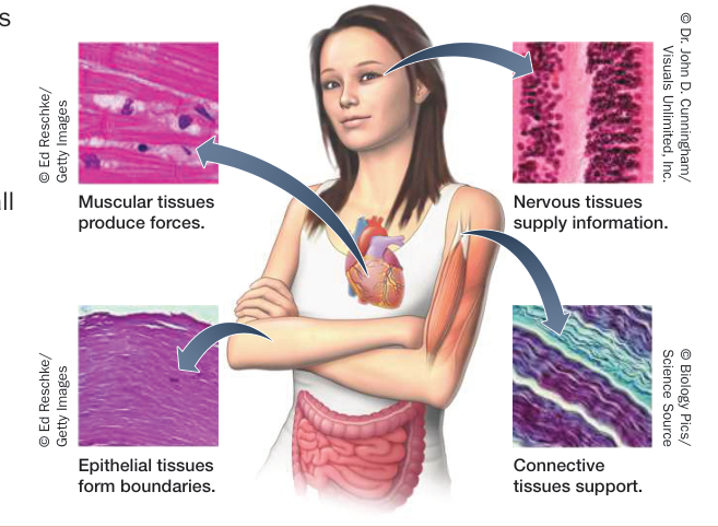
Figure 38.2
- ■ The internal environment consists of extracellular fluid, which comprises blood plasma and interstitial fluid.
- ■ Homeostasis is the maintenance of constancy of the internal environment. To maintain homeostasis, physiological regulatory systems compare feedback information to set points to generate command signals to effectors.
- ■ Negative feedback brings a function back to the set point, positive feedback moves the function farther from the set point, and feedforward information changes the set point.
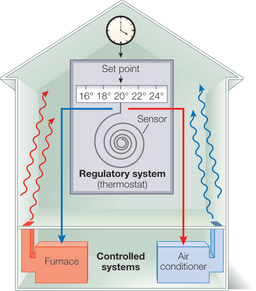
Figure 38.5
Questions
- What is the distribution of water in the human body?
- Compare the functions of negative, positive, and feedforward information, and give an example of each.
Biological Processes Are Temperature-Sensitive
- ■ For most living organisms, internal temperature must be within the range of 0°C–40°C.
- ■ The concept describes the effect of temperature on biochemical and living systems.
- ■ Thermoregulatory adaptations enable animals to maintain optimal internal temperature despite external conditions.
- ■ Animals can adjust to seasonal changes through mechanisms of acclimatization.
Questions
- What dangers do extreme temperatures pose for living organisms?
- If the rate of a chemical reaction doubles over a 10-degree-Celsius rise in temperature, what is its ?
- What adaptations might a fish have for surviving seasonal temperature changes and for surviving acute temperature changes?
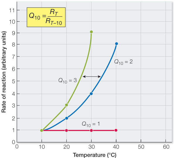
Figure 38.7
Body Temperature Depends on the Balance between Heat In and Heat Out of the Body
- ■ Thermoregulatory adaptations of ectotherms are mostly behavioral, whereas endotherms also have physiological means of maintaining a constant body temperature.
- ■ Heat exchanges between an animal and its environment occur via radiation, convection, conduction, and evaporation, and along with metabolism they make up the energy budget of an animal.
- ■ Control of blood temperature through metabolism and blood flow to the skin are important thermoregulatory mechanisms.
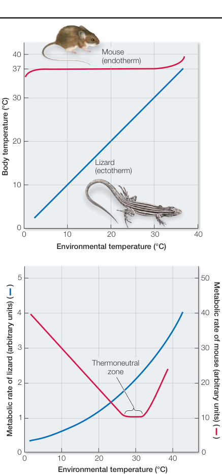
Figure 38.8 Questions
- Why does the metabolic rate of the mammal go up as environmental temperature goes down, but that of the lizard goes down?
- How does control of blood flow to the skin influence the components of the energy budget equation?
- How does countercurrent heat exchange enable a giant tuna to maintain a body temperature above sea temperature?
Body Temperature Is Regulated through Adaptations for Heat Production and Heat Loss
- ■ The basal metabolic rates (BMRs) of endotherms are measured within their thermoneutral zones, and when divided by body mass, they are lower as species get bigger.
- ■ When an animal is outside its thermoneutral zone, heat loss and heat production responses require energy expenditure.
- ■ Brown fat is a tissue that produces heat by uncoupling oxidative phosphorylation from ATP production.
- ■ The mammalian thermoregulatory system resides in the hypothalamus, which generates set points for thermoregulatory responses and uses its own temperature as negative feedback and skin temperature as well as other variables as feedforward information.
- ■ Mammalian hibernation is regulated hypothermia.
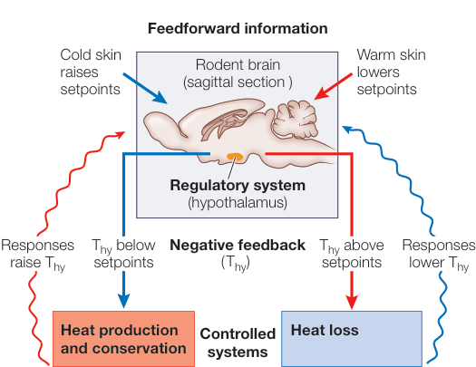
Figure 38.16 Questions
- Describe the mouse-to-elephant curve.
- What is meant by upper and lower critical temperatures?
- Why is the mammalian thermoregulatory system called a proportional thermostat with adjustable set points?