Chapter 39Animal Hormones

Key Concepts
39.1 Hormones Circulate around the Body and Affect Cells That Have Receptors
39.2 The Endocrine and Nervous Systems Work Together
39.3 Hormones Play Important Roles in Development
39.4 Hormones Regulate Metabolism and the Internal Environment
Sex hormones are responsible for the dramatic differences between male and female red deer (Cervus elaphus).
Investigating Life: The Smart and Healthy Hormone
Exercise, especially aerobic exercise, benefits many aspects of health, such as cardiovascular fitness, body weight management, and improved sleep. Improved cognition has been added recently to the list of benefits. How can exercise improve so many functions of the body, and even the brain? One mechanism for stimulating diverse cellular functions all over the body is hormonal communication. Hormones are signaling molecules that are produced and secreted by certain cells, diffuse into the blood, and circulate throughout the entire body. These signals have effects on any cells that have receptors for them, but the resulting actions depend on the nature of the receiving cells.
An interesting, new hormone discovered at Harvard University in 2012 could be the “exercise hormone.” It is a small protein of 112 amino acids that is released from exercising muscle into the extracellular fluid. Since this molecule was thought to have a signaling function, it was named irisin. Iris is the messenger of the Greek gods, and she travels the world with the speed of the wind. Irisin was shown to have several actions supporting physical activity. White fat is normally not a metabolically active tissue, but irisin transforms it to be more like brown fat (see Key Concept 38.5), which has more mitochondria, more blood flow, and greater ability to mobilize lipids. Irisin causes the “browning” of white fat and improves glucose metabolism and bone remodeling. Irisin seems to be the muscle’s way of telling other tissues to “shape up.”
Could irisin also be the signal that mediates the effects of exercise on brain function? One important factor that increases in the brain with regular exercise is BDNF (brain-derived neurotrophic factor). The benefits of increasing BDNF in the brain might not just help athletes play smarter, but BDNF could have huge impacts in aging populations by contributing to improvements in depression, epilepsy, stroke, Alzheimer’s disease, and Parkinson’s disease. What links does BDNF production have to exercise? Does the brain just “know” when we are exercising, or could irisin from the muscle be communicating that information? Normally proteins do not cross the blood–brain barrier, so is irisin produced in the brain during exercise, or is the brain receiving this exercise signal from the muscle in the blood?
QA In what ways does irisin mediate the health and cognitive benefits of exercise? (Find the answer on page 883.)
Hormones Circulate around the Body and Affect Cells That Have Receptors
Learning Objectives
- 39.1.1 Describe the structural feature of hormones that determines whether they act on cell surface or cytoplasmic receptors.
- 39.1.2 Explain how different target cells in the body can respond differently to the same hormone.
- 39.1.3 Discuss scientific thinking about the evolutionary history of hormones in multicellular animals.
In multicellular animals, physiological regulatory systems require information and cell-to-cell communication. Most intercellular communication is by means of chemical signals that bind to receptors, as described in Chapter 7. Some types of chemical signals discussed in other chapters include growth factors, morphogens, and cytokines. In subsequent chapters you will learn about chemical signals used by the nervous system called neurotransmitters. Each of these examples involves a chemical message secreted by cells and received by other cells (target cells) that have appropriate receptors. These four groups of chemical signals are named according to the types of actions they have: growth factors influence cell division, morphogens influence developmental processes, cytokines control immune system cells, and neurotransmitters enable nerve cells to communicate with each other and with target cells such as muscles. In this chapter we cover another broad class of chemical signals called endocrines. These chemical signals are secreted by epithelial cells directly into the extracellular fluid (ECF; see Key Concept 38.2). From the ECF, endocrines can diffuse locally and also into the blood, which distributes them throughout the body.
There are multiple chemical communication systems in the body
Some analogies might help differentiate how chemical signaling in the immune, nervous, and endocrine information systems works. The immune system (the topic of Chapter 40) operates like an army of private security guards. Immune system cells make their rounds of the body, and if they detect a security breach, they sound their alarms—cytokines—which activate the body’s defenses. The nervous system (see Chapters 43–45) operates like a landline telephone system: a central integration and command center receives incoming calls and sends signals along specific wires to specific receivers. The endocrine system is more like a radio, television, or cell-phone system, broadcasting signals that can be picked up by anyone who has an appropriate receiver that is turned on and tuned in. Some endocrine signals are weak, like those of a walkietalkie, and are picked up only locally, but others are strong, like those of a major broadcast station, and reach distant parts of the body. In all cases, the signals are received only by cells that have appropriate receptors, and the responses of those cells are determined by the cells’ internal signal transduction machinery.
Endocrine signaling can act locally or at a distance
Endocrine signals that enter the blood are called hormones (from a Greek word meaning to excite, arouse, or stimulate). Hormones activate target cells wherever they occur in the body (Figure 39.1A). You probably are familiar with several hormones, such as testosterone, estrogen, insulin, and norepinephrine. Those molecules are well known, as are their sites of release and their actions.
Some endocrine signals are released in such tiny quantities, or are so rapidly inactivated by enzymes or taken up so efficiently by local cells, that they never diffuse into the blood in sufficient amounts to act on distant cells (Figure 39.1B). Because these signals affect only target cells near their release site, they are called paracrines (para, “near”). An example of a paracrine signal is histamine, one of the mediators of inflammation. The most local action an endocrine signal can have is when it binds to receptors on or in the same cell that secreted it. An autocrine signal is so named because it influences the cell that secreted it. Hormones and paracrines can have autocrine functions as a means of providing negative feedback to control their own rates of secretion.
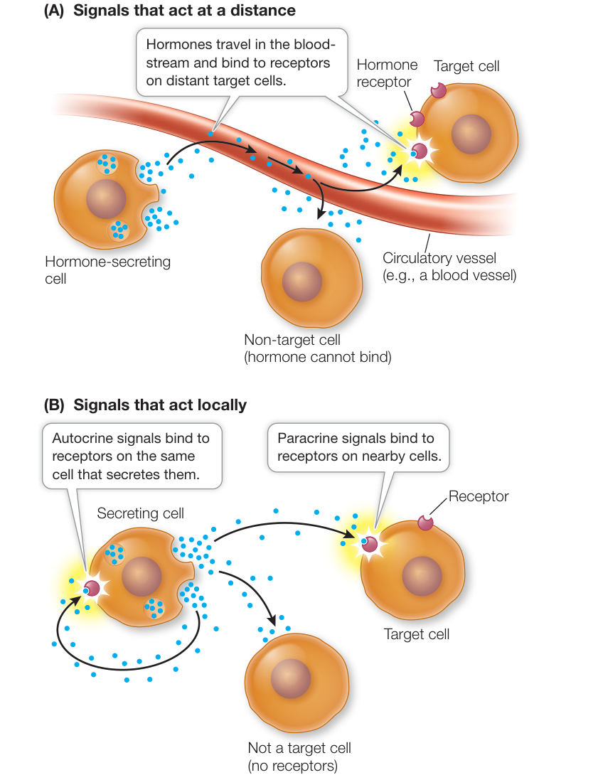
Figure 39.1 Chemical Signaling Systems (A) Hormones are distributed throughout the body in the bloodstream. (B) Paracrines and autocrines diffuse locally in the extracellular fluid. Paracrines influence nearby cells, and autocrines influence the same cells that release them.
Some endocrine cells exist as single cells within a tissue. Hormones of the digestive tract, for example, are secreted by isolated endocrine cells in the walls of the stomach and small intestine. Many hormones are secreted by aggregations of endocrine cells called endocrine glands. A single endocrine gland may secrete multiple hormones. Be sure you don’t confuse endocrine glands with exocrine glands. Endocrine glands secrete hormones within the body, whereas exocrine glands secrete substances through ducts to the outside of the body (sweat glands and salivary glands are examples).
Two other categories of chemical signals are neuroendocrines, or neurohormones, which are secreted by nerve cells and enter the circulation, and pheromones, which are released from the body of an animal to influence the behavior and physiology of other animals in its vicinity. We discuss pheromones in Chapter 51.
There are three chemical groups of hormones
The chemical structures of hormones are diverse, but they can be classified into three groups:
- Peptide and protein hormones are the majority of hormones. A peptide consists of 2 or more amino acids, and a protein is defined as having 50 or more amino acids. Insulin is therefore a small protein hormone (Figure 39.2A). Peptide and protein hormones are water-soluble and thus easily transported in the blood. Peptide and protein hormones are frequently packaged in vesicles within the cells that make them, and then released by exocytosis.
- Steroid hormones (such as estrogen and testosterone) are synthesized from cholesterol (Figure 39.2B), are lipid-soluble, and pass easily through cell membranes. Steroid hormones diffuse out of the cells that make them and are usually bound to carrier molecules in the blood.
- Amine hormones are mostly synthesized from the amino acid tyrosine (the thyroid hormone thyroxine is one example; Figure 39.2C). Some amine hormones are water-soluble and others are lipid-soluble; their modes of release differ accordingly.
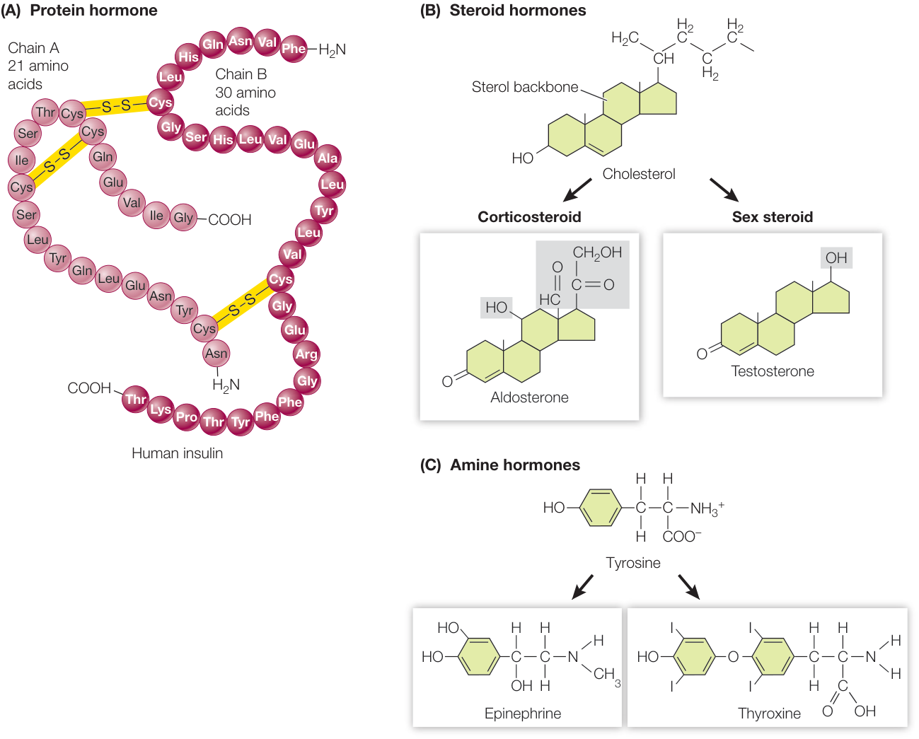
Figure 39.2 Three Classes of Hormones (A) The largest hormone molecules are peptides and proteins. An example of a protein hormone is the pancreatic hormone insulin. (B) Steroid hormones are modified from cholesterol molecules. They include the corticosteroids produced by the adrenal gland and the sex steroids produced primarily by the gonads. (C) Amine hormones are tiny molecules synthesized from a single amino acid. Both epinephrine and thyroxine are synthesized from tyrosine units, but thyroxine is lipid-soluble and epinephrine is water-soluble. Their modes of release and transport and the locations of their receptors differ accordingly.
Hormone action is mediated by receptors on or within their target cells
Water-soluble hormones cannot pass readily through cell membranes; instead they bind to receptors on the surfaces of target cells. These receptors are large transmembrane glycoprotein complexes with three domains:
- A binding domain that projects outside the cell membrane
- A transmembrane domain that anchors the receptor in the membrane
- A cytoplasmic domain that extends into the cytoplasm of the cell
When a hormone binds to the binding domain, the cytoplasmic domain initiates the target cell’s response, usually through second messengers. Second messengers activate cascades of intracellular events, eventually activating protein kinases or protein phosphatases (see Figures 7.6 and 7.7). In most cases these protein kinases and phosphatases activate or inactivate enzymes in the cytoplasm that lead to the cell’s response, but the signaling cascade initiated by the receptor can also generate signals that enter the nucleus and alter gene expression (see Figure 7.10).
Lipid-soluble hormones can diffuse through cell membranes, and therefore their receptors are usually inside cells, in either the cytoplasm or the nucleus. There are also some membrane-bound receptors for lipid-soluble hormones, but they are the exception rather than the rule. In most cases, the complexes formed by the lipid-soluble hormones and their receptors act by altering gene expression in the cell’s nucleus (see Figure 7.8).
Hormone action depends on the nature of the target cell and its receptors
Wherever a hormone encounters a cell with an appropriate receptor, it can bind to that receptor and trigger a response. The nature of the response depends on the signal transduction pathways of the responding cell. Therefore the same hormone can cause different responses in different types of cells.
Consider the amine hormone epinephrine, and the closely related amine hormone and neurotransmitter norepinephrine. Both of these molecules are involved in the fight-or-flight response (Figure 39.3). Suppose you are walking in the forest and almost step on a rattlesnake. You jump back, your heart starts to thump, and protective reactions are set in motion. The jump and the heart thumping are driven by almost instantaneous activation of your sympathetic nervous system (see Figure 45.8), which uses norepinephrine to signal target cells. Your sympathetic nervous system also stimulates endocrine cells in the adrenal glands located just above your kidneys. The adrenal glands secrete both epinephrine and norepinephrine, which diffuse rapidly into your blood, circulate around your body, and activate the many components of the fight-or-flight response.
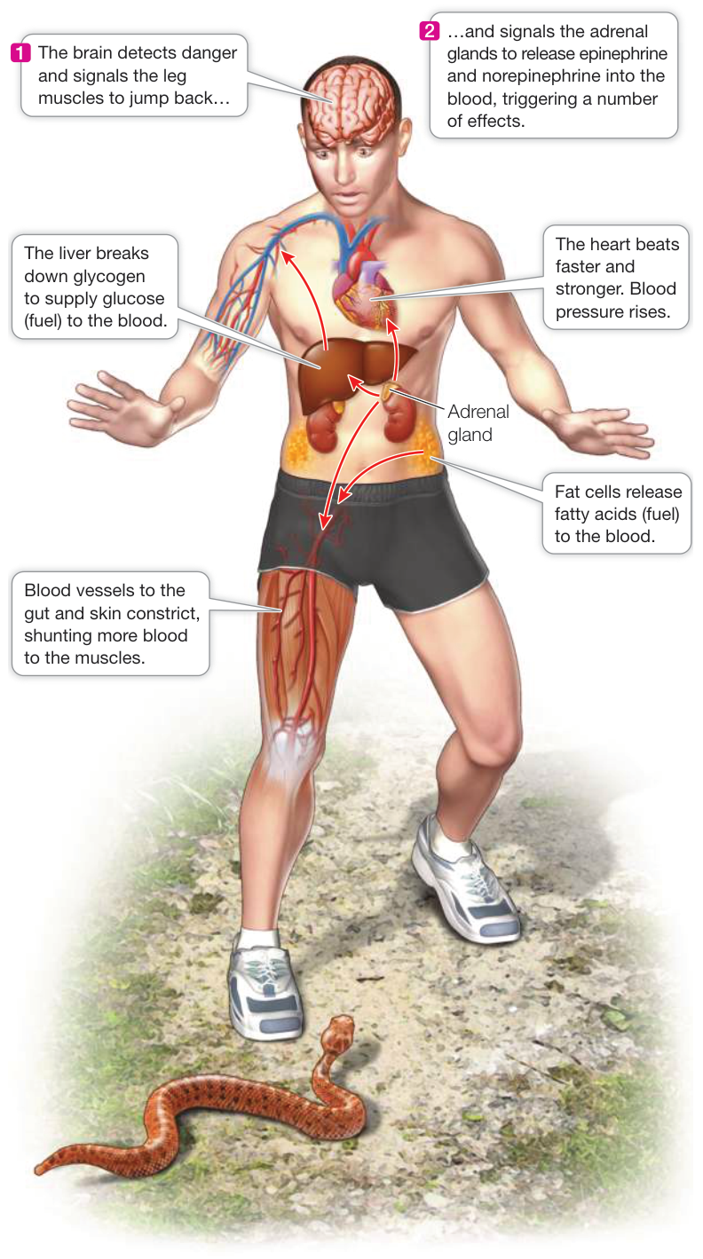
Figure 39.3 The Fight-or-Flight Response The brain of a person suddenly faced with a threatening situation sends a signal to the adrenal glands, which almost instantaneously release the hormones epinephrine and norepinephrine. Epinephrine and norepinephrine circulate around the body and induce the various components of the fight-or-flight response in different tissues.
Epinephrine and norepinephrine bind to receptors in your heart, causing a faster and stronger heartbeat. Your heart is now pumping more blood. Epinephrine also binds to receptors in certain blood vessels. By causing constriction of blood vessels supplying your skin, kidneys, and digestive tract—tissues not essential for immediate survival—epinephrine results in more blood being available for the muscles needed for your escape from danger.
Epinephrine binds to cells in the liver, stimulating them to break down glycogen and release glucose into the blood as a quick energy supply (see Figure 7.15). In fatty tissue, epinephrine stimulates the breakdown of fats to yield fatty acids—another source of energy. These are just some of the actions triggered by one hormone. In each case the cellular response depends on the cell’s receptors, its associated intracellular signaling cascade, and the mechanisms activated by that signaling cascade.
Hormone structure is conserved through evolution, but functions change
Intercellular chemical signaling was critical for the evolution of multicellularity, so hormones have a long evolutionary history. The least complex multicellular animals—the sponges—do not have nervous systems, but they do have intercellular chemical communication. Studying the evolution of hormonal signaling reveals an interesting generalization: the signal molecules themselves are highly conserved. The amino acid sequence of the hormone irisin discussed in the chapter opener is 100% identical in mice and humans. We find the same chemical compounds over broad groups of organisms, although their functions may differ. As organisms have evolved to occupy different environments and have different lifestyles, the same hormone—receptor systems have diversified to serve different functions. A good example of this evolutionary diversification is the hormone prolactin (Figure 39.4). The name prolactin reflects the role this hormone plays in mammals to stimulate the growth of mammary glands and the production of milk. Mammary glands are unique to mammals, but we find prolactin in all vertebrate groups—and in all cases it is involved in some way in reproduction.
The list of hormones known to exist is long and growing longer (as shown by the recent discovery of irisin). To make the subject manageable, we focus primarily on the endocrine system of mammals. Figure 39.5 presents the human endocrine glands and most of the hormones that are discussed in this and subsequent chapters.
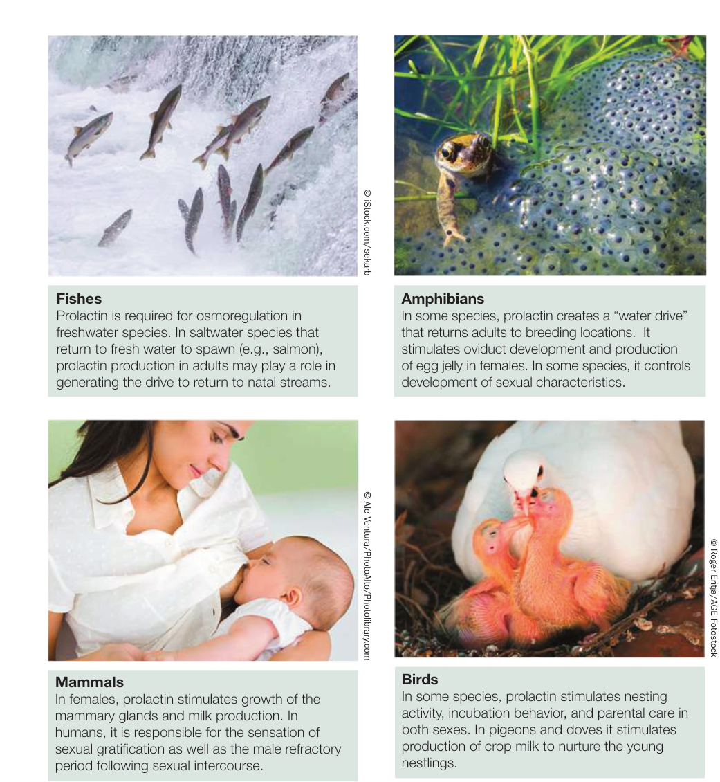
Figure 39.4 Prolactin’s Structure Is Conserved, but Its Functions Have Evolved The hormone prolactin is found in all vertebrate groups and has a long evolutionary history. Its probable function in early vertebrates was in regulating the body’s salt and water balance (osmoregulation). It maintains this function in some species, and has evolved in others to control several physiological processes, most of which are associated with reproduction. © ErnestoGravelpond/Alamy Stock Photo © ErnestoGravelpond/Alamy Stock Photo The hormone prolactin is found in all vertebrate groups and has a long evolutionary history. Its probable function in early vertebrates was in regulating the body’s salt and water balance (osmoregulation). It maintains this function in some species, and has evolved in others to control several physiological processes, most of which are associated with reproduction.
© Stock.com/sekaris
Figure 39.4 Prolactin’s Structure
Is Conserved, but Its Functions Have Evolved
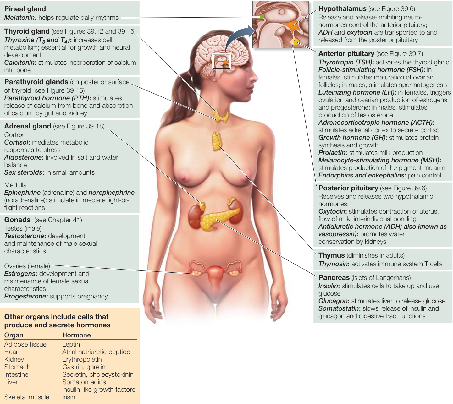
Figure 39.5 The Endocrine System of Humans Cells that produce and secrete hormones may be organized into discrete endocrine glands, or they may be embedded in the tissues of other organs, such as the digestive tract or kidneys. The hypothalamus is part of the brain, but it includes cells that secrete neurohormones into the extracellular fluid. Activity 39.1 The Human Endocrine Glands
Recap and Assess
Endocrine signals initiate responses in target cells that vary depending on the signal transduction pathways of the responding cells. Autocrines and paracrines act locally. Hormones are chemical signals released by endocrine cells into the extracellular fluid, where they diffuse into the blood and travel to distant target cells. The receptors for water-soluble hormones are on the surfaces of target cells; the receptors for most lipid-soluble hormones are inside the target cells. The responses activated by a hormone depend on the nature of the target cell, thus one hormone can have many functions. The chemical structures of signal molecules have been highly conserved over evolutionary time, but the same molecules can serve different functions in different organisms.
- Why are the effects of epinephrine more rapid than the effects of estrogen?
- Why is the presence of a particular hormone in two species not an indication of the closeness of their evolutionary relationship?
- How can a single hormone have two different effects in the same species?
The endocrine system and the nervous system are both information systems, but a major difference between them is their speed of communication. As we will see in Chapter 44, neural signals are very fast and reach distant parts of the body in hundredths or thousandths of a second. Neural signals are also fast to disappear and therefore can rapidly communicate complex information such as that necessary to type on a laptop. Endocrine signals have to be secreted and then have to diffuse and circulate, and they frequently act through intracellular mechanisms such as protein synthesis that take time. Thus the nervous system is good at generating complex rapid responses, and the endocrine system at generating sustained responses. The different characteristics of the two information systems make it advantageous for them to work together.
The Endocrine and Nervous Systems Work Together
Learning Objectives
- 39.2.1 Describe the developmental origins of the anterior pituitary and the posterior pituitary.
- 39.2.2 Diagram the anatomical and signaling relationships among the hypothalamus, the pituitary gland, and a peripheral endocrine gland.
- 39.2.3 Using an example, explain how a negative-feedback loop controls hormone secretion.
The nervous system and the endocrine system control and regulate virtually all physiological and behavioral mechanisms that contribute to homeostasis, survival, and reproductive success. Challenges to an organism usually trigger both physiological and behavioral responses, as we saw with respect to thermoregulation in Chapter 38 and also with the fight-or-flight reactions shown in Figure 39.3. Therefore the endocrine and nervous systems must work together in an integrated fashion, and indeed they do. The nervous system receives and integrates an enormous amount of information from the body and from the environment. The nervous system uses that information to control many tissue and organ functions as well as behavior. It is therefore not surprising that the nervous system regulates many endocrine functions. Conversely, hormones circulate to the nervous system, providing feedback information used in regulation. Hormones reaching the brain also stimulate behaviors that contribute to the health and well-being of an organism.
The pituitary is an interface between the nervous and endocrine systems
The pituitary gland sits in a depression at the bottom of the skull, just over the back of the roof of the mouth (Figure 39.6). It is attached by a stalk to the hypothalamus, a part of the brain involved in many physiological regulatory systems such as that of thermoregulation (see Key Concept 38.5). Through its close connection with the hypothalamus, the pituitary serves as the interface between the nervous system and the endocrine system and is involved in the hormonal control of many physiological processes.
The pituitary has two parts with different developmental origins. The anterior pituitary originates as an outpocketing of the roof of the embryonic mouth cavity. The posterior pituitary originates as an outpocketing of the floor of the developing brain. Thus the anterior pituitary originates from gut epithelial tissue and the posterior pituitary from neural tissue. Both parts interact with the nervous system but in different ways. The anterior pituitary is controlled by hypothalamic neurohormones that reach the anterior pituitary via the blood. The posterior pituitary contains long extensions of hypothalamic neurons that release their neurohormones in the posterior pituitary.
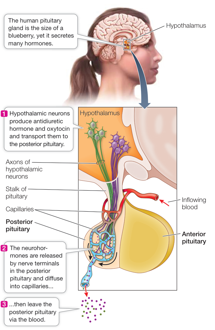
Figure 39.6 The Posterior Pituitary Releases Neurohormones Neurons in the hypothalamus produce two peptide neurohormones—antidiuretic hormone and oxytocin—which are stored in and released by nerve endings in the posterior pituitary.
THE POSTERIOR PITUITARY The long extensions of hypothalamic neurons into the posterior pituitary are called axons. The terminals of these axons in the posterior pituitary contain vesicles of neurohormones—either antidiuretic hormone or oxytocin. When these hypothalamic neurons are activated, they fire electrical signals (action potentials) that travel down their axons and cause the release of the neurohormones (see Figure 39.6).
Connect the Concepts As described in Key Concept 44.2, action potentials are sudden and transient electrical signals generated by voltage-gated ion channels in the cell membrane of the axon. These signals travel rapidly along axons, and their arrival at the axon terminals initiates the release of neurotransmitters.
The main action of antidiuretic hormone (ADH) in mammals and birds is to increase the amount of water conserved by the kidneys. When ADH secretion is high, the kidneys produce only a small volume of highly concentrated urine. When ADH secretion is low, the kidneys produce a large volume of dilute urine. The posterior pituitary increases its release of ADH when blood pressure falls or the blood becomes too salty. ADH is also known as vasopressin because at high concentrations it causes the constriction of peripheral blood vessels as a means of elevating blood pressure.
The hormone oxytocin is released from the posterior pituitary when a woman is about to give birth. Oxytocin stimulates contractions of the uterine muscles, which pushes the baby against the opening of the uterus, the cervix. Stretching of the cervix stimulates more oxytocin release, resulting in the delivery of the baby. Oxytocin also brings about the flow of milk from the mother’s breasts. The baby’s suckling stimulates neurons in the mother’s brain that cause the secretion of oxytocin. Even the crying of her baby can cause a nursing mother to secrete oxytocin, resulting in the release of breast milk—a good example of how the nervous system integrates information that regulates hormonally mediated processes.
THE ANTERIOR PITUITARY The anterior pituitary produces and releases into the circulation four peptide and protein hormones that act as tropic hormones, meaning they control the activities of other endocrine glands. These four tropic hormones are thyrotropin (thyroid-stimulating hormone), luteinizing hormone (LH), follicle-stimulating hormone (FSH), and adrenocorticotropic hormone (ACTH). Each is produced by a different type of pituitary cell. We discuss these tropic hormones later in this chapter and in Chapter 41 when we describe their target glands—the thyroid, testes, ovaries, and adrenal cortex. Other peptide and protein hormones produced by the anterior pituitary are prolactin (see Figure 39.4), growth hormone, enkephalins and endorphins, and melanocyte-stimulating hormone (MSH).
Growth hormone (GH) acts on a wide variety of tissues to promote growth and development. One of its important effects is to stimulate cells to take up amino acids. Growth hormone also promotes growth by stimulating the liver to produce somatomedins that stimulate the growth of bone and cartilage and other cells and tissues. Overproduction of GH in children causes gigantism; affected individuals may grow to nearly 8 feet tall. Underproduction of GH results in pituitary dwarfism, in which individuals fail to reach normal adult height.
Endorphins and enkephalins are the body’s natural painkillers. In the brain, these molecules act as neurotransmitters in pathways that control pain. Their production in the anterior pituitary is normally quite small and probably has little significant effect.
Melanocyte-stimulating hormone (MSH) stimulates the production of the pigment melanin in skin and hair. MSH is produced by a thin layer of cells between the anterior and posterior pituitary that is sometimes called the intermediate lobe of the pituitary.
Hypothalamic neurohormones control the anterior pituitary
Hypothalamic neurons do not extend into the anterior pituitary as they do into the posterior pituitary. So how do the neurohormones produced by hypothalamic neurons reach the anterior pituitary cells they control? The pituitary is connected to the brain by the pituitary stalk (Figure 39.7). Within that stalk are hypothalamic–pituitary portal blood vessels (blood vessels that begin and end in capillary beds). The hypothalamic cells secrete neurohormones at the bottom of the hypothalamus where the stalk begins. The neurohormones diffuse into the portal blood vessels that begin there, and are carried in the blood to where the portal vessels end in the anterior pituitary. Hypothalamic neurohormones are released in such tiny amounts that they were only hypothesized to exist before the technology was developed to isolate and characterize their structure.
In the 1960s two large teams of scientists led by Roger Guillemin at the Salk Institute and Andrew Schally at Baylor University initiated the search for these hypothalamic secretions. Massive numbers of hypothalami from pigs and sheep were collected from slaughterhouses and shipped to their laboratories. One extraction effort began with the hypothalami from 270,000 sheep and yielded only 1 milligram of purified thyrotropin-releasing hormone (TRH)—the first hypothalamic release-stimulating hormone to be isolated and characterized. TRH turned out to be a simple tripeptide consisting of glutamine, histidine, and proline. It causes certain anterior pituitary cells to release the tropic hormone thyrotropin, which in turn stimulates the activity of the thyroid gland.
Soon after discovering TRH, Guillemin’s and Schally’s teams identified gonadotropin-releasing hormone (GnRH), which stimulates certain anterior pituitary cells to release the tropic hormones that control the activity of the gonads (the ovaries and the testes). For these discoveries, Guillemin and Schally shared the 1977 Nobel Prize in Physiology or Medicine with Rosalyn Yalow, who invented the technique of radioimmunoassay, which made possible the measurement of miniscule amounts of specific molecules such as releasing hormones.
Many other hypothalamic neurohormones, including both releasing and release-inhibiting hormones, are now known. The major hypothalamic neurohormones that control anterior pituitary function are listed in Figure 39.7.
Negative-feedback loops regulate hormone secretion
In addition to being controlled by hypothalamic releasing and release-inhibiting hormones, the endocrine cells of the anterior pituitary are also under direct and indirect negative-feedback control by the hormones of the target glands they stimulate (see Figure 39.7). For example, cortisol, produced by the adrenal gland in response to ACTH secreted by the anterior pituitary, reaches the pituitary in the circulating blood and inhibits further release of ACTH. Cortisol also acts as a negative-feedback signal to the hypothalamus, inhibiting the release of corticotropin-releasing hormone (CRH). Because cortisol is the final hormone released in this chain of interacting structures called the hypothalamic–pituitary–adrenal axis, its feedback actions are called long-loop
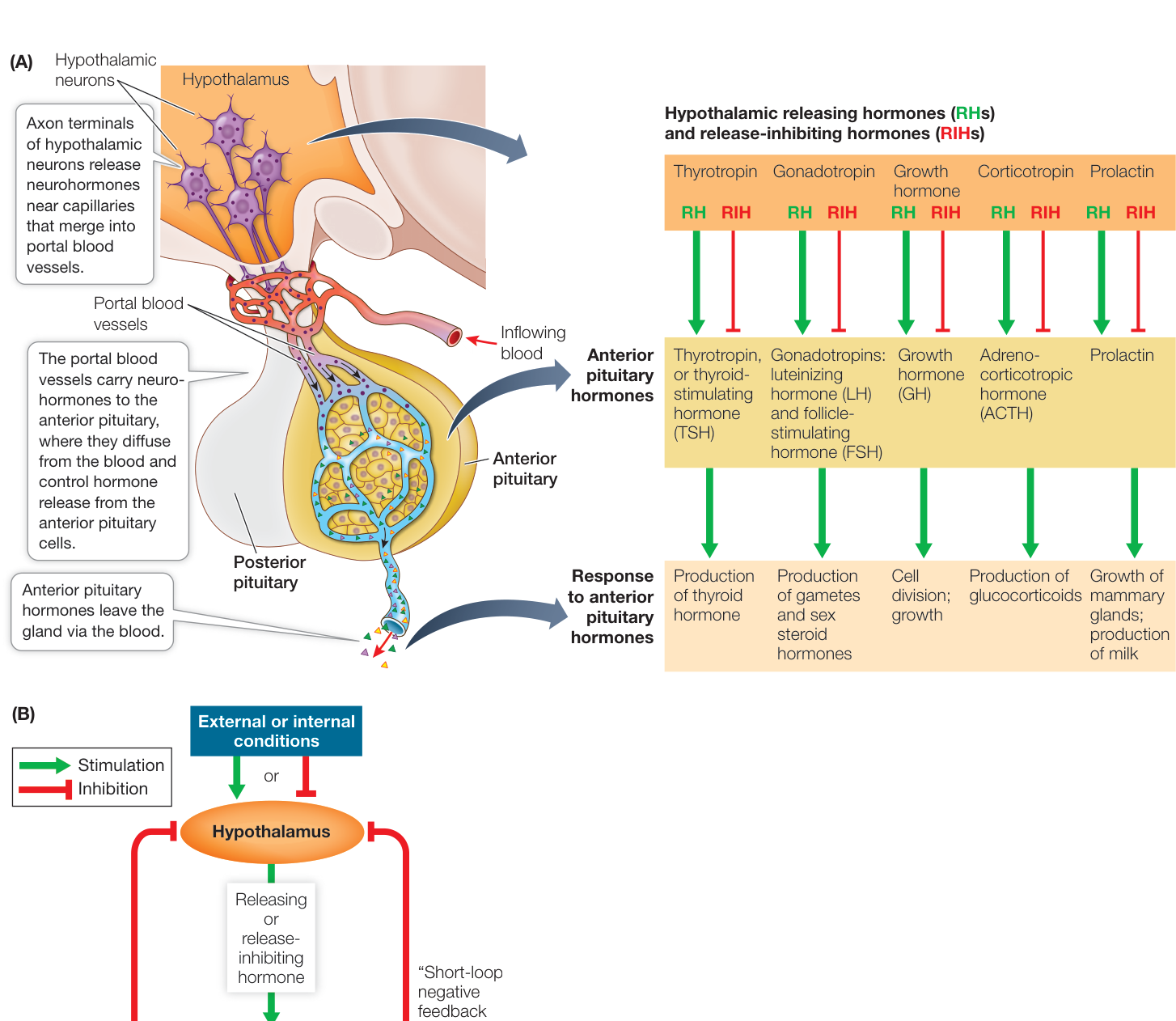
Figure 39.7 The Anterior Pituitary Is Controlled by the Hypothalamus Cells of the anterior pituitary produce tropic hormones that control other endocrine glands, as well as several other peptide and protein hormones. These cells are controlled by neurohormones produced in the hypothalamus and are called releasing or release-inhibiting hormones. These neurohormones are delivered through portal blood vessels that run between the hypothalamus and the anterior pituitary through the pituitary stalk. Q: Melanocyte-stimulating hormone (MSH) is an anterior pituitary hormone (not shown in Figure 39.7A). When anterior pituitary cells are grown in culture, they release MSH into the medium. If extracts of the hypothalamus are added to the medium, the release of MSH decreases. What do these results tell you about the hypothalamic control over MSH-producing cells?
Hormones influence the nervous system
Hormones have more influences on the nervous system than simply being negative-feedback signals controlling their own secretion. For example, the posterior pituitary hormone oxytocin, which plays a role in the birth process and in stimulating the flow of milk from the breasts, also has a strong behavioral effect in promoting bonding (see the story that opens Chapter 7). If oxytocin release is experimentally blocked, mammalian mothers, from rats to sheep, will reject their newborn offspring, but if a virgin rat is given a dose of oxytocin, she will adopt strange pups as if they were her own. Oxytocin promotes pair bonding in a variety of animals. In humans, oxytocin secretion rises with intimate sexual contact, and it has been nicknamed the “cuddle hormone.” Experiments using games involving financial exchanges have shown that puffs of oxytocin applied to the nostrils raise the level of trust between players. Thus circulating hormones can have a strong influence on behavior. Another example is the effects of sex hormones, which we will discuss in Key Concept 51.3.
negative feedback. In this system, the tropic hormone ACTH also exerts negative-feedback control on the hypothalamic cells that produce the corresponding releasing hormone (CRH). This action is called short-loop negative feedback because of the proximity of the pituitary and the hypothalamus.
The effects of hormones on the brain that we have been discussing here are mostly activation effects—they influence the likelihood that the organism will act in a particular way. The hormone irisin that we presented at the beginning of this chapter plays a broader and more general role in brain function by improving the cognitive ability of the brain. Thus this hormone is not influencing a specific behavior, but it is promoting the higher functions of the brain in processing and integrating information.
These effects are apparently mediated by BDNF and include the production of new brain cells, increased blood flow, and changes in the structures supporting communication between brain cells. Irisin may play a very important role in maintaining brain health, and therefore it is important to understand how exercise influences the release of BDNF in the brain. Investigating Life: How Could Irisin Mediate the Benefits of Exercise on Cognition? explores this interesting question.
Investigating Life: How Could Irisin Mediate the Benefits of Exercise on Cognition?
Experiment
Original Paper: C. D. Wrann et al. 2013. Exercise induces hippocampal BDNF through a PGC-1 /FNDC5 pathway. Cell Metab 18: 649–659.
Irisin secreted by exercising muscle is cleaved from a protein called FNDC5, and FNDC5 expression is controlled by the transcription factor PGC-1 . Irisin then circulates in the blood, but because of the blood–brain barrier (see Key Concept 43.1), it is unlikely to reach brain structures such as the hippocampus where it could enhance cognition. However, could irisin also be expressed in the brain? If so, exercise should result in similar changes in expression of FNDC5 and its transcription factor PGC-1 in the hippocampus as it does in skeletal muscle. First, we need to show in our experimental system and with our assays that exercise induces expression of PGC-1 and FNDC5 in exercising muscle. Then we can use the same experimental system to investigate expression in the hippocampus.
HYPOTHESIS Endurance exercise results in increased expression of PGC-1 and FNDC5 in skeletal muscle.
METHOD
- Have one group of mice exercise daily on running wheels for 30 days and a control group that does not. for each group.
- At the end of 30 days, measure PGC-1 and FNDC5 mRNA levels in leg muscle of both groups of mice.
- Plot the data for each variable (FNDC5 and PGC-1 ) for the exercised and control mice, and use an unpaired t-test (see Appendix B) to evaluate the significance of the results.
RESULTS Error bars indicate standard deviation.
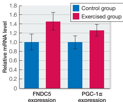
CONCLUSION Relative levels of FNDC5 mRNA were significantly higher in the exercised than in the non-exercised mice (). Relative levels of PGC-1 mRNA also were significantly higher in the exercised than in the non-exercised mice ().
Work with the Data
Similar measurements of expression of FNDC5 and PGC-1 mRNAs were made from the hippocampus of the exercised and the non-exercised mice. Again, for each group. The results are shown in the table.
| FNDC5 | PGC-1 | ||
|---|---|---|---|
| Control | Exercised | Control | Exercised |
| 1.02 | 1.33 | 1.22 | 1.23 |
| 1.10 | 1.24 | 0.84 | 1.09 |
| 0.92 | 1.68 | 1.06 | 1.35 |
| 0.93 | 1.30 | 1.26 | 1.18 |
| 1.21 | 1.34 | 1.04 | 1.40 |
| 0.93 | 1.33 | 1.12 | 1.18 |
| 0.96 | 1.50 | 1.04 | 1.46 |
| 1.01 | 1.29 | 1.09 | 1.19 |
| 1.00 | 1.44 | 1.02 | 1.54 |
| 0.97 | 1.41 | 0.54 | 1.16 |
QUESTIONS
- Plot the data as was done for the leg skeletal muscle results. Then you can apply the same analysis methods to these data that were applied to the leg muscle data in the preceding experiment. Did exercise have a significant effect on FNDC5 and PGC-1 expression in the hippocampus?
- The neurotrophic factor BDNF is a secreted protein that plays important roles in neural changes associated with learning and memory. How would you determine if the exercise protocol induced a significant increase in BDNF expression in the hippocampus, assuming you have an assay for BDNF?
- How could you determine if a noted increase in the expression of FNDC5 in the hippocampus is sufficient for an increased expression of BDNF in the hippocampus? Assume you can culture hippocampal neurons so that the expression of FNDC5 is stimulated or suppressed.
Recap and Assess
The pituitary is the interface between the nervous system and the endocrine system. The posterior pituitary releases two neurohormones, antidiuretic hormone (ADH) and oxytocin. The anterior pituitary, under the control of other neurohormones from the hypothalamus, releases four tropic hormones that control other endocrine glands. In addition, the anterior pituitary produces and releases five other hormones that act directly on non-endocrine tissues. The hypothalamus and pituitary are under negative-feedback control by the hormones of their target glands.
- Explain how the control of oxytocin release involves positive feedback control, whereas the control of ADH release involves negative feedback control.
- How do hypothalamic neurons control the release of hormones from the anterior pituitary?
- Explain what is meant by long-loop and short-loop negative feedback in the hypothalamic control of endocrine function.
The hypothalamic and pituitary hormones and some of the endocrine glands they control are easy to conceptualize because they share common features in how they are organized and function. But the entire list of hormones is diverse, making generalizations difficult. We cannot consider all of the hormones in this chapter. Instead, we focus on hormones that have some common functions—one of which is the coordination of developmental processes.
Hormones Play Important Roles in Development
Learning Objectives
- 39.3.1 Describe how hormones control complete and incomplete metamorphosis of arthropods.
- 39.3.2 Explain how an individual can have a male genotype but female body characteristics and genitalia.
- 39.3.3 Explain the role of negative feedback in the timing of puberty in humans.
The development of an animal from a fertilized egg requires the coordination of processes throughout the body. Imagine the problems that would arise if growth of the body were not symmetrical, or if the growth of organ systems did not match the demands placed on them by the growing body. Because hormones circulate in the blood to all parts of the body, they are ideal signals for integrating and coordinating the many diverse processes involved in development.
Insect development consists of either complete or incomplete metamorphosis
The changes that occur in a developing insect can be dramatic and involve major changes in body form. Insects such as bees and butterflies undergo a developmental process called complete metamorphosis. Starting out as a caterpillar (a larva), a butterfly feeds voraciously and goes through a series of growth stages called instars that are separated by molts. The last instar builds a cocoon and enters into a quiescent, transformative state called a pupa. The tissues of the pupa are extensively remodeled to form the adult body, which is entirely different from that of the larva. The larval butterfly crawls and eats vegetation; the adult butterfly flies and consumes flower nectar. Many other species of insects undergo incomplete metamorphosis, which involves less extreme changes of body form. These insects also go through a series of instars, getting bigger with each molt, but the last juvenile instar simply molts into the adult form with all of the essential adult body parts. Molting involves shedding the skin, or in many cases the rigid exoskeleton, and therefore requires coordination of cellular processes in the entire epidermis of the animal. Both complete and incomplete metamorphosis require integrated processes in all parts of the insect body.
Connect the Concepts Insect metamorphosis is described in more detail in Key Concept 30.4.
Experiments on insect development revealed hormonal signaling systems
Pioneering experiments of hormonal action and control in insects were done on the bloodsucking bug Rhodnius prolixus, which undergoes incomplete metamorphosis. Newly hatched Rhodnius lack certain adult features and molt five times before developing into a mature adult. A blood meal triggers each episode of molting and growth. Rhodnius is an amazingly hardy experimental animal—it survives for quite a long time after its head is cut off. If decapitated within an hour after a blood meal, Rhodnius can survive for up to a year, but it never molts. If decapitated a week after its blood meal, however, it does molt, but into an adult (Figure 39.8A).
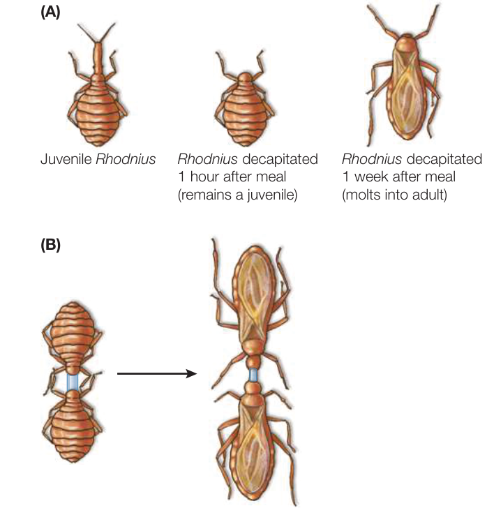
Figure 39.8 Diffusible Substance Controls Molting in a Bloodsucking Bug (A) Stimulation of molting in Rhodnius prolixus depends on signals coming from the head following a blood meal. (B) By connecting two bugs together with a glass tube, the experimenter showed that the molting signal was a diffusible substance.
This observation suggested that molting is triggered by some substance slowly diffusing from the head. To test this hypothesis, two decapitated bugs—one decapitated soon after a blood meal and one a week after a blood meal—were connected together with a glass tube that allowed body fluids to cross between the two bugs. Both molted into adults, proving that a diffusible substance from the bug’s head triggered molting (Figure 39.8B). Since those pioneering experiments on Rhodnius, the diffusible substances (hormones) controlling molting in arthropods have been identified chemically and their functions have been described.
Three hormones regulate molting and maturation in arthropods
An arthropod’s nervous system receives various types of information about the environment (e.g., day length, temperature, social cues, nutrition) that help determine the optimal timing for the stages of growth and development. When conditions are right, the brain signals the prothoracic gland to produce the hormones that orchestrate physiological processes involved in development and molting.
Two hormones, prothoracicotropic hormone (PTH) and ecdysone, work in sequence to regulate molting in arthropods. Cells in the brain produce PTTH, which is why it has also been called “brain hormone.” PTTH is transported to and stored in paired structures called the corpora cardiaca attached to the brain (Figure 39.9). After appropriate stimulation (which for Rhodnius is a blood meal), PTTH is released and diffuses through the extracellular fluid to an endocrine gland, the prothoracic gland. PTTH stimulates the prothoracic gland to secrete the hormone ecdysone. Ecdysone diffuses to target tissues and stimulates molting.
Ecdysone is a steroid hormone (see Figure 39.2B) and thus is related to the vertebrate hormones estrogen and testosterone (which also play roles in controlling growth and development). Ecdysone is lipid-soluble and readily passes through the cell membrane of its target cells (mostly cells of the epidermis). In the target cells, ecdysone binds to a receptor that induces expression of genes encoding enzymes involved in digesting the old cuticle and secreting a new one.
What determines whether an instar molts into another juvenile form or matures into an adult? Juvenile hormone is a molecule that is released continuously from the corpora allata (structures that are attached to the corpora cardiaca, which release PTTH). As long as juvenile hormone is present, Rhodnius molts into another juvenile instar. Normally Rhodnius stops producing juvenile hormone during the fifth instar and then molts into an adult.
The role of juvenile hormone is more complex in insects that undergo complete metamorphosis. An example of complete metamorphosis is provided by the silkworm moth Hyalophora cecropia (see Figure 39.9). As long as juvenile hormone is present in high concentrations, larvae molt into larger larvae. When the level of juvenile hormone falls, larvae spin cocoons and molt into pupae. Because no juvenile hormone is produced in pupae, they molt into adults. Many modern pesticides use juvenile hormone analogs to prevent larvae developing into adults.
These cases of insect development show how just a few hormones can coordinate major developmental effects that involve different cell types and tissues in the body. The hormonal signals reach all parts of the body simultaneously, and depending on the receptors on different cell types and the internal mechanisms driven by those receptors, the developmental processes are orchestrated.
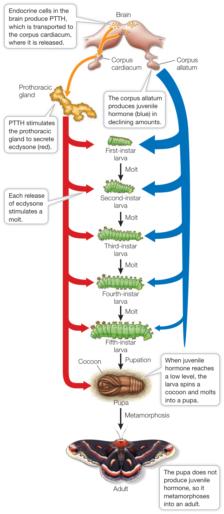
Figure 39.9 Hormonal Control of Metamorphosis Three hormones control molting and metamorphosis in the silkworm moth Hyalophora cecropia. (After C. M. Williams. 1952. Biol Bull 103: 120–138.)
Sex steroids control vertebrate sexual development
Hormones control many developmental processes in vertebrates. One example already mentioned in Key Concept 39.2 is the influence that the anterior pituitary growth hormone has on growth. Other examples are the hormones involved in sex development. The gonads—the testes of the male and the ovaries of the female—produce hormones as well as sperm and ova. The male steroid hormones are collectively called androgens, and the dominant hormone is testosterone. The female steroids are estrogens and progesterone. The dominant estrogen is estradiol, which is synthesized from testosterone. Males and females both synthesize testosterone, but females have an enzyme (aromatase) that converts testosterone to estradiol.
The sex steroids determine whether a mammalian embryo develops into a phenotypic female or male. In humans, the gonads of an early embryo are undifferentiated. Beginning in about the seventh week of development, the expression of genes on the Y chromosome of an XY individual normally causes the undifferentiated gonads to produce androgens. In response to androgens, the reproductive system develops the male phenotype. If no Y chromosome is present (i.e., the individual is genotype XX), androgens are not produced at this time and female structures develop (Figure 39.10). After birth, the sex steroids control the maturation of the reproductive organs and the development and maintenance of secondary sexual characteristics, such as breasts and facial hair.
Phenotypic sex determination provides a good example of the importance of receptors in hormonal control mechanisms. XY individuals are occasionally born who because of a mutation do not have functional androgen receptors—a condition called androgen insensitivity syndrome. These individuals have an XY genotype and internal testes, but they develop the external body form and genitalia of a female.
Puberty is the transition from juvenile body form and sexual functions to those of the adult. In juvenile humans, sex steroids are produced at low levels. At puberty (around age 12 or 13), sex steroid production increases rapidly, stimulating maturation of the secondary sexual characteristics (body forms, structures, and behavior). Why does this sudden increase occur? In both juvenile and adult humans, the activities of the gonads are controlled by the anterior pituitary tropic hormones luteinizing hormone (LH) and follicle-stimulating hormone (FSH), which together are the gonadotropins. The production of gonadotropins by the anterior pituitary is under the control of gonadotropin-releasing hormone (GnRH) produced by the hypothalamus (Figure 39.11). The production of GnRH is controlled by negative feedback from circulating sex steroids. Before puberty the circulating levels of sex steroids are low, but the hypothalamus is highly sensitive to that negative-feedback signal. As a result, only low levels of GnRH are released. Puberty is initiated by a reduction in the sensitivity of hypothalamic GnRH-producing cells to negative feedback. As a result, GnRH production increases, stimulating increased production of gonadotropins and hence increased production of sex steroids.
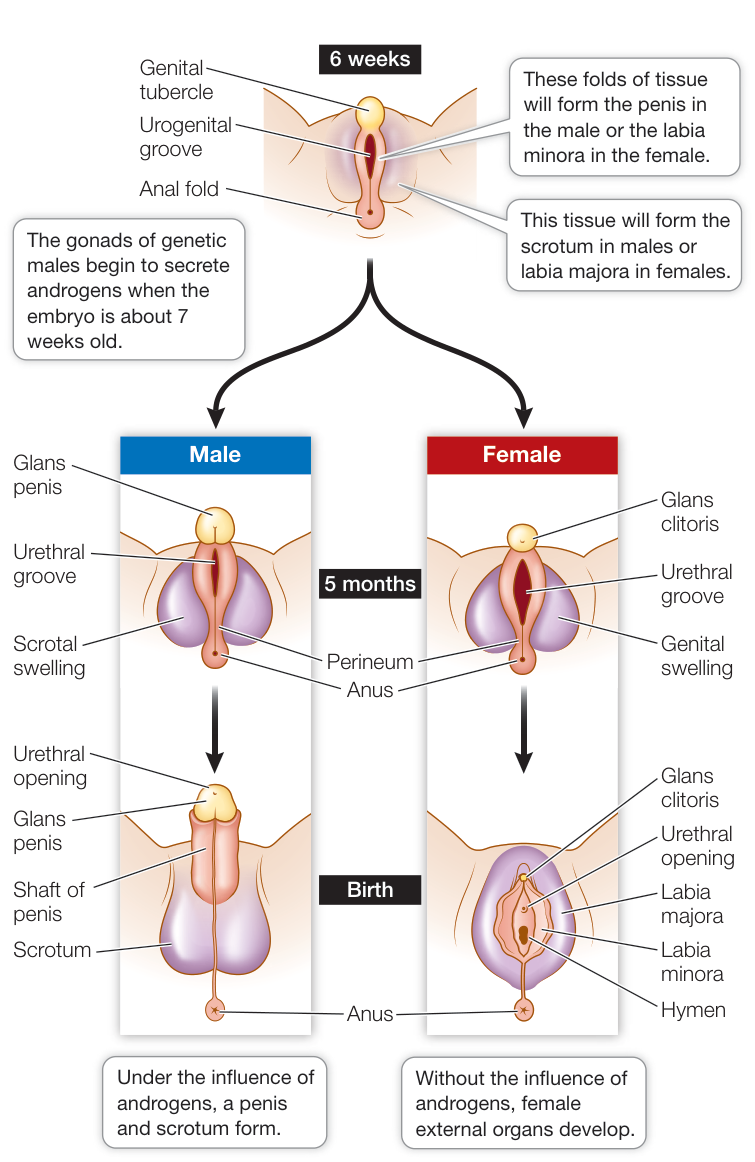
Figure 39.10 Sex Steroids Direct the Development of Human Sex Organs The external sex organs of early human embryos are undifferentiated. Androgens promote the development of male sex organs. In the absence of androgens, female sex organs form. Q: What do you think the effect of a mutation that results in non-functional androgen receptors would be?
In females, increasing levels of LH and FSH at puberty stimulate the ovaries to increase their production of the female sex hormones. The increased circulating levels of these hormones stimulate the development of the traits of a sexually mature woman: enlarged breasts, vagina, and uterus; broadened hips; increased subcutaneous fat; pubic hair; onset of menstrual and ovarian cycles (see Figure 41.14); and behavioral changes. In males, an increasing level of LH stimulates groups of cells in the testes to increase their synthesis of testosterone, which in turn initiates the physiological, anatomical, and behavioral changes associated with adolescence. The voice deepens, hair begins to grow on the face and body, and the testes and penis grow larger. Testosterone also stimulates bone and skeletal muscle growth. FSH in males stimulates production of sperm.
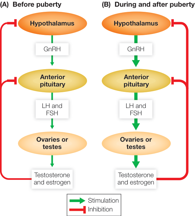
Figure 39.11 The Trigger for Puberty (A) In a prepubertal individual, the gonads produce sex steroids in small quantities, and because the hypothalamus is highly sensitive to the negative-feedback effects of those sex steroids, GnRH production is suppressed. (B) A sudden decrease in the sensitivity of the hypothalamus to this feedback causes increased production of GnRH and the onset of puberty.
The roles that sex steroids play in adult sexual behavior and reproduction as well as the control and regulation of these hormones will be described in more detail in Chapter 41.
Recap and Assess
Hormones play major roles in development. Because they circulate throughout the body, they can simultaneously affect many cells and tissues, but in different ways depending on the receptors and intracellular pathways in those cells. Thus hormones can coordinate the complex sets of responses involved in development. Arthropod development involves complete or incomplete metamorphosis controlled by ecdysone, juvenile hormone, and prothoracicotropic hormone (PTTH). Human gonadal and phenotypic sex development is controlled by steroid hormones.
- Why did decapitation of Rhodnius prevent molting when done 1 hour after feeding but not when done 1 week after feeding?
- How can a person with an XY genotype develop with the phenotype of a female?
- What is the role of negative feedback in the timing of puberty?
A common feature of the roles of hormones in development is that they coordinate the execution of events programmed into the genome. These functions have evolved to be rather stable in the face of changes in the internal and external environments. In contrast, hormonal systems involved in maintaining homeostasis of the internal environment have evolved to be very sensitive to changes in the internal or external environment that could compromise homeostasis. We look at some of these hormonal regulatory mechanisms in the next key concept.
Hormones Regulate Metabolism and the Internal Environment
Learning Objectives
- 39.4.1 Explain how both hyperthyroidism and hypothyroidism cause goiter.
- 39.4.2 Explain how the concentration of calcium in the blood is regulated.
- 39.4.3 Describe how insulin and glucagon regulate blood sugar levels.
- 39.4.4 Apply concepts about hormonal control of stress to explain why prolonged stress can have negative effects on the body.
Hormones participate in regulating all physiological systems. In this key concept we examine a few examples of hormonal actions that are critical in regulating the internal environment. For simplicity we focus on humans, but these systems are similar in all mammals.
Thyroxine stimulates many metabolic processes
Thyroxine is a crucial signal in the regulation of cellular energy metabolism, and it is produced and secreted by the thyroid gland. This gland wraps around the front of the windpipe (trachea) and expands into a lobe on either side (see Figures 39.5 and 39.15). There are two cell types in the thyroid gland, and they produce different hormones. Thyroxine is produced by epithelial cells that make up spherical, colloid-containing structures called follicles (Figure 39.12A). Between the follicles are cells that produce calcitonin, which is involved in regulating blood calcium, as we will describe shortly.
Follicle cells synthesize the glycoprotein thyroglobulin in the endoplasmic reticulum and package it in secretory vesicles in the Golgi apparatus (Figure 39.12B). Thyroglobulin is a polymer of the amino acid tyrosine. Follicle cells also take up iodide from the blood using cotransport and move it into the follicle lumen. The thyroglobulin vesicles are secreted into the lumen along with an enzyme that oxidizes the iodide to iodine. Iodine is highly reactive and combines with the tyrosine units of the thyroglobulin. When the thyroid gland is stimulated to release thyroxine, the follicle cells take up thyroglobulin from the lumen of the follicle by endocytosis. These bits of thyroglobulin are then cleaved to form smaller molecules consisting of only two tyrosine units, and these molecules leave the follicle cells and enter the blood (Figure 39.13).
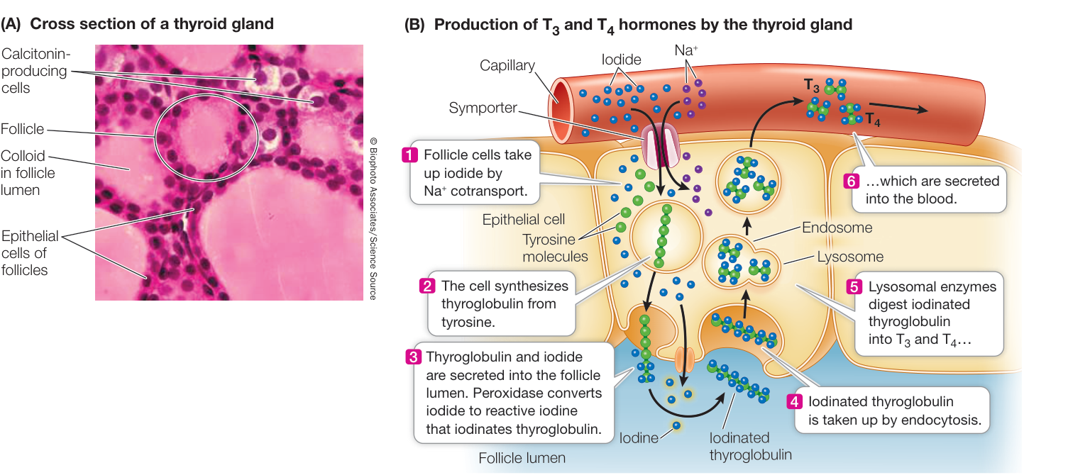
Figure 39.12 The Thyroid Gland Consists of Many Follicles (A) Cross section through a thyroid gland, showing numerous follicles bounded by epithelial cells. Calcitonin-secreting cells are located in the spaces between the follicles. (B) The epithelial cells of the follicle synthesize thyroglobulin and secrete it into the lumen of the follicle, where it is iodinated and stored until it is processed by the epithelial cells to generate and .
The thyroid usually releases about ten times as much as . However, is a more active hormone than , so when you read about the effects of thyroxine, keep in mind that the actions discussed are primarily those of . The difference in the activities of and makes it possible to control the effects of thyroxine in different tissues. Within target cells, can be converted to by an enzyme called deiodinase. A different deiodinase can convert into an inactive hormone called reverse . Deiodinase can also inactivate by converting it into or . Thus each target cell can have a unique sensitivity to thyroid hormones using these enzymes to control the conversion of to or to reverse , , or .
THE PRODUCTION AND RELEASE OF THYROXINE ARE REGULATED BY TSH AND TRH The tropic hormone thyroid-stimulating hormone (TSH), also known as thyrotropin is produced by the anterior pituitary. TSH activates the thyroxine-producing follicle cells in the thyroid. Thyrotropin-releasing hormone (TRH), produced in the hypothalamus and transported to the anterior pituitary through the portal blood vessels, activates the TSH-producing pituitary cells. The hypothalamus uses environmental information, such as temperature or day length, to determine whether to increase or decrease its secretion of TRH. This sequence of steps is regulated by a negative-feedback loop (see Figure 39.7B). Circulating thyroxine inhibits the response of pituitary cells to TRH, so less TSH is released when thyroxine levels are high, and more TSH is released when thyroxine levels are low. Circulating thyroxine also exerts negative feedback on the production and release of TRH by the hypothalamus.
Because thyroxine is lipid-soluble, it enters cells readily and binds to receptors in the nucleus. When bound to thyroxine, these receptors, which are found in most cells of the body, are transcription factors. They move to the nucleus and stimulate the transcription of numerous genes whose products are transport proteins, structural proteins, and enzymes involved in metabolic pathways; thus thyroxine elevates the metabolic rates of most cells and tissues. Exposure to cold for several days leads to an increased release of thyroxine, an increased conversion of to , and therefore an increased basal metabolic rate (see Key Concept 38.5).
During development and growth, thyroxine promotes amino acid uptake and protein synthesis. Insufficient thyroxine in a human fetus or growing child greatly retards physical and mental development, resulting in a condition known as cretinism.
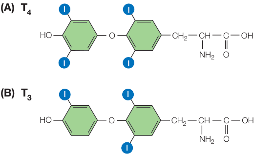
Figure 39.13 Cleavage of Thyroglobulin Yields Two Active Hormones (A) Tetraiodothyronine ( ) is iodinated at four locations. (B) Triiodothyronine ( ) is iodinated at only three locations.
GOITER An enlarged thyroid gland is called a goiter (Figure 39.14). A goiter can be caused by either hyperthyroidism (excess production of thyroxine) or hypothyroidism (thyroxine deficiency). The negative-feedback loop whereby thyroxine controls TSH release explains how two seemingly opposite conditions result in the same symptom.
- The most common cause of hyperthyroid goiter is Graves’ disease, an autoimmune disease involving an antibody to the TSH receptor. This antibody binds to and activates the TSH receptors on the follicle cells, causing uncontrolled production and release of thyroxine. Blood levels of TSH are low due to negative feedback from high levels of thyroxine, but the thyroid remains maximally stimulated and grows bigger. People with hyperthyroidism have high metabolic rates, usually feel hot, and may develop a buildup of fat behind the eyeballs that causes their eyes to bulge.

Figure 39.14 A Hypothyroid Goiter In this condition, dietary iodide deficiency leads to a lack of functional thyroxine, resulting in the oversynthesis of thyroglobulin and subsequent enlarged follicles.
- Hypothyroid goiter results when there is not enough circulating thyroxine to turn off TSH production. A common cause is deficiency of dietary iodide, without which the follicle cells cannot make active thyroxine ( or ). Without negative feedback from thyroxine, TSH levels remain high and the thyroid continues to produce thyroglobulin. Because sufficient iodine is not available, however, the thyroglobulin is poorly iodinated. When it is broken down by the follicle cells, it produces little functional or . As a result, TSH levels remain high and stimulate more and more synthesis of thyroglobulin, and the thyroid gets bigger. The symptoms of hypothyroidism are low metabolism, intolerance of cold, and general physical and mental sluggishness.
Goiter affects about 5% of the world’s population. The addition of iodide to table salt has greatly reduced the incidence of hypothyroid goiter in industrialized nations, but the condition is still common in other parts of the world and is a leading cause of intellectual impairment.
Three hormones regulate blood calcium concentrations
Blood calcium must be regulated within a narrow range or serious problems occur. If blood calcium falls below this range, the nervous system becomes overly excited, resulting in muscle spasms and even seizures. If blood calcium rises above this range, the nervous system becomes depressed and muscles—including the heart—weaken. Regulation of blood calcium is difficult because only about 0.1% of the calcium in the body is located in the extracellular fluid. About 1% is in cells, and almost 99% is in the bones. Therefore the body must maintain a tiny pool of calcium in the blood at a precise concentration, and that tiny pool can be influenced greatly by relatively small shifts in the much larger pools of calcium in the cells and bones.
The body has multiple mechanisms for changing blood calcium levels, including:
- Deposition or resorption of bone
- Excretion or retention of calcium by the kidneys
- Absorption of calcium from the digestive tract
These mechanisms are controlled by three hormones: calcitonin, parathyroid hormone, and calcitriol (synthesized from vitamin D).
CALCITONIN REDUCES BLOOD CALCIUM Remember that calcitonin is produced by cells in the thyroid gland. These cells are stimulated to release their hormone when blood calcium rises. Calcitonin acts to lower the concentration of calcium in the blood, mainly by stimulating calcium uptake by bone-forming cells (Figure 39.15). Bone is continuously remodeled through a dynamic process that involves both resorption of old bone and synthesis of new bone, as we will discuss in Key Concept 46.3. Cells called osteoclasts break down bone and release calcium into the blood, and cells called osteoblasts take up calcium from the blood and deposit it in new bone. Calcitonin decreases the activity of osteoclasts and thereby favors removal of calcium from the blood and its deposition in bone by osteoblasts. The turnover of bone in adult humans is not very high, so calcitonin does not play a major role in calcium homeostasis in adults. It is probably more important in young individuals whose bones are actively growing.
PARATHYROID HORMONE INCREASES BLOOD CALCIUM The parathyroid glands are four tiny structures embedded in the posterior surface of the thyroid gland (see Figure 39.15). Their single hormone product, parathyroid hormone (PTH), also called parathormone), is the most important hormone in the regulation of blood calcium levels. Circulating calcium activates receptors in the cell membrane of the parathyroid cells. When these receptors are activated, they inhibit the synthesis and release of PTH. A fall in blood calcium removes this inhibition and triggers the synthesis and release of PTH. PTH stimulates bone turnover by actions on both osteoclasts and osteoblasts. The end result of these actions of PTH is a net increase of calcium in the blood. PTH also maintains blood concentration of calcium by stimulating the kidneys to reabsorb it rather than excrete it in the urine.
CALCITRIOL INCREASES BLOOD CALCIUM In the past, fragile bones were common among people living at high latitudes, where winter days are short and the winter diet often lacks fish, dairy products, and fresh vegetables. Since the condition could be reversed by taking cod-liver oil, it was assumed that a vitamin D deficiency was involved. That vitamin was named vitamin D, but when its chemical identity was established, this molecule turned out not to be a vitamin at all.
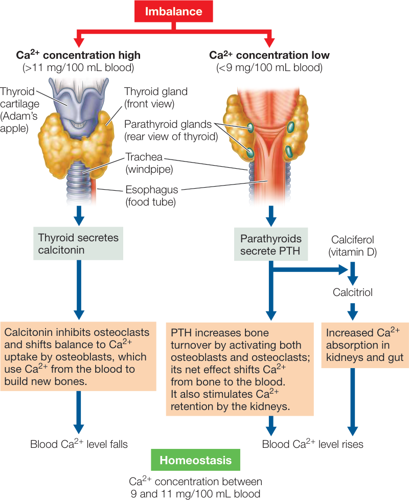
Figure 39.15 Hormonal Regulation of Calcium Calcitonin, parathyroid hormone (PTH), and calcitriol (the active form of calciferol, or vitamin D) regulate Ca2+ levels in the blood.
A vitamin D is a substance that the body requires in small quantities but cannot synthesize for itself and must therefore obtain from food (or from supplements such as vitamin pills). However, vitamin D—more accurately named calciferol—is synthesized naturally from cholesterol when skin cells receive ultraviolet light. Calciferol is not an active hormone, but through actions of enzymes in the liver and kidneys it is converted into the active form called calcitriol, which circulates in the blood and acts on distant cells (and therefore is a hormone). The conversion of calciferol to calcitriol is activated by PTH. Calcitriol promotes the absorption of calcium from food in the gut. Thus the combined actions of PTH and calcitriol raise blood calcium levels.
PTH lowers blood phosphate levels
Bones are made of phosphate as well as calcium. When PTH stimulates the release of calcium from bone, it also releases phosphate.
Normal blood concentrations of calcium and phosphate are just below the levels at which they precipitate out of solution as calcium phosphate salts. Even a small rise in concentrations may cause precipitation that leads to maladies such as kidney stones and calcium deposits in the arteries (hardening of the arteries). To reduce this risk, PTH acts on the kidneys to increase the elimination of phosphate via the urine.
Insulin and glucagon regulate blood glucose concentrations
Before the 1920s, the disease diabetes mellitus was fatal. The name diabetes refers to excessive urine production, and the name mellitus refers to the fact that the urine is sweet. These symptoms are both due to the large amounts of glucose in the blood and therefore in the urine. Other characteristics of the disease are weakness, lethargy, and a dramatic loss of body mass. Even before the discovery of hormones, diabetes was known to be connected with abnormal glucose metabolism and with the pancreas—a large gland located just below the stomach (see Figure 39.5). The exact links, however, were not clear. Today we recognize two forms of diabetes. The form that was mostly seen prior to the 1920s usually occurred in young people and was called juvenile diabetes. We now call it type I diabetes, and it is caused by a lack of the protein hormone insulin, which is produced and secreted by cells in the pancreas called cells.
Glucose enters cells by diffusion, but cell membranes are not very permeable to glucose. Glucose transporter proteins in cell membranes facilitate the movement of glucose into cells, and the glucose transporters called GLUT4 that are most common in muscle and adipose tissue are controlled by insulin. When insulin binds to its receptor on the cell membrane, it causes these glucose transporters to move from cytoplasmic vesicles to the cell membrane, thus making the cell more permeable to glucose (Fig. 39.16). When insulin is not present, these transporters are returned to the cytoplasmic pool through endocytosis.
Another form of diabetes, called adult onset or type II diabetes, is in epidemic proportions in the United States today. Type II diabetes is associated with high carbohydrate consumption, obesity, and lack of exercise. It is caused either by low production of insulin or by insensitivity to insulin. More than 90% of diabetes cases seen in the United States today are type II diabetes, and the numbers are rising alarmingly. Statistics from the Centers for Disease Control and Prevention show that the number of new cases of type II diabetes reported in the United States in 1991 was less than 600,000 and had been fairly stable since 1980. Between 1991 and 2012, the annual number of new cases tripled. Today the incidence of diabetes in the United States is over 30 million, more than 9% of the population. Diabetes is currently the seventh leading cause of death in the United States, and it is a common contributor to blindness, kidney failure, stroke, heart disease, and amputations resulting from ulcerations. This disease is largely preventable and even reversible with lifestyle changes.
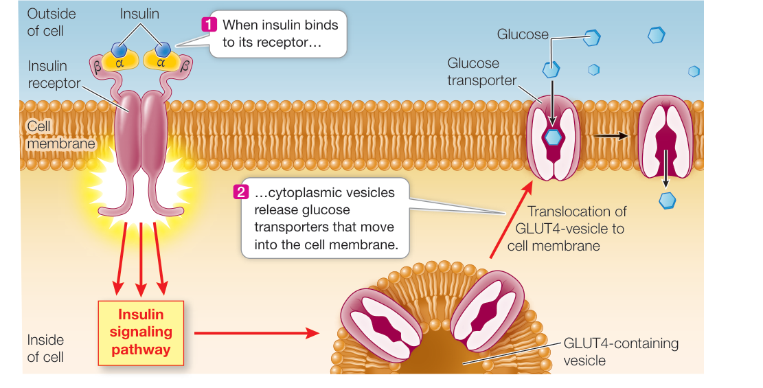
Figure 39.16 Glucose Transporters Are Controlled by Insulin Most tissues of the body depend on insulin to promote the uptake of glucose into cells. Glucose enters cells by diffusion, but that diffusion is facilitated by passive glucose transporters. When insulin binds to receptors on the cell surface, it stimulates the transfer of one type of glucose transporter (GLUT4) from intracellular sequestration to the cell membranes. When insulin levels fall, the glucose transporters are again sequestered intracellularly.
In both type I and type II diabetes, glucose entry into cells is impaired, resulting in so much glucose accumulating in the blood that it starts to spill over into the urine. A high concentration of glucose in the blood increases urine output by two mechanisms. First, it causes water to move from cells into the blood by osmosis, and this increase in blood volume results in increased urine production. Second, the increased glucose in the tubules of the kidneys pulls more water into the urine by osmosis. Diabetic individuals lose their metabolic fuel in their urine, and they can become dehydrated. Because glucose uptake by muscle and adipose tissue is impaired in the absence of insulin, muscle cells must depend on fat and protein for fuel, and adipose tissue cannot replenish its stores of triglycerides. The body wastes away if the condition is not treated.
For centuries, the prospects for people suffering with type I diabetes were bleak. A change came almost overnight in 1921, when the physician Frederick Banting and medical student Charles Best at the University of Toronto discovered they could reduce the symptoms of diabetes by injecting an extract prepared from pancreatic tissue. The active component of this extract was found to be insulin, a small protein consisting of just 51 amino acids. In the United States today, insulin replacement therapy using manufactured insulin allows more than 1.5 million people with type I diabetes to lead almost normal lives.
Much evidence now shows that type II diabetes can be treated with a combination of dietary changes and weight loss. Figure 39.17 looks at the intriguing question of whether the hormone irisin (introduced in the opening of this chapter) can mediate the beneficial effects of exercise on glucose metabolism in type II diabetes patients.
ISLETS OF LANGERHANS The pancreas is mostly an exocrine gland that produces digestive enzymes and secretes them through ducts into the gut (see Figure 49.13). But within the pancreas are clusters of endocrine cells called islets of Langerhans, after the German medical student who discovered them. They contain three types of cells, each of which produces a specific hormone:
- Beta () cells produce and secrete insulin.
- Alpha () cells produce and secrete glucagon, a hormone that has effects mostly opposite from those of insulin.
- Delta () cells produce the hormone somatostatin.
After a meal, the concentration of glucose in the blood rises, stimulating the cells of the islets to release insulin. Insulin causes target cells throughout the body to use circulating glucose as fuel and convert it into storage products such as glycogen and fat. When the gut is empty of food, blood glucose concentration falls and the islets stop releasing insulin. As a result, most cells shift to using glycogen and fat rather than glucose as fuel. If blood glucose concentration falls substantially below normal, the islet cells release glucagon, which stimulates the liver to break down stored glycogen and release glucose into the blood. We will discuss these actions in greater detail in Key Concept 49.4.
SOMATOSTATIN Somatostatin is released from the cells of the pancreas in response to rapid increases of glucose and amino acids in the blood. This hormone has paracrine functions within the islets, where it inhibits the release of both insulin and glucagon. Outside the pancreas it acts as a hormone, slowing the digestive activities of the gut and extending the period during which nutrients are absorbed. Somatostatin is also produced in very small amounts by cells in the hypothalamus. Hypothalamic somatostatin, also called growth hormone-inhibiting hormone (GHIH), is transported in the portal blood vessels to the anterior pituitary, where it inhibits the release of growth hormone and thyrotropin.
The adrenal gland is two glands in one
An adrenal gland sits above each kidney, just below the middle of your back. Functionally and anatomically, each adrenal gland consists of a gland within a gland (Figure 39.18). The core, or adrenal medulla, produces epinephrine (adrenalin) and, to a lesser degree, norepinephrine (noradrenalin). The medulla develops from nervous tissue and is under the control of the nervous system. Surrounding the medulla is the adrenal cortex, which produces steroid hormones. The cortex is under hormonal control by a tropic hormone produced by cells in the anterior pituitary.
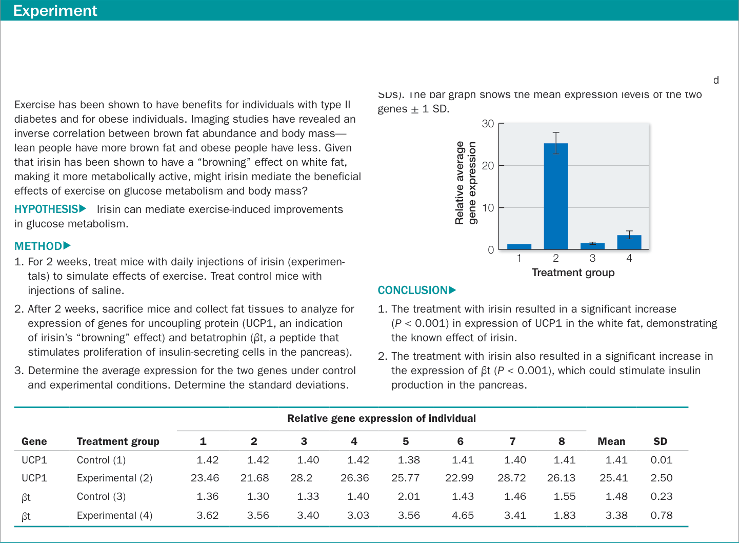
Figure 39.17A Effects of Exercise on Glucose Metabolism Original Paper: Y. Zhang et al. 2014. Irisin stimulates browning of white adipocytes through mitogen-activated protein kinase p38 MAP kinase and ERK MAP kinase signaling. Diabetes 63: 514–525. Do t-tests to determine if the treatment (irisin injections) had a significant effect on the expression of genes for UCP1 and for βt. RESULTS▸ The table shows gene expression (in arbitrary units) in white adipose tissue (simulated data based on published means an SD ) Th b h h th i l l f th t Figure 39.17A Effects of Exercise on Glucose Metabolism Original Paper: Y. Zhang et al. 2014. Irisin stimulates browning of white adipocytes through mitogen-activated protein kinase p38 MAP kinase and ERK MAP kinase signaling. Diabetes 63: 514–525. Do t-tests to determine if the treatment (irisin injections) had a significant effect on the expression of genes for UCP1 and for t. RESULTS The table shows gene expression (in arbitrary units) in white adipose tissue (simulated data based on published means and SDs). The bar graph shows the mean expression levels of the two genes SD. Figure 39.17B Effects of Exercise on Glucose Metabolism Original Paper: Y. Zhang et al. 2014. Irisin stimulates browning of white adipocytes through mitogen-activated protein kinase p38 MAP kinase and ERK MAP kinase signaling. Diabetes 63: 514–525.
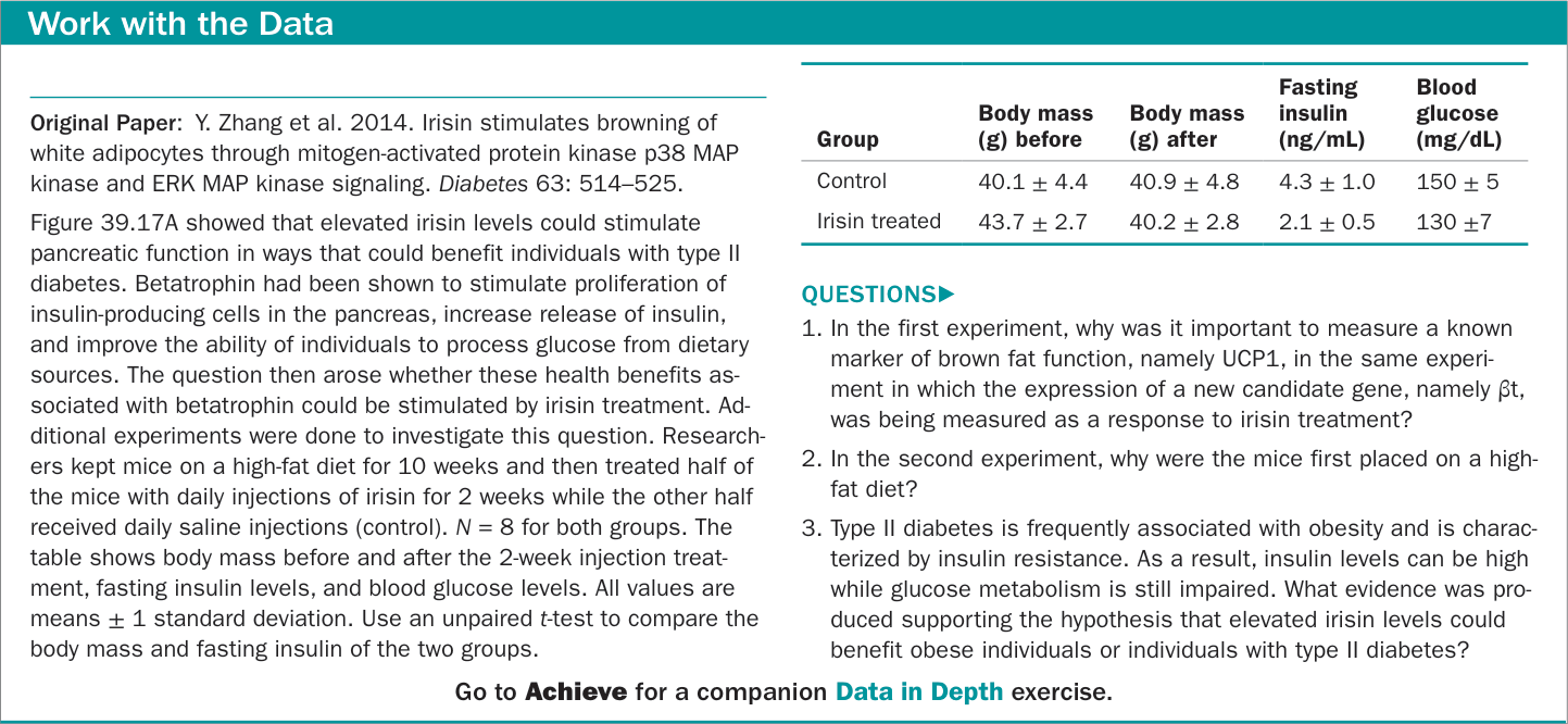
Figure 39.17B Effects of Exercise on Glucose Metabolism
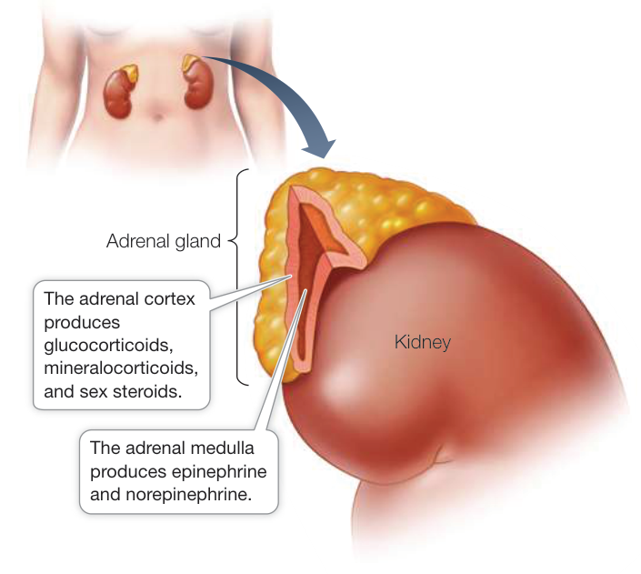
Figure 39.18 The Adrenal Is Really Two Glands An adrenal gland, consisting of an outer cortex and an inner medulla, sits above each kidney. The medulla and the cortex produce different hormones.
THE ADRENAL CORTEX The cells of the adrenal cortex use cholesterol to produce three classes of steroid hormones (see Figure 39.2B), collectively called corticosteroids:
- Mineralocorticoids influence the salt and water balance of the extracellular fluid.
- Glucocorticoids influence blood glucose concentrations as well as other aspects of fat, protein, and carbohydrate metabolism.
- Sex steroids play roles in sexual development, sexual behavior, and anabolism (tissue building).
In adult humans, the adrenal cortex secretes only negligible amounts of sex steroids. The major producers of sex steroids are the gonads, as we saw in Key Concept 39.3.
Aldosterone, the primary mineralocorticoid, stimulates the kidneys to conserve sodium and excrete potassium, as we will discuss in Chapter 50. If the adrenal glands are removed from an animal, sodium must be added to its diet, or its sodium will be depleted and it will die.
The main glucocorticoid in humans is cortisol, which is critical for mediating the body’s metabolic responses to stress. Within minutes of a stressful stimulus (one provoking fear or anger, for example), blood cortisol levels begin to rise. This response is much slower than the neurally mediated epinephrine and norepinephrine response to stress, but it lasts longer. Whereas epinephrine and norepinephrine cause sudden increases in blood pressure, cortisol causes a sustained increase in blood pressure. Cortisol stimulates tissues that are not critical for escaping the danger or threat to switch from using blood glucose for energy to using fats and proteins. This action preserves circulating glucose to fuel the actions of the nervous system and skeletal muscles necessary to meet the threat. Cortisol’s elevation of blood pressure increases delivery of oxygen to the muscles, preparing them for flight or fight. Cortisol also inhibits the immune system. Dealing with the immediate stressor is more important than feeling sick, having allergic reactions, or healing wounds. This explains why cortisol and drugs that mimic its actions are useful for reducing inflammation and allergic responses. Cortisol decreases activity of the gut and reproductive systems—you can digest lunch and reproduce later if you escape the threat. The actions of cortisol are adaptive in responding to acute stress, but they can cause problems if sustained over time, as sometimes occurs in modern society due to social, economic, and job-related stressors.
Cortisol release is controlled by adrenocorticotropic hormone (ACTH) from the anterior pituitary. ACTH release is controlled in turn by corticotropin-releasing hormone (CRH) from the hypothalamus (see Figure 39.7). The action of ACTH on the adrenal cortex is to stimulate the synthesis of cortisol. Like other steroid hormones, cortisol is not stored in vesicles and therefore is available for immediate release. As cortisol or other steroid hormones diffuse into the blood, they combine with carrier proteins, and their release from these proteins can have a long time course, thus stretching out their actions. Also, many of their actions stimulate gene expression in target cells, which takes time but has a long-lasting effect.
Turning off the stress responses activated by cortisol is as important as turning them on. A study of stress in rats showed that old rats could turn on these stress responses as effectively as young rats, but they lost the ability to turn them off as rapidly. As a result, they suffered from the well-known consequences of stress seen in humans: digestive system problems, cardiovascular problems, strokes, impaired immune system function, and increased susceptibility to cancers and other diseases. Acute stress responses are turned off by negative feedback from cortisol on both the ACTH-secreting cells of the anterior pituitary and the CRH-secreting cells of the hypothalamus. With chronic or prolonged stress, these control mechanisms become insufficient and cortisol exerts negative feedback through another brain region, the hippocampus. Prolonged exposure to cortisol, however, causes trauma to and loss of hippocampal cells, resulting in the decreased ability to turn off the stress response.
Loss of adrenal cortical function results in Addison’s disease, characterized by fatigue, muscle weakness, digestive problems, low blood sodium (hyponatremia), low blood pressure, and salt hunger. Without replacement hormone treatment, death is likely. An interesting symptom of Addison’s disease is darkening of many skin areas that normally are not exposed to the sun—for instance, the lining of the cheeks. You can understand the cause of this symptom by thinking about the source of ACTH and the control of its secretion. In the hypothalamic–pituitary–adrenal axis (see Figure 39.7), an end hormone (in this case cortisol) frequently serves as a negative-feedback signal to the pituitary and also the hypothalamus. If the hormones of the adrenal cortex are absent, that negative feedback is gone, and the system will call for more ACTH. The production of ACTH by pituitary cells involves the expression of a polypeptide called proopiomelanocortin which is then cleaved to produce multiple peptide hormones. These include ACTH, MSH, endorphins, and enkephalins. The excess production of MSH is the cause of the skin darkening in Addison’s disease.
THE ADRENAL MEDULLA Stress causes the adrenal medulla to release epinephrine and norepinephrine, which arouse the body to action. As we saw earlier in this chapter, epinephrine and norepinephrine increase heart rate and blood pressure and divert blood flow to active muscles and away from the gut and skin. Only about one-fifth as much norepinephrine is secreted from the adrenal medulla as epinephrine, but it has similar functions. Norepinephrine is also used by the nervous system as a neurotransmitter, and it is involved in controlling many physiological processes.
Epinephrine and norepinephrine are both water-soluble, and both bind to the same set of receptors on the surfaces of target cells. These adrenergic receptors are of two general types, -adrenergic and -adrenergic. The -adrenergic receptors respond more strongly to norepinephrine than to epinephrine, whereas -adrenergic receptors respond about equally to both epinephrine and norepinephrine. Because of this difference in receptor affinities, it is possible for drugs to blunt the flight-or-fight responses without disrupting physiological regulatory processes. Such drugs are called “beta blockers” because, by inhibiting -adrenergic receptors, they can reduce the fight-or-flight response to epinephrine without disrupting the physiological regulatory functions of norepinephrine mediated through the -adrenergic receptors. Beta blockers are commonly prescribed to reduce symptoms of anxiety such as dry mouth and elevated heart rate (palpitations).
Many chemicals may act as hormones
We have discussed the major mammalian endocrine glands and their hormones in this chapter, but many more hormones exist. As we discuss the organ systems of the body in the chapters that follow, we will frequently describe hormones that their tissues produce as well as hormones that control their functions.
The pineal gland has a daily cycle of melatonin release
Humans, like most vertebrate species, have a pineal gland located between the cerebral hemispheres. The pineal has a daily rhythm of production and secretion of the hormone melatonin. The functions of the pineal and melatonin have to do with coordinating daily rhythms of physiological functions and, in many species, seasonal changes such as reproduction timing, migration, and preparations for environmental stress such as cold weather.
Recap and Assess
The major endocrine glands of mammals include the hypothalamus, pituitary gland, thyroid gland, parathyroid glands, pancreas, adrenal glands, gonads, and pineal gland. Each of these glands secretes and responds to hormones that play crucial roles in controlling physiology and development. Thyroxine is a lipid-soluble hormone that influences the expression of genes involved in regulating cell metabolism. Calcitonin and parathyroid hormone (PTH) regulate blood calcium levels, mainly by regulating the remodeling of bone. Blood glucose levels are controlled by insulin. When blood glucose levels rise, insulin is released and promotes uptake of glucose by cells. When blood glucose levels fall, the lack of insulin decreases uptake of glucose by most cells. Glucagon is released in the event of serious hypoglycemia, and it stimulates breakdown of glycogen to raise blood glucose levels. The adrenal cortex produces steroid hormones, the most important of which is cortisol, which is involved in stress responses. The adrenal medulla produces and releases epinephrine and norepinephrine.
- If you analyzed the chemical composition of thyroglobulin from a hyper-and a hypothyroid patient, how would the two samples differ?
- Two symptoms of type II diabetes are insulin insensitivity and poor glucose tolerance (slow clearance of glucose from the blood). Explain the likely causes of these symptoms.
- Osteoporosis is a weakening of the bones due to an imbalance between bone resorption and bone formation. Explain why liver and kidney disease are associated with a higher risk of osteoporosis.
- Explain how chronic stress can lead to stress-related diseases.

Activity 39.2 Concept Review: Vertebrate Hormones
Investigating Life: QA In what ways could irisin mediate the health benefits of exercise?
The initial discovery of irisin and its functions revealed that it induces brown fat-like changes in white fat, including expression of the gene that codes for uncoupling protein (UCP1), which enables brown fat cells to metabolize lipids without producing ATP. As a result, metabolism in the brown fat cells is not inhibited by the buildup of ATP or the lack of ADP and the brown fat can simply metabolize excess energy and generate heat.
Exercise does more than promote weight loss and toned muscles. It also improves cognition and glucose metabolism. Improving glucose metabolism can help reverse the changes associated with type II diabetes—elevated blood glucose, elevated blood insulin, loss of sensitivity of the pancreas to glucose, and loss of sensitivity of body tissues to insulin. Investigating Life: How Could Irisin Mediate the Benefits of Exercise on Cognition? and Figure 39.17 present experiments showing that irisin is a mediator of benefits of exercise on cognition and glucose metabolism. It seems likely that irisin is a major integrating factor in a large suite of healthful benefits of exercise.
Future directions
The experiments explored in Investigating Life: How Could Irisin Mediate the Benefits of Exercise on Cognition? and Figure 39.17 suggest the interesting possibility that irisin could be developed as a therapeutic that might be useful in treating the national epidemic of obesity and type II diabetes. It might also prove to be beneficial in cognitive declines due to conditions such as stroke and Alzheimer’s disease. Before such treatments can be developed, however, research is needed on such questions as: Are there potential negative side effects of abnormal elevations of irisin? Is there the potential for abuse of irisin, similar to what has been seen with other growth-promoting agents such as growth hormone and testosterone? It would seem that the safest way to benefit from the actions of irisin would be to use the natural approach of regular exercise to promote its normal production and release.

Visual Summary

© Tierfotoagentur/Alamy Stock Photo
You should be able to relate each summary to the adjacent figures. If you go to this visual summary in Achieve, you can follow links to figures, animations, activities, and simulations that will help you consolidate the material.
Hormones Circulate around the Body and Affect Cells That Have Receptors
- ■ Endocrine cells secrete chemical signals that induce responses in other cells that have receptors for those molecules.
- ■ Hormones are endocrine signals that circulate in the blood and activate distant target cells.
- ■ Protein and peptide hormones are water-soluble and have cell surface receptors. Steroid hormones are lipid-soluble and have cytoplasmic receptors. Some amine hormones are water-soluble and some are lipid-soluble. The same hormone can cause different responses in different target cells.
- ■ Chemical structures of hormones are highly conserved, but functions change.
Questions
- What are the functional consequences of insulin being water-soluble and estrogen being lipid-soluble?
- How and why does epinephrine cause glycogen to be broken down in the liver, but also cause decreased activity in the gut?
The Endocrine and Nervous Systems Work Together
- ■ The pituitary gland is the interface between the nervous and endocrine systems.
- ■ The posterior pituitary secretes antidiuretic hormone (ADH) and oxytocin. The anterior pituitary secretes four tropic hormones, as well as growth hormone (GH), prolactin, endorphins, enkephalins, and melanocyte-stimulating hormone (MSH).
- ■ The anterior pituitary is controlled by hypothalamic neurohormones.
- ■ Hormone release is controlled by negative-feedback loops.
Questions
- If a young woman had an accident that destroyed her pituitary, what effects would you expect to see on her metabolic rate, menstrual cycle, and urine production, and why?
- ADH acts on the kidney and on blood vessels. How do these two different actions contribute to the same physiological effect?
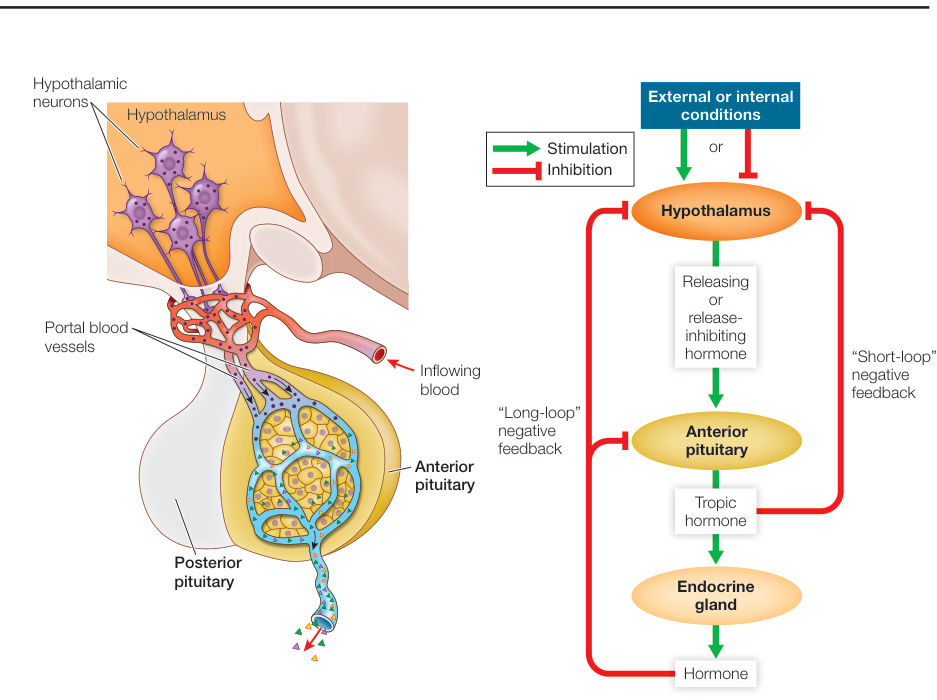
Figure 39.7
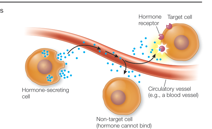
Figure 39.1
Hormones Play Important Roles in Development
- ■ Prothoracicotropic hormone (PTH) and ecdysone control molting in arthropods, and juvenile hormone prevents maturation.
- ■ In mammals, androgens, estrogens, and progesterone control sexual development, secondary sexual characteristics, and reproductive functions.
Questions
- Why do many insecticides contain analogs of juvenile hormone?
- If you injected the gonadotropins LH and FSH into a juvenile mammal, would you expect to see responses in the testes and ovaries? Why or why not?
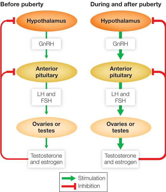
Figure 39.11
Hormones Regulate Metabolism and the Internal Environment
- ■ The thyroid gland produces and secretes and , which control many aspects of cell metabolism.
- ■ Blood calcium is regulated by three hormones: calcitonin, parathyroid hormone (PTH), and calcitriol.
- ■ Three hormones secreted by the pancreas—insulin, glucagon, and somatostatin—regulate blood glucose.
- ■ The adrenal medulla produces and releases epinephrine and norepinephrine.
- ■ The adrenal cortex produces aldosterone, which stimulates the kidneys to conserve sodium and excrete potassium; cortisol, which mediates stress responses; and small amounts of sex steroids.
- ■ The pineal gland releases melatonin, a hormone involved in controlling biological rhythms.
Questions
- Why are thyroid hormones well suited for mediating seasonal adaptations?
- Why is PTH considered to be the most important regulator of blood calcium?
- Why are -adrenergic blockers rather than -adrenergic blockers used to control anxiety?
- Why is it adaptive that muscle and fat require insulin for adequate uptake of glucose, but the brain does not?
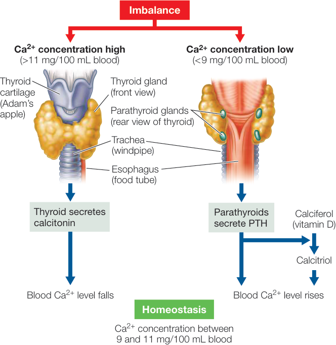
Figure 39.15