Chapter 40Immunology: Animal Defense Systems
Key Concepts
40.1 Animals Use Innate and Adaptive Mechanisms for Defense
40.2 Innate Defenses Are Nonspecific
40.3 Adaptive Defenses Are Specific
40.4 The Humoral Adaptive Response Involves Antibodies
40.5 The Cellular Adaptive Response Involves T Cells and Receptors
40.6 Malfunctions in Immunity Can Be Harmful
Soldiers being vaccinated against smallpox in the nineteenth century. Using vaccines continues to be important in protecting an army as it is exposed to pathogens in the environment.
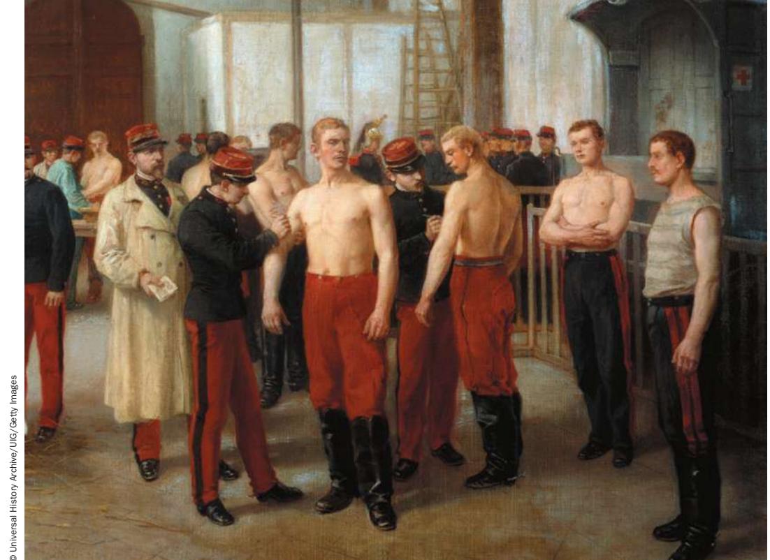
© Universal History Archive/UIG/Getty Images
Investigating Life: Vaccines and Immunity
Vials in two deep freezers, one in Atlanta, Georgia, and one in Siberia, are all that is left of the smallpox virus. Smallpox killed as many as 500 million people in the twentieth century, but the disease made its last appearance in 1978 and was declared eradicated in 1980, thanks to vaccination. A vaccine is usually an inactive form of a pathogen or toxin that provokes the immune system to produce antibodies: specific proteins directed against the target. The immune system destroys whatever is bound to the antibodies.
The eradication of smallpox was a spectacular international accomplishment. So why hasn’t vaccination for other potentially lethal diseases such as the flu (influenza) been widely accepted by the public? More than one-third of Americans refuse flu shots, and a significant number of parents refuse vaccination for their children.
Unfortunately, those who refuse vaccination may harm people other than themselves. A vaccination program can control or eradicate a disease only if a high percentage (typically above 80%) of people are vaccinated, thus disrupting the chain of infection from person to person. A high level of vaccination results in “herd immunity,” a state in which even those who cannot be vaccinated or who have weak immune systems are protected from infection. This protection is lost if the vaccination rate falls below the level needed for herd immunity. The elderly or sick, and infants with their immature immune systems, are most at risk.
The best way to develop herd immunity in a population is compulsory vaccination. In many countries, vaccination is a prerequisite for school enrollment and military enlistment and is required during epidemics. For example, during the periodic smallpox epidemics in the twentieth century in the United States, doctors accompanied by police would go into neighborhoods where the disease raged, vaccinating all those who were uninfected and removing infected people to quarantine. You can imagine the reaction of parents whose children were taken away, often to die. Henning Jacobson was arrested when he refused vaccination during a smallpox epidemic. He took his case to the U.S. Supreme Court, which ruled in 1905 that each state is entitled to protect its citizens. This provided a legal framework for compulsory vaccination that continues to this day. But opposition and court challenges continue.
QA What are the mechanisms and implications of long-lasting immunity? (Find the answer on page 909.)
Animals Use Innate and Adaptive Mechanisms for Defense
Learning Objectives
- 40.1.1 Describe the three phases of an internal response to a pathogen.
- 40.1.2 Distinguish between innate and adaptive immunity.
- 40.1.3 Describe how the Toll-like receptor (TLR) pathway is activated in innate immunity.
- 40.1.4 List the three categories of white blood cells and gives examples of each.
Animals have several ways of defending themselves against pathogens—harmful organisms and viruses that can cause disease. Once a pathogen breaches the physical defenses designed to keep it out, internal defense systems are activated. These internal systems are based on differences between self and nonself, or foreign, molecules. The internal defensive response involves three phases:
- Recognition phase. The organism recognizes the pathogen as foreign because it can discriminate between self and nonself.
- Activation phase. The recognition event leads to a mobilization of cells and molecules to fight the invader.
- Effector phase. The mobilized cells and molecules destroy the invader.
There are two general types of defense mechanisms:
- Innate defenses, or nonspecific defenses, are mechanisms that provide the first line of defense against pathogens. Innate defenses typically act very rapidly and include physical barriers such as the skin, molecules that are toxic to invaders, and phagocytic cells that ingest invaders. (Recall from Key Concept 6.5 that phagocytosis is a form of endocytosis, in which a cell engulfs a large particle or another cell.) The innate immune system recognizes broad classes of organisms and molecules, such as viruses, bacteria, fungi, protists, parasitic worms, and various toxins.
- Adaptive defenses are aimed at specific pathogens and are activated by cells in the innate immune system. For example, the adaptive immune system can make an antibody protein that will recognize, bind to, and aid in the destruction of a specific pathogen, if that specific pathogen ever enters the body. Adaptive defenses are typically slower to develop than innate defenses and longer-lasting.
An organism that is able to avoid the detrimental effects of attack by a pathogen is said to have immunity to that pathogen.
Innate defenses evolved before adaptive defenses
All animals have innate defenses against their enemies. For example, horseshoe crabs first appeared in the fossil record about 400 million years ago. These arthropods, represented today by four living species, rely only on innate defenses. These defenses include barriers, defensive cells, and defensive molecules (Figure 40.1):
- Barriers include physical mechanisms for resisting infections. The hard exoskeleton that is characteristic of arthropods prevents pathogens from getting inside horseshoe crabs.
- Defensive cells include those that bind to microbial pathogens, ingest them by endocytosis, and destroy them by hydrolysis. Amebocytes in the blood of horseshoe crabs fulfill this defensive role.
- Horseshoe crabs have a wide array of defensive molecules that are released from cells in their blood and are toxic to invading pathogens. These molecules include peptides that disrupt bacterial cell membranes, rendering them permeable; and peptides that bind to bacterial surfaces and cross-link them.
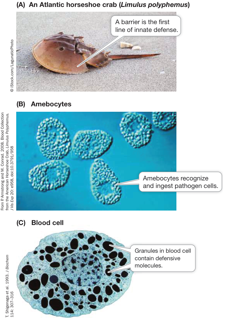
Figure 40.1 Innate Defenses in Horseshoe Crabs (A) A hard, impermeable exoskeleton acts as a barrier against pathogens. (B) Phagocytic amebocytes ingest and hydrolyze invading pathogens. (C) Blood cells release chemicals harmful to invading pathogens.
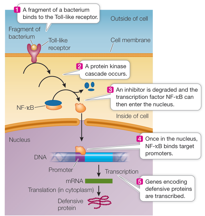
Figure 40.2 Cell Signaling and Defense Binding of a pathogenic molecule or fragment to the Toll-like receptor initiates a signal transduction pathway that results in the transcription of genes whose products are involved in innate defenses.
Studies of internal innate immune defenses, along with genome sequencing, have revealed that the recognition and activation phases evolved very early in animals. For example, animals as diverse as humans and fruit flies share a class of receptors called Toll-like receptors (TLRs) that participate in innate defense responses. These receptors recognize foreign molecules called pathogen-associated molecular patterns (PAMPs). In vertebrates, each TLR recognizes and binds to a specific molecule that is found in a broad class of pathogens, such as a component of the bacterial cell wall. Binding triggers a signal transduction pathway that ends with the expression of genes for anti-pathogen molecules (Figure 40.2). This pathway exists in some form in many animal groups, including humans.
Connect the Concepts The recognition of PAMPs is widespread in immunity and is therefore an evolutionarily ancient mechanism. Plants have an immune response to invading pathogens that also involves PAMPs; see Key Concept 37.2.
Mammals have both innate and adaptive defenses
Mammals, like other vertebrates, have both kinds of defense mechanisms, and these mechanisms are the focus of this chapter. In mammals, the innate and adaptive mechanisms operate together as a coordinated defense system. Table 40.1 gives an overview of these defenses. Innate immunity is the body’s first line of defense, because the adaptive defenses often require days or even weeks to become effective.
Table 40.1 Innate and Adaptive Immune Responses to an Infection
| Role | System | Mechanisms |
|---|---|---|
| Prevent invasion | Innate, nonspecific (first line) | Physical barrier (skin and lining of organs) Dryness, low pH Mucus Lysozyme, defensins |
| Early response to invasion (< 96 hr) | Innate, nonspecific (second line) | Inflammation Phagocytosis Natural killer cells Complement system Interferons |
| Late response to invasion (> 96 hr) | Adaptive, specific | Humoral immunity (antibodies from B cells) Cellular immunity (T cells) |
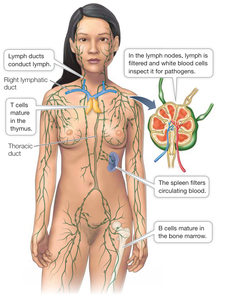
Figure 40.3 The Human Lymphatic System A network of ducts and vessels collects lymph from body tissues and carries it toward the heart, where it mixes with blood to be pumped back to the tissues. Other lymphoid tissues, including the lymph nodes, thymus, spleen, and bone marrow, are also essential to the body’s defense system.
Blood and lymph tissues play important roles in defense
The components of the mammalian defense system are dispersed throughout the body and interact with almost all of its other tissues and organs. The lymphatic system is an essential part of the immune system and includes the thymus, bone marrow, spleen, lymph nodes, lymphatic vessels, and lymph (Figure 40.3). The lymphatic system functions in partnership with the circulatory system, which includes the heart and blood vessels. Blood and lymph have nondefensive functions that will be discussed in Chapter 48. They each have central roles in defense as well.
Blood and lymph both consist of liquids in which cells are suspended:
- Blood plasma is a yellowish solution containing ions, small molecule solutes, and soluble proteins. Suspended in the plasma are red blood cells, white blood cells, and platelets (cell fragments essential to blood clotting). Whereas red blood cells are normally confined to the closed circulatory system (the heart, arteries, capillaries, and veins), white blood cells and platelets are also found in the lymph.
- Lymph is a fluid that is derived from the blood (but lacking red blood cells) and other tissues that accumulates in intercellular spaces throughout the body. From these spaces, the lymph moves slowly into the vessels of the lymphatic system. Tiny lymph capillaries conduct this fluid to larger ducts that eventually join together, forming two large vessels, the thoracic duct and right lymphatic duct, which join a major vein (the left subclavian vein) near the heart. By this system of vessels, the lymph is eventually returned to the blood and the circulatory system.
At many sites along the lymph vessels are small, roundish structures called lymph nodes, which contain large numbers of a type of white blood cell called lymphocytes. As lymph passes through a lymph node, the lymphocytes encounter foreign cells and molecules that have entered the body, and if they are recognized as foreign, an immune response is initiated.
White blood cells play many defensive roles
One milliliter of human blood typically contains about 5 billion red blood cells and 7 million of the larger white blood cells (also called leukocytes). All of these cells originate from multipotent stem cells (constantly dividing undifferentiated cells that can form several different cell types) in the bone marrow. There are three major groups of white blood cells: granulocytes, monocyte-derived cells, and lymphocytes (Figure 40.4). Granulocytes include basophils, eosinophils, and neutrophils, all of which circulate in the blood, and the mast cells, which are found in various tissues around the body. Monocyte-derived cells include monocytes and the macrophages and dendritic cells that monocytes can develop into. Macrophages and dendritic cells are both found in tissues and play important roles in stimulating an adaptive immune response (after traveling to lymph nodes). Lymphocytes include the B cells and T cells, which are the workhorses of the adaptive immune response and reside primarily in the lymph nodes, and the natural killer cells, which are found in various tissues and are responsible for attacking virus-infected and tumor cells. Neutrophils, monocytes, macrophages, and dendritic cells are all able to phagocytose microbial particles. Both defensive and cell signal proteins play fundamental roles in the interactions and functioning of these cells.
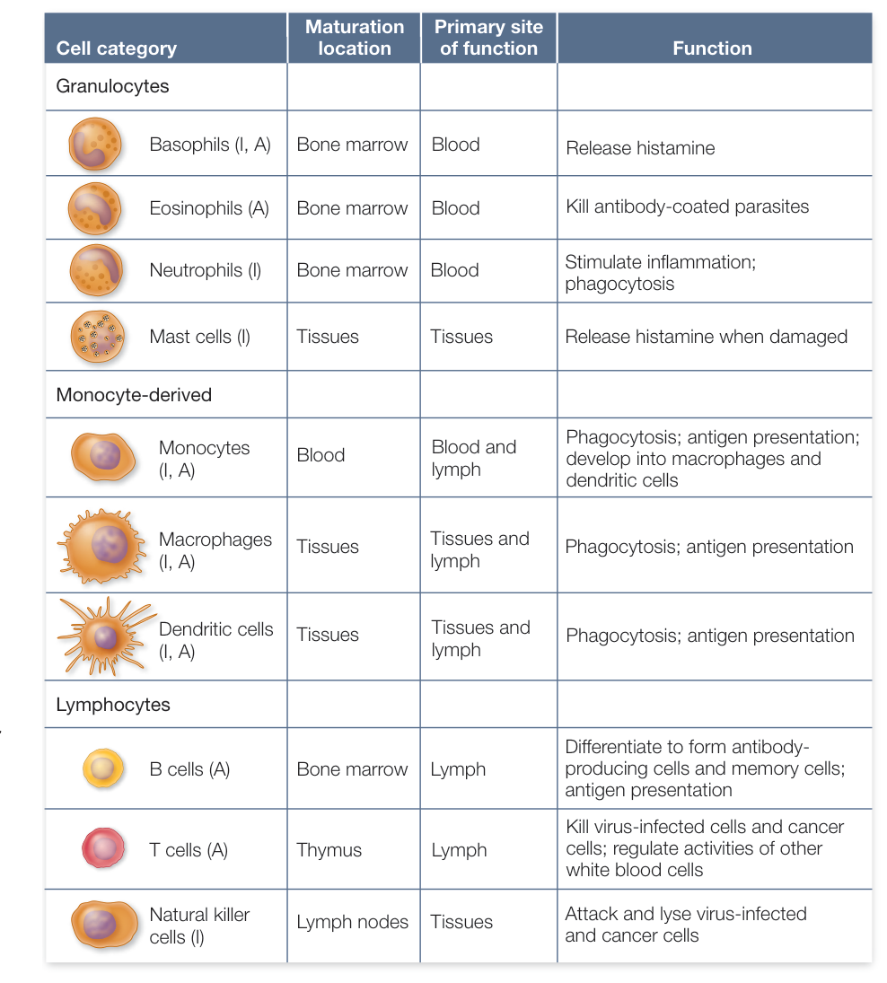
Figure 40.4 White Blood Cells White blood cells have key roles in both innate (I) and adaptive (A) immunity.
Immune system proteins bind pathogens or signal other cells
The cells that defend mammalian bodies work together, interacting with one another and with the cells of invading pathogens. These cell–cell interactions are accomplished by a variety of key proteins, including receptors, other cell surface proteins, and signal molecules. Four of the major players are listed here and are discussed in more detail later in the chapter.
- Antibodies are proteins that bind specifically to certain substances identified by the immune system as being foreign. The specific molecules that bind to antibodies are called antigens. Antibodies are produced by B cells.
- Major histocompatibility complex (MHC) proteins are used to display antigens on the surfaces of self cells, so that the antigens can be detected by the T cells of the immune system. MHC proteins also function as important self-identifying labels.
- T cell receptors are integral membrane proteins on the surfaces of T cells. They recognize and bind to antigens presented by the MHC proteins on the surfaces of other cells.
- Cytokines are soluble signal proteins released by many cell types. They bind to cell surface receptors and alter the behavior of their target cells.
Recap and Assess
All animals have innate defenses against pathogens, and vertebrates have innate and adaptive defenses. Both kinds of mechanisms are based on the ability to differentiate self from nonself. Innate defenses target a broad range of foreign molecules and organisms, whereas adaptive defenses target specific molecules and pathogens. Both innate and adaptive defenses involve specialized white blood cells.
- Describe three characteristics of innate immunity and adaptive immunity that distinguish them from each other.
- Some people have a rare genetic disease that results in defective signaling in the TLR pathway. What would be the phenotype of such individuals?
- If you compared the genomes of an insect and a human with regard to the presence of genes involved in immunity, what might you find?
The outcome of an infectious disease—the life or death of the host—often depends on the success of both rapid, innate responses and long-lasting, adaptive responses to invading pathogens. We turn now to the innate defenses that protect vertebrates from disease.
Innate Defenses Are Nonspecific
Learning Objectives
- 40.2.1 Summarize the various mechanisms for preventing entry into the body by a pathogen.
- 40.2.2 Explain how the innate defense system defends against a pathogen once it has entered the body.
- 40.2.3 List the steps in the inflammatory response.
Innate defenses are general protection mechanisms that attempt to stop pathogens from entering the body or to quickly eliminate those that do manage to enter. They are “innate,” meaning no previous experience with the pathogen is required, and are thus “ready to go,” in contrast to adaptive responses, which take time to develop after a pathogen or toxin has been encountered and recognized as being foreign.
Innate immunity is sometimes described as nonspecific immunity because it is applied indiscriminately to any potentially harmful invader. There are two lines of innate defense (Figure 40.5). The first line of innate defense is encountered by a potential pathogen as soon as it lands on the surface of an animal.
Preventing invasion is the first line of defense
A pathogen that cannot enter the body can do little harm. Regions where the body is in contact with the environment are areas where innate defenses are deployed to prevent invasion. Consider the formidable challenges that a pathogenic bacterium faces to invade the body:
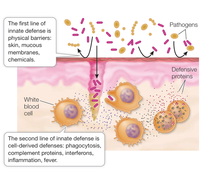
Figure 40.5 Innate Immunity Several innate factors act to prevent invasion. If pathogens do enter the body, other innate factors quickly provide nonspecific defenses against them.
Media Clip 40.1 The Chase Is On: Phagocyte versus Bacteria Life12e.com/mc40.1
- The physical barrier: A bacterium has to penetrate a tissue in order to invade the body. Cells in tissues that are at body surfaces in contact with the environment, such as gut cells, are held together by tight junctions, making entry difficult. Skin is about 30 cell layers thick, making it especially difficult to penetrate; it also sheds constantly and is salty and dry—conditions that inhibit microbial growth. For a bacterium to penetrate the skin, a break is usually required, which is why cuts and scrapes increase the risk of infection.
- The presence of mucus: Mucus is a slippery secretion produced by mucous membranes found on the surfaces of many internal epithelial tissues in contact with the external environment, such as in the digestive, respiratory, and urogenital systems (see Key Concept 38.1). Mucus traps microorganisms so they can be removed by the beating of cilia (see Figure 5.19), which continuously move the mucus and its trapped debris away.
- The presence of lysozyme: Lysozyme is an enzyme made by cells of the mucous membranes that is secreted into mucus and attacks the cell walls of many bacteria, causing them to lyse (burst open).
- The presence of defensins: Defensins, also made by mucous membranes, are peptides of 18–45 amino acids that contain hydrophobic domains. They are toxic to a wide range of pathogens, including bacteria, microbial eukaryotes, and enveloped viruses. Defensins insert themselves into the cell membranes of these organisms and make the membranes permeable, thus killing the invaders. Defensins are also produced in phagocytes, where they kill pathogens ingested by phagocytosis. Plants also produce defensins in response to pathogen exposure.
- The presence of extreme conditions: Harsh conditions in the gut can also kill pathogens. For example, gastric juice in the stomach is a deadly environment for many bacteria because of the hydrochloric acid and proteases that are secreted into it.
- The presence of normal flora: Bacteria and fungi that normally live and reproduce in great numbers on our body surfaces without causing disease will compete with pathogens for space and nutrients.
Connect the Concepts Key Concept 37.2 describes constitutive and induced responses to pathogens in plants. Among the latter are the pathogenesis-related (PR) proteins, which are similar to defensins produced by animals. These small peptides bind to fungal membranes and are toxic to a wide range of fungal targets, but they are not toxic to plant or animal cells. Other PR proteins may serve as alarm signals to plant cells that have not yet been attacked.
Recognition and response involve a variety of cells and proteins
The second line of innate defense involves recognition of a pathogen or damage. Several types of white blood cells, including neutrophils, monocyte-derived cells, and mast cells (see Figure 40.4), express receptors that give them the ability to recognize molecules associated with pathogens or with cell damage (as might be produced with trauma). Natural killer cells also have the ability to recognize self cells that are expressing abnormal proteins on their cell surface, as might occur when the cells are infected by a bacterium or exhibiting a cancer phenotype. The response to recognition varies depending on the cell type but often involves a variety of proteins, including complement proteins and interferons.
PHAGOCYTES Some phagocytes, such as neutrophils and monocytes, travel freely in the circulatory and lymphatic systems; others, such as macrophages and dendritic cells, move out of blood vessels and adhere to certain tissues. When these cells recognize a pathogenic cell, virus, pathogen molecule, or molecule produced by cell damage, they ingest it by phagocytosis (Figure 40.6A). Inside the phagocyte the phagosome (vesicle with ingested particle) fuses with a lysosome to form a phagolysosome (or secondary lysosome), which results in the digestion of the ingested pathogen (Figure 40.6B; see also Figure 5.12). In addition, phagocytes may release cytokines to recruit other phagocytes. Finally, several phagocytes, especially monocyte-derived dendritic cells, are antigen-presenting cells that travel to the lymph nodes to initiate an adaptive immune response. Dendritic cells act as messengers between the innate and adaptive immune systems. The dendritic cell “presents” an antigenic fragment from an ingested pathogen on its surface, along with class II MHC proteins, to T cells (see Key Concept 40.5). The dendritic cell also secretes signals that activate other cells of the adaptive immune system.
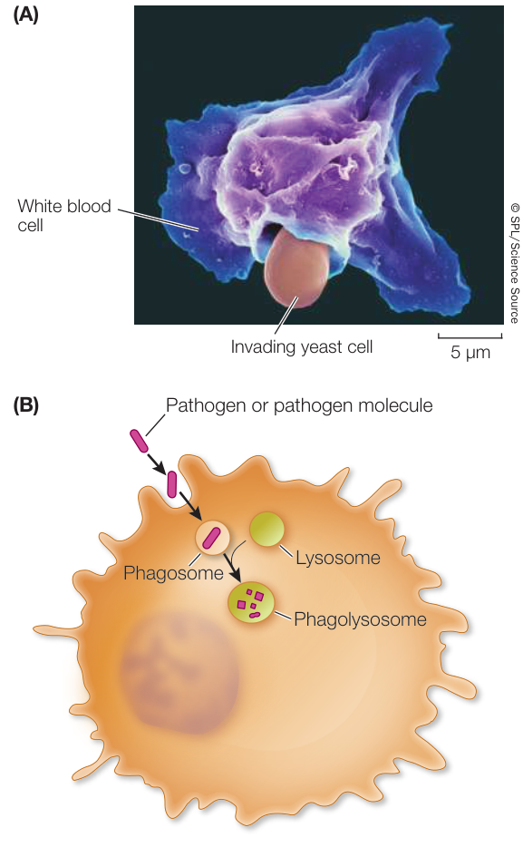
Figure 40.6 Phagocytes Endocytose Pathogens and Related Molecules Several types of white blood cells are phagocytic and will endocytose cells, viruses, and various molecules as part of an immune response. (A) A white blood cell in the process of ingesting an invading yeast cell. (B) Once ingested in a vesicle (phagosome), fusion with a lysosome causes degradation of the invading pathogen.
NATURAL KILLER CELLS One type of lymphocyte, the natural killer cells, can distinguish virus-infected cells and some tumor cells from their normal counterparts and initiate apoptosis (programmed cell death) of these target cells. In addition to exhibiting this innate defense action, natural killer cells interact with the adaptive immune system by lysing antibody-labeled target cells.
MAST CELLS Mast cells are found predominantly in peripheral tissues in contact with the environment. They are stimulated by pathogens or injury. One of their functions is to initiate the inflammatory response by releasing cytokines and other chemical signals (see the next section). Mast cells also interact with the adaptive immune system and are able to respond to particular antigens after they bind a specific class of antibodies (IgE; see Key Concept 40.4) produced during an adaptive response.
COMPLEMENT PROTEINS Vertebrate blood contains more than 20 different proteins that make up the antimicrobial
complement system. This system can be activated by various mechanisms, including both innate and adaptive defense responses. The proteins act in a characteristic sequence, or cascade, with each protein activating the next:
- First, the proteins attach to specific components on the surface of a microbe or to an antibody that has already bound to the microbe’s surface. In either case, binding helps phagocytes recognize and destroy the microbe.
- Then, complement proteins activate the inflammatory response (see Figure 40.7) and attract phagocytes to the site of infection.
- Finally, complement proteins lyse invading cells (such as bacteria).
INTERFERONS When a cell is infected by a pathogen, it produces small amounts of signal proteins called interferons that increase the resistance of neighboring cells to infection. Interferons are a class of cytokines and have been found in many vertebrates. Various molecules, including double-stranded (virus) RNA, induce the production of interferons. Thus interferons are particularly important as a defense against viruses. Interferons bind to receptors on the cell membranes of uninfected cells, stimulating a signaling pathway that inhibits virus reproduction if the cells are
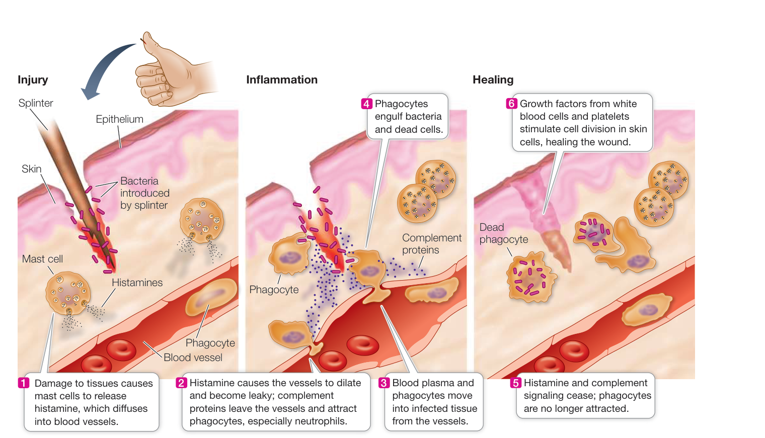
Figure 40.7 Interactions of Cells and Chemical Signals Result in Inflammation Histamine and other signals are released from mast cells to initiate the inflammatory response. The chemical signals associated with inflammation attract phagocytes, which digest the pathogens and damaged cells.
Q: Antihistamines are a class of drugs widely used to treat people with inflammation. How do you think they work?
Activity 40.2 Inflammatory Response subsequently infected. In addition, interferons stimulate the cells to hydrolyze bacterial or virus proteins to peptides, an initial step in adaptive immunity (see Key Concept 40.3).
Inflammation is a coordinated innate response to infection or injury
When mammalian tissue is damaged because of infection or injury, the body responds with inflammation. This response can happen almost anywhere in the body, internally as well as on the surface. Inflammation is an important phenomenon: it isolates the damaged area to stop the spread of the damage; it recruits cells and molecules to the damaged location to kill potential invaders; and it promotes healing. The first responders to tissue damage are mast cells, which adhere to the skin and the linings of organs and release numerous chemical signals, including:
- Tumor necrosis factor, a cytokine protein that kills target cells and activates immune cells.
- Prostaglandins, fatty acid derivatives involved in various responses, including the widening of blood vessels. Prostaglandins interact with nerve endings and are partly responsible for the pain caused by inflammation.
- Histamine, an amino acid derivative that leads to the important process of dilation and leakiness of blood vessels. When released in excess, as in some allergic reactions, histamines cause itchy, watery eyes and rashes.
The redness and heat of inflammation result from the dilation and leakiness of blood vessels in the infected or injured area (Figure 40.7). Phagocytes, especially neutrophils, which are abundant in the blood, enter the inflamed area, where they engulf the invaders and dead tissue cells. Phagocytes are responsible for most of the healing associated with inflammation. They produce several cytokines, which (among other functions) can signal the brain to produce a fever. This rise in body temperature accelerates B and T cell production and phagocytosis, thereby speeding the immune response. In some cases, pathogens are temperature-sensitive and their growth is inhibited. The pain of inflammation results from increased pressure due to swelling, the action of leaked enzymes on nerve endings, and the action of prostaglandins, which increase the sensitivity of the nerve endings to pain.
Following inflammation, pus may accumulate. Pus is a mixture of leaked fluid and dead cells: bacteria, neutrophils (the most abundant white blood cells), and damaged body cells. Pus is a normal result of inflammation and is gradually consumed and further digested by macrophages.
Recap and Assess
Innate immunity is the first line of defense against pathogens. Innate immunity includes barriers to prevent invasion, such as skin, mucus, enzymes, and chemicals, and after invasion, cellular responses involving the recognition of various molecules. Recognition by white blood cells of molecules produced by pathogens or cell trauma leads to coordinated responses such as the production of defensive proteins and inflammation.
A pathogenic bacterium lands on a cell in one of your nasal sinuses. Outline the innate defenses that come into play.
- Your immune system does not mount an innate defense response to a speck of dust that lands in your eye, but it may mount an innate response to a bacterium. Why?
- When a splinter of wood permeates the skin, the skin swells up. Outline the events by which this occurs.
Often the innate immune system, with its nonspecific defenses, is adequate to prevent or fight off a pathogenic infection. But in many cases this system works together with adaptive immunity, which detects and responds to specific pathogens. We now turn to the development and functioning of adaptive immunity.
Adaptive Defenses Are Specific
Learning Objectives
- 40.3.1 Distinguish between the recognition, activation, and effector phases in adaptive immunity.
- 40.3.2 Explain the function of antigen-presenting, T-helper, cytotoxic T, and B cells in an adaptive immune response.
- 40.3.3 Summarize how vaccines provide long-lasting immunity to pathogens.
More than a century ago, experiments showed that guinea pigs that had recovered from a bacterial infection that causes diphtheria produced a substance in their blood that could protect other guinea pigs from infection with lethal doses of the diphtheria bacterium. This observation led to the concept of adaptive immunity and to the understanding that factors promoting immunity are found in blood.
In this key concept we outline the main features of the adaptive immune system. We consider the two major types of adaptive responses: the humoral immune response, which produces antibodies against pathogen antigens that are not associated with particular cells; and the cellular immune response, which produces a response against antigens present on cells that are infected. These two responses work in concert.
What are the key features of adaptive immunity?
Four important features of the adaptive immune system are:
- Specificity
- The ability to distinguish self from nonself
- The ability to respond to an enormous diversity of foreign molecules
- Immunological memory
SPECIFICITY The B and T cell lymphocytes are crucial components of adaptive immunity. T cell receptors and the antibodies produced by B cells recognize and bind to specific foreign substances (antigens), and this interaction initiates an adaptive immune response. The specific sites on antigens that the immune system recognizes are called antigenic determinants, or epitopes (Figure 40.8). An antigenic determinant is a specific portion of a large molecule, such as a certain sequence of amino acids that may be present in a protein. Antigens are usually proteins or polysaccharides, and there can be multiple antigens on a single invading bacterium. A single antigenic molecule can have multiple, different antigenic determinants. The host animal responds to the presence of an antigen with highly specific defenses involving T cell receptors and antibodies. These receptors and soluble proteins bind to the antigenic determinants. Each T cell and each antibody is specific for a single antigenic determinant.
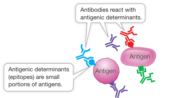
Figure 40.8 Antibodies Bind Particular Parts of Antigens Antibodies bind to a small region of an antigen called an antigenic determinant, or epitope. A single antigen has numerous potential epitopes and can thus be recognized by more than one antibody.
DISTINGUISHING SELF FROM NONSELF You have seen how the innate immune system identifies foreign molecules and thus distinguishes between self and nonself. The adaptive immune system has a different set of mechanisms for distinguishing self from nonself. The human body contains tens of thousands of different molecules, each with a specific three-dimensional structure capable of generating immune responses. Thus every cell in the body has a tremendous number of antigens. A crucial requirement of an individual’s adaptive immune system is that it recognizes the body’s own antigens and does not attack them.
DIVERSITY Challenges to the immune system are numerous. Pathogens take many forms: viruses, bacteria, protists, fungi, and multicellular parasites. Furthermore, each pathogenic species usually exists as many subtly different genetic strains, and each strain possesses multiple surface features. Estimates vary, but a reasonable guess is that a single person can respond specifically to 10 million different antigens. Upon recognizing an antigen, the adaptive immune system responds by activating B and T cell lymphocytes of the appropriate specificity.
IMMUNOLOGICAL MEMORY After the adaptive immune system responds to a particular type of pathogen once, the adaptive immune system “remembers” that pathogen and can usually respond more rapidly and powerfully to the same threat in the future. This immunological memory usually saves us from repeats of childhood diseases such as chicken pox and is the basis for protection using vaccination.
All four of these features of adaptive immune defense characterize both the humoral immune response and the cellular immune response.
Monocyte-derived cells play a key role in activating the adaptive immune system
As you have seen, one way to eliminate pathogens with the innate immune system is phagocytosis. After ingesting a pathogenic organism or infected host cell, monocyte-derived cells display fragments of the pathogen on their cell surfaces. These fragments function as antigens, and antigen presentation is one way that components of the innate immune system communicate with the adaptive immune system. Monocyte-derived cells play a key role in activating the adaptive immune system. After engulfing pathogens or infected host cells, these cells migrate to lymph nodes, where they present antigen to immature (previously unexposed) T cells. In addition, the antigen-presenting cells secrete cytokines and other signals that stimulate the activation and differentiation of the T cells.
Two types of adaptive immune responses interact
The adaptive immune system mounts two types of responses against invaders: the humoral immune response and the cellular immune response. These two responses work simultaneously and cooperatively, sharing many mechanisms. In particular, a robust B cell response cannot happen without T cells. We will use the example of a viral infection in an overview of these two types of responses (Figure 40.9). These responses also occur when bacteria or other pathogens invade the body and grow inside cells.
B cells that make antibodies are the workhorses of the humoral immune response, and cytotoxic T (TC) cells that bind and cause the death of antigen-expressing cells are the workhorses of the cellular immune response. There are three phases in both types of adaptive immune responses:
- Recognition phase: In both cellular and humoral immunity, recognition occurs when an antigen is inserted into the cell membrane of an antigen-presenting cell, with the unique antigen structure protruding from the cell membrane. The antigen-presenting white blood cells denoted in Figure 40.4 are particularly good at presenting antigens to T-helper (TH) cells. However, many different cell types, when infected, can present antigens to TC cells. A virus-infected host cell may present antigen to a TC cell or it may be phagocytosed by an antigen-presenting white blood cell. Free virus particles or molecules may also be phagocytosed. Antigen-presenting cells partially degrade pathogen molecules, leaving smaller antigens, which are often oligopeptides of about ten amino acids that are directed to the external surface of the cell membrane. Several different antigens from a virus may be presented on the surface of an antigen-presenting cell. Each antigen is recognized by a TH cell or a TC cell bearing a T cell receptor protein that is able to recognize (bind) the antigen. In both humoral and cellular immunity, binding initiates the activation phase.
- Activation phase: When the TH cell recognizes an antigen on an antigen-presenting cell, it propagates and releases cytokines that stimulate B cells and cells bearing receptors that can bind the same antigen to divide. The result is a clone of B cells that function in humoral immunity and a clone of cells that function in cellular immunity. cells can also be directly stimulated upon binding to an antigen-presenting cell; no involvement of cells is needed in this case.
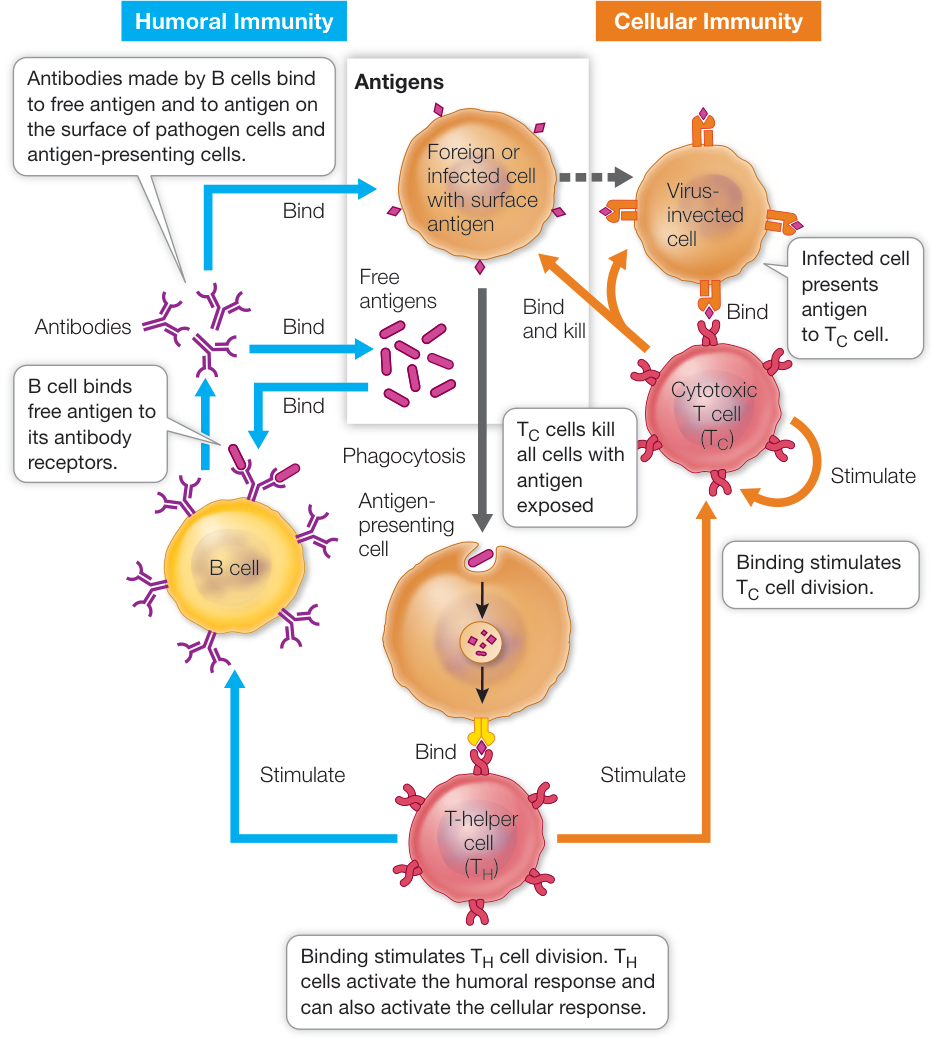
Figure 40.9 The Adaptive Immune System Humoral immunity involves the production of antibodies by B cells. Cellular immunity involves the activation of cytotoxic T cells that bind to cells expressing the antigen. Both antibodies and T cell receptors are specific for a particular antigen. For further details, see Figure 40.18. Q: In AIDS, a virus infects cells and kills them. What would be the effects on adaptive immunity?
3. Effector phase: In the humoral immune response, cells of the B clone produce antibodies that bind to virus particles or virus-infected cells. The bound antibodies attract phagocytes and complement proteins that engulf and destroy the virus and the virus-infected cells. In cellular immunity, cells of the clone bind to virus-infected cells and cause their death, often by stimulating apoptosis.
Adaptive immunity develops as a result of clonal selection
Before the humoral and cellular immune responses can take place, the body needs to generate a vast diversity of B and T cells that have the ability to bind different antigens. How does this tremendous diversity arise? As we will discuss in Key Concept 40.4, this diversity is generated primarily by DNA changes—chromosomal rearrangements and other mutations—that occur shortly after the B and T cells are formed in the bone marrow. Millions of different B cells develop, each of which can produce only one kind of antibody. Similarly, there are millions of different T cells, each with one specific kind of T cell receptor. Thus the adaptive immune system is “predeveloped”—all of the machinery available to respond to an immense diversity of antigens is already there, even before the antigens are ever encountered.
As we have described, when a pathogen enters the vertebrate body it stimulates the innate immune system. In addition to triggering its own defensive responses, the innate immune system triggers adaptive defensive responses via specific antigens that are presented on the surfaces of antigen-presenting cells, particularly dendritic cells. This triggers the proliferation of B and T cells that are specific for those particular antigens. How does this proliferation occur? The answer lies in the process of clonal selection: antigen binding “selects” a particular B or T cell for proliferation. When an antigen fits the surface receptor on a B or T cell and binds to it, that cell is activated. It divides to form a clone of cells (a genetically identical group derived from a single cell), all of which recognize and react to the same antigen. This process is illustrated for B cells in Figure 40.10. Binding and activation select a particular B or T cell, while proliferation generates the clone, hence the term “clonal selection.”
Clonal deletion helps the immune system distinguish self from nonself
Normally the body is tolerant of its own molecules. One way that the immune system does this is through the process of clonal deletion. This occurs during the early differentiation of T and B cells, when these cells encounter self antigens. There are mature cells expressing self antigens in the areas where T and B cells develop. The presence of self antigens allows developing B and T cells to be tested for binding to self antigens early in their development. Any immature B or T cell that expresses an antibody or T cell receptor that can bind to a self antigen undergoes apoptosis within a short time and is removed from the lymphocyte pool (see Figure 40.10).
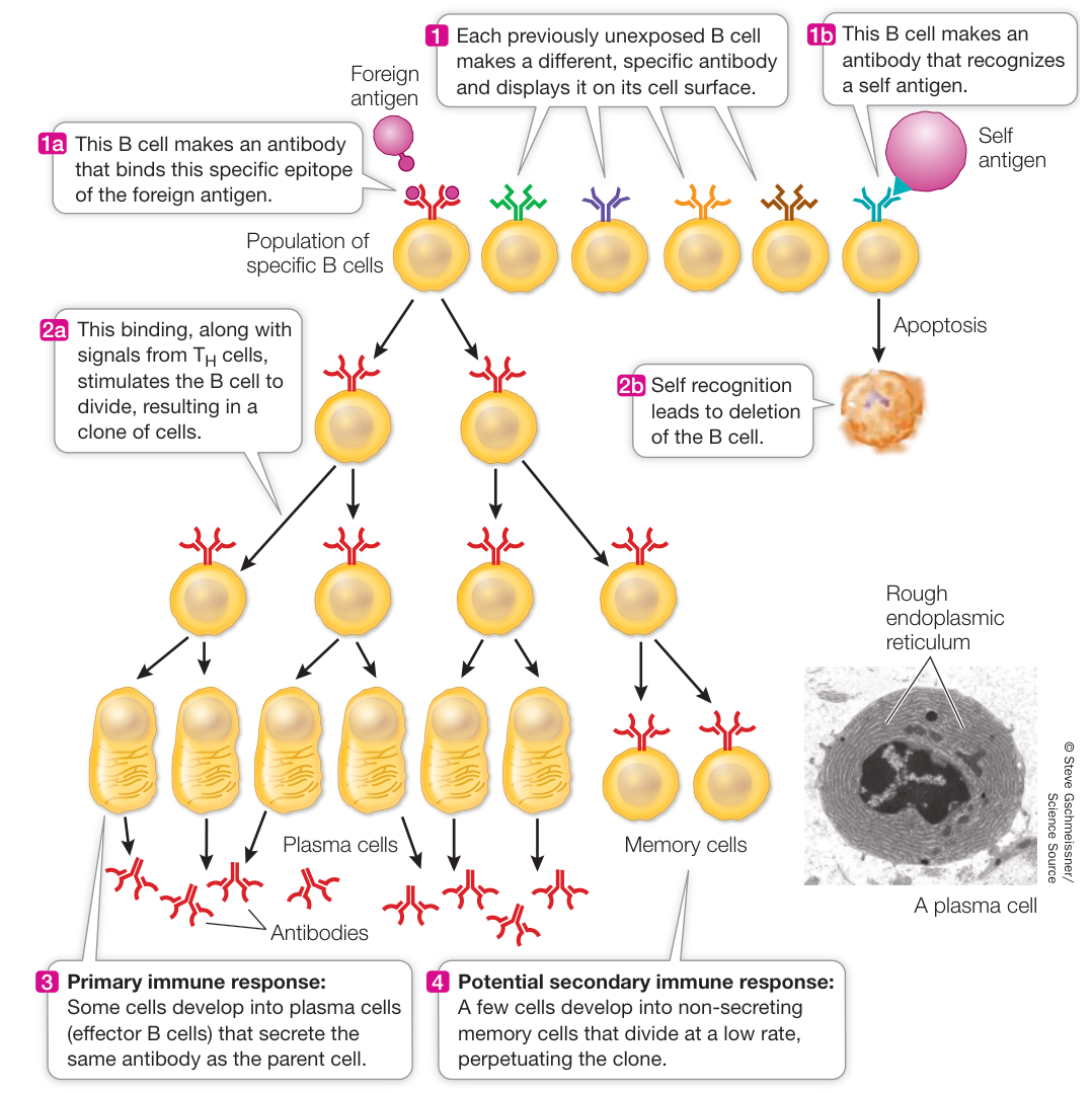
Figure 40.10 Clonal Selection and Deletion in B Cells Left side: The binding of a foreign antigen to a specific receptor on the surface of a B cell stimulates that cell to divide, producing a clone of genetically identical cells to fight that invader. Most cells of the clone are plasma cells containing extensive rough endoplasmic reticulum for synthesizing antibodies. Some cells become long-lived memory cells that can last for years or decades in the lymph system (especially in the spleen). Right side: The binding of a self antigen on a B cell leads to apoptosis and loss of the cell. Q: You may never have been exposed to Ebola virus. Do you have cells that make antibodies against this virus? Explain.
Immunological memory results in a secondary immune response
The first time a vertebrate animal is exposed to a particular antigen there is a time lag (usually several days) before the B cell-produced antibody molecules and T cells specific to that antigen slowly increase. But for years afterward—sometimes for life—the immune system “remembers” that particular antigen, allowing the body to mount a faster response the next time it encounters the antigen. How does this happen?
The answer lies in the fact that activated B and T cells divide and differentiate to produce two types of daughter cells: effector cells and memory cells.
- Effector cells carry out the attack on the antigen. Effector B cells, called plasma cells, secrete antibodies. Effector T cells release cytokines and other molecules that initiate reactions that destroy nonself or altered cells. Effector cells live only a few days.
- Memory cells (see Figure 40.10) are long-lived cells that retain the ability to start dividing on short notice to produce more effector and more memory cells. Memory B and T cells may survive in the body for decades, rarely dividing.
Effector cells and memory cells can respond to an antigen in two different ways:
- When the body first encounters a particular antigen, a primary immune response is activated, in which the previously unexposed B and T cells that recognize that antigen proliferate to produce clones of effector and memory cells.
- After a primary immune response to a particular antigen, subsequent encounters with the same antigen will trigger a much more rapid and powerful secondary immune response. The memory cells that bind with that antigen proliferate, launching a huge army of plasma cells and effector T cells. The secondary immune response is more rapid because memory cells are present in higher concentration than are naive B and T cells that have never been activated.
Vaccines are an application of immunological memory
Thanks to immunological memory, exposure to many diseases (including childhood diseases such as chicken pox) provides a natural immunity to those diseases. Furthermore, it is possible to provide artificial immunity against many life-threatening diseases by vaccination: the introduction of antigen into the body in a form that does not cause disease.
Vaccination initiates a primary immune response, generating memory cells without making the person ill. Later, if a pathogen carrying the same antigen attacks, specific memory cells already exist. They recognize the antigen and quickly overwhelm the invaders with a massive production of B and T cells and antibodies (Figure 40.11). The success of vaccines at reducing the frequency of many infectious diseases cannot be overstated; several pathogens that used to cause common childhood or adult illnesses—many of them deadly—have become minimal public health issues thanks to vaccination.
Because of mutation, an antigenic determinant on a pathogen can change over time. If the change is significant in terms of three-dimensional structure, a primary immune response directed against an antigen may not result in a secondary immune response with an altered antigen. For pathogens that have rapidly evolving antigens, vaccination will be less effective. You are familiar with this with regard to influenza. More often than not, the annual flu season features a genetic strain of the flu virus that is not sufficiently related to the previous season’s vaccine; so a new vaccine, which selects new B cell clones, must be made. A remarkable recent finding is that memory cells can last a long time and help in a response to new, related antigens. People who were exposed to a particular strain of flu retain antibody-making memory cells against that strain for decades and develop a rapid immune response to a new, genetically different strain that appears years later. In the case of flu, the memory cells are making antibodies that bind to not just the first strain of flu they responded to, but to later ones as well. Characterizing these antibodies may provide a clue to developing a vaccine that is broadly effective and obviates the need to develop new ones (Investigating Life: What Are the Mechanisms and Implications of Long-Lasting Immunity?).
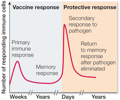
Figure 40.11 The Timing and Magnitude of an Adaptive Immune Response The primary immune response, initiated by pathogen invasion or by vaccination, leads to a moderate response that may take days to weeks. Reinfection by a pathogen expressing the same antigens results in a much more robust and rapid response that prevents disease.
Investigating LIFE What Are the Mechanisms and Implications of Long-Lasting Immunity?
Experiment
Original Paper: M. Miller et al. 2013. Neutralizing antibodies against previously encountered influenza virus strains increase over time: A longitudinal analysis. Sci Transl Med 5: 1–9.
The Framingham Heart Study was begun in 1948 with the objective of finding out the causes of heart disease and stroke. About 5,000 people ages 30–62 and living in the town Framingham, Massachusetts, have been subjected every 2 years to extensive medical exams in an attempt to relate lifestyle factors to heart disease. Subsequent generations have been added to the study ever since. Not only has this study led to major findings about the causes of heart disease, but it has provided a wealth of data on other medical conditions as well. Peter Palese of the Icahn School of Medicine at Mount Sinai Hospital in New York City measured antibodies in blood to flu virus strains from 40 people in the Framingham study, born between 1917 and 1952. They had been exposed to major flu outbreaks in 1957, 1968, and 1977, as well as to the typical flu that comes up every year. Blood samples were examined between 1987 and 2008 to see if these people still had antibodies against the three major flu outbreak strains, and whether these antibody amounts increased over time as people fought off the new, less virulent strains that arise every year.
HYPOTHESIS Exposure to new strains of flu stimulates antibody production against strains to which an individual was exposed previously.
METHOD
1 Obtain blood samples from people born between 1917 and 1952 who had been exposed to Asian flu (1957), Hong Kong flu (1968), and Russian flu (1977).
2 Measure antibodies against the three strains in blood samples taken between 1987 and 2008.
RESULTS

CONCLUSION People exposed to flu virus develop long-lasting memory cells that make antibodies that have broad specificity to other flu strains.
Investigating LIFE Work with the Data follows on the next page.
Work with the Data
QUESTIONS
- Blood samples were taken from 1987 to 2008, and the people’s reactivity against various flu strains was tested. Figure A shows the results for antibodies against the Russian flu strain of 1977 in numerous individuals. The black points, connected by black lines, are the means for each sample period. Note that these people were exposed to many flu strains after 1977. The data shown are typical for the way specific immune reactions are measured. The titer is presented as a reciprocal. For example, a titer of 2 means that undiluted serum sample gave an antibody reaction (titer 1), and so did a dilution of 1 part serum and 1 part water (dilution of 1/2, reciprocal = 2). If the sample was diluted to a titer of 4 (1 part serum, 3 parts water), there was no reaction because the concentration of antibody was too low. The higher the titer number, the more the blood serum could be diluted to still react with the antigen; that is, a higher titer number means there were more antibodies in the original serum sample. What can you conclude about the titer of anti-Russian flu strain over the years 1987–2008?
- The flu virus mutates rapidly. In contrast, another virus to which people are exposed, cytomegalovirus (CMV), does not mutate rapidly. Antibody titers against CMV were measured, and the data are shown in Figure B. The blue points, connected by blue lines, are the means for each sample period. What can you conclude from these data? Compare the antibody response to CMV over time with that against the Russian flu virus strain and comment.
- A protein on the surface of the flu virus called hemagglutinin (HA) is exposed to the immune system because of its prominent location (Figure C). When the flu virus genes mutate, the “head” of the protein tends to accumulate more changes than the stalk. Thus the amino acid sequences of stalk regions of various flu strains are similar. The investigators measured antibodies to the stalk region that were either broadly reactive to various flu strains, or not broadly reactive. The results are shown in the table. What can you conclude about the broadly reactive antibodies? How does this provide a guide for the development of a broadly based flu vaccine?
| Types of antibodies to stalk region | Titer |
|---|---|
| Broadly reactive | 4,080 |
| Not broadly reactive | 1,024 |

Figure A
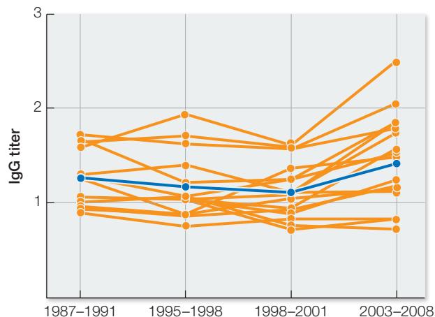
Figure B
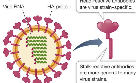
Figure C
Recap and Assess
The adaptive immune system reacts against foreign antigens—parts of molecules that can bind antibodies or T cell receptors. An amazing diversity of both antibodies (produced by B cells) and of T cell receptors are produced, resulting in a huge variety of antigens that can be bound. In the primary immune response, B cells and T cells that recognize a particular antigen proliferate by clonal selection. Immunological memory prepares the body for a much stronger secondary immune response.
- A fellow passenger sneezes on the train and the common cold virus enters your nose and infects cells lining your nasal passage. Outline how your body responds, noting the type and stages of the adaptive immune response.
- In 2009 the H1N1 strain of influenza spread around the world. People who had been alive at the time of the 1918 epidemic had fewer symptoms. Explain this in terms of immunological memory.
- How do vaccines make use of immunological memory?
The general features of the adaptive system are that it is premade to respond specifically to virtually any antigen, and it involves an initial primary response to the antigen and a more powerful secondary response when the same antigen is encountered in the future. As we noted, the adaptive system has a humoral response with B cells making antibodies and a cellular response with T cells expressing specific receptors. We now turn to these two types of adaptive responses, beginning with the humoral system.
The Humoral Adaptive Response Involves Antibodies
Learning Objectives
- 40.4.1 Compare the antigen-binding site of an antibody with other proteins such as enzymes that bind other molecules.
- 40.4.2 Describe the processes that give rise to antibody diversity.
- 40.4.3 Compare the diversity of specific antibodies in an individual with the diversity of other biological molecules.
Every day in the human body, billions of B cells survive the test of clonal deletion and are released from the bone marrow into the circulatory system and the lymphatic system. B cells are the basis for the humoral immune response.
A B cell begins as an unexposed B cell with a receptor protein on its cell surface that is specific for a particular antigen. The cell is activated by antigen binding to this receptor, and after stimulation by a cell, it gives rise to a clone of plasma cells that make antibodies as well as to a smaller number of memory cells (see Figure 40.10). The stimulation occurs after the B cell presents antigen to a cell expressing a receptor that can recognize the antigen. The cell then secretes cytokines that stimulate the B cell to divide.
Activated plasma cells (effector B cells) can synthesize and secrete large amounts of antibody proteins—up to 2,000 molecules per second! All the plasma cells arising from a given B cell produce antibodies that are specific for the antigen that originally bound to the parent B cell. Thus antibody specificity is maintained as B cells proliferate.

Animation 40.3 Humoral Immune Response
Antibody protein structure reflects function
Antibodies belong to a class of proteins called immunoglobulins. There are several types of immunoglobulins, but all contain a tetramer consisting of four polypeptide chains: two “heavy chains” and two “light chains” (Figure 40.12). In each immunoglobulin molecule, the two light chains are identical, and the two heavy chains are identical. Disulfide bonds hold the chains together, characteristically in a Y-like shape.
Each of the four polypeptide chains has a constant region and a variable region (see Figure 40.12A):
- The amino acid sequences of the constant regions are similar among the immunoglobulins. They determine the destination and function—the class—of each immunoglobulin.
- The amino acid sequences of the variable regions are different for each specific immunoglobulin. Their three-dimensional antigen-binding sites are determined by their amino acid sequences and are responsible for antibody specificity.
Antigen–antibody binding is noncovalent. The attractive forces involved in holding antigen and antibody together include hydrogen bonds, ionic bonds, hydrophobic interactions, and van der Waals forces (see Key Concept 2.2). These attractive forces are generated from particular amino acids at the binding site on both molecules. So for an antibody protein, the sequence of amino acids at the binding site is crucial to its specificity. Generating antibody diversity is thus a molecular genetics challenge, as we will soon discuss.
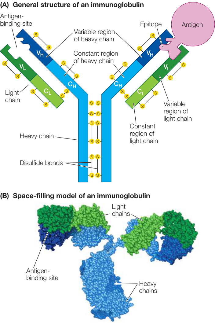
Figure 40.12 The Structure of an Immunoglobulin Four polypeptide chains (two light, two heavy) make up an immunoglobulin molecule. Both diagrammatic (A) and space-filling (B) representations of immunoglobulin are shown here. Activity 40.3 Immunoglobulin Structure

The two antigen-binding sites on each immunoglobulin molecule are identical, making the antibody bivalent (bi, “two,” + valent, “binding”), meaning the molecule can bind two antigen molecules at once. Bivalency, in addition to the presence of multiple epitopes on the surfaces of many antigens (including large proteins, viruses, and bacteria, as you saw in the case of the influenza virus explored in Investigating Life: What Are the Mechanisms and Implications of Long-Lasting Immunity?), permits antibodies to form large complexes with the antigens. These complexes are easy targets for ingestion and breakdown by phagocytes (Figure 40.13).
There are five classes of immunoglobulins
While the variable regions are responsible for the specificity of an immunoglobulin, the constant regions of the heavy chain determine the class of the immunoglobulin. Table 40.2 describes the five classes of immunoglobulins and their functions. The most abundant class in the circulatory system is IgG; these soluble antibody proteins make up about 80% of the total immunoglobulin content of the bloodstream. They are made in greatest quantity during a secondary immune response. IgG molecules defend the body in several ways. For example, after some IgG molecules bind to antigens, they become attached by their heavy chains to phagocytes. This attachment permits the phagocytes to destroy the antigens by phagocytosis (see Figure 40.13B).
Immunoglobulin diversity results from DNA rearrangements and other mutations
Each mature B cell makes antibodies targeted to only one single antigen. And there are millions of possible antigens to which an individual can be exposed. How can the genome encode enough different antibodies to protect the body against all the possible pathogens? Although a single individual makes millions of immunoglobulins that differ in their amino acid sequences, there are not millions of different immunoglobulin genes. It turns out that instead of a single gene encoding each complete immunoglobulin, the genome of the differentiating B cell has multiple different coding regions for each domain of the protein, and diversity is generated by putting together different combinations of these regions. Shuffling of this genetic deck generates the enormous immunological diversity that characterizes each individual mammal.
This shuffling may remind you of the alternative splicing that occurs during RNA processing (see Key Concept 16.5). However, the difference with immunoglobulins is that shuffling to generate antibody diversity occurs at the DNA level: every B cell clone produces a different antibody sequence because it has a different DNA sequence at its heavy and light antibody genes. Given that every cell in the body begins with essentially identical DNA sequences, how do these DNA sequence differences arise?
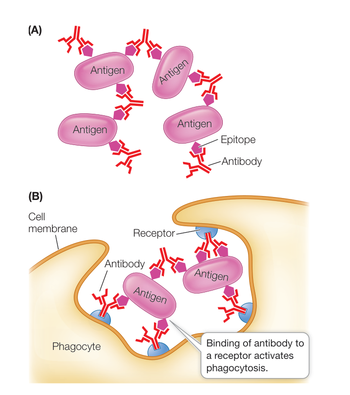
Figure 40.13 Antibodies Cause Aggregation of Antigenic Targets Because antibodies are bivalent, each one can bind to two antigen molecules at once, aggregating them (A) and making them easy targets for phagocytes (B).
Each gene encoding an immunoglobulin chain is in reality a “supergene” assembled by means of genetic recombination from several
Table 40.2 Immunoglobulin Classes
| Class | General structure | Location | Function |
|---|---|---|---|
| IgG | Monomer | Free in blood plasma; about 80% of circulating antibodies | Most abundant antibody in primary and secondary immune responses; crosses placenta and provides immunity to fetus |
| IgM | Pentamer | Surface of B cell; free in blood plasma | Antigen receptor on B cell membrane; first class of antibodies released by B cells during primary response |
| IgD | Monomer | Surface of B cell | Cell surface receptor of mature B cell; important in B cell activation |
| IgA | Dimer | Saliva, tears, breast milk, and other body secretions | Protects mucosal surfaces; prevents attachment of pathogens to epithelial cells |
| IgE | Monomer | Secreted by plasma cells in skin and tissues lining gastrointestinal and respiratory tracts | Binds to mast cells and basophils to sensitize them to subsequent binding of antigen, which triggers release of histamine that contributes to inflammation and some allergic responses |
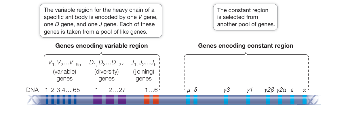
Figure 40.14 Antibody Supergene Human immunoglobulin heavy chains have four domains, each of which is coded for by one of several possible genes selected from a cluster of similar genes. Each immunoglobulin heavy-chain protein has one gene from each of the four clusters (V, D, J and constant).
clusters of smaller genes scattered along part of a chromosome (Figure 40.14). Every cell in the body has tens of immunoglobulin genes located in separate clusters that are potentially capable of participating in the synthesis of both the variable and constant regions of immunoglobulin chains. In most body cells and tissues, these genes remain intact and separated from one another. But during B cell development, these genes are cut out, rearranged, and joined together in DNA recombination events. One gene from each cluster is chosen randomly for joining, and the others are deleted. In the case of one multigene set, the J genes, some of the extra sequences are removed by RNA splicing (Figure 40.15).
In this manner, a unique immunoglobulin supergene is assembled from randomly selected “parts.” Each B cell precursor assembles two supergenes, one for a specific heavy chain and the other, assembled independently, for a specific light chain. Each B cell also rearranges, and expresses, genes from a single chromosome for each chain—the other chromosome is not rearranged unless the process fails for the first chromosome. This remarkable example of genetic differentiation generates an enormous diversity of immunoglobulins from the same genome. It is a major exception to the generalization that all somatic cells derived from the fertilized egg have essentially identical DNA sequence.
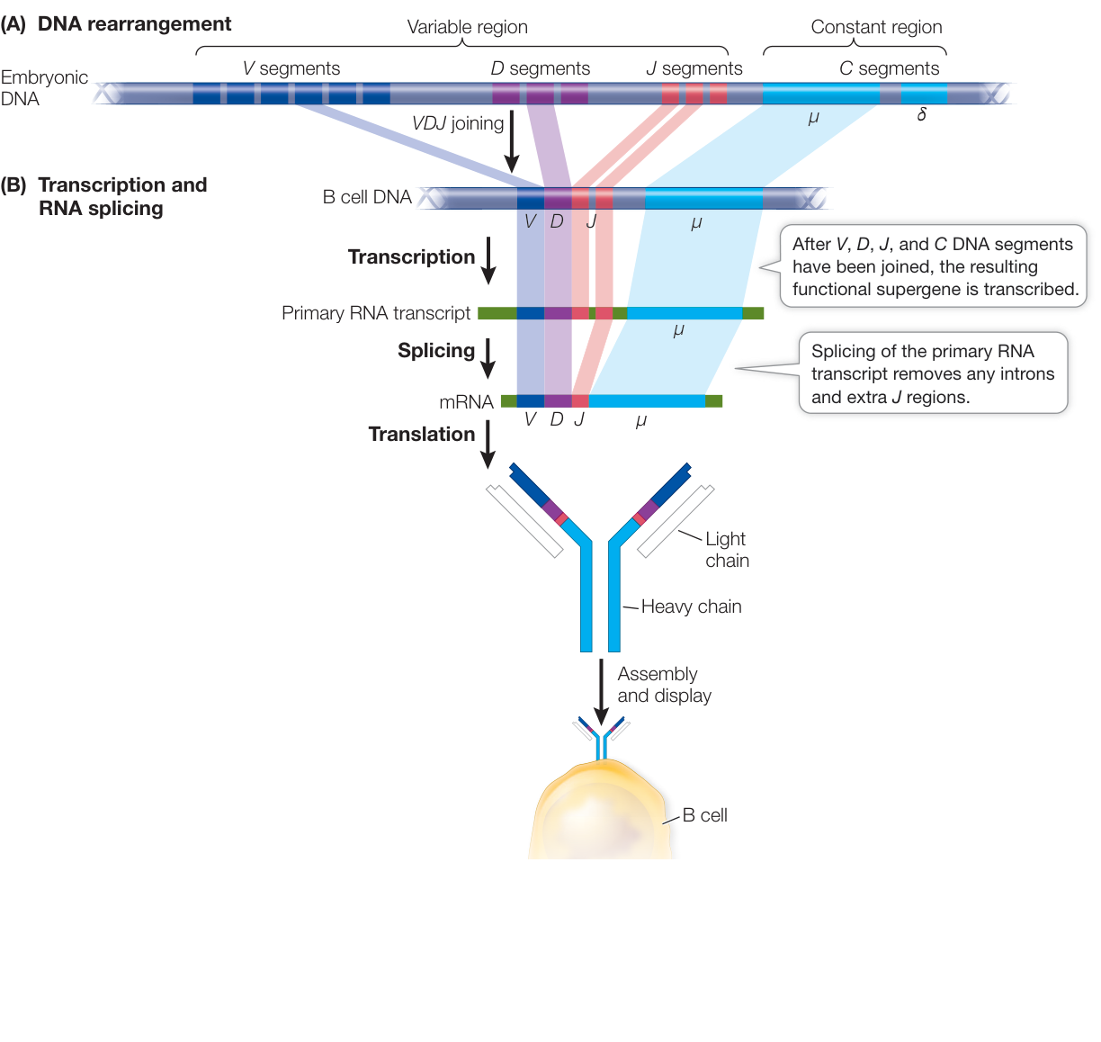
Figure 40.15 Heavy-Chain Gene Recombination and RNA Splicing Two types of rearrangements in the heavy-chain gene clusters are required for antibody formation. (A) Prior to transcription, DNA is rearranged to join one each of the V, D, and J genes into a variable region supergene. (B) After transcription, RNA splicing joins the VDJ region to the constant region and removes extra J regions, if present.
Figure 40.14 illustrates the gene families that encode the constant and variable regions of the heavy chain in humans. Multiple genes encode each of the three parts of the variable region: , , and genes, though these numbers can vary across chromosomes. In addition, the sequences of the , , and regions on one copy of a chromosome are likely to differ from those on the other copy because there is a lot of allelic diversity at these genes. Each B cell randomly selects one gene from each of these gene clusters in one of the two chromosome copies to make the final coding sequence () of the heavy-chain variable region. So the number of different heavy chains that can be made through this random recombination process is quite large:
Now consider that the light chains are similarly constructed, although they differ in that there are two supergenes (on different chromosomes), either of which can be used, and the part of the variable region is not present in the light chain. The two supergenes have and and and genes respectively, yielding copies of each supergene = 640 possible combinations. The number of possible combinations of light-and heavy-chain variable regions is thus:
Other mechanisms generate even more diversity:
- When the DNA sequences that encode the , , and regions are rearranged so that they are next to one another, the recombination event is not precise, and errors occur at the junctions. This imprecise recombination can create codon deletions, codon insertions, and frame-shift mutations, with resulting amino acid changes.
- After the DNA sequences are cut and before they are rejoined, the enzyme terminal transferase often adds some nucleotides to the free ends of the DNA pieces. These additional bases create insertion mutations.
- There is a very high spontaneous somatic mutation rate in immunoglobulin genes, 10,000-fold higher than for other genes. Once again, this process creates many new alleles and adds to antibody diversity.
Connect the Concepts When they occur in the coding regions of proteins, point mutations can cause silent, missense, nonsense, loss-of-stop, or frame-shift mutations (see Figure 15.3).
When we include these possibilities with the millions of combinations that can be made by random DNA rearrangements, it is not surprising that the immune system can mount a response to almost any natural or artificial substance.
Once the DNA rearrangements are completed, each supergene is transcribed and then translated to produce an immunoglobulin light chain or heavy chain. These chains combine to form an active immunoglobulin protein. Importantly, each B cell produces a single heavy and light chain so that all of its antibodies bind the same antigen.
DNA rearrangements in B cells occur in the bone marrow, where they are synthesized. In contrast, DNA changes in T cells occur in the thymus, which is where T cell precursors travel after synthesis in the bone marrow. During your lifetime, you continue to synthesize functional B cells in your bone marrow. However, because your thymus rapidly diminishes in size after puberty, you make substantially fewer T cells once you reach adulthood. The repertoire of T cells available at puberty represents the ones you will have at your disposal to mount an immune response for the rest of your life.

Animation 40.4 A B Cell Builds an Antibody
The constant region is involved in immunoglobulin class switching
Generally, a B cell makes only one class of immunoglobulin at a time. But class switching can occur, in which a B cell changes the immunoglobulin class it synthesizes.
Early in its life, a B cell makes IgM and IgD antibodies. The pre-mRNA that the B cell synthesizes for the heavy chain contains the constant regions for both of these antibodies (the and genes in Figure 40.14), and alternative splicing generates mature mRNAs with either the IgM or IgD constant region. If the B cell later becomes a plasma cell during a humoral immune response, a rearrangement occurs in the cell’s DNA, positioning the variable region genes (consisting of the same , , and genes) next to a constant region gene farther away on the original DNA molecule. Such a DNA deletion results in the production of a new immunoglobulin with a different constant region of the heavy chain, and therefore a different function (see Table 40.2). However, this immunoglobulin has the same variable regions—and therefore the same antigen specificity—as the IgM and IgD produced by the parent B cell. The new immunoglobulin protein falls into one of the other three classes (IgA, IgE, or IgG), depending on which of the constant region genes is placed adjacent to the variable region genes.
Recap and Assess
The humoral immune response is based on the synthesis by B cells of specific immunoglobulins. The specificity of an immunoglobulin derives from the amino acid sequence of its variable regions on the light and heavy chains. B cells can make millions of immunoglobulins with different specificities through DNA rearrangement of the , , and genes, imprecise recombination, addition of nucleotides, and high mutation rates. The particular class of immunoglobulin produced by a B cell is determined by its constant region.
- Describe the part of an antibody molecule that interacts with an antigen. How is it similar to the active site of an enzyme? How does it differ?
- Why don’t B cells use alternative RNA splicing to generate antibody diversity?
- Discuss the diversity of antibody specificities in an individual in relation to the diversity of enzymes. Does every cell in an animal contain genetic information for all of that organism’s enzymes? For all of the immunoglobulins? Explain your answer.
By making antibodies, B cells are the major players in the humoral immune response. We now turn to the cellular immune response, and the T cells responsible for it.
The Cellular Adaptive Response Involves T Cells and Receptors
Learning Objectives
- 40.5.1 Summarize the role of MHC proteins in the activation of T cells.
- 40.5.2 Explain how T-helper cells are involved in activating the humoral immune response.
- 40.5.3 Explain how cytotoxic T cells kill virus-infected cells.
- 40.5.4 Describe how Tregs suppress some cellular and humoral immune responses.
Two types of effector T cells (T-helper cells and cytotoxic T cells) are involved in the cellular immune response. They work along with proteins of the major histocompatibility complex (the MHC proteins), which present antigens on the surfaces of cells and contribute to the immune system’s tolerance for the body’s own cells.
Like B cells, T cells possess specific membrane receptors. The T cell receptor is not an immunoglobulin, but a glycoprotein with a molecular weight of about half that of an IgG. It is made up of two polypeptide chains, each encoded by a separate gene (Figure 40.16). The two chains have distinct regions with constant and variable amino acid sequences. The variable regions consist of 42 V and 61 J regions for one polypeptide and 48 V, 3 D, and 7 J regions for the other chain, yielding 2,562 possible combinations from one chromosome and 1,008 from the other. Thus the T cell receptor dimer can produce more than 5 million (2,562 x 1008 x 2 copies of each chromosome) different amino acid sequences from recombination alone. In addition, as with B cell antibodies, the recombination process is error-prone and terminal transferases add base pairs during recombination. Together, these processes massively increase the number of possible T cell receptors, but unlike B cell antibodies, T cell receptors do not exhibit somatic hypermutation to further increase diversity. As in the immunoglobulins, the variable regions provide the site for specific binding to antigens. But there is one major difference: whereas an antibody can bind to an antigen whether it is present on the surface of a cell or not, a T cell receptor binds only to an antigen displayed by an MHC protein on the surface of an antigen-presenting or target cell.
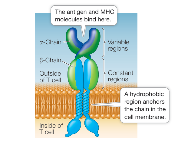
Figure 40.16 A T Cell Receptor The receptors on T cells are smaller than those on B cells. The two polypeptides on the T cell receptor contain both variable and constant regions. The constant regions fix the receptor in the cell membrane, while the variable regions establish the specificity for binding to antigen.
When a T cell is activated by contact with a specific antigen, it proliferates and forms a clone. Its descendants form clones of two types of effector T cells (see Figure 40.9):
- T-helper () cells assist both the cellular and the humoral immune responses.
- Cytotoxic T () cells recognize virus-infected or mutated cells and kill them by inducing lysis.
MHC proteins present antigen to T cells, causing recognition
Both and cells express T cell receptors that bind to antigen on the cell surface. But the response of each cell type to binding is quite different. binding results in activation of the adaptive immune response, whereas binding results in death of the cell carrying the antigen. MHC proteins form complexes with antigens on cell surfaces and assist with recognition by the T cells, so that the appropriate type of T cell binds.
The MHC proteins are cell membrane glycoproteins. Two types of MHC proteins function to present antigens to the two different types of T cells:
- Class I MHC proteins are present on the surface of every nucleated cell in the mammalian body. These proteins are used to present antigens to cells. Thus, any nucleated cell can potentially present antigen to a cell. The antigens can be cytosolic fragments of virus proteins in virus-infected cells or abnormal proteins made by cancer cells as a result of somatic mutations. A cell with the appropriate T cell receptor binds to the antigen–MHC complex. To ensure binding, the cell also has a cell surface receptor protein called CD8 that recognizes and binds to MHC I.
- Class II MHC proteins are on the surfaces of antigen-presenting monocyte-derived cells and B cells. They present antigens to cells. Unlike cells that present antigens using class I MHC proteins, antigen presentation by these cells is one of their primary functions. These cells ingest antigens and break them down in the phagolysosome (see Figure 40.6B); one of the resulting fragments then binds to MHC II for presentation (Figure 40.17). A cell with the appropriate T cell receptor binds to the antigen–MHC complex. To ensure binding, the cell also has a cell surface receptor protein called CD4 that recognizes and binds to MHC II.
T cells are synthesized in the bone marrow but mature in the thymus, where all of the gene rearrangements occur. During maturation, it is important that the T cells end up with either a CD8 or CD4 receptor, for recognizing MHC I and II respectively, and that the T cell receptor does not recognize self antigens. Most immature T cells that enter the thymus are lost during this process of “thymic selection” for cells expressing the appropriate receptors that are unable to recognize self antigens as foreign.
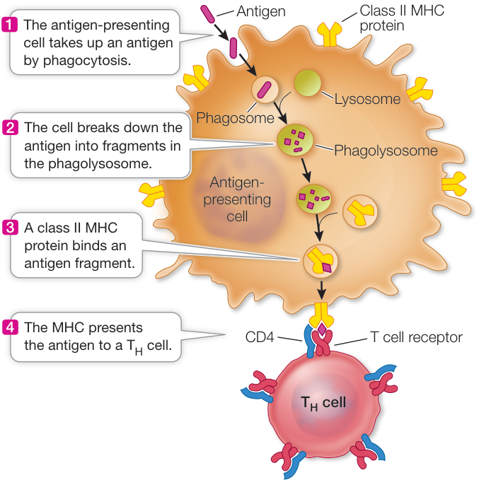
Figure 40.17 Monocyte-Derived Cells and B Cells Are Antigen-Presenting Cells A monocyte-derived cell or B cell endocytoses an antigen, breaks it into fragments, and then displays one of the fragments on an MHC II protein at the cell surface. T cell receptors, with their CD4 cell surface receptor proteins, on a specific cell can then bind to and interact further with the antigen–MHC II complex.
In humans there are three genetic loci for class I MHC proteins and three for class II MHC proteins. Each of these six loci has as many as 100 different alleles in the human population. With so many alleles, the number of possible genotypes is enormous; it is not surprising that different people are likely to have different MHC genotypes. This is why it can be difficult to find a good “match” for organ donations; a donor MHC genotype that does not match the recipient’s MHC genotype will be recognized as foreign and stimulate an immune response. To accomplish its role in antigen presentation, an MHC protein has an antigen-binding site that can hold a peptide of about 10–20 amino acids.
Information on MHC proteins, the cellular origins of antigens, and T cells is summarized in Table 40.3.
T-helper cells and MHC II proteins contribute to the humoral immune response
When a cell that has survived the selection processes in the thymus binds to an antigen-presenting cell, it releases cytokines that cause the cell to proliferate (an autocrine signal; see Key Concept 7.1), producing a clone of cells with the same specificity. The steps to this point constitute the activation phase of the humoral immune response, and they occur in the lymphoid tissues. Next comes the effector phase, in which the cells activate previously unexposed B cells with the same specificity to produce antibodies.
As noted earlier, B cells are also antigen-presenting cells. B cells take up antigens bound to their surface immunoglobulin receptors by endocytosis, break them down, and display antigenic fragments on class II MHC proteins. When a cell binds to the displayed antigen–MHC II complex, it releases cytokines that cause the B cell to produce a clone of plasma cells and memory cells (Figure 40.18A). Finally, the plasma cells secrete antibodies, completing the effector phase of the humoral immune response. A humoral immune response thus requires that an antigen be recognized by a cell and a B cell. Without recognition by both, an effective response will not occur.
Cytotoxic T cells and MHC I proteins contribute to the cellular immune response
Class I MHC proteins play a role in the cellular immune response that is similar to the role played by class II MHC proteins in the humoral immune response. In a virus-infected or mutated cell, foreign or abnormal proteins or peptide fragments in the cytosol are carried to the ER where they combine with class I MHC molecules. The resulting complex is displayed on the cell surface and presented to cells. When a cell recognizes and binds to this antigen–MHC I complex, the cell is activated to proliferate (Figure 40.18B). In some cases a cell that binds the same antigen-presenting cell is needed to activate cell proliferation.
In the effector phase of the cellular immune response, cells recognize and bind to cells bearing the same antigen–MHC I complex. These bound cells produce a substance called perforin, which ultimately results in apoptosis of the bound target cell. In addition, the cells can bind to a specific receptor (called Fas) on the target cell that initiates apoptosis in that cell. These two mechanisms work in concert to eliminate the antigen-containing host cell. Because cells recognize MHC proteins complexed with nonself antigens, they help rid the body of its own virus-infected or cancer cells.
Regulatory T cells suppress the humoral and cellular immune responses
A third class of T cells called regulatory T cells (Tregs) ensures that the immune system does not attack self cells and molecules indiscriminately. Tregs are similar to other T cells: they mature in the thymus gland, carry CD4 receptors, and become activated if they bind to antigen–MHC complexes. But Tregs differ in one
Table 40.3 The Interaction between T Cells and Antigen-Presenting Cells
| Presenting cell type | Antigen presented | MHC class | T cell type | T cell surface protein |
|---|---|---|---|---|
| Any nucleated cell | Cytosolic protein fragment | Class I | Cytotoxic T () cell | CD8 |
| Monocyte-derived cells and B cells | Fragments from phagocytosed proteins | Class II | T-helper () cell | CD4 |
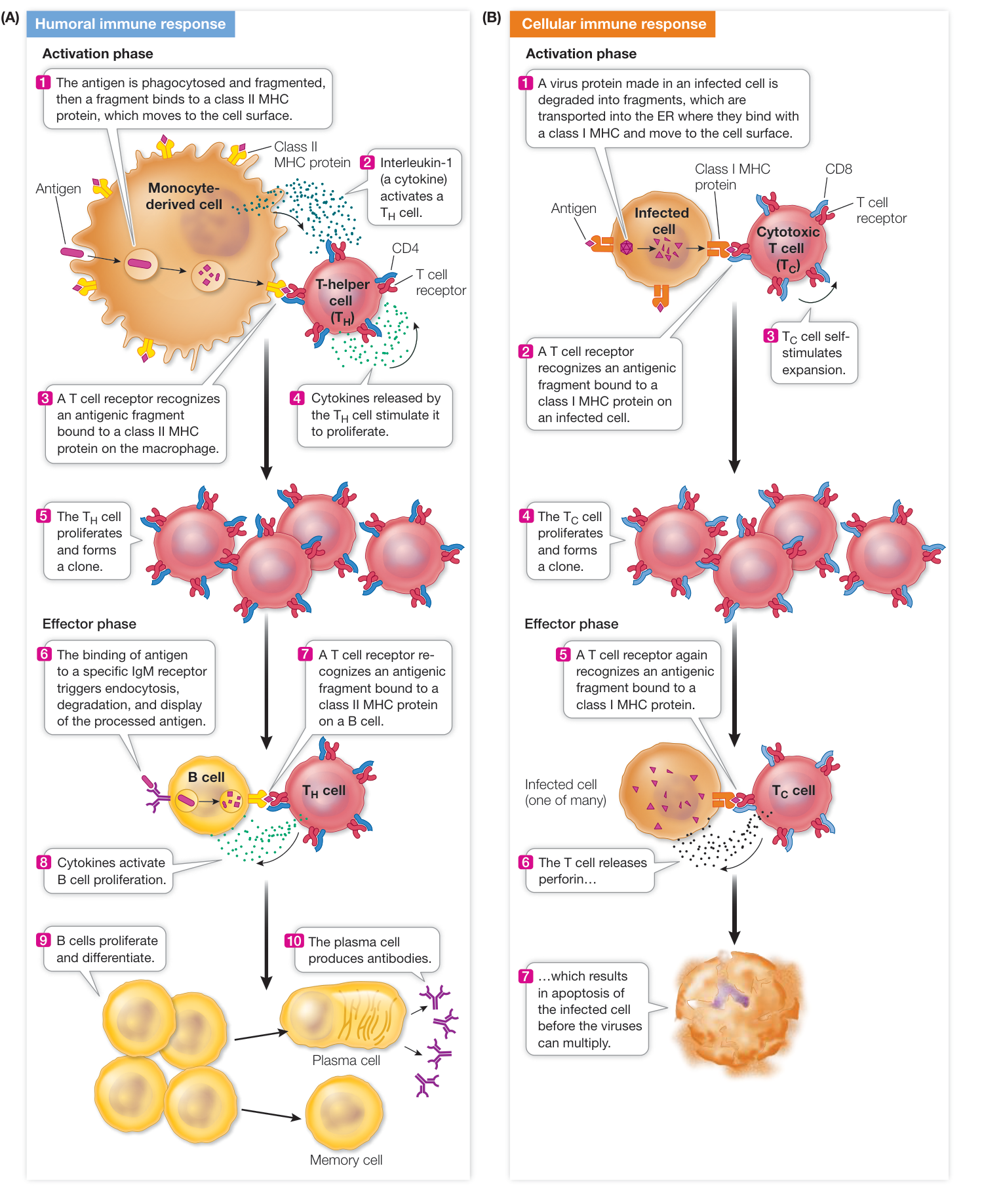
Figure 40.18 Phases of the Humoral and Cellular Immune Responses Both the humoral (A) and the cellular (B) immune responses have activation and effector phases, all of which involve T cells.
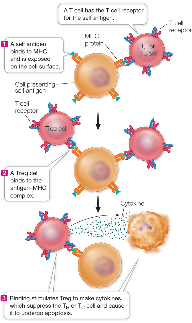
Figure 40.19 Tregs and Tolerance A special class of T cells called regulatory T cells (Tregs) inhibits the activation of the immune system in response to self antigens.
important way: the antigens that Tregs recognize are self antigens. The activation of Tregs causes them to secrete cytokines, which block the activation of T cells that are bound to the same antigen-presenting cell (Figure 40.19). Thus Tregs constitute another mechanism for distinguishing self from nonself.
Recap and Assess
The cellular immune response acts against pathogen-infected or mutated body cells. Specific receptors on T cells bind to antigen-MHC complexes displayed on cell surfaces. T-helper cells bind antigens presented on MHC II proteins by monocyte-derived and B cells and contribute to the humoral immune response. Cytotoxic T cells bind to antigens presented on MHC I proteins by any nucleated cell and contribute to the cellular immune response. Tregs suppress immune responses to self antigens.
MHC proteins almost always differ among unrelated people, so organ transplants usually provoke a cellular immune response by the host, causing rejection. People receiving transplants are given a drug called cyclosporine, which inhibits cytokine release by T cells. Why do you think cytokine inhibition by cyclosporine reduces rejection?
- When a virus infects a cell, the immune system mounts an adaptive response to this infection. What are the roles of the T cell receptor and MHC protein in this response?
- Cancer cells often express proteins that the body has never seen, so they are targeted by the cellular adaptive immune system. In some cancers, however, Tregs increase in numbers and activity. Why would this indicate a poor prognosis?
Given the numerous and complex cellular interactions that activate the immune system and generate antibody diversity, you may have perceived many points at which the immune system could fail. We now turn to several situations in which one or more components of this complex system malfunction.
Malfunctions in Immunity Can Be Harmful
Learning Objectives
- 40.6.1 Summarize the responses that occur during immediate and delayed hypersensitivity.
- 40.6.2 Describe how desensitization can be used to minimize allergic reactions.
- 40.6.3 Summarize the hypotheses that explain how autoimmunity can occur.
- 40.6.4 Explain how HIV infection causes immune deficiency.
Sometimes the immune system overreacts, as in an allergic response. Sometimes it attacks self antigens, as in autoimmune disease. And sometimes it fails to act, or acts weakly, as in immune deficiency disease.
The immune system mounts an excessive response in allergic reactions
An allergic reaction arises when the human immune system overreacts to (is hypersensitive to) a dose of antigen. Although the antigen itself may present no danger to the host, the inappropriate immune response may produce inflammation and other symptoms, which can cause serious illness or even death. Allergic reactions are the most familiar examples of this phenomenon. Allergic reactions may involve immediate hypersensitivity or delayed hypersensitivity.
IMMEDIATE HYPERSENSITIVITY Immediate hypersensitivity arises when an allergic individual is exposed to an antigen (in this case referred to as an allergen) from the environment, such as a food, pollen, or the venom of an insect. In response to the allergen, the individual makes large amounts of IgE. When this happens, mast cells in tissues and basophils in the blood bind the constant end of the IgE. If that individual is exposed to the same allergen again, binding of the allergen to the IgE causes the mast cells and basophils to rapidly release a large amount of histamine (Figure 40.20). This results in symptoms such as dilation of blood vessels, inflammation, and difficulty breathing. If not treated with antihistamines, a severe allergic reaction can lead to death. It is not known why some people produce excessive amounts of IgE in response to allergens. There is some evidence for genetic factors predisposing people to allergic responses.
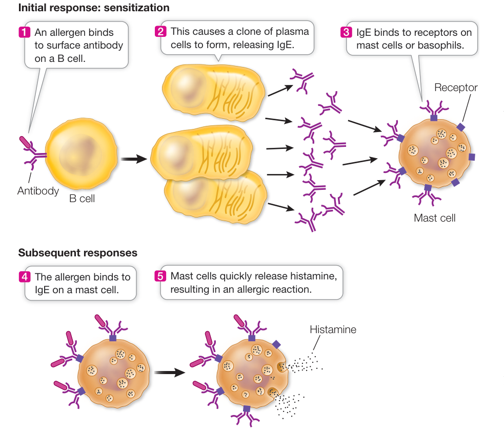
Figure 40.20 An Allergic Reaction An allergen is an antigen that stimulates B cells to make large amounts of IgE antibodies, which bind to mast cells and basophils. When the body encounters the allergen again, these cells produce large amounts of histamine, which has harmful physiological effects.
Allergy to pollen can be treated using a process called desensitization. The process involves injecting small amounts of the allergen (typically just an extract of the offending plant tissue) into the skin—enough to stimulate IgG production but not enough to stimulate IgE production. The next time the person is exposed to the allergen, IgG binds to it, tying it up before IgE can bind it and stimulate mast cells to release histamine.
Desensitization does not work well for food allergens because the IgE response to those substances is so strong that even a small amount of antigen provokes it. The best approach for those with food allergies is to avoid foods containing the allergens. This can be difficult, but food labels listing all the ingredients are helpful. Molecular biologists are beginning to identify the antigens that act as allergens, with the hope of developing vaccines or genetically modified foods that lack the allergenic epitopes.
DELAYED HYPERSENSITIVITY Delayed hypersensitivity is an allergic reaction that does not begin until hours after exposure to an antigen. In this case the antigen is taken up by antigen-presenting cells, and a T cell response is initiated. A cell produces a clone of cells that secrete various cytokines, which cause such reactions as inflammation and rash. These events take time (hence the term “delayed”). An example is the rash that develops after exposure to poison ivy.
Autoimmune diseases are caused by reactions against self antigens
Errors in the selection of T cells in the thymus can result in T cells that bind to antigen–MHC complexes that carry self antigens. Although the precise origin of autoimmunity is not known, there are at least two hypotheses:
- Failure of clonal deletion. B or T cells that recognize self antigens and should have been destroyed by clonal deletion are not destroyed.
- Molecular mimicry. T cells that recognize a nonself antigen, such as a virus, also recognize something on a self antigen that has a similar structure.
Autoimmunity does not always result in disease, but several autoimmune diseases are common:
People with systemic lupus erythematosus (SLE) have antibodies to many cellular components, including DNA and nuclear proteins released from dying cells. These antinuclear antibodies can cause serious damage when they bind to normal tissue antigens and form large circulating antigen–antibody complexes, which become stuck in tissues and provoke inflammation.
People with rheumatoid arthritis have difficulty in shutting down a T cell response to self antigens. These patients may have low activity of CTLA4, an inhibitory protein that blocks T cells from reacting to self antigens. This results in inflammation of the joints and other tissue damage.
Hashimoto’s thyroiditis is the most common autoimmune disease in women over 50. Immune cells attack thyroid tissue, resulting in fatigue, depression, weight gain, and other symptoms.
Insulin-dependent diabetes mellitus, or type I diabetes, occurs most often in children. It is caused by an immune reaction against several proteins in the cells of the pancreas that manufacture the protein hormone insulin. This reaction kills the insulin-producing cells, so people with type I diabetes must take insulin daily in order to survive.
AIDS is an immune deficiency disorder
There are several inherited and acquired immune deficiency disorders. In some individuals, T or B cells never form; in others, B cells lose the ability to give rise to plasma cells. In either case, the affected individual is unable to mount an adaptive immune response and thus lacks a major line of defense against pathogens.
The cell is perhaps the most central component of the immune system because of its essential roles in both the humoral and cellular immune responses (see Figure 40.9). This cell is the target of human immunodeficiency virus (HIV), the retrovirus that results in acquired immune deficiency syndrome (AIDS).
HIV can be transmitted from person to person in body fluids containing the virus (such as blood, semen, or vaginal fluid). The recipient tissue is either blood (by transfusion) or a mucous membrane lining an organ (the mucus contains a high concentration of B and T cells). HIV initially infects macrophages, cells, and antigen-presenting dendritic cells in the blood and tissues. At first there is an immune response to the viral infection, and cells are activated. But because HIV infects the cells, they are killed both by HIV itself and by cells that lyse infected cells. Consequently cell numbers decline after the first month or so of infection. Meanwhile, the extensive production of HIV by infected cells activates the humoral immune system. Antibodies bind to HIV, and the complexes are removed by phagocytes. The HIV level in blood goes down. There is still a low level of infection, however, because of the depletion of cells (Figure 40.21). This process reaches a low, steady-state level called the “set point.” This point varies among individuals and is a strong predictor of the rate of progression of the disease. For most people it takes 8–10 years without treatment for the more severe manifestations of AIDS to develop. In some it can take as little as a year; in others, 20 years.
During this dormant period, people carrying HIV generally feel fine, and their cell levels are adequate for them to mount immune responses. Eventually, however, as the virus continues to destroy the cells, their numbers fall to the point where the infected person is susceptible to infections that the cells would normally eliminate. These infections result in conditions such as Kaposi’s sarcoma, a skin tumor caused by a herpes virus; pneumonia caused by the fungus Pneumocystis jirovecii; and lymphoma tumors caused by the Epstein–Barr virus. These conditions result from opportunistic infections because the pathogens take advantage of the crippled immune system of the host. They lead to death within a year or two.
HIV has been intensively studied. This has resulted in the development of drugs targeted to HIV proteins, such as the reverse transcriptase that makes cDNA from the virus RNA, and the virus protease that cuts the large precursor virus protein into its final active proteins. Treatment with combinations of such drugs has had spectacular success. Before the 1990s, getting AIDS was a death sentence, with few sufferers surviving beyond a year or two. Today the survival of a treated, infected person is decades longer. Unfortunately, like many medical treatments, HIV drugs are not available to all who need them—particularly in poor regions of the world where AIDS is prevalent. Since AIDS is caused by a viral infection, a vaccine would seem an appropriate preventive agent. Unfortunately, despite a huge international effort that is ongoing, a vaccine against HIV has not been developed, probably because the virus “hides out” inside immune cells and is prone to a high mutation rate. There are about 1.2 million deaths per year worldwide from AIDS.
Recap and Assess
Failures of the immune system include allergic reactions (caused by hypersensitivity to antigens), autoimmune diseases (caused by reactions against self antigens), and immune deficiency disorders.
- Some people are allergic to bee stings. Outline the events that characterize the immune reactions involved.
- There has been recent progress in treating bee sting allergy by desensitization. How does this work?
- A new cancer treatment involves promoting autoimmunity by designing a drug to inhibit CTLA4, a protein on T cells that normally blocks them from mounting an autoimmune response. How does this treatment work? What might be its side effects?
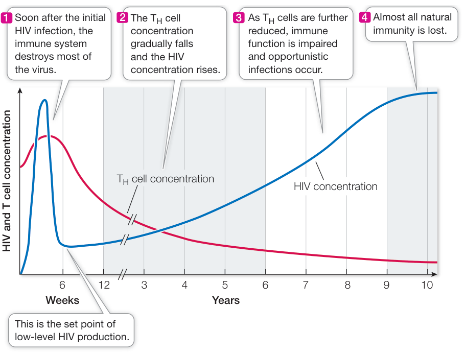
Figure 40.21 The Course of an HIV Infection An HIV infection may be carried, unsuspected, for many years before the onset of symptoms. (After M. Kogan and J. Rappaport. 2011. Retrovirology 8:25. doi:10.1186/1742-4690-8-25.)
Investigating Life: QA What are the mechanisms and implications of long-lasting immunity?
You have seen in this chapter that lasting immunity against a pathogen can be achieved in two ways, both of which involve exposure to an antigen associated with the pathogen and the resulting production of memory cells. The first way is natural: in Investigating Life: What Are the Mechanisms and Implications of Long Lasting Immunity?, we showed that people exposed to virulent strains of flu viruses retain their immunity, and even increase it when exposed to less harmful flu strains. The second way is artificial: as we described in the opening story, vaccines have been spectacularly successful at inducing lasting immunity and even eradicating diseases when enough people are vaccinated. Why, then, do people refuse vaccination? There appear to be several reasons. One is complacency—the threat of disease may seem remote. For example, measles, which used to kill thousands of children every year in the United States and still does in poor countries, is no longer a highly visible threat. Second, some people believe that vaccines, although exhaustively tested and proven safe, are unsafe and cause disease. The internet is full of such incorrect assertions. Third, false alarms have led people to dismiss vaccination advisories. The discovery of the H1N1 flu virus (swine flu) in Mexico in 2009 led to a high alert and a mass vaccination program that turned out not to be necessary. In addition, some people are suspicious of governmental programs in general. Vaccination is a scientific success in terms of bolstering immunity and eradicating disease. But like any technology, its acceptance is a political issue.
Future directions
Following activation of the immune system, many effector cells are mobilized. Most eventually die, but some survive as memory cells. Researchers are investigating how this happens. At the University of Wisconsin, Marulasiddappa Suresh and his colleagues have shown that a single transcription factor called FoxO1 plays an important role in the effector-to-memory-cell transition. This molecule is not needed for the production of effector cells, but if it is not there, memory cells do not form. The genes regulated by FoxO1 are being characterized and may hold the key to the memory cell. Meanwhile, at Yale University, Susan Kaech and her colleagues have shown that an intercellular signal molecule, interleukin-7 (IL-7), binds to effector cells and results in changes that may allow memory cell survival. Specifically, the target cells produce a membrane channel that allows rapid uptake of glycerol, the backbone of fats. Production of fats in the memory cell may provide the energy needed for long-term survival. Understanding how memory cells form and survive may provide valuable information for the production of better vaccines.

© Universal History Archive/UIG/Getty Images
You should be able to relate each summary to the adjacent figures. If you go to this visual summary in Achieve, you can follow links to figures, animations, activities, and simulations that will help you consolidate the material.
Visual Summary
Animals Use Innate and Adaptive Mechanisms for Defense
and Animation 40.1
- ■ Innate defenses are present in all animals and respond to a broad range of pathogens.
- ■ Adaptive defenses are present in vertebrates and target specific pathogens.
- ■ A variety of white blood cell types and their receptors are involved in both responses.
- When a pathogen enters the body, it triggers an internal response. What are the three phases of this response?
- Several Toll-like receptors (TLRs) are found in the genome. What would be the effect of knocking out (rendering nonfunctional) both alleles of (a) a gene encoding a TLR or (b) the gene encoding the Nf- B transcription factor, which is one of the main targets of the TLR signal transduction pathway?
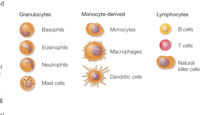
Figure 40.1


Innate Defenses Are Nonspecific
- ■ Organisms use a variety of physical, chemical, and enzyme barriers and the host microbiome to prevent pathogen entry.
- ■ A pathogen that enters the body is recognized by the foreign macromolecules it expresses.
- ■ Innate responses to pathogens include phagocytosis and the release of complement proteins and cytokines.
- ■ Inflammation causes mast cells to release chemicals that recruit phagocytes.
Questions
- Why is lysozyme concentration high in tears? Do you think it is also high in breast milk?
- When an invader is recognized as foreign, how might a white blood cell first respond to it?
- When you cut yourself, the wound often swells up. What has happened to cause this?
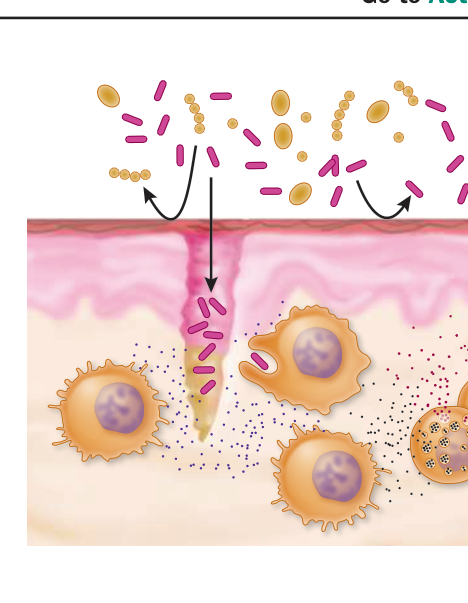
Figure 40.5
Adaptive Defenses Are Specific
- ■ An enormous diversity of receptors able to bind specific antigens are produced by B and T cells; cells expressing self-recognizing receptors are deleted.
- ■ Initiating an adaptive immune response requires antigen binding and often involves antigen-presenting cells.
- ■ An adaptive immune response can be humoral or cellular.
- ■ Clonal selection produces effector cells that respond to the current infection and memory cells ready to respond to a repeated infection by the same pathogen.
- What is the difference between clonal deletion and clonal selection? When does each of these events happen?
- B cells can bind antigen. Why must T-helper cells recognize the same antigen to get a humoral response?
- Vaccination requires the development of memory cells. Why do you think some vaccines require “boosters”?
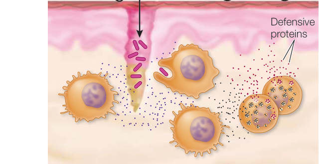
Figure 40.10
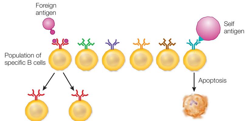
The Humoral Adaptive Response Involves Antibodies
- ■ Immunoglobulins come in five classes and may be B cell membrane receptors or secreted from B cells. Class switching involves DNA rearrangements.
- ■ Immunoglobulins show enormous diversity in the amino acid sequences of their variable regions due to rearrangements of the V, D, and J genes.
Questions
- Suppose a mammal possesses 50 V, 20 D, and 5 J genes for its heavy chain and 50 V and 5 J genes for its light chain. How many possible antibodies can it make from rearranging these genes?
- The number of antibodies actually produced by the mammal in question 1 is greater than 50 million. Where does this additional diversity come from?
- A B cell that never undergoes class switching produces both IgM and IgD antibodies. How is it able to produce two classes of antibodies?
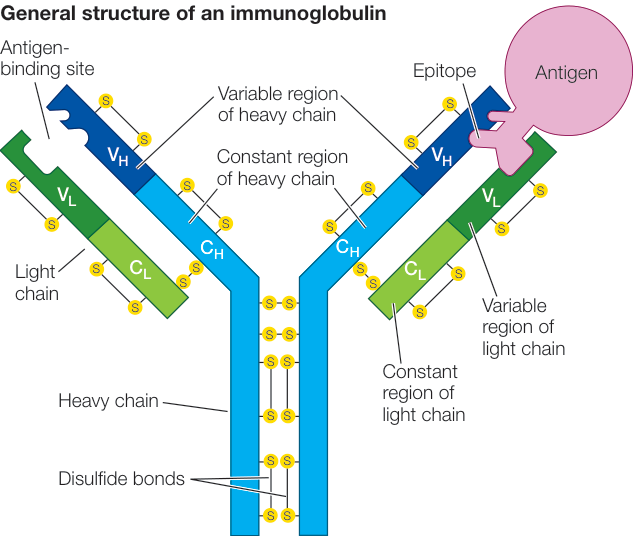
Figure 40.12
The Cellular Adaptive Response Involves T Cells and Receptors
- ■ T cell receptor proteins have extreme diversity due to rearrangements of the V, D, and J genes in their variable regions.
- ■ Class I and II MHC proteins present antigens to CD8-expressing cytotoxic T cells and CD4-expressing T-helper cells respectively and are thus involved in both the humoral and cellular immune responses.
- ■ In humans, MHC proteins are encoded at six genes, each of which is quite variable due to allelic variation (more than 100 alleles in the human population).
- ■ Regulatory T cells (Tregs) destroy other T cells expressing self antigens.
- Why is a CD4 or CD8 receptor required for T cell activation?
- What are the similarities and difference in generating diversity in T cell receptors and B cell receptors (antibodies)?
- Antigen presentation can occur to either or a cells. Outline the similarities and differences of antigen presentation and the responses obtained by each of these cell types.
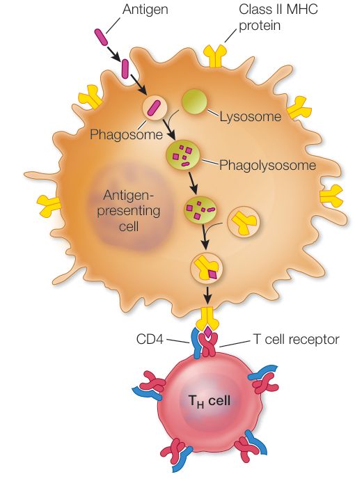
Figure 40.17


Malfunctions in Immunity Can Be Harmful
- ■ Immediate hypersensitivity is due to a rapid and substantial release of histamines from mast cells that are bound to an IgE antibody that recognizes the allergen (antigen).
- ■ Delayed hypersensitivity is due to a T-helper cell response that release cytokines.
- ■ Desensitization leads to the increase in circulating IgG antibodies that bind an allergen before it can bind to IgE on mast cells.
- ■ Autoimmunity occurs when T or B cells recognize self antigens.
- ■ Human immunodeficiency virus (HIV) targets T-helper cells, weakening the immune system, and if untreated eventually leads to acquired immune deficiency syndrome (AIDS).
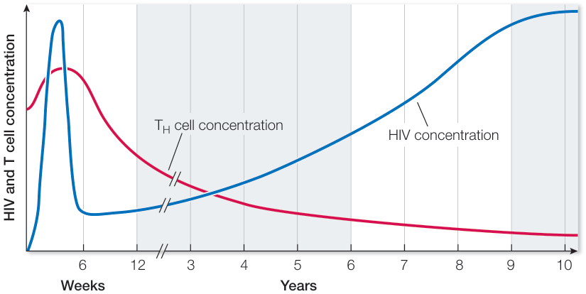
Figure 40.21
Questions
- Why do some individuals show more extreme allergic reactions with every exposure to an allergen?
- Allergic reactions often involve the release of large amounts of histamine. How might antihistamines work to reduce an allergic reaction?
- HIV is particularly effective at disrupting an individual’s ability to mount an immune response. Why is this the case?