Chapter 42Animal Development
Key Concepts
- 42.1 Fertilization Activates Development
- 42.2 Mitosis Divides Up the Early Embryo
- 42.3 Gastrulation Generates Multiple Tissue Layers
- 42.4 Organs Develop from the Three Germ Layers
- 42.5 Extraembryonic Membranes Nurture Avian and Mammalian Embryos
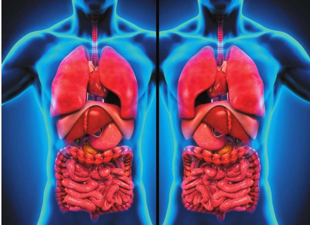
Our internal organization is not symmetrical, and some individuals are born with the mirror-image organization of what is seen in most people—a condition called situs inversus.
Investigating Life: Go with the Flow
Place your hand over your heart. Point to your appendix; point to your pancreas. Surely your hand went to the left side of your chest, then to the right side of your lower abdomen, and last to the left side of your upper abdomen. Vertebrates are bilaterally symmetrical, but our symmetry is not absolute. Some internal organs are oriented to the left and some to the right. This bilateral asymmetry arises from events very early in the development of the embryo. However, in about 1 out of every 7,000 people, bilateral asymmetry is reversed—a condition known as situs inversus (“location inverted”). Most people with situs inversus lead normal lives and are unaware of their condition until they have diagnostic imaging or surgery.
You will learn in this chapter that a mammalian embryo goes from a single layer of cells to two layers of cells by forming a midline slit. Cells migrate through that slit to form the second cell layer. At one end of the slit is an area called the node. Each cell at the node has a primary cilium. The primary cilia of some of these cells are nonmotile and serve sensory functions. The primary cilia of other nodal cells are motile, and they beat in a clockwise motion that moves extracellular fluid across the surface of the node. That fluid movement is from right to left. Why?
Imagine that a cilium moves like the hands of a clock (but much faster!). Twelve o’clock points to the embryo’s future head. The cilia protrude from the cells at an angle such that they are closest to the cell surface at the 6 o’clock position. Thus when the cilia rotate through the 6 o’clock position, they are closer to the cell surface and experience greater resistance than at the 12 o’clock position. As a result, the circular beating of cilia at the node creates left–right flow of extracellular fluid and therefore left–right positional information.
Does that simple left–right positional information generated by beating nodal cilia instruct the development of left–right anatomical asymmetry? The answer is probably yes, because in strains of mice that have no nodal cilia or have only non-motile nodal cilia, about half of the population has reversed organ asymmetry. Similarly, in humans there is a spectrum of rare genetic disorders called Kartagener’s syndrome that is characterized by nonmotile cilia. About half of these individuals have situs inversus.
Q&A How does the embryo know left from right? (Find the answer on page 958.)
Fertilization Activates Development
Learning Objectives
- 42.1.1 List the contributions of the sperm and the egg to the zygote.
- 42.1.2 Explain how sperm entry converts frog egg symmetry from radial to bilateral.
- 42.1.3 Describe how the movement of cytoplasm, proteins, and organelles following fertilization of the frog egg instructs early development.
Fertilization is the joining of sperm and egg to produce a diploid zygote. You might think of fertilization as the event that begins development. But recall two things from Chapter 41: (1) in sexually reproducing animals, development proceeds without fertilization, and (2) in sexually reproducing animals, fertilization is preceded by events in the maturing egg that set the stage for development but are paused. Thus fertilization activates multicellular development in sexually reproducing animals.
Fertilization does more than restore a full diploid complement of maternal and paternal genes. The fusion of sperm and egg cell membranes:
- triggers ion fluxes across the egg membrane.
- creates blocks to the entry of additional sperm into the egg.
- changes the pH of the egg cytoplasm.
- increases egg metabolism and stimulates protein synthesis.
- initiates the rapid series of cell divisions that produce a multicellular embryo.
We described the mechanisms of fertilization in Key Concept 41.2. Here we take a closer look at the cellular and molecular interactions of sperm and egg that are the first steps of development.
The sperm and the egg make different contributions to the zygote
Eggs are much larger than sperm. Egg cytoplasm is well stocked with organelles, nutrients, and a variety of molecules, including cytoplasmic determinants such as transcription factors and mRNAs. Nearly everything the embryo needs during its early stages of development comes from the mother, including its mitochondria (and therefore all of its mitochondrial DNA), its ribosomes, and other organelles. Cytoplasmic determinants in the egg play important roles in setting up the signaling cascades that orchestrate the four major events of development: determination, differentiation, morphogenesis, and growth. Determination involves processes that limit the patterns of gene expression in the earliest cells of the embryo, which are totipotent—each one has the capacity to develop into a whole organism. Differentiation is when those limited patterns of gene expression are initiated and the cells take on specialized characteristics of a tissue type, such as the appearance of contractile proteins in muscle cells. Morphogenesis is the stage of development when tissues interact and change shape to form organs. Growth is maturing and getting larger to have the size and capabilities of the adult organism.
The sperm makes only two major contributions to the zygote: its haploid nucleus and, in most species, a centriole. That centriole produces the zygote’s centrosome, which organizes the mitotic spindles for subsequent cell divisions (see Figure 11.9). Centrioles are also the origin of the microtubules of the primary cilia, which are important in cell signaling, as you will see when we return to the opening question about how embryos know right from left.
Fertilization sets the stage for determination
The large size of amphibian eggs and the fact that they can be fertilized and go through early stages of development in culture media make them ideal models for studying how rearrangements of egg cytoplasm lead to determination. Determination sets the developmental fates of cells and precedes differentiation—the expression of different cell properties. The molecules in the cytoplasm of the amphibian egg are not homogeneously distributed. The entry of the sperm into the egg stimulates rearrangements of the egg cytoplasm that introduce additional organization to the egg. One such cytoplasmic difference is the emergence of distinct “top” and “bottom” ends of an organism or structure, a difference that establishes polarity. Sperm entry establishes the polarity of the zygote, and informational molecules in the egg cytoplasm are organized with respect to that polarity. Therefore when cell divisions begin, these informational molecules—which guide subsequent development—are not divided equally among daughter cells.
Rearrangement of egg cytoplasm following fertilization is easily observed in some frog species because of pigments in the cytoplasm. The nutrients in an unfertilized frog egg are dense yolk granules that are concentrated by gravity in the lower half of the egg, called the vegetal hemisphere. The haploid nucleus of the egg is located at the opposite end, in the animal hemisphere. The outermost (cortical) cytoplasm of the animal hemisphere is more pigmented than the underlying cytoplasm. The vegetal hemisphere is not pigmented. Because of these differences, rearrangement of the cytoplasm after fertilization is easy to observe.
The unfertilized frog egg is radially symmetrical. You can turn it on its vegetal–animal pole axis, and all sides are the same. Sperm-binding sites are localized on the surface of the animal hemisphere. When a sperm binds to and enters the egg, the egg’s radial symmetry is converted to bilateral (left–right) symmetry with a dorsal–ventral axis. The cortical (superficial) cytoplasm rotates toward the site of sperm entry (Figure 42.1). This rotation brings the animal and vegetal regions of cytoplasm into contact with each other, producing a band of lightly pigmented cytoplasm on the side opposite the site of sperm entry. This band, called the gray crescent, marks the location of important developmental events in some species of amphibians.
The centriole that was the sperm’s contribution to the egg initiates cytoplasmic reorganization that coincides with the appearance of the gray crescent. The centriole organizes microtubules in the vegetal hemisphere cytoplasm into a parallel array that guides the movement of the cortical cytoplasm as well as proteins and organelles.
The movement of cytoplasm, proteins, and organelles changes the distribution of critical signals. A key transcription factor in early development is -catenin produced from maternal mRNA that was produced and stored in the egg while it was maturing in the ovary. -Catenin is found throughout the egg cytoplasm (Figure 42.2). Also present throughout the egg cytoplasm is a protein kinase, glycogen synthase kinase-3 (GSK-3), that phosphorylates -catenin and thereby targets it for degradation. An inhibitor of GSK-3 is localized in the vegetal cortex of the egg. After sperm entry, this inhibitor moves along microtubules to the gray crescent, where it prevents the degradation of -catenin. As a result, the concentration of -catenin is higher on the dorsal side of the developing embryo than on the ventral side (the side where the sperm entered). The regional differences in -catenin concentration set up differences within the egg cytoplasm that have developmental consequences.
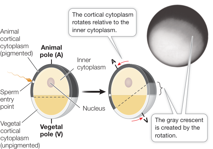
Figure 42.1 The Gray Crescent In amphibian eggs, cortical rotation and rearrangement of the cytoplasm after fertilization create the gray crescent opposite the point of sperm entry. These events are important for specifying the body axes and other important events in later development. (After R. P. Elinson and B. Rowning, 1988. Dev Biol 128: 185–197.)
Recap and Assess
The haploid egg has a full complement of cell organelles and is stocked with nutrients and informational molecules that power and direct the early stages of development. Sperm entry into the egg contributes a haploid nucleus and a centriole to the zygote. Fertilization restores diploidy, activates the egg, and stimulates rearrangement of the cytoplasm, setting up the body axes and organizing positional information that will control determination and differentiation.
- Apart from its nucleus, what is the major contribution of the sperm to subsequent cell signaling in the embryo?
- How is the gray crescent generated in the fertilized frog egg?
- How does -catenin become more concentrated on the dorsal side of the amphibian embryo?
The uneven distribution of informational molecules in the cytoplasm of the fertilized egg is essential to later events in development. In the next key concept you will see how these informational molecules end up in different cells of the developing embryo.
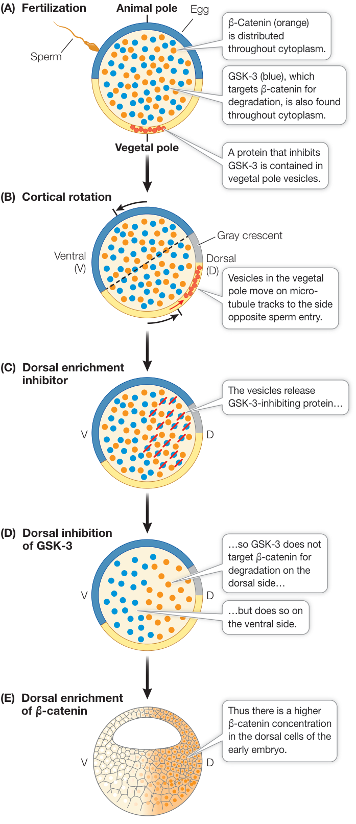
Figure 42.2 Cytoplasmic Factors Set Up Signaling Cascades Cytoplasmic movement changes the distributions of critical developmental signals. In the fertilized frog egg, the interaction of the protein kinase, the GSK-3 inhibitor, and the protein -catenin results in differential concentrations of -catenin in blastomeres following cleavage, thereby specifying the dorsal–ventral axis of the embryo.
Mitosis Divides Up the Early Embryo
Learning Objectives
- 42.2.1 Compare four types of cleavage.
- 42.2.2 Explain the difference between regulated and autonomous specification.
- 42.2.3 Explain the difference between identical and non-identical twins.
- 42.2.4 Describe the difference between determination in reproductive germ cells and other types of cells in the embryo.
-Catenin plays a major role in the cell–cell signaling cascade that begins the process of cell determination and the formation of the embryo. But cell–cell signaling requires more than one cell. The single-celled zygote must become a multicellular embryo.
Cleavage produces a multicellular embryo
Cleavage transforms the diploid zygote into a mass of undifferentiated cells that develop as the embryo. Zygote cytoplasm is not homogeneous, and these first cell divisions result in the differential distribution of nutrients and cytoplasmic determinants in the early embryo.
In most animals, cleavage proceeds with rapid DNA replication and mitosis but with no cell growth and little gene expression. The embryo becomes a solid ball of smaller and smaller cells. Eventually this ball forms a central fluid-filled cavity called a blastocoel. At this developmental stage the embryo is called a blastula, and its individual cells are blastomeres. The pattern of cleavage in different species influences the form of their blastulas.
- Complete cleavage occurs in eggs that have little yolk, such as the eggs of amphibians. Early cleavage furrows divide the egg completely. In the amphibian egg, the concentration of yolk in the vegetal pole causes unequal cytoplasm division, and animal hemisphere blastomeres are smaller than those in the vegetal hemisphere (Figure 42.3A).
- Incomplete cleavage occurs in eggs with a lot of yolk, and the cleavage furrows do not penetrate it completely. Discoidal cleavage is a type of incomplete cleavage common in fishes as well as in birds and other reptiles. In the yolk-rich eggs of these species, the embryo forms as a disc of cells, or blastodisc, that sits on top of the dense yolk mass (Figure 42.3B).
- Superficial cleavage is a variation of incomplete cleavage that occurs in insects such as fruit flies (Drosophila). Early in development, cycles of mitosis occur without cell division, producing a syncytium—a single cell with many nuclei (Figure 42.3C). The nuclei eventually migrate to the periphery of the egg, after which the cell membrane of the egg grows inward, creating a blastoderm by partitioning the nuclei into individual cells surrounding a core of yolk.
- Rotational cleavage is a unique form of complete cleavage that occurs only in placental (eutherian) mammals.
The positions of the mitotic spindles during cleavage are not random but are defined by cytoplasmic determinants produced from the maternal genome and stored in the egg. The orientation of the mitotic spindles can determine the planes of cleavage and the arrangement of the blastomeres.
In complete cleavage, if the mitotic spindles of successive cell divisions form parallel or perpendicular to the animal–vegetal axis of the zygote, a pattern of radial cleavage occurs. The first two cell divisions are parallel to the animal–vegetal axis, and the third is perpendicular to it (see Figure 42.3A). Spiral cleavage results when the mitotic spindles are at oblique angles to the animal–vegetal axis. In spiral cleavage, each new cell layer is shifted to the left or right, depending on the orientation of the mitotic spindles. Most mollusks have spiral cleavage, reflected in some species, such as snails, by a coiling shell pattern.
Cleavage in mammals is unique
Several features of early cell divisions in eutherian mammals are very different from those seen in other animal groups. (Note: unless stated otherwise, all mammals referred to in this chapter are eutherians). First, this process in mammals is very slow. Cell divisions are 12–24 hours apart, compared with tens of minutes to a few hours in non-mammalian species. Also, the cell divisions of mammalian blastomeres are not in synchrony with each other. Because the blastomeres do not undergo mitosis at the same time, the number of cells in the embryo does not increase in the regular progression (2, 4, 8, 16, 32, etc.) typical of other species. The slowness of mammalian cleavage means that genes expressed during cleavage can play roles in cleavage. In animals such as sea urchins and frogs in which cleavage progresses rapidly, very little if any gene transcription occurs in the blastomeres. Instead, cleavage is directed by molecules that were present in the egg before fertilization.
The pattern of mammalian cleavage is called rotational cleavage. The first cell division is parallel to the animal–vegetal axis as in radial cleavage, but in the second cell division, the two blastomeres divide at right angles to one other. One blastomere divides parallel to the animal–vegetal axis, while the other divides perpendicular to this axis (Figure 42.4A). As in other animals that have complete cleavage, the early cell divisions in a mammalian zygote produce a loosely associated ball of cells. After the eight-cell stage, however, the behavior of the mammalian blastomeres changes. They change shape to maximize their surface contact with one another, form tight junctions (see Figure 6.6), and become a compact mass of cells (Figure 42.4B).
Soon after the transition to the 32-cell stage, the cells of the mammalian embryo separate into two groups. The inner cell mass develops as the embryo, while the surrounding outer cells become an encompassing sac called the trophoblast. Trophoblast cells secrete fluid, creating a cavity—the blastocoel—with the inner cell mass at one end. At this stage the mammalian embryo is called a blastocyst, distinguishing it from the blastulas of other animal groups (Figure 42.4C). The pluripotent cells of the inner cell mass are known as embryonic stem cells and are the subject of much research because of their therapeutic potential.
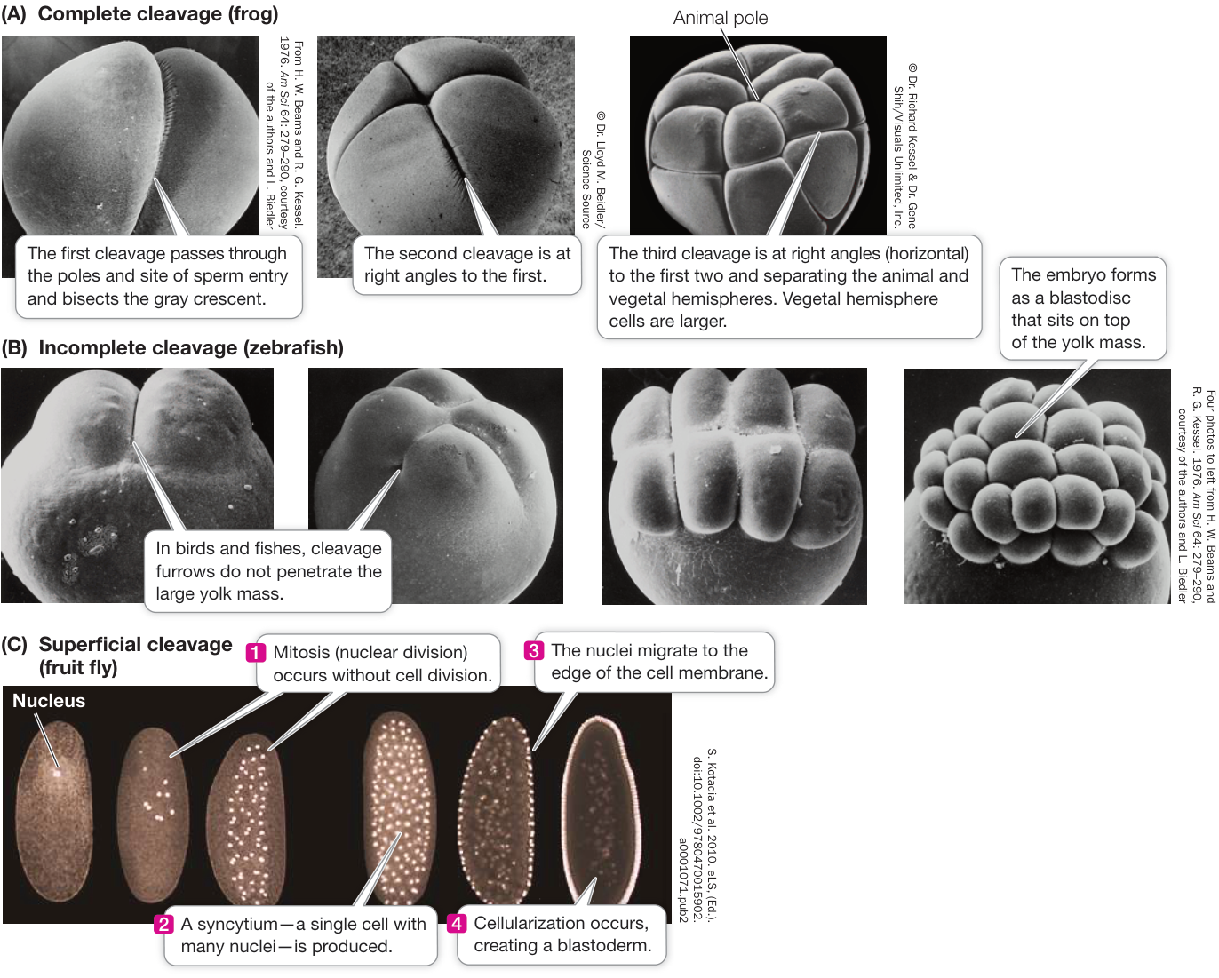
Figure 42.3 Some Patterns of Cleavage (B) Incomplete cleavage (zebrafish) (C) Superficial cleavage (fruit fly) Differences in patterns of early embryonic development reflect differences in the way the egg cytoplasm is organized. (A) The frog is a model organism representing complete cleavage in these scanning electron micrographs (SEMs). (B) SEMs of zebrafish (Danio rerio) embryos illustrate incomplete cleavage, in which the large yolk mass limits the planes of cleavage. (C) Nuclear staining reveals the syncytial nuclei characteristic of the early embryo of a fruit fly. These nuclei migrate to the periphery. Cleavage furrows then move inward to separate the nuclei into individual cells, forming the blastoderm.
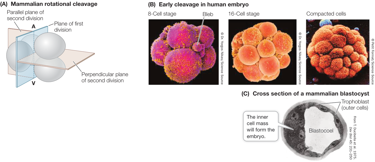
Figure 42.4 Becoming a Blastocyst (A) Mammals have rotational, complete cleavage, in which the plane of the first cleavage is parallel to the animal–vegetal (A–V) axis, but the second cell division involves two planes (beige) at right angles to each other. (B) Scanning electron micrographs (color added) of early cleavage (leading to the formation of the blastocyst) in a human embryo. The cells’ outer surfaces are covered with cilia (bright yellow). The small spheres, or “blebs,” of cytoplasmic material, prominent at the eight-cell stage, disintegrate as cleavage progresses. (C) Seen in cross section under a light microscope, a mammalian blastocyst consists of an inner cell mass adjacent to a fluid-filled blastocoel and surrounded by trophoblast cells. (A after B. J. Gulyas. 1975. J Exp Zool 193: 235–247. doi:10.1002/jez.1401930212)
Why is mammalian cleavage so different? A key factor is that mammalian eggs contain little or no yolk and must derive all nutrients from the mother. To support the developing embryo, a connection develops between the circulatory systems of the embryo and the mother. As you will see later in this chapter, the structures that provide this connection are the placenta and the umbilical cord. Thus the blastocyst of mammals must produce both the embryo (from the inner cell mass) and its support structures (from the trophoblast).
Fertilization in mammals occurs in the upper reaches of the oviduct, and cleavage occurs as the zygote travels down the oviduct to the uterus (Figure 42.5). When the blastocyst arrives in the uterus, the trophoblast adheres to the lining of the uterus (the endometrium), beginning the process of implantation. In humans, implantation begins about 6 days after fertilization and is aided by adhesion molecules and enzymes secreted by the trophoblast.
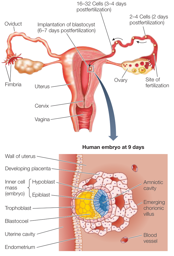
Figure 42.5 A Human Blastocyst at Implantation Adhesion molecules and proteolytic enzymes secreted by trophoblast cells allow the blastocyst to burrow into the endometrium. Once the blastocyst is implanted in the wall of the uterus, the trophoblast cells send out numerous projections—the chorionic villi—which increase the embryo’s area of contact with the mother’s bloodstream. The inner cell mass divides into two embryonic tissues, the hypoblast and the epiblast. The epiblast splits to form the amniotic cavity.
As the blastocyst moves down the oviduct to the uterus, it must not embed itself in the oviduct (fallopian tube) wall, or the result will be an ectopic, or tubal, pregnancy—a very dangerous condition. Early implantation is prevented by the zona pellucida that surrounded the egg (see Figure 41.6) and remains around the cleaving ball of cells. At about the time the blastocyst reaches the uterus, it hatches from the zona pellucida so implantation can occur.
The fates of blastomeres depend on the cytoplasm they receive during cleavage
Cleavage results in a repackaging of the egg cytoplasm into a large number of small cells surrounding the fluid-filled blastocoel. There is little gene expression during early cleavage divisions in most species with mammals being the exception. Even without gene expression, however, cells in different regions of the blastula possess different complements of the nutrients and cytoplasmic determinants that were present in the egg. For example, Figure 42.2 illustrated the processes by which -catenin becomes localized in the region of the amphibian zygote that will become the dorsal side of the embryo.
The blastocoel prevents cells from different regions of the blastula coming into contact and interacting, but that soon changes. During the next stage of development, the cells of the blastula move around and come into new associations with one another, communicate instructions to one another, and begin to differentiate. In many animals, these movements of the blastomeres are so regular and well orchestrated that it is possible to label specific blastomeres with a dye, thus producing fate maps that identify the tissues and organs formed from each blastomere’s progeny (Figure 42.6).
Blastomeres become determined—committed to specific fates—at different times in different species. In some species, such as roundworms, the fates of blastomeres are restricted as early as the two-cell stage. This early determination depends on informational molecules that were originally present in the egg cytoplasm and is called autonomous specification. If one of these blastomeres is experimentally removed, a particular portion of the embryo will not form. The resulting developmental pattern has been called mosaic development because each blastomere seems to contribute a specific set of “tiles” to the final “mosaic” that is the adult animal. Mosaic development is the common form of development in invertebrates.
The fates of blastomeres in most vertebrate embryos are determined by information they receive from their environment and their neighboring cells. This mechanism of determination is called regulated specification, and it gives rise to regulative development. Unlike in mosaic development, the loss of some cells during cleavage in regulative development does not affect the developing embryo, because the remaining cells compensate for the loss. A subset of the remaining cells may add a cell division, or they may even change their fate to compensate for the cells lost. Because development is regulative in humans, a single blastomere can be removed from early embryos without harming the remaining blastomeres or disrupting normal development. Cells removed from embryos produced by in vitro fertilization can be used for preimplantation genetic diagnosis to ensure that healthy embryos are selected for implantation in the mother.
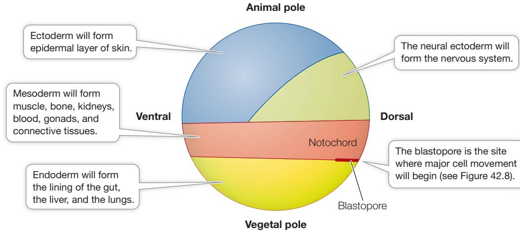
Figure 42.6 Fate Map of a Typical Amphibian Blastula
If some blastomeres can change their fate to compensate for the loss of other cells during cleavage and blastula formation, can those cells form an entire embryo? Yes, in species with regulative development, they can. If during cleavage or early blastula formation, the blastomeres are physically separated into two groups, both groups can produce complete embryos. Since the two embryos come from the same zygote, they will be monozygotic twins—genetically identical.
Non-identical twins occur when two separate eggs are fertilized by two separate sperm. Thus although identical twins are always the same sex, non-identical twins have a 50% chance of being the same sex (the same chance as two non-twin siblings). If genetic or environmental factors cause the blastula or blastocyst to split partially, the result is twins who are conjoined at some point on their bodies and usually share some of their organs and limbs. Conjoined twins occur in about 1 out of 50,000 human pregnancies.
Reproductive germ cells are determined early in cleavage
Molecules present in the egg cytoplasm determine which lineage of cells will eventually populate the gonads and become the reproductive stem cells—oogonia and spermatogonia. In fruit flies, at the ninth nuclear division (recall that the egg is a multinucleate syncytium at this stage; see Figure 42.3C), a group of nuclei migrate to the posterior pole of the egg where they become surrounded by pole plasm—cytoplasm containing a complex mixture of fibrils, mitochondria, specific proteins, and mRNAs. As the cellularization of the blastoderm proceeds, the nuclei within the pole plasm give rise to the lineage of cells that will eventually migrate to the gonads (when they form) and produce germ cells (eggs and sperm).
As in fruit flies, the germ cell lineage in frogs starts with a special type of cytoplasm—the germ cell plasm—localized to one part of the egg. As a result of cleavage, the germ cell plasm becomes enclosed within some of the cells in the vegetal hemisphere; descendants of these cells will eventually migrate to the gonads once those structures form. Germ cell plasm includes inhibitors of transcription and translation that prevent these cells from differentiating into anything other than germ cells.
Recap and Assess
Cleavage divides up the cytoplasm of the zygote such that different blastomeres contain different combinations of informational molecules. The amount of nutrients stored in the egg influences the pattern of cell cleavage that produces the blastula. Blastulation in mammals is different than in other vertebrates. The cells that will give rise to germ cell lineages are set aside very early in development.
- What characteristic of the eggs of different species relates to whether cleavage is complete or incomplete, and what is the exception?
- Why can gene expression during cleavage play developmental roles in mammals but not in sea urchins and frogs?
- Why can identical twins arise from a single egg in mammals but not in most invertebrates?
- Why is there a longer time between determination and differentiation in germ cells than in skin cells?
Of the next stage of development—gastrulation—the developmental biologist Lewis Wolpert once said, “It is not birth, marriage, or death, but gastrulation which is the most important time in your life.” During gastrulation, cell movements create new cell-to-cell contacts, which in turn set up signaling cascades that initiate the differentiation of cells and tissues and set the stage for the emergence of the body plan.
Gastrulation Generates Multiple Tissue Layers
Learning Objectives
- 42.3.1 Compare gastrulation in sea urchins, frogs, and reptiles.
- 42.3.2 Describe the experiment that revealed the role of the dorsal lip of the amphibian blastopore in embryonic development.
- 42.3.3 Describe cell movements that occur during gastrulation in sea urchins and amphibians.
- 42.3.4 Summarize how organizer cells achieve a pattern by appropriately expressing growth factor antagonists.
- 42.3.5 Explain the meaning of necessity and sufficiency in investigating potential inductive signals.
The blastula is typically a fluid-filled ball of cells. How does this simple ball of cells become an embryo made up of multiple tissue layers with head and tail ends and dorsal and ventral sides?
Gastrulation is the process whereby the blastula is transformed by massive movements of cells into an embryo with multiple tissue layers and distinct body axes. The resulting spatial relationships between tissues make possible the inductive interactions between cells that trigger differentiation and organ formation.
In the triploblastic animals (see Key Concept 29.1), three germ layers (also called cell layers or tissue layers, and not to be confused with germ cells) form during gastrulation:
The endoderm is the innermost germ layer, created as some blastomeres move to the inside of the embryo. The endoderm gives rise to the epithelial lining of the digestive tract, respiratory tract, pancreas, thyroid, and liver.
- The ectoderm is the outer germ layer, formed from those cells remaining on the outside of the embryo. The ectoderm contributes to the nervous system, including the eyes and ears; and to the epidermal layer of the skin and structures derived from skin, such as hair, feathers, nails or claws, sweat glands, oil glands, and even teeth and other tissues of the mouth.
- The mesoderm is the middle layer and is made up of cells that migrate between the endoderm and the ectoderm. The mesoderm contributes to many organs, including the heart, blood vessels, muscles, and bones.
The three germ layers are illustrated for a very early embryo in the fate map shown in Figure 42.6.
Some of the most interesting and important challenges in animal development have dealt with two related questions: what directs the cell movements of gastrulation, and what is responsible for the resulting patterns of cell differentiation and organ formation? Scientists have made significant progress in answering both these questions at the molecular level. In the following discussion we begin with sea urchin gastrulation because it is the simplest to conceptualize in spatial terms. We then describe the more complex pattern of gastrulation in frogs, and then the still more complex patterns in reptiles (including birds) and mammals.
Invagination at the vegetal pole initiates gastrulation in the sea urchin
The sea urchin blastula is a hollow ball of cells only one cell layer thick. At the end of blastulation the rate of mitosis slows, and the beginning of gastrulation is marked by a flattening of the vegetal hemisphere (Figure 42.7). Some cells at the vegetal pole break away from neighboring cells and migrate into the cavity to become mesenchyme—cells of the middle germ layer,
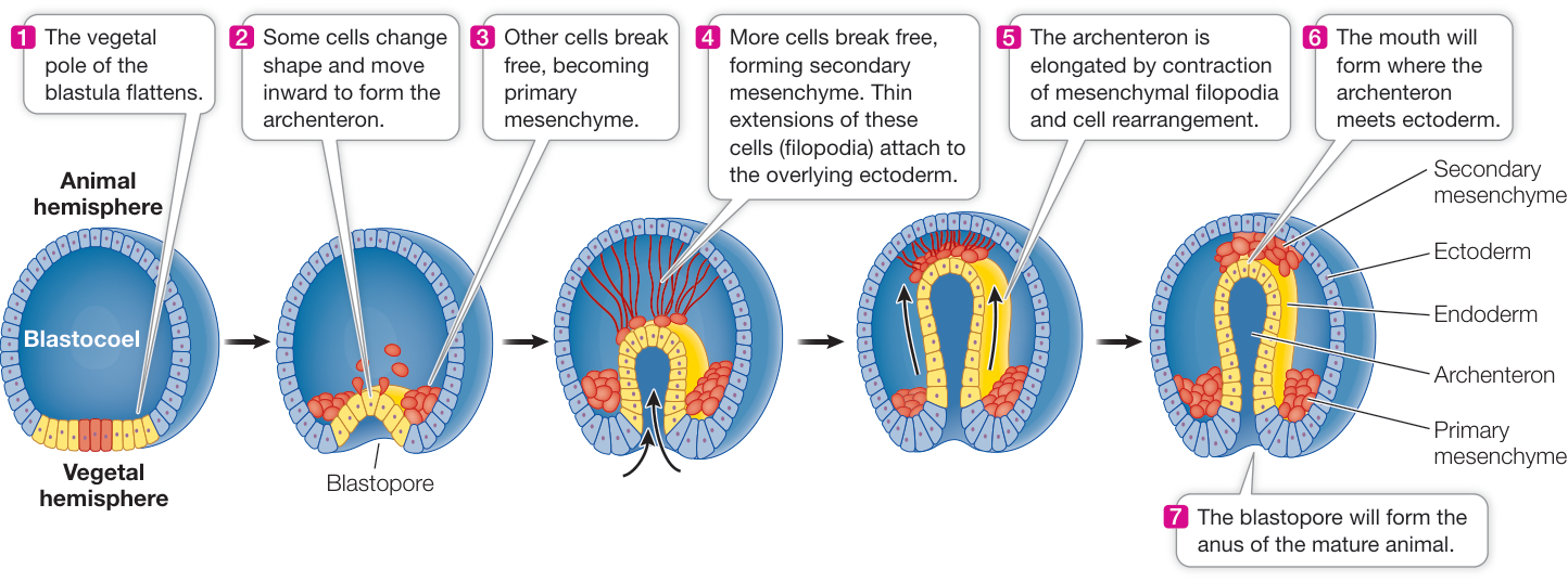
Figure 42.7 Gastrulation in Sea Urchins During gastrulation, cells move to new positions and form the three germ layers from which differentiated tissues develop.
Q: How do cells on the surface of the vegetal pole become the cells lining the gut?
the mesoderm. Mesenchymal cells are not organized in tightly packed sheets or tubes like epithelial cells are; they act as independent units, migrating into and among the other tissue layers. These early mesenchymal cells will contribute to the skeletal structures of the sea urchin.
The flattening at the vegetal pole results from changes in the shape of individual blastomeres. These cells, which are originally rather cuboidal, become wedge-shaped, with smaller outer edges and larger inner edges. As a result, the vegetal pole bulges inward, or invaginates, as if someone were poking a finger into a hollow ball (see Figure 42.7). The invaginating cells become endoderm and form the primitive gut, called the archenteron. The archenteron begins as a short stubby invagination, but its cells extend, flatten, interdigitate, and migrate over one another to form a long thin tube. This process is called convergent extension. As the archenteron gets closer to the opposite side of the blastocoel, more mesenchymal cells migrate out of it into the blastocoel. These cells remain attached to the tip of the archenteron and send out extensions called filopodia that adhere to the overlying ectoderm. The filopodia contract and pull the archenteron toward the overlying ectoderm. The mouth of the animal will form where the archenteron makes contact with this overlying ectoderm. The opening created by the invagination of the vegetal pole is called the blastopore, and it will become the anus of the animal.
Connect the Concepts Recall from Key Concept 29.1 that where the mouth and anus form during gastrulation distinguishes the two major groups of animals that have bilateral symmetry, the protostomes and the deuterostomes. If the blastopore becomes the mouth, the animals are classified as protostomes (“mouth first”); if the blastopore becomes the anus, the animals are classified as deuterostomes (“mouth second”).
What mechanisms control the various cell movements of sea urchin gastrulation? The immediate answer is that specific properties of particular blastomeres change. For example, some vegetal cells change shape and bulge into the blastocoel, and these cells become mesenchyme. Once they lose contact with their neighboring cells on the surface of the blastula, they send out filopodia that then move along an extracellular matrix of proteins laid down by the cells lining the blastocoel.
A deeper understanding of gastrulation requires that we discover the molecular mechanisms whereby different blastomeres develop different properties. Cleavage systematically divides the cytoplasm of the egg. The sea urchin blastula at the 64-cell stage is radially symmetrical, but it has polarity and consists of tiers of cells. As in the frog blastula, the top is the animal pole and the bottom is the vegetal pole. If the embryo is cut in half horizontally, only the cells from the vegetal pole are capable of initiating the development of a complete sea urchin. Such experiments show that the different cells of the blastula already have different developmental potentials. Why is this so? We now know that many transcriptional regulatory proteins are not uniformly distributed in the egg cytoplasm, and as cleavage progresses, these proteins end up in different groups of cells. Therefore specific sets of genes are activated in different cells, determining their different developmental capacities.
Frog gastrulation begins at the gray crescent
Amphibian blastulas are more than one cell layer thick and they contain stored nutrients called yolk; gastrulation is therefore more complex in amphibians than in sea urchins. Variation is considerable across different species of amphibians, so this brief account combines results from studies done on different species to produce a generalized picture of amphibian development.
Amphibian gastrulation begins when certain cells in the gray crescent region (see Figure 42.1) change their shapes and cell-adhesion properties. These cells bulge inward toward the blastocoel while they remain attached to the outer surface of the blastula by slender necks; because of their shape, they are called bottle cells. Bottle cells mark the spot where the dorsal lip of the blastopore will form (Figure 42.8).
As the bottle cells move inward, the dorsal lip is created, and a sheet of cells moves over it into the blastocoel. This process is called involution. One group of involuting cells is the prospective endoderm; these cells form the primitive gut, or archenteron. Another group will move between the endoderm and the outermost cells to form mesoderm. As with the extension of the archenteron in the sea urchin, these cell rearrangements involve convergent extension. The cells elongate in the direction of growth, but they also intercalate (move in between each other). As gastrulation proceeds, cells from the animal hemisphere flatten and move toward the site of involution in a process called epiboly. The blastopore lip widens and spreads ventrally, and eventually forms a complete circle surrounding a “plug” of yolk-rich cells. As cells continue to move inward through the blastopore, the archenteron grows, gradually displacing the blastocoel.
As gastrulation comes to an end, the amphibian embryo consists of three germ layers: ectoderm on the outside, endoderm on the inside, and mesoderm in between. The embryo also has a dorsal–ventral and anterior–posterior organization. Most important, the fates of specific regions of the endoderm, mesoderm, and ectoderm have been determined. The beautiful experiments revealing how determination takes place in the amphibian embryo are an old but exciting story.
The dorsal lip of the blastopore organizes the formation of the amphibian embryo
In the early 1900s the German biologist Hans Spemann was studying the development of salamander eggs. He was interested in finding out whether the nuclei of blastomeres remain capable of directing the development of complete embryos or whether these nuclei lose some developmental potential. With great patience and dexterity, he formed loops from single hairs taken from his baby daughter and tied them around fertilized eggs along the plane of the first cell division, effectively dividing the eggs in half, with the nucleus restricted to one side. The side containing the nucleus went through cell divisions and developed into a salamander; the other half simply degenerated. Up until the 16-cell stage, if one nucleus escaped to the other side of the constriction, twin salamanders could develop. Thus each of the nuclei of the blastula (at least up to the 16-cell stage) was capable of directing and supporting development of the whole organism (regulative development).
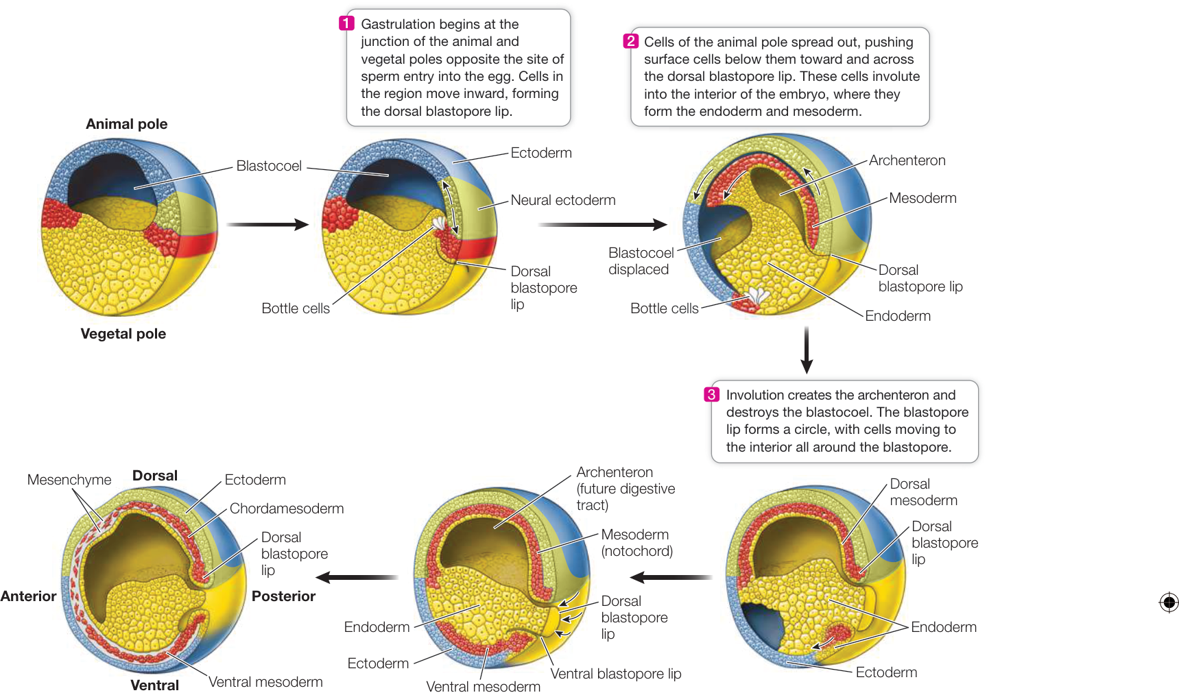
Figure 42.8 Gastrulation in the Frog Embryo Yellow, blue, green, and red in this diagram are matched to those colors in Figure 42.6, the frog fate map. (After R. E. Keller. 1986. In Developmental Biology: A Comprehensive Synthesis, Vol. 2, L. Browder (ed.), pp. 241–327. Springer: New York.)
Media Clip 42.1 Frog Gastrulation Time-Lapse Life12e.com/mc42.1
Q: Epiboly results in the formation of which tissues in the adult frog?
As often happens in science, Spemann’s bisection experiments revealed a new phenomenon. Sometimes the half of the blastula receiving an escaped nucleus did not develop. When his loops bisected the gray crescent of the egg, both halves of the zygote developed into a complete embryo. When he tied the loops so the gray crescent was on only one side of the constriction, however, only that half of the zygote developed into a complete embryo (Figure 42.9). When the half lacking gray crescent material received a nucleus, it underwent cell division but formed only a clump of undifferentiated cells that Spemann called a “belly piece.” Spemann hypothesized that cytoplasmic factors unequally distributed in the fertilized egg were necessary for gastrulation and the development of a normal salamander.
To further test the hypothesis that cells receiving different complements of cytoplasmic factors had different developmental fates, Spemann transplanted pieces of early gastrulas to various locations on other gastrulas. Guided by fate maps (see Figure 42.6), he was able to take a piece of ectoderm he knew would develop into the epidermis of the skin and transplant it to a region that normally becomes part of the nervous system, and vice versa.
When he performed these transplants in early gastrulas—when the blastopore was just beginning to form—the transplanted pieces always developed into tissues that were appropriate for the location where they were placed. Transplanted cells destined to become epidermis in their original location developed into nervous system tissue, and transplanted cells destined to become nervous system tissue in their original location developed into host epidermis. Thus Spemann learned that the fates of the transplanted cells had not been determined before the transplantation.
Simulation 42.1 Cell Fates
In late gastrulas, however, the same experiment yielded opposite results. Transplanted cells destined to become epidermis in their original location produced patches of skin cells in the host nervous system, and the transplanted cells from regions that would develop into nervous system tissue produced nervous tissue in the skin of the recipient. At some point during gastrulation, the fates of the embryonic cells had become determined.
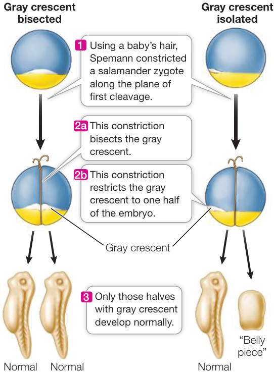
Figure 42.9 Gastrulation and the Gray Crescent Spemann’s research revealed that gastrulation and subsequent normal development in salamanders depend on cytoplasmic determinants localized in the region of the blastula that corresponds to the gray crescent region in the fertilized egg. (After H. Spemann. 1938. Embryonic Development and Induction. Yale University Press: New Haven, CT.)
Spemann’s next experiment, done with his student Hilde Mangold, produced momentous results: they transplanted the dorsal lip of the blastopore (Figure 42.10). When this small piece of tissue was transplanted into the presumptive belly area of another gastrula, it stimulated a second site of gastrulation—and a second complete embryo formed belly-to-belly with the original embryo. Because the dorsal lip of the blastopore was apparently capable of inducing the host tissue to form an entire embryo, Spemann and Mangold called the dorsal lip tissue the primary embryonic organizer, or simply the organizer. For more than 100 years, what has come to be called the Spemann–Mangold organizer has been an active area of research.
Transcription factors and growth factors underlie the organizer’s actions
The primary embryonic organizer has been studied intensively with molecular methods to discover the mechanisms involved in its action. The distribution of the transcription factor -catenin in the late blastula corresponds to the location of the organizer in the early gastrula, so -catenin is a candidate for the initiator of organizer activity. To prove that a protein is an inductive signal, it has to be shown that it is both necessary and sufficient for the proposed effect. In other words, the effect should not occur if the candidate protein is not present (necessity), and the candidate protein should be capable of inducing the effect where it would otherwise not occur (sufficiency).
The criteria of necessity and sufficiency have been satisfied for -catenin. If -catenin mRNA transcripts are depleted by injections of antisense RNA into the egg (see Key Concept 18.4), gastrulation does not occur. If -catenin is experimentally overexpressed in another region of the blastula, it can induce a second axis of embryo formation, as the transplanted dorsal lip did in the Spemann–Mangold experiments. Thus -catenin appears to be both necessary and sufficient for the formation of the primary embryonic organizer. But, -catenin is only one component of a complex signaling process. How the presence of -catenin creates the organizer, and how the organizer induces the beginnings of the body plan, involves a complex series of interactions between transcription factors and growth factors that control gene expression.
Properties of organizer cells change as they migrate from the dorsal lip
Organizer cells begin the process of formation of the dorsal lip of the blastopore. Specifically, these cells are at the center of the dorsal lip and involute, moving forward on the midline (i.e., the middle of the anterior–posterior axis). The first organizer cells to enter the embryo migrate anteriorly to become the head endoderm and head mesoderm. Here they induce neighboring cells to participate in making structures of the head. Organizer cells that involute later in gastrulation induce structures of the trunk, and the last of the organizer cells to move inward from the dorsal lip induce structures of the tail. How do the organizer cells change to enable them to induce head, trunk, or tail structures?
Inductive tissue interactions can suppress as well as activate. Goosecoid is a transcription factor expressed by early organizer cells. Goosecoid activates genes encoding certain signaling factors. As the early organizer cells move forward in the blastocoel, they come into contact with new populations of cells that produce several different growth factors. For head structures to form, certain of these growth factors have to be suppressed. The most anterior organizer cells, under the influence of Goosecoid, produce and release antagonists to those growth factors.
The induction of trunk structures requires suppression of a different set of growth factors. In organizer cells that involute later than the head organizers, Goosecoid is no longer the dominant transcription factor, and these cells express different growth factor antagonists. The induction of tail structures requires still different activities of the organizer cells that involute last. Thus the organizer cells express appropriate sets of growth factor antagonists at the right times to achieve different patterns of differentiation on the anterior–posterior axis.
The initiation of nervous system development also involves a suppressive tissue interaction. For a long time it was thought that the involuting organizer cells actively induced the overlying ectoderm to form nervous tissue rather than becoming epidermis. We now know, however, that epidermis is not the default state of the dorsal ectoderm. Rather, the underlying mesoderm secretes factors called BMP proteins that induce the ectoderm to become epidermis. The role of the involuting organizer cells is to block that induction, allowing the overlying ectodermal cells to follow what is really their default pathway—differentiation into nervous tissue.
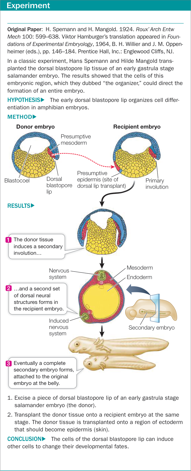
Figure 42.10 The Dorsal Lip Induces Embryonic Organization
The amount of yolk influences gastrulation
The eggs of reptiles, including birds, contain a massive amount of yolk, and the blastulas of these groups develop as a disc of cells on top of the yolk (see Figure 42.3B). We will use the chicken egg to show how gastrulation proceeds in a flat disc of cells rather than in a ball of cells.
Cleavage in the chick results in a flat, circular layer of cells called a blastodisc (Figure 42.11). Between the blastodisc and the yolk mass is a fluid-filled space. Some cells from the blastodisc break free and move into this space. These cells come together to form a continuous layer called the hypoblast, which will later contribute to extraembryonic membranes that support and nourish the developing embryo. The overlying cells make up the epiblast, from which the embryo will form. Thus the avian blastula is a flattened structure consisting of an upper epiblast and a lower hypoblast, which are joined at the margins of the blastodisc. The blastocoel is the fluid-filled space between the epiblast and hypoblast.
Gastrulation begins with a thickening in the posterior region of the epiblast, caused by the movement of cells toward the midline and then forward along the midline (see Figure 42.11). The result is a midline ridge called the primitive streak. A depression called the primitive groove forms along the length of the primitive streak. The primitive groove functions as the blastopore, and cells migrate through it into the blastocoel to become endoderm and mesoderm.
In the chick embryo, no archenteron forms, but the endoderm and mesoderm migrate forward to form the gut and other structures. At the anterior end of the primitive groove is a thickening called Hensen’s node, which is the equivalent of the dorsal lip in the amphibian blastopore. Many signaling molecules found in the frog organizer are also expressed in Hensen’s node. Cells moving into the blastocoel and moving anteriorly from Hensen’s node become the notochord and organize the chick embryo in a manner similar to that of the frog embryo.
Gastrulation in mammals is similar to avian gastrulation
Mammalian embryos (with the exception of protherians) derive their nourishment from the maternal circulation, and therefore mammalian eggs do not have large amounts of yolk constraining their cleavage and early development. Nevertheless, mammals evolved from reptilian ancestors, so it is not surprising that mammals and reptiles, including birds, share certain patterns of early development. Earlier we described the development of the mammalian inner cell mass (the equivalent of the avian blastodisc) and the outer trophoblast.
As in birds, in eutherians the inner cell mass splits into an upper layer called the epiblast and a lower layer called the hypoblast. The embryo forms from the epiblast, while the hypoblast contributes to the extraembryonic membranes that will encase the developing embryo and help form the placenta (see Figure 42.5). The epiblast also contributes to the extraembryonic membranes; specifically, it splits off an upper layer of cells that forms the amnion. The amnion grows to surround the developing embryo as a membranous sac filled with amniotic fluid. Gastrulation occurs in the mammalian epiblast just as it does in the avian epiblast. A primitive groove forms, and epiblast cells migrate through the groove to become layers of endoderm and mesoderm. At the top of the groove is the node which is homologous with Hensen’s node in birds.
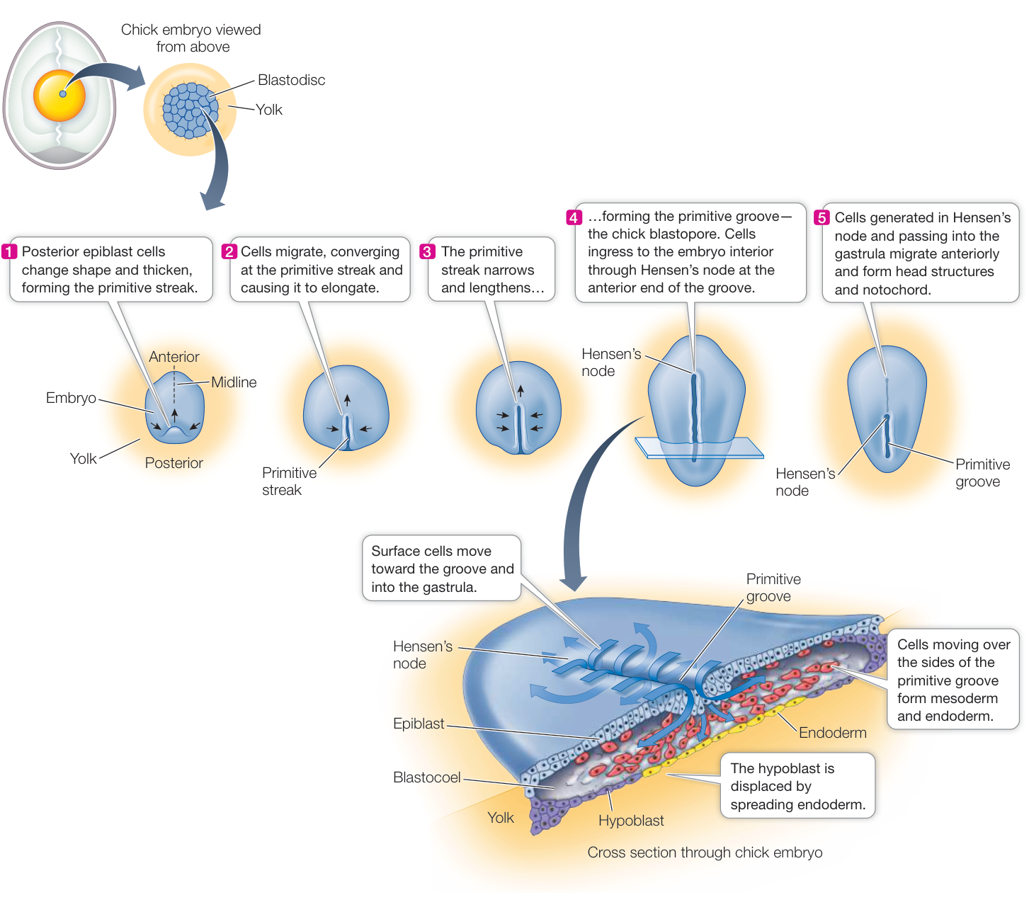
Figure 42.11 Gastrulation in Birds Because their eggs contain a large yolk mass, bird and other reptilian embryos have a flat-tened blastodisc and display a pattern of gastrulation very different from that of amphibians. (After B. I. Balinsky. 1975. An Introduction to Embryology. W. B. Saunders: Philadelphia.)
How is bilateral symmetry broken?
Gastrulation results in the anterior–posterior and dorsal–ventral patterning of the embryo, but all of the mechanisms discussed produce bilateral symmetry. At the opening of this chapter we introduced an early mechanism that breaks that symmetry to produce the well-known left–right asymmetries of the body. The human heart is on the left; the major lobe of the liver is on the right; the pancreas and spleen are on the left; the appendix is on the right; the major blood vessel leaving the heart, the aorta, bends to the left; the right lung has three lobes and the left lung has two; and so on. At the very beginning of gastrulation, the location where the involution of cells begins in a mammal is marked by a node of cells. The ventral surface of the node has ciliated cells. Some of these cilia are motile and others are not. The motile cilia create a leftward flow of extracellular fluid over the node. The experiments in Investigating Life: Does the Direction of Nodal Flow Influence the Development of Left–Right Asymmetry in the Mouse Embryo? investigate whether that directional flow is both necessary and sufficient to initiate signaling processes that break bilateral symmetry of the embryo.
Investigating Life: Does the Direction of Nodal Flow Influence the Development of Left-Right Asymmetry in the Mouse Embryo?
Experiment
Original Paper: S. Nonaka et al. 2002. Determination of left-right patterning of the mouse embryo by artificial nodal flow. Nature 418: 96–99.
HYPOTHESIS The leftward flow of extracellular fluid created by the beating of nodal primary cilia is the stimulus for breaking bilateral symmetry in organ development in the mouse.
METHOD
- Mount wild-type mouse embryos at different stages of development (presomite and 1, 2, or 3 somites; see Figure 42.13) in a culture medium-filled chamber with their nodal ends pointed upward.
- Artificially pump culture medium through the chamber either from the left (normal) or from the right (reversed) and at slow or fast flow rates.
- After 4 days assess the embryos as to the direction of looping (normal or reversed) of their developing heart tubes.
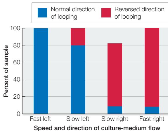
RESULTS Since there were different numbers of embryos in different conditions, and some embryos did not survive, the data are displayed as percent of the total sample under each condition that showed normal or reversed direction of looping of their heart tubes. For example, in the slow left column there were 16 total embryos: 12 had normal direction of looping and 4 had reversed looping. The graph displays how heart-looping patterns were affected in the presomite stage by the direction of culture-medium flow.
| Speed and direction of culture-medium flow a | ||||
|---|---|---|---|---|
| Genotype, stage | Fast left | Slow left | Slow right | Fast right |
| Wild type, presomite | 9N, OR, 10T | 12N, 4R, 16T | 13N, 3R, 16T | 2N, 21R, 24T |
aN = normal heart looping; R = reversed looping; T = total embryos in sample.
CONCLUSION When extracellular fluid moved across the node from right to left, the direction of heart-tube looping was normal. If this signal was weak (slow left flow), or if a weak reversed signal was applied (slow right flow), there was a small effect on normal direction of heart-tube looping, but when the reversed signal was strong, the heart-tube looping was in the reversed direction. These results support the conclusion that nodal flow is an important signal in breaking bilateral symmetry in the mouse embryo.
Work with the Data
The experiment illustrated in the graph was done on presomite embryos—before any somites had formed. Developmental signals usually have a time window of effectiveness given that developmental events are sequential and different events must be coordinated. Is there a time window within which nodal flow determines left-right symmetry in the embryo? To investigate the timing of sensitivity to nodal flow, another experiment was conducted using only fast right flow on early embryos at different stages of development.
| Genotype, stage | Results |
|---|---|
| Wild type, presomite | 2N, 21R, 24T |
| Wild type, 1-somite | 9N, OR, 12T |
| Wild type, 2-somite | 22N, OR, 22T |
| Wild type, 3-somite | 14N, OR, 14T |
QUESTIONS
- Plot the data as the percent of responses on the y axis and developmental stage on the x axis.
- Why did this experiment not include left flow as a stimulus?
- Do these data provide evidence for a sensitive period for nodal flow determining left-right asymmetry?
- What do you conclude from the results on the 1-, 2-, and 3-somite wild-type embryos?
Recap and Assess
The cell movements of gastrulation convert the blastula into an embryo with three tissue layers. New contacts between cells set up inductive signaling interactions that determine cell fates. Dorsal lip tissue is the source of organizer cells that induce development of head, trunk, and tail structures.
- The sea urchin blastula develops into a radially symmetrical larva, but the frog blastula develops into a bilateral embryo. How does this difference relate to processes of gastrulation in these two types of animals?
- What is meant by the criteria of necessity and sufficiency, and how have modern methods shown that the Spemann–Mangold primary embryonic organizer satisfies those criteria?
- How do the cells of the primary embryonic organizer induce different structures on the anterior–posterior axis as they migrate from the dorsal lip to the head region of the embryo?
- How does gastrulation in reptiles compare with that in amphibians?
We have described how the fertilized egg develops into an embryo with three germ layers and how cellular signals trigger different patterns of differentiation. In the next key concept we describe how organs and organ systems develop.
Organs Develop from the Three Germ Layers
Learning Objectives
42.4.1 Describe how nervous system development begins with neurulation.
42.4.2 Describe the formation of somites and their role in development.
42.4.3 Explain how genetic and environmental factors can influence neural tube formation.
42.4.4 Explain how a limited number of Hox genes can organize complex anterior–posterior patterning of the vertebrate embryo.
Gastrulation produces an embryo with three germ layers that are positioned to influence one another through inductive tissue interactions. During the next phase of development, called organogenesis, organs and organ systems develop simultaneously and in coordination with each other. In the chordates (see Key Concept 31.1), an early process of organogenesis is neurulation, the initiation of the nervous system. We examine neurulation in the amphibian embryo, but it occurs in a similar fashion in reptiles (including birds) and in mammals.
The organizer sets the stage for organogenesis
One group of cells that passes over the dorsal lip of the blastopore moves anteriorly and becomes the endodermal lining of the digestive tract. Another group of cells that passes over the dorsal lip on the midline becomes chordamesoderm, so named because it forms a rod of mesoderm—the notochord—that extends down the center of the embryo. These cells also have important organizer functions (see Figure 42.8). The notochord gives structural support to the developing embryo, and in vertebrates it is replaced by the vertebral column. The organizing capacity of the chordamesoderm enables the overlying ectoderm to become neural ectoderm.
Neurulation involves the formation of an internal neural tube from an external sheet of cells. The first signs of neurulation are flattening and thickening of the ectoderm overlying the notochord; this thickened area forms the neural plate (Figure 42.12A). The edges of the neural plate that run in an anterior–posterior direction continue to thicken to form ridges or folds. Between these neural folds, a groove forms and deepens as the folds roll over it to converge on the midline. The folds fuse, forming a cylinder, the neural tube, and a continuous overlying layer of epidermal ectoderm (Figure 42.12 B–D).
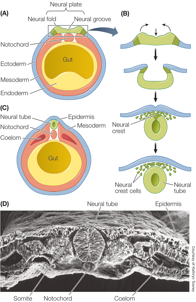
Figure 42.12 Neurulation in a Vertebrate (A) At the start of neurulation, the ectoderm of the neural plate (green) is flat. (B) The neural plate invaginates and folds, forming a tube. (C, D) The completely formed neural tube seen (C) in diagrammatic form and (D) in a scanning electron micrograph of a chick embryo.
Cells from the most lateral portions of the neural plate do not become part of the neural tube, but disassociate from it and come to lie between the neural tube and the overlying epidermis. These neural crest cells migrate outward to lead the development of the connections between the central nervous system (brain and spinal cord) and the rest of the body. Neural crest cells generate many diverse structures, including jaws, skull, face, pigment cells, glands, smooth muscle, and many others.
The neural tube develops bulges at the anterior end, which become the major divisions of the brain; the rest of the tube becomes the spinal cord. In humans, failure of the neural folds to fuse in this posterior region results in spina bifida, a birth defect in which the spinal cord is exposed because the vertebrae do not fuse. If the folds fail to fuse at the anterior end, an infant can develop without a forebrain (a condition called anencephaly). Although several genetic factors can cause these defects, other factors are environmental, including maternal diet. The incidence of neural tube defects in the United States in the early 1900s was as high as 1 in 300 live births; today it is less than 1 in 1,000. A major factor in this improvement has been the inclusion of folate (a B vitamin) in the mother’s diet. It is essential for pregnant women to ingest sufficient folate.
Body segmentation is an early feature of vertebrate development
The vertebrate body plan, like that of arthropods, consists of repeating segments that are modified during development. These segments are most evident as the repeating patterns of vertebrae, ribs, nerves, and muscles along the anterior–posterior axis.
As the neural tube forms, mesodermal tissues gather along the sides of the notochord to form separate, segmented blocks of cells called somites (Figure 42.13). Somites produce cells that will become the vertebrae, ribs, muscles of the trunk, and limbs.
Nerves that connect the brain and spinal cord with tissues and organs throughout the body are also arranged segmentally. The somites help guide the organization of these peripheral nerves, but the nerves are not of mesodermal origin. As you saw earlier, when the neural tube fuses, the neural crest cells break loose and migrate inward between the epidermis and the somites and also through the somites. These neural crest cells have diverse fates, including the development of peripheral nerves.
As development progresses, the different segments of the body change. Regions of the spinal cord differ, regions of the vertebral column differ in that some vertebrae grow ribs of various sizes and others do not, forelegs arise in the anterior part of the embryo, and hind legs arise in the posterior region.
Hox genes control differentiation along the anterior–posterior axis
How is mesoderm in the anterior part of a mouse embryo programmed to produce forelegs rather than hind legs? In Key Concept 21.4 you saw how homeotic genes control body segmentation in Drosophila. You also learned that all homeotic genes contain a DNA sequence called the homeobox. In vertebrates, the homeotic genes that control differentiation along the anterior–posterior body axis are called Hox genes
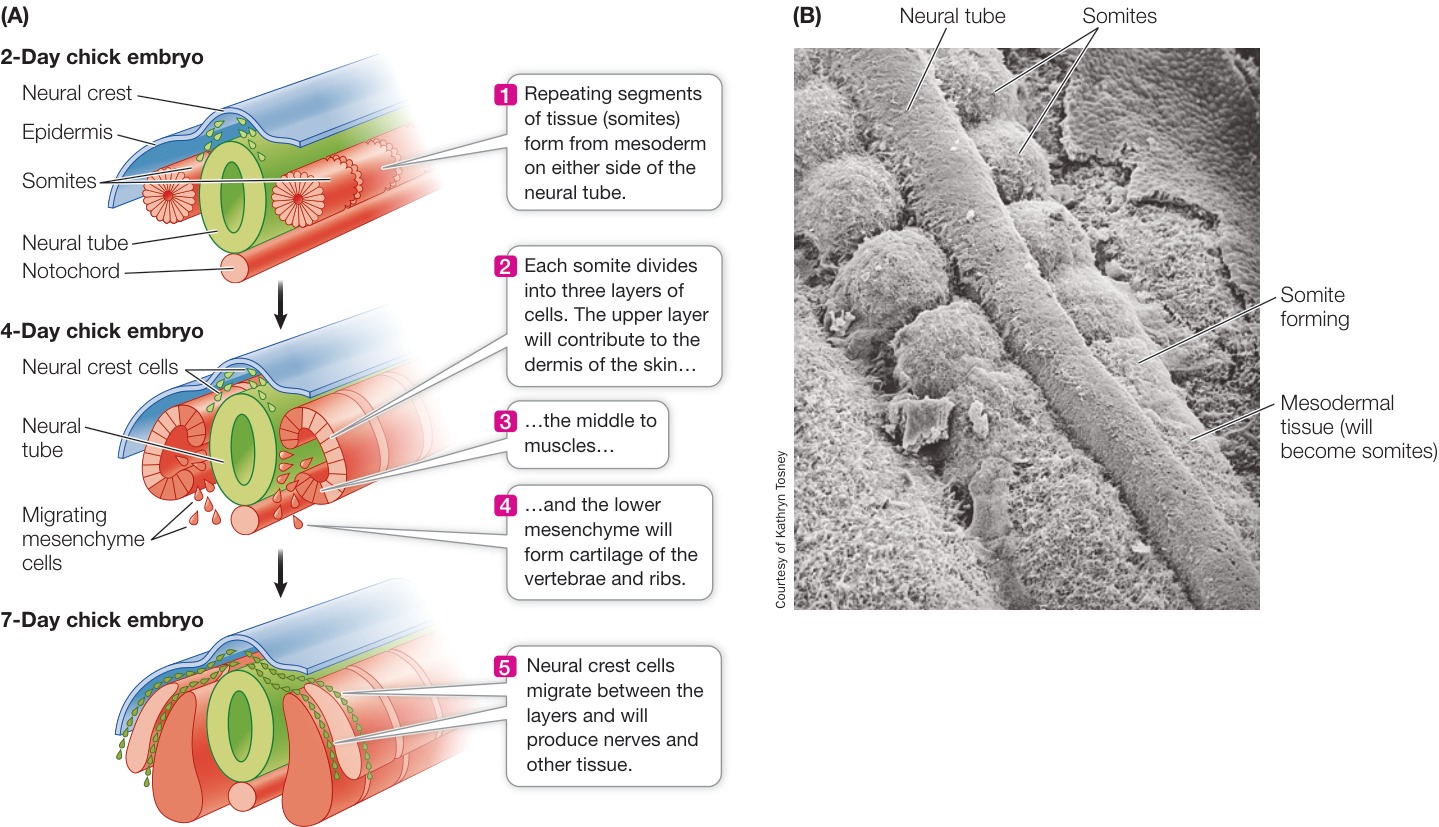
Figure 42.13 Developing Body Segmentation (A) Repeating blocks of tissue called somites form on either side of the neural tube. Muscle, cartilage, bone, and the inner layer of the skin form from the somites. (B) In this scanning electron micrograph of somite formation in a chick embryo, the overlying ectoderm has been removed and the neural tube and somites are seen from above.
In mammals, four Hox gene complexes reside on different chromosomes in clusters of about ten genes each. Remarkably, the temporal and spatial expression of these genes generally follows the same pattern as their linear order on their chromosome. That is, the Hox genes closest to the 3’ end of each gene complex are expressed first and in the anterior of the embryo. The Hox genes at the 5’ end of the gene complex are expressed later and in a more posterior part of the embryo. As a result, different segments of the embryo receive different combinations of Hox gene products, which serve as transcription factors (Figure 42.14; see also Figure 21.15).
Whereas Hox genes give cells information about their position on the anterior–posterior body axis, other genes provide information about their dorsal–ventral position. Tissues in each segment of the body differentiate according to their dorsal–ventral location. The notochord provides many of these signals.
After body segmentation develops, the formation of organs and organ systems progresses rapidly. The development of an organ involves extensive inductive interactions. An example is the ability of the chordamesoderm to induce overlying ectoderm to become neural ectoderm, and form the neural tube (see Figure 42.12). These inductive interactions are a current focus of study for developmental biologists.
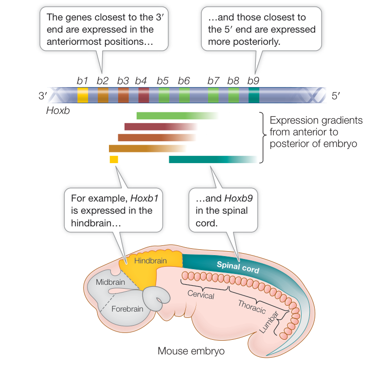
Figure 42.14 Hox Genes Control Body Segmentation Hox genes are expressed along the anterior–posterior axis of the embryo in the same order as their arrangement between the 3’ and 5’ ends of the gene complex. As a result of gene duplication during evolution, vertebrates have four copies of the Hox gene complex shown.
Recap and Assess
Gastrulation sets up tissue interactions that initiate organogenesis. Neurulation is initiated by organizer mesoderm that forms the notochord. Neurulation results in the formation of the central nervous system, and associated neural crest cells form the peripheral nerves. Suites of Hox genes are expressed sequentially in vertebrates, but in overlapping patterns providing a variety of signaling molecule patterns that govern differentiation on the anterior–posterior axis.
- How does the ectoderm overlying the notochord respond to chordamesoderm signals to form a neural tube as well as neural crest cells?
- What might be some consequences of a disruption in somite formation in the embryo?
- How do Hox genes generate a diversity of signals sufficient to control differentiation of tissues and organs on the anterior–posterior axis?
You have seen how the basic structure of the developing embryo arises, through the establishment of the anterior–posterior and dorsal–ventral axes, the formation of the neural tube, and the emergence of a segmented body plan. In the next key concept we examine the developmental events that result in the formation of structures that support the developing embryo: the extraembryonic membranes and the placenta.
Extraembryonic Membranes Nurture Avian and Mammalian Embryos
Learning Objectives
42.5.1 Describe the formation of the yolk sac, amnion, chorion, and allantois, using the chick as an example.
42.5.2 Explain how the placenta forms.
42.5.3 Explain why a first-trimester embryo is particularly sensitive to environmental risks for birth defects.
There is more to a developing reptile or mammal than the embryo itself. The embryos of these vertebrates are surrounded by several extraembryonic membranes that originate from the embryo but are not part of it. Extraembryonic membranes function in nutrition, gas exchange, and waste removal. In mammals they interact with tissues of the mother to form the placenta. The evolutionary relationships between the extraembryonic membranes of birds and mammals were discussed in Key Concept 31.4.
Birds develop four extraembryonic membranes
The chicken provides a good example of how extraembryonic membranes form from the germ layers created during gastrulation. In the chick, four membranes form—the yolk sac, the allantois, the amnion, and the chorion. The yolk sac is the first to form, and it does so by extension of the hypoblast layer along with some adjacent mesoderm. The yolk sac grows to enclose the entire body of yolk in the egg (Figure 42.15). It constricts at the top to create a tube that is continuous with the gut of the embryo. However, yolk does not pass through this tube. Yolk is digested by the cells of the yolk sac, and the nutrients are transported to the embryo through blood vessels that form from mesoderm and line the outer surface of the yolk sac. The allantois is also an outgrowth of the extraembryonic endoderm plus adjacent mesoderm. It forms a sac for storage of metabolic wastes.
Ectoderm and mesoderm combine and extend beyond the limits of the embryo to form the other extraembryonic membranes.
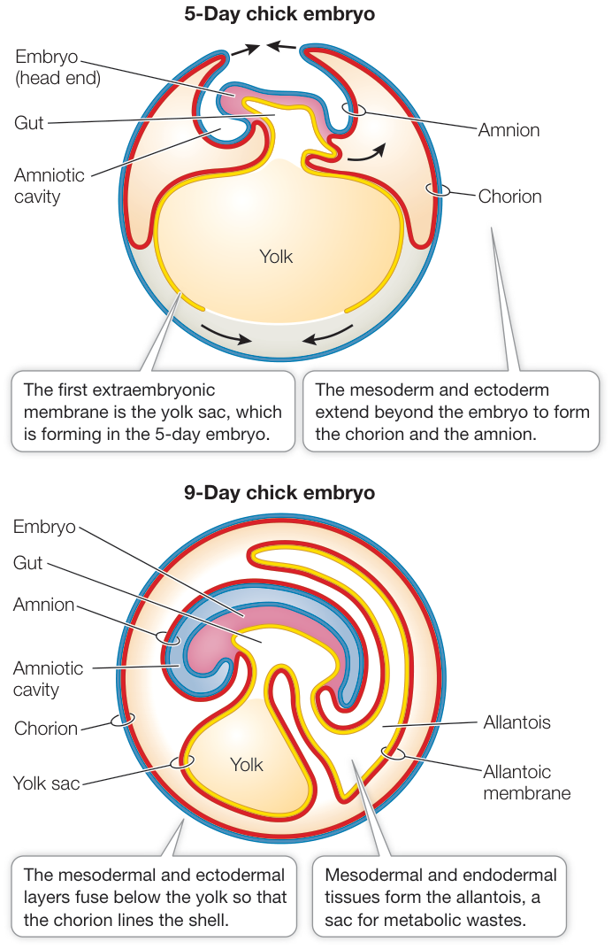
Figure 42.15 The Extraembryonic Membranes In birds (and other reptiles) and mammals, the embryo constructs four extraembryonic membranes. In birds, the yolk sac encloses the yolk, and the amnion and chorion enclose the embryo. Fluids secreted by the amnion fill the amniotic cavity, providing an aqueous environment for the embryo. The chorion, along with the allantois, mediates gas exchange between the embryo and its environment. The allantois stores the embryo’s waste products. (See also Figure 31.19.)
Two layers of cells extend all along the inside of the eggshell, both over the embryo and below the yolk sac. Where they meet, they fuse, forming two membranes, the inner amnion and the outer chorion. The amnion surrounds the embryo, forming the amniotic cavity. The amnion secretes fluid into the cavity, providing a protective environment for the embryo. The outer membrane, the chorion, forms a continuous membrane just under the eggshell (see Figure 42.15) that limits water loss from the egg. The exchange of and between the embryo and the environment requires diffusion of those gases across the chorion and the allantois.
The mammalian placenta forms from extraembryonic membranes
In eutherian mammals, the first extraembryonic membrane to form is the trophoblast (see Figures 42.4C and 42.5). When the blastocyst reaches the uterus and hatches from its encapsulating zona pellucida, trophoblast cells interact directly with the endometrium. Adhesion molecules expressed on the surfaces of these cells attach them to the uterine wall. By secreting proteolytic enzymes, the trophoblast burrows into the endometrium, beginning the process of implantation. Eventually the entire trophoblast is within the wall of the uterus. The trophoblast cells send out numerous projections, or villi, to increase the surface area of contact with maternal blood.
Meanwhile, the hypoblast cells proliferate to form what in the bird would be the yolk sac. But there is virtually no yolk in eggs of eutherian mammals; instead the yolk sac contributes mesodermal tissues that interact with trophoblast tissues to form the chorion. The chorion, along with tissues of the uterine wall, produces the placenta, the organ that exchanges nutrients, respiratory gases, and metabolic wastes between the mother and the embryo (Figure 42.16).
At the same time the yolk sac is forming from the hypoblast, the epiblast produces the amnion, which grows to enclose the entire embryo in a fluid-filled amniotic cavity. The rupturing of the amnion and chorion and the loss of amniotic fluid (the “water breaks”) herald the onset of labor in humans.
An allantois also develops in mammals, but its importance depends on how well nitrogenous wastes can be removed by the blood vessels of the placenta. The human allantoic sac is small because the human placenta deals effectively with the fetal nitrogenous wastes. In contrast, the pig placenta is less effective at clearing nitrogenous wastes from the fetus, so the pig’s allantoic sac is large. In humans and other eutherian mammals, the allantois contributes to the formation of the umbilical cord, by which the embryo is attached to the chorionic placenta (see Figure 31.19B). It is through the blood vessels of the umbilical cord that nutrients and oxygen from the mother reach the developing fetus, and wastes, including carbon dioxide and urea, are removed.
Human gestation is divided into trimesters
In humans, gestation, or pregnancy, lasts about 266 days, or 9 months. Gestation is shorter in smaller mammals (21 days in mice) and longer in larger mammals (600 days in elephants). The events of human gestation (Figure 42.17) are divided into three trimesters of roughly 3 months each. The first trimester
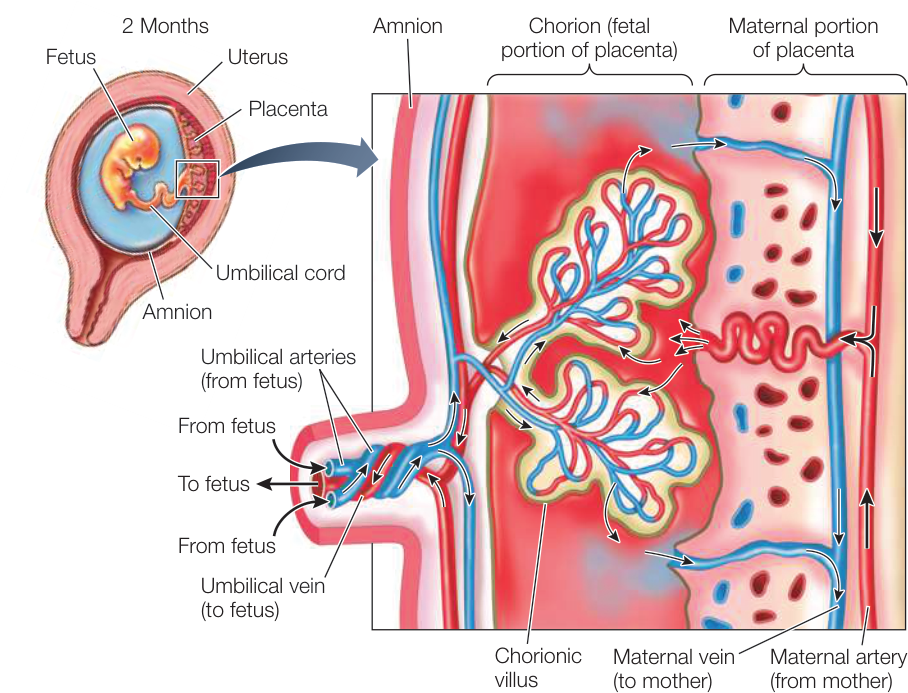
Figure 42.16 The Mammalian Placenta In humans and most other mammals, nutrients and wastes are exchanged between maternal and fetal blood in the placenta, which forms from the chorion and tissues of the uterine wall. The embryo is attached to the placenta by the umbilical cord. Embryonic blood vessels invade the placental tissue to form fingerlike chorionic villi. Maternal blood flows into the spaces surrounding the villi, and placental blood flows through the villi so nutrients and respiratory gases can be exchanged between the maternal and fetal blood.
(see Figure 42.17A) is a time of rapid cell division and tissue differentiation. Because signal transduction cascades that determine sequences of developmental processes are in their early stages, the first trimester is the period when the embryo is most sensitive to damage from radiation, drugs, chemicals, and pathogens that can cause birth defects. An embryo can be damaged before the mother even realizes she is pregnant. A classic case is that of thalidomide, a drug widely prescribed in Europe in the late 1950s to treat nausea. Women who took this drug in the fourth and fifth weeks of pregnancy, when the embryo’s limbs are beginning to form, gave birth to children with missing or severely malformed arms and legs.
During the second trimester the fetus grows rapidly; its limbs elongate, and its fingers, toes, and facial features become well formed (see Figure 42.17B). Eyebrows and fingernails grow, and the fetus’s nervous system develops rapidly. Fetal movements are first felt by the mother early in the second trimester and become progressively stronger and more coordinated.
The fetus grows rapidly during the third trimester. As this final stage approaches its end (see Figure 42.17C), the internal organs mature. The digestive system begins to function, the liver stores glycogen, the kidneys produce urine, and the brain undergoes cycles of sleep and waking. A human infant is born when the last of its critical organs—the lungs—mature.
Although the first-trimester embryo is the most susceptible to adverse effects of drugs, chemicals, and diseases, the potential for serious effects from exposure to environmental factors exists throughout pregnancy. Protein malnutrition and exposure to alcohol and cigarette smoke are examples of factors that can result in low birth weight, intellectual disability, and other developmental complications.
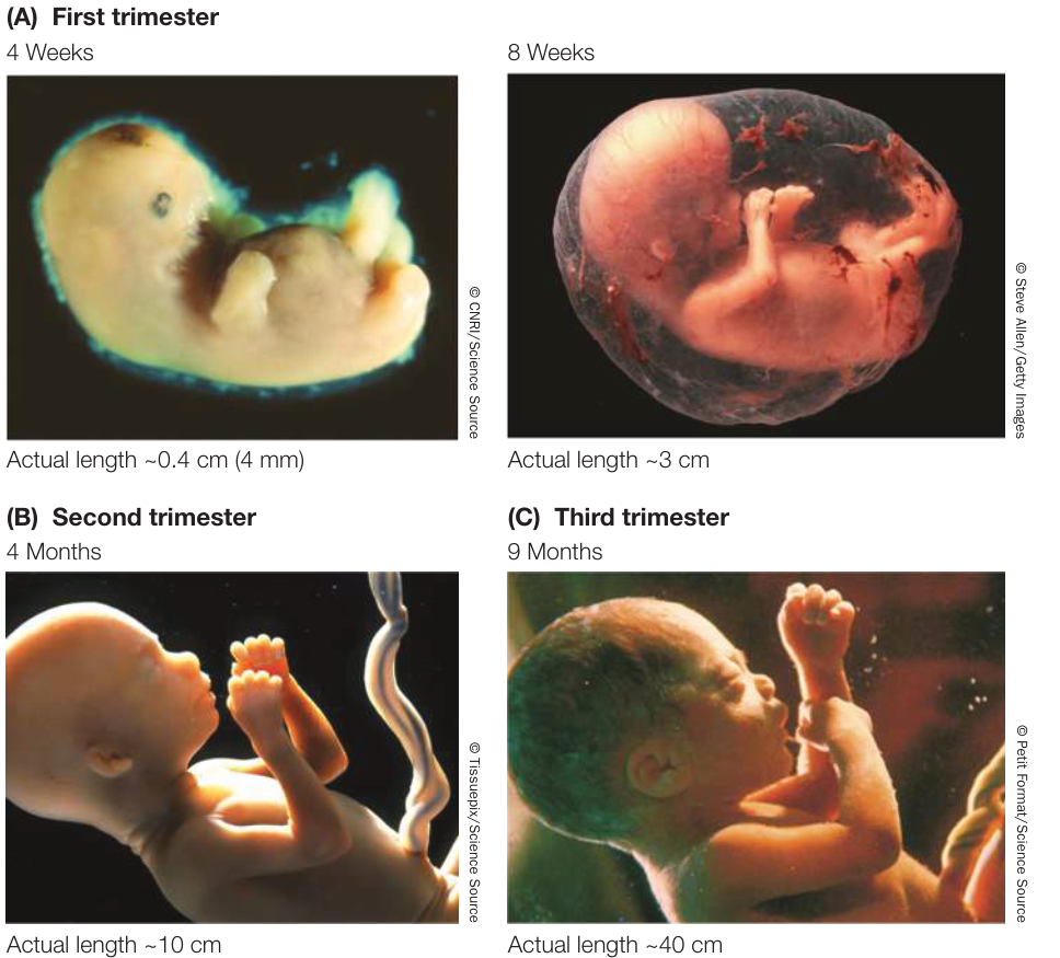
Figure 42.17 Stages of Human Development (A) At 4 weeks, most of the embryo’s organ systems have been formed and the heart is beating. The body structures of this 8-week-old embryo are forming rapidly. (B) At 4 months, the fetus has fully formed limbs with fingers and toes and moves freely within the amniotic cavity. (C) This fetus is well along in its ninth month. Soon its lungs will be mature enough to trigger the onset of contractions and birth.
Recap and Assess
The extraembryonic membranes of reptiles and mammals sustain the growing embryo. In reptiles these membranes surround the embryo within the shelled egg. In mammals the extraembryonic membranes form the placenta, an organ that exchanges nutrients, respiratory gases, and metabolic wastes between the mother and the embryo. Human gestation lasts 9 months. At the end of the first trimester, the fetus is very small but most of its organs have begun to form. In the second trimester, limbs elongate and the fetus moves. By the end of the third trimester, most organs have begun to function.
- Which germ layers participate in the development of the chick’s yolk sac, allantois, chorion, and amnion?
- What is the origin of the placenta in mammalian development?
- Why is a first-trimester embryo particularly sensitive to environmental risks?
▶ Investigating LIFE
QA How does the embryo know left from right?
At the beginning of gastrulation in the mammal, when midline cells of the epiblast begin to move inward to create the primitive groove, a region called the node forms at the anterior end of the groove. Motile cilia at the node create a flow of extracellular fluid across the node. Nonmotile nodal cilia are believed to sense the direction of fluid flow across the node. There are two hypotheses about how these cilia function, one chemical and the other mechanical. Both sensory mechanisms are used by cilia elsewhere in the body. In the nose, cilia of the olfactory cells sense chemicals. In the ear, cilia are bent by sound waves, which stimulates ion channels to open. The leftward flow of fluid across the node imposes mechanical forces on the nonmotile cilia that differ on the two sides of the node. Other research, however, supports the possibility of chemical signaling. Research on the mouse node shows that the beating of the cilia causes proteins of a certain size range to form a concentration gradient across the node. More important, it was discovered that nodal cells secrete small vesicles that are swept to the left side of the node by the flow, and these vesicles burst when they contact the nonmotile cilia of surrounding cells. The contents of these vesicles could be the chemical signals that initiate left-right asymmetry in gene expression and development.
Future directions
A major player in the determination of left-right asymmetry is the primary cilium of cells in the node that forms during gastrulation. However, just about every cell in the body has a primary cilium. Until fairly recently, the primary cilia were thought to be vestigial structures or simply organelles to sequester the centriole and thereby inhibit cell division. We now know that the primary cilia of cells function widely in mechanisms of cell signaling and in transduction of physical and chemical stimuli. Just about every organ in the body uses primary cilia for essential functions. Primary cilia have been implicated in many medical conditions such as cancer, kidney disease, obesity, blindness, and developmental disorders. In the near future we will be seeing lots of new discoveries about the functions of primary cilia.

© Nerthuz/Getty Images
Visual Summary

You should be able to relate each summary to the adjacent figures. If you go to this visual summary in Achieve, you can follow links to figures, animations, activities, and simulations that will help you consolidate the material.
Fertilization Activates Development
- ■ The sperm contributes a haploid nucleus and a centriole to the zygote. The egg contributes a haploid nucleus, nutrients, organelles, mRNAs, and proteins.
- ■ The amphibian egg is radially symmetrical, with an animal and vegetal hemisphere. The site of sperm entry defines the anterior-posterior axis and therefore the left-right sides of the zygote.
- ■ Sperm entry into the amphibian egg stimulates rotation of the cortical cytoplasm, forming the gray crescent on the side opposite the site of sperm entry.
- ■ Microtubules organized by the centriole move organelles and proteins, including a GSK-3 inhibitor, from the vegetal area toward the gray crescent. GSK-3 breaks down -catenin, so its inhibition enhances -catenin concentration in the gray crescent area, instructing the first stages of development.
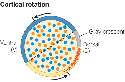
Figure 42.2
Questions
- Why is mitochondrial DNA especially useful for investigating the genealogical histories?
- What event following sperm entry breaks the radial symmetry of the frog egg?
- How does the sperm contribute to the formation of the gray crescent?
Mitosis Divides Up the Early Embryo
- Complete cleavage completely divides the egg; incomplete cleavage does not, leaving the yolk undivided. Complete cleavage can be radial, with successive mitotic spindles at right angles, or spiral, with successive mitotic spindles at oblique angles. In rotational cleavage, the type of complete cleavage seen in mammals, the mitotic planes of the second division are at right angles to each other. Superficial cleavage divides nuclei, which later are partitioned into individual cells.
- Cleavage results in autonomous specification if the fates of individual blastomeres are determined, and in regulated specification if the fates of individual blastomeres are dependent on their environment.
- Identical twins develop from the same fertilized egg. Non-identical twins develop simultaneously from separate fertilized eggs.
- Reproductive germ cells remain undetermined and totipotent throughout development and migrate into the gonads once they have formed.
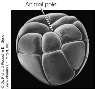
Figure 42.3
Questions
- In a species with autonomous specification, is it possible for an individual to have identical twins? If not, why?
- Why is cleavage in birds and fishes called discoidal?
- Why is it possible to genotype human blastocysts growing in culture without damaging the developing embryo?
Gastrulation Generates Multiple Tissue Layers
- Gastrulation involves massive cell movements that turn a ball (sea urchin, frog) or sheet (reptile) of cells into a cellular mass with three germ layers: endoderm, ectoderm, and mesoderm.
- Transplanting the dorsal lip of the blastopore from one salamander gastrula to the belly region of another resulted in two belly-to-belly embryos and led to the concept of the primary embryonic organizer.
- The transcription factor -catenin is responsible for inducing the primary embryonic organizer. If it is depleted in the egg by antisense RNA, gastrulation does not occur (demonstrating necessity), but if it is overexpressed in a region of the blastula, a second embryo forms (demonstrating sufficiency).
- As cells from the surface of the amphibian gastrula move over the dorsal lip of the blastopore and migrate forward, they release transcription factors that antagonize locally produced growth factors, thus altering the developmental fates of the cells in those areas.
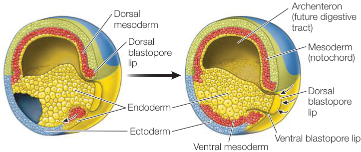
Figure 42.8 1. Why did Spemann’s constriction of a frog egg through the gray crescent result in two tadpoles, whereas constriction that did not bisect the gray crescent resulted in only one tadpole? 2. What are the three germ layers formed by gastrulation in the frog, what are their origins, and what are their fates? 3. How and why are amphibian and avian gastrulation different?
Organs Develop from the Three Germ Layers
- ■ Development of the nervous system begins by folding of the dorsal neural plate to form a neural tube. Mesodermal cells adjacent to the neural tube form segmentally arranged blocks of cells called somites, which will become skin, muscle, and bone.
- ■ Neural crest cells break away from the neural tube and migrate between somites to become peripheral nerves and many other structures.
- ■ Genetic, nutritional, and environmental factors can cause abnormalities in neural tube development.
- ■ Anterior–posterior patterning of the vertebrate embryo is controlled by clusters of Hox genes that are arranged linearly on different chromosomes. Hox genes are expressed in the embryo in the same sequence as they occur on the chromosomes, which results in different segments of the embryo receiving different combinations of transcription factors.
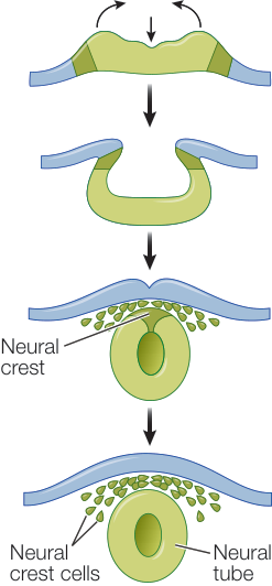
Figure 42.12
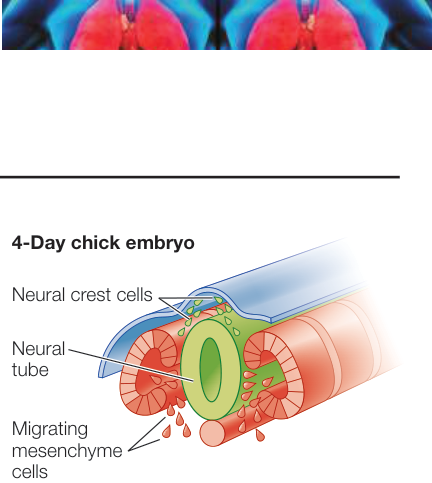
Figure 42.13 Questions
- How does a plate of dorsal ectoderm become the neural tube of the amphibian embryo?
- What are neural crest cells, and what do they do?
- What is the relationship between the positions of Hox genes on their chromosomes and their location of expression in the embryo?
Extraembryonic Membranes Nurture Avian and Mammalian Embryos
- ■ The embryos of reptiles and mammals are protected and nurtured by four extraembryonic membranes. In reptiles the yolk sac surrounds the yolk and provides nutrients to the embryo, the chorion lines the eggshell and participates in gas exchange, the amnion surrounds the embryo and encloses it in an aqueous environment, and the allantois stores metabolic wastes.
- ■ In mammals the chorion and the trophoblast cells interact with the maternal uterus to form a placenta, which provides the embryo with nutrient and gas exchange.
- ■ The first-trimester embryo is particularly sensitive to environmental risks because this is a time of rapid cell divisions and of many signaling cascades responsible for tissue differentiation and organ formation.
- ■ Human pregnancy, or gestation, lasts 9 months. The embryo forms in the first trimester; during this time, it is most vulnerable to environmental factors that can lead to birth defects. During the second and third trimesters the fetus grows, external features form, and the organ systems mature.
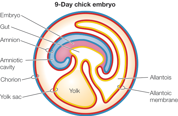
Figure 42.15 Questions
- What is the function of the allantois in the avian embryo, and why is an allantois less important in the mammalian embryo?
- Compare the function of the chorion in birds and mammals.
- Why are fetal movements not felt by the mother in the first trimester?