Chapter 44Sensory Systems
Key Concepts
44.1 Sensory Receptor Cells Convert Stimuli into Action Potentials
44.2 Chemoreceptors Respond to Specific Molecules
44.3 Mechanoreceptors Respond to Physical Forces
44.4 Photoreceptors Respond to Light
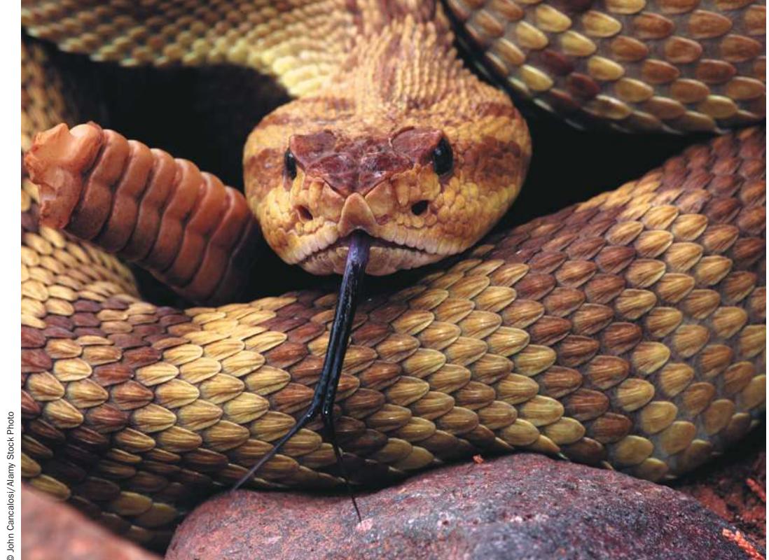
The “holes” below the eyes of this rattlesnake are the pit organs that detect infrared radiation from the snake’s preferred prey—small rodents—with precision, even in total darkness. The forked tongue also provides information, picking up chemical signals (odors) that are detected by a specialized organ in the roof of the snake’s mouth.
© John Cancales/Alany Stock Photo
Investigating Life: Seeing in the Dark
A rattlesnake can see to strike a running rodent in complete darkness. How can this be, when “seeing” means using the eyes to detect light, and “complete darkness” means no light? But these definitions are based on human capabilities. What we experience as light is only a small portion of the spectrum of electromagnetic radiation. We see wavelengths that we call red, orange, yellow, green, blue, indigo, and violet, but other animals see wavelengths humans cannot. Some insects, for example, perceive patterns on flowers that reflect ultraviolet wavelengths invisible to humans (see Figure 27.15). Similarly, rattlesnakes “see” infrared wavelengths that we cannot (although at high enough levels of intensity, humans feel infrared wavelengths as heat).
It is not the snake’s eyes that perceive infrared light. Pit vipers, rattlesnakes, and their relatives have pit organs located between the nostril and the eye on each side of the head that contain high densities of infrared-sensitive neurons. The two pits are positioned in such a way that sensory receptor cells in the pits receive directional information. Information from the pit organs goes to the same region of the brain as information from the eyes, so rattlesnakes actually do “see” the world in a range of electromagnetic radiation that is different from the human visual spectrum.
Our definition of silence is as human-oriented as our definition of darkness. “Sound” is actually pressure waves in the environment, and many animals are sensitive to pressure waves with frequencies, or pitches, we cannot hear. Elephants communicate using sound waves that are below human hearing range; such long waves travel great distances, an advantage to large animals that roam over extensive areas. Bats emit incredibly loud, brief sound pulses that are above our range of hearing. A flying bat hears echoes of these pulses bouncing off objects in the environment and can therefore avoid obstacles and catch prey in total darkness.
“Reality” to us is what our eyes see, our ears hear, our noses smell, and what we touch and taste. Humans sense only a limited range of the information available. Animals with different ranges of sensitivity process different sources of information and perceive the world quite differently than we do. A challenge for neurobiologists is to understand how neurons are adapted to detect different types of information in the environment.
QanA How do pit vipers “see” in the dark? (Find the answer on page 1003.)
Sensory Receptor Cells Convert Stimuli into Action Potentials
Learning Objectives
- 44.1.1 Describe the roles of receptor potentials and action potentials in sensory systems.
- 44.1.2 Explain how the same membrane event, an action potential, can result in sensations of heat, light, sound, touch, or taste.
- 44.1.3 Describe the various ways that environmental stimuli can generate receptor potentials.
- 44.1.4 Explain the functional significance of sensory adaptation.
Sensory receptor cells, usually called sensors or receptors, transduce (convert) physical and chemical stimuli such as light and sound waves, pressure (e.g., touch), and odorant and taste molecules into neuronal signals. These signals are then transmitted to the central nervous system (CNS) for processing and interpretation. The first step in this process of sensory transduction is a change in the membrane potential of the receptor cell in response to a specific type of stimulus.
Sensory transduction involves changes in membrane potentials
Sensory transduction typically begins with a receptor protein that opens or closes ion channels in response to a specific stimulus such as heat, light, chemicals, mechanical force, or electric fields. The resulting change in membrane permeability to ions alters the receptor cell’s membrane potential. A change in the membrane potential of a receptor cell in response to a stimulus is a receptor potential. Receptor potentials are graded membrane potentials that spread over only short distances. To signal over long distances in the nervous system, receptor potentials must generate action potentials, which they can do in two ways:
- The receptor potential may trigger action potentials in the receptor cell itself.
- The receptor potential may cause the receptor cell to release neurotransmitters that induce a postsynaptic neuron to generate action potentials.
A good model of how a receptor cell generates action potentials is the stretch receptor of a crayfish (Figure 44.1). Stretching the muscle to which the stretch receptor is attached causes receptor potentials. These receptor potentials spread to the base of the cell’s axon, where they generate action potentials that travel down the axon to the CNS. The rate at which action potentials are fired depends on the magnitude of the receptor potential; that magnitude, in turn, depends on how much the muscle is stretched.
In a receptor cell that does not fire action potentials (such as the photoreceptors in the vertebrate eye), the spreading receptor potential reaches a presynaptic patch of cell membrane and induces the release of a neurotransmitter. The intensity of the stimulus influences how much neurotransmitter is released. That neurotransmitter binds to receptor proteins on an associated sensory neuron, altering its membrane potential and causing it to increase or decrease its rate of firing action potentials. In a few cases, this second cell also does not generate action potentials, but simply changes the rate at which it releases neurotransmitter onto another neuron. Eventually, however, the stimulation of a sensory cell is always coded as a change in firing of action potentials in a sensory circuit.
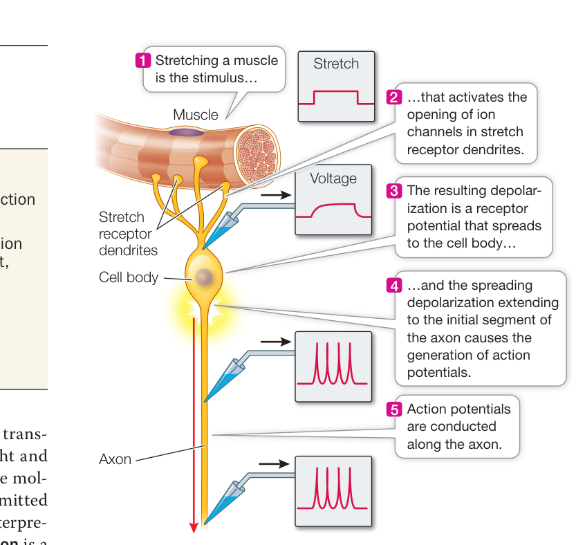
Figure 44.1 Stimulating a Sensory Cell Produces a Receptor Potential Signal transduction in the stretch receptor of a crayfish can be investigated by measuring the membrane potential at different places on the stretch receptor neuron while stretching the muscle innervated by that sensory neuron.
Sensory receptor proteins act on ion channels
Sensory receptor proteins respond to stimuli by directly or indirectly opening or closing ion channels in the sensory cell (Figure 44.2), leading either to an action potential or to the release of neurotransmitter. Those sensory receptor proteins that act directly on membrane potential are either ion channels themselves or directly affect the opening of an associated ion channel. Examples are receptors that respond to physical force (mechanoreceptors) and those that respond to temperature (thermoreceptors). Electroreceptors do not have receptor proteins, but their cell membranes are sensitive to voltage changes and respond to depolarization by opening voltage-gated channels, and the triggers neurotransmitter release. Sensory receptor proteins that influence ion channels indirectly usually do so by activating G proteins and second messengers, as described in Key Concepts 7.2 and 7.3. Examples are most chemoreceptors and photoreceptors.
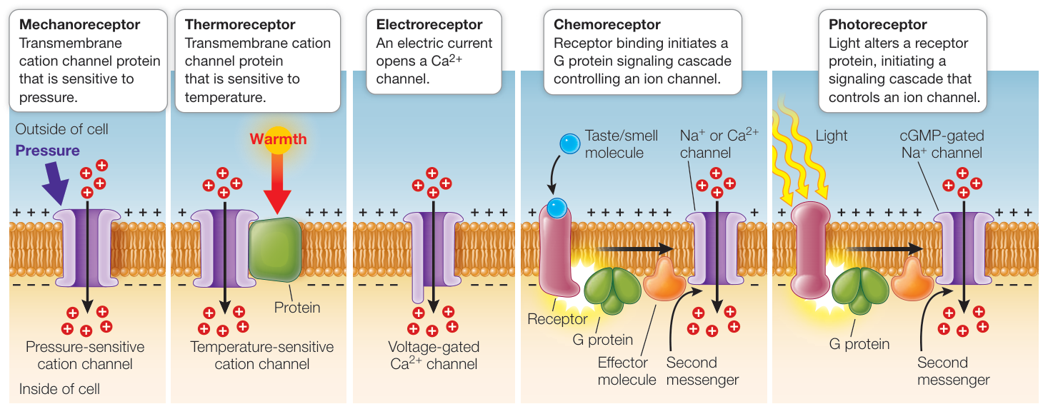
Figure 44.2 Sensory Cell Membrane Receptor Proteins Respond to Stimuli Mechanoreceptors and thermoreceptors have transmembrane proteins that are also cation channels. Changes in their respective stimuli, pressure or heat, alter the permeability of the channels.
To discover the molecular mechanisms by which infrared radiation is detected, a large superfamily of receptor proteins called transient receptor potential (TRP) channel proteins are the focus of investigation. TRP channels generate large initial responses to stimuli and then gradually fade even as the stimuli remain constant. Seven classes of TRP channels have been identified, along with about 28 different molecular structures. Each type of TRP channel is specialized to respond to a different stimulus, such as stretch, pressure, temperature, light, or certain chemicals. Some respond to both physical and chemical stimuli, such as one that responds to heat, capsaicin (an active ingredient in chili peppers), and wasabi. Could a heat-sensitive TRP channel be responsible for the infrared-detection function of the pit organs of rattlesnakes? We pursue this question in Investigating Life: How Do Pit Vipers “See” in the Dark?
Sensation depends on which neurons receive action potentials from sensory cells
All sensory systems process information in the form of action potentials. But the sensations we perceive—heat, pressure, light, smell, sound—differ because the messages from different kinds of sensory cells arrive at different places in the CNS. Action potentials arriving in the visual cortex of the brain are interpreted as light, whereas those that arrive in the olfactory bulb are perceived as smells.
A small patch of skin on your arm contains some sensory receptor cells that increase their firing rates when the skin is warmed and others that increase their activity when the skin is cooled. Other sensory cells in the same patch of skin respond to touch, irritants such as insect bites, and painful stimuli. These receptor cells transmit their messages through axons that enter the CNS at the spinal cord. The synapses made by those axons in the spinal cord and the subsequent pathways of transmission determine whether the stimulation of the skin on your arm is perceived as warmth, cold, touch, itch, or pain; even though the action potentials carried by all of these sensory axons are the same, the connectivity of each axon is specific for a given sensory modality. The intensity of a given sensation is coded by the frequency of the action potentials.
Some sensory receptors transmit information about the body’s internal conditions of which we are not consciously aware. The brain continuously receives information about body temperature, the concentrations of carbon dioxide and oxygen in the blood, arterial pressure, muscle tension, and the position of the limbs. All of this information is important for homeostasis but does not necessarily result in conscious sensation.
Some sensory receptor cells are assembled with other types of cells into sensory organs, such as eyes, ears, and noses, that enhance the ability of sensory cells to collect, filter, and amplify stimuli. Sensory systems include the sensory cells, their associated organs, and the neuronal networks that process the information.
Many receptors adapt to repeated stimulation
Some sensory receptor cells give gradually diminishing responses to maintained or repeated stimulation. This phenomenon, known as sensory adaptation, enables an animal to ignore background or unchanging conditions while remaining sensitive to changes and new information. (Note that this use of the term “adaptation” is different from its application in an evolutionary context.) When you get dressed, you feel each item of clothing touch your skin, but the sensation of clothes touching your skin is not constantly on your mind throughout the day. You are immediately aware of new sensations, however, such as your shoe coming untied or someone touching your back.
Some sensory receptor cells adapt very little or very slowly; examples are some types of pain receptors and the mechanoreceptors that control balance. You do not want to ignore pain, which usually is signaling that something is wrong in your body, and to
Investigating Life: How Do Pit Vipers “See” in the Dark?
Experiment
RESULTS►
CONCLUSION►
maintain equilibrium you must continuously know (albeit unconsciously) the tensions and forces on all of your joints and muscles.
Original Paper: E. O. Gracheva et al. 2010. Molecular basis of infrared detection by snakes. Nature 464: 1006–1012.
Neurons of the pit organ have their cell bodies in a neuronal ganglion in the head—the trigeminal ganglion (TG). Sensory neurons conducting information from the body of the snake have their cell bodies in neuronal ganglia associated with the spinal cord—the dorsal root ganglia (DRGs). Any receptor protein that is involved in sensing heat by the pit organ or by temperature sensors in the skin should be expressed in the TG or the DRGs, respectively.
HYPOTHESIS Different transient receptor potential (TRP) channel proteins responsive to heat are expressed in the TG and the DRGs, and they have different properties.
METHOD Many neurons in the TG of rattlesnakes innervate the pit organs, and therefore the RNA transcripts for any protein that is abundant in the pit organ nerve endings should be highly expressed in the TG. Similarly, RNA transcripts for sensory receptor proteins in the skin of the body should be highly expressed in the DRGs. Whole genome RNA sequencing (RNA-seq) was used to create the RNA profiles of the TG and the DRGs of the rattlesnake. Of particular interest were specific TRP RNA reads (specific RNA sequences) that were differentially expressed in the TG and the DRGs. If there are more reads for a certain TRP channel in the TG versus the DRGs, that TRP channel might play a role in the pit organ’s ability to sense infrared radiation. Because the range of read values was so large, the data are plotted as logarithmic values (log-log plot).
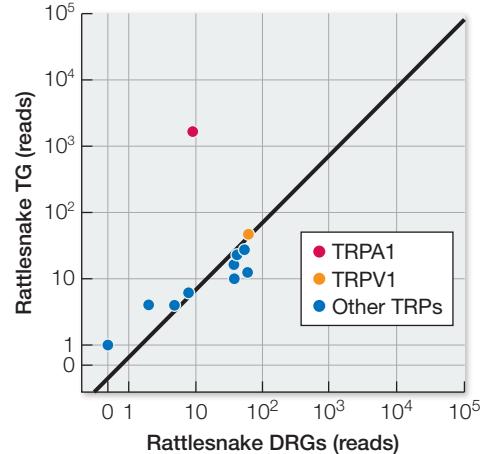
- TRPV1 and “Other TRPs” are expressed equally in the TG and the DRGs, so they probably are not responsible for the unique infrared-sensing properties of the pit organ.
- TRPA1 is expressed more in the TG than in the DRGs. This channel could therefore serve a unique function in the pit organ.
Recap and Assess
Sensory receptor cells have receptor proteins that respond to specific stimuli from the external or internal environment by directly or indirectly opening or closing ion channels, causing the generation of action potentials or neurotransmitter release in sensory neurons. How the resulting information is processed and interpreted depends on the neuronal circuitry that is activated.
- In the crayfish stretch receptor, what creates the receptor potential? Where and how does it stimulate an action potential?
- How are we able to perceive action potentials—all of which are essentially the same—as different sensations?
- Peppers have a distinctive flavor, but they also can be hot if they contain the molecule capsaicin. Explain the difference in how the flavor versus the hotness of a pepper is sensed.
- Why is adaptation or the lack thereof important in different sensory modalities?
Now that you have a general view of how sensory systems code and process information, let’s turn to how sensory systems gather and filter stimuli, transduce specific stimuli into action potentials, and transmit action potentials to the CNS, which perceives them as different types of information.
Work with the Data
The data from the experiment comparing TRP channel expression in the TG and the DRGs support the conclusion that the TRPA1 channel could be the infrared (IR) sensor in the pit organ. Another piece of evidence would be finding that the TRPA1 gene is not highly expressed in the TG of snakes without pit organs, or that it is highly expressed in snakes not closely related to rattlesnakes but that have pit organs. Accordingly, the investigators performed RNA-seq analysis of the TG and the DRGs of rat snakes (no pits) and pythons (pits).
RESULTS (Simulated data, arbitrary units)
| Channel | Rat snake | Python | ||
|---|---|---|---|---|
| TG | DRGs | TG | DRGs | |
| TRPA1 | 105 | 110 | 900,000 | 50 |
| TRPV1 | 85 | 90 | 15 | 9 |
QUESTIONS►
- Compare the relative expression of TRPV1 and TRPA1 in the TG and the DRGs of the rat snake (no pits) and the python (pits). What is the major difference?
- Explain how these results do or do not support the conclusion that the TRPA1 channel is the IR sensor in the pit organ.
- What functional data on the TRPA1 channel would further support the conclusion that TRPA1 is the IR sensor in the pit organ?
Chemoreceptors Respond to Specific Molecules
Learning Objectives
44.2.1 Explain how a large but limited number of olfactory receptor proteins can discriminate a much larger number of odorant molecules, as well as their concentrations.
44.2.2 Explain how the five classes of taste receptors generate action potentials in their sensory neurons.
A colony of corals responds to a small amount of meat extract in seawater by extending its coral polyps and their tentacles in search of food; a solution of a single type of amino acid can stimulate this response. Conversely, a small amount of seawater in which corals were crushed will stimulate a defensive retraction of the coral polyps. Humans also react strongly to certain chemical stimuli. When you smell freshly baked bread you salivate and feel hungry, and when you smell rotting meat you feel nauseated.
All animals receive information about chemical stimuli through chemoreceptors, which are receptor proteins that bind to specific molecules (their ligands) and are responsible for smell and taste. Chemoreceptors are also responsible for monitoring some aspects of the internal environment, such as the level of carbon dioxide in the blood.
Olfaction is the sense of smell
The sense of smell, olfaction, depends on chemoreceptors. In vertebrates the olfactory receptors are neurons embedded in a layer of epithelial tissue in the uppermost region of the nasal cavity (Figure 44.3). The dendrites of these neurons project as olfactory cilia on the surface of the nasal epithelium, and their axons extend through holes in the overlying bone into the olfactory bulb
(the olfactory integration area of the brain). A protective layer of mucus covers the nasal cavity epithelium. Molecules from the environment must diffuse through this mucus to reach the receptor proteins on the olfactory cilia. When you have a cold, the amount of mucus in your nasal cavity increases, and the epithelium swells. With this in mind, you can easily understand why respiratory infections cause loss of your sense of smell.
An odorant is a molecule in the environment that binds to and activates an olfactory receptor protein on the cilia of olfactory receptor neurons (ORNs). Different olfactory receptor proteins bind specific subsets of odorant molecules. An odorant molecule binding to its receptor on an ORN activates a G protein. The G protein then activates an enzyme that causes an increase of a second messenger (cAMP in vertebrates) in the cytoplasm. The second messenger binds to and opens cation channels in the ORN’s cell membrane, causing an influx of into the ORN, which depolarizes to threshold and fires action potentials. An interesting feature of ORNs is that they are continuously regenerated. ORNs have to be constantly replaced because they are embedded in nasal epithelium, which sheds regularly, as do other epithelial linings.
Connect the Concepts Key Concept 7.2 describes how G protein-coupled receptors transmit signals from a variety of stimuli outside a cell to its interior. Ligand binding of the receptor activates the G protein on the cytoplasmic surface of the membrane. A subunit of the G protein separates and travels along the membrane to activate an effector protein.
The olfactory world has an enormous number of odors and a correspondingly large number of olfactory receptor proteins. In the 1990s Linda Buck and Richard Axel discovered a family of about 1,000 genes in mice (about 3% of the mouse genome) that code for olfactory receptor proteins. Each receptor protein that is expressed is found in a limited number of ORNs in the olfactory epithelium, and each ORN expresses just one receptor type. The investigators matched specific gene products with the odorants they detect. For their discoveries of the molecular nature of the olfactory system, Buck and Axel received a Nobel Prize in 2004.
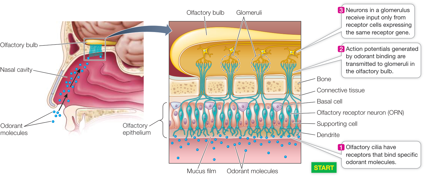
Figure 44.3 Olfactory Receptors Communicate Information Directly to the Brain The receptor cells of the human olfactory system are embedded in epithelial tissues lining the nasal cavity and send their axons to the olfactory bulb of the brain.
Olfactory sensitivity involves discrimination of many more odorants than there are olfactory receptors. An odorant molecule can be quite complex, and different regions of that molecule may bind to different receptor proteins. The next stage of processing olfactory information occurs in the olfactory bulb, where axons from ORNs expressing the same receptor protein make synapses with olfactory bulb neurons that contribute their axons to the olfactory tract. The globular tangles of axons from ORNs and dendrites from olfactory bulb neurons are called olfactory glomeruli (see Figure 44.3). A complex odorant molecule can activate a unique combination of glomeruli in the olfactory bulb, so an olfactory system with hundreds of different receptor proteins can discriminate an astronomically large number of smells. The more odorant molecules that bind to ORNs, the greater the frequency of action potentials and thus the greater the intensity of the perceived smell.
Humans have a sensitive olfactory system, but in comparison with most mammals we depend far more on vision than on olfaction. The nasal epithelium of a typical dog is 15–20 times larger than a human’s and has about 300 million odorant receptors, compared with about 6 million in the average human. For some scents, the threshold sensitivity of the dog is 100,000 times lower; a dog’s nose reveals a huge amount of information not available to people.
Some chemoreceptors detect pheromones
A chemical signal used for communication among conspecifics (individuals of the same species) is called a pheromone. Individual animals secrete pheromone molecules into the environment, triggering behavioral responses in other individuals of the species. Pheromones may attract mates, communicate alarm signals, mark food trails, or define territories, among many other uses. Their function in mate attraction illustrates the remarkable sensitivity of chemosensory systems, and the silkworm moth Bombyx mori is an extensively studied example.
To attract a mate, the female silkworm moth releases a pheromone called bombykol from a gland at the tip of her abdomen (Figure 44.4A). The male silkworm moth has about 10,000 receptors for this molecule on each of his feathery antennae (Figure 44.4B). A single molecule of bombykol may be sufficient to generate action potentials in the antennal nerve, which transmits the signal to the male’s CNS. The extreme sensitivity of the male bombykol receptors ensures that the sexual message sent by a female moth is likely to reach any male within a huge downwind area. The rate of firing in the male’s sensory nerves is proportional to the concentration of bombykol in the air, so by flying upwind he can follow an airborne concentration gradient to “home in” on the signaling female.
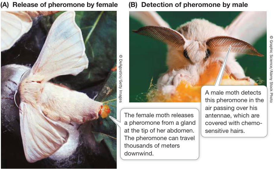
Figure 44.4 Pheromones Can Communicate over Great Distances Mating in silkworm moths of the genus Bombyx is coordinated by a pheromone called bombykol. Q: Molecules have different diffusion coefficients. Bombykol has a high diffusion coefficient. How do you think its diffusion coefficient would compare with that of a territory-marking pheromone, and why might they be different?
The vomeronasal organ contains chemoreceptors
The vomeronasal organ (VNO) is a small, paired tubular structure embedded in the nasal epithelium of many species of amphibians, reptiles, and mammals (not including humans). In mammals the VNO is located on the septum dividing the two nostrils (see Figure 51.3) and has a pore that opens into the nasal cavity. The VNO pulsates and draws a sample of nasal fluid over the chemoreceptors embedded in its walls. The information from these chemoreceptors goes to an accessory olfactory bulb in the brain, and from there to brain regions involved in sexual and other instinctive behaviors. The VNO of a rodent detects pheromones that signal the sex, strain, identity, and sexual receptivity of another individual.
In snakes the VNO opens into the roof of the mouth cavity. Each time the snake’s forked tongue darts in and out, the forks fit into the VNO openings and present the chemoreceptors located there with a sample of molecules from the surrounding air (see the chapter opening photo). Thus the snake uses its tongue to smell its environment, not to taste it. Why doesn’t the snake simply use the flow of air to and from its lungs, as we do, to smell the environment? In snakes, air flows to and from the lungs slowly (and can even stop entirely for long periods of time), but the tongue can dart in and out rapidly. It is a quick source of olfactory information used in social communication and hunting prey.
Gustation is the sense of taste
In humans and other vertebrates, the sense of taste, gustation, depends on clusters of chemoreceptors called taste buds. The taste buds of terrestrial vertebrates are confined to the mouth cavity, but some fishes have taste buds in the skin that enhance their ability to sense their environment. Some fishes living in murky water are very sensitive to small amounts of amino acids in the water and can find food without the use of vision. The duck-billed platypus, a prototherian mammal (see Figure 31.26B), has a similar ability as a result of taste buds on the sensitive skin of its bill.
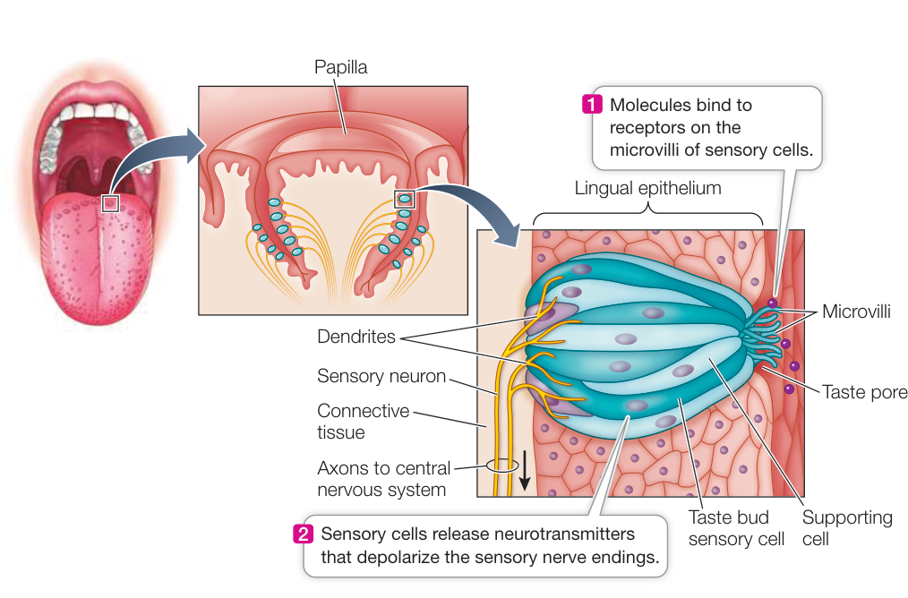
Figure 44.5 Taste Buds Are Clusters of Sensory Cells A human tongue has as many as 10,000 taste buds, most of which are found on the raised papillae.
The human tongue has 5,000–10,000 taste buds embedded in the epithelium. Most of them are found on the sides of the papillae (Figure 44.5). (Look at your tongue in a mirror—the papillae make it look fuzzy.) The outer surface of a taste bud has a pore that exposes the tips of the sensory receptor cells. Microvilli (tiny hairlike projections) increase the surface area of these cells where their tips converge at the pore. These chemoreceptor cells generate action potentials and release neurotransmitter at their bases, where they form synapses with sensory neurons that convey the signals to the CNS.
The tongue does a lot of hard work, so its epithelium, along with cells of its taste buds, is shed and replaced at a rapid rate. Individual taste bud cells last about 10 days before they are replaced, but the sensory neurons associated with them live on, constantly forming new synapses as new taste buds form.
Gustation begins with the membrane receptor proteins of the microvilli of the taste bud sensory cells (see Figure 44.5). Humans perceive five taste classes: sweet, salty, sour, bitter, and umami. However, taste buds can distinguish among a variety of sweet-tasting molecules and a variety of bitter-tasting molecules. Umami is a savory, meaty taste that is stimulated by amino acids, including monosodium glutamate (MSG), a commonly used flavor enhancer. Sweet, bitter, and umami tastes are the functions of a small family of G protein-coupled TRP channels. Other TRP channels related to the ones you saw associated with heat sensation in Investigating Life: How Do Pit Vipers “See” in the Dark? are responsible for spicy and hot tastes. Still other TRP channels that act as cold sensors are activated by minty, cool-tasting foods. Saltiness receptor proteins allow to diffuse into taste bud sensory cells through open channels, depolarizing the sensory cell. Sourness receptors are activated by a direct effect of ions on channels.
Various selective pressures led to the evolution of diversity in gustatory receptors. Receptors for bitter tastes may have evolved as protective mechanisms enabling animals to detect toxic plant compounds such as quinine, caffeine, and nicotine. Because many such toxic compounds evolved in plants as protections against herbivorous predators, having a variety of receptors in those herbivores is a product of the continuous “arms race” between predators and prey (see Key Concept 54.1). Also, a large number of molecules in food could indicate nutritional value, so having a variety of receptors is advantageous. The diversity of sweet receptors helps explain why it has been possible to invent many chemically distinct artificial sweeteners.
Regardless of the mechanism of taste transduction by the receptors of the taste buds, all these cells release neurotransmitter onto sensory neurons. These neurons then generate action potentials that are conducted to the CNS, where they are interpreted as specific taste sensations. The full complexity of the chemosensitivity that enables us to enjoy the subtle flavors of food comes from the combined activation of gustatory and olfactory receptors, which is why you may lose your sense of taste when you have a cold.
Recap and Assess
All animals receive information about chemical stimuli through chemoreceptors, which have diverse structures and bind to a tremendous variety of ligands. Chemoreceptors are the basis of the sensations of olfaction and gustation and the reception of pheromones. They also monitor some aspects of an animal’s internal environment.
- Different odorants have different cell surface receptors, but these receptors activate the same intracellular G protein signaling pathway. How does the olfactory system discriminate different odors?
- How does the olfactory system discriminate between the whiff of a skunk, a repulsive odor of skunk, and the smell of a skunk cabbage?
- The concentration thresholds for sensing bitter tastes are lower than for salt or sweet. Offer an evolutionary explanation for why this is so.
- With only five classes of taste receptors, how can we discriminate so many different tastes?
You have now seen how chemoreceptors give rise to the sensations of smell and taste, and how some animals use chemoreception to communicate with others of their species. Next we describe the sensory cells that respond to mechanical forces, including the vibrations you perceive as sound.
Mechanoreceptors Respond to Physical Forces
Learning Objectives
- 44.3.1 Describe the various touch sensors and their unique properties.
- 44.3.2 Describe how tension in a muscle is sensed and adjusted for load.
- 44.3.3 Explain how sound waves of different frequencies are heard as sounds of different pitches.
The cell membranes of mechanoreceptors have cation channels that open when the membrane is disturbed by physical forces. The intensity of the physical force is registered as a graded receptor potential, which in turn leads to either the release of neurotransmitter or the generation of action potentials. The amount of neurotransmitter release or the rate of action potentials tells the CNS the strength of the stimulus to the mechanoreceptor. A diversity of mechanoreceptor cells has evolved in different sensory systems. The functions of these cells include interpreting skin sensations, sensing blood pressure, hearing, and maintaining balance.
Many different receptor cells respond to touch and pressure
Human skin (and that of other animals) is packed with diverse mechanoreceptors that generate varied sensations (Figure 44.6).
The most important tactile receptors, found in both hairy and non-hairy skin of humans, are Merkel’s discs, which adapt rather slowly and provide continuous information about anything touching the skin. Meissner’s corpuscles, found primarily in non-hairy skin, are very sensitive but adapt rapidly; they provide information about changes in things touching the skin. The rapid adaptation of Meissner’s corpuscles is why you roll a small object between your fingers (rather than holding it still) to discern its shape and texture: as you roll it, the object continually stimulates the Meissner’s corpuscles.
Deeper in the skin, Ruffini endings adapt slowly and are good at providing information about vibrating stimuli of low frequencies, while Pacinian corpuscles, which adapt rapidly, provide information about vibrating stimuli of higher frequencies. Even deeper in the skin, the dendrites of sensory neurons wrap around hair follicles. When the surface hairs are displaced, those neurons are stimulated.
The density of tactile mechanoreceptor cells varies across the body’s surface. By touching the skin with two toothpicks simultaneously (when a person’s eyes are closed!), you can determine how far apart two stimuli have to be before the person can tell whether the sensations are produced by one toothpick or by two. On back skin, stimuli have to be relatively far apart before they are perceived as two discrete stimuli. But when this same “two-point spatial discrimination test” is applied to the lips or fingertips, a person can identify two stimuli as separate even when they are quite close together, because the receptor density is much greater in these regions.
Mechanoreceptors are also found in muscles, tendons, and ligaments
An animal receives information from mechanoreceptors about the position of its limbs and the stresses on its muscles and joints. These mechanoreceptors supply information continuously to the CNS, and this information is essential for postural control and the coordination of movements.
The mechanoreceptors in skeletal muscle are the muscle spindles. These stretch receptors are modified muscle cells embedded in connective tissue inside muscles and innervated by sensory neurons (Figure 44.7A). When a muscle is stretched, the muscle spindles are stretched as well, signaling the spindle neurons to transmit action potentials to the CNS. Figure 44.1 showed how crayfish stretch receptors transduce physical force into action potentials; the actions of muscle spindles are similar. The CNS uses the information from muscle spindles to adjust the strength of the muscle contraction so that it matches the load put on the muscle; thus a person can hold a mug steady while it is being filled.
Another type of mechanoreceptor, the Golgi tendon organ, is found in tendons and ligaments and provides information about the force generated by a contracting muscle (Figure 44.7B). When a contraction becomes too forceful, action potentials from the Golgi tendon organ inhibit the spinal cord motor neurons innervating that muscle, causing the muscle to relax and protecting it from tearing. (You may recall a cell organelle called the Golgi apparatus. What these two very different structures have in common is their discovery in the late nineteenth century by the Italian anatomist Camillo Golgi.)
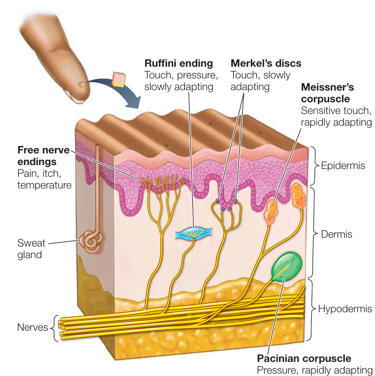
Figure 44.6 The Skin Feels Many Sensations Even a very small patch of skin contains a variety of sensory cells, making the skin a multimodal receptor that can sense temperature, pressure, texture, pain, touch, and itch.
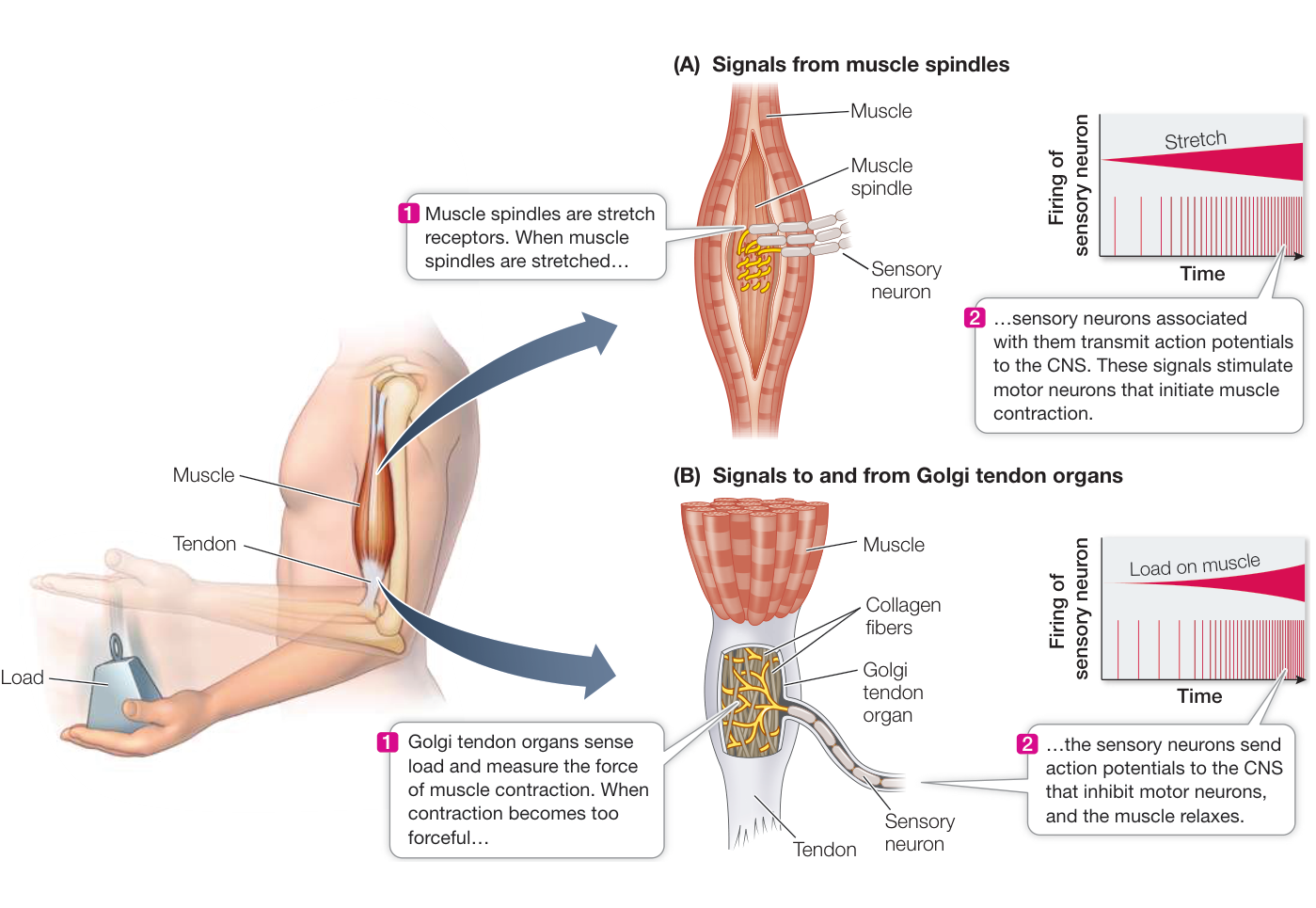
Figure 44.7 Stretch Receptors Stretch receptors provide information about the stresses on muscles and joints in an animal’s limbs. (A) Signals from muscle spindles to the CNS initiate muscle contraction. (B) Golgi tendon organs in tendons and ligaments inhibit a contraction that becomes too forceful, triggering a reduction in muscle tension and protecting the muscle from tearing.
Hair cells are mechanoreceptors of the auditory and vestibular systems
Hair cells are the mechanoreceptors for the vertebrate auditory (sound-perceiving) and vestibular (equilibrium-maintaining) systems. Both of these systems are housed in the complex structures of the vertebrate ear. Stereocilia—fingerlike extensions of the cell membrane stiffened by cross-linked actin filaments—project from the surface of each hair cell like a set of organ pipes (Figure 44.8). Stereocilia bend in response to waves of pressure; bending of the stereocilia in one direction depolarizes the hair cell, and bending in the other direction hyperpolarizes it.
Measurements with microelectrodes have shown that the bending of stereocilia creates local electric currents near their tips, indicating that ion channels near the tips must be opening or closing. Electron microscope images reveal minute filaments that connect the tip of each stereocilium to its taller neighbor. It is hypothesized that these filaments are fine molecular attachments to the ion channels, and that they act like springs that open the channels. If the taller neighboring stereocilium is bent away, the spring tightens and the ion channel is opened. If the taller neighbor bends toward its shorter neighbor, the spring is relaxed and the channel closes (see Figure 44.8).
Auditory systems use hair cells to sense sound waves
The stimuli that animals perceive as sounds are pressure waves. Auditory systems use mechanoreceptors to convert pressure waves into receptor potentials. Auditory systems include special structures that gather sound waves, direct them to the sensory organ, and amplify their effect on the mechanoreceptors. A good example of an auditory system is the human ear (Figure 44.9), which can be divided into three major areas: the outer, middle, and inner ear (see Figure 44.9A).
OUTER EAR The outer ear consists of the pinna (plural pinnae) and the auditory canal. The pinnae collect sound waves and direct them into the auditory canals; watch a dog change the orientation of its ears to focus on a particular sound to get the idea of the role pinnae play in hearing. Covering the end of the auditory canal is the tympanic membrane (commonly called the eardrum), which vibrates in response to pressure waves traveling down the canal, thus converting the pressure waves to physical forces in the middle ear.
MIDDLE EAR On the other side of the tympanic membrane is the middle ear, an air-filled cavity connected to the throat at the back of the mouth through the eustachian tube (also called the pharyngotympanic tube). Because the eustachian tube is also filled with air, pressure can equilibrate between the middle ear and the environment. When you have a cold, the tube can be blocked by mucus or tissue swelling and you may have difficulty equilibrating the pressure in the middle ear with the outside air pressure (as you have to do when changing altitude in an airplane).
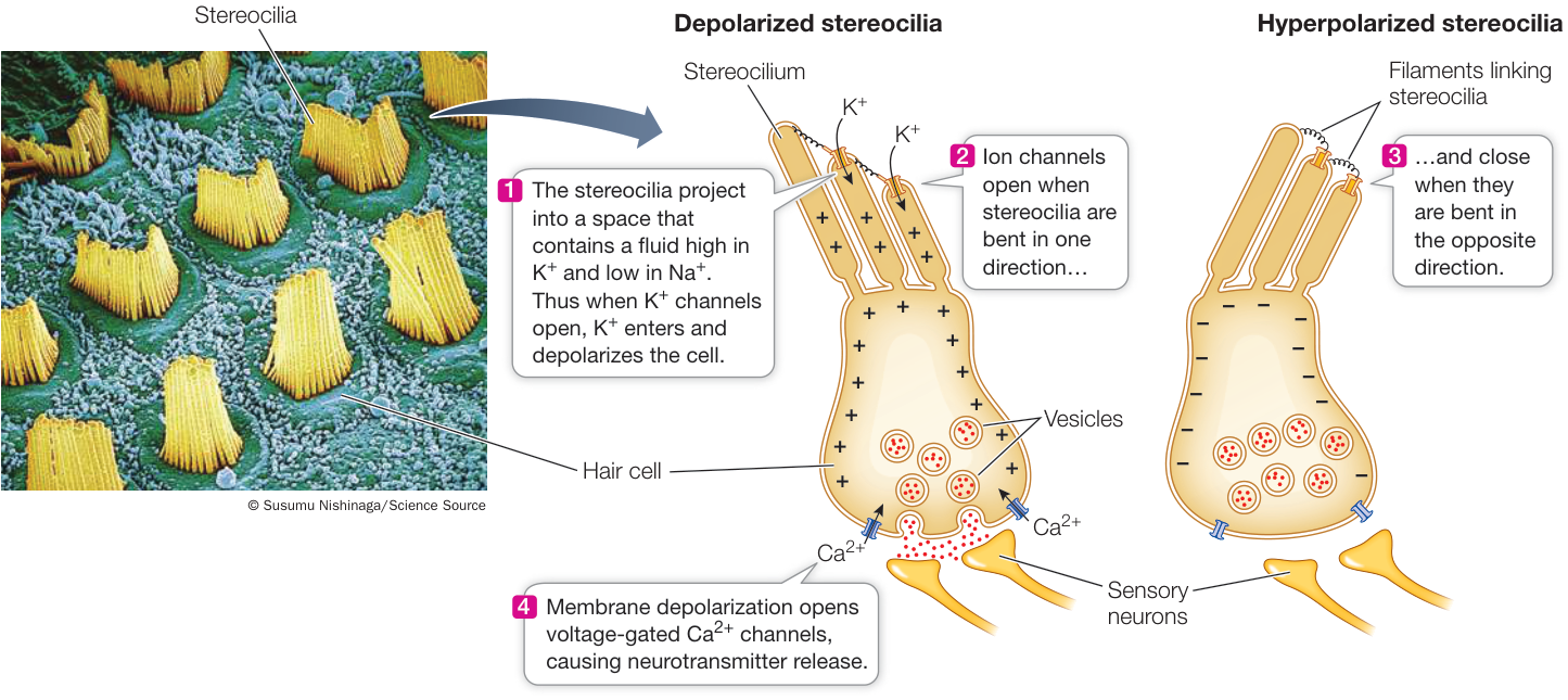
Figure 44.8 Hair Cells Have Mechanosensors on Their Stereocilia Hair cells have stereocilia that are connected to each other by small filaments. When a cilium is bent in one direction, the filament opens a cation channel at the tip of the neighboring cilium, resulting in depolarization of membrane potential. When depolarized, the hair cell releases neurotransmitter onto a sensory neuron. Movement of the hair cell in the opposite direction closes the cation channel and the cell hyperpolarizes, causing a reduction in its release of neurotransmitter.
The middle ear contains the ossicles, three delicate bones individually named the malleus (“hammer”), incus (“anvil”), and stapes (“stirrup”) (see Figure 44.9B). The ossicles transmit the vibrations of the tympanic membrane to another flexible membrane, the oval window. The ossicles act as a lever (like a hammer pulling out a nail), translating a large movement of the tympanic membrane into a smaller movement of the oval window, but a movement of greater force. Because the oval window is much smaller than the tympanic membrane, the pressure the stapes transmits to the oval window is more than 20 times greater than the pressure exerted by a sound wave on the tympanic membrane.
Behind the oval window lies the fluid-filled inner ear. Movements of the oval window impart pressure changes to that enclosed fluid. These pressure waves are transduced into action potentials.
INNER EAR The inner ear is a bony structure consisting of two sets of canals. One is the organ of balance, the vestibular system, and the other is the organ of hearing, the cochlea. The cochlea (“snail” or “spiral shell”) is a long, tapered, coiled structure. A cross section of the cochlea reveals that it is composed of three parallel canals separated by two membranes, the vestibular membrane and the basilar membrane (see Figure 44.9C). Sitting on the basilar membrane is the organ of Corti, which transduces pressure waves into action potentials. The organ of Corti contains hair cells with stereocilia (see Figure 44.8). The tips of the hair cells are embedded in a gelatinous overhanging shelf called the tectorial membrane all enclosed in the cochlear canal (see Figure 44.9D).
Stereocilia are not motile, but because their tips are attached to the more rigid tectorial membrane, stereocilia bend when the basilar membrane flexes. As shown in Figure 44.8, when the stereocilia are bent in one direction, the hair cell is depolarized by an inflow of ions. This effect of opening channels happens because the fluid in the cochlear canal has a higher concentration than does the intracellular fluid of the hair cell. The response of the hair cell is a graded membrane potential. The hair cells do not fire action potentials, but the changes in their membrane potential alter the rate at which the hair cells release neurotransmitter onto the sensory neurons. Those sensory neurons fire action potentials, and their axons, which make up the auditory nerve, transmit those action potentials to the brain.
The vestibular and tympanic canals are separate until they reach the apical end of the cochlea (the end farthest from the oval window), where they join; thus they form one continuous canal that turns back on itself (see Figure 44.10). Just as the oval window is a flexible membrane at the beginning of the vestibular canal, the round window is a flexible membrane at the end of the tympanic canal. When the oval window vibrates, the waves of fluid pressure create traveling waves of flexion in the basilar membrane.
Flexion of the basilar membrane is perceived as sound
What causes the basilar membrane to flex, and how does this mechanism distinguish sounds of different frequencies? Air is highly compressible but fluids are not; therefore a pressure wave can travel through air without displacing much air, whereas a pressure wave in fluid displaces that fluid. When the stapes pushes on the oval window, the fluid in the vestibular canal is displaced. If the movement of the oval window occurs very slowly, the cochlear fluid pressure wave travels down the vestibular canal, around the bend, and back through the tympanic canal (Figure 44.10). At the end of the tympanic canal, the displacement pressure is dissipated by the outward bulging of the round window.
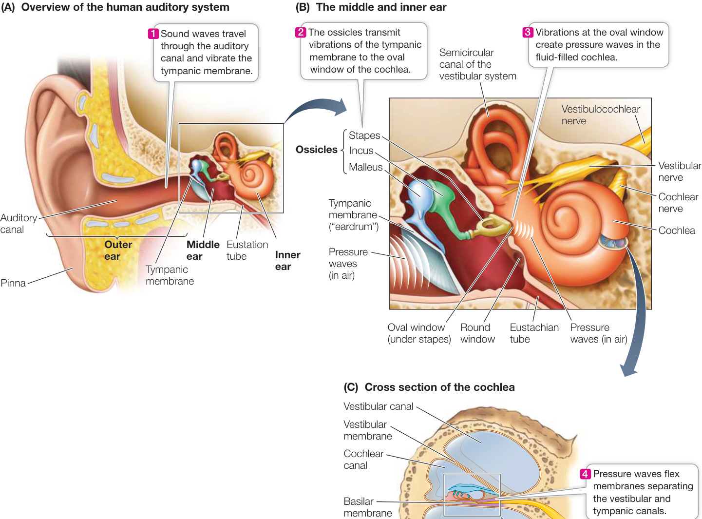
Figure 44.9 Structures of the Human Ear (A) The pinnae direct sound waves down the auditory canal to impinge on the tympanic membrane. The tympanic membrane mechanically transmits these pressure waves into movements of the ossicles in the middle ear. (B) The ossicles transmit their movement into pressure waves in the fluid of the cochlea at the oval window. (C) The cochlea is divided into fluid-filled canals; pressure waves from the ossicles cause the membranes between the canals to flex. (D) Flexing of the basilar membrane bends stereocilia on hair cells in the organ of Corti. Q: How can the ears detect directionality of a sound source?
The basilar membrane is not uniform—it is thicker and stiffer at its base and wider and thinner at its apical end. Pressure waves in the cochlear fluid have different frequencies and set up different patterns of traveling waves. High-frequency waves cause maximal flexion at the basal end of the basilar membrane, whereas low-frequency pressure waves result in maximal flexion at the apical end (see Figure 44.10). Thus different frequencies of pressure waves flex the basilar membrane at different locations and activate different sets of hair cells. Action potentials stimulated by different sets of hair cells along the organ of Corti code for different frequencies of sound and send that information along the cochlear nerve to the auditory cortex, where those action potentials are interpreted as different pitches of sound.
Various types of damage can result in hearing loss
There are two general types of acquired hearing loss, or deafness. Conduction deafness is caused by the loss of function of
(D) The organ of Corti
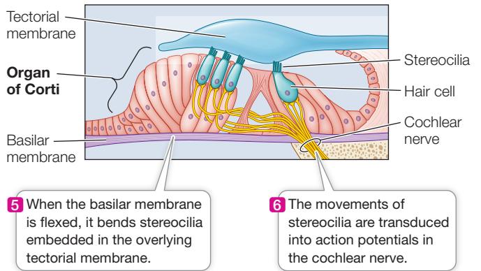
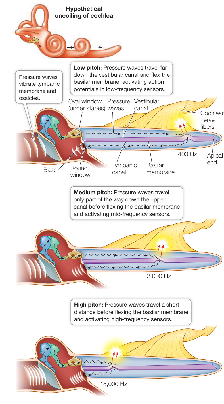
Figure 44.10 Sensing Pressure Waves in the Inner Ear Pressure waves of different frequencies flex the basilar membrane at different locations. Information about sound frequency is specified by which hair cells are activated. For simplicity, this representation illustrates the cochlea as uncoiled. Animation 44.1 Sound Transduction in the Human Ear
the tympanic membrane or the ossicles of the middle ear. Repeated infections of the middle ear can cause scarring of the tympanic membrane and stiffening of the connections between the ossicles. The consequence is less efficient conduction of sound waves from the tympanic membrane to the oval window. With increasing age, the ossicles inevitably stiffen, resulting in a gradual loss of the ability to hear high-frequency sounds.
Nerve deafness is caused by damage to the inner ear or the auditory pathways. A common cause of nerve deafness is damage to the hair cells of the delicate organ of Corti by exposure to loud sounds such as jet engines, pneumatic drills, or highly amplified music. Consistent exposure to sounds above 85 decibels can damage hearing; this damage is cumulative and irreversible. Even using earphones can put you at risk for hearing loss because they generate high-pressure sound waves close to the tympanic membrane. Personal stereo earphones can reach 120 decibels, and people commonly use them at 100 decibels (equivalent to being at a rock concert).
The vestibular system uses hair cells to detect forces of gravity and momentum
Hair cells in the vestibular system of the inner ear detect the position and movement of the head—information essential for maintaining equilibrium (balance). Information from the vestibular system is also crucial for the control of eye movements. When you look at something, you can move your head while staying focused on the object because of your vestibulo-ocular reflex.
In the mammalian inner ear, the vestibular system consists of three bony semicircular canals and a bony chamber called the vestibule. Within each canal is a membranous semicircular duct, and within the vestibule are the membranous saccule and the utricle. The ducts and the saccule and utricle are filled with fluid. In the semicircular ducts, the fluid shifts when the head changes position (Figure 44.11A). Since the three semicircular canals have different orientations, the fluid in their ducts responds differentially to the direction of movement. Projecting into the base of each duct is a cupula, a gelatinous swelling enclosing a cluster of hair cell stereocilia. When the shifting fluid pushes on the cupula, it bends the stereocilia and causes a graded potential in the hair cell membranes.
The stereocilia in the saccule and utricle are bent in a different way. These stereocilia are embedded in gelatinous membranes that contain otoliths (“ear stones”), which are crystals of calcium carbonate. When the head changes position or when it accelerates or decelerates, gravitational forces are exerted on the otoliths and the stereocilia bend (Figure 44.11B).
As in the cochlea, the hair cells of the vestibular system do not fire action potentials, but they release neurotransmitter at synapses with sensory neurons, which in turn fire action potentials.
Motion sickness is a common malady experienced when riding in a boat, car, plane, or amusement park ride, or when experiencing any situation in which the vestibular system and the visual system produce conflicting information. The vestibular system may be indicating movement when the visual system is not, or vice versa.
Simulation 44.1 Mechanoreceptors
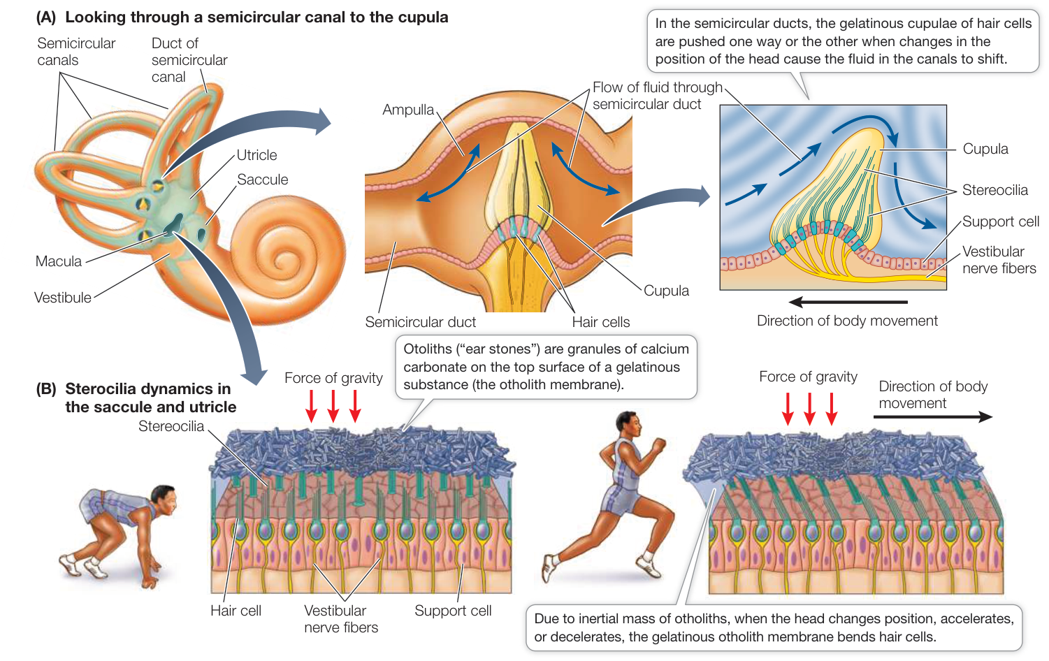
Figure 44.11 Organs of Equilibrium The vestibular system consists of bony chambers and fluid-filled canals. (A) Each semicircular duct has a cupula containing stereocilia. When fluid moves against the cupula, the stereocilia bend. (B) In the maculae of the saccule and utricle, vestibular nerve endings synapse with hair cells whose stereocilia are embedded in a gelatinous layer covered with otoliths. Forces that move the otoliths bend the stereocilia.
Recap and Assess
Sensations that derive from mechanoreceptors include touch, tickle, pressure, joint position, muscle load, hearing, and equilibrium. The membranes of mechanoreceptors have ion channels that are opened by distortion of the membrane, resulting in graded receptor potentials that cause the cell to release neurotransmitter or to fire action potentials. Hair cells involved in hearing and equilibrium have stereocilia that are bent by pressure waves, resulting in receptor potentials.
- Why are there different types of mechanoreceptor cells that have different rates of adaptation?
- Explain the role of specific mechanoreceptors when you are (a) holding a glass that is being filled and (b) lifting heavy furniture.
- How do different frequencies of pressure waves in the air result in action potentials being fired in different acoustic neurons?
We next turn to vision, perhaps the most elaborate of the senses.
Photoreceptors Respond to Light
Learning Objectives
- 44.4.1 List the steps whereby a photon of light is transduced into a change in membrane potential.
- 44.4.2 Describe the properties and locations of rod and cone cells in the retina.
- 44.4.3 Describe the structural adaptations enabling movement detection and visual acuity.
- 44.4.4 Explain ganglion cell receptive fields in terms of the lateral processing afforded by the horizontal and amacrine cells.
Sensitivity to light—photosensitivity—confers on the simplest animals the ability to orient to the Sun and sky, and it gives more complex animals rapid and extremely detailed information about objects in their environments. Photosensitivity is ubiquitous in the animal kingdom, and the molecular basis for that sensitivity is a family of visual pigments that have been evolutionarily conserved.
These photosensitive pigments are contained in photoreceptor cells in the eyes, the organs that gather light energy and initiate its conversion into action potentials. The photoreceptors in the eyes are associated with several additional cell types that integrate the information from the visual receptors. Visual pigments with different spectral sensitivities enable color vision.
Invertebrates have a variety of visual systems
Photoreceptors and visual pigments are incorporated into a variety of visual systems, from simple to complex. Flatworms obtain directional information about light from photoreceptor cells that are organized into eye cups—paired, bilateral structures, each partly shielded from light by a layer of pigmented cells lining the cup. The photoreceptors on the two sides of the animal are unequally stimulated unless the animal is facing directly toward or away from a light source. The flatworm generally uses directional information from the eye cups to move away from light.
Arthropods have compound eyes that provide them with information about patterns in the environment. These eyes are called compound eyes because each eye consists of many optical units called ommatidia (singular ommatidium), each with its own narrow-angle lens (Figure 44.12). In contrast, a vertebrate eye consists of just one optical unit with a wide-angle lens. The number of ommatidia in a compound eye varies from only a few in some ants to 800 in fruit flies and to 30,000 in some dragonflies.
Each ommatidium has a lens that directs light onto photoreceptor cells. Flies, for example, have eight elongated photoreceptors in each ommatidium. The inner borders of the photoreceptors are covered with microvilli that contain a photosensitive pigment that traps light. Axons from the photoreceptors send the light information to the nervous system. Since each ommatidium of a compound eye is directed at a slightly different part of the visual world, only a low-resolution (pixilated) image can be communicated from the compound eye to the CNS.
Image-forming eyes evolved independently in vertebrates and cephalopods
Both vertebrates and cephalopod mollusks (such as squid and octopuses) have eyes with exceptional abilities to form detailed images of the visual world. Like cameras, both of these eye types focus inverted images on an internal surface that is sensitive to light. Considering that they evolved completely independently of each other, their degree of similarity is remarkable (Figure 44.13).
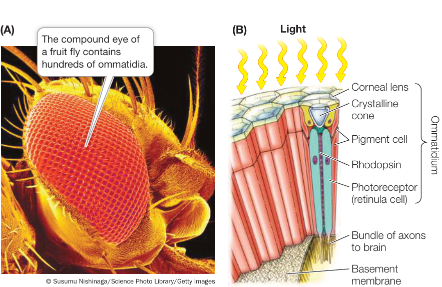
Figure 44.12 Ommatidia: The Functional Units of Insect Eyes (A) The micrograph shows the compound eye of a fruit fly. (B) The rhodopsin-containing retinula cells are the photoreceptors in ommatidia. (B after M. Friedrich. 2006. Arthropod Struct Dev 35: 357–378.)
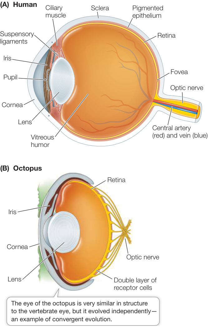
Figure 44.13 Convergent Evolution of Eyes The lenses of vertebrate (A) and cephalopod (B) eyes focus images on layers of photoreceptor cells. Q: Humans commonly experience “floaters” in their eyes—tissue fragments that float around in the field of vision. Where in the structure of the eye would you expect floaters to exist?
The vertebrate eye (see Figure 44.13A) is a spherical, fluid-filled structure bounded by a tough connective tissue layer called the sclera. The sclera at the front of the eye forms the transparent cornea, through which light passes to enter the eye. Just inside the cornea is the pigmented iris, which gives the eye its color. The iris controls the amount of light that reaches the photoreceptor cells at the back of the eye. The central opening of the iris is the pupil. The iris is under neural control. In bright light, the iris constricts and the pupil is very small. As light levels fall, the iris opens and the pupil enlarges.
Behind the iris is the crystalline protein lens that makes fine adjustments in the focus of images falling on the photosensitive layer—the retina—at the back of the eye. The cornea and the gel-like mass (vitreous humor) within the eye bend light rays passing through them so that they are focused on the retina. The lens makes fine adjustments to the focus and allows the eye to accommodate—that is, to focus on objects at various locations in the near visual field. To focus a camera on objects close at hand, you adjust the distance between the lens and the internal surface sensitive to light. Fishes, amphibians, and non-avian reptiles accommodate in a similar manner, moving the lenses of their eyes closer to or farther from their retinas. Mammals and birds use a different method; they alter the shape of the lens.
The mammalian lens is contained in a connective tissue sheath that tends to keep it in a spherical shape, but the sheath is attached to suspensory ligaments that pull the lens into a flatter shape. Circular ciliary muscles counteract the pull of the suspensory ligaments, permitting the lens to round up. When the ciliary muscles are at rest, the flatter lens has the correct optical properties to focus distant images on the retina. Contracting the ciliary muscles rounds up the lens, changing its light-bending properties to bring close images into focus (Figure 44.14).
Lenses become less elastic with age, so we lose the ability to focus on objects close at hand without the help of corrective lenses. Most people over the age of 45 need the assistance of reading glasses or bifocal lenses.
The vertebrate retina receives and processes visual information
During embryonic development, nervous tissue grows out from the brain to form the retina. In addition to a layer of photoreceptor cells, the retina includes other cells arranged in layers that process visual information from the photoreceptors (see Figure 44.20). Light must pass through all the layers of retinal cells before being captured by photosensors. In humans and other day-active animals, the light that is not captured is absorbed by a black-pigmented epithelial tissue layer behind the retina.
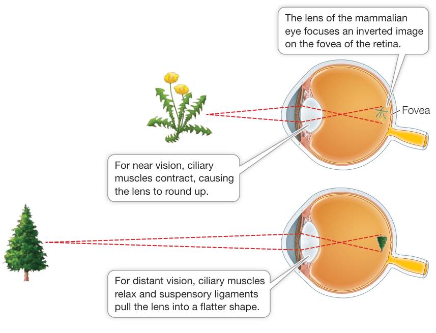
Figure 44.14 Staying in Focus Mammals and birds focus their eyes by changing the shape of the lens depending on the eye’s distance from the object of focus.
In contrast, nocturnal animals such as deer and raccoons have an iridescent reflective layer behind their retinas, which maximizes the capture of photons by reflecting them back onto the photoreceptors. This is why a deer in the headlights or your pet dog or cat in a flash photo appears to have bright white eyes. In contrast, humans do not have a white reflective layer in the retina, so flash photographs often have a “red-eye effect,” caused by the flash being reflected by the abundant blood vessels in the retina.
Opsins are the universal photoreceptor molecule in animals
Photosensitivity in animals depends on the ability of opsin molecules to absorb photons of light and activate a G protein. Opsins are proteins that are not themselves photosensitive, but they contain covalently bound functional groups called 11-cis-retinal (Figure 44.15). Retinal absorbs photons of light, and the color of light it receives depends on the structure of its opsin. There are several different opsins. The most common one that gives humans sensitivity to low levels of light, but not color, is called rhodopsin.
When 11-cis-retinal absorbs a photon of light energy, it changes into a different isomer of retinal, called all-trans-retinal. This change puts a strain on the bonds between retinal and opsin, changing the conformation of opsin and signaling the detection of light. In vertebrate eyes, the retinal and the opsin eventually separate from each other (a process called bleaching), which causes the opsin to lose its photosensitivity. A series of enzymatic reactions returns the all-trans-retinal to the 11-cis isomer, which then recombines with opsin so that it once again becomes photosensitive.
How does the conformational change of opsin transduce light into a cellular response? After retinal is converted from the 11-cis to the all-trans form, its interactions with opsin pass through several unstable intermediate stages. One of these stages triggers a G protein signaling cascade that results in the alteration of membrane potential that is the photoreceptor cell’s response to light.
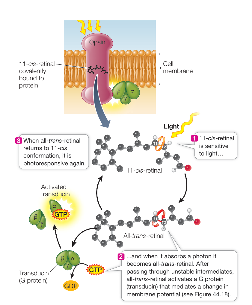
Figure 44.15 Light Changes the Conformation of Rhodopsin The light-absorbing molecule 11-cis-retinal bonds with the protein opsin to form the vertebrate visual pigment rhodopsin.
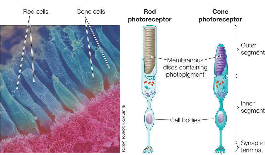
Figure 44.16 Rods and Cones This scanning electron micrograph of photoreceptors in the retina of a mud puppy (an amphibian) shows cylindrical rods and tapered cones.
Rod and cone cells are the photoreceptors of the vertebrate retina
The photoreceptors of the vertebrate retina are two types of modified neurons called rod cells and cone cells based on their shapes (Figure 44.16). Rod cells are highly light-sensitive and perceive shades of gray in dim light. Cone cells function at high light levels and are responsible for high-acuity color vision. The human retina has about 5 million cones and 100 million rods, but the density of each type varies across the retina.
ROD CELLS Rod cells do not produce action potentials, but they release neurotransmitter from their bases, where they form synapses with the next neurons in the visual pathway. Each rod cell has an outer segment, an inner segment, and a synaptic terminal (see Figure 44.16). The highly specialized outer segment contains stacks of discs made up of cell membranes densely packed with rhodopsin. These discs capture photons of light. The inner segment contains the cell nucleus, mitochondria, and other organelles. The synaptic terminal is where the rod cell communicates with other neurons.
To see how a rod cell responds to light, we can penetrate a single rod cell with an electrode and record its membrane potential in the dark and in the light, as shown in Figure 44.17. From what you have learned about other types of sensory receptors, you might expect that stimulation of the rod cell by light would make its membrane potential less negative, but the opposite is true—it becomes more negative, that is, it becomes hyperpolarized.
Rod cell outer segments have cyclic GMP (cGMP) gated sodium channels. In the dark, abundant cGMP in the cell keeps these channels open, creating the dark current. When light is flashed on the dark-adapted rod cell, these channels close in proportion to the intensity of the light, and the cell’s membrane potential becomes more negative—it hyperpolarizes (see Figure 44.17). The rate of neurotransmitter release from the base of the cell changes as membrane potential changes. As the rod cell hyperpolarizes, its release of neurotransmitter decreases.
How does the absorption of light by rhodopsin hyperpolarize the rod cell? As illustrated in Figure 44.15, when rhodopsin is excited by light it undergoes conformational changes that initiate a signaling cascade. The dark-adapted rod cell has open channels—the depolarizing dark current (Figure 44.18). Light photons excite rhodopsin, which activates a G protein called transducin. Activated transducin activates a phosphodiesterase (PDE). Activated
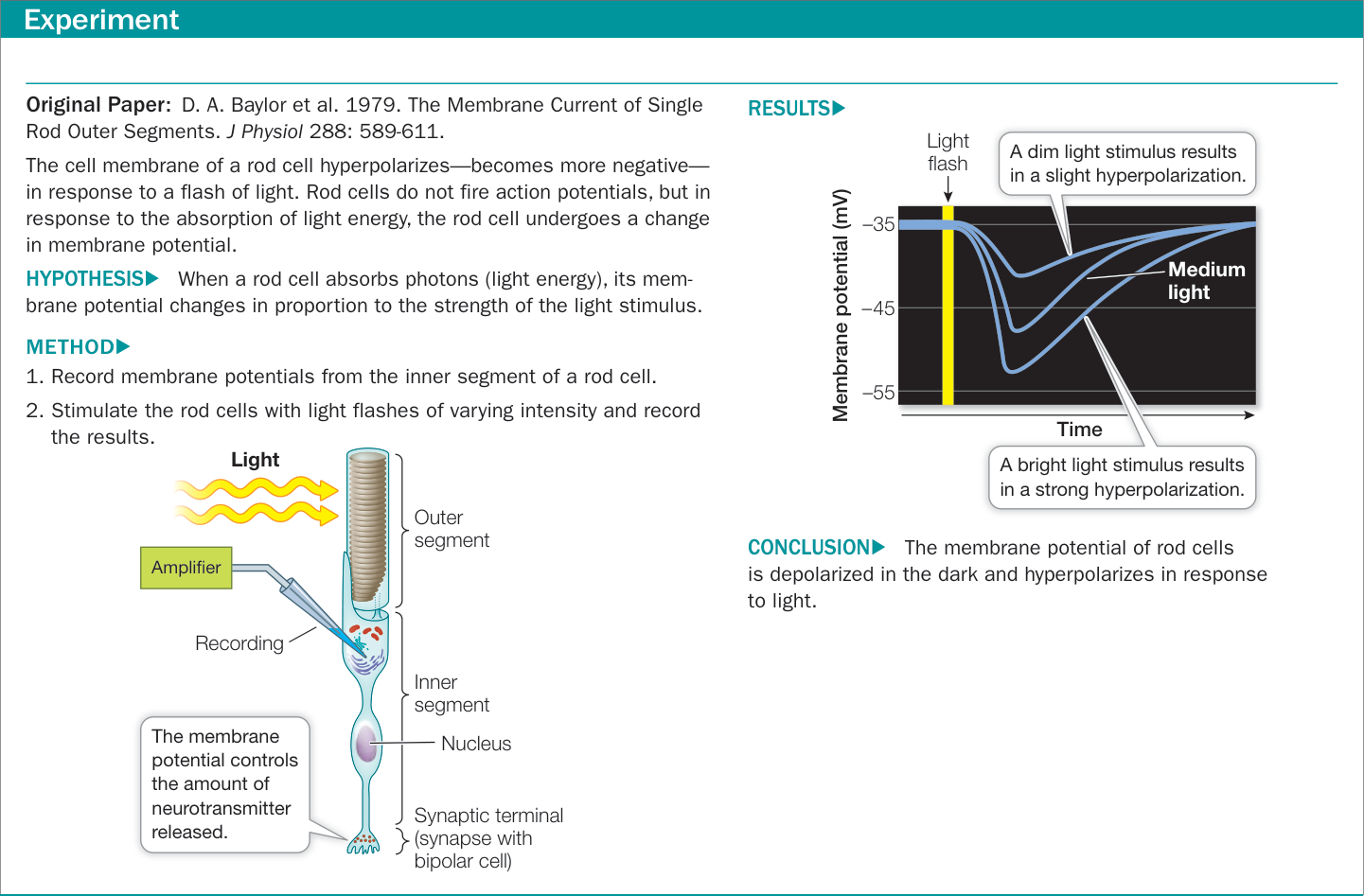
Figure 44.17 How Light Stimulates Rod Cells
PDE converts cGMP to GMP, which causes the cGMP gated channels to close, changing the balance between the inward flow of and the outward flow of and causing the cell to hyperpolarize.
This cascade may seem like a roundabout way of doing business, but its advantage is its enormous amplification ability. Each molecule of light-excited rhodopsin can activate several hundred transducin molecules, thus activating a large number of PDE molecules. The catalytic capacity of PDE is great—one PDE molecule can hydrolyze several hundred molecules of cGMP per second. The bottom line is that a single photon of light can result in the closure of a huge number of channels.
CONE CELLS Cone cells are responsible for the high-acuity color vision of day-active vertebrates such as humans. It is therefore logical that the highest density of cone cells is in the area of the retina that receives light from the center of the visual field, a region called the fovea (see Figure 44.13A). The human fovea has about 160,000 cones per square millimeter. But humans are not the champions of high-acuity vision; a hawk’s fovea has almost twice that number of cones, making the hawk’s vision much sharper than ours. Birds also have two foveae in each eye; one receives light from straight ahead, the other from a more lateral field of vision. The forward-looking foveae make binocular vision possible, while the lateral-looking foveae provide high-acuity vision. Birds use both sets of foveae by frequently turning their heads slightly; they cannot move their eyes in the sockets as humans can.
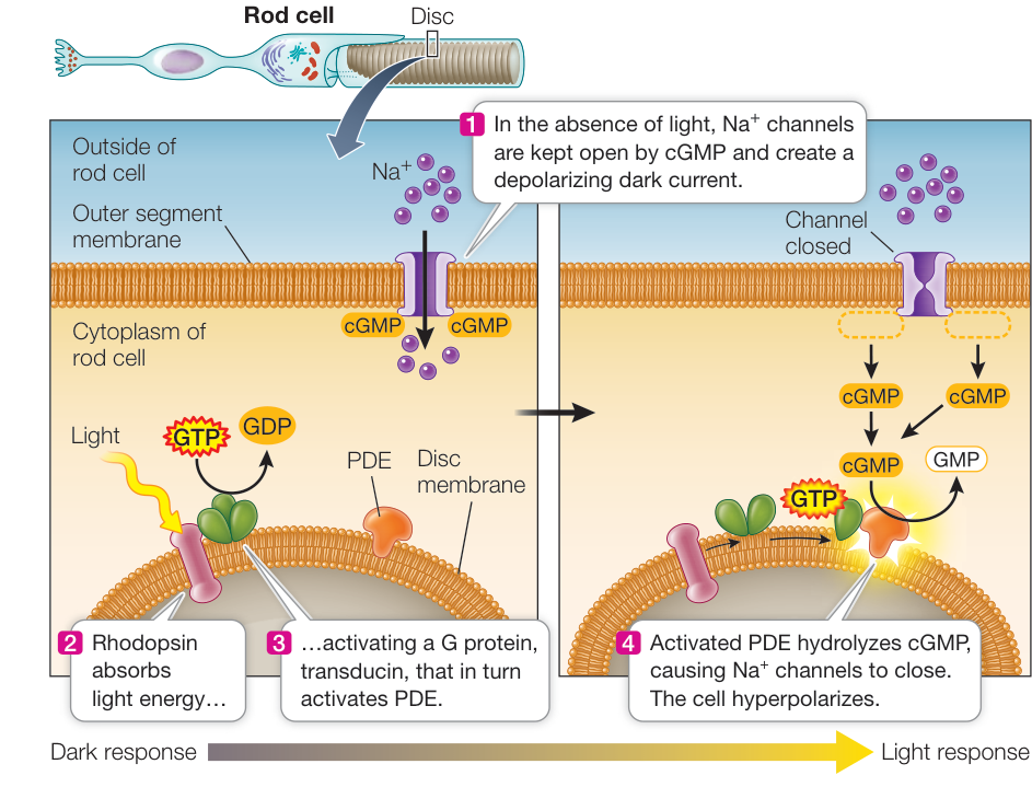
Figure 44.18 Light Absorption Closes Sodium Channels The absorption of light by rhodopsin initiates a signaling cascade that hyperpolarizes the rod cell.
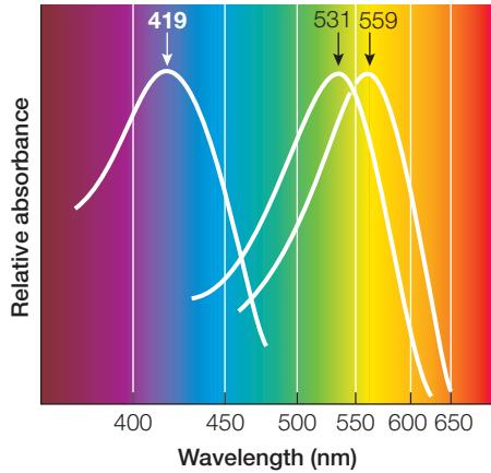
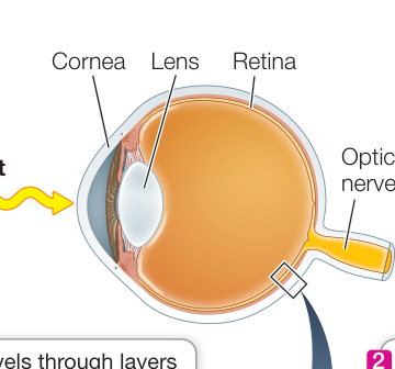
Figure 44.19 Absorption Spectra of Cone Cells The three kinds of cone cells contain slightly different visual pigments that absorb different wavelengths of light. (After J. K. Bowmaker et al. 1980. J Physiol 298: 131–143.)
Cones have low sensitivity to light and contribute little to night vision. Night vision depends mostly on rod cells, which is why vision in dim light is mostly in shades of gray and acuity is low. You may have trouble seeing a small object at night when you are looking straight at it—that is, when its image is falling on your fovea. If you look a little to the side, so that the image falls on a rod-rich area of your retina, you see the object better. Astronomers looking for faint objects in the sky learned this trick a long time ago. The retinas of nocturnal animals, such as flying squirrels, contain a high percentage of rods. By contrast, some animals that are active only during the day (such as chipmunks) have mostly cones in their retinas.
The human retina usually has three kinds of cone cells, each containing slightly different opsin molecules that differ in the wavelengths of light they absorb best. Although the same 11-cis-retinal group is the light-absorber in all three kinds of cones, its molecular interactions with opsin determine the spectral sensitivity of the cone cell as a whole (Figure 44.19). Those that absorb maximally around 419 nanometers (nm) are called S-cones for short wavelength (blue range). Those that absorb maximally around 531 nm are called M-cones for medium wavelength (green range), and those that absorb maximally around 559 nm are called L-cones for long wavelength (yellow–red range). Because different wavelengths of light are differentially absorbed by the different cone cell visual pigments, the brain interprets the relative inputs from the different classes as a full range of color. Color blindness in humans results from the absence or dysfunction of one or more of the three classes of cone cells. Since the genes for the medium (green) and short (yellow/red) opsins are on the x chromosome in humans, and males have only one x chromosome, it is more likely that men will be colorblind for red and/or green than will women.
Information is integrated by the layers of neurons in the retina
The human retina is organized into layers of neurons that receive visual information and process it before sending it to the brain (Figure 44.20). Closest to the lens (and thus to light input) is a layer of ganglion cells; a central layer contains three neuronal types, bipolar cells, horizontal cells, and amacrine cells; and at the “rear” of the retina lie the photoreceptors (rods and cones). The layers of cells between the photoreceptors and the ganglion cells process information about the visual field.
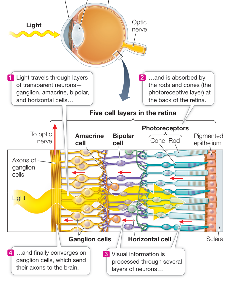
Figure 44.20 The Human Retina Five layers of neurons receive and process visual information. The rods and cones are photoreceptors. The other four layers are the ganglion cells, the bipolar cells, the horizontal cells, and the amacrine cells, all of which are involved in transmitting light signals received by the photoreceptors to the brain. Media Clip 44.1 Into the Eye Life12e.com/mc44.1 Activity 44.3 Structure of the Human Retina
GANGLION AND BIPOLAR CELLS From our discussion of rod cells, you know that the photoreceptor cells at the back of the retina hyperpolarize in response to light and do not generate action potentials. The ganglion cells at the front of the retina do, however, fire action potentials. The axons of ganglion cells form the optic nerves that travel to the brain.
The ganglion cells are connected to the photoreceptors by bipolar cells. Changes in the membrane potential of rods and cones in response to light alter the rates at which the rods and cones release neurotransmitter at their synapses with the bipolar cells. In response to this neurotransmitter, the membrane potentials of the bipolar cells change, altering the rate at which they release neurotransmitter onto ganglion cells. The neurotransmitter release from the bipolar cells determines the rate at which ganglion cells fire action potentials. Thus the direct flow of information in the retina is from photoreceptor to bipolar cell to ganglion cell. The ganglion cells send the information to the brain via the optic nerves.
Each human eye contains about 1.2 million ganglion cells but more than 100 million rods and cones. Therefore there must be convergence of information as it passes from the photoreceptors to the ganglion cells. A given bipolar cell can receive input from multiple rods or multiple cones, but not from both. The relationship among photoreceptors, bipolar cells, and ganglion cells depends on their location on the retina. In the fovea, a ganglion cell may receive input from as few as five photoreceptors, but in the periphery of the retina, a ganglion cell may receive input from thousands of photoreceptors. Visual acuity is a reflection of these quantitative relationships.
The patch of photoreceptors that communicates with a ganglion cell forms a circular receptive field. When light falls on a receptive field, its ganglion cell can be either excited or inhibited. As already mentioned, each ganglion cell sends an axon to the brain in an optic nerve. Thus the information coming from the retina to the brain is about the pattern of patches of light and dark falling on the retina. We’ll return to a discussion of how the brain constructs the visual field from the input from receptive fields in Chapter 45.
HORIZONTAL AND AMACRINE CELLS The other two cell layers, the horizontal cells and the amacrine cells, consist of interneurons that communicate laterally across the retina. Horizontal cells form synapses with neighboring photoreceptors and bipolar cells. Thus light falling on one photoreceptor can influence the sensitivity of its neighbors to light. This lateral flow of information enables the retina to sharpen the perception of contrast between light and dark patterns. Amacrine cells form local interconnections between bipolar cells and ganglion cells. Some amacrine cells are highly sensitive to changing illumination or to motion. Others assist in adjusting the sensitivity of the eyes according to the overall level of light falling on the retina. When background light levels change, amacrine cell connections to the ganglion cells adjust the range of intensities to which they are sensitive. Thus even with large changes in background illumination such as going from outdoors into a building, the eyes are still sensitive to small, rapid changes in the pattern of light falling on the retina. The light reflected from this page can vary by nine orders of magnitude (1,000,000,000-fold) between a sunny outdoor environment and a moonless night. Even though our eyes function best over a light-intensity range of only two orders of magnitude, modulation by amacrine cells can rapidly shift that functional range to accommodate a wide range of light intensities.
Recap and Assess
Vertebrates have image-forming eyes in which a lens focuses incoming light onto the photosensitive retina. Opsin molecules are responsible for light sensitivity in all animals, and in vertebrates opsins are contained in receptor cells called rods and cones. Slight differences in the opsin molecules result in differences in spectral sensitivity, which is the basis for color vision. Rods and cones do not fire action potentials, but their graded membrane potentials alter neurotransmitter release onto retinal bipolar cells. The bipolar cells in turn influence ganglion cells that do fire action potentials in their axons, which travel in the optic nerves to the brain. Two other types of retinal cells, horizontal and amacrine cells, sharpen contrast and adjust sensitivity of the retina, respectively.
- Explain why a compound eye is better at detecting movement than at forming images.
- How do photons of light change the receptor potential of rod cells?
- Explain how rods and the various cones differ in their spectral sensitivity.
- If you are looking straight ahead and someone holds up a colored card on your right or left side, why can’t you tell what the color is?
- Give two reasons why your vision is impaired when you come from bright outside light into a dimly lit room.
Knowing the path of information from sensory receptor cells to the CNS still does not tell us how that information is processed by the brain. What does the eye tell the brain, for example, in response to a pattern of light falling on the retina? In Chapter 45 you will learn how the mammalian brain reassembles sensory information into our perception of the world.
Investigating Life: Qancana How do pit vipers “see” in the dark?
The pit organs of rattlesnakes are the infrared detectors that signal the presence of warm-blooded prey. In this chapter we considered how the pit organs transduce infrared radiation into neuronal information. Infrared is a portion of the electromagnetic radiation spectrum, as is visible light. So could the receptor cells in the pit organs be modified photoreceptors? Or have they evolved from the types of sensory neurons in the skin that sense temperature? The temperature-sensitive neuron theory seems more likely since there is no rhodopsin in the pit organs. But how did such neurons acquire an acute sensitivity to minute changes in temperature of the pit organ caused by distant infrared emitters? The experiments covered in Investigating Life: How Do Pit Vipers “See” in the Dark? identified a particular TRP channel that is highly expressed in the trigeminal ganglia but not in the dorsal root ganglia of rattlesnakes. These experiments also revealed that the TRPA1 channel is not highly expressed in the TG or DRGs of non-pit snakes, but is highly expressed in a distantly related pit snake, the python. In the online exercise to Investigating Life: How Do Pit Vipers “See” in the Dark? it was determined that expression of the TRPA1 gene in frog oocytes causes them to have a steep temperature sensitivity in the range of 34°C–42°C. Thus we can conclude that the TRPA1 channel has evolved along with the pit organ to enable pit vipers such as the rattlesnake to detect warm-blooded prey.
Future directions
The identification of specific genes that convey specific sensory capabilities—for example, identification of the different opsin genes for color vision and the TRPA1 gene for infrared detection—raises the possibility that genetic engineering efforts could be directed toward expanding the sensory capabilities of animals, including humans. What would it be like to be able to see in the infrared and ultraviolet ranges or to hear infrasound and ultrasound? Would the human brain be able to process and integrate that huge amount of additional information? How would our conscious perception of reality be changed?

© John Cancalosi/Alamy Stock Photo
You should be able to relate each summary to the adjacent figures. If you go to this visual summary in Achieve, you can follow links to figures, animations, activities, and simulations that will help you consolidate the material.
Visual Summary
Sensory Receptor Cells Convert Stimuli into Action Potentials
- ■ Receptor potentials are graded membrane potentials that can generate action potentials or cause the release of neurotransmitter.
- ■ Receptor proteins cause ion channels to open or close, affecting the receptor cell’s membrane potential. Different receptor proteins respond to physical forces, light, chemicals, temperature, or electric charge.
- ■ Transient receptor potential (TRP) genes are responsible for many modalities of sensory transduction.
- ■ Sensory adaptation enables the nervous system to ignore irrelevant or continuous stimuli while remaining responsive to relevant or new stimuli.
- What is a graded membrane potential, and how can it integrate inputs from multiple receptor proteins?
- Which would generate a faster response, an electroreceptor or a chemoreceptor? Why?
- Which type of receptor protein enables a pit viper to see in the dark?
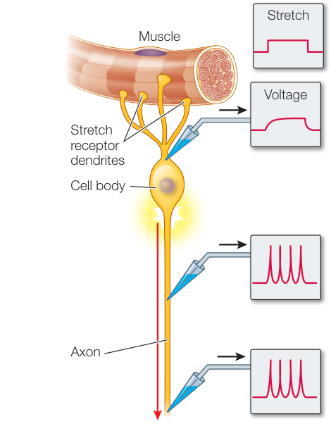
Figure 44.1
Chemoreceptors Respond to Specific Molecules
- ■ Chemoreceptors are responsible for olfaction, gustation, and the sensing of pheromones.
- ■ There are thousands of olfactory receptor genes, but each mammalian olfactory receptor neuron (ORN) expresses only one of these genes. A molecule binding to that receptor protein causes the cell to fire an action potential.
- ■ ORNs project to the olfactory bulb of the brain, and ORNs for a particular odorants project to specific olfactory glomeruli.
- ■ Pheromones communicate information among individuals of the same species.
- ■ Taste buds in the mouth cavities of terrestrial vertebrates are responsible for gustation. The five basic tastes are sweet, salty, sour, bitter, and umami.
Questions
- What is the function of a glomerulus in the olfactory bulb?
- How can pheromones be released by a female silkworm moth provide directional information enabling a male to find her?
- What are similarities and differences in which taste receptors detect and signal sweet versus salty tastes?
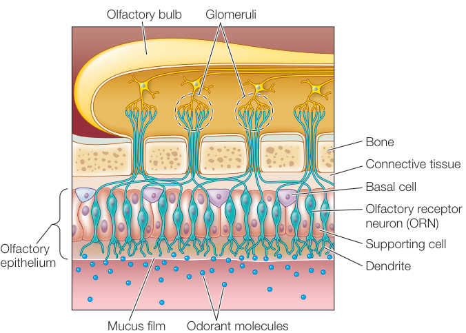
Figure 44.3
Mechanoreceptors Respond to Physical Forces
- ■ Skin mechanoreceptors respond to touch and pressure. The density of mechanoreceptors determines the sensitivity of a skin area.
- ■ Stretch receptors in muscle spindles and in Golgi tendon organs inform the CNS of the positions of and loads on parts of the body.
- ■ In mammals, ear pinnae collect and direct sound waves to the tympanic membrane, which vibrates in response to sound waves. The movements of the tympanic membrane are amplified by the ossicles that conduct the vibrations to the oval window. Movements of the oval window create pressure waves in the fluid-filled cochlea.
- ■ Pressure waves of different frequencies in the cochlea flex its basilar membrane in different locations. These flexions bend the stereocilia of the hair cells in the organ of Corti causing them to cells to release neurotransmitter onto auditory sensory neurons that fire action potentials in the auditory nerve.
- ■ Hair cells are also the mechanoreceptors in the organs of equilibrium in the mammalian vestibular system—the semicircular canals and the saccula and utricle.
- Ruffini endings adapt slowly, whereas Pacinian corpuscles adapt rapidly. Both of these mechanoreceptors sense vibrations applied to the skin. Which one is best at sensing high-frequency vibrations, and why?
- You read that the muscle spindle has a sensory neuron. It also has motor neurons that cause the spindle fibers to
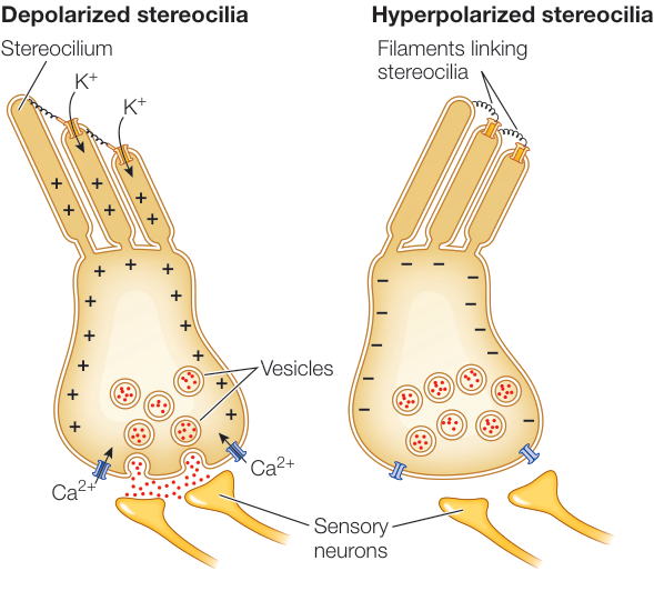
Figure 44.8 contract and stretch their mechanosensors. What would be the response of the arm flexor muscle in response to action potentials in the motor neurons to its muscle spindles?
- What is the function of otoliths, and how do they achieve that function?
Photoreceptors Respond to Light
- ■ Eye cups of flatworms sense the direction of a light source. Compound eyes of arthropods detect shapes, patterns, and movement. Eyes of vertebrates and cephalopods form high-resolution images.
- ■ Vertebrate and cephalopod eyes focus images of the visual field onto dense arrays of photoreceptor cells that transduce the image into neuronal signals.
- ■ Photons of light are absorbed by opsins, visual pigments that bind light-absorbing 11-cis-retinal. Absorption of light changes conformation of retinal, resulting in changes in the membrane potential of the photoreceptor cell.
- ■ Vertebrates have two types of photoreceptors, rod cells and cone cells.
- ■ Rhodopsin is the visual pigment of rod cells. Cone cells have three different opsins, which gives them different spectral sensitivities.
- ■ The photoreceptor cells of vertebrates are at the rear of the retina. They communicate with ganglion cells through bipolar cells. Ganglion cell axons form the optic nerves.
Questions
- If you had an electrode in a rod cell and you shone a light on the cell, what would you see in the recording from that electrode, and how would that response be communicated to the next retinal cell involved in visual transduction?
- If you are trying to put a key in a lock in very dim light, why does it help to look a little bit to the side of the keyhole?
- What is transducing and how does it work?
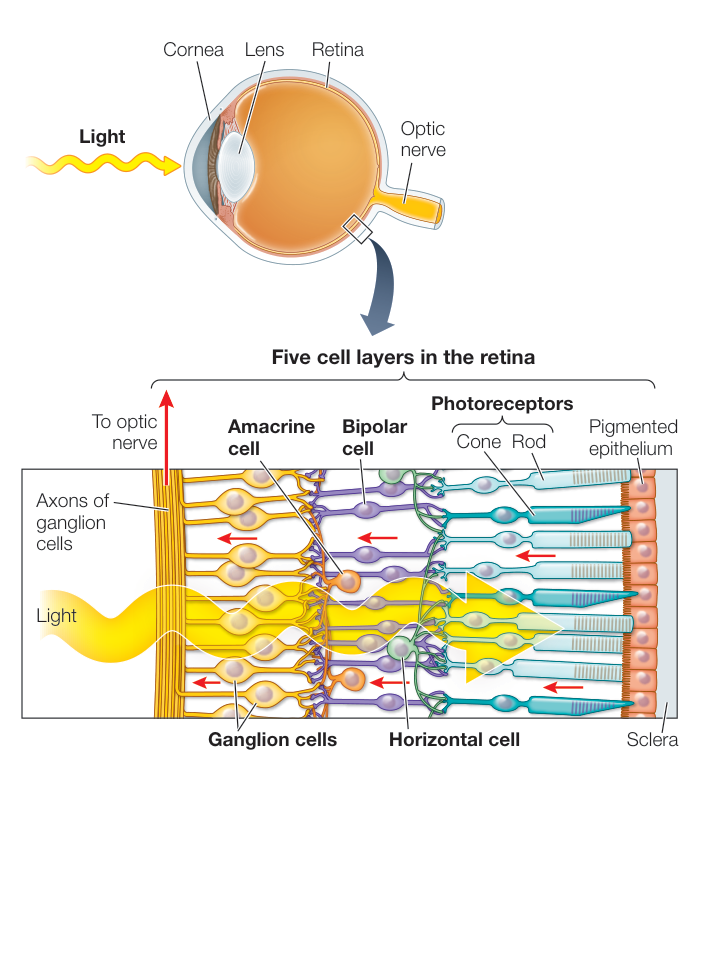
Figure 44.20