26
PREDICTION OF PROTEIN-PROTEIN INTERACTIONS FROM EVOLUTIONARY INFORMATION
INTRODUCTION
The more we know about the molecular biology of the cell, the more we see genes and proteins as part of networks or pathways instead of as isolated entities and understand their function as a variable of the cellular context. To fulfill this new paradigm, genomic sequences offer a catalog of building blocks, and protein interactions provide the first information for the challenging task of understanding the functions of these genes and proteins within their situation in networks and pathways. To further refine the information on the context and timing in which these observed biological functions take place, additional insight from genetic regulatory mechanisms and genetic specificity (cell-type specificity, individual differences, etc.) will become increasingly important.
The study of protein interactions can be divided into two complementary aspects, the determination of the residues or regions implicated in the interactions and the deciphering of the identity of the interaction partners (which proteins interact with which ones). These two problems have been typically addressed by biophysical and biochemical techniques, such as binding studies (chromatographic isolation of complexes, coimmunoprecipitation, protection, cross-linking studies, etc.) and indirect genetic methods (gene suppression studies, systematic mutagenesis, and interspecies exchanges). The development of genomic and postgenomic technologies has changed the panorama considerably, with the possibility of obtaining systematically massive amounts of data about protein interactions. Progress has been done in the automation of experimental approaches such as yeast-two-hybrid based methods and mass spectrometry determination of components of macromolecular complexes. At the same time, a number of new bioinformatics techniques have been developed based on the considerable amount of sequence and genomic information that is being accumulated in databases. In the following, we review the status of these bioinformatics approaches to the study of protein interactions, including the prediction of interaction partners and protein-protein binding regions.
Evolutionary Features Related with Structure and Function
Multiple sequence alignments (MSA) represent the main source of evolutionary information available. The relation between equivalent positions of homologous proteins compiles the result of the changes allowed by the evolution of the corresponding proteins at different positions. The interplay of structural and functional requirements contributing to the stabilization of natural variants creates complex patterns of mutational behavior of the positions that have been exploited by a number of computational methods in the search for structural and/or functional constraints (Figure 26.1).
Conserved Positions. The information most widely extracted from multiple sequence alignments is related with conserved positions1. These invariable positions are interpreted as important residues for the structure or function of the protein since apparently changes were disallowed during evolution (Figure 26.1). Conserved positions usually are located in structural cores (i.e., structurally important positions) and active/binding sites (i.e., positions directly involved in protein function). Although the problem of locating conserved positions could look trivial at first sight, there are many different approaches that produce different results depending, for example, on the treatment of conservative changes2. Modern methods for detecting conserved sites incorporate complex evolutionary models taking into account the phylogeny of the sequences in the MSA3, so as to avoid artifacts due to the peculiarities or uneven distribution of sequences in the alignment (i.e., highly similar sequences resulting in most of the positions being conserved). Despite a number of attempts to analyze the relationship between binding sites, conserved positions, and type of amino acid in those positions4, 5, the complex nature of the relations indicates that much remains to be done in this area.
Figure 26.1. Sequence features related with structure and function extracted from multiple sequence alignments. MSA and its implicit phylogeny are shown. Conserved positions are invariant throughout all the sequences. In the family-dependent conserved positions, a different amino acid type is conserved within each one of the subfamilies. For coevolving pairs of positions, changes in one of the positions are correlated with the corresponding changes in the other.
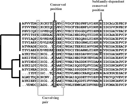
Family-Dependent Conserved Positions. There is a more subtle kind of conservation, the family-dependent conservation. These positions are typically conserved in the subfamilies that form well-defined branches of the phylogenetic tree and differ in the chemical type of the amino acids that characterize each subfamily (Figure 26.1). We have previously used the term tree-determinants to describe these positions given their drastic in fluence in the structure of the corresponding phylogenetic trees. Family-dependent conserved positions would be most likely related with the specific features of each subfamily that in most cases turn out to be the differential binding to other proteins and substrates.
One of the first approaches to the detection of this kind of functional residues, implemented in the sequencespace program, was developed by Casari, Sander, and Valencia6 and later followed by a plethora of other methods7, 8, 9, 10 (Mesa, Pazos, and Valencia, 2003). The underlying principle in all these methods is the detection of positions in multiple sequence alignments with a differential conservation in the different groups of sequences that form part of a larger protein family. All these methods use the subfamily classification implicit in the phylogeny represented by the MSA (Figure 26.1). Recently, some methods were developed that do not rely on that implicit classification but are able to incorporate an external classification of the sequences according to some known functional property11, 12.
Coevolving Positions. Another sequence-based approach for the prediction of protein structure and molecular complexes is based on the detection of correlated mutations in multiple sequence alignments and their use as distance constraints between residues belonging to the same or different proteins. Correlated mutations correspond to pairs of positions with a clear pattern of covariation (Figure 26.1). The underlying evolutionary model for explaining their relation with space neighboring assumes that part of the detected correlated pairs correspond to compensatory mutations, where in particular sequences of the multiple sequence alignments the mutation of one residue was compensated along the evolution by a mutation of a neighbor residue, most likely to keep proteins (or protein complexes) in permissible limits of protein stability. Coevolving positions have been demonstrated to have an important functional role together with conserved positions13.
The method proposed in 199414 for detecting these types of positions was a weak predictor of proximity between residues in protein structures. Despite their low accuracy, these predicted contacts have been demonstrated to be useful, for example, in filtering structural models15 or driving ab initio simulations16.
PREDICTION OF INTERACTING REGIONS
Structure and Sequence Characteristics of the Interaction Sites
In general, the interaction surface (interface) is difficult to distinguish from the rest of the surface, from either a sequence or structure point of view. The interaction sites are not clearly enriched in any particular type of amino acid17. Nevertheless, some particular types of complexes do have preferences for some amino acids at the interfaces, that is, hydrophobic residues in homodimers18, 19. Similarly, structural characteristics are only discriminative for some type of complexes, that is, nonplanar interfaces for enzyme inhibitor and large planar interfaces for homodimers18, 20. For these reasons, complex machine learning systems are required for dealing with the problem of predicting interaction sites from sequence or structure information.
Structure-Based Methods
The problem of determining the physical structure of protein complexes when the structure of the members is known is part of the problem of docking molecules, such as proteins with small molecules (Chapter 27). Despite the considerable efforts that have gone in solving this problem, directed at the design of new drugs, the solutions in the case of protein-protein interaction are still far from optimal.
Despite these difficulties, various groups have achieved considerable progress in the development of physical docking programs. Most of the current approaches consider proteins as rigid bodies and have the physical matching of the surfaces as their main guide. Only a few approaches integrate conformational flexibility, allowing the interacting surfaces to adapt one to the other, normally at the expenses of reducing the search space for possible interacting surfaces. Some programs take into account other features for predicting regions of interaction, like hydrophobicity or electrostatics (Chapter 24). For recent reviews on docking, see Schneidman-Duhovny, Nussinov, and Wolfson21 and Gray22. An interesting effort to compare the various protein docking approaches in a blind test is organized by Janin et al. (CAPRI: http://capri.ebi.ac.uk/). The results of this experiment, if enough structures of protein complexes become available for the comparison, will be important for updating our view of the capability of current docking methods. A pressing question now is that the structural genomics efforts are on the way to solve a substantially larger number of isolated proteins and protein domains (Chapter 40).
There are other structure-based techniques for predicting interacting regions apart from physical docking. Some approaches are based on the fact that in many cases binding or active sites are energetically unstable. This is due to many factors, including the need to put together energetically unfavorable combinations of residues due to functional requirements (e.g., two residues with the same charge in a catalytic site) or to maintain unstable interacting regions that are stabilized upon binding, favoring in this way the bound form in the binding equilibrium. The uneven distribution of the stabilization energy in proteins has been related with binding sites and cooperativity effects23. Relationships between functionally important residues and those that negatively contribute to the electrostatic stabilization energy have also been found24.
New concepts taken from graph theory have been applied to the detection of interaction sites in 3D structures. Within these methods, a protein structure is represented as an undirected graph where the nodes are the residues and the edges represent contacts between them. Within this graph, residues (nodes) with special connectivity characteristics such as high “centrality” have been associated with functional and binding sites25.
The combined data on massive protein interactions (see the section “Experimental Techniques and High-Throughput Applications”) and structural information can be mined in the search for short motifs or patterns frequently involved in interactions26. These motifs can be used for predicting new interacting regions.
With the increasing availability of protein 3D structures, methods that use structural alignments for predicting binding and functional sites are emerging. These methods are conceptually similar to the ones based on sequence alignments (see the section “Sequence-Based Methods”) but use structural alignments to relate more distant proteins27, 28.
Sequence-Based Methods
Ab Initio Methods. We term ab initio methods in this context as methods that only use the sequence of a protein to predict residues involved at the binding interface for intermolecular recognition.
This category of methods includes the detection of intrinsically unstructured segments in protein sequences, also termed “disordered regions”; see Bracken et al.29 and Chapter 38. To some extent opposing the paradigm that structure determines function, what determines the function of these proteins is actually their lack of structure. The functional importance of these regions arises from their frequent involvement in protein-protein interactions. In many cases, these unstructured segments become structured on binding, favoring the bound form of the interaction equilibrium. This type of entropy-driven interaction has the unique specificity/affinity characteristics required for some biological complexes30. These intrinsically unstructured regions are to some extent related to the low complexity segments in the primary sequence31, 32. Nevertheless, specific predictors have been developed to locate these regions33, 34.
Another method that uses a single sequence as the primary input to identify interacting regions is the neural network (NN) developed by Ofran and Rost35. This NN is trained with sequence segments of interaction sites, and it correctly predicts at least one interaction site for 20% of the complexes in its test set.
Methods Based on Multiple Sequence Alignments. Most sequence-based methods for predicting protein interaction regions use evolutionary information in the form of multiple sequence alignment. For example, Ofran and Rost’s method commented in the previous section obtains better results when MSA-derived information is incorporated36.
The use of evolutionary conservation, a strategy often employed to identify residues of functional importance, has also been used for detecting interaction sites. Although the best results are obtained when combining conservation with structural information (see the section “Hybrid Methods”), we include in this section of sequence-based methods predictors that combine residue conservation with predicted structural features that are often based on MSA. For example, ConSeq uses a sophisticated calculation of conservation together with predicted solvent accessibility for predicting functional residues and binding sites37.
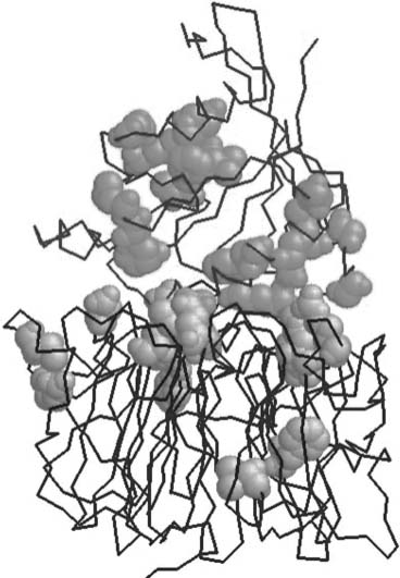
The relation between tree-determinant residues (see the section “Family-Dependent Conserved Positions”) and interacting surfaces has been analyzed in detail in some well-characterized systems6, 7, 38, 39, 40. More importantly, it has also been demonstrated by direct experiments how exchanging tree-determinant residues between protein subfamilies lead to a switch in the observed specificity of interaction of the corresponding proteins. This demonstration has been performed on two different proteins in the ras family of small GTPases41, 42. In Figure 26.2, the tree-determinant residues extracted from the multiple sequence alignments of Ran and Rcc1 are marked in the structure of the complex formed by these two proteins. It can be seen that many of these residues, predicted from sequence information alone and before the structure of the complex was solved, map in the interaction surface, especially in Rcc1.
Figure 26.2. Complex between Ran (upper chain) and Rcc1 (lower chain) marking the treedeterminant residues (in spacefill) for the two proteins (PDB ID: 1I2M). Tree-determinant residues were calculated with the sequencespace algorithm6. Figure courtesy of Juan A.G. Ranea. Figure also appears in the Color Figure section.
Coevolving positions (see the section “Coevolving Positions”) have also been used for detecting interfaces43, 44, 45. In spite of the weak power of correlated mutations in predicting neighboring residues, it was demonstrated that, in many cases, it is enough to discriminate the right arrangement of two protein chains from many alternatives43. Recently, it has been argued that the observed correlation appears only in permanent (not transient) complexes46.
Hybrid Methods
Methods that incorporate both structural and sequence information to predict interaction surfaces demonstrated significant improvement from the equivalent ones that use only sequence information. The simplest way of combining sequence and structural information for predicting binding regions is by simply mapping the sequence-based predicted sites (conserved, family-dependent conserved, correlated, etc.) into the 3D structure of the protein to assess whether the predicted positions have the structural characteristics expected for a functional or binding site (clustered or accessible to the solvent) (Figure 26.3). For example, the ConSurf program looks for conserved residues that map in the surface of the protein, taking into account the distribution of sequences in the MSA for calculating conservation47. Panchenko et al.’s method also looks for clusters of conserved residues48. The method developed by Aloy et al. searches for conserved apolar residues that cluster in the surface of the protein49. Distant sequences in the MSA are successively removed until the set of conserved residues forms a cluster. Landgraf et al.’s method looks for surface regions of the protein following the same phylogeny as the whole family50. Structural information is used to define those regions (as sets of residues close in the surface of the protein). 3D information can also be incorporated in other methods for detecting family-dependent conserved positions simply by imposing clustering51, 52. Some methods map sequence conservation on 3D structures but using a simplified alphabet that reflects the functional groups of the amino acids instead of considering all the 2053.
Figure 26.3. Prediction of binding sites combining multiple sequence alignments (MSA) and structural information. Conserved positions in the MSA (black) and family-dependent conserved positions (gray) have been extensively used as predictors of interaction and functional sites. If available, structural information can be used a priori or a posteriori. If used a priori (left), the structure is used to define surface patches (gray). The sequence features of members in this patch are encoded (i.e., amino acid frequency vectors in this case) and used as input features to a machine learning system such as a neural network (bottom). The machine learning system is trained to distinguish patches involved in interaction from non-interacting ones. A posteriori (right), the structure can be used to assess whether a set of positions predicted from sequence has the characteristic 3D features of a binding site such as being clustered in proximity and exposed to the solvent.
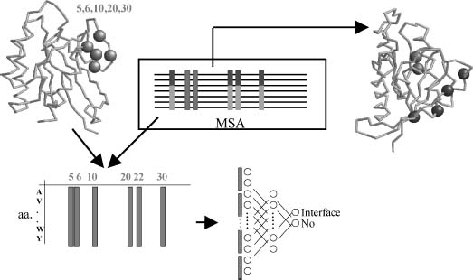
A much direct way of combining sequence and structural information for predicting binding sites is by using machine learning systems, such as neural networks or support vector machines (SVM). This general strategy was introduced by Zhou and Shan54 and Fariselli et al.55, and later followed by many other authors56, 57, 58. These methods use the structure of a protein to define surface patches of neighbor residues and the multiple sequence alignment to obtain the sequence profiles (or other sequence-based features) for the members of the patch (Figure 26.3). The machine learning system is then trained with that information for a training set of proteins with known interaction surfaces. After the training process, surface patches (plus their corresponding sequence features) of proteins with known structure but unknown interaction surface are presented to the system to identify which surface patches are involved in intermolecular recognition. The reported accuracy of these methods is higher than 70%. Nevertheless, it is difficult to test these methods and compare their performance due to the absence of a good test set of protein interfaces (nonredundant in sequence and structure, covering all types of complexes, etc).
PREDICTION OF INTERACTION PARTNERS
Experimental Techniques and High-Throughput Applications
The experimental approaches for the determination of interaction partners have undergone dramatic improvements during the last few years, particularly through the systematic application of different strategies based on the yeast-two-hybrid protocol59 and the tandem affinity purification (TAP) of complexes followed by mass spectrometry (MS) analysis60. These approaches were used to determine large proportions of the interactomes of a number of model organisms, ranging from bacteria such as H. pylori61 to human62, covering unicellular eukaryotes such as yeast60, 63, 64 or multicelular organisms such as C. elegans65 and D. melanogaster66. These interactomes, in spite of having provided a lot of information on living systems67, still have a high degree of error68, 69 and their determination is very expensive in terms of time and money (see the section “Future Trends”).
Computational Methods Based on Genomic Information
In parallel to these experimental approaches for the detection of interacting pairs of proteins, a number of bioinformatics techniques have also been developed. These methods, based on genomic and sequence features intuitively related with protein interaction, usually detect pairs of proteins that may interact physically or functionally (i.e., proteins that form part of the same signaling or metabolic pathway).
The results of many of these methods can be accessed online at repositories such as STRING70 (http://string.embl.de). These data can be used for inferring the functional role of a protein by identifying its potential interactors. This is called “context-based” function prediction and is orthogonal and complementary to the classic sequence-based functional transfer.
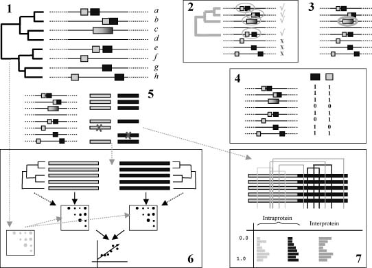
The main methods based on sequence and genomic features for predicting interacting pairs of proteins are depicted in Figure 26.4.
Phylogenetic Profiling. This method is based on the detection of pairs of genes that have a similar species distribution. That is, they are present/absent in the same species71. The idea behind this approach is that proteins that need each other to perform a given function will either be simultaneously present or absent. In the second case, loss of interacting partners can be observed as a consequence of “reductive evolution” where the organism (especially bacteria) would eliminate genes when the corresponding interacting partner is not present. As a result, interacting or functionally related genes would tend to have similar phylogenetic profiles, that is, similar patters of presence/absence (Figure 26.4, panel 4).
In their first versions, phylogenetic profiles were coded as binary vectors with “1” coding for the presence of a given gene and “0” coding for its absence (Figure 26.4, panel 4). Later, quantitative information was incorporated by encoding the similarity of a protein sequence in a given organism with respect to an organism of reference into the positions of the vector, representing in this way not only the presences/absences of the genes but their relative divergences as well72. Another improvement came from the incorporation of information on the phylogeny of the species involved, together with an evolutionary model of gene gain and loss. This allows to naturally exclude profile similarities not due to functional reasons but rather to the underlying evolutionary73, 74.
Not only are similar profiles informative but also the “anticorrelated” ones (one protein is present whenever the other is absent and the other way around). These anticorrelated profiles have been related with enzyme “displacement” in metabolic pathways75. Furthermore, this versatile technique has recently been extended to triplets of proteins, allowing the search for more complicated patterns of presence/absence (e.g., “protein C is present if A is absent and B is also absent”), which allows the detection of interesting cases representing biological phenomena beyond binary functional interactions, like complementation76.
This successful approach has two main limitations. First, it cannot be applied to many essential proteins, since they are present in all organisms and hence they create noninformative profiles. Second, this methodology requires fully sequenced genomes in order to accurately determine whether a given gene is present in an organism or not.
Conservation of Gene Neighboring. The conservation of the proximity of genes along the genome between distantly related species to predict interaction is also being utilized by several methods77, 78. A typical example would be proteins encoded by genes within a conserved operon where, if present together in another bacteria, would contain the sufficient, necessary components to execute the functional mechanism related with the activity of that operon.
Figure 26.4. Theme of strategies implemented in computational methods for assessing the possible interaction between two proteins. (1) Sequence and genomic information about two proteins (yellow and blue) is used to assess their possible interaction. The inputs for these methods are the sequences and genomic positions of the orthologs of the two proteins, as defined by a given orthology criteria, in a number of organisms (related by a phylogeny). (2) Conservation of gene neighboring: the number of genomes where both proteins are close (according with a given distance cutoff) and their phylogeny are used to assess whether the proteins are interacting or not. (3) Gene fusion: a search for genomes where both proteins appear as part of a single polypeptide is performed. (4) Phylogenetic profiling: phylogenetic profiles for both proteins are constructed by assessing the presence [1] or absence [0] of the two proteins in the set of genomes, and the similarity between these profiles is evaluated. (5) Multiple sequence alignments for the two proteins are built using the sequences of the organisms where both are present. These MSA are used by the following two methods. (6) Similarity of phylogenetic trees: the multiple sequence alignments ofthe proteins (5) are used to generate distance matrices for both sets of orthologs. Alternatively, these multiple sequence alignments can be used to generate the actual phylogenetic trees and extract the distance matrices from them. The similarity of these distance matrices is used as an indicator of interaction. Eventually, the phylogenetic distances between the species involved can be incorporated in the method for correcting the background similarity expected between the trees due to the underlying speciation events. (7) Correlated mutation: the accumulation of correlated mutations between the two multiple sequence alignments is used as an indicator of interaction. Figure also appears in the Color Figure section.
The limitation of this technique is that this approach is reserved for bacterial genomes where there is a clear tendency for the clustering of functionally related genes in operons to allow cotranscription. Inferences can only be made for eukaryotic proteins only if homologues are clearly established in a prokaryotic organism.
Gene Fusion. A third group of methods is related to the presence of fused genes in various genomes79, 80. This strategy uses the observation that some interacting protein pairs are encoded by two independent genes in some organisms whereas they are encoded by a single gene in other organisms. In such cases, it will be logical to conclude that the two proteins, as independent entities or as domains of the same protein, would be functionally related.
Marcotte et al.80 proposed an evolutionary hypothesis for explaining such fusion events: if two proteins have to interact in order to perform a given function, the concentration of the active complex would be much higher if the two proteins are fused together than if the two proteins are separated and hence having to rely on random diffusion to find each other and form the active complex.
The fact that two proteins are fused is a clear indication of their functional relationship (except for “promiscuous domains”). Hence, this approach produces almost no false positives. The disadvantage, however, is the range of applicability because these fusion events, in spite of being very informative, are not very frequent.
Similarity of Phylogenetic Trees. The observation that interacting proteins tend to have topologically similar phylogenetic trees has been used by some methods to predict interaction partners. This similarity was qualitatively observed in sporadic examples of interacting families such as insulin and insulin receptors81, and first quantified for two proteins by Goh et al.82. Pazos and Valencia83 statistically demonstrated the relation between the similarity of phylogenetic trees and interaction in large sets of interacting proteins. Figure 26.5 shows an example of two interacting proteins (the nuoE and nuoF subunits of the NADH dehydrogenase complex) that have similar phylogenetic trees.
The hypothesis behind this approach is that interacting proteins would be subject to a process of coevolution that would be translated into a stronger than expected similarity between their phylogenetic trees84, 85. The first generation algorithms of this approach measure similarity between the phylogenetic trees by indirectly evaluating it as the similarity between the distance matrices of the two families, using a correlation formulation83.
Since it was proposed, this simple and intuitive method (mirrortree) has been applied to many proteins, and different implementations and variations of it were developed84, 85. For example, this concept of similarity of trees was used to look for the correct mapping between two families of interacting proteins (e.g., to select which ligand within a family interacts with which receptor within another family). The idea is that the correct mapping (set of relationships between the leaves of both trees) will be the one maximizing the similarity between both trees86, 87. Another obvious extension of the method has been to incorporate information on the phylogeny of the species involved in the trees in order to correct the “background” similarity expected between any pair of trees due to the underlying speciation process88, 89. Recently, it has also been shown that incorporating information on the whole network of interprotein co-evolutions drastically improves the accuracy of this methodology (Juan et al., 2008b).
Figure 26.5. Example of two interacting proteins that have similar phylogenetic trees. The E. coli proteins are the nuoE (gray) and nuoF (black) subunits of the N module of the NADH reductase. The structure corresponds to the T. thermophilus orthologs (chains B and A in PDB 2FUG). The phylogenetic trees of these two proteins are shown at the top. The species correspondence is indicated with dotted lines.
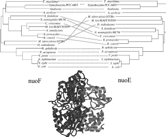
One important limitation of this method is that it can only be applied to pairs of proteins where the orthologs are found in many common species. Only the leaves of the trees corresponding to species where both proteins are present can be used (Figure 26.4, panel 6).
Other Sequence-Based Methods. There are a number of other methods for inferring protein interactions from genomic information.
Coevolving positions (see the section “Coevolving Positions”) have been used for predicting interacting pairs of proteins90, extending their previous usage for detecting interacting surfaces (see the section “Methods based on Multiple Sequence Alignments”). The idea is that pairs of multiple sequence alignments corresponding to interacting proteins would be more enriched in interprotein correlated mutations than pairs that do not interact.
The methods described so far do not involve training, that is, they do not “learn” from examples of known interactions and noninteractions. On the contrary, there are other methods that are trained with examples91, 92, 93, 94, 95. In general, the input is a set of characteristics (descriptors) of the proteins or protein pairs (including experimental data in some cases). Based on a set of known interactions, amachine learning classifier “learns” to distinguish interacting from noninteracting pairs using the values of these descriptors. For example, Sprinzak and Margalit91 use pairs of sequence signatures extracted from known interactions to predict new ones. One of the descriptors used by some of these methods is the domain composition of the proteins95, 96. The idea is that some combinations of domains are more prone to interact.
Computational Methods Based on Structural Information
There are also computational methods for the prediction of interaction partners that use structural information. Most of these methods are intended to predict whether the homologues of two proteins known to interact will interact too or not. Aloy et al. derived statistical potentials from known interactions and then used them to score the possible interactions between the homologues of the members of a given complex97, 98. In a similar way, the energetic feasibility of different complexes between members of the Ras family and different families of Ras effectors was evaluated using the FOLD-X program99.
FUTURE TRENDS
Deciphering the complex network of protein interactions behind cellular processes is crucial for understanding many aspects of living systems and is being tackled with ongoing experimental and computational techniques in need of further developments. Currently, the experimental techniques for the massive characterization of these networks still have several important drawbacks. First, there is a surprisingly low overlap between the results of similar experiments67. Second, experimental techniques such as yeast two hybrid often yield a large number of false positives with an accuracy estimated to be as low as ~ 10% in some cases68. Third, experimental approaches are still far from reaching a “high-throughput” state since the intrinsic drawbacks of the methodologies allow them to test a fraction of all possible pairs of proteins67. Finally, other limitations of these techniques paradoxically arise from their experimental nature. These include the tendency to preferentially detect interactions between highly expressed proteins or between proteins belonging to a particular cellular compartment in detriment of others68.
Computational methods, while having their own set of limitations, are free from many of these drawbacks faced by experimental approaches. Thus, these two classes of methods complement each other. A common limitation of the computational methods for detecting interaction partners is the dependency on the quantity and quality of the genomic data. For this reason, we expect the accuracy of these methods to improve as more sequences and genomes become available. The use of structural information can help improving the performance of current methodologies. For example, HMM derived from structural alignments (Chapters 16 and 22) are widely used for detecting remote homologues. The localization of homologues is common to many of these computational techniques.
We are already seeing a combination of results from the new experimental powerful techniques, the new bioinformatics approaches for the prediction of interacting partners, the relations established between genes with similar expression patterns in DNA array experiments, and the systematic mining of available information about protein interactions in databases and literature repositories100. Combining and finding consensus within these accumulated information obtained from different sources, we will quickly approach a situation in which it will be possible to glimpse the structure of the complex network of protein interactions that govern cell life in relatively simple model systems such as yeast and bacteria.
The methods for predicting interaction partners and detecting interacting regions are helping in the interpretation of the massive amounts of sequence and genomic information in functional terms. The promise is that the combination of improved experimental techniques and computational methods along with an effective consensus interpretation of accumulated data from widely disparate sources would lead not only to a better understanding of cell function but also to better ways of manipulating it with new and more specialized drugs designed specifically for that purpose.
REFERENCES
- Zuckerkandl E, Pauling L (1965): Evolutionary divergence and convergence in proteins. In: Bryson V, Vogel HJ, editors. Evolving Genes And Proteins. New York: Academic Press, pp 97–166.
- Valdar WS (2002): Scoring residue conservation. Proteins 48:227–241.
- Pupko T, Bell RE, Mayrose I, Glaser F, Ben-Tal N (2002): Rate4Site: an algorithmic tool for the identification of functional regions in proteins by surface mapping of evolutionary determinants within their homologues. Bioinformatics 18:S71-S77.
- Villar HO, Kauvar LM (1994): Amino-acid preferences at protein binding sites. FEBS Lett 349:125–130.
- Ouzounis C, Perez-Irratxeta C, Sander C, Valencia A (1998): Are binding residues conserved? Pac Symp Biocomput 401–412.
- Casari G, Sander C, Valencia A (1995): A method to predict functional residues in proteins. NatStruct Biol 2:171–178.
- Lichtarge O, Bourne HR, Cohen FE (1996): An evolutionary trace method defines binding surfaces common to protein families. J Mol Biol 257:342–358.
- Andrade MA, Casari G, Sander C, Valencia A (1997): Classification of protein families and detection of the determinant residues with an improved self-organizing map. Biol Cybern 76:441–450.
- Kalinina OV, Mironov AA, Gelfand MS, Rakhmaninova AB (2004): Automated selection of positions determining functional specificity of proteins by comparative analysis of orthologous groups in protein families. Protein Sci 13:443–456.
- Mihalek I, Res I, Lichtarge O (2004): A family of evolution-entropy hybrid methods for ranking protein residues by importance. J Mol Biol 336:1265–1282.
- Hannenhalli SS, Russell RB (2000): Analysis and prediction of functional sub-types from protein sequence alignments. J Mol Biol 303:61–76.
- Pazos F, Rausell A, Valencia A (2006): Phylogeny-independent detection of functional residues. Bioinformatics 22:1440–1448.
- Socolich M, Lockless SW, Russ WP, Lee H, Gardner KH, Ranganathan R (2005): Evolutionary information for specifying a protein fold. Nature 437:512–518.
- Göbel U, Sander C, Schneider R, Valencia A (1994): Correlated mutations and residue contacts in proteins. Proteins 18:309–317.
- Olmea O, Rost B, Valencia A (1999): Effective use of sequence correlation and conservation in fold recognition. J Mol Biol 293:1221–1239.
- Ortiz A, KolinskiA, RotkiewiczP, IlkowskiB, Skolnick J (1999): Abinitio folding of proteins using restraints derived from evolutionary information. Proteins S3:177–185.
- Chakrabarti P, Janin J (2002): Dissecting protein-protein recognition sites. Proteins 47:334–343.
- Jones S, Thornton JM (1997): Analysis of protein-protein interaction sites using surface patches. J Mol Biol 272:121–132.
- Ofran Y, RostB (2003a): Analysing six types of protein-protein interfaces. J Mol Biol 325:377–387.
- Lo Conte L, Chothia C, Janin J (1999): The atomic structure of protein-protein recognition sites. J Mol Biol 285:2177–2198.
- Schneidman-Duhovny D, Nussinov R, Wolfson HJ (2004): Predicting molecular interactions in silico: II. Protein-protein and protein-drug docking. Curr Med Chem 11:91–107.
- Gray JJ (2006): High-resolution protein-protein docking. Curr Opin Struct Biol 16:183–193.
- Luque I, Freire E (2000): Structural stability of binding sites: consequences for binding affinity and allosteric effects. Proteins S4:63–71.
- Elcock A (2001): Prediction of functionally important residues based solely on the computed energetics of protein structure. J Mol Biol 312:885–896.
- Del Sol A, O’Meara P (2005): Small-world network approach to identify key residues in protein— protein interaction. Proteins 58:672–682.
- Neduva V, Russell RB (2006): Peptides mediating interaction networks: new leads at last. Curr Opin Biotechnol 17:465–471.
- Pazos F, Sternberg MJE (2004): Automated prediction of protein function and detection of functional sites from structure. Proc Natl Acad Sci USA 101:14754–14759.
- Chung JL, Wang W, Bourne PE (2006): Exploiting sequence and structure homologs to identify protein-protein binding sites. Proteins 62:630–640.
- Bracken C, Iakoucheva LM, Romero PR, Dunker AK (2004): Combining prediction, computation and experiment for the characterization of protein disorder. Curr Opin Struct Biol 14:570–576.
- Vucetic S, Brown CJ, Dunker AK, Obradovic Z (2003): Flavors of protein disorder. Proteins 52: 573–584.
- Wootton JC, Federhen S (1996): Analysis of compositionally biased regions in sequence databases. Methods Enzymol 266:554–571.
- Romero P, Obradovic Z, LiX, Garner EC, Brown CJ, Dunker AK (2001): Sequence complexity of disordered protein. Proteins 42:38–48.
- Iakoucheva LM, Dunker AK (2003): Order, disorder, and flexibility: prediction from protein sequence. Structure 11:1316–1317.
- Ward JJ, Sodhi JS, McGuffin LJ, Buxton BF, Jones DT (2004): Prediction and functional analysis of native disorder in proteins from the three kingdoms of life. J Mol Biol 337:635–645.
- Ofran Y, Rost B (2003b): Predicted protein-protein interaction sites from local sequence information. FEBS Lett 544:236–239.
- Ofran Y, Rost B (2007): ISIS: interaction sites identified from sequence. Bioinformatics. 23:e13-e16.
- Berezin C, Glaser F, Rosenberg J, Paz I, Pupko T, Fariselli P, Casadio R, Ben-TalN(2004): ConSeq: the identification of functionally and structurally important residues in protein sequences. Bioinformatics 20:1322–1324.
- AtrianS, Sanchez-Pulido L, Gonzalez-Duarte R, Valencia A (1997): Shaping of Drosophila alcohol dehydrogen as through evolution. Relationship with enzyme functionality. JMol Evol47:211–221.
- Pazos F, Sanchez-Pulido L, Garcfa-Ranea JA, Andrade MA, Atrian S, Valencia A (1997b): Comparative analysis of different methods for the detection of specificity regions in protein families. In: Lundh D, Olsson B, Narayanan A, editors. Biocomputing and Emergent Computation. Singapore: World Scientific, pp 132–145.
- Pereira-Leal JB, Seabra MC (2001): Evolution of the Rab family of small GTP-binding proteins. J Mol Biol 313:889–901.
- Stenmark H, Valencia A, Martinez O, Ulrich O, Goud B, Zerial M(1994): Distinct structural elements of rab5 define its functional specificity. EMBO J 13:575–583.
- Azuma Y, Renault L, Garcia-Ranea JA, Valencia A, Nishimoto T, Wittinghofer A (1999): Model of the Ran-RCC1 interaction using biochemical and docking experiments. J Mol Biol 289: 1119–1130.
- Pazos F, Helmer-Citterich M, Ausiello G, Valencia A (1997a): Correlated mutations contain information about protein-protein interaction. J Mol Biol 271:511–523.
- Yeang CH, Haussler D (2007): Detecting coevolution in and among protein domains. PLoS Comput Biol 3:e211.
- Madaoui H, Guerois R (2008): Coevolution at protein complex interfaces can be detected by the complementarity trace with important impact for predictive docking. Proc Natl Acad Sci USA 105:7708–7713.
- Mintseris J, Weng Z (2005): Structure, function, and evolution of transient and obligate protein-protein interactions. Proc Natl Acad Sci USA 102:10930–10935.
- Armon A, Graur D, Ben-TalN (2001): ConSurf: an algorithmic tool for the identification of functional regions in proteins by surface mapping of phylogenetic information. J Mol Biol 307:447–463.
- Panchenko AR, Kondrasho vF, Bryant S (2004): Prediction of functional sites by analysis of sequence and structure conservation. Protein Sci 13:884–892.
- Aloy P, Querol E, Aviles FX, Sternberg MJE (2001): Automated structure-based prediction of functional sites in proteins: applications to assessing the validity of inheriting protein function from homology in genome annotation and to protein docking. J Mol Biol 311:395–408.
- Landgraf R, Xenarios I, Eisenberg D (2001): Three-dimensional cluster analysis identifies interfaces and functional residue clusters in proteins. J Mol Biol 307:1487–1502.
- MadabushiS, Yao H, MarshM, Kristensen DM, Philippi A, Sowa ME, LichtargeO (2002): Structural clusters of evolutionary trace residues are statistically significant and common in proteins. J Mol Biol 316:139–154.
- YaoH, Kristensen DM, Mihale kI, Sowa ME, Shaw C, Kimmel M, Kavraki L, Lichtarge O (2003): An accurate, sensitive, and scalable method to identify functional sites in protein structures. JMol Biol 326:255–261.
- Innis CA, Anand AP, Sowdhamini R (2004): Prediction of functional sites in proteins using conserved functional group analysis. J Mol Biol 337:1053–1068.
- Zhou H-X, Shan Y (2001): Prediction of protein interaction sites from sequence profile and residue neighbor list. Proteins 44:336–343.
- Fariselli P, Pazos F, Valencia A, Casadio R (2002): Prediction of protein-protein interaction sites in heterocomplexes with neural networks. Eur J Biochem 269:1356–1361.
- Koike A, Takagi T (2004): Prediction of protein-protein interaction sites using support vector machines. Protein Eng Des Sel 17:165–173.
- Bordner AJ, Abagyan R (2005): Statistical analysis and prediction of protein-protein interfaces. Proteins 60:353–366.
- Bradford JR, Westhead DR (2005): Improved prediction of protein-protein binding sites using a support vector machines approach. Bioinformatics 21:1487–1494.
- Fields S, Song O (1989): A novel genetic system to detect protein-protein interactions. Nature 340:245–246.
- Gavin AC, Bosche M, Krause R, Grandi P, Marcioch M, Bauer A, Schult zJ, Rick JM, Michon AM, Cruciat CM, et al. (2002): Functional organisation of the yeast proteome by systematic analysis of protein complexes. Nature 415:141–147.
- Rain JC, Selig L, De Reuse H, Battaglia V, Reverdy C, Simon S, Lenzen G, Petel F, Wojcik J, Schächter V, et al. (2001): The protein-protein interaction map of Helicobacter pylori. Nature 409:211–215.
- Stelzl U, Worm U, Lalowski M, Haenig C, Brembeck FH, Goehler H, Stroedicke M, ZenknerM, Schoenherr A, Koeppen S, et al. (2005): A human protein-protein interaction network: a resource for annotating the proteome. Cell 122:957–968.
- Ito T, Tashiro K, Muta S, Ozawa R, Chiba T, Nishizawa M, Yamamoto K, Kuhara S, Sakaki Y (2000): Toward a protein-protein interaction map of the budding yeast: a comprehensive system to examine two-hybrid interactions in all possible combinations between the yeast proteins. Proc Natl Acad Sci USA 97:1143–1147.
- UetzP, GiotL, Cagney G, Mansfield TA, JudsonRS, Knight JR, LockshonD, NarayanV, Srinivasan M, Pochart P, et al. (2000): A comprehensive analysis of protein-protein interactions in Saccharomyces cerevisiae. Nature 403:623–631.
- Li S, Armstrong CM, Bertin N, Ge H, Milstein S, Boxem M, Vidalain PO, Han JD, Chesneau A, Hao T, et al. (2004): A map of the interactome network of the metazoan C. elegans. Science 303:540–543.
- Giot L, Bader JS, Brouwer C, Chaudhuri A, Kuang B, Li Y, Hao YL, Ooi CE, Godwin B, Vitols E, et al. (2003): A protein interaction map of Drosophila melanogaster. Science 302:1727–1736.
- Uetz P, Finley RL Jr(2005): From protein networks to biological systems. FEBS Lett 579:1821–1827.
- von Mering C, Krause R, Snel B, Cornell M, Oliver SG, Fields S, Bork P (2002): Comparative assessment of large scale data sets of protein-protein interactions. Nature 417:399–403.
- Aloy P, Russell RB (2002b): Potential artefacts in protein-interaction networks. FEBS Lett 530:253–254.
- von Mering C, Huynen M, Jaeggi D, Schmidt S, Bork P, Snel B (2003): STRING: a database of predicted functional associations between proteins. Nucleic Acids Res 31:258–261.
- Pellegrini M, Marcotte EM, Thompson MJ, Eisenberg D, Yeates TO (1999): Assigning protein functions by comparative genome analysis: protein pylogenetic profiles. Proc Natl Acad Sci USA 96:4285–4288.
- Date SV, Marcotte EM (2003): Discovery of uncharacterized cellular systems by genome-wide analysis of functional linkages. Nat Biotechnol 21:1055–1062.
- Zhou Y, Wang R, Li L, Xia X, Sun Z (2006): Inferring functional linkages between proteins from evolutionary scenarios. J Mol Biol 359:1150–1159.
- Barker D, Meade A, PagelM(2007): Constrained models of evolution lead to improved prediction of functional linkage from correlated gain and loss of genes. Bioinformatics 23:14-20.
- Morett E, Korbel JO, Rajan E, Saab-Rincon G, Olvera L, Olvera M, Schmidt S, Snel B, Bork P (2003): Systematic discovery of analogous enzymes in thiamin biosynthesis. Nat Biotechnol 21:790–795.
- Bowers PM, Cokus SJ, Eisenberg D, Yeates TO (2004): Use of logic relationships to decipher protein network organization. Science 306:2246–2249.
- Dandekar T, Snel B, Huynen M, Bork P (1998): Conservation of gene order: a finger print of proteins that physically interact. Trends Biochem Sci 23:324–328.
- Overbeek R, Fonstein M, D’Souza M, Pusch GD, Maltsev N (1999): Use of contiguity on the chromosome to predict functional coupling. In Silico Biol 1:93–108.
- Enright AJ, Iliopoulos I, Kyrpides NC, Ouzounis CA (1999): Protein interaction maps for complete genomes based on gene fusion events. Nature 402:86–90.
- Marcotte EM, Pellegrini M, Thompson MJ, Yeates TO, Eisenberg D (1999): A combined algorithm for genome-wide prediction of protein function. Nature 402:83–86.
- Fryxell KJ (1996): The coevolution of gene family trees. Trends Genet 12:364–369.
- GohC-S, Bogan AA, Joachimiak M, Walther D, Cohen FE (2000): Co-evolution of proteins with their interaction partners. J Mol Biol 299:283–293.
- Pazos F, Valencia A (2001): Similarity of phylogenetic trees as indicator of protein-protein interaction. Protein Eng 14:609–614.
- Juan D, Pazos F, Valencia A(2008a): Co-evolution and co-adaptation in protein networks. FEBS Lett 582:1225–1230.
- Pazos F, Valencia A (2008): Protein co-evolution, co-adaptation and interactions. Embo J 27: 2648–2655.
- Ramani AK, Marcotte EM (2003): Exploiting the co-evolution of interacting proteins to discover interaction specificity. J Mol Biol 327:273–284.
- Izarzugaza JM, Juan D, Pons C, Ranea JA, Valencia A, Pazos F (2006): TSEMA: interactive prediction of protein pairings between interacting families. Nucleic Acids Res 34: W315-319.
- Pazos F, Ranea JAG, Juan D, Sternberg MJE (2005): Assessing protein co-evolution in the context of the tree of life assists in the prediction of the interactome. J Mol Biol 352:1002–1015.
- Sato T, Yamanishi Y, Kanehisa M, Toh H (2005): The inference of protein-protein interactions by co-evolutionary analysis is improved by excluding the information about the phylogenetic relationships. Bioinformatics 21:3482–3489.
- Pazos F, Valencia A (2002): In silico two-hybrid system for the selection of physically interacting protein pairs. Proteins 47:219-227.
- Sprinzak E, Margalit H (2001): Correlated sequence-signatures as markers of protein-protein interactions. J Mol Biol 311:681–692.
- Jansen R, Yu H, Greenbaum D, Kluger Y, Krogan NJ, Chung S, Emili A, Snyder M, Greenblatt JF, Gerstein M (2003): A Bayesian networks approach for predicting protein-protein interactions from genomic data. Science 302:449–453.
- Yamanishi Y, Vert JP, Kanehisa M (2004): Protein network inference from multiple genomic data: a supervised approach. Bioinformatics 20:I363-I370.
- Ben-Hur A, Noble WS (2005): Kernel methods for predicting protein-protein interactions. Bioin-formatics 21:i38-46.
- Chen XW, Liu M (2005): Prediction of protein-protein interactions using random decision forest framework. Bioinformatics 21:4394–4400.
- Bornberg-Bauer E, Beaussart F, Kummerfeld SK, Teichmann SA, Weiner J3rd(2005): The evolution of domain arrangements in proteins and interaction networks. Cell Mol Life Sci 62:435–445.
- Aloy P, Russell RB (2002a): Interrogating protein interaction networks through structural biology. Proc Natl Acad Sci USA 99:5896–5901.
- Aloy P, Russell RB (2003): InterPreTS: protein interaction prediction through tertiary structure. Bioinformatics 19:161–162.
- Kiel C, Foglierini M, Kuemmerer N, Beltrao P, Serrano L (2007): A genome-wide Ras-effector interaction network. J Mol Biol 370:1020–1032.
- Hoffmann R, Valencia A (2004): A gene network for navigating the literature. Nat Genet 36:664.
- del Sol Mesa A, Pazos F, Valencia A (2003): Automatic methods for predicting functionally important residues. J Mol Biol 326:1289–1302.