29
PREDICTION OF PROTEIN STRUCTURE IN 1D: SECONDARY STRUCTURE, MEMBRANE REGIONS, AND SOLVENT ACCESSIBILITY
INTRODUCTION
No General Prediction of 3D Structure from Sequence, Yet
The hypothesis that the 3D structure1 of a protein (the fold) is uniquely determined by the specificity of the sequence has been verified for many proteins1. While particular proteins (chaperones) often play an important role in folding2, it is still generally assumed that the final structure is at the free-energy minimum3. Thus, all information about the native structure of a protein is coded in the amino acid sequence and its native solution environment. Can we decipher the code; that is, can we predict 3D structure from sequence? In principle, the code could by deciphered from physicochemical principles4, 5. In practice, the inaccuracy in experimentally determining the basic parameters and the limited computing resources prevent prediction of protein structure from first principles6. Hence, the only successful structure prediction tools are knowledge-based, using a combination of statistical theory and empirical rules. The field of protein structure prediction has advanced significantly over the past 15 years (see Chapter 28) with improved methods7, 8. However, possibly the most important change was the growth of the PDB9 that increases the odds of finding local 3D matches to build comparative models10. We can, however, still not predict structure from sequence reliably without finding related sequences of known structure in the databases8.
Structure Prediction in 1D has become Increasingly Accurate and Important
An extreme simplification of the prediction problem is to project 3D structure onto strings of structural assignments. For example, we can assign a secondary structure state, marked by one symbol, for each residue, or we can assign a number for the accessibility of that residue. Such strings of per-residue assignments are essentially one-dimensional (1D). In fact, arguably the most surprising improvements in bioinformatics over the last decade may have been achieved by methods predicting protein structure in 1D. The key to this breakthrough was the wealth of evolutionary information contained in ever-growing databases. Moreover, prediction accuracy continues to rise11. This success is crucial for target selection in structural genomics12, for using structure prediction to get clues about function13, 14, and for using simplified predictions for more sensitive database searches and predictions of higher dimensional aspects of protein structure (see below).
Apologies to Developers!
This brief synopsis of methods predicting protein structure in 1D has no chance of being fair to all developing methods for 1D protein structure prediction. Even a MEDLINE search restricted to “secondary structure prediction” revealed more than 100 publications over the last year. Consequently, the review will be somehow unfair to the majority of developers. Instead, the focus lies on the small subset of most accurate or most widely used methods.
METHODS
Secondary Structure Prediction Methods
Basic Concept. The principal idea underlying most secondary structure prediction methods is the fact that segments of consecutive residues have preferences for certain secondary structure states15, 16. Thus, the prediction problem becomes a pattern classification problem tractable by pattern recognition algorithms. The goal is to predict whether a residue is in a helix, in a strand, or in none of the two (no regular secondary structure, often referred to as the “coil” or “loop” state). The first-generation prediction methods in the 1960s and ’70s were all based on single amino acid propensities17, 18, 19, 20, 21. Basically, these methods compiled the probability of a particular amino acid for a particular secondary structure state. The second-generation methods dominated the scene until the early 1990s; they extended the principal concept to compiling propensities for segments of adjacent residues; that is, taking the local environment of the residues into consideration. Typically methods used segments of 3-51 adjacent residues22, 23, 24, 25, 26, 27, 28, 29 (Nishikawa and Ooi, 1986). Basically, any imaginable theoretical algorithm had been applied to the problem of predicting secondary structure from sequence: physicochemical principles, rule-based devices, expert systems, graph theory, linear and multilinear statistics, nearest-neighbor algorithms, molecular dynamics, and neural networks20, 21, 30, 31. However, it seemed that prediction accuracy stalled at levels around 60% of all residues correctly predicted in either of the three states helix, strand, or other. It was argued that the limited accuracy resulted from the fact that all methods used only information local in sequence (input: about 3-51 consecutive residues). Local information was estimated to account for roughly 65% of the secondary structure formation. Two additional problems were common to most methods developed from 1957 to 1993. First, predicted secondary structure segments were, on average, only half as long as observed segments. Historically, this problem was solved for the first time through a particular combination of neural networks32, 33. Second, strands were predicted at levels of accuracy only slightly superior to random predictions. Again, the argument for this deficiency was that the hydrogen bonds determining the formation of sheets (note: paired strands form a sheet) are less local in sequence than the bonds responsible for helices (Chapter 19). Again, this problem was first solved through neural networks32, 34. The solution was rather simple: we realized that about 20% of the correctly predicted residues were in strands, about 30% in helices, and about 50% in nonregular secondary structure. These values are similar to the percentage of the respective classes in proteins. This observation prompted us to simply bias the database used for training neural networks by presenting each class equally often. The result was a prediction well balanced between the three classes, that is, about 60% of the strand residues were correctly predicted. In practice, this was an important advance. However, it also cast an important spotlight onto the explanation that secondary structure formation is partially determined by nonlocal interactions. Clearly, sheets are nonlocal structures. Nevertheless, the preferences for a segment to form a strand or a helix appear similarly strong because both can be predicted at similar levels of accuracy designing the appropriate prediction method16, 33, 35.
Evolutionary Information Key to Significantly Improved Predictions. On the one hand, about 67 out of 100 residues can be exchanged in a protein without changing structure36. On the other hand, exchanges of very few residues often destabilize a protein structure. The explanation for this ostensible contradiction is simple: evolution has realized the unlikely by exploring all neutral mutations that do not prevent structure formation. Thus, the residue exchange patterns extracted from an aligned protein family are highly indicative of specific structural details. This also implies that a profile of a few consecutive residues taken from alignments implicitly contains nonlocal information. The source of this information is that evolutionary selection acts on a 3D object (the protein) rather than on an abstracted 1D construct (the sequence). Early on, it was realized that this information could improve predictions37, 38 (Maxfield and Scheraga, 1979). However, the breakthrough of the third-generation methods to levels above 70% accuracy required a combination of larger databases with more advanced algorithms31, 34. It was also recognized very early on that information from the position-specific evolutionary exchange profile of a particular protein family facilitates discovering more distant members of that family37. Automatic database search methods successfully used position-specific profiles for searching39. However, the breakthrough to large-scale routine searches has been achieved by the development of PSI-BLAST40 and hidden Markov models (HMMs)41, 42. Since the improvement of secondary structure prediction relies significantly on the information content of the family profile used, today’s larger databases and better search techniques resulted in pushing prediction accuracy even higher. The current top-of-the-line secondary structure prediction methods are all based on extended profiles11, 43.
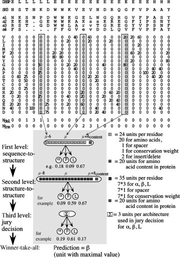
Key Players. PHD was the program that surpassed the level of 70% accuracy first33 (Rost and Sander, 1994). It uses a system of neural networks to achieve a performance well balanced between all secondary structure classes (Figure 29.1). Although still widely used, PHD is no longer the most accurate method43, 44. David Jones pioneered using automated, iterative PSI-BLAST searches45. The most important step climbed by the resulting method PSIPRED has been the detailed strategy to avoid polluting the profile through unrelated proteins. To avoid this trap, the database searched has to be filtered first46. Other than the advanced use of PSI-BLAST, PSIPRED achieves its success through a neural network system similar to that implemented in PHD. At the CASP meeting at which David Jones introduced PSIPRED, Kevin Karplus and colleagues presented their prediction method (SAM-T99sec/SAM-T04) finding more diverged profiles through HMMs47. The most important prediction method used by SAM-T99sec is a simple neural network with two layers of hidden units. However, the major strength of the method appears to be the quality of the alignment used. Some recent methods improve accuracy significantly not through more divergent profiles but through the particular algorithm; examples are the SSpro series of programs, the latest is SSpro448, 49, and Porter50. The principal idea is to overcome the limitations of feed-forward neural networks with an input window of relative small and fixed size with bidirectional recurrent neural networks (BRNN) capable of taking the entire protein chain as input51. The latest implementation SSpro4 uses this concept in combination with advanced PSI-BLAST profiles to become one of the best existing methods. Porter50 goes beyond SSpro4 by also using details in the alignment more cleverly and by combining a variety of different networks. Porter may be the most accurate existing method for the prediction of secondary structure. Another method that reaches high accuracy is SABLE252, 53 that combines the prediction of secondary structure and solvent accessibility; PROFsec and PROFacc realize the same idea in a similar way54. One recent method reached very high levels of prediction accuracy without paying for this achievement by complexity: YASPIN combines HMMs with neural networks in a very simple and efficient way55. Quite a different route toward secondary structure prediction is taken by the HMMSTR/I-sites programs56.
Specialized Methods: Coiled Coil Predictions. A coiled coil is abundle of several helices assuming a side-chain packing geometry often referred to as “knobs-into-holes”57. The “knobs” are the side chains of one helix that pack into the hole created by four side chains surrounding the facing helix. This supercoil slightly alters the helix periodicity from 3.6 to 3.5 and results in the coiled-coil-specific symmetry in which every seventh residue occupies a similar position on the helix surface. The first and fourth of the seven residues are typically hydrophobic, the other four hydrophilic, frequently exposing the helix to solvent. These specific sequence features are at the base of accurate predictions for coiled coil helices58, 59. The most widely used program is COILS that is based on amino acid preferences compiled for the few coiled coil proteins that were known at high resolution a decade ago60. The program detects coiled coil preferences in windows of 14, 21, and 28 residues. The longer the window the better the distinction between proteins that have coiled coil regions and those that do not58. If we know the precise location of the coiled coil regions and the multimeric state, we can predict 3D structure for coiled coil regions at levels of accuracy that resemble experimentally determined structures61, 62 (below 2.5 Å). O’Donoghue and Nilges used the experimentally known boundaries of the coiled coil regions and their known multimeric state for prediction. It reains to be tested how sensitive that 3D prediction is with respect to errors in predicting the coiled coil regions. Recently, Wolf and coworkers developed a method to predict the multimeric state of a coiled coil region63, 64. When labeling all likely coiled coil proteins in entire proteomes, we found that about 8-10% of all eukaryotic proteins and 2-10% of all proteins in archaebacteria and prokaryotes contain at least one coiled coil region65.
Figure 29.1. Neural network system for secondary structure prediction (PHDsec). From the multiple alignment (here guide sequence SH3 plus four other proteins a1–a4; note that lower case letters indicate deletions in the aligned sequence), a profile of amino acid occurrences is compiled. To the resulting 20 values at one particular position m in the protein (one column), three values are added: the number of deletions and insertions, as well as the conservation weight (CW). Thirteen adjacent columns are used as input. The whole network system for secondary structure prediction consists of three layers: two network layers and one layer averaging over independently trained networks.
Other Alphabets. Most secondary structure prediction methods focus on the standard DSSP alphabet of eight states. Many groups have tried to directly predict all eight states and have found that this approach did not bear fruit, possibly because this detailed separation is not supported by data, or more importantly is not conserved in evolution. In fact, we possibly should define the goals of simplifying structure prediction exactly by what is evolutionarily conserved66, 67. This is exactly what Kevin Karplus and colleagues realized when sampling the space of local alphabets to identify those that are most conserved in evolution and to develop methods that predict exactly these alphabets68. The concept is successful. So far it has remained an exceptional case possibly only because the approach has remained so unique that very few groups have worked on ways of how to use the information predicted by such methods directly.
Solvent Accessibility Prediction Methods
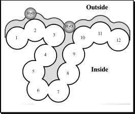
Basic Concept. It has long been argued that if the segments of secondary structure could be accurately predicted, the 3D structure could be predicted by simply trying different arrangements of the segments in space69, 70. One criterion for assessing each arrangement could be to use predictions of residue solvent accessibility71, 72, 73. The principal goal is to predict the extent to which a residue embedded in a protein structure is accessible to solvent. Solvent accessibility can be described in several ways71, 72, 73. The most detailed fast method compiles solvent accessibility by estimating the volume of a residue embedded in a structure that is exposed to solvent (Figure 29.2; this method was developed by Connolly73 and later implemented in DSSP74). Different residues have a different possible accessible area. The most extreme simplification for accessibility accounts for this by normalizing (dividing observed value by maximally possible value) to a two-state description distinguishing between residues that are buried (relative solvent accessibility less than 16%) and exposed (relative solvent accessibility ≥16%). The precise choice of the threshold is not well defined75, 76. The classical method to predict accessibility is to assign either of the two states, buried or exposed, according to residue hydrophobicity, that is, very hydrophobic stretches are predicted to be buried77, 78. However, more advanced methods have been shown to be superior to simple hydrophobicity analyses79, 80, 81, 82, 83. Typically, these methods use similar ways of compiling propensities of single residues or segments of residues to be solvent accessible, as secondary structure prediction methods. For particular applications, such as using predicted solvent accessibility to predict glycosylation sites, it seems beneficial to train neural networks on different definitions of accessibility84, 85. In particular, Hansen et al.84 realized alternative compilations by changing the size of the water molecule used in DSSP (Figure 29.2). In contrast to the situation for secondary structure, most of the information needed to predict accessibility is contained in the preference of single residues (Rost and Sander, 1994). Nevertheless, using windows of adjacent residues also improves solvent accessibility prediction significantly86 (Rost and Sander, 1994).
Figure 29.2. Measure accessibility. Residue solvent accessibility is usually measured by rolling a spherical water molecule over a protein surface and summing the area that can be accessed by this molecule on each residue (typical values range from 0 to 300Å2). To allow comparisons between the accessibility of long extended and spherical amino acids, typically relative values are compiled (actual area as percentage of maximally accessible area).Amore simplified description distinguishes two states: buried (here residues numbered 1–3 and 10–12) and exposed (here residues 4–9) residues. Since the packing density of native proteins resembles that of crystals, values for solvent accessibility provide upper and lower limits to the number of possible inter-residue contacts.
Evolutionary Information Improves Accessibility Prediction. Solvent accessibility at each position of the protein structure is evolutionarily conserved within sequence families. This fact has been used to develop methods for predicting accessibility using multiple alignment information86, 87, 88. The two-state (buried, exposed) prediction accuracy is above 75%, that is, more than four percentage points higher than for methods not using alignment information. Predictions of solvent accessibility have also been used successfully for prediction-based threading, as a second criterion toward 3D prediction, by packing secondary structure segments according to upper and lower bounds provided by accessibility predictions, and as a basis for predicting functional sites89.
Available Key Players. Possibly, the exclusion of methods predicting solvent accessibility from the CASP meetings (see Section “Practical Aspects”) slowed down the progress of the field. In particular, few of the methods developed are readily available through public servers. Prominent exceptions are the solvent accessibility predictions by PHD76 and PROFphd54 available through the PredictProtein server16, 90. Both use systems of neural networks with alignment information. The improvement of PROFphd over PHD was achieved by (1) training the neural networks only on high-resolution structures and by (2) using predicted secondary structure as additional input11, 54. Technically, both PROFphd and PHD are the only available methods predicting real values for relative solvent accessibility on a grid of 0, 1, 4, 9, 16, 25, 36, 49, 64, 81 (percentage relative accessibility). Another method that improved prediction accuracy considerably over older programs is embedded in the JPred2 server88. It uses PSI-BLAST profiles as input to neural networks predicting accessibility in two states (buried/exposed). FKNNacc is based on the identification of related local stretches (k-fuzzy nearest-neighbor methods)91. More recently several methods have combined the prediction of secondary structure and solvent accessibility; for examples, PROFacc, SABLE252, and ACCpro4 (analogue of SSpro4)48, 49.
Related Task: Prediction of Ooi/Coordination Number. The prediction of the residue coordination or Ooi number92 is directly related to that of the solvent accessibility. This number estimates how many residues surround a particular residue in a sphere. Thereby, the Ooi number provides two boundaries: one for the number of internal bounds and the other for the degree of accessibility to solvent. Several of the recent methods that target the prediction of the Ooi number are based on neural networks48, 49, 93. A related method simply estimated the contact density based on inter-residue contact predictions succeeded in predicting folding rates94. Thus suggesting that the direct relationship between residue coordination and solvent accessibility can also be used to make inferences about the protein folding rates.
Transmembrane Region Prediction Methods
The Task. Even in the optimistic scenario that in the near future most protein structures will be experimentally determined, one class of proteins will still represent a challenge for experimental determination of 3D structure: transmembrane proteins. The major obstacle with these proteins is that they do not crystallize and are hardly tractable by NMR spectroscopy. Consequently, for this class of proteins structure prediction methods are needed even more than for globular water-soluble proteins. Fortunately, the prediction task is simplified by strong environmental constraints on transmembrane proteins: the lipid bilayer of the membrane reduces the degrees of freedom making the prediction almost a 2D problem95, 96. Two major classes of integral membrane proteins are known: the first insert transmembrane helices (TMH) into the lipid bilayer (Figure 29.3) and the second form pores by transmembrane beta-strand barrels (TMB)97, 98, 99. While predicting transmembrane helices is simpler than predicting globular helices, there is ample evidence that prediction accuracy has been significantly overestimated100, 101.
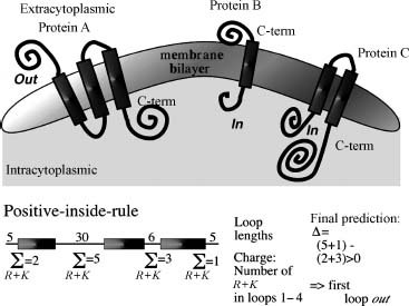
Basic Concept. We can use a number of observations that constrain the problem of predicting membrane helices. (1) TM helices are predominantly apolar and between 12 and 35 residues long102. (2) Globular regions between membrane helices are typically shorter than 60 residues65, 103. (3) Most TMH proteins have a specific distribution of the positively charged amino acids arginine and lysine, coined the positive-inside rule by Gunnar von Heijne104, 105. Connecting loop regions at the inside of the membrane have more positive charges than loop regions at the outside (Figure 29.3). (4) Long globular regions (more than 60 residues) differ in their composition from those globular regions subject to the positive-inside rule. Most methods simply compile the hydrophobicity along the sequence and predict a segment to be a transmembrane helix if the respective hydrophobicity exceeds some given threshold106, 107, 108, 109, 110, 111, 112, 113, 114, 115, 116. In addition, some methods also explore the hydrophobic moment97, 107, 117 or other membranespecific amino acid preferences118, 119, 120, 121. The most important step is to adequately average hydrophobicity values over windows of adjacent residues111, 122. One of the major problems of hydrophobicity-based methods appears the poor distinction between membrane and globular proteins100, 123. A number of methods use the positive-inside rule to also predict the orientation of membrane helices110, 115, 124, 125, 126, 127.
Figure 29.3. Topology of helical membrane proteins. In one class of membrane proteins, typically apolar helical segments are embedded in the lipid bilayer oriented perpendicular to the surface of the membrane. The helices can be regarded as more or less rigid cylinders. The orientation of the helical axes, that is, the topology of the transmembrane protein, can be defined by the orientation of the first N-terminal residues with respect to the cell. Topology is defined as out when the protein N-term (first residue) starts on the extracytoplasmic region (protein A) and as in if the N-term starts on the intracytoplasmic side (proteins B and C). The lower part explains the “inside-out rule.” The differences between the positive charges are compiled for all even and odd nonmembrane regions. If the even loops have more positive charges, the N-term of the protein is predicted outside. This rule holds for most proteins of known topology.
Evolutionary Information Improves Prediction Accuracy. Using evolutionary information also improves TMH predictions, significantly123, 125, 128, 129. However, the growth of the sequence databases seems to have reversed the advantage of using evolutionary information101. Until around 1997, most membrane helices were conserved in the following sense. Assume protein A has a TMH at positions N1-N2. Since the number of membrane helices is important for the function of the protein, we expect that all proteins A’ that are found to be similar to A in a database search will also have a membrane helix at the corresponding positions N1-N2. However, this assumption is no longer correct102. The practical result is that alignment-based predictions are much less accurate when based on the large merger of SWISS-PROT and TrEMBL130 than when based on the smaller SWISS-PROT only102. Interestingly, we can explore the power of using evolutionary information by carefully filtering the results from PSI-BLAST searches101.
Available Key Players. TopPred2 is one of the classics in the field. It averages the GES-scale of hydrophobicity109 using a trapezoid window122, 124. MEMSAT110 introduced a dynamic programming optimization to find the most likely prediction based on statistical preferences. TMAP131 uses statistical preferences averaged over aligned profiles. PHD combines a neural network using evolutionary information with a dynamic programming optimization of the final prediction123, 129. DAS optimizes the use of hydrophobicity plots132. SOSUI112 uses a combination of hydrophobicity and amphiphilicity preferences to predict membrane helices. TMHMM is the most advanced, and seemingly most accurate, current method to predict membrane helices126. It embeds a number of statistical preferences and rules into a hidden Markov model to optimize the prediction of the localization of membrane helices and their orientation114 (note: similar concepts are used for HMMTOP). MINNOU133 is based on a combination of the predictions of secondary structure and solvent accessibility, such as SABLE, with the objective of predicting transmembrane helices. Phobius134, 135 combines the prediction of signal peptides through HMMs and thereby improves in both tasks over many other methods. THUMPUP is a toolkit that, amongst other tasks, also predicts membrane helices136.
The group of Rita Casadio pushed the application of machine-learning devices to the task of predicting beta-barrel membrane proteins (TMB)137, 138. They developed two methods: B2TMR, based on neural networks137, and HMM-B2TMR, based on hidden Markov models138. PROFtmb applied this concept and improved by a detailed, accurate distinction between upward and downward strands, that is, by effectively predicting the topology of TMBs139, 140. transFold141 is also based on HMMs; however, it is restricted to short TMB proteins as it performs additional energy calculations. This limitation comes with an important added benefit: tranFold also predicts strand pairings. TMB-HUNT applied a variant of the k-nearest neighbor method to the task, and BOMP142 utilized simple statistics; another statistics/Bayesian network-based approach appears rather accurate143.
Integral Membrane or not?. The first question when scanning large data sets often is which proteins are likely associated with the membrane. The general methods are motif and domain based, and potentially identify the protein as one of a subtype of TMH or TMB proteins. TMH- or TMB-specific methods are designed to identify features common to all TMH (or all TMB) proteins and do not identify subtypes. InterProScan144 is a portal that allows querying the general methods at once. UniProt145 provides a comprehensive view of previously analyzed results on many proteins and accompanying experimental information on structure or function.
TMB-Specific Methods. BOMP (beta-barrel outer membrane protein predictor)142, 146, TMB-HUNT147, and PROFtmb140 are specially designed to identify TMB proteins in a pool. They have all been evaluated for accuracy in discriminating TMBs from background. Unfortunately, a definitive comparison is complicated by the fact that the evaluations are all done on different data sets. It is recommended that you submit your query to all three and scrutinize the results. Taking a consensus of predictors has been found consistently to yield better accuracy than relying on one individual predictor.
TMH-Specific Methods. Of the six TMH-specific methods, only TMHMM has been rigorously evaluated for accuracy in discriminating TMH proteins from others. While all methods implicitly predict whether a protein is a TMH by the presence of one or more predicted TM-helices, because the others are not evaluated for accuracy, it is not recommended to use these methods to screen a pool for potential TMH proteins.
PROGRAMS AND PUBLIC SERVERS
All methods described are available through public servers. A list of URLs and the contact addresses is summarized in Table 29.1. Most programs, except HMMSTR and PSIPRED, listed in Table 29.1 are also available by single click: META-PP allows you to fill out a form with the sequence and your e-mail address once and to simultaneously submit your protein to a number of high-quality servers148. This concept of accessing many servers through one has been pioneered by the BCM-Launcher149 supposedly accessing the largest number of different methods. Other combinations are given by NPSA150, META-Poland151, and ProSAL152 In contrast to all others, META-PP attempts to (i) return as few results as possible by filtering out technical messages and to (ii) combine only high-quality methods. Note that both the BCM launcher and the current GCG package153 return predictions of secondary structure from methods that are neither state ofthe art nor competitive with the best method from a decade ago without indicating this to the user.
TABLE 29.1. Availability of Prediction Methodsa
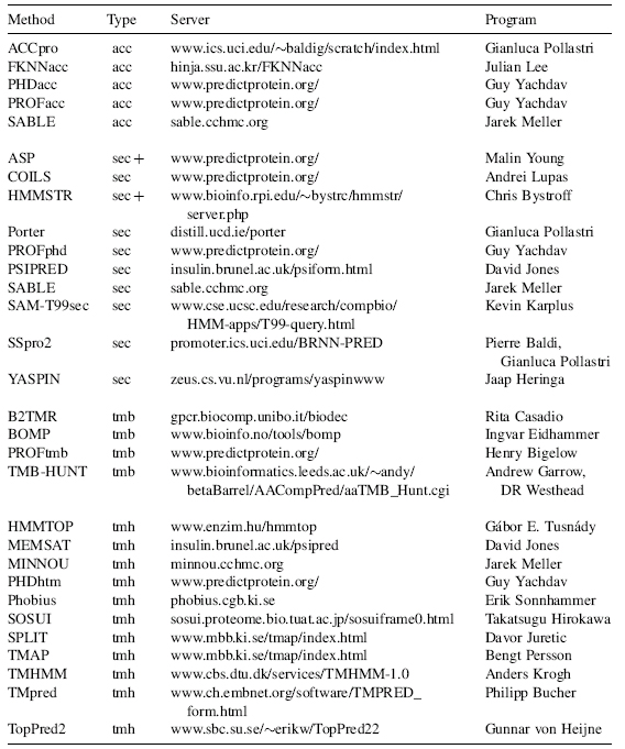
aAcronyms: prediction of: acc—solvent accessibility; sec—secondary structure; tmb—transmembrane beta strands; tmh—transmembrane helices.
PRACTICAL ASPECTS
Evaluation of Prediction Methods Correctly Evaluating Protein Structure Prediction is Difficult
Developers of prediction methods in bioinformatics may significantly overestimate their performance because of the following reasons. First, it is difficult and time consuming to correctly separate data sets used for developing and testing. Second, estimates of performance of the different methods are often based on different data sets. This problem frequently originates from the rapid growth of the sequence and structure databases. Third, single scores are usually not sufficient to describe the performance of a method. The lack of clarity is particularly unfortunate at a time when an increasing number of tools are made easily available through the Internet and many of the users are not experts in the field of protein structure prediction. Two prominent examples illustrate this problem. (1) Trans-membrane helix predictions have been estimated to yield levels above 95% per-residue accuracy that is more than 18 percentage points above actual performance on subsequent test cases102. It is not unlikely that the recent methods for the prediction of beta-barrel membrane proteins are also overestimated. (2) Many publications on predicting the secondary structural class from amino acid composition allowed correlations between “training” and testing sets. Consequently, levels of prediction accuracy published, close to 100%, exceeded by far the theoretical possible margins—around 60%154.
Although overestimates of performance can slow down experimental and theoretical progress substantially, sometimes methods perform more than anticipated by the authors. For instance, PROFtmb was published as a method that fails to identify TMB proteins in eukaryotes when used with default thresholds. Nevertheless, experimental groups have used this tool to identify eukaryotic TMB proteins. Similarly, PHDsec, PSIPRED, and PROFsec were published with the warning that they work exclusively for globular, nonmembrane proteins. There is evidence that these tools actually also work for integral membrane proteins155.
CASP: How Well do Experts Predict Protein Structure?. Altogether seven CASP experiments have attempted to address the problem of overestimated performance8, 156, 157, 158, 159, 160. The procedure used by CASP is the following: (1) Experimentalists who are about to determine the structure of a protein send the sequence to the CASP organizers159. (2) Sequences are distributed to the predictors. The deadline for returning results is given by the date that the structure will be published. (3) All predictions are evaluated in a meeting at Asilomar. CASP resolves the bias resulting from using known protein structures as targets. However, it often cannot provide statistically significant evaluations since the number of proteins tested is too small44, 161, 162. Nevertheless, CASP provides valuable insights into the performance of prediction methods and has become the major source of development in the field of protein structure prediction. Due to the fact that “failing at CASP is bad for the CV,” most predictions are submitted only after experts have studied the data in detail. Thus, CASP intrinsically evaluates how well the best experts in the field can predict structure.
EVA and LiveBench: Automatic, Large-Scale Evaluation of Performance. The limitations of CASP prompted two efforts at creating large-scale and continuously running tools that automatically assess protein structure prediction servers: EVA161, 163, 164, 165 and LiveBench166, 167. LiveBench specializes in the evaluation of fold recognition, while EVA analyzes comparative modeling, contact prediction, fold recognition, and secondary structure prediction. The EVA results for secondary structure prediction methods were essential to conclude that these methods have improved significantly and to isolate the particular reasons for the improvements (mostly due to growing databases)11, 43, 44.
Secondary Structure Prediction in Practice
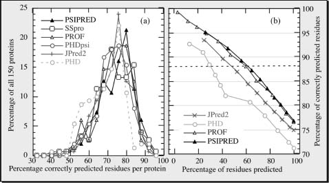
77% Right Means 33% Wrong!. The best current methods (Porter, SABLE, SAM-T04, PSIPRED, PROFphd, SSpro) reach levels around 77-80% accuracy (percentage of residues predicted correctly in one of the three states: helix, strand, or other)11, 54, 163, 168. Five observations are important for using prediction methods. (1) Levels of accuracy are averages over many proteins (Figure 29.4a). Hence, the accuracy for the prediction of your protein may be much lower, or much higher, than 77%. (2) Stronger predictions are usually more accurate (Figure 29.4b). This allows, to some extent, to find out whether or not the prediction for your protein is more likely to be above or below average. (3) Often predictions go badly wrong, that is, helices are incorrectly predicted as strands and vice versa. In fact, the best current methods confuse helices and strands for, on average, about 3% of all residues163. Encouragingly, some of these “bad errors” are in fact not so severe, after all, since some of these are due to regions that can switch structural conformations in response to environmental changes (see below). (4) Prediction accuracy is rather sensitive to the information contained in the alignment used for the prediction: differences between single-sequence-based predictions and optimal alignment-based predictions can exceed more than 25 percentage points43. (5) Ifon average 77% of the residues are correctly predicted, this trivially implies that 33% are wrong. Often, it is extremely instructive to form an expert opinion about where these wrong predictions are169.
Figure 29.4. Prediction accuracy varies but stronger predictions are better! All results are based on 150 novel protein structures not used to develop any of the methodshown161, 163. For all methods shown, the three-state per-residue accuracy varies significantly between these proteins, with one standard deviation in the order of 10%(a). This implies that it is difficult for users to estimate the “actual” accuracy for their protein. However, most methods now provide an index measuring the reliability of the prediction for each residue. Shown is the accuracy versus the cumulative percentages of residues predicted at a given level of reliability (coverage versus accuracy). For example, PSIPRED and PROFphd reach a level above 88% for about 60% of all residues (dashed line). This particular line is chosen since secondary structure assignments by DSSP agree to about 88% for proteins of similar structure. Although JPred2 is only marginally less accurate than PSIPRED and PROFphd, it reaches this level of accuracy for less than half of all residues.
Latest Improvement: 4 Parts Database Growth, 3 Better Search, 4 Other. Jones solicited two causes for the improved accuracy of PSIPRED: (1) training and (2) testing the method on PSI-BLAST profiles. Cuff and Barton88 examined in detail how different alignment methods improve. However, which fraction of the improvement results from the mere growth of the database, which from using more diverged profiles, and which from training on larger profiles? Using the PHD version from 1994 to separate the effects43, we first compared a noniterative standard BLAST170 search against SWISS-PROT130 with one against SWISS-PROT + TrEMBL130 + PDB9. The larger database improves performance by about two percentage points43. Secondly, we compared the standard BLAST against the big database with an iterative PSI-BLAST search. This yielded less than two percentage points additional improvement43. Thus, overall, the more divergent profile search against today’s databases supposedly improves any method using alignment information by almost four percentage points. The improvement through using PSI-BLAST profiles to develop the method is relatively small: PHDpsi was trained on a small database of not very divergent profiles in 1994; for example, PROFphd was trained on PSI-BLAST profiles of a 20 times larger database in 2000. The two differ by only one percentage point, and part of this difference resulted from implementing new concepts into PROF54.
Averaging Over Many Methods May Help. All methods predict some proteins at lower levels of accuracy than others34, 44, 171. Nevertheless, for most proteins there is a method that predicts secondary structure at a level higher than average44. The latter is applied when averaging over prediction methods. In fact, such averages are helpful as long as compiled over good methods172. Thus, using ALL available programs is a rather bad idea!
Solvent Accessibility Prediction in Practice
Very few of the seemingly more accurate methods predicting solvent accessibility are publicly available. Furthermore, there is no EVA-like evaluation of methods based on large sets and identical conditions. The first case scenario in which methods are compared based on different data sets, and different ways to define accessibility, is the rule rather than the exception. Thus, it is important to view values published with caution. Most methods predict accessibility in two states (exposed and buried). Levels of prediction accuracy vary significantly according to choice of the thresholds to distinguish between the two states88. If we define all residues that are less than 16% solvent accessible as “exposed,” the best current methods reach levels around 75 ± 10% accuracy35, 43, 88, 173. Using alignment information significantly improves prediction accuracy. However, accessibility predictions are more sensitive to alignment errors than secondary structure predictions35, 43. A reason for this may be that accessibility is evolutionarily less well conserved than is secondary structure43.
Transmembrane Region Prediction in Practice
Caution: No Appropriate Estimate of Performance Available!. The appropriate evaluation of methods predicting membrane helices is even more difficult than the evaluation of other categories of structure prediction. Three major problems prevent adequate analyses. (1) We do not have enough high-resolution structures to allow a statistically significant analysis102. (2) Low-resolution experiments (gene fusion) differ from high-resolution experiments (crystallography) almost as much as prediction methods do102. Thus, low-resolution experiments do not suffice to evaluate prediction accuracy. (3) All methods optimize some parameters. Since there are so few high-resolution structures, all methods use as many of the known ones, as possible. However, those methods perform much better on proteins for which they were developed than on new proteins was impressively demonstrated—and overlooked in a recent analysis of prediction methods100.
Crude Estimates for Where We Are at in the Field. The best current methods (HMMTOP2, PHDhtm, and TMHMM2) predict all helices correct for about 70% of all proteins102. MINNOU133 and Phobius134, 135 are likely to reach similar or even higher performance. For more than 60% of the proteins, the topology is also predicted correctly100, 102. PHDhtm getting about 70% ofthe observed TMH residues right102 achieves the most accurate per-residue prediction. All methods based on advanced algorithms tend to underestimate transmembrane helices; thus, about 86% of the TMH residues predicted by the best methods in this category PHD and DAS are correct102. Most methods tend to confuse signal peptides with membrane helices; the best separation is achieved by a system predicting subcellular localization ALOM2174. Almost as accurate are PHD and TopPred2 (followed by TMHMM)100. Surprisingly, most methods have also been overestimated in their ability to distinguish between globular and helical membrane proteins; particularly, most methods based only on hydrophobicity scales incorrectly predict membrane helices in over 90% of a representative set of globular proteins101. TMHMM, SOSUI, and PHDhtm appear to yield the most accurate distinction between membrane and globular proteins (less than 2% false positives)102. The most accurate hydrophobicity index appears to be the one recently developed in the Ben-Tal group175. All methods fail to distinguish membrane helices from signal peptides to the extent that the best methods still falsely predict membrane helices for 25% (PHDhtm) to 34% (TMHMM2) of all signal peptides tested. The good news for the practical application are that we have an accurate method detecting signal peptides176 and that most incorrectly predicted membrane helices start closer than 10 residues to N-terminal methionine residues, that is, could be corrected by experts.
Genome Analysis: Many Proteins Contain Membrane Helices. Despite the overestimated performance, predictions of transmembrane helices are valuable tools to quickly scan entire genomes for membrane proteins. A few groups base their results only on hydrophobicity scales, known to have extremely high error rates in distinguishing globular and membrane proteins. Nevertheless, the averages published for entire genomes are surprisingly similar between different authors65, 103, 129, 177, 178, 179, 180. About 10-30% of all proteins appear to contain membrane helices. One crucial difference, however, is that more cautious estimates do not perceive a statistically significant difference in the percentage of TMH proteins across the three kingdoms: eukaryotes, prokaryotes, and archaea181. However, preferences between particular types of membrane proteins differ; in particular, eukaryotes have more 7TM proteins (receptors), whereas prokaryotes have more 6- and 12TM proteins (ABC transporters)65, 103.
EMERGING AND FUTURE DEVELOPMENTS
Regions Likely to Undergo Structural Change Predicted Successfully
Young et al.182 have unraveled an impressive correlation between local secondary structure predictions and global conditions. The authors monitor regions for which secondary structure prediction methods give equally strong preferences for two different states. Such regions are processed combining simple statistics and expert rules. The final method is tested on 16 proteins known to undergo structural rearrangements and on a number of other proteins. The authors report no false positives, and they identify most known structural switches. Subsequently, the group applied the method to the myosin family identifying putative switching regions that were not known before but appeared reasonable candidates183. I find this method most remarkable in two ways: (1) it is the most general method using predictions of protein structure to predict some aspects of function and (2) it illustrates that predictions may be useful even when structures are known (as in the case of the myosin family). Recently, several group have begun to predict local flexibility directly;for example, Wiggle184 identifies flexible regions through a rather unique, innovative combination of molecular dynamics and machine learning. PROFbval185 predicts normalized B-values directly from sequence alignments through neural networks; like many recent state-of-the-art methods, PROFbval uses other 1D predictions as input. GlobPlot186 uses the correlation between low-complexity and flexibility.
Aspects of Protein Function Predicted by Expert Analysis of Secondary Structure
The typical scenario in which secondary structure predictions help us to learn more about function comes from experts combining predictions and their intuition, most often to find similarities to proteins of known function but insignificant sequence similarity187, 188, 189, 190, 191, 192, 193, 194, 195, 196, 197, 198, 199. Usually, such applications are based on very specific details about predicted secondary structure (some examples in Table 29.1). Thus, successful correlations of secondary structure and function appear difficult to incorporate into automatic methods.
Exploring Secondary Structure Predictions to Improve Database Searches
Initially, three groups independently applied secondary structure predictions to fold recognition, that is, the detection of structural similarities between proteins of unrelated sequences200, 201, 202. A few years later, almost every other fold recognition/threading method has adopted this concept46, 203, 204, 205, 206, 207, 208, 209, 210, 211. Two recent methods extended the concept by not only refining the database search but also by actually refining the quality of the alignment through an iterative procedure212, 213, 214, 215, 216. A related strategy has been explored by Ng and Henikoffs to improve predictions and alignments for membrane proteins217; the concept has been adopted by others218, 219, 220, 221, 222.
From 1D Predictions to 2D, and 3D Structure
Are secondary structure predictions accurate enough to help predict higher order aspects of protein structure automatically? 2D (inter-residue contacts) predictions: Baldi et al.223 have recently improved the level of accuracy in predicting beta-strand pairings over earlier work224 by using another elaborate neural network system. 3D predictions: the following list of five groups exemplifies that secondary structure predictions have now become a popular first step toward predicting 3D structure. (1) Ortiz et al.225 successfully use secondary structure predictions as one component of their 3D structure prediction method. (2) Eyrich and coworkers226, 227 minimize the energy of arranging predicted rigid secondary structure segments. (3) Lomize et al.228 also start from secondary structure segments. (4) Chen, Singh, and Altman, 1999 suggest using secondary structure predictions to reduce the complexity of molecular dynamics simulations. (5) Levitt and coworkers229, 230 combine secondary structure-based simplified presentations with a particular lattice simulation attempting to enumerate all possible folds.
Using Accessibility to Predict Aspects of Function
Features of multiple alignments can reveal aspects of protein function231, 232, 233, 234, 235, 236, 237. To simplify the story, residues can be conserved because of structural and functional reasons. If we could distinguish between the two, we could predict the functional residues. Obviously, residues that are exposed and conserved are likely to reveal functional constraints. This suggests using predicted accessibility and combination with alignments to predict functional residues. One particular application is the identification of residues involved in transient protein-protein interactions237, 238, 239, 240. Once again, methods combining evolutionary information and 1D predictions as input perform best239; in fact, they are accurate enough to even predict interaction hot spots241. Another possible application of predicted accessibility is the prediction of subcellular localization: the surface compositions differ significantly between extracellular, cytoplasm, and nuclear proteins242. Many methods have recently used this correlation to better the prediction of localization243, 244, 245, 246, 247, 248, 249, 250. The combination of 1D predictions and predictions of localization can be used to annotate experimental protein-protein interactions as, for example, exemplified in the PiNAT system251 that annotates protein networks.
Using 1D Predictions for Target Selection in Structural Genomics
Structural genomics proposes to experimentally determine one high-resolution structure for every known protein252, 253, 254, 255, 256, 257. Obviously, this goal could be reached faster if we could avoid all proteins of known structure. This is relatively straightforward181, 254, 258. More difficult is the task of avoiding proteins that do not express, do not purify, or are longer than 200 residues, and do not crystallize (or do not diffract well enough). One way toward this goal is to exclude all proteins with membrane helices65, 258. Can bioinformatics do more than that? A whole battery of approaches is being applied to steer the target selection and structure exploitation in the operating structural genomics centers12, 184, 238, 245, 250, 259, 260, 261, 262, 263, 264, 265, 266, 267, 268, 269, 270, 271, 272, 273, 274, 275, 276, 277. Many of these approaches are based on 1D predictions.
Eukaryotes Full of Disorder?
At the point of the first edition of this book, it had been shown that regions of low-complexity, as predicted by the program SEG278, are the rule rather than the exception in the protein universe279, 280, 281, 282, 283, 284, 285. Using predictions of secondary structure, we had found that there are many proteins that do not have low-complexity regions but nevertheless appear to have long (more than 70 residues) regions without regular secondary structure (helix/strand, dubbed NORS). Such NORS proteins appear to be significantly more abundant in eukaryotes than in all other kingdoms, reaching levels around 25% of the entire genome181. We found many of the NORS to be evolutionarily conserved, suggesting that these may in fact be proteins with induced structure rather than without structure. These findings had confirmed earlier analyses283 and have been confirmed by subsequent work286.
Since the publication of the first edition of the book, the field has exploded. Over the last years, many studies have shown that often the lack of a unique, native 3D structure in physiological conditions can be crucial for function287, 288, 289, 290, 291, 292, 293, 294. Such proteins are often referred to as disordered, unfolded, natively unstructured,or intrinsically unstructured proteins. A typical example is a protein that adopts a unique 3D structure only upon binding to an interaction partner and thereby performs its biochemical function287, 288, 290. The better our experimental and computational means of identifying such proteins, the more we realize that they come in a great variety: some adopt regular secondary structure (helix or strand) upon binding, some remain loopy; some proteins are almost entirely unstructured, others have only short unstructured regions. The more our ability to recognize short unstructured regions increases, the more we realize that the label “unstructured protein” would be rather misleading, as most “unstructured proteins” have relatively short unstructured regions. There is no single one way to define “unstructured regions.” Many methods have been developed that predict such natively unstructured or disordered regions based on a great diversity of aspects184, 186, 295, 296, 297, 298, 299, 300, 301, 302, 303, 304, 305, 306. Increasingly, it becomes apparent that unstructured regions are an important means of increasing complexity of molecular interactions. It is rather remarkable that methods developed for the prediction of regular 1D structure become once again crucial for the exploration of regions that appear to pose an altogether different challenge to the advance of structural biology over the next decade.
FURTHER READING
Review articles:
- Secondary structure, old21: Goldmine for finding citations for very early prediction methods.
- Secondary structure, new11: Brief walk through the recent highlights in the field of protein secondary structure prediction.
- Secondary structure for alignments307: Reviews using secondary structure predictions to improve alignment methods.
- Coiled coil helices59: Analysis of the performance of various programs predicting coiled coil helices based on new structures.
- Transmembrane predictions96, 97, 308: Reviews of methods predicting transmembrane regions from different perspectives.
- Using 1D predictions for function prediction13, 14: Reviews methods that extend beyond database comparisons to predict aspects of protein function.
- Prediction of natively unstructured/disordered regions290, 291, 309 (Oldfield et al., 2005)
Original articles (sorted by subject/year):
(1) Secondary structure, PHDsec33, 35: Original papers describing the first method that surpassed the threshold of 70% prediction accuracy through combining neural networks and evolutionary information.
(2) Secondary structure, PSIPRED45: The alignments used by PHD are replaced by PSI-BLAST alignments. This improves prediction accuracy significantly. However, possibly the most important aspect is the description of ways to run PSI-BLAST automatically without finding too many wrong hits.
(3) Secondary structure, SSpro/Porter48, 49, 50, 310: The most complicated and seemingly most successful architecture for using neural networks to predict secondary structure is presented here.
(4) Secondary structure, switches182: A data set of 16 protein sequences having functions that involve substantial backbone rearrangements are analyzed with respect to the ambivalence of predicted secondary structure. They find all segments involved in conformational switches to have ambivalent predictions, measured by the similarity in prediction probabilities for helix, sheet, and loop, as reported by PHD.
(5) Secondary structure applied to classify genomes311: Does a protein’s secondary structure determine its three-dimensional fold? This question is tested directly by analyzing proteins of known structure and constructing a taxonomy based solely on secondary structure. The taxonomy is generated automatically, and it takes the form of a tree in which proteins with similar secondary structure occupy neighboring leaves.
(6) Solvent accessibility, PHDacc76: Analysis of the evolutionary conservation of solvent accessibility and description of an alignment-based neural network prediction method.
(7) Transmembrane helices, TMHMM126: Most advanced and seemingly most accurate method predicting transmembrane helices through cyclic hidden Markov models.
(8) Transmembrane helices, genome analysis103: The authors analyze all membrane helix predictions for a number of entirely sequenced genomes. They conclude that more complex organisms use more helical membrane proteins than simpler organisms.
(9) Using 1D predictions for genome analysis65: 28 entirely sequenced genomes are compared based on predictions of coiled coil proteins58 (COILS), membrane helices16 (PHD), and functional classes312 (EUCLID). In contrast to many other publications, the correlation between the complexity of an organism and its use of helical membrane proteins is not confirmed.
ACKNOWLEDGMENTS
Thanks to the EVA team that enabled to quote some of the numbers given: Volker Eyrich (Schroedinger, USA), Marc Marti-Renom (Valencia, Spain), Andrej Sali (UCSF, USA), and Osvaldo Grana and Alfonso Valencia (Madrid, Spain). The work of BR was supported by the grants 2-R01-LM07329, U54-GM074958, GM75026, and U54-GM072980 from the National Institutes of Health (NIH). His work was made possible by the atmosphere created by a dream team at Columbia, to thank just a few of those: Jinfeng Liu (now Genentech, USA), Yanay Ofran (now Tel Aviv University, Israel), Dariusz Przybylski, Rajesh Nair, Marco Punta, Guy Yachdav, Yana Bromberg, Henry Bigelow, Sven Mika, Nancy Sosa, Eyal Mozes, Avner Schlessinger, Andrew Kernytsky, Kazimierz Wrzeszczynski, Ta-Tsen Soong, Phil Carter (now London, England), Claus Andersen (now Siena Biotech, Italy), and Volker Eyrich (now Schroedinger, USA). Last, not the least, thanks to all those who deposit their experimental data in public databases and to those who maintain these databases, in particular thanks to the teams around Phil Bourne, Helen Berman, Rolf Apweiler, and Amos Bairoch who started to clean it up!
REFERENCES
- Anfinsen CB (1973): Principles that govern the folding of protein chains. Science 181:223-230.
- Ellis RJ, Dobson C, Hartl U (1998): Sequence does specify protein conformation. Trends Biochem Sci 23(12):468.
- Dobson CM, Karplus M (1999): The fundamentals of protein folding: bringing together theory and experiment. Curr Opin Struct Biol 9(1):92-101.
- Levitt M, Warshel A (1975): Computer simulation of protein folding. Nature 253:694-698.
- Hagler AT, Honig B (1978): On the formation of protein tertiary structure on a computer. Proc Natl Acad Sci USA 75:554-558.
- van Gunsteren WF (1993): Molecular dynamics studies of proteins. Curr Opin Struct Biol 3:167-174.
- Kryshtafovych A, Venclovas C, Fidelis K, Moult J (2005): Progress over the first decade of CASP experiments. Proteins 61 (Suppl 7):225-236.
- Moult J, Fidelis K, Rost B, Hubbard T, Tramontano A (2005): Critical assessment of methods of protein structure prediction (CASP): round 6. Proteins 61(S7):3-7.
- Berman HM, Battistuz T, Bhat TN, Bluhm WF, Bourne PE, Burkhardt K, Feng Z, Gilliland GL, Iype L, Jain S, Fagan P, Marvin J, Padilla D, Ravichandran V, Schneider B, Thanki N, Weissig H, Westbrook JD, Zardecki C (2002): The Protein Data Bank. Acta Crystallogr D 58 (Pt 6 No 1):899-907.
- Chance MR, Fiser A, Sali A, Pieper U, Eswar N, Xu G, Fajardo JE, Radhakannan T, Marinkovic N (2004): High-throughput computational and experimental techniques in structural genomics. Genome Res 14(10B):2145-2154.
- Rost B (2001): Protein secondary structure prediction continues to rise. J Struct Biol 134:204-218.
- Liu J, Hegyi H, Acton TB, Montelione GT, Rost B (2004): Automatic target selection for structural genomics on eukaryotes. Prot Struct Funct Bio 56:188-200.
- Rost B, Liu J, Nair R, Wrzeszczynski KO, Ofran Y (2003): Automatic prediction of protein function. Cell Mol Life Sci 60(12):2637-2650.
- Ofran Y, Punta M, Schneider R, Rost B (2005): Beyond annotation transfer by homology: novel protein function prediction methods that can assist drug discovery. Drug Disc Today 10(21): 1475-1482.
- Brändén C, Tooze J (1991): Introduction to Protein Structure, New York: Garland Publ.
- Rost B (1996): PHD: predicting one-dimensional protein structure by profile based neural networks. Method Enzymol 266:525-539.
- Chou PY, Fasman GD (1974): Prediction of protein conformation. Biochemistry 13:211-215.
- Robson B (1976): Conformational properties of amino acid residues in globular proteins. J Mol Biol 107:327-356.
- Garnier J, Osguthorpe DJ, Robson B (1978): Analysis of the accuracy and implications of simple methods for predicting the secondary structure of globular proteins. J Mol Biol 120: 97-120.
- Schulz GE, Schirmer RH, (1979): Prediction of secondary structure from the amino acid sequence. In Principles of Protein Structure, Berlin: Springer-Verlag, pp. 108-130.
- Fasman GD (1989): The development of the prediction of protein structure. In: Fasman GD, editor. Prediction of Protein Structure and the Principles of Protein Conformation, New York, London: Plenum Press, pp. 193-303.
- Nishikawa K, Ooi T (1982): Correlation of the amino acid composition of a protein to its structural and biological characteristics. J Biochem 91:1821-1824.
- Deleage G, Roux B (1987): An algorithm for protein secnodary structure prediction based on class prediction. Protein Engin 1:289-294.
- Biou V, Gibrat JF, Levin JM, Robson B, Garnier J (1988): Secondary structure prediction: combination of three different methods. Prot Engin 2:185-191.
- Bohr H, Bohr J, Brunak S, Cotterill RMJ, Lautrup B, Nørskov L, Olsen OH, Petersen SB (1988): Protein secondary structure and homology by neural networks. FEBS Letters 241:223-228.
- Gascuel O, Golmard JL (1988): A simple method for predicting the secondary structure of globular proteins: implications and accuracy. CABIOS 4:357-365.
- Levin JM, Garnier J (1988): Improvements in a secondary structure prediction method based on a search for local sequence homologies and its use as a model building tool. Biochim Biophys Acta 955:283-295.
- Qian N, Sejnowski TJ (1988): Predicting the secondary structure of globular proteins using neural network models. J Mol Biol 202:865-884.
- Garnier J, Robson B (1989): The GOR method for predicting secondary structure in proteins. In: Fasman, GDeditor. Prediction of Protein Structure and the Principles of Protein Conformation, New York: Plenum Press, pp. 417-465.
- Rost B, Sander C (1996): Bridging the protein sequence-structure gap by structure predictions. Annual Review of Biophysics and Biomolecular Structure 25:113-136.
- Rost B, Sander C (2000): Third generation prediction of secondary structure. Methods Mol Biol 143:71-95.
- Rost B, Sander C (1992): Exercising Multi-Layered Networks on Protein Secondary Structure. Paper presented at the Neural Networks: From Biology to High Energy Physics, Elba, Italy.
- Rost B, Sander C (1993): Prediction of protein secondary structure at better than 70% accuracy. J Mol Biol 232:584-599.
- Rost B, Sander C, Schneider R (1993): Progress in protein structure prediction? TIBS 18:120-123.
- Rost B, Sander C (1994a): Combining evolutionary information and neural networks to predict protein secondary structure. Prot Struct Funct Genet 19:55-72.
- Rost B (1999): Twilight zone of protein sequence alignments. Protein Engin 12(2):85-94.
- Dickerson RE, Timkovich R, Almassy RJ (1976): The cytochrome fold and the evolution ofbacterial energy metabolism. J Mol Biol 100:473-491.
- Zvelebil MJ, Barton GJ, Taylor WR, Sternberg MJE (1987): Prediction of protein secondary structure and active sites using alignment of homologous sequences. J Mol Biol 195:957-961.
- Barton GJ (1996): Protein sequence alignment and database scanning. In: Sternberg MJE, editor. Protein structure prediction, 1st edn. , Vol. 170. Oxford: Oxford University Press, pp. 31-64.
- Altschul SF, Madden TL, Schaeffer AA, Zhang J, ZhangZ, Miller W, Lipman DJ (1997): GappedBlast and PSI-Blast: a new generation of protein database search programs. Nucleic Acids Res 25:3389-3402.
- Eddy SR (1998): Profile hidden Markov models. Bioinformatics 14(9):755-763.
- Karplus K, Barrett C, Hughey R (1998): Hidden Markov models for detecting remote protein homologies. Bioinformatics 14:846-856.
- Przybylski D, Rost B (2002): Alignments grow, secondary structure prediction improves. Prot Struct Funct Bio 46:195-205.
- Rost B, Eyrich V (2001): EVA: large-scale analysis of secondary structure prediction. Prot Struct Funct Genet 45 (Suppl 5):S192-S199.
- Jones DT (1999b): Protein secondary structure prediction based on position-specific scoring matrices. J Mol Biol 292(2):195-202.
- Jones DT, Tress M, Bryson K, Hadley C (1999): Successful recognition of protein folds using threading methods biased by sequence similarity and predicted secondary structure. Prot Struct Funct Genet 37(S3):104-111.
- Karplus K, Barrett C, Cline M, Diekhans M, Grate L, Hughey R (1999): Predicting protein structure using only sequence information. Prot Struct Funct Genet S3:121-125.
- Pollastri G, Baldi P, Fariselli P, Casadio R (2002a): Prediction of coordination number and relative solvent accessibility in proteins. Proteins 47(2):142-153.
- Pollastri G, Przybylski D, Rost B, Baldi P (2002b): Improving the prediction of protein secondary structure in three and eight classes using recurrent neural networks and profiles. Prot Struct Funct Bio 47:228-235.
- Pollastri G, McLysaght A (2005): Porter: a new, accurate server for protein secondary structure prediction. Bioinformatics 21(8):1719-1720.
- Pollastri G, Vullo A, Frasconi P, Baldi P (2006): Modular DAG-RNN architectures for assembling coarse protein structures. J Comput Biol 13(3):631-650.
- Adamczak R, Porollo A, Meller J (2004): Accurate prediction of solvent accessibility using neural networks-based regression. Proteins 56(4):753-767.
- Adamczak R, Porollo A, Meller J (2005): Combining prediction of secondary structure and solvent accessibility in proteins. Proteins 59(3):467-475.
- Rost B (2005): How to use protein 1D structure predicted by PROFphd. In: Walker JE, editor. The Proteomics Protocols Handbook, Totowa, NJ: Humana, pp. 875-901.
- Lin K, Simossis VA, Taylor WR, Heringa J (2005): A simple and fast secondary structure prediction method using hidden neural networks. Bioinformatics 21(2):152-159.
- Bystroff C, Thorsson V, and Baker D (2000). HMMSTR: a hidden Markov model for local sequence-structure correlations in proteins. J Mol Bio 301:173-190.
- Crick FHC (1953): The packing of a-helices: simple coiled-coils. Acta Crystallogr A 6:689-697.
- Lupas A (1996): Prediction and analyis of coiled-coil structures. Method Enzymol 266:513-525.
- Lupas A (1997): Predicting coiled-coil regions in proteins. Curr Opin Struct Biol 7:388-393.
- Lupas A, Van Dyke M, Stock J (1991): Predicting coiled coils from protein sequences. Science 252:1162-1164.
- Nilges M, Brünger AT (1993): Successful prediction of coiled coil geometry of the GCN4 leucine zipper domain by simulated annealing: comparison to the X-ray. Prot Struct Funct Genet 15:133-146.
- O’Donoghue SI, Nilges M (1997): Tertiary structure prediction using mean-force potentials and internal energy functions: successful prediction for coiled-coil geometries. Folding & Design 2(4):S47-S52.
- Wolf E, Kim PS, Berger B (1997): MultiCoil: a program for predicting two- and three-stranded coiled coils. Protein Sci 6(6):1179-1189.
- Newman JR, Wolf E, Kim PS (2000): A computationally directed screen identifying interacting coiled coils from Saccharomyces cerevisiae. Proc Natl Acad Sci USA 97(24):13203-13208.
- Liu J, Rost B (2001): Comparing function and structure between entire proteomes. Protein Sci10(10):1970-1979.
- Rost B, Sander C, Schneider R (1994): Redefining the goals of protein secondary structure prediction. J Mol Biol 235:13-26.
- Zemla A, Venclovas C, Fidelis K, Rost B (1999): A modified definition of SOV, a segment-based measure for protein secondary structure prediction assessment. Prot Struct Funct Genet 34:220-223.
- Karchin R, Cline M, Karplus K (2004): Evaluation of local structure alphabets based on residue burial. Proteins 55(3):508-518.
- Mumenthaler C, Braun W (1995): Predicting the helix packing of globular proteins by self-correcting distance geometry. Protein Sci 4:863-871.
- Cohen FE, Presnell SR (1996): The combinatorial approach. In: Sternberg MJE, editor. Protein Structure Prediction, 1st edn., Vol. 170: Oxford: Oxford University Press, pp. 207-228.
- Lee BK, Richards FM (1971): The interpretation of protein structures: estimation of static accessibility. J Mol Biol 55:379-400.
- Chothia C (1976): The nature of the accessible and buried surfaces in proteins. J Mol Biol 105:1-12.
- Connolly ML (1983): Solvent-accessible surfaces of proteins and nucleic acids. Science 221: 709-713.
- Kabsch W, Sander C (1983): Dictionary of protein secondary structure: pattern recognition of hydrogen bonded and geometrical features. Biopolymers 22:2577-2637.
- Hubbard TJP, Blundell TL (1987): Comparison of solvent-inaccessible cores of homologous proteins: definitions useful for protein modelling. Protein Engin 1:159-171.
- Rost B, Sander C (1994b): Conservation and prediction of solvent accessibility in protein families. Prot Struct Funct Genet 20(3):216-226.
- Kyte J, Doolittle RF (1982): A simple method for displaying the hydrophathic character of a protein. J Mol Biol 157:105-132.
- Sweet RM, Eisenberg D (1983): Correlation of sequence hydrophobicities measures similarity in three-dimensional protein structure. J Mol Biol 171:479-488.
- Holbrook SR, Muskal SM, Kim S-H (1990): Predicting surface exposure of amino acids from protein sequence. Protein Engin 3:659-665.
- Mucchielli-Giorgi MH, Hazout S, Tuffery P (1999): PredAcc: prediction of solvent accessibility. Bioinformatics 15(2):176-177.
- Carugo O (2000): Predicting residue solvent accessibility from protein sequence by considering the sequence environment. Protein Engin 13(9):607-609.
- Li X, Pan XM (2001): New method for accurate prediction of solvent accessibility from protein sequence. Prot Struct Funct Genet 42(1):1-5.
- Naderi-Manesh H, Sadeghi M, Arab S, Moosavi Movahedi AA (2001): Prediction of protein surface accessibility with information theory. Prot Struct Funct Genet 42(4):452-459.
- Hansen J, Lund O, Tolstrup N, Gooley AA, Williams KL, Brunak S (1998): NetOglyc: prediction of mucin type O-glycosylation sites based on sequence context and surface accessibility. Glycoconj J 15:115-130.
- Gupta R, Jung E, Gooley AA, Williams KL, Brunak S, Hansen J (1999): Scanning the available Dictyostelium discoideum proteome for O-linked GlcNAc glycosylation sites using neural networks. Glycobiology 9(10):1009-1022.
- Thompson MJ, Goldstein RA (1996): Predicting solvent accessibility: higher accuracy using Bayesian statistics and optimized residue substitution classes. Prot Struct Funct Genet 25:38-47.
- Rost B (1994): Conservation and prediction of solvent accessibility in protein families. Prot Struct Funct Genet 20:216-226.
- Cuff JA, Barton GJ (2000): Application of multiple sequence alignment profiles to improve protein secondary structure prediction. Prot Struct Funct Genet 40(3):502-511.
- Rost B, O’Donoghue SI (1997): Sisyphus and prediction of protein structure. Comput Appl Biol Sci 13:345-356.
- Rost B, Yachdav G, Liu J (2004): The PredictProtein server. Nucleic Acids Res 32 (Supplement 2): W321-W326.
- Sim J, Kim SY, Lee J (2005): Prediction of protein solvent accessibility using fuzzy k-nearest neighbor method. Bioinformatics 21(12):2844-2849.
- Nishikawa K, Ooi T (1986b): Radial location of amino acid residues in a globular protein: correlation with the sequence. J Biochem 100:1043-1047.
- Fariselli P, Olmea O, Valencia A, Casadio R (2001): Prediction of contact maps with neural networks and correlated mutations. Protein Engin 14(11):835-843.
- Punta M, Rost B (2005): Protein folding rates estimated from contact predictions. J Mol Biol 348(3):507-512.
- Taylor WR, Jones DT, Green NM (1994): A method for α-helical integral membrane protein fold prediction. Prot Struct Funct Genet 18:281-294.
- Bowie JU (2005): Solving the membrane protein folding problem. Nature 438(7068):581-589.
- von Heijne G (1996): Prediction of transmembrane protein topology. In: Sternberg MJE, editor., Protein Structure Prediction, 1st edn., Vol. 170: Oxford: Oxford University Press, pp. 101-110.
- Seshadri K, Garemyr R, Wallin E, von Heijne G, Elofsson A (1998): Architecture of beta-barrel membrane proteins: analysis of trimeric porins. Protein Sci 7(9):2026-2032.
- Buchanan SK (1999): β-Barrel proteins from bacterial outer membranes: structure, function and refolding. Curr Opin Struct Biol 9:455-461.
- Möller S, Croning DR, Apweiler R (2001): Evaluation of methods for the prediction of membrane spanning regions. Bioinformatics 17(7):646-653.
- Chen CP, Kernytsky A, Rost B (2002): Transmembrane helix predictions revisited. Protein Sci 11:2774-2791.
- Chen CP, Rost B (2002): State-of-the-art in membrane prediction. Appl Bioinf1:21-35.
- Wallin E, von Heijne G (1998): Genome-wide analysis of integral membrane proteins from eubacte- rial, archaean, and eukaryotic organisms. Protein Sci 7:1029-1038.
- von Heijne G (1986): The distribution of positively charged residues in bacterial inner membrane proteins correlates with the trans-membrane topology. EMBO J 5:3021-3027.
- von Heijne G (1989): Control of topology and mode of assembly of apolytopic membrane protein by positively charged residues. Nature 341:456-458.
- Tanford C (1980): The Hydrophobic Effect: Formation ofMicelles and Biological Membranes. New York: John Wiley & Sons.
- Eisenberg D, Schwartz E, Komaromy M, Wall R (1984): Analysis of membrane and surface protein sequences with the hydrophobic moment plot. J Mol Biol 179:125-142.
- Klein P, Kanehisa M, De Lisi C (1985): The detection and classification of membrane-spanning proteins. Biochim Biophys Acta 815:468-476.
- Engelman DM, Steitz TA, Goldman A (1986): Identifying nonpolar transbilayer helices in amino acid sequences of membrane proteins. Ann Rev Biophys Biophys Chem 15:321-353.
- Jones DT, Taylor WR, Thornton JM (1994): A model recognition approach to the prediction of all- helical membrane protein structure and topology. Biochemistry 33:3038-3049.
- von Heijne G (1994): Membrane proteins: from sequence to structure. Ann Rev Biophys Biomol Struct 23:167-192.
- Hirokawa T, Boon-Chieng S, Mitaku S (1998): SOSUI: classification and secondary structure prediction system for membrane proteins. Bioinformatics 14(4):378-379.
- Phoenix DA, Stanworth A, Harris F (1998): The hydrophobic moment plot and its efficacy in the prediction and classification of membrane interactive proteins and peptides. Membr Cell Biol 12 (1):101-110.
- Tusnady GE, Simon I (1998): Principles governing amino acid composition of integral membrane proteins: application to topology prediction. J Mol Biol 283(2):489-506.
- Harris F, Wallace J, Phoenix DA (2000): Use of hydrophobic moment plot methodology to aid the identification of oblique orientated alpha-helices. Mol Memb Biol 17(4):201-207.
- Lio P, Vannucci M (2000): Wavelet change-point prediction of transmembrane proteins. Bioinformatics 16(4):376-382.
- Liu LP, Deber CM (1999): Combining hydrophobicity and helicity: a novel approach to membrane protein structure prediction. Bioorg Med Chem 7(1):1-7.
- Ben-Tal N, Honig B, Miller C, McLaughlin S (1997): Electrostatic binding of proteins to membranes. Theoretical predictions and experimental results with charybdotoxin and phospholipid vesicles. Biophys J 73(4):1717-1727.
- Monne M, Hermansson M, von Heijne G (1999): A turn propensity scale for transmembrane helices. J Mol Biol 288(1):141-145.
- Pasquier C, Promponas VJ, Palaios GA, Hamodrakas JS, Hamodrakas SJ (1999): A novel method for predicting transmembrane segments in proteins based on a statistical analysis of the SwissProt database: the PRED-TMR algorithm. Protein Engin 12(5):381-385.
- Pilpel Y, Ben-TalN, Lancet D (1999): kPROT: a knowledge-based scale for the propensity of residue orientation in transmembrane segments. Application to membrane protein structure prediction. J Mol Biol 294(4):921-935.
- von Heijne G (1992): Membrane protein structure prediction. J Mol Biol 225:487-494.
- Rost B, Casadio R, Fariselli P, Sander C (1995): Prediction of helical transmembrane segments at 95% accuracy. Protein Sci 4:521-533.
- Sipos L, von Heijne G (1993): Predicting the topology of eukaryotic membrane proteins. Eur J Biochem 213:1333-1340.
- Persson B, Argos P (1997): Prediction of membrane protein topology utilizing multiple sequence alignments. J Protein Chem 16(5):453-457.
- Sonnhammer ELL, von Heijne G, Krogh A (1998): A hidden Markov model for predicting transmembrane helices in protein sequences’, paper presented to Sixth International Conference on Intelligent Systems for Molecular Biology (ISMB98), Montreal, Canada.
- Tusnady GE, Simon I (2001): Topology of membrane proteins. J Chem Inf Comput Sci41(2):364-368.
- Neuwald AF, Liu JS, Lawrence CE (1995): Gibbs motif sampling: detection of bacterial outer membrane protein repeats. Protein Sci 4:1618-1631.
- Rost B, Casadio R, Fariselli P (1996): Topology prediction for helical transmembrane proteins at 86% accuracy. Protein Sci 5:1704-1718.
- Bairoch A, Apweiler R (2000): The SWISS-PROT protein sequence database and its supplement TrEMBL in 2000. Nucleic Acids Res 28(1):45-48.
- Persson B, Argos P (1996): Topology prediction of membrane proteins. Protein Sci 5:363-371.
- Cserzö M, Wallin E, Simon I, von Heijne G, Elofsson A (1997): Prediction of transmembrane a-helices in prokaryotic membrane proteins: the dense alignment surface method. Protein Engin 10:673-676.
- Cao B, Porollo A, Adamczak R, Jarrell M, Meller J (2006): Enhanced recognition of protein transmembrane domains with prediction-based structural profiles. Bioinformatics 22(3):303-309.
- Kall L, Krogh A, Sonnhammer EL (2004): A combined transmembrane topology and signal peptide prediction method. J Mol Biol 338(5):1027-1036.
- Kall L, Krogh A, Sonnhammer EL(2005): An HMM posterior decoder for sequence feature prediction that includes homology information. Bioinformatics 21 (Suppl 1):i251-i257.
- Zhou H, Zhang C, Liu S, Zhou Y (2005): Web-based toolkits for topology prediction of transmembrane helical proteins, fold recognition, structure and binding scoring, folding-kinetics analysis and comparative analysis of domain combinations. Nucleic Acids Res 33(Web server issue): W193-W197.
- Jacoboni I, Martelli PL, Fariselli P, De Pinto V, Casadio R (2001): Prediction of the transmembrane regions of beta-barrel membrane proteins with a neural network-based predictor. Protein Sci 10:779-787.
- Martelli PL, Fariselli P, Krogh A, Casadio R (2002): A sequence-profile-based HMM for predicting and discriminating beta barrel membrane proteins. Bioinformatics 18 (Suppl 1):S46-S53.
- Bigelow H, Petrey D, Liu J, Przybylski D, Rost B (2004): Prediction of transmembrane beta-barrels for entire proteomes. Nucleic Acids Res 32:2566-2577.
- Bigelow H, Rost B (2006b): PROFtmb: a web server for predicting bacterial transmembrane beta barrel proteins. Nucleic Acids Res 34(Web server issue):W186-W188.
- Waldispuhl J, Berger B, Clote P, Steyaert JM (2006): Predicting transmembrane beta-barrels and interstrand residue interactions from sequence. Proteins 65(1):61-74.
- Berven FS, Flikka K, Jensen HB, Eidhammer I (2004): BOMP: a program to predict integral beta-barrel outer membrane proteins encoded within genomes of Gram-negative bacteria. Nucleic Acids Res 32(Web server issue):W394-W399.
- Gromiha MM, Suwa M (2006): Discrimination of outer membrane proteins using machine learning algorithms. Proteins 63(4):1031-1037.
- Zdobnov EM, Lopez R, Apweiler R, Etzold T (2002): The EBI SRS server: recent developments. Bioinformatics 18(2):368-373.
- Bairoch A, Apweiler R, Wu CH, Barker WC, Boeckmann B, Ferro S, Gasteiger E, Huang H, Lopez R, Magrane M, Martin MJ, Natale DA, O’Donovan C, Redaschi N, Yeh LS (2005): The Universal Protein Resource (UniProt). Nucleic Acids Res 33(Database issue):D154-D159.
- Berven FS, Karlsen OA, Straume AH, Flikka K, Murrell JC, Fjellbirkeland A, Lillehaug JR, Eidhammer I, Jensen HB (2006): Analysing the outer membrane subproteome of Methylococcus capsulatus (Bath) using proteomics and novel biocomputing tools. Arch Microbiol 184(6):362-377.
- Garrow AG, Agnew A, Westhead DR (2005): TMB-Hunt: a web server to screen sequence sets for transmembrane beta-barrel proteins. Nucleic Acids Res 33(Web server issue): W188-W192.
- Eyrich VA, Rost B (2003): META-PP: single interface to crucial prediction servers. Nucleic Acids Res 31(13):3308-3310.
- Smith RF, Wiese BA, Wojzynski MK, Davison DB, Worley KC (1996): BCM Search Launcher: an integrated interface to molecular biology data base search and analysis services available on the World Wide Web. Genome Res 6(5):454-462.
- Combet C, Blanchet C, Geourjon C, Deléage G (2000): NPS@: network protein sequence analysis. Trends Bio Sci 25(3):147-150.
- Rychlewski L (2000): META server. Warsaw: IIMCB.
- Kleywegt G, (2001): ProSAL: Protein sequence analysis launcher. Swedish Structural Biology Network.<http//alpha2bmcuuse/~gerard/srf/prosalhtml>.
- Devereux J, Haeberli P, Smithies O (1984): GCG package. Nucleic Acids Res 12:387-395.
- Wang Z-X, Yuan Z (2000): How good is prediction of protein structural class by the component- coupled method? Prot Struct Funct Genet 38:165-175.
- Forrest LR, Tang CL, Honig B (2006): On the accuracy of homology modeling and sequence alignment methods applied to membrane proteins. Biophys J 91(2):508-517.
- Moult J, Pedersen JT, Judson R, Fidelis K (1995): A large-scale experiment to assess protein structure prediction methods. Prot Struct Funct Genet 23(3):ii-v.
- Moult J, Hubbard T, Bryant SH, Fidelis K, Pedersen JT (1997): Critical assessment of methods of protein structure prediction (CASP): round II. Prot Struct Funct Genet (Suppl 1): 2-6.
- Moult J, Hubbard T, Bryant SH, Fidelis K, Pedersen JT (1999): Critical assessment of methods of protein structure prediction (CASP): round III. Prot Struct Funct Genet (Suppl 3): 2-6.
- Zemla A, Venclovas C, Fidelis K (2001): Protein structure prediction center. Lawrence Livermore National Laboratory, <http//PredictionCenterllnlgov/>.
- Moult J, Fidelis K, Zemla A, Hubbard T (2003): Critical assessment of methods of protein structure prediction (CASP): round V. Prot Struct Funct Genet 53 (Suppl 6):334-339.
- Eyrich V, Martí-Renom MA, Przybylski D, Fiser A, Pazos F, Valencia A, Sali A, Rost B (2001): EVA: continuous automatic evaluation of protein structure prediction servers. Bioinformatics 17:1242-1243.
- Marti-Renom MA, Madhusudhan MS, Fiser A, Rost B, Sali A (2002): Reliability of assessment of protein structure prediction methods. Structure 10:435-440.
- Eyrich VA, Koh IYY, Przybylski D, Graña O, Pazos F, Valencia A, Rost B (2003): CAFASP3 in the spotlight of EVA. Prot Struct Funct Bio 53 (Suppl 6):548-560.
- Koh IYY, Eyrich VA, Marti-Renom MA, Przybylski D, Madhusudhan MS, Narayanan E, Graña O, Valencia A, Sali A, Rost B (2003): EVA: evaluation of protein structure prediction servers. Nucleic Acids Res 31(13):3311-3315.
- Grana O, Eyrich VA, Pazos F, Rost B, Valencia A (2005): EVAcon: a protein contact prediction evaluation service. Nucleic Acids Res 33(Web server issue):W347-W351.
- Rychlewski L, Fischer D, Elofsson A (2003): LiveBench-6: large-scale automated evaluation of protein structure prediction servers. Prot Struct Funct Genet 53 (Suppl 6):542-547.
- Rychlewski L, Fischer D (2005): LiveBench-8: the large-scale, continuous assessment of automated protein structure prediction. Protein Sci 14(1):240-245.
- Rost B, Baldi P (2001): New Improvements In Protein Secondary Structure Prediction. CUBIC_2001_11, Columbia University.
- Hubbard T, Tramontano A, Barton G, Jones D, Sippl M, Valencia A, Lesk A, Moult J, RostB, SanderC, Schneider R, Lahm A, Leplae R, Buta C, Eisenstein M, Fjellstrom O, Floeckner H, Grossmann JG, Hansen J, Helmer-Citterich M, Joergensen FS, Marchler-Bauer A, Osuna J, Park J, Reinhardt A, Ribas de Pouplana L, Rojo-Dominguez A, Saudek V, Sinclair J, Sturrock S, Venclovas C, Vinals C (1996): Update on protein structure prediction: results of the 1995 IRBM workshop. Folding & Design 1:R55-R63.
- Altschul SF, Gish W (1996): Local alignment statistics. Methods Enzymol 266:460-480.
- Lesk AM, Lo Conte L, Hubbard TJP (2001): Assessment of novel folds targets in CASP4: predictions of three-dimensional structures, secondary structures, and interresidue contacts. Prot Struct Funct Genet 45(Suppl 5):98-118.
- Rost B, Baldi P, Barton G, Cuff J, Eyrich V, Jones D, Karplus K, King R, Ouali M, Pollastri G, Przybylski D (2001): Simple Jury Predicts Protein Secondary Structure Best. CUBIC_2001_10, Columbia University.
- Lesk AM (1997): CASP-2: report on ab initio predictions. Prot Struct Funct Genet 151-166.
- akai K, Kanehisa M (1992): A knowledge base for predicting protein localization sites in eukaryotic cells. Genomics 14:897-911.
- Kessel A, Ben-Tal N (2002): Free energy determinants of peptide association with lipid bilayers. In: Simon S, McIntosh T, editors. Peptide-Lipid Interactions, Vol. 52. San Diego, NY: Academic Press,
- Nielsen H, Engelbrecht J, Brunak S, von Heijne G (1997): Identification of prokaryotic and eukaryotic signal peptides and prediction of their cleavage sites. Protein Engin 10:1-6.
- Goffeau A, Slonimski P, Nakai K, Risler JL (1993): How many yeast genes code for membrane-spanning proteins? Yeast 9:691-702.
- Arkin IT, Brunger AT, Engelman DM (1997): Are there dominant membrane protein families with a given number of helices? Prot Struct Funct Genet 28:465-466.
- Frishman D, Mewes HW (1997): Protein structural classes in five complete genomes. Nat Struct Bio 4:626-628.
- Jones DT (1998): Do transmembrane protein superfolds exist? FEBS Letters 423(3):281-285.
- Liu J, Tan H, Rost B (2002): Loopy proteins appear conserved in evolution. J Mol Biol 322:53-64.
- Young M, Kirshenbaum K, Dill KA, Highsmith S (1999): Predicting conformational switches in proteins. Protein Sci 8:1752-1764.
- Kirshenbaum K, Young M, Highsmith S (1999): Predicting allosteric switches inmyosins. ProteinSci 8:1806-1815.
- Gu J, Gribskov M, Bourne PE (2006): Wiggle-predicting functionally flexible regions from primary sequence. PLoS Comput Biol 2(7):e90.
- Schlessinger A, Yachdav G, Rost B (2006): PROFbval: predict flexible and rigid residues in proteins. Bioinformatics 22(7):891-893.
- Linding R, Jensen LJ, Diella F, Bork P, Gibson TJ, Russell RB (2003a): Protein disorder prediction: implications for structural proteomics. Structure 11(11):1453-1459.
- Brautigam C, Steenbergen-Spanjers GC, Hoffmann GF, Dionisi-Vici C, van den Heuvel LP, Smeitink JA, Wevers RA (1999): Biochemical and molecular genetic characteristics of the severe form of tyrosine hydroxylase deficiency. Clin Chem 45(12):2073-2078.
- Davies GP, Martin I, Sturrock SS, Cronshaw A, Murray NE, Dryden DT (1999): On the structure and operation of type I DNA restriction enzymes. J Mol Biol 290(2):565-579.
- de Fays K, Tibor A, Lambert C, Vinals C, Denoel P, De Bolle X, Wouters J, Letesson JJ, Depiereux E (1999): Structure and function prediction of the Brucella abortus P39 protein by comparative modeling with marginal sequence similarities. Protein Engin 12(3):217-223.
- Di Stasio E, Sciandra F, Maras B, Di Tommaso F, Petrucci TC, Giardina B, Brancaccio A (1999): Structural and functional analysis of the N-terminal extracellular region of beta-dystroglycan. Biochem Biophys Res Commun 266(1):274-278.
- Gerloff DL, Cannarozzi GM, Joachimiak M, Cohen FE, SchreiberD, BennerSA(1999): Evolutionary, mechanistic, and predictive analyses of the hydroxymethyldihydropterin pyrophosphokinase family of proteins. Biochem Biophys Res Commun 254(1):70-76.
- Juan HF, Hung CC, Wang KT, Chiou SH (1999): Comparison of three classes of snake neurotoxins by homology modeling and computer simulation graphics. Biochem Biophys Res Commun257(2):500-510.
- Laval V, Chabannes M, Carriere M, Canut H, Barre A, Rouge P, Pont-Lezica R, Galaud J (1999): A family of Arabidopsis plasma membrane receptors presenting animal beta-integrin domains. Biochim Biophys Acta 1435(1-2):61-70.
- Seto MH, Liu HL, Zajchowski DA, Whitlow M (1999): Protein fold analysis of the B30.2-like domain. Prot Struct Funct Genet 35(2):235-249.
- Xu H, Aurora R, Rose GD, White RH (1999): Identifying two ancient enzymes in Archaea using predicted secondary structure alignment. Nat Struct Biol 6(8):750-754.
- Jackson RM, Russell RB (2000): The serine protease inhibitor canonical loop conformation: examples found in extracellular hydrolases, toxins, cytokines and viral proteins. J Mol Biol 296(2):325-334.
- Paquet JY, Vinals C, Wouters J, Letesson JJ, Depiereux E (2000): Topology prediction of Brucella abortus Omp2b and Omp2a porins after critical assessment of transmembrane beta strands prediction by several secondary structure prediction methods. J Biomol Struct Dyn 17(4):747-757.
- Shah PS, Bizik F, Dukor RK, Qasba PK (2000): Active site studies of bovine alpha1 → 3- galactosyltransferase and its secondary structure prediction. Biochim Biophys Acta 1480(1-2):222-234.
- Stawiski EW, Baucom AE, Lohr SC, Gregoret LM (2000): Predicting protein function from structure: unique structural features of proteases. Proc Natl Acad Sci USA 97(8):3954-3958.
- Rost B (1995): TOPITS: Threading One-dimensional Predictions in to Three-dimensional Structures. Paper presented at the Third International Conference on Intelligent Systems for Molecular Biology, Cambridge, England.
- Fischer D, Eisenberg D (1996): Fold recognition using sequence-derived properties. Protein Sci 5:947-955.
- Russell RB, Copley RR, Barton GJ (1996): Protein fold recognition by mapping predicted secondary structures. J Mol Biol 259:349-365.
- Ayers DJ, Gooley PR, Widmer-Cooper A, Torda AE (1999): Enhanced protein fold recognition using secondary structure information from NMR. Protein Sci 8(5):1127-1133.
- de la Cruz X, Thornton JM (1999): Factors limiting the performance of prediction-based fold recognition methods. Protein Sci 8(4):750-759.
- Di Francesco V, Munson PJ, Garnier J (1999): FORESST: fold recognition from secondary structure predictions of proteins. Bioinformatics 15(2):131-140.
- Hargbo J, Elofsson A (1999): Hidden Markov models that use predicted secondary structures forfold recognition. Prot Struct Funct Genet 36(1):68-76.
- Jones DT (1999a): GenTHREADER: an efficient and reliable protein fold recognition method for genomic sequences. J Mol Biol 287(4):797-815.
- Koretke KK, Russell RB, Copley RR, Lupas AN (1999): Fold recognition using sequence and secondary structure information. Prot Struct Funct Genet 37(S3):141-148.
- Ota M, Kawabata T, Kinjo AR, Nishikawa K (1999): Cooperative approach for the protein fold recognition. Proteins 37(S3):126-132.
- Panchenko A, Marchler-Bauer A, Bryant SH (1999): Threading with explicit models for evolutionary conservation of structure and sequence. Prot Struct Funct Genet (Suppl 3): 133-140.
- Kelley LA, MacCallum RM, Sternberg MJ (2000): Enhanced genome annotation using structural profiles in the program 3D-PSSM. J Mol Biol 299(2):499-520.
- Heringa J (1999): Two strategies for sequence comparison: profile-preprocessed and secondary structure-induced multiple alignment. Comput Chem 23(3-4):341-364.
- Jennings AJ, Edge CM, Sternberg MJ (2001): An approach to improving multiple alignments of protein sequences using predicted secondary structure. Protein Engin 14(4):227-231.
- Simossis VA, Heringa J (2005): PRALINE: a multiple sequence alignment toolbox that integrates homology-extended and secondary structure information. Nucleic Acids Res 33(Web server issue):W289-W294.
- Simossis VA, Kleinjung J, Heringa J (2005): Homology-extended sequence alignment. Nucleic Acids Res 33(3):816-824.
- Sammeth M, Heringa J (2006): Global multiple-sequence alignment with repeats. Proteins 64(1):263-274.
- Ng P, Henikoff J, Henikoff S (2000): PHAT: a transmembrane-specific substitution matrix. Bioinformatics 16(8):760-766.
- Muller T, Rahmann S, Rehmsmeier M (2001): Non-symmetric score matrices and the detection of homologous transmembrane proteins. Bioinformatics 17(Suppl 1):S182-S189.
- Hedman M, Deloof H, Von Heijne G, Elofsson A (2002): Improved detection of homologous membrane proteins by inclusion of information from topology predictions. Protein Sci 11(3):652-658.
- Sutormin RA, Rakhmaninova AB, Gelfand MS (2003): BATMAS30: amino acid substitution matrix for alignment of bacterial transporters. Proteins 51(1):85-95.
- Yu YK, Altschul SF (2005): The construction of amino acid substitution matrices for the comparison of proteins with non-standard compositions. Bioinformatics 21(7):902-911.
- Sadovskaya NS, Sutormin RA, Gelfand MS (2006): Recognition of transmembrane segments in proteins: review and consistency-based benchmarking of Internet servers. J Bioinform Comput Biol 4(5):1033-1056.
- Baldi P, Pollastri G, Andersen CA, Brunak S (2000): Matching protein beta-sheet partners by feedforward and recurrent neural networks. Ismb 8:25-36.
- Hubbard TJP, Park J (1995): Fold recognition and ab initio structure predictions using hidden Markov models and P-strand pair potentials. Prot Struct Funct Genet 23:398-402.
- Ortiz AR, Kolinski A, RotkiewiczP, Ilkowski B, Skolnick J (1999): Ab initio folding of proteins using restraints derived from evolutionary information. Prot Struct Funct Genet Suppl 3:177-185.
- Eyrich VA, Standley DM, Felts AK, Friesner RA (1999a): Protein tertiary structure prediction using a branch and bound algorithm. Prot Struct Funct Genet 35(1):41-57.
- Eyrich VA, Standley DM, Friesner RA (1999b): Prediction of protein tertiary structure to low resolution: performance for a large and structurally diverse test set. J Mol Biol 288(4):725-742.
- Lomize AL, Pogozheva ID, Mosberg HI (1999): Prediction of protein structure: the problem of fold multiplicity. Prot Struct Funct GenetSuppl 3: (3) 199-203.
- Samudrala R, Xia Y, Huang E, Levitt M(1999): Ab initio protein structure prediction using a combined hierarchical approach. Prot Struct Funct GenetSuppl 3 (3):194-198.
- Samudrala R, Huang ES, Koehl P, Levitt M (2000): Constructing side chains on near-native main chains for ab initio protein structure prediction. Protein Engin 13(7):453-457.
- Casari G, Sander C, Valencia A (1995): A method to predict functional residues in proteins. Nat Struct Biol 2:171-178.
- Lichtarge O, Bourne HR, Cohen FE (1996): Evolutionarily conserved Galphabetagamma binding surfaces support a model of the G protein-receptor complex. Proc Natl Acad Sci USA 93(15):7507-7511.
- Lichtarge O, Yamamoto KR, Cohen FE (1997): Identification of functional surfaces of the zinc binding domains of intracellular receptors. J Mol Biol 274(3):325-337.
- Pazos F, Sanchez-PulidoL, Garcia-Ranea JA, Andrade MA, Atrian S, Valencia A (1997): Comparative analysis of different methods for the detection of specificity regions in protein families. Paper presented at BCEC97: Bio-Computing and Emergent Computation, 1-2 September 1997, Skövde, Sweden.
- Marcotte EM, Pellegrini M, Ng HL, Rice DW, Yeates TO, Eisenberg D (1999): Detecting protein function and protein-protein interactions from genome sequences. Science 285(5428):751-753.
- Irving JA, Pike RN, Lesk AM, Whisstock JC (2000): Phylogeny of the serpin superfamily: implications of patterns of amino acid conservation for structure and function. Genome Res 10(12): 1845-1864.
- Wodak SJ, Mendez R (2004): Prediction of protein-protein interactions: the CAPRI experiment, its evaluation and implications. Curr Opin Struct Biol 14(2):242-249.
- Yao H, Kristensen DM, Mihalek I, Sowa ME, Shaw C, Kimmel M, Kavraki L, Lichtarge O (2003): An accurate, sensitive, and scalable method to identify functional sites in protein structures. J Mol Biol 326(1):255-261.
- Ofran Y, Rost B (2007a): ISIS: Interaction sites identified from sequence. Bioinformatics 23(ECCB): e5-e12.
- Porollo A, Meller J (2007): Prediction-based fingerprints of protein-protein interactions. Prot Struct Funct Genet 66(3):630-645.
- Ofran Y, Rost B (2007b): Protein-protein interaction hot spots carved into sequences. PLoS Computat Biol 3(7):e119.
- Andrade MA, O’Donoghue SI, Rost B (1998): Adaptation of protein surfaces to subcellular location. J Mol Biol 276:517-525.
- Nair R, Rost B (2003): Better prediction of sub-cellular localization by combining evolutionary and structural information. Prot Struct Funct Bio 53(4):917-930.
- Nair R, Rost B (2003): LOC3D: annotate sub-cellular localization for protein structures. Nucleic Acids Res 31(13):3337-3340.
- Nair R, Rost B (2004): LOCnet and LOCtarget: sub-cellular localization for structural genomics targets. Nucleic Acids Res 32 (Supplement 2):W517-W521.
- Yu CS, Lin CJ, Hwang JK (2004): Predicting subcellular localization of proteins for Gram-negative bacteria by support vector machines based on n-peptide compositions. Protein Sci 13(5):1402-1406.
- Nair R, Rost B (2005): Mimicking cellular sorting improves prediction of subcellular localization. J Mol Biol 348(1):85-100.
- Lee S, Lee B, Jang I, Kim S, Bhak J (2006): Localizome: a server for identifying transmembrane topologies and TM helices of eukaryotic proteins utilizing domain information. Nucleic Acids Res 34(Web server issue):W99-103.
- Pierleoni A, Martelli PL, Fariselli P, Casadio R (2006): BaCelLo: a balanced subcellular localization predictor. Bioinformatics 22(14):e408-e416.
- Rossi A, Marti-Renom MA, Sali A (2006): Localization of binding sites in protein structures by optimization of a composite scoring function. Protein Sci 15(10):2366-2380.
- Ofran Y, Yachdav G, Mozes E, Soong T-t, Nair R, Rost B (2006): Create and assess protein networks through molecular characteristics of individual proteins. Bioinformatics 22(12):e402-e407.
- Gaasterland T (1998): Structural genomics taking shape. Trends Genet 14(4):135.
- Rost B (1998): Marrying structure and genomics. Structure 6:259-263.
- Sali A (1998): 100 000 protein structures for the biologist. Nat Struct Biol 5(12):1029-1032.
- Burley SK, Almo SC, Bonanno JB, Capel M, Chance MR, Gaasterland T, Lin D, Sali A, Studier FW, Swaminathan S (1999): Structural genomics: beyond the human genome project. Nat Genet 23(2):151-157.
- Shapiro L, Harris T (2000): Finding function through structural genomics. Curr Opin Biotechol 11 (1):31-35.
- Thornton J (2001): Structural genomics takes off. Trends Biochem Sci 26(2):88-89.
- Liu J, Rost B (2002): Target space for structural genomics revisited. Bioinformatics 18:922-933.
- Bray JE, Marsden RL, Rison SC, Savchenko A, Edwards AM, Thornton JM, Orengo CA (2004): A practical and robust sequence search strategy for structural genomics target selection. Bioinformatics.
- Chelliah V, Chen L, Blundell TL, Lovell SC (2004): Distinguishing structural and functional restraints in evolution in order to identify interaction sites. J Mol Biol 342(5):1487-1504.
- DeWeese-Scott C, Moult J (2004): Molecular modeling of protein function regions. Proteins 55(4):942-961.
- Grant A, Lee D, Orengo C (2004): Progress towards mapping the universe of protein folds. Genome Biol 5(5):107.
- Jones S, Thornton JM (2004): Searching for functional sites in protein structures. Curr Opin Chem Biol 8(1):3-7.
- Liu J, Rost B (2004a): CHOP proteins into structural domains. Prot Struct Funct Bio 55(3):678-688.
- Liu J, Rost B (2004b): Sequence-based prediction of protein domains. Nucleic Acids Res 32(12):3522-3530.
- Chandonia J-M, Brenner SE (2005): Implications of structural genomics target selection strategies: Pfam5000, whole genome, and random approaches. Proteins 58(1):166-179.
- Gutteridge A, Thornton JM (2005): Understanding nature’s catalytic toolkit. Trends Biochem Sci 30 (11):622-629.
- Kifer I, Sasson O, Linial M (2005): Predicting fold novelty based on ProtoNet hierarchical classification. Bioinformatics 21(7):1020-1027.
- Nimrod G, Glaser F, Steinberg D, Ben-Tal N, Pupko T (2005): In silico identification of functional regions in proteins. Bioinformatics 21(Suppl 1):i328-i337.
- Oldfield CJ, Ulrich EL, Cheng Y, Dunker AK, Markley JL (2005b): Addressing the intrinsic disorder bottleneck in structural proteomics. Prot Struct Funct Bio 59(3):444-453.
- Pal D, Eisenberg D (2005): Inference of protein function from protein structure. Structure (Camb) 13 (1):121-130.
- Sasson O, Linial M (2005): ProTarget: automatic prediction of protein structure novelty. Nucleic Acids Res 33(Web server issue):W81-W84.
- Bhattacharya A, Tejero R, Montelione GT (2006): Evaluating protein structures determined by structural genomics consortia. Proteins 66(4):778-795.
- Kouranov A, Xie L, de la Cruz J, Chen L, Westbrook J, Bourne PE, Berman HM (2006): The RCSB PDB information portal for structural genomics. Nucleic Acids Res 34(Database issue):D302-D305.
- Levitt M (2007): Growth of novel protein structural data. Proc Natl Acad Sci USA 104(9):3183-3188.
- Liu J, Montelione GT, Rost B (2007): Novel leverage of structural genomics. Nat Biotechnol 25:849-851.
- Marti-Renom MA, Pieper U, Madhusudhan MS, Rossi A, Eswar N, Davis FP, Al-Shahrour F, Dopazo J, Sali A (2007): DBAli tools: mining the protein structure space. Nucleic Acids Res Maxfield FR, Scheraga HA (1979): Improvements in the prediction of protein topography by reduction of statistical errors. Biochemistry 18:697-704.
- Wootton JC, Federhen S (1996): Analysis of compositionally biased regions in sequence databases. Method Enzymol 266:554-571.
- Saqi M (1995): An analysis of structural instances of low complexity sequence segments. Protein Engin 8:1069-1073.
- Garner E, Cannon P, Romero P, Obradovic Z, Dunker AK (1998): Predicting disordered regions from amino acid sequence: common themes despite differing structural characterization. Genome Inform 9:201-214.
- Romero P, Obradovic Z, Kissinger C, Villafranca JE, Garner E, Guilliot S, Dunker AK (1998):Thousands of proteins likely to have long disordered regions. Pac Symp Biocomput 3:437-448.
- Wright PE, Dyson HJ (1999): Intrinsically unstructured proteins: re-assessing the protein structure-function paradigm. J Mol Biol 293(2):321-331.
- Dunker AK, Lawson JD, Brown CJ, Williams RM, Romero P, Oh JS, Oldfield CJ, Campen AM, Ratliff CM, Hipps KW, Ausio J, Nissen MS, Reeves R, Kang C, Kissinger CR, Bailey RW, Griswold MD, Chiu W, Garner EC, Obradovic Z (2001): Intrinsically disordered protein. J Mol Graph Model 19(1):26-59.
- Dunker AK, Obradovic Z (2001): The protein trinity-linking function and disorder. Nat Biotechol 19 (9):805-806.
- Romero P, Obradovic Z, Li X, Garner EC, Brown CJ, Dunker AK (2001): Sequence complexity of disordered protein. Prot Struct Funct Genet 42(1):38-48.
- Ward JJ, Sodhi JS, McGuffin LJ, Buxton BF, Jones DT (2004): Prediction and functional analysis of native disorder in proteins from the three kingdoms of life. J Mol Biol 337(3):635-645.
- Dyson HJ, Wright PE (2004): Unfolded proteins and protein folding studied by NMR. Chem Rev 104 (8):3607-3622.
- Fuxreiter M, Simon I, Friedrich P, Tompa P (2004): Preformed structural elements feature in partner recognition by intrinsically unstructured proteins. J Mol Biol 338(5):1015-1026.
- Dunker AK, Cortese MS, Romero P, Iakoucheva LM, Uversky VN (2005): Flexible nets. The roles of intrinsic disorder in protein interaction networks. Febs J 272(20):5129-5148.
- Dyson HJ, Wright PE (2005): Intrinsically unstructured proteins and their functions. Nat Rev Mol Cell Biol 6(3):197-208.
- Tompa P (2005): The interplay between structure and function in intrinsically unstructured proteins. FEBS Letters 579(15):3346-3354.
- Vucetic S, Obradovic Z, Vacic V, Radivojac P, Peng K, Iakoucheva LM, Cortese MS, Lawson JD, Brown CJ, Sikes JG, Newton CD, Dunker AK (2005): DisProt: a database of protein disorder. Bioinformatics 21(1):137-140.
- Liu J, Perumal NB, Oldfield CJ, Su EW, Uversky VN, Dunker AK (2006): Intrinsic disorder in transcription factors. Biochemistry 45(22):6873-6888.
- Radivojac P, Vucetic S, O’Connor TR, Uversky VN, Obradovic Z, Dunker AK (2006): Calmodulin signaling: analysis and prediction of a disorder-dependent molecular recognition. Proteins 63(2):398-410.
- Tompa P (2002): Intrinsically unstructured proteins. Trends Biochem Sci 27(10):527-533.
- Jones DT, Ward JJ (2003): Prediction of disordered regions in proteins from position specific score matrices. Prot Struct Funct Genet 53(Suppl 6):573-578.
- Vucetic S, Brown CJ, Dunker AK, Obradovic Z (2003): Flavors of protein disorder. Proteins 52 (4):573-584.
- Radivojac P, Obradovic Z, Smith DK, Zhu G, Vucetic S, Brown CJ, Lawson JD, Dunker AK (2004): Protein flexibility and intrinsic disorder. Protein Sci 13:71-80.
- Beltrao P, Serrano L (2005): Comparative genomics and disorder prediction identify biologically relevant SH3 protein interactions. PLoS Comput Biol 1(3):e26.
- Coeytaux K, Poupon A (2005): Prediction of unfolded segments in a protein sequence based on amino acid composition. Bioinformatics 21(9):1891-1900.
- Dosztanyi Z, Csizmok V, Tompa P, Simon I (2005): The pairwise energy content estimated from amino acid composition discriminates between folded and intrinsically unstructured proteins. J Mol Biol 347(4):827-839.
- Lise S, Jones DT (2005): Sequence patterns associated with disordered regions in proteins. Proteins 58(1):144-150.
- Oldfield CJ, Cheng Y, Cortese MS, Brown CJ, Uversky VN, Dunker AK (2005a): Comparing and combining predictors of mostly disordered proteins. Biochemistry 44(6):1989-2000.
- Vullo A, Bortolami O, Pollastri G, Tosatto SC (2006): Spritz: a server for the prediction of intrinsically disordered regions in protein sequences using kernel machines. Nucleic Acids Res 34(Web server issue):W164-W168.
- Schlessinger A, Liu J, Rost B (2007a): Natively unstructured loops differ from other loops. PLoS Comput Biol 3(7):e140.
- Schlessinger A, Punta M, Rost B (2007b): Natively unstructured regions in proteins identified from contact predictions. Bioinformatics 23(18):2376-2384.
- Simossis VA, Heringa J (2004): Integrating protein secondary structure prediction and multiple sequence alignment. Curr Protein Pept Sci 5(4):249-266.
- Punta M, Forrest LR, Bigelow H, Kernytsky A, Liu J, Rost B (2006): Membrane protein prediction methods. Methods 41(4):460-474.
- Fink AL (2005): Natively unfolded proteins. Curr Opin Struct Biol 15(1):35-41.
- Baldi P, Brunak S, Frasconi P, SodaG, Pollastri G (1999): Exploiting the past and the future in protein secondary structure prediction. Bioinformatics 15(11):937-946.
- PrzytyckaT, Aurora R, RoseGD (1999): A protein taxonomy based on secondary structure. Nat Struct Biol 6(7):672-682.
- Tamames J, Ouzounis C, Casari G, Sander C, Valencia A (1998): EUCLID: automatic classification of proteins in functional classes by their database annotations. Bioinformatics 14(6):542-543.
- Chen CC, Singh JP, Altman RB (1999): Using imperfect secondary structure predictions to improve molecular structure computations. Bioinformatics 15(1):53-65.
- Linding R, Russell RB, Neduva V, Gibson TJ (2003b): GlobPlot: exploring protein sequences for globularity and disorder. Nucleic Acids Res 31(13):3701-3708.
- Liu J, Rost B (2003): NORSp: predictions of long regions without regular secondary structure. Nucleic Acids Res 31(13):3833-3835.
- Nishikawa K, Ooi T (1986a): Amino acid sequence homology applied to the prediction of protein secondary structure, and joint prediction with existing methods. Biochim Biophys Acta 871:45-54.