Assignment 2.5: Cell division
Your answers are saved in this browser. Check them against the downloaded answer key, or open the explanation when you are ready.
Lecture 2.5
Transcript
HAZEL SIVE: In this part of our class organization of the cell,
you should now have some familiarity
with the notion of a lipid bilayer
and some familiarity with the organelles
that the cell contains and that indeed makes the cell a cell.
Let's go on then to the last topic
we're going to cover in our class, which
is the topic of cell division.
One of the really cool things about cells
is that they make more of themselves.
Cell division makes more cells.
And this is one of the attributes of life
that is not true for inanimate objects, of course.
When we think of cells making more cells,
you should be familiar with something
called the cell division cycle, which briefly has two parts.
This involves firstly replication
and making more of the DNA, taking
the DNA, the genes that are in one cell,
making a whole other set of them.
So you've now got two sets.
So DNA replication, which makes two sets of the genes--
since as we discussed, the genes are DNA--
and secondly, DNA partitioning or segregation.
And this partitioning divides the DNA between daughter cells.
So there are two parts to DNA--
to cell division that we've listed here that we're
going to expand upon.
But before we do, we need a bit more terminology
that you should be familiar with so
that we can talk about cell division in its correct sense.
The genes which are made of DNA are not just
floating free in the nucleus.
They're organized in large units where there are
lots of genes joins together.
And they are organized as chromosomes.
These are called chromosomes.
The genes-- reminder, DNA--
are organized in chromosomes.
And I'm going to abbreviate chromosomes
as chr for the rest of this part of our class
because it's a long word to write out.
So we're going to abbreviate this as chr.
In your cells, each chromosome actually
has a matching chromosome.
There's two of each, two of each.
So let's write body cells contain two of each chr, two
of each chromosome.
So there are two chromosome number ones, two chromosome
number twos, two chromosome number threes, et cetera.
This twofold number gets a special name.
It is called diploid.
And it's abbreviated 2n, 2 little n.
Each of the chromosomes that kind of matches the two number
ones and the two number twos and the two number threes
are called homologs or homologous chromosomes.
So the matching chromosomes are called homologs or homologous.
It means that they match.
On the other hand, the germ cells, the eggs and the sperm,
only have one of each chromosome.
Germ cells, sperm and egg, contain one of each chromosome.
This is called haploid.
And you write it n, lowercase n.
That is a little background that you
need to know before we talk about the next details of cell
division.
There are two kinds of cell division.
There's something called mitosis and something called
meiosis, mitosis and meiosis.
And they are somewhat different from one another
although they're both forms of cell division.
Let's start with mitosis.
This occurs in body cells.
And even as we're speaking, there
are thousands and millions of your cells that
are undergoing cell division right now,
hundreds, hundreds of thousands of cell divisions per second
in our bodies.
The outcome of mitosis is two cells
that are identical to the parent cell.
Outcome two cells-- they are always called daughter cells--
two cells identical to the parent cell.
The process of cell division with mitosis
has again two phases.
Firstly, the DNA replicates.
And we can think now about the chromosomes replicating
these collections of DNA segments all joined together.
Same thing, DNA replication.
We can also refer to chromosome replication.
The chromosomes then do something amazing.
They kind of line up at the equator
of the cell on a special structure called a spindle.
And then once they're lined up properly, the cell knows.
And it pulls apart one of each chromosome to the daughter
cell.
So the chromosomes line up on a special structure
called a spindle.
And again, you don't have to know exactly what this is now.
I want you to have heard of it at this point.
The spindle, for those of you who want to know,
is made of a particular type of cellular structure called
microtubules.
And what is really important is that one copy
of each homologous chromosome is going to go
to each of the daughter cells.
One copy of each chromosome is partitioned
to the daughter cell.
So you start off with a mother cell or a parent
cell that is diploid.
And you land up with two daughter cells
that are diploid also.
You get these daughter cells, finally,
because cell membrane partitions the new cells from each other.
And let's just make sure that we've written it.
The mother and the daughter cells
or the parent and the daughter cells are all diploid or 2n.
Let's look at a slide or so.
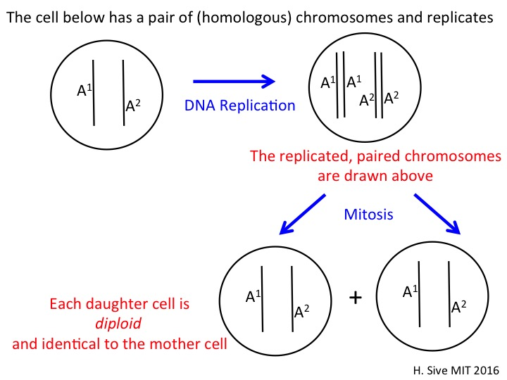
We can diagram this starting with a very simple cell that
has just one pair of chromosomes.
In our cells, we have 23 pairs or mostly pairs of chromosomes.
And in this cell, I've just shown you one pair.
The chromosomes are called A. That's
the name of the chromosome.
I've got chromosome one, but I've
called it A and the two copies I've
designated as A superscript 1 and A superscript 2.
They are homologous.
They are identical or similar to one another, largely identical.
The mother cell is diploid.
And the first thing that happens is that the cell
undergoes DNA replication.
And I've drawn the replicated chromosomes.
You can see the two A1s and the two A2s
are connected or next to one another at this point.
They will then line up on the structure of the spindle.
And one copy of A1 will go to one of the daughter cells.
One copy of A2 will go to one of the daughter cells.
So you land up with two daughter cells
that have the identical chromosome set as the mother.
They are all diploid.
And the actual chromosomes are the same
in the daughters as the mother.
This is a hugely important process.
It preserves the character of one cell
from one cell generation to the next.
And it's why during your life, your skin cells
and your intestinal cells stay much the same
because you're making more of the same
every time the cells divide.
On this slide, you can see some steps in mitosis.
I want you to have heard of mitosis at this point.
The specific steps are really descriptive of the process
of how the chromosomes are segregating.
You can see the chromosomes lining up
on the spindle at the stage called metaphase.
And then you can see them pulling apart
at the phase called anaphase.
And if you want to know what it really looks like,
what all of the spindle and the chromosomes pulling apart
look like, you can see these beautiful images
of mitosis in a sea urchin where the chromosomes have lined up
on the spindle in the top left hand image.
And then over time, you can see the chromosomes
pulling apart and being divided into two daughter cells.
And the things that look like flames or streaks in the cell
are these structures called microtubules
that make up the spindle, the part of the factory
of the cell that pulls the chromosomes into the daughter
cells.
The last type of cell division I want to tell you about
is called meiosis.
Meiosis leads to the production of the germ cells, the egg
and the sperm.
And the difference between meiosis and mitosis
is that at the end of meiosis, you get four cells.
And those four cells have each got the haploid number
of chromosomes.
They've each only got one of the normal paired chromosomes.
So the outcome, the outcome of meiosis, are four cells.
They are not identical to the parent,
not identical to the parent.
And each of these--
and they're not identical to the parent,
I should say, for a number of reasons.
But one of the reasons is that they
have the haploid number of chromosomes,
one copy of each chromosome instead of two.
How does this work?
As before, there is DNA replication,
chromosome replication so that you
get double the amount of DNA to start so that you've got DNA
that you can spread around.
And then there are some strange things
that happen during meiosis.
The chromosomes pair up together.
So you've got, at the end of DNA replication,
you've actually got two copies of chromosome A1
and two copies of chromosome A2 as I showed you in our diagram
on the screen.
And I'll show you another diagram in a moment.
And those two copies come really close together.
And they pair with one another.
And I mentioned that those copies
of chromosome 1 or chromosome A were similar but not
entirely identical.
They can exchange bits of DNA with one another.
So a bit of DNA from the chromosome A1
can now jump on to chromosome A2.
And the matching bit from A2 will go into A1.
So you land up with chromosomes that are not
exactly the same as the parental chromosome, either.
That's another difference with meiosis.
So the DNA, there's DNA replication.
The chromosomes pair and very often exchange DNA segments.
It is crazy, but this is what they do.
And then there are two steps to meiosis.
In meiosis I, the same pair, one replicated chromosome pair,
goes to the daughter cell.
And you land up with two cells that are diploid,
but they're diploid not like the parent.
So you get the outcome here is two cells.
And then there is a second division.
Those two cells in meiosis II will divide again.
And a single copy of each chromosome
will go to the daughter cells.
So they divide again, and a single homolog
goes to each daughter cell.
You do not need to know the details of this at this point.
I want you to have the general idea.
So let's go through this animation, which
will give you the general idea.
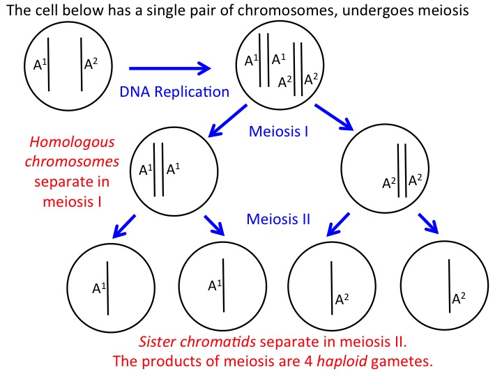
We start again with a mother cell that is diploid.
It undergoes DNA replication.
You have now two copies of chromosome A1,
two copies of chromosome of A2.
And these four copies, these four chromosomes,
pair with one another.
They come real close together.
And they pair with one another.
And that's when they may exchange pieces of DNA
in a process that is very complicated.
Meiosis I then happens.
And look.
And see you'll be able to contrast this with mitosis.
You've got both copies of chromosome A1 going
to one daughter cell and both copies of chromosome A2
going to another daughter cell.
So that is really different from mitosis.
But then what's also different is
that there's another division.
And you now land up with four cells
such that each of the cells has got one copy of chromosome A1,
or one copy of chromosome A2.
These cells are haploid.
They are different from the parent because of this haploidy
but also because they've had this funny DNA exchange
during the process of meiosis.
So these are the two kinds of cell division
that occur throughout life.
And now you've heard of them.
And you know some differences between the processes.
For your last exercise in class today, I'd
like you to look at this assignment and practice what
you know about cell division.
Reference Summary
Mitosis
Mitosis is the process of cell division by which 2 daughter cells are produced from 1 mother cell.
The daughter cells have the same ploidy and are genetically identical to both each other and the mother cell.
Meiosis
Meiosis is the process of cell division by which 4 gametes are produced from 1 mother cell.
The gametes' ploidies are half of the mother cell's ploidy. The gametes are NOT genetically identical to each other NOR to the mother cell.
Gametes are cells with half of the normal ploidy (haploid for humans). Examples of gametes are eggs (ova) and sperm cells.
Exercise 1 (Haploid v. Diploid)
Exercise 2 (Diagramming Mitosis)
Exercise 3 (Diagramming Meiosis)
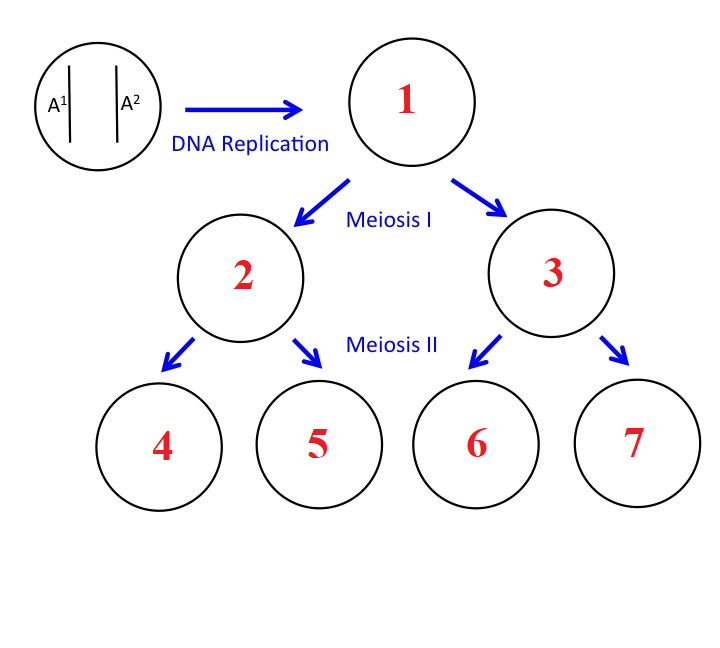
The cell below has a single pair of chromosomes and undergoes meiosis.
MIT OpenCourseWare / MIT Open Learning Library. Hazel Sive and Diviya Ray. Adapted for local study under Creative Commons Attribution–NonCommercial–ShareAlike 4.0. License terms.