Assignment 5.1: Restriction digests
Your answers are saved in this browser. Check them against the downloaded answer key, or open the explanation when you are ready.
Lecture 5.1
Transcript
HAZEL SIVE: Welcome back to Getting Up to Speed in Biology.
Today is our last discussion.
And the topic today is building with DNA.
We're going to cover four topics today.
And I think you will find them both interesting and useful.
The first is genetic engineering.
I'll talk about what that is.
We'll then talk about something called
restriction endonucleases.
We'll talk about vectors and ligation.
And then we'll talk about something
that has the abbreviation PCR.
Let's start with the topic of genetic engineering.
I bet you've heard of this.
Another word for genetic engineering
is recombinant DNA technology, recombinant DNA technology.
And what it really means is making DNA constructions
in the lab.
Building with DNA, the title of our discussion today,
DNA construction in the lab--
something that people do to build DNA molecules.
And one of the aspects of genetic engineering,
the subtopic that is really crucial,
is called molecular cloning.
And that refers to making lots of the same DNA,
making lots of the same DNA.
And lots of the same DNA or molecules of DNA
that are all very similar to one another or identical
are called clones.
Now, why would you want to do that?
Let's take a moment to look at some
of the power of genetic engineering
and give you a sense of why we are even having this discussion
today and why this is part of your getting up
to speed in biology.
Genetic engineering is everywhere.
There are genetically-modified animals.
Some of them are kind of fun, like fish
that glow in the dark different colors.
Some of them are for research like mice
that have been genetically engineered to glow green
or to glow green in different tissues.
Some of them are for food, cattle
that have been engineered so that they produce
much leaner meat or chickens that have been engineered
so they don't have any feathers and are easier to process
after they have been killed.
Plants are genetically engineered,
again, some of them for food, for example rice, which
is one of the staple crops of our planet,
lacks a specific substance called beta carotene.
And beta carotene is required for vitamin A production, which
is essential for life.
You can genetically engineer rice
so that it produces beta carotene.
And this is not scary.
It's just useful.
And then finally, there is a lot of health use
of genetic engineering.
One can manufacture particular human proteins
by genetic engineering, for example, insulin.
One can think about going and curing
human diseases that are caused by variant genes that
have bad effects, deleterious effects,
maybe even by replacing the faulty gene
with the normal gene, although that
is right out there at the limits of what
is ethically permissible.
And it's presently not something that we're doing in humans.
But genetic engineering is everywhere.
It is really interesting.
It is really cool.
And it is really fundamental to biology today.
Let us think therefore how you do genetic engineering.
That's what we're going to talk about today.
How do you build with DNA?
And let's consider the kind of global process of building
a new DNA molecule with the use of genetic engineering
techniques, and then we'll go through step by step
how you do each of the steps involved.
Here then are some basic steps of building a recombinant DNA
molecule where the term recombinant means
that the DNA comes from two or more sources.
The first thing that you need to do
is to think about what you're going to engineer, obviously.
And you'll usually have some gene of interest,
which we'll abbreviate GOI.
You're going to isolate the DNA that
encodes the gene of interest or part of it.
And to do that, you usually cut out the gene of interest
from some larger piece of DNA.
Remember, we talked about chromosomes
being long strings of genes and other DNA all joined together.
If you're going to get your gene of interest or part of it,
you have to be able to isolate it from the chromosome.
So that is one of the first steps, this cutting
out of the gene of interest.
And then you want to take your gene of interest
and put it in some environment so
that you can grow lots of it.
And the way you do that is to paste it--
paste your gene of interest--
into a vector, where a vector is a carrier.
A vector is a carrier of DNA, of extra DNA.
It itself is DNA.
And it has the property that it replicates very avidly.
And so you can grow lots of your clone.
So a vector is a carrier of extra DNA that replicates.
And because it replicates, you can
get lots of your clone DNA, lots of clone DNA.
That's the basic process.
And what we're going to talk about today
are two parts of this, really.
We're going to talk about cutting out
your gene of interest.
And we're going to talk about pasting your gene of interest
into a vector.
If you look at the schematic that I drew for you,
the different steps are shown here starting off with cells.
We open up the cells.
We break them open.
We extract the DNA.
We cut and isolate the gene of interest,
and then we insert or paste the gene of interest into a vector.
That gives us a recombinant clone.
Because the vector can replicate,
we can grow a lot of gene of interest DNA,
and then we can do things with that DNA.
It can be used to make RNA, which then can then
be translated into a protein of interest.
And that protein of interest, maybe insulin,
can then be purified in the lab and modified if necessary
and used as a human therapeutic.
We're going to start by talking about cutting the DNA.
And that is done with reagents called
restriction endonucleases, restriction endonucleases.
These are enzymes.
They're proteins.
And they're enzymes, biological catalysts,
that precisely cut DNA.
Sometimes, they're called molecular scissors.
And they do so because the proteins that
are the restriction endonucleases
recognize particular DNA sequences, sequences
of bases in the DNA.
They bind to those sequences, and then they have activities
so they can break the double-stranded DNA
in very precise ways.
Let's write that down.
These are enzymes that precisely cut DNA.
They recognize.
They bind, and they cut specific DNA sequences.
And once they've done their cutting,
there are two types of ends of the cut DNA.
These ends are either called blunt
or they are called sticky.
And you're going to need to know the difference.
And we'll follow through this on the next couple of boards.
Let's start off by talking about blunt restriction endonucleases
and blunt ends after restriction enzyme or endonuclease cutting.
And we'll use, for example, an enzyme that is called SmaI.
So, blunt-ended restriction endonucleases,
I'm going to abbreviate restriction endonuclease RE.
And the example I'll use is S, big S, small maI.
Sma1 refers to a particular bacterium
from which these restriction endonucleases--
this restriction endonuclease-- was isolated.
Restriction enzymes are part of the bacterial defense system
so that if the bacterium is invaded
by a virus that is a DNA virus, these enzymes
will cut up the viral DNA and render it nonpathogenic.
And now we use these enzymes for genetic engineering.
The SmaI recognition site looks like this--
CCC GGG 5 prime to 3 prime on the top strand.
And you know now what the complementary strand
looks like.
It is 3 prime GGG CCC 5 prime.
When SmaI recognizes this site, it makes two cuts.
It cuts one strand here and one strand opposite.
We can also indicate these by arrows and a vertical line.
There are a number of different notations
you can use to indicate where the restriction enzyme cuts.
And out of this come two DNA fragments.
One of them looks like 5 prime CCC 3
prime, and on the other strand, of course, GGG 3 prime
to 5 prime antiparallel.
And the other fragment looks like 5 prime GGG 3
prime and 3 prime CCC 5 prime.
You can see that the ends of the two pieces are flush.
There's no nucleotide sticking out,
no base sticking out on either end.
That's why this is called a blunt cutter,
a blunt-ended restriction enzyme cutter.
So both of these are made.
You can also see that the SmaI site is a palindrome.
This is the recognition site to SmaI.
It is a palindrome.
And many restriction recognition sites are.
Good.
Let's look at a sticky-ended site now.
So a sticky end restriction enzyme.
And the example we'll use here is called EcoRI.
And it has a recognition site that
goes 5 prime GAATC 3 prime and on its other strand CTTAAG
5 prime.
You can see now one of the many reasons
you need to know about complementarity is
so you can easily fill in the other strand.
EcoRI cuts in a different way than SmaI.
It cuts off this G on that strand
and the matching G on the other strand.
And we can put an arrow here if we want.
And I sometimes put a line right across where
the cut is going to be made.
And now we have to figure out after the cut what
are the two pieces of DNA that are made look like.
And the easiest way to do this is start at the cut
either at the arrow or your slash line and kind of peel
away the DNA until you get to the place
where the other strand is cut.
So let's draw this now.
We'll have 5 prime G on the one strand.
And on the other strand, we'll have CTTAA.
5 prime, 3 prime--
never forget to put in those 5 primes and those 3 primes.
That's what the piece from the top looks like.
How about the piece from the bottom?
There, we have 5 prime AATTC 3 prime.
And we have on the other strand a G. That's what we get.
These ends are sticky because there is single-stranded DNA
that is left after the cut.
And the single-stranded DNA is able to base pair.
And it is therefore sticky with regard to its ability
to base pair.
That's where the term "sticky end" comes from.
So here again is the recognition site for EcoRI.
And again it's a palindrome.
EcoRI leaves a 5 prime overhang.
That means that there is more DNA towards the 5 prime end
than the 3 prime end in the sticky cut.
There are also enzymes--
and I'll show you on a slide in a moment--
that leave a 3 prime overhang.
So this region here is a 5 prime overhang.
And there are-- I'll make the note-- there are both 5 prime
and 3 prime overhang sticky-ended restriction
endonucleases, REs.
Let's take a look at a couple of slides here.
There are thousands of restriction endonucleases.
As I mentioned, they were all named
after the bacterium from which they
were isolated, so EcoRI from E. coli, PstI from Providencia,
and so on.
You can see the double-stranded recognition site
in the DNA and, on this diagram, the structure
of the cleaved products.
You can look through books of restriction endonucleases.
And you can find something that cuts exactly where you
want it to in the DNA.
And you can get therefore a very precise cut in a piece of DNA
when you want to do a cut and paste
to make a recombinant clone.
I've redrawn for you the SmaI, SmaI or EcoRI sites.
And then on the bottom of the slide,
I've also put in another site which leaves
sticky 3 prime overhang ends.
And this is PstI.
And you can see here that there is
extra unpaired single-stranded DNA sticking out
towards the 3 prime end of each of the ends of the cut DNA.
One thing I want to emphasize is that these restriction
endonuclease sites are in a whole big piece of DNA.
We draw them just as the six base pair or the four base
pair or the eight base pair cut site.
But of course, they are joined to long pieces of DNA
on either side.
Good.
Now you've had some information about restriction
endonucleases, I want you to go to the class assignment
and practice doing some restriction enzyme work.
Reference Summary
Restriction Endonucleases
A restriction endonuclease, sometimes referred to simply as a restriction enzyme, cuts double-stranded DNA at palindrome sequences. Each restriction endonuclease cuts at one specific sequence. After being cut, the double-stranded DNA is left with "sticky ends". Different restriction endonucleases leave different types of sticky ends, which are reviewed below.
Types of Sticky Ends
SmalI leaves blunt ends:

EcoRI leaves sticky (5' overhang) ends:

PstI leaves sticky (3' overhang) ends:

Exercise 1 (Identifying products)
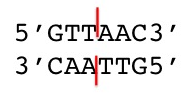
Here is the Hpa1 restriction cut site.
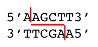
Here is the HindIII restriction cut site.
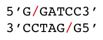
Here is the BamHI restriction cut site.
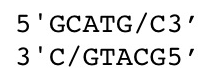
Here is the SphI restriction cut site.
Exercise 2 (Types of sticky ends)
Here is the AluI restriction cut site.
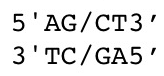
Here is the KpnI restriction cut site.
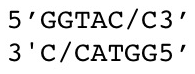
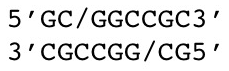
Here is the NotI restriction cut site.
MIT OpenCourseWare / MIT Open Learning Library. Hazel Sive and Diviya Ray. Adapted for local study under Creative Commons Attribution–NonCommercial–ShareAlike 4.0. License terms.