Chapter 6Cell Membranes
Key Concepts
- 6.1 Biological Membranes Are Lipid-Protein Bilayers
- 6.2 The Cell Membrane Is Important in Cell Adhesion and Recognition
- 6.3 Substances Can Cross Membranes by Passive Processes
- 6.4 Active Transport across Membranes Requires Energy
- 6.5 Large Molecules Enter and Leave a Cell through Vesicles
Blood, Sweat, and Membranes Water lost through sweating, leaves body tissues by passing through their cell membranes.

© Scott Barbour/Getty Images
Investigating Life: Sweating and Membranes
As you saw in Chapter 2, sweating is a way to reduce body heat by using excess heat to evaporate water. At peak activity, in the gym or doing other physical work, you may lose as much as 2 liters of water an hour. Total water content of a typical person (it varies with body weight, fat content, etc.) is about 30 liters.
The sweat glands lie just below the surface of the skin. They are essentially cell-lined tubes surrounded by extracellular fluid. When sweating is triggered, these tubes fill with water and dissolved substances. To get from the extracellular fluid into the tube, water must go through the cells that line the tube.
A hallmark of living cells is the ability to regulate what enters and leaves the cytoplasm. This is a function of the cell membrane, a hydrophobic lipid bilayer with associated proteins. Because it is insoluble in the aqueous environment both inside and outside cells, the membrane is a physical barrier. But it is also a functional barrier—that is, the cell membrane is selectively permeable. Whereas water is polar, the interior of the membrane is nonpolar, so water has a natural tendency to avoid the membrane. The rate of movement of water across a lipid bilayer is modest. When you engage in normal activities such as reading this book, the cell membranes enclosing the cells lining the sweat glands do not allow much water to enter or leave. But when you exercise vigorously, tiny membrane-enclosed vesicles inside the cells fill with water and some dissolved salts. In a process called exocytosis, these vesicles fuse with the cell membrane and release their watery contents (sweat) into the tubes. From there the sweat flows to the surface of the skin and evaporates.
Vesicles are not the only way to get polar water across a non-polar membrane. For most types of animal cells, the modest rate of movement of water across the cell membrane is sufficient to meet the changing needs of the cell. But in some organs, like the mammalian kidney, and in plant roots, stems, and leaves, water needs can be so great and urgent that movement through the lipid bilayer is not enough. Instead, these organs have special pores called aquaporins present in the cell membrane. Water can flow through these pores readily because the proteins lining the channel have a hydrophilic inner surface.
QA What is the importance of aquaporin membrane channels? (Find the answer on page 129.)
Biological Membranes Are Lipid-Protein Bilayers
Learning Objectives
- 6.1.1 Explain how the hydrophobic and hydrophilic regions of phospholipids are involved in membrane formation.
- 6.1.2 Differentiate among integral, peripheral, and anchored proteins.
- 6.1.3 Design an experiment to compare the properties of membranes in different types of cells.
- 6.1.4 Give an example of information provided by cell fusion experiments.
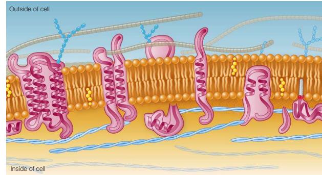
The physical organization and functioning of all biological membranes depend on their constituents: lipids, proteins, and carbohydrates. You are already familiar with these molecules from Chapter 3 and with membranes that enclose cells and organelles from Chapter 5. Lipids establish the physical integrity of the membrane and create a barrier to the rapid passage of hydrophilic materials such as polar molecules and ions. In addition, the phospholipid bilayer serves as a lipid “lake” in which a variety of proteins “float” (Figure 6.1). This general design is known as the fluid mosaic model. It is mosaic because it is made up of many discrete components, and fluid because the components can move freely.
Although the fluid mosaic model is largely valid for membrane structure, it does not say much about membrane composition.
As you read about the various molecules in membranes in the next sections, keep in mind that some membranes have more protein than lipids, others are lipid-rich, others have significant amounts of cholesterol or other sterols, and still others are rich in carbohydrates.
Lipids form the hydrophobic core of the membrane
Phospholipids are the major lipid components of biological membranes. Phospholipids form thin bilayers, with their charged, hydrophilic “heads” on the external surface interacting with the aqueous environment, and their nonpolar, fatty acid “tails” interacting with each other on the inside of the membrane (Figure 6.2).
Connect the Concepts Recall from Key Concept 2.2 that some compounds are hydrophilic (“water-loving”) and others are hydrophobic (“water-hating”), and from Key Concept 3.4 that a phospholipid molecule has both hydrophilic and hydrophobic regions.
The thickness of a biological membrane is about 8 nanometers (0.008 µm), which is twice the length of a typical phospholipid—another indication that the membrane consists of a lipid bilayer. To give you an idea of the scale, a typical sheet of paper is about 8,000 times thicker than a membrane.
Although all biological membranes have a similar structure, membranes from different cells and different organelles within a cell may vary greatly in their lipid composition. For example, phospholipids can differ in fatty acid chain length (number of carbon atoms), degree of unsaturation (number of double bonds) in the fatty acids, and the polar groups present (see Key Concept
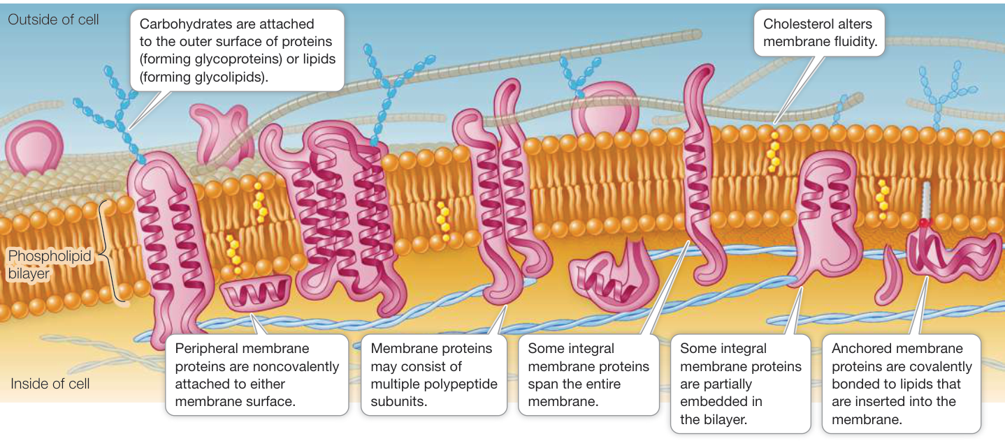
Figure 6.1 The Fluid Mosaic Model The general molecular structure of a biological membrane is a continuous phospholipid bilayer that has proteins embedded in or associated with it.
Q: What kinds of chemical interactions hold some membrane proteins embedded in the membrane and others on the membrane surface?
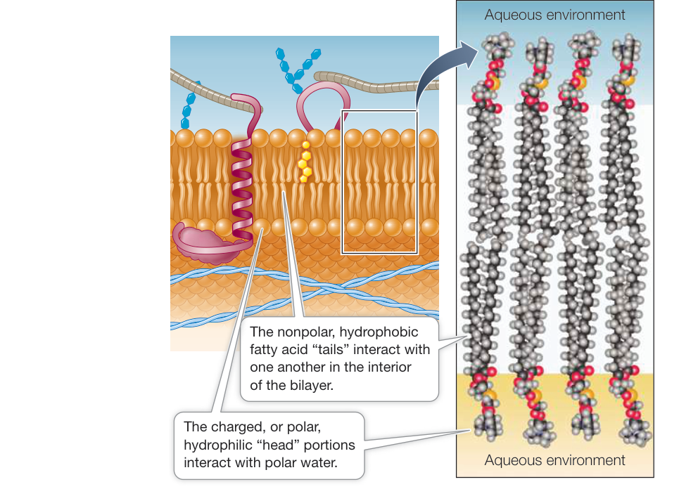
Figure 6.2 Phospholipid Bilayer The phospholipid bilayer separates two aqueous regions. The eight phospholipid molecules shown on the right represent a small cross section of a membrane bilayer. (Right after L. Stryer. 1981. Biochemistry, 2nd Ed., W. H. Freeman: New York.)
3.4). The saturated chains allow close packing of fatty acids in the bilayer, whereas the “kinks” in unsaturated fatty acids (see Figure 3.22) make for a less dense, more fluid packing.
The steroid cholesterol (see Figure 3.24) is an abundant component (20%–50%) of animal cell membranes. The hydroxyl group of cholesterol interacts with the polar ends of phospholipids, and the nonpolar rings and hydrocarbon chain insert into the fatty acids chains in the interior of the membrane. Cholesterol is important for membrane integrity and modulates membrane fluidity; the cholesterol in your membranes is not hazardous to your health.
The fatty acids of the phospholipids make the hydrophobic interior of the membrane somewhat fluid—about as fluid as light-weight olive oil. This fluidity permits some molecules to move laterally within the plane of the membrane. A given phospholipid molecule in the cell membrane can travel from one end of the cell to the other in a little more than a second! However, a phospholipid molecule in one half of the bilayer is unlikely to spontaneously flip over to the other side. For that to happen, the polar part of the molecule would have to move through the hydrophobic interior of the membrane. Since spontaneous phospholipid flip-flops are rare, the inner and outer halves of the bilayer may be quite different in the kinds of phospholipids they contain.
Membrane fluidity is affected by several factors, two of which are particularly important:
Lipid composition: Cholesterol and long-chain, saturated fatty acids pack tightly beside one another, with little room for movement. This close packing results in less fluid membranes. A membrane with shorter-chain fatty acids, unsaturated fatty acids, or less cholesterol is more fluid.
- Temperature: Because molecules move more slowly and fluidity decreases at reduced temperatures, cellular processes that take place within the membrane may slow down or stop under cold conditions in organisms that cannot keep their bodies warm. To address this problem, in some organisms the lipid composition of their membranes changes when they get cold, replacing saturated with unsaturated fatty acids and using fatty acids with shorter tails. These changes play a role in the survival of plants, bacteria, and hibernating animals during the winter.
Membrane proteins are asymmetrically distributed
Typically, cell membranes have 1 protein molecule for every 25 lipid molecules. This ratio varies depending on membrane function. In the inner membrane of the mitochondrion, which is specialized for energy processing, there is 1 protein for every 15 lipids. However, myelin—a modified cell membrane that encloses portions of some neurons (nerve cells) and acts as an electrical insulator—has only 1 protein for every 70 lipids.
Membrane proteins are very diverse. In fact, about one-fourth of the protein-coding genes in the eukaryotic genome encode membrane proteins. There are three general types of membrane proteins: integral proteins, peripheral proteins, and anchored proteins.
Integral membrane proteins are at least partly embedded in the phospholipid bilayer (see Figure 6.1). Like phospholipids, these proteins have both hydrophilic and hydrophobic regions (domains) (Figure 6.3).
- Hydrophilic domains: Stretches of amino acids with hydrophilic side chains (R groups; see Table 3.2) give certain regions of the protein a polar character. These hydrophilic domains interact with water and stick out into the aqueous environment inside or outside the cell or organelle.
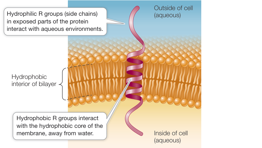
Figure 6.3 Interactions of Integral Membrane Proteins An integral membrane protein is held in the membrane by the distribution of the hydrophilic and hydrophobic side chains on its amino acids.
- Hydrophobic domains: Stretches of amino acids with hydrophobic side chains give other regions of the protein a nonpolar character. These domains interact with the fatty acids in the interior of the phospholipid bilayer, away from water.
Peripheral membrane proteins lack exposed hydrophobic groups and are not embedded in the bilayer. Instead, they have polar or charged regions that interact with exposed parts of integral membrane proteins, or with the polar heads of phospholipid molecules (see Figure 6.1).
Anchored membrane proteins are covalently attached to fatty acids or other lipid groups. The hydrophobic lipid groups insert into the phospholipid bilayer and hold these proteins in association with the membrane.
Proteins are asymmetrically distributed on the inner and outer surfaces of membranes, giving the two surfaces different properties. An integral protein that extends all the way through the phospholipid bilayer and protrudes on both sides is known as a transmembrane protein. In addition to having one or more transmembrane domains that extend through the bilayer, such a protein may have domains with other specific functions on the inner and outer sides of the membrane. Peripheral membrane proteins, by contrast, are located on one side of the membrane or the other. This asymmetrical arrangement of membrane proteins gives the two surfaces of the membrane different properties. As you will see, these differences have great functional significance.
Like lipids, some membrane proteins move around relatively freely within the phospholipid bilayer. Experiments that involve the technique of cell fusion illustrate this migration dramatically. When two cells are fused, a single continuous membrane forms and surrounds both cells, and some proteins from each cell distribute themselves uniformly around this membrane (Figure 6.4).
Although some proteins are free to migrate in the membrane, others are not, but rather are confined to a specific region of the membrane. These membrane regions are like an enclosure of animals at the zoo: the animals are free to move around within the fenced area but not outside it. An example is the protein in the cell membrane of a muscle cell that recognizes a chemical signal from a neuron. This protein is normally found only at the specific region where the neuron meets the muscle cell. Proteins inside the cell can restrict the movement of proteins within a membrane. The cytoskeleton has components just below the inner face of the membrane that are attached to membrane proteins protruding into the cytoplasm. The stability of the cytoskeletal components may thus restrict movement of attached membrane proteins.
Membranes are constantly changing
Membranes in eukaryotic cells are constantly forming, transforming from one type to another, fusing with one another, and breaking down. As we discussed in Chapter 5, fragments of membrane move, in the form of vesicles, from the endoplasmic reticulum (ER) to the Golgi apparatus, and from the Golgi apparatus to the cell membrane (see Figure 5.11). Secondary lysosomes form when primary lysosomes from the Golgi apparatus fuse with phagosomes from the cell membrane.
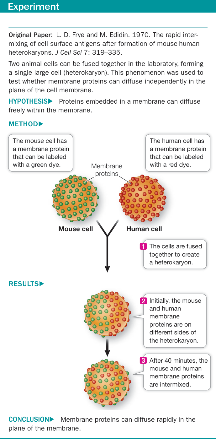
Figure 6.4 Rapid Diffusion of Membrane Proteins
Because all membranes appear similar under the electron microscope, and because they interconvert readily, we might expect all subcellular membranes to be chemically identical. However, that is not the case: there are major chemical differences among the membranes of even a single eukaryotic cell. Membranes are changed chemically when they form parts of certain organelles. In the Golgi apparatus, for example, the membranes of the cis face closely resemble those of the ER in chemical composition, but those of the trans face are more similar to the cell membrane.
Cell membrane carbohydrates are recognition sites
In addition to lipids and proteins, the cell membrane contains carbohydrates (see Figure 6.1). The carbohydrates are located on the outer surface of the cell membrane and serve as recognition sites for other cells and molecules, as you will see in Key Concept 6.2.
Membrane-associated carbohydrates may be covalently bonded to lipids or to proteins:
- A glycolipid consists of a carbohydrate covalently bonded to a lipid. Extending out from the cell surface, the carbohydrate may serve as a recognition signal for interactions between cells. For example, the carbohydrates on some glycolipids change when cells become cancerous. This change may allow white blood cells to target cancer cells for destruction.
- A glycoprotein consists of one or more short carbohydrate chains covalently bonded to a protein. The bound carbohydrates are oligosaccharides, usually not exceeding 20 monosaccharide units in length (see Key Concept 3.3). A proteoglycan (see Key Concept 5.4) is a more heavily glycosylated protein: it has more carbohydrate molecules attached to it, and the carbohydrate chains are often longer than they are in glycoproteins. The carbohydrates of glycoproteins and proteoglycans often function in cell recognition and adhesion.
The “alphabet” of monosaccharides on the outer surfaces of membranes can generate a large diversity of messages. Recall from Key Concept 3.3 that monosaccharides are simple carbohydrates, often containing five or six carbons in a ring structure and which can bond with one another in various configurations. They may form linear or branched oligosaccharides with many different three-dimensional shapes. An oligosaccharide of a specific shape on one cell can bind to a complementary shape on an adjacent cell. This binding is the basis of cell–cell adhesion.
Recap and Assess
The fluid mosaic model applies to the cell membrane and the membranes of organelles. In the aqueous environment of the cell, phospholipids spontaneously assemble into a bilayer. The cell membrane is considered fluid because the phospholipids and some of the proteins that compose it can move within their monolayer. An integral membrane protein has both hydrophilic and hydrophobic domains, which affect its position and function in the membrane. Peripheral membrane proteins are bound to one side or the other of the membrane. Anchored membrane proteins are covalently attached to membrane lipids. Carbohydrates that attach to lipids and proteins on the outside of the membrane serve as recognition and adhesion sites for adjacent cells.
- How do the hydrophobic and hydrophilic regions of phospholipids form a membrane bilayer?
- What differentiates an integral protein from a peripheral protein?
- If a cell membrane is frozen and then fractured so that the two leaflets of the cell membrane are peeled away from each other, bumps appear in electron microscopy. What are the bumps?
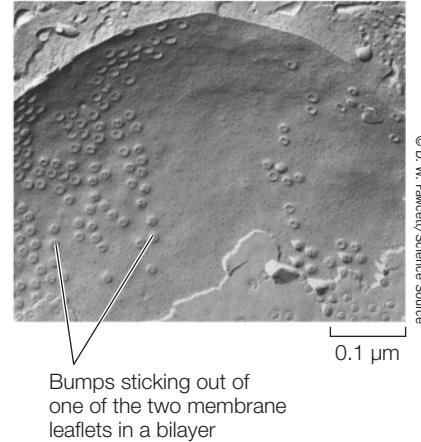
- When a normal lung cell becomes a lung cancer cell, there are several important changes in cell membrane properties. How would you investigate the observation that the cancer cell membrane is more fluid, with more rapid diffusion in the plane of the membrane of both lipids and proteins?
Now that you understand the structure of biological membranes, let’s see how their components function. In the next key concept we’ll focus on the membrane that surrounds individual cells—the cell membrane—and look at how the cell membrane allows individual cells to be grouped together into multicellular systems of tissues.
The Cell Membrane Is Important in Cell Adhesion and Recognition
The Cell Membrane Is Important in Cell Adhesion and Recognition
Learning Objectives
- 6.2.1 Design investigations to analyze the function of proteins and carbohydrates in cell adhesion and cell recognition.
- 6.2.2 Compare and contrast the three types of cell junctions found in animals.
Often the cells of multicellular organisms exist in specialized groups with similar functions, called tissues. Your body has about 37 trillion cells organized into various kinds of tissues—such as muscle, nerve, and epithelium. Two processes allow cells to arrange themselves in groups:
- Cell recognition, in which one cell specifically recognizes and binds to another cell of a certain type
- Cell adhesion, in which the connection between the two cells is strengthened
Simple organisms are useful in understanding cell recognition and adhesion
Both cell adhesion and cell recognition involve the cell membrane. One way to study these processes is to break down a tissue into its individual cells and then allow them to adhere to one another again. This type of experiment is most easily done in relatively simple organisms, such as sponges, which provide good models for studying processes that also occur in the complex tissues of larger species.
Sponges are multicellular marine animals that have only a few distinct cell layers (see Key Concept 29.1). The cells of a sponge adhere to one another but can be separated mechanically by passing the animal several times through a fine wire screen (Figure 6.5). Through this process, what was a single animal becomes hundreds of individual cells suspended in seawater. If such cells are stirred gently for a few hours, cell recognition occurs: the cells bump into and recognize one another, sticking together in the same shape and tissue organization as in the original sponge. This recognition is species-specific; if disaggregated sponge cells from two different species are placed in the same container and shaken, individual cells will stick only to other cells of the same species. Two different sponges form, just like the ones at the start of the experiment.
Tissue-specific and species-specific cell recognition and cell adhesion are essential to the formation and maintenance of tissues in multicellular organisms. Think of your own body. What keeps muscle cells bound to muscle cells and skin to skin? Specific cell adhesion is so obvious a characteristic of complex organisms that it is easy to overlook. You will see many examples of specific cell adhesion throughout this book; here we describe its general principles. As you will see, cell recognition and cell adhesion depend on cell membrane proteins.
Cell recognition and adhesion involve proteins and carbohydrates at the cell surface
The molecules responsible for cell recognition and adhesion in sponges are proteoglycans (often 80% carbohydrate by molecular weight) that carry two kinds of carbohydrates. One kind is relatively small and binds to membrane components, keeping the proteoglycan attached to the cell. The other kind of carbohydrate is a larger, sulfated polysaccharide. If the sulfated polysaccharide from a particular species of sponge is purified and attached to cellulose beads, the beads will aggregate together or with sponge cells—but only with cells of the same species from which the polysaccharide was purified. This demonstrates that the sulfated polysaccharide is responsible for both the specific recognition and adhesion of the sponge cells.
Cell adhesion can result from interactions between the carbohydrates that are parts of glycolipids, glycoproteins, or proteoglycans—as is the case in sponge cells. In other cases, a carbohydrate on one cell interacts with a membrane protein on another cell. Or two proteins can interact directly. As we described in Key Concept 3.2, a protein not only has a specific shape, it also has specific chemical groups exposed on its surface where they can interact with other substances, including other proteins. Both of these features allow binding to other specific molecules. Cell adhesion occurs in all kinds of multicellular organisms. In plants, cell adhesion may be mediated by both integral membrane proteins and specific carbohydrates in the cell walls.
Usually, the binding of cells in a tissue is homotypic; that is, the same molecule sticks out of both cells, and the exposed surfaces fit together and bind to each other. This is a factor that keeps skin cells together in a sheet of cells. But heterotypic binding (between different molecules on different cells) also occurs. In this case, chemical groups on different surface molecules have an affinity for one another. For example, when the mammalian sperm meets the egg, different proteins on the two types of cells have complementary binding surfaces. Similarly, some algae form male and female reproductive cells (analogous to sperm and eggs) that have flagella to propel them toward each other. Male and female cells can recognize each other by heterotypic glycoproteins on their flagella.
Three types of cell junctions connect adjacent cells
In a complex multicellular organism, cell recognition molecules allow specific types of cells to bind to one another. Often, after the initial binding, both cells contribute material to form additional membrane structures that connect them to one another. These specialized structures, called cell junctions, are most evident in electron micrographs of animal epithelial tissues, which are layers of cells that line body cavities or cover body surfaces. These surfaces often receive stresses or must retain their contents under pressure, so it is particularly important that their cells adhere tightly. We will examine three types of cell junctions that enable animal cells to seal intercellular spaces, reinforce attachments to one another, and communicate with each other. Tight junctions, desmosomes, and gap junctions, respectively, perform these three functions (Figure 6.6).
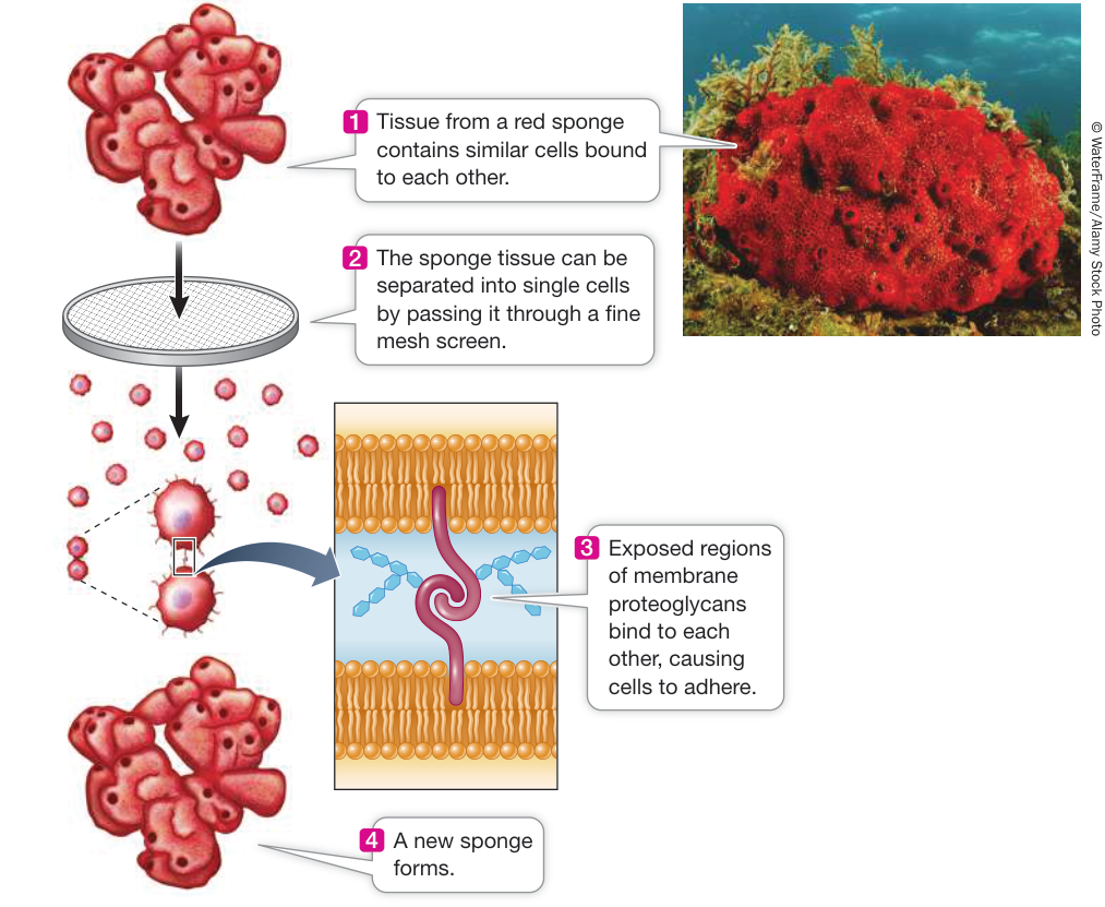
Figure 6.5 Cell Recognition and Adhesion In most cases (including the aggregation of animal cells into tissues), the binding between molecules is homotypic (same to same).
Tight junctions prevent substances from moving through the spaces between cells. For example, cells lining the bladder have tight junctions so urine cannot leak out into the body cavity. Another important function of tight junctions is to maintain distinct faces of a cell within a tissue by restricting the migration of membrane proteins over the cell surface from one face to the other. This may result in a certain function (e.g., endocytosis) being confined to one region of the cell surface.
Desmosomes hold neighboring cells firmly together, acting like spot welds or rivets. Materials can still move around in the extracellular matrix. This provides mechanical stability for tissues such as skin that receive physical stress.
Gap junctions are channels that run between membrane pores in adjacent cells, allowing substances to pass between cells. In the heart, for example, gap junctions allow the rapid spread of electric current (mediated by ions) so the heart muscle cells beat in unison.
Cell membranes adhere to the extracellular matrix
In Key Concept 5.4 you learned about the extracellular matrix of animal cells, which is composed of collagen protein arranged in fibers in a gelatinous matrix of proteoglycans. The attachment of a cell to the extracellular matrix is important in maintaining the integrity of a tissue. In addition, some cells can detach from their neighbors, move, and attach to other cells; this is often mediated by interactions with the extracellular matrix.
A transmembrane protein called integrin often mediates the attachment of epithelial cells to the extracellular matrix (Figure 6.7). More than 24 different integrins have been described in human cells. All of them bind to a protein in the extracellular matrix outside the cell, and to actin filaments, which are part of the cytoskeleton, inside the cell. So in addition to playing a role in adhesion, integrin has a role in maintaining cell structure via its interaction with the cytoskeleton. The binding of integrin to the extracellular matrix is noncovalent and reversible. When a cell moves its location within a tissue or organism, the integrin-matrix interaction is released and the cell is free from the constraints of the matrix. When the cell arrives at the new location, a new integrin may be made to allow the cells to interact with the particular extracellular matrix in that tissue.
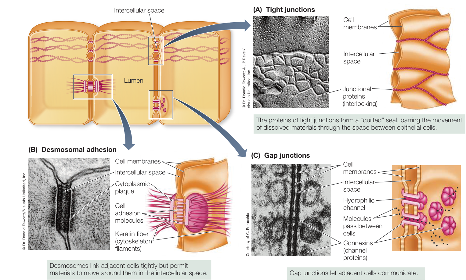
Figure 6.6 Junctions Link Animal Cells Together Tight junctions (A) and desmosomes (B) are abundant in epithelial tissues. Gap junctions (C) are also found in some muscle and nerve tissues, in which rapid communication between cells is important. Although all three junction types are shown in the cell at the top left, all three are not necessarily seen at the same time in actual cells.
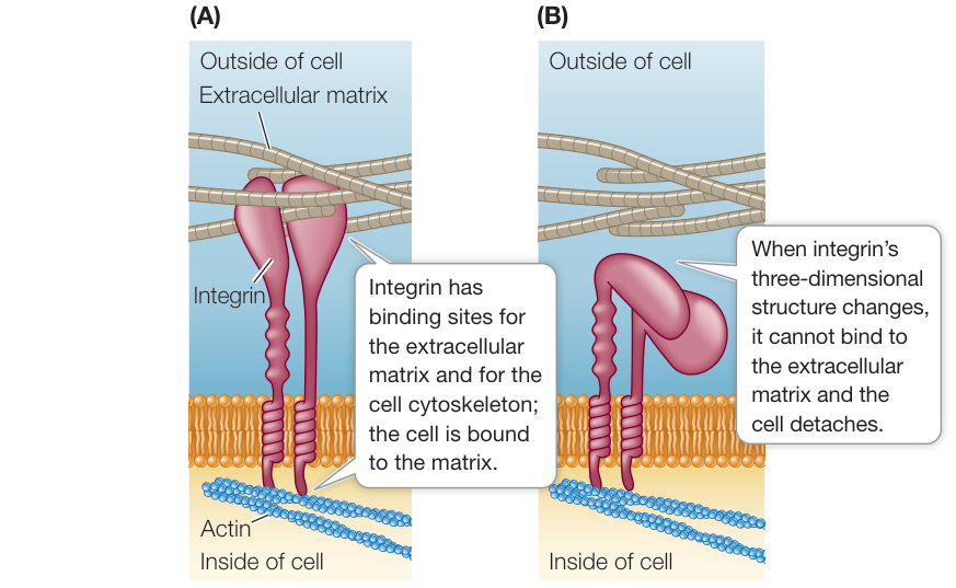
Figure 6.7 Integrins and the Extracellular Matrix (A) Integrins mediate the attachment of cells to the extracellular matrix. (B) When integrins no longer mediate attachment, the cell can separate itself from its extracellular matrix. Q: When a tumor spreads in the body, cells detach from the extracellular matrix and then reattach at another organ in the body. For each tumor type, there are preferred organs to which it spreads. What does this indicate about integrins and the extracellular matrix?
Recap and Assess
In multicellular organisms, cells arrange themselves into tissues via the processes of cell recognition and cell adhesion. These processes are mediated by membrane-associated proteins and carbohydrates. Cell membrane proteins also interact with the extracellular matrix. Cell junctions assist in strengthening tissues and allow cells to communicate with one another.
- Describe an experiment showing a role for carbohydrates in cell recognition and adhesion.
- To which of the three types of animal cell junctions are plasmodesmata most similar (see Figure 5.9)?
You have seen how the cell membrane and molecules associated with it facilitate binding between cells and the maintenance of cell adhesion. We’ll turn now to another major function of membranes: regulating the substances that enter or leave a cell or organelle.
Substances Can Cross Membranes by Passive Processes
Learning Objectives
- 6.3.1 Predict rates of diffusion of substances under different conditions.
- 6.3.2 Discuss the effect of different environmental conditions on the movement of water into and out of living cells.
- 6.3.3 Explain the role of channel proteins in facilitating diffusion across membranes.
Membranes separate an internal environment from the external environment, giving cells or organelles the ability to regulate their internal composition. Biological membranes allow some substances, but not others, to pass through them. This characteristic of membranes is called selective permeability. Selective permeability allows the membrane to determine what substances enter or leave a cell or organelle.
There are two fundamentally different processes by which substances cross biological membranes:
- The processes of passive transport do not require the input of chemical-bond energy to drive them.
- The processes of active transport are driven by chemical-bond energy (metabolic energy).
This key concept focuses on the passive processes by which substances cross membranes. The energy for the passive transport of a substance comes from the difference between its concentration on one side of the membrane and its concentration on the other—its concentration gradient. Passive transport can involve either of two types of diffusion: simple diffusion through the phospholipid bilayer, or facilitated diffusion via channel proteins or carrier proteins.
Diffusion is the process of random movement toward a state of equilibrium
In a solution, there is a tendency for all of the components to be evenly distributed. You can see this when a drop of food coloring falls into a glass of water. Initially the pigment molecules are very concentrated, but they will move about at random, slowly spreading until the intensity of the color is eventually the same throughout the glass (Figure 6.8). A solution in which the solute particles are uniformly distributed is said to be at equilibrium because there will be no future net change in their concentration. Being at equilibrium does not mean that the particles have stopped moving; it just means that they are moving in such a way that their overall distribution does not change.
Diffusion is the process of random movement toward a state of equilibrium. Although the motion of each individual particle is absolutely random, the net movement of particles is directional until equilibrium is reached. Diffusion is thus a net movement from regions of higher concentration to regions of lower concentration. In a solution with many different substances dissolved in it (solutes), the diffusion of each solute is independent of those of the others. How fast a substance diffuses depends on five factors:
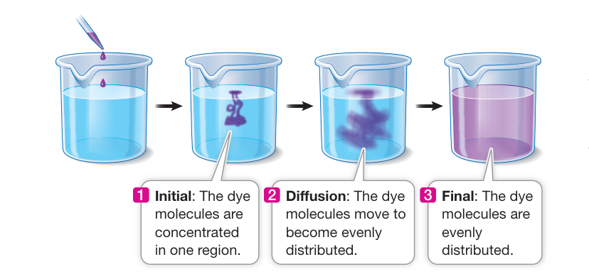
Figure 6.8 Diffusion Molecules tend to move from a region of higher concentration to one of lower concentration, until the molecules are evenly distributed in the environment, as shown here by dye molecules in a beaker of water. Q: The food dye in this experiment has a molecular weight of 600. If a dye with a molecular weight of 10,000 was used instead, what would the result be?
- The size and mass of the molecules or ions: smaller molecules diffuse faster.
- The temperature of the solution: higher temperatures lead to faster diffusion because ions or molecules have more energy, and thus move more rapidly, at higher temperatures.
- The density of the solution: as the density of the solution through which a substance is diffusing increases, the diffusion rate decreases.
- The concentration gradient in the system—that is, the change in solute concentration with distance in a given direction: the greater the concentration gradient, the more rapidly a substance diffuses.
- The area across which a substance diffuses and the distance it diffuses: a larger surface area permits more rapid diffusion. As you will see in Chapter 30, keeping the distance a substance must diffuse as short as possible maximizes the diffusion rate.
In our example in Figure 6.8, the gradient is the concentration of food coloring in the droplet when first dropped into the water as compared with the concentration of food coloring in the water on the edge of the glass. When the food coloring is first dropped into the water, there is a high (or steep) concentration gradient. All the coloring molecules are in one region. As time passes and the coloring molecules diffuse through the solution, the concentration gradient decreases.
DIFFUSION WITHIN CELLS AND TISSUES In a small volume such as that inside a cell, solutes distribute themselves rapidly by diffusion. Small molecules and ions may move from one end of a cell to another in a millisecond ( s, or one-thousandth of a second). However, the usefulness of diffusion as a transport mechanism declines drastically as distances become greater. In the absence of mechanical stirring, diffusion across more than a centimeter may take an hour or more, and diffusion across meters may take years! Diffusion would not be adequate to distribute materials over the length of a human body, much less than of a larger organism. But within our cells or across layers of one or two cells, diffusion is rapid enough to distribute small molecules and ions almost instantaneously.
DIFFUSION ACROSS MEMBRANES In a solution without barriers, all the solutes diffuse at rates determined by temperature, their physical properties, their concentration gradients, the density of the solution, and the area and distance of diffusion. If a biological membrane divides the solution into separate compartments (e.g., the cytoplasm of a cell vs. the extracellular environment), then the movement of the different solutes can also be affected by the properties of the membrane. The membrane is said to be permeable to solutes that can cross it more or less easily, but impermeable to substances that cannot move across it. Molecules to which the membrane is impermeable remain in separate compartments, and their concentrations may be different on the two sides of the membrane. Molecules to which the membrane is permeable diffuse from one compartment to the other until their concentrations are equal on both sides of the membrane, and equilibrium is reached. After that point, individual molecules will continue to pass through the membrane, but there will be no net change in concentration.
Simple diffusion takes place through the phospholipid bilayer
In simple diffusion, small molecules pass through the phospholipid bilayer of the membrane. A molecule that is itself hydrophobic, and is therefore soluble in lipids, enters the membrane readily and is able to pass through it. The more lipid-soluble the molecule is, the more rapidly it diffuses through the membrane bilayer. This statement holds true over a wide range of molecular weights.
By contrast, electrically charged or polar molecules, such as amino acids, sugars, and ions, do not pass readily through a membrane, for two reasons. First, such charged or polar molecules are not very soluble in the hydrophobic interior of the bilayer. Second, such substances form many hydrogen bonds with water and ions in the aqueous environment, be it the cytoplasm or the cell exterior. The multiplicity of these hydrogen bonds prevents the substances from moving into the hydrophobic interior of the membrane. Figure 6.9 summarizes these phenomena.
Osmosis is the diffusion of water across membranes
Movement of water molecules through a membrane toward a region of higher solute concentration is called osmosis. This is a passive process that uses no metabolic energy. Because large or polar solute molecules can’t cross the membrane, their otherwise random movement becomes biased away from the membrane (see Figure 6.9). If the solute concentration on one side of the membrane is higher, and all other factors are equal, then more solute molecules move away from the membrane on the side with the
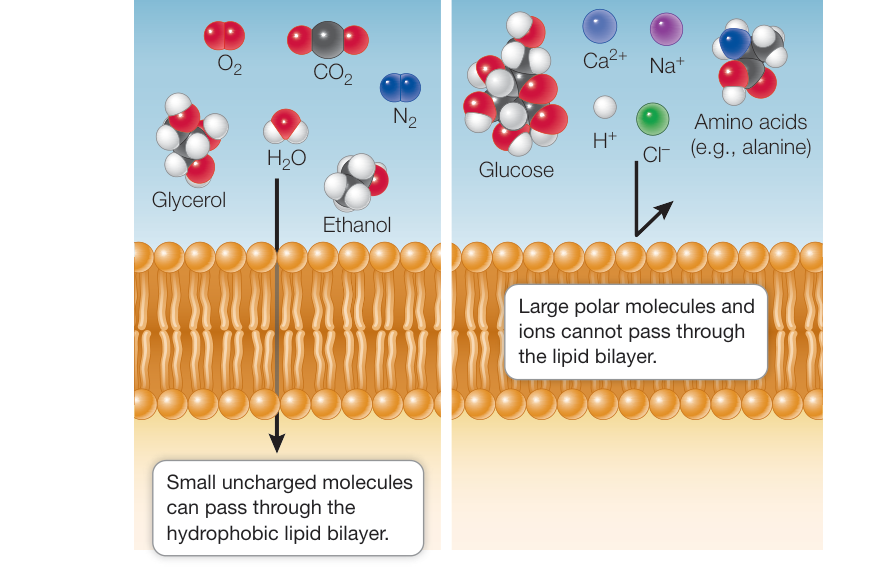
Figure 6.9 Permeability of Phospholipid Bilayers Small uncharged molecules can diffuse through the membrane, but ions and large polar molecules cannot.
In general, when we discuss tonicity in cells, we refer to the outside solution in comparison with the inside of the cell. Therefore a solution that has a higher concentration of solutes than the concentration inside the cell is hypertonic. To reach equilibrium, water would move out of the cell into the surrounding solution. Water moves from a hypotonic solution across a membrane to a hypertonic solution.
When we say that “water moves,” bear in mind that we are referring to the net movement of water. Since it is so abundant, water is constantly moving across the cell membrane into and out of cells. What concerns us here is whether the overall movement is greater in one direction or the other.
The total concentration of solutes in the environment determines the direction of osmosis. Consider red blood cells (see Figure 6.10). The environment outside the cell, the blood plasma, must be isotonic to the blood cells if the cells are not to burst (if the environment is hypotonic) or shrink (hypertonic). Regulation of the solute concentration of the blood plasma is therefore an important process for maintaining the integrity of the red blood cells. As you will see in Chapter 50, the body expends a great deal of energy maintaining greater solute concentration, and there is a net movement of water toward the side with the higher solute concentration.
Connect the Concepts Osmosis plays key roles in plant physiology (e.g., in the root; see Key Concept 33.1) and in animal physiology (e.g., in the kidney; see Key Concept 50.1).
Three terms are used to compare the total solute concentrations of two solutions separated by a membrane:
- Isotonic solutions have equal solute concentrations (Figure 6.10A).
- A hypotonic solution has a lower solute concentration than the other solution with which it is being compared (Figure 6.10B).
- A hypertonic solution has a higher solute concentration than the other solution with which it is being compared (Figure 6.10C).
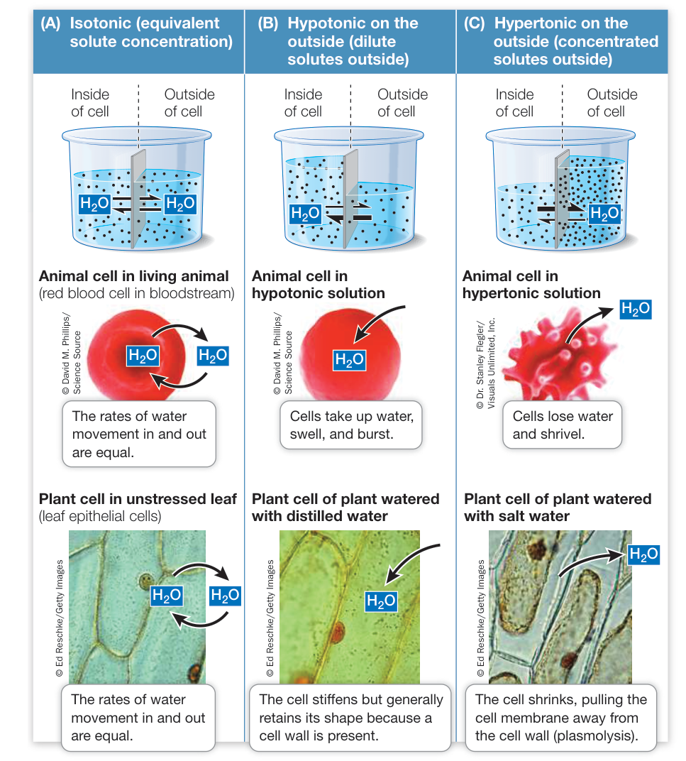
Figure 6.10 Osmosis Can Modify the Shapes of Cells In a solution that is isotonic with the cytoplasm (A), a plant or animal cell maintains a consistent, characteristic shape because there is no net movement of water into or out of the cell. In these models, it is assumed that the solutes do not move across the membrane. In a solution that is hypotonic to the cytoplasm (B), water enters the cell. An environment that is hypertonic to the cytoplasm (C) results in water leaving the cell. Q: If you overfertilize the soil of a potted plant, the plant often wilts. Why?
this environment. To put it more generally, cells and organisms strive to maintain an isotonic external environment.
Unlike animal cells, the cells of plants, archaea, bacteria, fungi, and some protists have cell walls outside their cell membrane that limit their volumes. Cells with sturdy walls take up a limited amount of water, and in so doing they build up internal pressure against the cell wall, which prevents further water from entering. This pressure within the cell is called turgor pressure. Turgor pressure keeps plants upright (and lettuce crisp) and is the driving force for the enlargement of plant cells. It is a normal and essential component of plant growth. If enough water leaves the cells, turgor pressure drops and the plant wilts. Turgor pressure reaches about 100 pounds per square inch ()—several times greater than the pressure in automobile tires. This pressure is so great that the cells would change shape and detach from one another were it not for adhesive molecules in the plant cell walls and the plasmodesmata linking the cell cytoplasms (see Key Concept 5.4).
Diffusion may be aided by channel proteins
As we saw earlier, polar or charged substances such as water, amino acids, sugars, and ions do not readily diffuse across membranes. But they can cross the hydrophobic phospholipid bilayer passively (that is, without the input of energy) in one of two ways, depending on the substance:
- Channel proteins are integral membrane proteins that form channels across the membrane through which certain substances can pass. Note that these are not channels in the widely used sense of the word (meaning open to the environment like a canal) but tunnels (surrounded by the membrane protein).
- Carrier proteins bind substances and speed up their diffusion through the phospholipid bilayer.
Diffusion that is aided by channel proteins or carrier proteins is called facilitated diffusion. The substances diffuse according to their concentration gradients, but their diffusion is facilitated by protein channels or carriers. Although these proteins enhance the rate of diffusion, facilitated diffusion, like simple diffusion, does not require any input of energy.
ION CHANNELS The best-studied channel proteins are the ion channels. As you will see in later chapters, the movement of ions across membranes is important in many biological processes such as respiration within the mitochondria, the electrical activity of the nervous system, and the opening of pores in leaves to allow gas exchange with the environment. Several types of ion channels have been identified—each specific for a particular ion. All show the same basic structure of a hydrophilic pore that allows a particular ion to move through its center.
Connect the Concepts Ion channels play an important role in the excitability of nervous tissues. See Key Concept 44.2.
Just as a fence may have a gate that can be opened or closed, most ion channels are gated: they can be opened or closed to ion passage (Figure 6.11). A gated channel opens when a stimulus causes a change in the three-dimensional shape of the channel. In some cases, this stimulus is the binding of a chemical signal, or ligand. Channels controlled in this way are called ligand-gated channels. Other channels are mechanically gated and open in response to physical stimuli such as sound waves in the ear, or are light-gated, as in the eye. A voltage-gated channel is stimulated to open or close by a change in the voltage (electric charge difference) across the membrane.
AQUAPORINS FOR WATER As you saw in the opening story of this chapter, water can cross membranes through protein channels called aquaporins. These channels function as a cellular plumbing system for moving water. The aquaporin channel is highly specific. Water molecules move in single file through the channel, which excludes ions so that the electrical properties of the cell are maintained. Aquaporins were first identified when a protein from red blood cell membranes was inserted into frog oocytes (immature egg cells). The membranes of these cells are normally impermeable to water, but the membranes of the cells treated with aquaporins became much more permeable (Investigating Life: Aquaporins Increase Membrane Permeability to Water).
Carrier proteins aid diffusion by binding substances
As we mentioned earlier, another type of facilitated diffusion involves the binding of the transported substance to a membrane protein called a carrier protein. Like channel proteins, carrier proteins facilitate the passive diffusion of substances into or out of cells or organelles. Carrier proteins transport polar molecules such as sugars and amino acids.
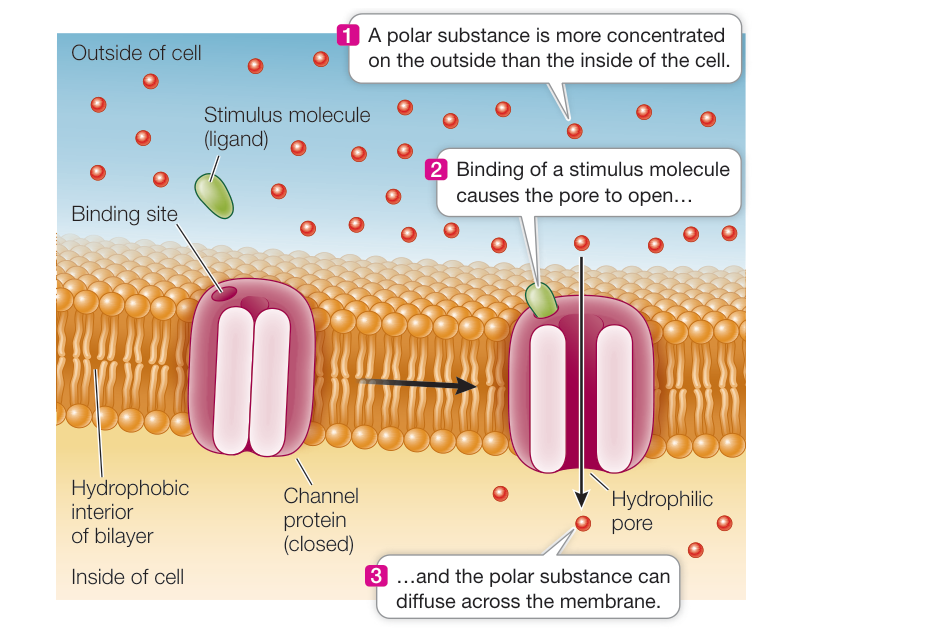
Figure 6.11 A Gated Channel Protein Opens in Response to a Stimulus The channel protein has a pore of polar amino acids and water. It is anchored in the hydrophobic bilayer by its outer coating of nonpolar R groups. The protein changes its three-dimensional shape when a stimulus molecule (ligand) binds to it, opening the pore so that specific hydrophilic substances can pass through. Other channels open in response to an electric potential (voltage) or mechanical stimuli. Q: Are gated ion channels directional, allowing ion transport either in or out of the cell?
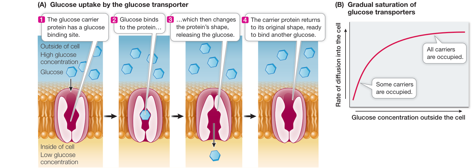
Figure 6.12 A Carrier Protein Facilitates Diffusion The glucose transporter is a carrier protein that allows glucose to enter the cell at a faster rate than would be possible by simple diffusion. (A) The transporter binds to glucose, brings it into the membrane interior, then changes shape, releasing glucose into the cell cytoplasm. (B) The graph shows the rate of glucose entry via a carrier versus the concentration of glucose outside the cell. As the glucose concentration increases, the rate of diffusion increases until the point at which all the available transporters are being used (the system is saturated).
Investigating Life: Aquaporins Increase Membrane Permeability to Water
Experiment
Original Paper: G. M. Preston et al. 1992. Appearance of water channels in Xenopus oocytes expressing red cell CHIP28 protein. Science 256: 385–387.
A protein was isolated from the membranes of cells in which water diffuses rapidly across the membranes. When the protein was inserted into oocytes, which do not normally have it, the water permeability of the oocytes was greatly increased.
HYPOTHESIS Aquaporin increases membrane permeability to water.
METHOD
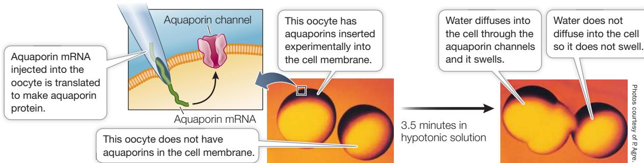
CONCLUSION Aquaporin increases the rate of water diffusion across the cell membrane.
Work with the Data
Although diffusion can account for limited water movement across cell membranes, simple diffusion seemed unlikely to explain the considerable water movement in kidney and red blood cells. By chance, Peter Agre and his colleagues found a major membrane protein, CHIP28, shared by these two cell types, and hypothesized that it was responsible for cell membrane water transport. So they did a “what if” experiment, taking the mRNA for CHIP28 and injecting it into frog oocytes that do not normally make the protein.
QUESTIONS
- Frog oocytes in isotonic liquid were injected with either a small amount of water (control) or with CHIP28 mRNA in a small amount of water. The oocytes were then transferred to a hypotonic medium, and changes in relative cell volume were measured by microscopy. Table A shows the results.
Table A
| Time (min) | Cell volume | |
|---|---|---|
| CHIP28 mRNA | Water only (control) | |
| 0 | 1.0 | 1.0 |
| 0.5 | 1.05 | 1.0 |
| 1 | 1.15 | 1.02 |
| 1.5 | 1.23 | — |
| 2 | 1.32 | 1.02 |
| 2.5 | 1.36 | — |
| 3 | 1.41 | 1.02 |
| 4 | (burst) | 1.03 |
Plot the data on a graph of relative cell volume versus time after injection. How do the mRNA and control eggs compare? What explains the increase in the volume of the mRNA-injected oocytes? What explains the situation with both cells at 4 minutes after injection?
- Water permeability () was calculated by the rate of osmotic swelling. Table B shows the results when increasing amounts of CHIP28 mRNA were injected.
Table B
| Amount of mRNA (ng) | (cm/sec ) a |
|---|---|
| 0 | 13.7 (3.3) |
| 0.1 | 50.0 (10.1) |
| 0.5 | 112 (29.2) |
| 2.0 | 175 (38.4) |
| 10.0 | 221 (14.8) |
aNumbers in parentheses indicate +/- standard deviation.
What can you conclude from these data? What statistical test (see Appendix B) would you do to show the significance of your conclusion?
- To further investigate the role of CHIP28 in water transport, oocytes were given a known inhibitor of protein-mediated water transport, mercuric chloride, and then tested for water permeability with CHIP28 mRNA. Some of the eggs were also treated with mercaptoethanol, a molecule that overcomes the inhibition of water transport by mercuric chloride. The question was, would the inhibitor actually block water transport mediated by CHIP28, and would mercaptoethanol restore transport? The results are shown in Table C.
Table C
| mRNA injected | Mercuric chloride | Mercaptoethanol | (cm/sec ) |
|---|---|---|---|
| None | None | None | 27.9 |
| None | Yes | None | 20.3 |
| None | Yes | Yes | 25.4 |
| CHIP28 | None | None | 210 |
| CHIP28 | Yes | None | 80.7 |
| CHIP28 | Yes | Yes | 188 |
What can you conclude about the molecular nature of water transport mediated by CHIP28 mRNA? What data support your conclusion? Explain all the controls that were done.
Glucose is the major energy source for most cells, and living systems require a great deal of it. Glucose is polar and cannot readily diffuse across membranes. Eukaryotic cell membranes contain a carrier protein—the glucose transporter—that facilitates glucose uptake into the cell. Binding of glucose to a specific three-dimensional site on one side of the transporter protein causes the protein to change its shape and release glucose on the other side of the membrane (Figure 6.12A). Since glucose is either broken down or otherwise removed almost as soon as it enters a cell, there is almost always a strong concentration gradient favoring glucose entry (that is, a higher concentration outside the cell than inside).
Transport by carrier proteins is different from simple diffusion. In simple diffusion, the rate of movement depends on the concentration gradient across the membrane. This is also true for carrier-mediated transport, up to a point. In carrier-mediated transport, as the concentration gradient increases, the diffusion rate also increases, but its rate of increase slows, and a point is reached at which the diffusion rate becomes constant. At this point, the facilitated diffusion system is said to be saturated (Figure 6.12B). This is explained by the fact that a particular cell has a specific number of carrier protein molecules in its cell membrane.
The rate of diffusion reaches a maximum when all the carrier molecules are fully loaded with solute molecules. Think of waiting for the elevator on the ground floor of a hotel with 50 other people. You can’t all get in the elevator (carrier) at once, so the rate of transport (say, ten people at a time) is at its maximum, and the transport system is “saturated.” As a consequence, cells that require large amounts of energy, such as muscle cells, have high concentrations of glucose transporters in their membranes so that the maximum rate of facilitated diffusion is greater. Likewise, the human brain has high glucose needs, and the blood vessels that nourish it have high concentrations of glucose transporters.
Recap and Assess
Diffusion is the movement of ions or molecules from a region of higher concentration to a region of lower concentration. Osmosis is the diffusion of water through a selectively permeable cell membrane. Channel proteins and carrier proteins can facilitate the diffusion of charged and polar substances, including water, across cell membranes.
- What properties determine whether, and how fast, a substance will diffuse across a membrane?
- What is the effect on red blood cells if the kidney removes too many ions from the blood and makes it hypotonic to the cells?
- Cells in the nervous system may have a higher concentration of ions inside the cell than outside. What is the role of the cell membrane in maintaining this imbalance? How can membrane channels eliminate this imbalance?
The process of diffusion tends to equalize the concentrations of substances outside and inside cells. However, one hallmark of a living cell is that it can have an internal composition quite different from that of its environment. To achieve this, a cell must sometimes move substances against their concentration gradients. This process requires an input of energy and is known as active transport.
Active Transport across Membranes Requires Energy
Learning Objectives
- 6.4.1 Compare and contrast diffusion and active transport.
- 6.4.2 Explain why active transport requires energy.
- 6.4.3 Describe and give examples of the different types of proteins that carry out active transport.
- 6.4.4 Differentiate between primary and secondary active transport.
In many biological situations, the concentration of a particular ion or small molecule inside the cell differs from that outside the cell. Such imbalances are maintained by a protein in the cell membrane that moves the substance against its concentration and/or electrical gradient. Movement of molecules or ions against a gradient is called active transport because it requires an input of energy. Often the energy source is adenosine triphosphate (ATP), which has chemical-bond energy stored in its terminal phosphate bond. In eukaryotes, ATP is produced in the mitochondria. Energy is released in a hydrolysis reaction that breaks the terminal phosphate bond of ATP, converting it to adenosine diphosphate (ADP). We will detail how ATP provides energy to cells in Key Concept 8.2.
The differences between diffusion and active transport are summarized in Table 6.1.
Active transport is directional
In diffusion, ions or molecules can move down their concentration gradients in either direction across the cell membrane. In contrast, active transport is directional, and moves a substance either into or out of the cell or organelle, depending on need. There are three kinds of membrane proteins that carry out active transport (Figure 6.13):
- A uniporter moves a single substance in one direction. For example, a calcium-binding protein found in the cell membrane and the ER membranes of many cells actively transports to locations where it is more highly concentrated, either outside the cell or inside the lumen of the ER.
Table 6.1 Membrane Transport Mechanisms
| Simple diffusion | Facilitated diffusion (through channel or carrier) | Active transport | |
|---|---|---|---|
| Cellular energy required? | No | No | Yes |
| Driving force | Conc. gradient | Conc. gradient | ATP hydrolysis |
| Membrane protein required? | No | Yes | Yes |
| Specificity | No | Yes | Yes |
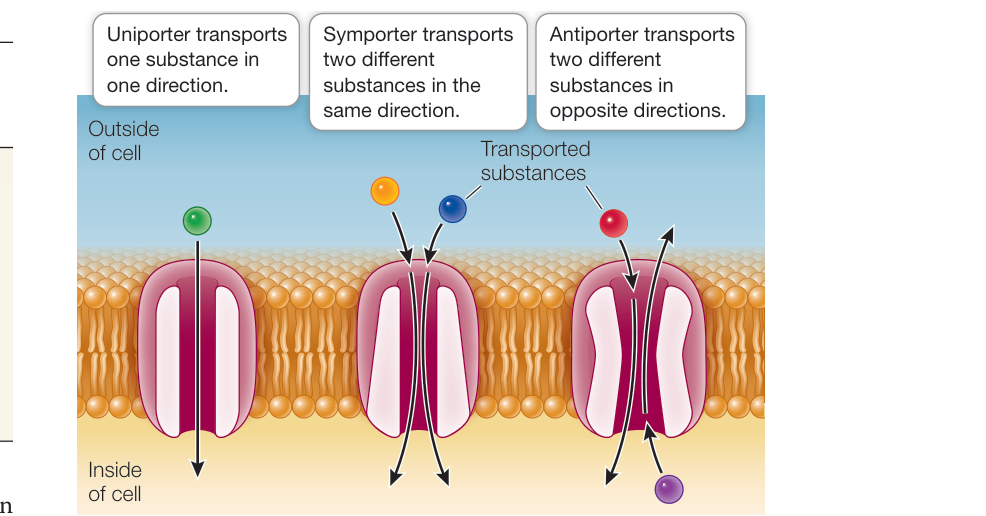
Figure 6.13 Three Types of Proteins for Active Transport Note that in each of the three cases, transport is directional. Symporters and antiporters are examples of coupled transporters. All three types of transporters are coupled to energy sources in order to move substances against their concentration gradients.
- A symporter moves two substances in the same direction. For example, a symporter in the cells that line the intestine must bind in addition to an amino acid in order to absorb amino acids from the intestine.
- An antiporter moves two substances in opposite directions, one into the cell (or organelle) and the other out of the cell (or organelle). For example, many cells have a sodium–potassium pump that moves out of the cell and into it.
Symporters and antiporters are also known as coupled transporters because they move two substances at once.
Different energy sources distinguish different active transport systems
There are two basic types of active transport:
- Primary active transport involves the direct hydrolysis of ATP, which provides the energy required for transport.
- Secondary active transport (co-transport) does not use ATP directly. Instead, its energy is supplied by an ion concentration gradient established by primary (ATP-driven) active transport.
In primary active transport, energy released by the hydrolysis of ATP drives the movement of specific ions against their concentration gradients. For example, we mentioned earlier that concentrations of potassium ions () inside a cell are often much higher than in the fluid bathing the cell. However, the concentration of sodium ions () is often much higher outside the cell. A protein in the cell membrane pumps out of the cell and into the cell against these concentration gradients, ensuring that the gradients are maintained (Figure 6.14). This sodium–potassium () pump is found in all animal cells. The pump is an integral membrane glycoprotein. It breaks down a molecule of ATP to ADP and a free phosphate ion () and uses the energy released to bring two ions into the cell and export three ions. The pump is thus an antiporter because it moves two substances in different directions.
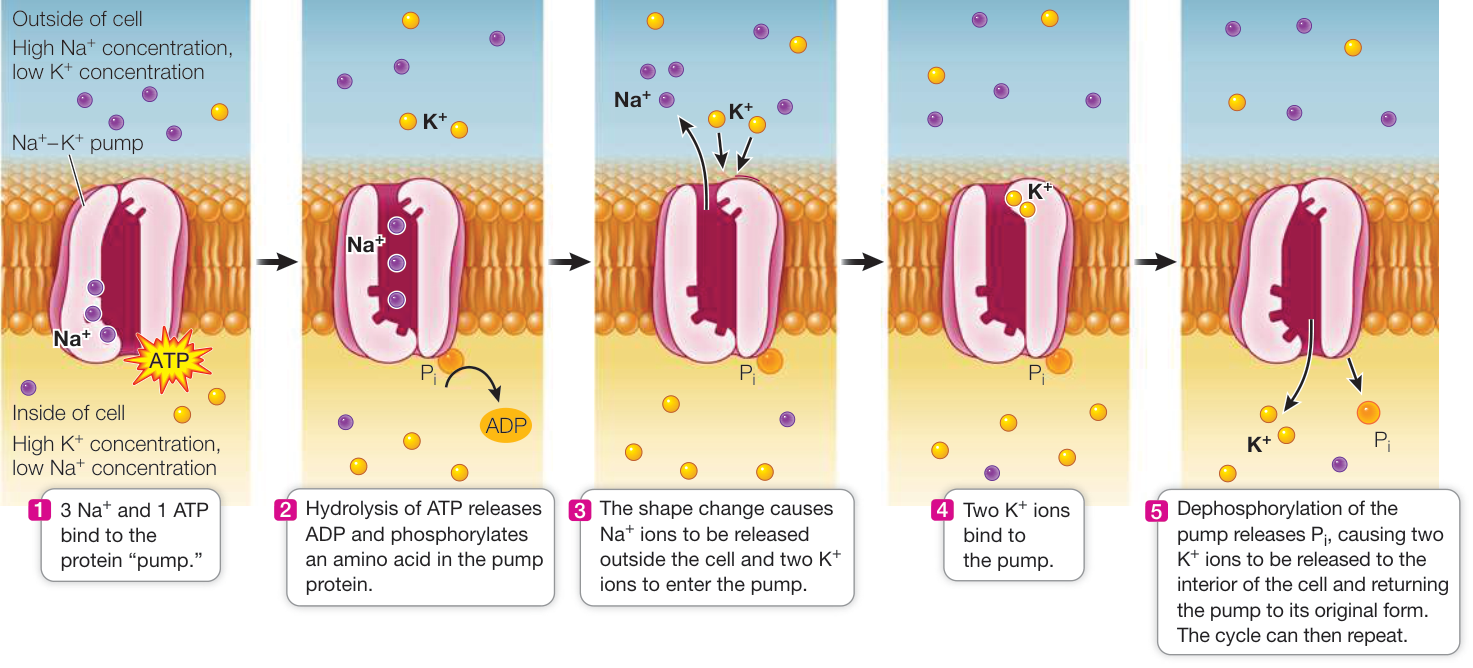
Figure 6.14 Primary Active Transport: The Sodium–Potassium Pump In active transport, energy is used to move a solute against its concentration gradient. Here, energy from ATP is used to move and against their concentration gradients.
Q: Ouabain is a natural product from a tree that has been used by traditional cultures in eastern Africa as a poison on arrows. Ouabain binds to a cell membrane protein. Tissues treated with ouabain have greatly increased intracellular . How does ouabain work?
An important drug used to treat a weak heart inhibits the – pump. Digitalis, purified from the foxglove plant, blocks the release of phosphate ions () from the protein (see Figure 6.14, step 5). This “freezes” the shape of the pump, and it can no longer bind . So accumulates inside heart cells. This ultimately leads to a greater force of contraction of heart muscle, which aids a patient whose heart is not pumping well.
In secondary active transport (co-transport), the movement of a substance against its concentration gradient is accomplished using energy “regained” by letting ions move across the membrane with their concentration gradients. Think of it this way: A large dam may prevent a river from flowing. The accumulated water represents energy, and when the water is allowed to flow past the dam, the energy it releases can be used to drive turbines that make electricity. It’s similar for secondary active transport. Consider, for example, the absorption of glucose into the bloodstream from the digestive tract. Once the – pump establishes a concentration gradient of sodium ions, the passive diffusion of some back into the cell can provide energy for the secondary active transport of glucose into the cell (Figure 6.15). Secondary active transport aids in the uptake of amino acids and sugars, which are essential raw materials for cell maintenance and growth. Both types of coupled transport proteins—symporters and antiporters—are used for secondary active transport.
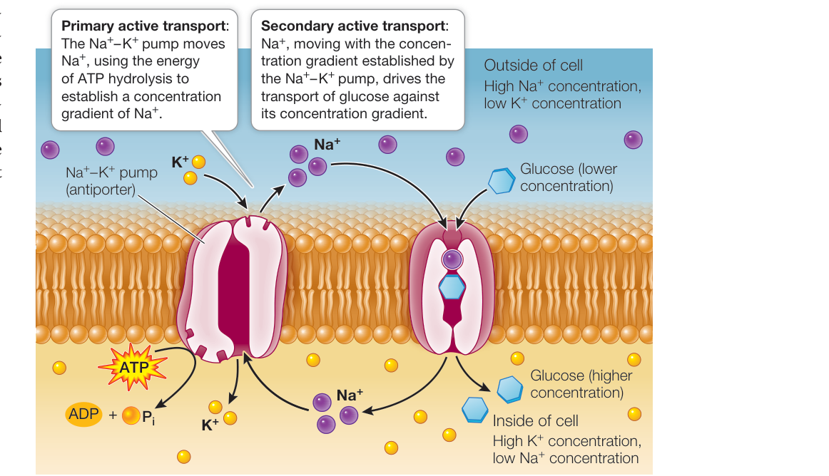
Figure 6.15 Secondary Active Transport (Co-Transport) The concentration gradient established by primary active transport by the sodium–potassium pump (left) powers the secondary active transport of glucose (right). A symporter protein couples the movement of glucose across the membrane against its concentration gradient to the passive movement of into the cell. Q: If the – pump is blocked by a drug, how will intracellular concentrations of and glucose be affected in cells with this co-transport mechanism?
Knowledge of co-transport was applied in developing oral rehydration therapy (ORT). Childhood diarrhea, often a result of infections, is a major problem throughout the world, especially in poor regions. As you know, diarrhea results in a lot of fluid loss from the intestine. The loss of water with dissolved ions can have severe consequences. When the mechanism of /glucose transport was described, physicians invented ORT, whereby water is consumed with dissolved and glucose. When this fluid reaches the intestine, cells in the intestinal lining use the – pump, which, along with the added in the oral fluid, keeps the level of high. The diffusion of into the intestinal cells is accompanied by the rapid diffusion of glucose. These solutes are accompanied by water, accomplishing rehydration. ORT costs less than 10 cents U.S. per liter and has saved millions of lives.
Recap and Assess
Active transport across a membrane is directional and requires an input of energy to move substances against their concentration gradients. Active transport allows a cell to maintain small molecules and ions at concentrations very different from those in the surrounding environment.
- Why is energy required for active transport?
- Why is the sodium–potassium ( – ) pump classified as an antiporter?
- What are the differences between primary active transport and secondary active transport?
- What are the similarities and differences between active transport and facilitated diffusion?
We have examined several passive and active ways in which ions and small molecules can enter and leave cells. But what about large molecules such as proteins? Many proteins are so large that they diffuse very slowly, and their bulk makes it difficult for them to pass through the phospholipid bilayer. A completely different mechanism is needed to move intact large molecules across membranes.
Large Molecules Enter and Leave a Cell through Vesicles
Learning Objectives
- 6.5.1 Compare and contrast the three types of endocytosis.
- 6.5.2 Describe the process of receptor-mediated endocytosis.
- 6.5.3 Provide examples of processes that involve endocytosis and/or exocytosis.
Macromolecules such as proteins, polysaccharides, and nucleic acids are too large and too charged or polar to pass through biological membranes. This is actually fortunate—think of the consequences if such molecules diffused out of cells: a red blood cell would not retain its hemoglobin! As you saw in Chapter 5, the development of a selectively permeable membrane was essential for the functioning of the first cells when life on Earth began. The interior of a cell can be maintained as a separate compartment with a different composition from that of the exterior environment, which is subject to abrupt changes. However, cells must sometimes take up or secrete (release to the external environment) intact large molecules. In Key Concept 5.3 we described phagocytosis, the mechanism by which solid particles can be brought into the cell by means of vesicles that pinch off from the cell membrane. The general terms for the mechanisms by which substances enter and leave the cell via membrane vesicles are endocytosis and exocytosis.

Animation 6.3 Endocytosis and Exocytosis
Macromolecules and particles enter the cell by endocytosis
Endocytosis is a general term for a group of processes that bring small molecules, macromolecules, large particles, and even small cells into the eukaryotic cell (Figure 6.16A). Generally, there are three types of endocytosis: phagocytosis, pinocytosis, and receptor-mediated endocytosis. In all three, the cell membrane invaginates (folds inward), forming a small pocket around materials from the environment. The pocket deepens, forming a vesicle. This vesicle separates from the cell membrane and migrates with its contents to the cell’s interior.
- In phagocytosis (“cellular eating”), part of the cell membrane engulfs large particles or even entire cells. Unicellular protists use phagocytosis for feeding, and some white blood cells use phagocytosis to defend the body by engulfing foreign cells and substances. The food vacuole or phagosome that forms usually fuses with a lysosome, where its contents are digested (see Figure 5.12).
- In pinocytosis (“cellular drinking”), vesicles also form. However, these vesicles are smaller, and the process operates to bring fluids and dissolved substances into the cell. Like phagocytosis, pinocytosis can be relatively nonspecific regarding what it brings into the cell. For example, pinocytosis occurs constantly in the endothelium, the single layer of cells that separates a tiny blood capillary from the surrounding tissue. Pinocytosis allows cells of the endothelium to rapidly acquire fluids and dissolved solutes from the blood.
- In receptor-mediated endocytosis, molecules at the cell surface recognize and trigger the uptake of specific materials.
Receptor-mediated endocytosis is highly specific
Receptor-mediated endocytosis is used by animal cells to capture specific macromolecules from the cell’s environment. This process depends on receptor proteins, which are proteins that can bind to specific molecules within the cell or in the cell’s external environment. In receptor-mediated endocytosis, the receptors are integral membrane proteins located at particular regions on the extracellular surface of the cell membrane. These membrane regions are called coated pits because they form slight depressions in
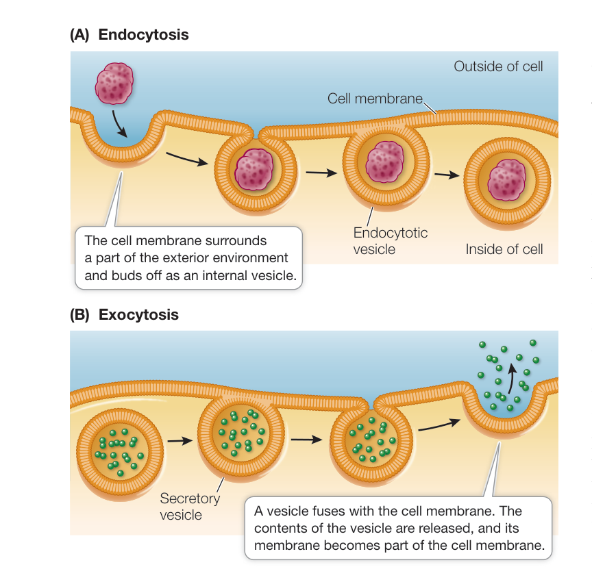
Figure 6.16 Endocytosis and Exocytosis Endocytosis (A) and exocytosis (B) are used by eukaryotic cells to take up and release fluids, large molecules, and particles. Smaller cells, such as invading bacteria, can be taken up by endocytosis.
Media Clip 6.1 An Amoeba Eats by Phagocytosis Life12e.com/mc6.1 the cell membrane and their cytoplasmic surfaces are coated by other proteins, such as clathrin. The uptake process is similar to that in phagocytosis.
When a receptor protein binds to its specific ligand (in this case, the macromolecule to be taken into the cell), its coated pit invaginates and forms a coated vesicle around the bound macromolecule. The clathrin molecules strengthen and stabilize the vesicle, which carries the macromolecule away from the cell membrane and into the cytoplasm (Figure 6.17). Once inside, the vesicle loses its clathrin coat and may fuse with a lysosome, where the engulfed material is digested (by the hydrolysis of polymers to monomers) and the products are released into the cytoplasm. Because of its specificity for particular macromolecules, receptor-mediated endocytosis is an efficient method of taking up substances that may exist at low concentrations in the cell’s environment.
An example of receptor-mediated endocytosis is the way in which cholesterol is taken up by many mammalian cells. Cholesterol and triglycerides, which have a low solubility in water, are packaged by liver cells into lipoprotein particles (see Figure 3.23C), which are released into the bloodstream. When a particular cell requires cholesterol, it produces specific low-density lipoprotein (LDL) receptors, which are inserted into the cell membrane in clathrin-coated pits. Binding of LDLs to the receptor proteins triggers the uptake of the LDLs via receptor-mediated endocytosis. Within the resulting vesicle, the LDL particles are freed from the receptors. The receptors segregate to a region that buds off and forms a new vesicle, which is recycled to the cell membrane. The freed LDL particles remain in the original vesicle, which fuses with a lysosome. There, the LDLs are digested and the cholesterol made available for cell use.
In healthy individuals, the liver takes up unused LDLs for recycling. People with the inherited disease familial hypercholesterolemia have a deficient LDL receptor in their livers. This prevents receptor-mediated endocytosis of LDLs, resulting in dangerously high levels of cholesterol in the blood. The cholesterol builds up in the arteries that nourish the heart and causes heart attacks. In extreme cases where only the deficient receptor is present, children and teenagers can have severe cardiovascular disease.
Exocytosis moves materials out of the cell
Exocytosis is the process by which materials packaged in vesicles are secreted from a cell when the vesicle membrane fuses with the cell membrane (Figure 6.16B). This fusing allows the vesicle contents to be released into the environment, and the vesicle membrane is smoothly incorporated into the cell membrane.
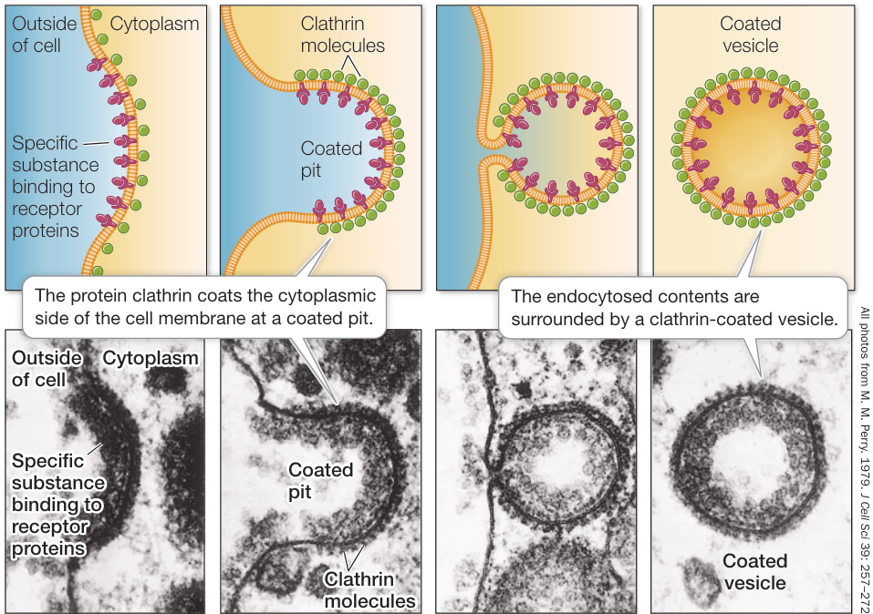
Figure 6.17 Receptor-Mediated Endocytosis The receptor proteins in a coated pit bind specific macromolecules, which are then carried into the cell by a coated vesicle.
Table 6.2 Endocytosis and Exocytosis
| Type of process | Example |
|---|---|
| ENDOCYTOSIS | |
| Receptor-mediated endocytosis | Specific uptake of large molecules (e.g., LDL) |
| Pinocytosis | Nonspecific uptake of extracellular fluid (e.g., fluids and dissolved substances from blood) |
| Phagocytosis | Nonspecific uptake of large undissolved particles (e.g., invading bacteria by cells of the immune system) |
| EXOCYTOSIS | |
| Release of large molecules | Vesicle fusion with cell membrane (e.g., digestive enzymes in the pancreas) |
| Release of small molecules | Vesicle fusion with cell membrane (e.g., neurotransmitters at the synapse) |
In another form of exocytosis, the vesicle touches the cell membrane and a pore forms, releasing the vesicle’s contents. There is no membrane fusion in this process, termed “kiss and run.” We saw an example of exocytosis in describing sweat glands at the start of the chapter.
Table 6.2 summarizes examples of endocytosis and exocytosis.
Recap and Assess
Endocytosis and exocytosis are the processes by which large particles and molecules are transported into and out of the cell. Endocytosis may be mediated by a receptor protein in the cell membrane.
- What are the differences between phagocytosis and pinocytosis?
- Describe receptor-mediated endocytosis and give an example of a process involving this phenomenon.
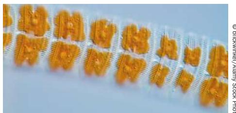
- Key Concept 25.2 describes the diatoms, which are protists that have complex glassy structures in their cell walls (see Figure 25.8 and the photo below of eight diatom cells inside their ornate cell walls). These structures form within the Golgi apparatus. How do they reach the cell wall without having to pass through a membrane?
© blickwinkel/Alanny Stock Photo
Investigating Life: QA What is the importance of aquaporin membrane channels?
There are ten genes encoding aquaporins in humans, each channel of the “plumbing system” having its particular location in the body. Genetic deficiency resulting in a lack of one of the aquaporins can have significant consequences. For example, one of the aquaporins occurs in the part of the kidney where water is removed from the developing urine and returned to the blood. When this aquaporin is absent, there is more water in the urine and the person urinates more and more often; bedwetting is a common result. The existence of aquaporins in plants was indicated when plant roots failed to take up water from the soil when they were exposed to solutions of mercuric chloride, a poison that binds to and inactivates membrane proteins. The proteins in the root cell membranes were identified as aquaporins; others have been found in the membrane that encloses the large central vacuole. Root aquaporins form channels for water on its way from the soil to the interior tissues, from which it is transported to the rest of the plant. In the vacuole, aquaporin channels are important in the organelle swelling, resulting in turgor pressure, which is important in plant organ structure and plant cell expansion. All of these aquaporins, and those of other organisms, share a common structure that spans the cell membrane and has a channel through which water molecules pass in single file.
Future directions
The water relations of plants, especially crop plants, are under intense study. Heavy use of chemical fertilizers that dissolve in water can lead to a hypertonic environment in the soil relative to the interior of plant root cells. Water tends to leave plant roots by osmosis through aquaporins. However, some plants can adapt to this by gating the aquaporin channel, shutting it off to preserve cellular water. Similarly, some plants can shut off their root aquaporins to preserve water during periods of drought, which are increasing in some regions due to climate change. Understanding these mechanisms and applying them to other plants can be important in future food production. Efforts are underway to insert aquaporins into synthetic membranes used for industrial applications. Because aquaporins allow only water and low concentrations of ions to pass through, such membranes could be used to purify contaminated fresh water, or to desalinate water, which as you know has very high ion concentrations. Aquaporins are expressed in skin, where they play a role in keeping the skin moist. This is also a goal of some cosmetics (moisturizers) and has led to a search for, and patenting of, small molecules that stimulate aquaporins. These molecules are components of some widely marketed skin creams.


© Scott Barbour/Getty Images
You should be able to relate each summary to the adjacent figures. If you go to this visual summary in Achieve, you can follow links to figures, animations, activities, and simulations that will help you consolidate the material.
Visual Summary
Biological Membranes Are Lipid-Protein Bilayers
- ■ Biological membranes consist of lipids, proteins, and carbohydrates. The fluid mosaic model of membrane structure describes a phospholipid bilayer in which proteins can move within the plane of the membrane.
- ■ The properties of membranes depend on the nonpolar lipids as well as integral membrane proteins, peripheral membrane proteins, and anchored membrane proteins inserted in or associated with the bilayer.
Questions
- For an integral membrane protein, which class of amino acids might you expect to find on the portion of the protein embedded in the membrane (see Table 3.2)? Give examples.
- What will happen if a cell is incubated in a nonpolar detergent?
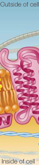
Figure 6.1
The Cell Membrane Is Important in Cell Adhesion and Recognition
- ■ In order for cells to assemble into tissues, they must recognize and adhere to one another. Cell recognition and cell adhesion depend on membrane-associated proteins and carbohydrates.
- ■ Cell junctions connect adjacent cells. In some animal cells, tight junctions prevent the passage of molecules through the intercellular spaces between cells, and they restrict the movement of membrane proteins over the cell surface. Desmosomes cause cells to adhere firmly to one another. Gap junctions provide channels for communication between adjacent cells.
Questions
- If a sponge is disaggregated and the cells are allowed to reaggregate in the presence of an enzyme that digests exposed cell membrane proteins, what will happen?
- Matrix metalloproteases are molecules made by cancer cells that hydrolyze collagen and proteoglycans in the extracellular matrix. Why is it significant that these molecules are made by cancer cells that migrate to other locations in the body?
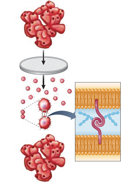
Figure 6.5
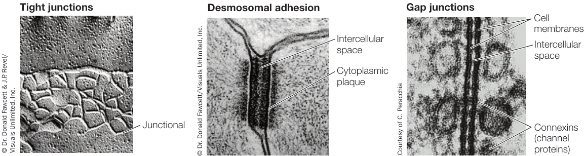
Figure 6.6
Substances Can Cross Membranes by Passive Processes
and Simulation 6.2
- ■ Membranes exhibit selective permeability, regulating which substances pass through them.
- ■ Diffusion is the movement of a solute from a region of higher concentration to a region of lower concentration.
- ■ In osmosis, water diffuses across a membrane from a region of higher water concentration to a region of lower water concentration. In an isotonic environment, total solute concentrations on both sides of the cell membrane are equal. If the solution surrounding a cell is hypotonic to the cell interior, more water enters the cell than leaves it, causing it to swell. In a hypertonic solution, more water leaves the cell than enters it, causing it to shrivel.
- ■ A substance can diffuse passively across a membrane by either simple diffusion or facilitated diffusion, via a channel protein or a carrier protein.
Questions
- Proteins and ions do not readily diffuse across the cell membrane. For each case, what is the major property of the chemical that limits its diffusion?
- The total concentration of substances dissolved (solutes) in red blood cells is 0.3 M. What is the total concentration of solutes in the blood plasma surrounding the red blood cells? If this concentration is reduced by 33%, what will happen to the cells?
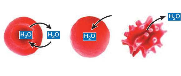
Figure 6.10
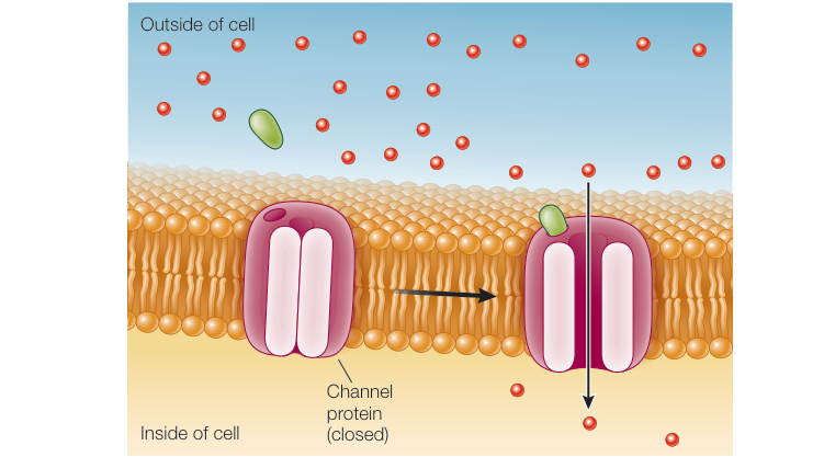
Figure 6.11
Active Transport across Membranes Requires Energy
- ■ Active transport requires the use of chemical-bond energy to move substances across membranes against their concentration or electrical gradients.
- ■ In primary active transport, energy from the hydrolysis of ATP is used to move ions into or out of cells. The sodium-potassium ( - ) pump is an example.
- ■ Secondary active transport (co-transport) couples the passive movement of one substance down its concentration gradient to the movement of another substance against its concentration gradient through active transport.
Questions
- The concentration of calcium ions () in the endoplasmic reticulum of an unfertilized egg cell of the marine brown alga Fucus is much higher than that in the surrounding cytosol. When the egg is fertilized, is released rapidly into the cytosol. What two types of membrane transport are involved?
- The imbalance of and concentrations, with the former higher and the latter lower outside than inside cells, is important to the functioning of the nervous system. Why is the nervous system dependent on a supply of energy via ATP?
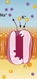
Figure 6.14
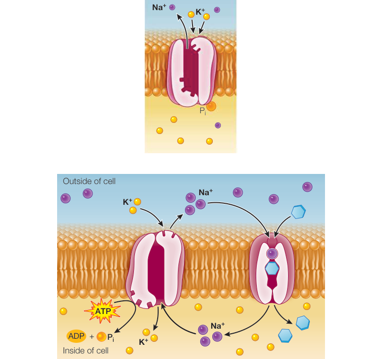
Figure 6.15
Large Molecules Enter and Leave a Cell through Vesicles
- ■ Endocytosis is the transport of small molecules, macromolecules, large particles, and small cells into eukaryotic cells via invagination of the cell membrane and the formation of vesicles.
- ■ In exocytosis, materials in vesicles are secreted from the cell when the vesicles fuse with the cell membrane.
- ■ In receptor-mediated endocytosis, a specific receptor protein on the cell membrane binds to a particular macromolecule.
Questions
- Why do the fatty acid and protein compositions of lipids differ in endocytotic and exocytotic vesicles?
- Developing red blood cells require iron to make hemoglobin. They take up iron carried by the protein transferrin via receptor-mediated endocytosis. If you mapped transferrin receptors on the cell membrane, would they be evenly distributed over the cell surface?
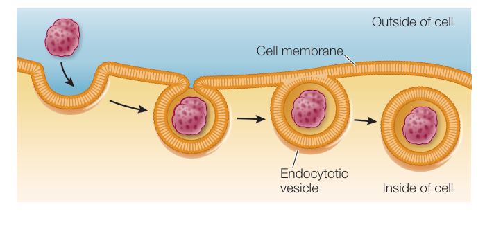
Figure 6.16
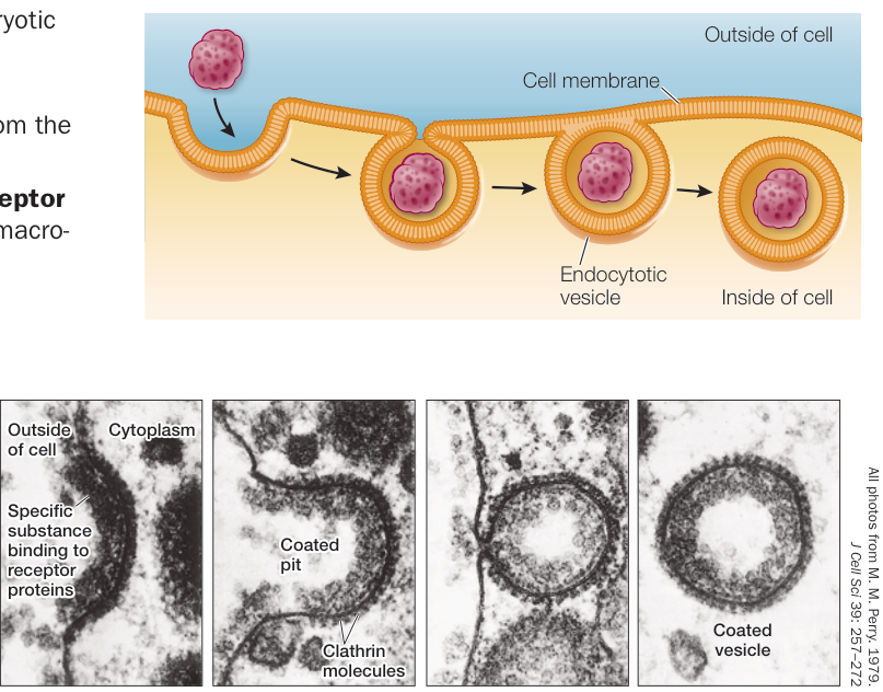
Figure 6.17