Chapter 5Cells: The Working Units of Life
PART TWO
Cells
Key Concepts
5.1 Cells Are the Fundamental Units of Life
5.2 Prokaryotic Cells Are the Simplest Cells
5.3 Eukaryotic Cells Contain Organelles
5.4 Extracellular Materials Provide Structure
5.5 Eukaryotic Cells Evolved in Several Steps
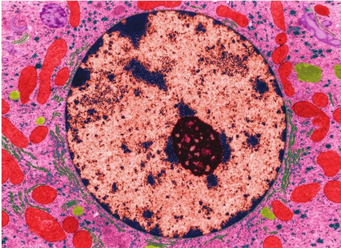
Compartmentalization is a prominent feature of this liver cell, which shows the prominent nucleus at the center and energy-producing mitochondria (red). Also visible are ribosomes (purple dots) which synthesize proteins. The cytosol (pink) is where much of the chemistry of the cell occurs.
Investigating Life: Natural Sunscreen
Sunlight plays a key role in life, providing the energy that is transformed into stored chemical-bond energy in green plants through the process of photosynthesis. When we eat food, the energy stored in its chemical bonds ultimately came from the Sun. But sunlight has a nasty side effect: part of it, the highly energetic ultraviolet light, damages the genetic material, DNA. Even without knowing this chemistry, some people take measures to minimize exposure to excess sunlight, either by staying in the shade or using chemical sunscreens. But there is also a biological mechanism that has evolved to reduce the Sun’s damage to organisms. In most animals, dark brown or black chemical pigments called melanins are made inside cells, the basic units of life. Melanins absorb ultraviolet light, thereby protecting DNA.
In humans, melanin is made in certain skin cells called melanocytes. Humans first evolved in Africa, and the intense sunlight near the equator acted as an evolutionary selective agent for the development of large numbers of melanocytes. As some of these dark-skinned people migrated to more northern regions where the Sun is not as intense, the selection pressure for melanocytes was reduced, and over millennia, genetic changes leading to fewer melanocytes persisted in offspring, resulting in lighter skin. Exposure to intense sunlight in these people results in the production of more melanin, the familiar tanning response.
Melanocytes are specialized cells. You find them in the skin and the eye but not typically in internal organs. We will return to the process of how different cells have different functions in later chapters. But even inside the melanocyte, there is specialization. Melanin is made inside a special cell compartment called the melanosome. These compartments can be expelled from melanocytes and transferred to other skin cells, so they too become loaded with pigment. This occurs spontaneously in dark-skinned people, while in paler people it is stimulated by exposure to intense sunlight.
The observation that the chemistry of life takes place within cells, and in some cases within specialized compartments in them, is a key concept of biological science.
QA Are pigments compartmentalized in all types of cells? (Find the answer on page 109.)
Cells Are the Fundamental Units of Life
Learning Objectives
- 5.1.1 List the three tenets of the original cell theory.
- 5.1.2 Explain how surface area-to-volume ratio constrains cell size.
- 5.1.3 Explain the importance of membranes in cell function.
Chapter 1 introduced some of the characteristics of life: chemical complexity, growth and reproduction, the ability to refashion substances from the environment, and the ability to move specific substances into and out of the organism. Cells have all of these characteristics. Just as atoms are the building blocks of chemistry, cells are the building blocks of life.
What is the cell theory?
The cell theory is an important unifying principle of biology. There are three components of the cell theory:
- Cells are the fundamental units of life.
- All living organisms are composed of cells.
- All cells come from preexisting cells.
To the original cell theory, first stated in 1838, should be added:
- Modern cells evolved from a common ancestor.
Cells contain water and other small and large molecules, which we examined in Chapters 2–4. Each cell contains at least 10,000 different types of molecules, most of them present in many copies. Cells use these molecules to transform matter and energy, to respond to their environments, and to reproduce. The cell theory has three important implications:
- Studying cell biology is in some sense the same as studying life. The principles that underlie the functions of the single cell of a bacterium are similar to those governing the approximately 37 trillion human cells of your body.
- Life is continuous. All the cells in your body came from a single cell, a fertilized egg (zygote). That zygote came from the fusion of two cells, a sperm and an egg, from your biological parents. The cells of your parents’ bodies were all derived from their parents, and so on back through generations and evolution to the initial cell.
- The origin of life on Earth was marked by the origin of the first cells (see Chapter 4).
Even the largest creatures on Earth are composed of cells, but the cells themselves are usually too small for the naked eye to see. Why are cells so small?
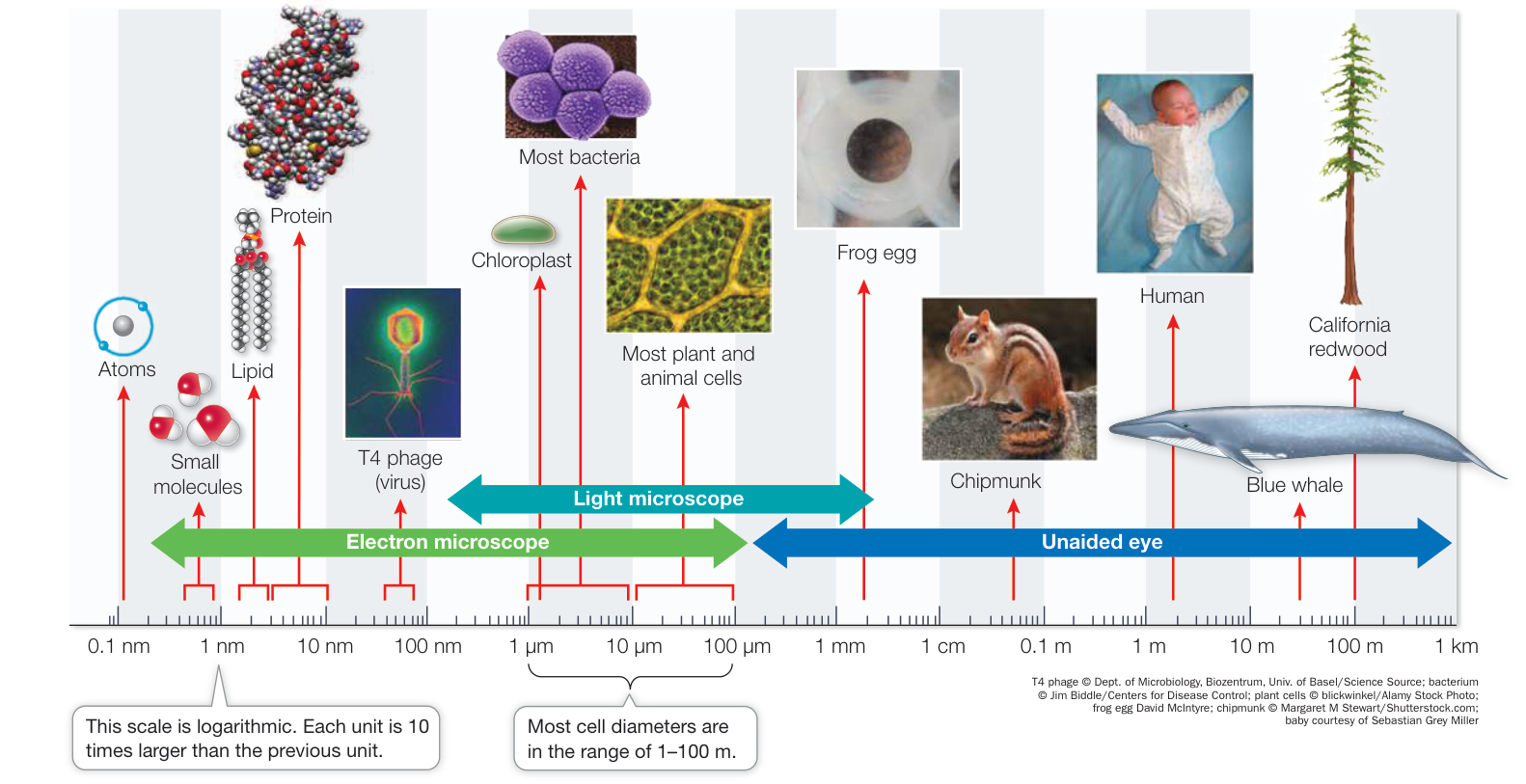
Figure 5.1 The Scale of Life This logarithmic scale shows the relative sizes of molecules, cells, and multicellular organisms. Protein data from PDB 1IVM. T. Obita et al. 2003. Cell Mol Life Sci 60: 176–184)
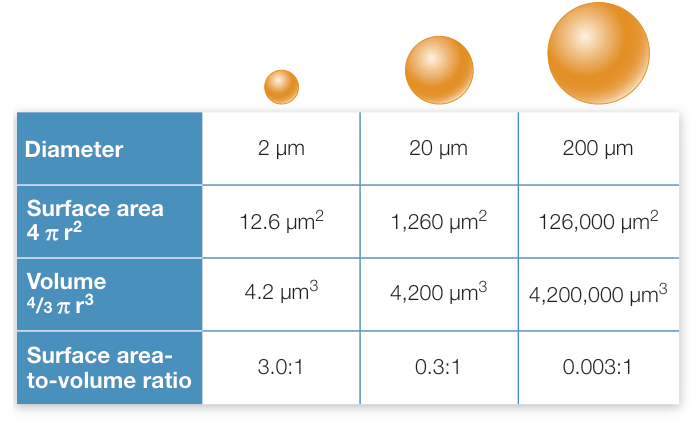
Figure 5.2 Why Cells Are Small As an object grows larger, its volume increases more rapidly than its surface area. Cells must maintain a large surface area-to-volume ratio in order to function. This explains why large organisms are composed of many small cells rather than a few huge ones. Q: If a cell with a diameter of 200 has many folds in its cell membrane and increases its surface area tenfold, what will its surface area-to-volume be?
Cell size is limited by the surface area-to-volume ratio
In 1665 Robert Hooke estimated that in 1 square inch of cork, which he examined under his magnifying lens, there were 1,259,712,000 cells! The diameters of typical cells range from about 1 to 100 micrometers (). There are some exceptions: the eggs of fishes and amphibians (several millimeters in diameter) and of birds and other reptiles (several centimeters in diameter) are large single cells that store nutrients for development. Some individual cells of several types of algae and bacteria are large enough to be seen with the unaided eye (Figure 5.1).
Small cell size is a practical necessity arising from the change in the surface area-to-volume ratio of any object as it increases in size. As an object increases in volume, its surface area also increases, but not at the same rate (Figure 5.2). This phenomenon has great biological significance. To appreciate this point, let’s assume that the amount of chemical activity carried out by a cell is proportional to its volume. The surface area of the cell determines the amount of substances that can enter it from the outside environment, and the amount of waste products that can exit to the environment.
As a living cell grows larger, its chemical activities, and thus its need for resources and its rate of waste production, usually increase faster than its surface area, assuming the shape remains the same. The surface area, being two-dimensional, increases in proportion to the square of the radius, whereas the volume, being three-dimensional, increases much more—in proportion to the cube of the radius. This explains why large organisms must consist of many small cells: cells must be small in volume in order to maintain a large enough surface area-to-volume ratio and an ideal internal volume. The large surface area represented by the many small cells of a multicellular organism allows the organism to carry out the many different functions required for survival.
While surface area-to-volume ratio limits cell size, some cells change their shape as they become larger, allowing them to maintain a sufficiently high ratio. Shape change can involve increased infoldings of the cell membrane, or size increases that occur in only one dimension. For example, nerve cells in humans, which can be a meter long, have very narrow projections, keeping the surface area-to-volume ratio high. Some cells get around the surface area-to-volume challenge by simply increasing the rate at which substances move across their boundary into and out of the cells.
Microscopes reveal the features of cells
Most cells are too small to be seen with the unaided eye. Microscopes allow cells and details within them to be seen. The property that allows detail to be seen is called resolution. Formally defined, resolution is the minimum distance two objects can be apart and still be seen as two objects. Resolution for the human eye is about 0.2 mm (200 ).
There are two basic types of microscopes—light microscopes and electron microscopes—that use different forms of radiation (see Figure 5.4). While the resolution is better in electron microscopy, only dead cells are visualized because they must be prepared in a vacuum. Light microscopes, by contrast, can be used to visualize living cells (for example, by phase-contrast microscopy; see Figure 5.4).
Before we look at cell structure, it is useful to consider the many uses of microscopy. Here is an example: an entire branch of medicine, pathology, makes use of many different methods of microscopy to aid in the analysis of cells and the diagnosis of diseases. For instance, a surgeon might remove from a body some tissue suspected of being cancerous. The pathologist might:
- examine the tissue quickly by phase-contrast microscopy to determine the size, shape, and spread of the cells;
- stain the tissue with a general dye and examine it by bright-field microscopy to bring out features such as the shapes of the nuclei, or cell division characteristics (Figure 5.3);
- examine the tissue under the transmission electron microscope to observe internal structures such as the mitochondria or the chromatin (these are described in Key Concept 5.3);
- stain the tissue with a specific dye and examine it by microscopy for the presence of proteins that are diagnostic of particular cancers. The results can influence the choice of therapy.
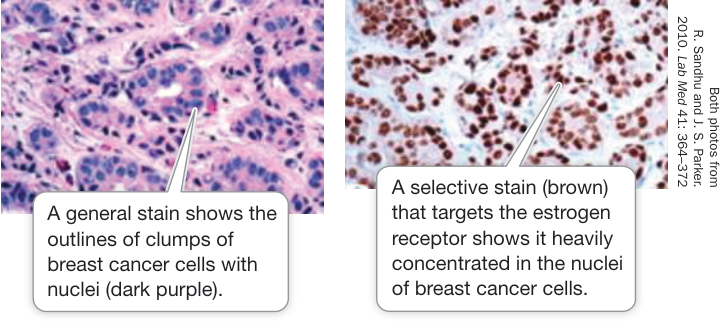
Figure 5.3 Staining Cells for Microscopy Stains can reveal cell and tissue structure, as well as composition.
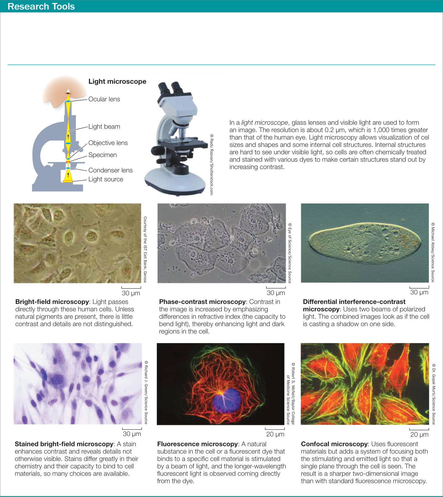
Figure 5.4 Looking at Cells The six images on this page show some techniques used in light microscopy. The three images on the following page were created using electron microscopes. All of these images are of a particular type of cultured cell known as HeLa cells. Note that the images in most cases are flat, two-dimensional views. As you look at images of cells, keep in mind that they are three-dimensional structures.
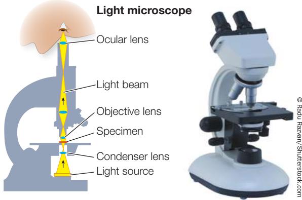
Most cells are much smaller than 200 µm and thus are invisible to the human eye. Microscopes magnify and increase resolution so that cells and their internal structures can be seen clearly (Figure 5.4).
The cell membrane forms an outer boundary of every cell
While light microscopy shows many types of cells and even some structure inside them, the cell membrane is best observed with
Research Tools (continued)
Transmission electron microscope
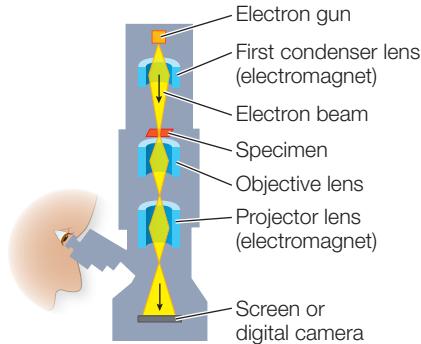
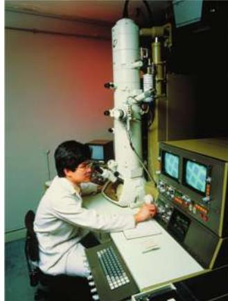
In an electron microscope, electromagnets are used to focus an electron beam, much as a light microscope uses glass lenses to focus a beam of light. Since we cannot see electrons, the electron microscope directs them through a vacuum at a fluorescent screen or digital camera to create a visible image. The resolution of electron microscopes is about 2 nm, which is about 100,000 times greater than that of the human eye. This resolution permits the details of many subcellular structures to be distinguished.
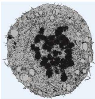
© Dr. Gopal Murt/Visuals Unimited, Inc.
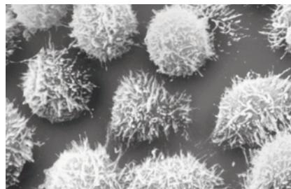
© Keith R. Porter/Science Source
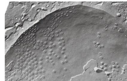
© D. W. Fawcett/Science Source
Transmission electron microscopy (TEM):
A beam of electrons is focused on the object by magnets. Objects appear darker if they absorb the electrons. If the electrons pass through they are detected on a fluorescent screen.
Scanning electron microscopy (SEM):
Directs electrons to the surface of the sample, where they cause other electrons to be emitted. These electrons are viewed on a screen. The three-dimensional surface of the sample can be visualized.
Freeze-fracture microscopy:
Cells are frozen and then a knife is used to crack them open. The crack often passes through the interior of cell and internal membranes. The “bumps” that appear are usually large proteins or aggregates embedded in the interior of the membrane.
an electron microscope. This very thin structure forms the outer boundary of the cytoplasm of every cell, and it has similar thickness and molecular structure in all cells. You will learn about membranes in more detail in Chapter 6. For now, keep in mind that (with the exception of some species of archaea discussed in Chapter 24) the cell membrane consists of a phospholipid bilayer (see Key Concept 3.4), and that a variety of proteins are embedded within the bilayer.
The membrane has several important roles:
The cell membrane acts as a selectively permeable barrier, preventing some substances from crossing it while permitting other substances to enter and leave the cell.
By regulating transport across it, the cell membrane allows the cell to maintain a more or less constant internal environment. The maintenance of a constant internal environment (known as homeostasis) is a key characteristic of life and will be discussed in detail in Chapter 38.
As the cell’s boundary with the outside environment, the cell membrane is important in communicating with adjacent cells and receiving signals from the environment. You will learn about this in Chapter 7.
The cell membrane often has proteins protruding from it that are responsible for binding and adhering to adjacent cells. Thus the cell membrane plays an important structural role and contributes to cell shape.
Everything inside the cell, except for the nucleus if the cell has one, is termed the cytoplasm. The cytosol is the fluid cytoplasm not contained inside another compartment.
Cells may be classified as either prokaryotic or eukaryotic
As you saw in Key Concept 1.1, biologists classify all living things into three domains: Archaea, Bacteria, and Eukarya. The organisms in Archaea and Bacteria are collectively called prokaryotes, and they have in common a prokaryotic cell organization. A prokaryotic cell typically does not have membrane-enclosed internal compartments; in particular, it does not have a nucleus. The first cells were probably similar in organization to those of modern prokaryotes.
Eukaryotic cell organization is found in members of the domain Eukarya (eukaryotes), which includes the protists, plants, fungi, and animals. In contrast to prokaryotic cells, eukaryotic cells contain membrane-enclosed compartments called organelles. The most noticeable organelle is the cell nucleus, where most of the cell’s DNA is located and where gene expression begins (Figure 5.5).
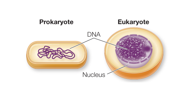
Figure 5.5 Prokaryotic versus Eukaryotic The presence of a cell nucleus and other membrane-bound internal compartments, or organelles, distinguishes eukaryotic cells from prokaryotic cells. Q: Which two components are present in both eukaryotic and prokaryotic cells?
Just as a cell is an enclosed compartment, separating its contents from the surrounding environment, so each organelle provides a compartment that separates molecules and biochemical reactions from the rest of the cell. This “division of labor” provides possibilities for regulation and efficiency that were important in the evolution of complex organisms and helps explain the complexity of eukaryotic cells relative to prokaryotic cells.
Recap and Assess
The cell theory is a unifying principle of biology. Cell size is limited in order to maintain a high surface area-to-volume ratio. Both prokaryotic and eukaryotic cells are enclosed within a cell membrane, but prokaryotic cells lack the membrane-enclosed organelles that are found in eukaryotic cells.
- How does cell biology embody all the principles of life?
- What is the importance of the cell membrane and the membranes that surround organelles?
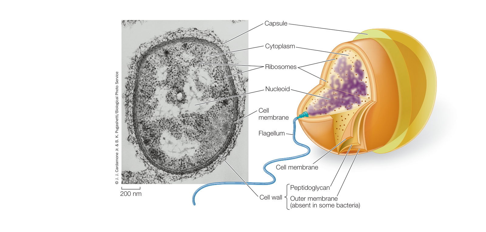
Figure 5.6 A Prokaryotic Cell This electron micrograph and drawing of the bacterium Pseudomonas aeruginosa illustrate the typical structures shared by all prokaryotic cells. In addition, this bacterium has a protective outer membrane that is not present in all prokaryotes. The flagellum and capsule are also structures found in some, but not all, prokaryotic cells.
As we mentioned in this key concept, there are two structural themes in cell architecture: prokaryotic and eukaryotic. We will now turn to the organization of prokaryotic cells.
Prokaryotic Cells Are the Simplest Cells
Learning Objectives
- 5.2.1 Describe the major features of a prokaryotic cell.
- 5.2.2 Identify specialized features found in some prokaryotic cells.
With diameters or lengths in the range of 1–10 µm, prokaryotic cells are generally smaller than eukaryotic cells, whose diameters are usually in the range of 10–100 µm. Each individual prokaryote is a single cell, but many types of prokaryotes form chains or small clusters of cells, and some occur in large clusters containing hundreds of cells. In this key concept we will first consider the features shared by cells in the domains Bacteria and Archaea. Then we will examine structural features that are found in some, but not all, prokaryotes.
Prokaryotic cells share certain features
All prokaryotic cells have the same basic structure (Figure 5.6):
- The cell membrane encloses the cell, regulating the traffic of materials into and out of the cell, and separating its interior from the external environment.
- The nucleoid is a region in the cell where the DNA is located. As we described in Key Concept 4.1, DNA is the hereditary material that controls cell growth, maintenance, and reproduction.
- The rest of the material enclosed in the cell membrane is the cytoplasm.
- Ribosomes are complexes of RNA and proteins in the cytoplasm that are about 25 nanometers (nm) in diameter. They can be visualized only with the electron microscope. They are the sites of protein synthesis, where information coded for in nucleic acids directs the sequential linking of amino acids to form proteins.
Connect the Concepts In terms of sheer numbers, prokaryotes are the most successful organisms on Earth. As we examine prokaryotic cells in this key concept, bear in mind that there are vast numbers of prokaryotic species, and that bacteria and archaea are distinguished in numerous ways. These differences, and the vast diversity of organisms in these two domains, are the subject of Chapter 24.
The cytoplasm is not static. Rather, the substances in this environment are in constant motion. For example, a typical protein moves around the entire cell within a minute, and it collides with many other molecules along the way. This motion helps ensure that biochemical reactions proceed at rates sufficient to meet the needs of the cell. Although they are structurally less complex than eukaryotic cells, prokaryotic cells are functionally complex, carrying out thousands of biochemical reactions.
Specialized features are found in some prokaryotes
As they evolved, some prokaryotes developed specialized structures that gave them a selective advantage: cells with these structures were better able to survive and reproduce in particular environments than cells lacking them.
CELL WALL Most prokaryotic cells have a cell wall located outside the cell membrane. The rigid wall supports the cell and determines its shape. The cell walls of most bacteria, but not archaea, contain peptidoglycan, a polymer of amino sugars that is linked at regular intervals to short peptides. Cross-linking among these peptides results in a single giant molecule around the entire cell. In some bacteria, another layer, the outer membrane (a polysaccharide-rich phospholipid membrane), encloses the peptidoglycan layer (see Figure 5.6). Unlike the cell membrane, this outer membrane is not a major barrier to the movement of molecules across it. Enclosing the cell wall in some bacteria is a slimy layer composed mostly of polysaccharides and referred to as a capsule. In some cases these capsules protect the bacteria from attack by white blood cells in the animals they infect. Capsules also help keep the cells from drying out, and sometimes they help bacteria attach to other cells.
INTERNAL MEMBRANES Some groups of bacteria—including the cyanobacteria—carry out photosynthesis: they use energy from the Sun to convert carbon dioxide and water into carbohydrates. These bacteria have an internal membrane system that contains molecules needed for photosynthesis. The development of photosynthesis, which requires membranes, was an important event in the early evolution of life on Earth. Other prokaryotes have internal membrane folds that are attached to the cell membrane. These folds may function in cell division or in various energy-releasing reactions.
FLAGELLA AND PILI Some prokaryotes swim by using appendages called flagella, which sometimes look like tiny corkscrews (Figure 5.7A). In bacteria, the filament of the flagellum is made of a protein called flagellin. (As you will see in Key Concept 5.3, the flagella of eukaryotes are quite different in structure, but similar in function.) A complex motor protein spins the bacterial flagellum on its axis like a propeller, driving the cell along. The motor protein is anchored to the cell membrane and, in some bacteria, to the outer membrane of the cell wall (Figure 5.7B). We know that the flagella cause the motion of cells because if they are removed, the cells do not move.
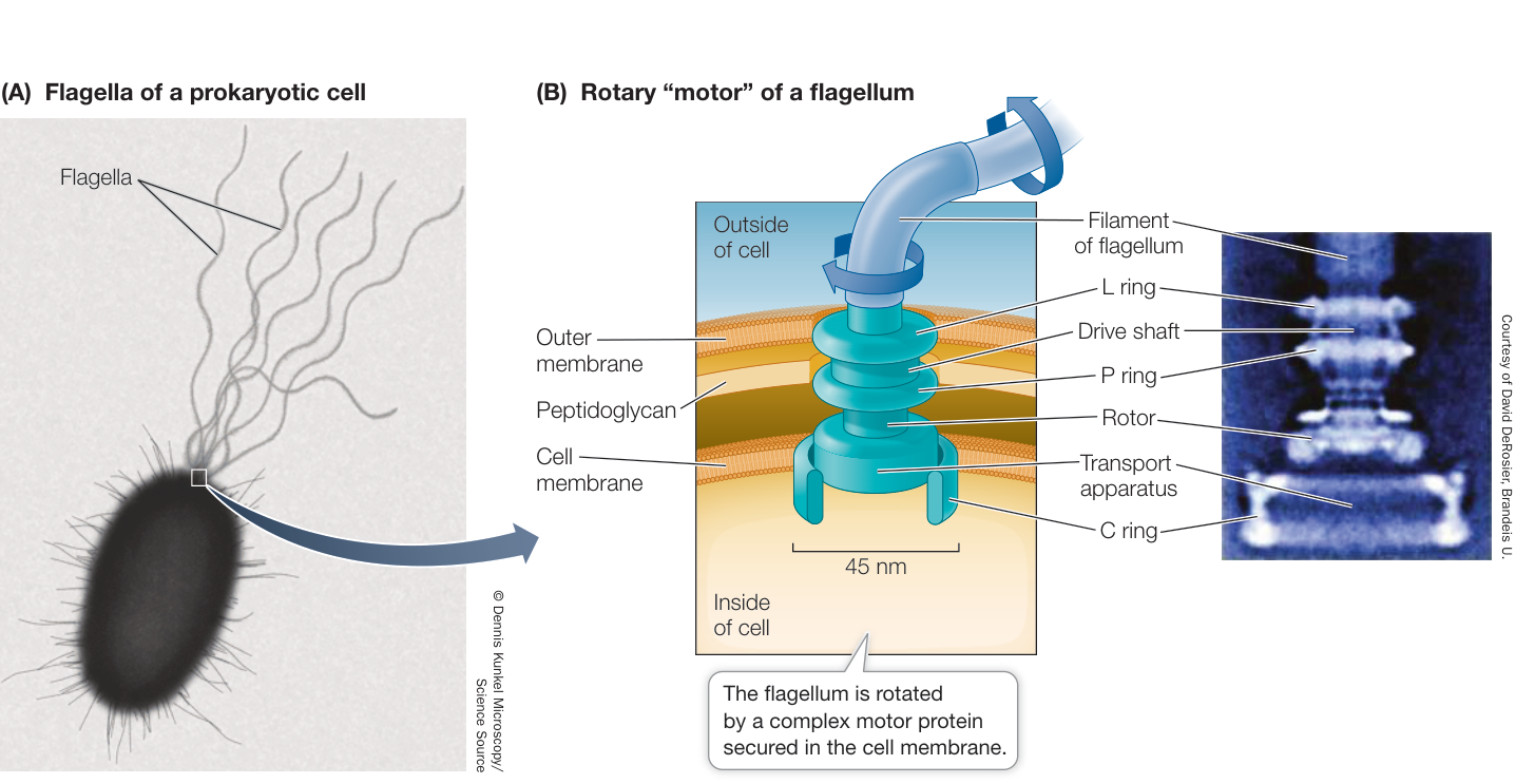
Figure 5.7 Prokaryotic Flagella - (A) Flagella contribute to the movement and adhesion of prokaryotic cells. - (B) Complex protein ring structures anchored in the cell membrane form a motor unit that rotates the flagellum and propels the cell.
Pili are structures made of protein that project from the surfaces of some types of bacterial cells. These hairlike structures are shorter than flagella and are used for adherence. Conjugative pili (sex pili) help bacteria join to one another to exchange genetic material. Fimbriae are composed of the same proteins as pili but are shorter, and help cells adhere to surfaces such as animal cells, for food and protection.
CYTOSKELETON The cytoskeleton is the collective name for protein filaments that play roles in cell division, cell movement, and in maintaining the shapes of cells. Prokaryotes have filamentous proteins that are homologous (similar in amino acid sequence) to the proteins that make up the cytoskeleton in eukaryotic cells. These proteins include FtsZ, which forms a contractile ring in the center of the cell when it divides, and MreB, which forms filaments just inside the cell membrane and maintains the shape of the cell.
Recap and Assess
Prokaryotic cells share basic features, including the cell membrane and a region called the nucleoid. The rest of the material enclosed by the cell membrane, including the ribosomes, is called the cytoplasm. Other features, such as cell walls, internal membranes, and flagella, are present in some but not all prokaryotes.
- What are the structure and function of the following specialized prokaryotic cell features: cell wall, capsule, and flagellum?
- What are the chemical compositions of the nucleoid and cytoplasm in a prokaryotic cell?
As we have already noted, the prokaryotic cell is one of two types of cells recognized in cell biology. The other is the eukaryotic cell. Eukaryotic cells are more structurally and functionally complex than prokaryotic cells.
Eukaryotic Cells Contain Organelles
Learning Objectives
- 5.3.1 Discuss the importance of compartmentalization in eukaryotic cells.
- 5.3.2 Compare the sizes, structures, typical numbers, and roles of subcellular structures.
- 5.3.3 Trace the path of a molecule as it moves between organelles in a eukaryotic cell.
Eukaryotic cells generally are larger than prokaryotes, and as noted in Figure 5.5, the distinguishing feature of eukaryotic cells is the presence of membrane-bound subcellular compartments called organelles. As you will see throughout this book, there are many types of eukaryotic cells in terms of structure and function. Here we will emphasize the features they have in common.

Activity 5.3 Eukaryotic Cell Tour
Compartmentalization is important to eukaryotic cell function
The most obvious organelle in almost all eukaryotic cells is the nucleus, which houses the genetic material, DNA, and thereby controls overall cell functions. Other organelles carry out particular functions, and you will see that separating these biochemical reactions from the rest of the cell is important. For example, one type of organelle hydrolyzes polymers such as proteins and lipids. If its reactions were not compartmentalized, the proteins and lipids of the rest of the cell would also be broken down. Another type of organelle, in plant cells, carries out the reactions of photosynthesis, converting light energy into chemical-bond energy which is then used to synthesize monosaccharides. In still another organelle, its membrane is used to separate electric charges between the interior of the organelle and the surrounding cytoplasm, allowing the production of molecules rich in chemical-bond energy. As we noted in Key Concept 4.4 when discussing the role of a membrane in separating and concentrating molecules to favor their interaction in chemical reactions, compartmentalization within a cell favors the reactions inside that compartment.
Organelles can be studied by microscopy or isolated for chemical analysis
Cell organelles and structures were first detected by light and then by electron microscopy (see Figure 5.4). The functions of the organelles could sometimes be inferred by observations and experiments, leading, for example, to the hypothesis (later confirmed) that the nucleus contains the genetic material. Later, the use of stains targeted to specific macromolecules allowed cell biologists to determine the chemical compositions of organelles (see Figure 5.4).
Another way to analyze cells is to take them apart in a process called cell fractionation. This process permits cell organelles and other cytoplasmic structures to be separated from each other and examined using chemical methods. Cell fractionation begins with the destruction of the cell membrane, which allows the cytoplasmic components to flow out into a test tube. The various organelles can then be separated from one another on the basis of size or density (Figure 5.8). Biochemical analyses can then be done on the isolated organelles. Microscopy and cell fractionation have complemented each other, giving us a more complete picture of the composition and function of each organelle and structure.
Microscopy of eukaryotic cells has revealed that many of the organelles are similar in appearance in each cell type (Figure 5.9). By comparing Figures 5.9 and 5.6 you can see some of the prominent differences between eukaryotic cells and prokaryotic cells.
Ribosomes are factories for protein synthesis
The ribosomes of prokaryotes and eukaryotes are similar in that both types
RESEARCH TOOLS
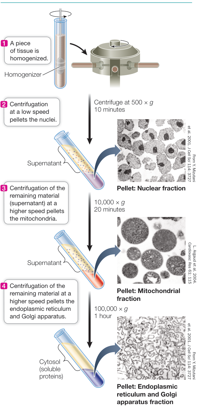
Figure 5.8 Cell Fractionation Organelles can be separated from one another after cells are broken open and their contents suspended in an aqueous medium. The medium is placed in a tube and spun in a centrifuge, which rotates about an axis at high speed. Centrifugal forces (measured in multiples of gravity, ) cause particles to sediment (form a pellet) at the bottom of the tube, which may be collected for biochemical study. Heavier particles sediment at lower speeds (lower centrifugal forces) than lighter particles. By adjusting the speed of centrifugation, researchers can separate and partially purify cellular organelles.
Q: How would you use this method to isolate ribosomes?
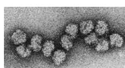
consist of two different-sized subunits, are about 25 nm in diameter, and can number up to thousands per cell. Because they lack membranes and are not a compartment, ribosomes are not classified as organelles. Rather, like the cytoskeleton (discussed later in this key concept), they are called cell structures.
Chemically, ribosomes consist of a special type of RNA called ribosomal RNA (rRNA). Ribosomes also contain more than 50 different protein molecules, which fit together noncovalently, employing forces for protein interaction such as hydrophobic interactions (see Figure 3.13). Additionally, some of the proteins bind noncovalently to the rRNA.
Connect the Concepts As discussed in Key Concept 14.5, the ribosome is the molecular workbench where mRNA is translated into protein.
In prokaryotic cells, ribosomes generally exist unattached in the cytoplasm. In eukaryotic cells, they are found in several places: in the cytoplasm; attached to the surface of the endoplasmic reticulum (a membrane-bound organelle, discussed later in this key concept); and inside other organelles, including the mitochondria and chloroplasts in plant cells. In each of these locations, the ribosomes are molecular factories where proteins are synthesized.
The nucleus contains most of the genetic information As we discussed in Chapter 4, hereditary information is stored in the sequence of nucleotides in DNA molecules. Most of the DNA in a eukaryotic cell resides in the single nucleus, about 5 µm in diameter (Figure 5.10A). The nucleus has several functions in the cell:
- It is the location of most of the cell’s DNA and the site of DNA replication.
- It is the site where gene transcription is turned on or off.
- A region within the nucleus, the nucleolus, is where ribosomes (see the previous section) begin to be assembled from RNA and proteins.
The contents of the nucleus, aside from the nucleolus, are referred to as the nucleoplasm. Similar to the cytosol, the nucleoplasm consists of the liquid content of the nucleus and the insoluble molecules suspended within it. The nucleus is enclosed by an integrated structure composed of two membranes, called the nuclear envelope. The two membranes of the nuclear envelope are perforated by thousands of nuclear pores (Figure 5.10B), each measuring approximately 9 nm in diameter, which connect the nucleoplasm with the cytoplasm.
The pores are not really porous. Instead, they act as “traffic cops,” allowing some molecules to pass into and out of the nucleus and blocking others. This allows the nucleus to regulate its information-processing functions. Small substances, including ions and other molecules with molecular weights of less than 10,000 daltons, move through the pores. But larger molecules, such as most proteins that are made in the cytoplasm, can’t get through. In some cases, however, there is a special transport mechanism to get such proteins into the nucleus; you’ll learn about this in Chapter 14.
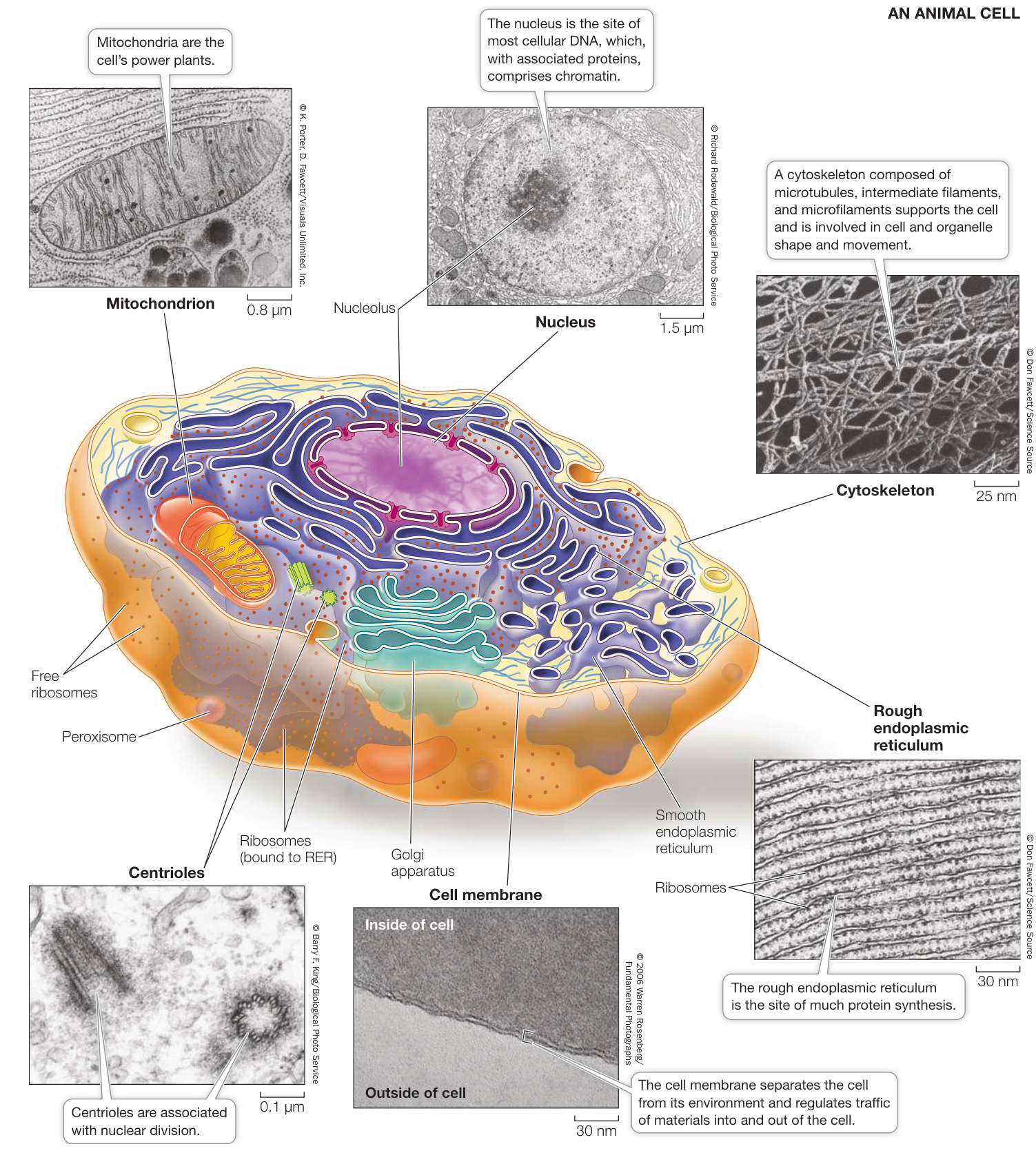
Figure 5.9 Eukaryotic Cells In electron micrographs, many plant cell organelles are nearly identical in form to those observed in animal cells. Cellular structures found in plant but not animal cells include the cell wall and the chloroplasts. Note that the images are two-dimensional “slices,” whereas cells are three-dimensional structures.
A PLANT CELL
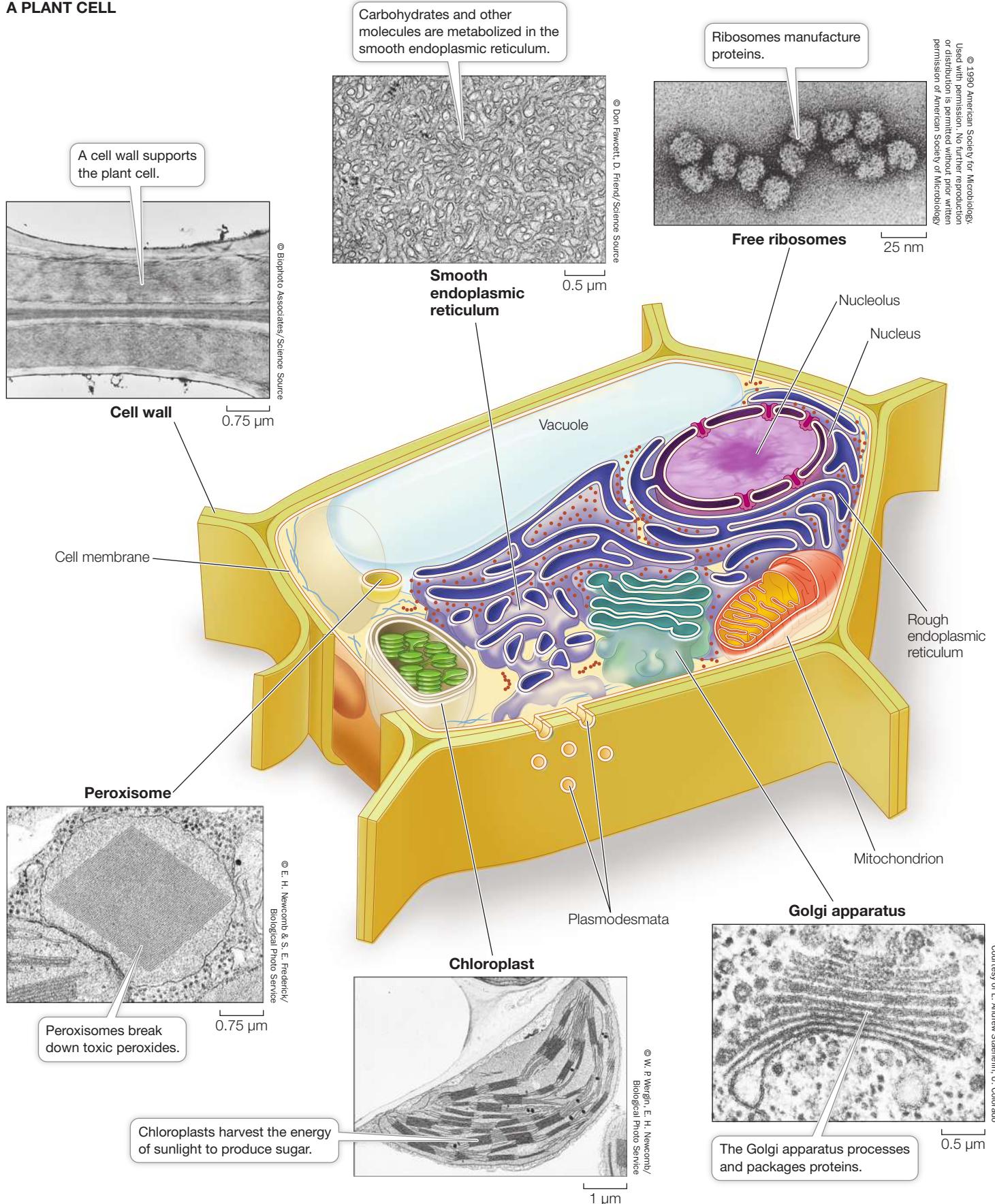
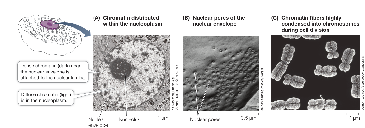
Figure 5.10 The Nucleus, Chromatin, and Chromosomes (A) Chromatin consists of nuclear DNA and the proteins associated with it. When the cell is not dividing, the chromatin is dispersed throughout the nucleus. This two-dimensional image was made using a transmission electron microscope. (B) The nuclear envelope has many pores that regulate traffic of large molecules such as RNA and proteins in and out of the nucleus. (C) The chromatin in dividing cells becomes highly condensed, so that the individual chromosomes can be seen. This three-dimensional image of isolated metaphase chromosomes was produced using a scanning electron microscope.
Inside the nucleus, each DNA molecule (in humans there are 46 of them) combines with proteins to form a fibrous complex called chromatin, which makes up long, thin threads called chromosomes. Despite the seeming disorganization of the nucleus as seen in Figure 5.10A, there is an underlying architecture, with chromosomes arranged specifically. We know this because if a laser beam is directed at a small region of the nucleus, only one or two chromosomes—the ones in that region—are damaged. The effects of this subnuclear organization are only beginning to be understood. One of them seems to be that chromosomes near one another may swap DNA regions; in some cases this leads to abnormal cell reproduction and cancer.
If and when a cell reproduces—a process called cell division—the chromatin becomes tightly compacted and condensed so that the individual chromosomes are visible under a light microscope (Figure 5.10C). At the inside edge of the nucleus, the chromatin is attached to a protein meshwork, called the nuclear lamina, which is formed by the polymerization of proteins called lamins into long, thin structures called intermediate filaments (discussed later in this key concept). The nuclear lamina maintains the shape of the nucleus by its attachment to both the chromatin and the nuclear envelope. On the outside of the nucleus, the outer membrane of the nuclear envelope folds outward into the cytoplasm and is continuous with the membrane of another organelle, the endoplasmic reticulum, which we will discuss next.
The endomembrane system is a group of interrelated organelles
Much of the volume of some eukaryotic cells is taken up by an extensive endomembrane system. This is an interconnected system of membrane-enclosed compartments that are sometimes flattened into sheets and sometimes have other characteristic shapes (see Figure 5.9). The endomembrane system includes:
- the cell membrane
- the nuclear envelope
- the endoplasmic reticulum
- the Golgi apparatus
- the lysosomes
Tiny, membrane-surrounded droplets called vesicles shuttle substances between the various components of the endomembrane system (Figure 5.11). In drawings and electron microscope pictures, this system appears static, fixed in space and time. But these depictions are just snapshots; in the living cell, the membranes and the materials they contain are in constant motion. Membrane components have been observed to shift from one organelle to another within the endomembrane system. Thus these membranes and compartments are functionally related.
ENDOPLASMIC RETICULUM Electron micrographs of eukaryotic cells reveal networks of interconnected membranes branching throughout the cytoplasm, forming tubes and flattened sacs. These membranes are collectively called the endoplasmic reticulum, or ER. The interior compartment of the ER, called the lumen, can compose up to 10% of the interior volume of the cell, and its folds result in a surface area many times greater than that of the cell membrane. There are two types of endoplasmic reticulum, the so-called rough and smooth.
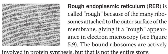
Rough endoplasmic reticulum (RER) is called “rough” because of the many ribosomes attached to the outer surface of the membrane, giving it a “rough” appearance in electron microscopy (see Figure 5.9). The bound ribosomes are actively involved in protein synthesis, but that is not the entire story:
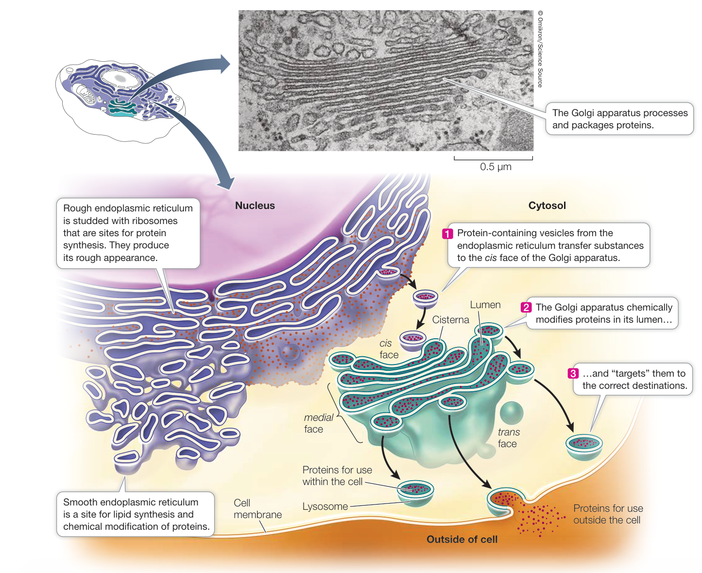
Figure 5.11 The Endomembrane System Membranes of the nucleus, endoplasmic reticulum, lysosomes, and Golgi apparatus form a network connected by vesicles.
Q: What processes in the endomembrane system are mediated by vesicles?
- The RER receives into its lumen certain newly synthesized proteins (including proteins destined for lysosomes, the cell membrane, and export from the cell), segregating them away from the cytoplasm. The RER also participates in transporting these proteins to other locations in the cell.
- While inside the RER, proteins can be chemically modified to alter their functions and “tag” them for delivery to specific cellular destinations.
- Proteins are sent to destinations elsewhere in the cell enclosed within vesicles that pinch off from the RER.
- Most membrane-bound proteins are made in the RER.
A protein enters the lumen of the RER through a pore as it is synthesized. As with a protein passing through a nuclear pore, there is a special transport mechanism to get proteins into the
ER, which you’ll learn about in Chapter 14. Once in the lumen of the RER, proteins undergo several changes, including the formation of disulfide bridges and folding into their tertiary structures (see Figure 3.6).
Some proteins become covalently linked to carbohydrate groups in the RER, thereby becoming glycoproteins. In the case of proteins directed to the lysosomes, the carbohydrate groups are part of an “addressing” system that ensures that the right proteins are directed to those organelles. This addressing system is very important because the enzymes within the lysosomes are some of the most destructive the cell makes. Were they not properly addressed and contained, they could destroy the cell. Cells that make a lot of proteins, such as the cells that make digestive enzymes for the human intestine and antibodies for the immune system, are full of RER.
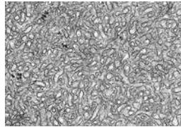
Smooth endoplasmic reticulum (SER) lacks ribosomes and is more tubular (and less like flattened sacs) than the RER, but it shows continuity with portions of the RER (see Figure 5.11). The SER has several important roles:
- It is responsible for the chemical modification of small molecules taken in by the cell that may be toxic to the cell. These modifications make the targeted molecules more polar, so they are more water-soluble and easily removed.
- It is the site for glycogen degradation in animal cells. We will discuss this important process in Chapter 9.
- It is the site where lipids and steroids are synthesized, as well as some polysaccharides in plant cells.
- It stores calcium ions, which when released trigger numerous cell responses.
Connect the Concepts Regulating muscle contraction is one of the important roles played by the calcium ions stored by the SER. See Key Concept 46.1.
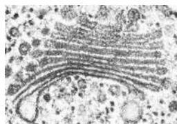
GOLGI APPARATUS The Golgi apparatus is another part of the diverse, dynamic, and extensive endomembrane system (see Figure 5.11 insert).
This structure was named after its discoverer, Camillo Golgi. Its appearance varies, but it almost always consists of two components: flattened membranous sacs called cisternae (singular cisterna) that are piled up like saucers, and small membrane-enclosed vesicles. The entire apparatus is about 1 m long.
The Golgi apparatus has several roles:
- It receives protein-containing vesicles from the RER.
- It modifies, concentrates, packages, and sorts proteins before they are sent to their cellular or extracellular destinations.
- It adds carbohydrates to proteins and modifies other carbohydrates that were attached to proteins in the RER.
- It is where some polysaccharides for the plant cell wall are synthesized.
- It cuts certain large precursor proteins transferred from the RER into smaller, functional fragments. For example, in humans the pituitary gland makes a 241-amino acid polypeptide called proopiomelanocortin. In the Golgi apparatus, this is cut into several molecules, including corticotropin (39 amino acids), melanocyte-stimulating hormone (22 amino acids), and -endorphin (31 amino acids). You will learn more about these molecules in Chapter 39.
The cisternae of the Golgi apparatus have three functionally distinct regions, called faces:
The cis face lies nearest to the nucleus or a patch of RER.
- The trans face lies closest to the cell membrane.
- The medial face lies in between (see Figure 5.11).
The terms cis, trans, and medial derive from Latin words meaning, respectively, “on the same side,” “on the opposite side,” and “in the middle.” These three parts of the Golgi apparatus contain different enzymes and perform different functions.
Protein-containing vesicles from the RER fuse with the cis membrane of the Golgi apparatus, releasing their cargo into the lumen of the Golgi apparatus cisternae. Other vesicles may move between the cisternae, transporting proteins, and it appears that some proteins move from one cisterna to the next through tiny channels. Vesicles budding off from the trans face carry their contents away from the Golgi apparatus. These vesicles go to the cell membrane or to the lysosome, another organelle in the endomembrane system.
How do the vesicles in this system recognize their targets for fusion? In other words, when a vesicle buds off from the ER, how does it “know” to fuse with the cis-Golgi membrane? The answer lies in proteins on both membranes appropriately called SNAREs. Pairs of SNAREs are complementary—that is, they bind to one another like a lock and key. So the SNARE on the ER surface that forms a vesicle can bind to a SNARE on the cis-Golgi.
LYSOSOMES The primary lysosomes originate from the Golgi apparatus. They contain digestive enzymes and are the sites where macromolecules—proteins, polysaccharides, nucleic acids, and lipids—are hydrolyzed into their monomers (see Figure 3.4).
Lysosomes are about 1 m in diameter, are surrounded by a single membrane, and have a densely staining, featureless interior (Figure 5.12). There may be dozens of lysosomes in a cell, depending on its needs. The interior of the lysosome is more acidic (pH 5) than the external cytoplasm (pH 7.2).
Lysosomes are cellular “disposals,” compartments for the breakdown of food, other cells, or foreign objects that are taken up by the cell. These materials enter the cell by a process called phagocytosis (phago, “eat,” + cytosis, “cellular”). In this process, a pocket forms in the cell membrane and then deepens and encloses material from outside the cell. The pocket becomes a small vesicle called a phagosome, containing food or other material, which breaks free of the cell membrane to move into the cytoplasm. The phagosome fuses with a primary lysosome to form a secondary lysosome, where digestion occurs. These reactions are enhanced by the acidity of the lysosome’s interior. The products of digestion pass through the membrane of the lysosome, providing energy and raw materials for other cellular processes. For example, a protease might hydrolyze proteins to amino acids, which are released to the cytoplasm for reassembly into other proteins. The “used” secondary lysosome, now containing undigested particles, then moves to the cell membrane, fuses with it, and releases the undigested contents to the environment, a process called exocytosis.
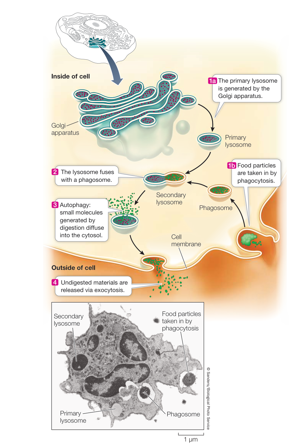
Figure 5.12 Lysosomes Isolate Digestive Enzymes from the Cytoplasm Lysosomes are sites for the hydrolysis of material taken into the cell by phagocytosis. Discarded components exit the cell by exocytosis. Activity 5.4 Lysosomal Digestion
Phagocytes (see Key Concept 40.1) are specialized cells that have an essential role in taking up and breaking down materials; they are found in nearly all animals and many protists. You will encounter them and their activities again at many places in this book, but at this point one example suffices: in the human liver and spleen, phagocytes digest approximately 10 billion aged or damaged blood cells each day! The digestion products are then used to make new cells to replace those that are digested.
Lysosomes are active even in cells that do not perform phagocytosis. Cells are dynamic systems; some cell components are continually being broken down and replaced by new ones. The programmed destruction of cell components is called autophagy, and lysosomes are where the cell breaks down its own materials. With the proper signal, lysosomes can engulf entire organelles, hydrolyzing their constituents.
How important is autophagy? An entire class of human diseases called lysosomal storage diseases are caused by the failure of lysosomes to digest specific cellular components; these diseases are invariably harmful or fatal. An example is Tay-Sachs disease, in which a particular lipid called a ganglioside is not broken down in the lysosomes and instead accumulates in brain cells. In the most common form of this disease, a baby starts exhibiting neurological symptoms and becomes blind, deaf, and unable to swallow after 6 months of age. Death occurs before age 4.
Plant cells do not appear to contain lysosomes, but the vacuole of a plant cell (which we describe later in this key concept) may function in an equivalent capacity because it, like lysosomes, hydrolyzes macromolecules.
Some organelles transform energy
A cell requires energy for activities such as growth, reproduction, responsiveness, and movement. Energy is harvested from fuel molecules in the mitochondria (found in all eukaryotic cells) and from sunlight in the chloroplasts of plant cells. Energy in biological systems is stored and transferred in the form of covalent bonds in organic molecules.
MITOCHONDRIA In eukaryotic cells, the breakdown of energy-rich molecules such as glucose begins in the cytoplasm. The molecules that result from this partial degradation enter the mitochondria (singular mitochondrion), whose primary function is to harvest the chemical-bond energy of those molecules in a form that the cell can use, namely the energy-rich molecule ATP (adenosine triphosphate) (see Key Concept 8.2). The production of ATP in the mitochondria, using fuel molecules and molecular oxygen (), is called cellular respiration (see Chapter 9).
Typical mitochondria are somewhat less than 1.5 m in diameter and are 2–8 m in length—about the size of many bacteria (Figure 5.13). They can reproduce and divide independently of the central nucleus. The number of mitochondria per cell ranges from one large organelle in some unicellular protists to a few hundred thousand in large egg cells. A typical human liver cell contains more than 1,000 mitochondria. Cells that are active in movement and growth require the most chemical-bond energy and tend to have the most mitochondria.
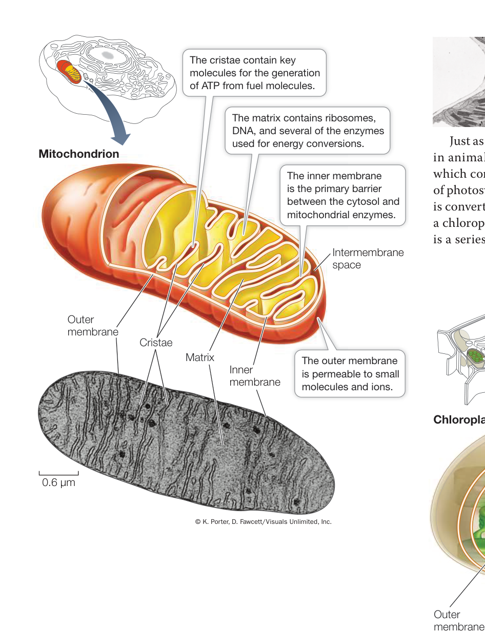
Figure 5.13 A Mitochondrion Converts Energy from Fuel Molecules into ATP The electron micrograph is a two-dimensional slice through a three-dimensional organelle. As the drawing emphasizes, the cristae are extensions of the inner mitochondrial membrane. Q: What kinds of human cells would you expect to have a lot of mitochondria?
Mitochondria have two membranes. The outer membrane is smooth in appearance and permits the movement of substances into and out of the organelle. Immediately inside the outer membrane is an inner membrane, which folds inward in many places and thus has a surface area much greater than that of the outer membrane. The folds tend to be quite regular, giving rise to shelflike structures called cristae (from the Latin word for “crest” or “plume”). The inner membrane exerts much more control over what enters and leaves the space it encloses than does the outer membrane. Embedded in the inner mitochondrial membrane are many large protein complexes that participate in cellular respiration.
The space enclosed by the inner membrane is called the mitochondrial matrix. In addition to many molecules involved in cellular respiration, the matrix contains ribosomes and DNA that are used to make some of the mitochondrial proteins needed for cellular respiration.
PLASTIDS Plastids occur only in the cells of plants and certain protists. Like mitochondria, plastids can divide independently of the cell nucleus. There are several types of plastids, with different functions.
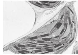
Just as melanosomes are a compartment for melanin pigment in animal cells (see the chapter opening), so are chloroplasts, which contain the green pigment chlorophyll and are the sites of photosynthesis (Figure 5.14). In photosynthesis, light energy is converted into chemical-bond energy. Like a mitochondrion, a chloroplast is enclosed by two membranes. In addition, there is a series of internal membranes that often look like stacks of flat, hollow pita bread. Each stack is called a granum (plural grana) and the pita bread—like compartments are called thylakoids (see Figure 5.14). Thylakoid lipids are distinctive: only 10% are phospholipids, whereas the rest are galactose-substituted diglycerides and sulfolipids. Because of the abundance of chloroplasts, these are the most abundant lipids in the biosphere.
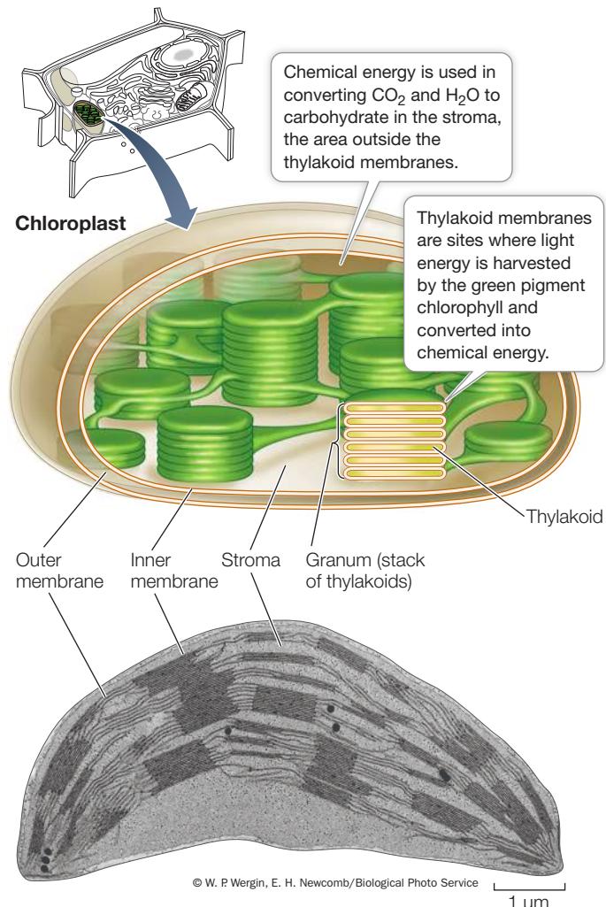
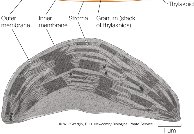
Figure 5.14 Chloroplasts Feed the World The electron micrograph shows a chloroplast from a leaf of corn. Chloroplasts are large compared with mitochondria and contain extensive networks of thylakoid membranes. These membranes contain the green pigment chlorophyll, and are the sites where light energy is converted into chemical energy for the synthesis of carbohydrates from and . Q: Chloroplasts are present only in plant cells. Why do these cells also contain mitochondria?
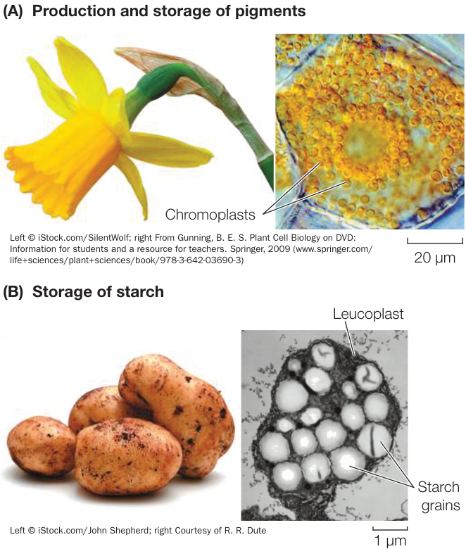
Figure 5.15 Other Types of Plastids (A) Chromoplasts in flower petals contain pigments. (B) Leucoplasts in potatoes store starch.
In addition to lipids and proteins, the membranes of the thylakoids contain chlorophyll and other pigments that harvest light energy for photosynthesis (we will see how they do this in Key Concept 10.2). The thylakoids of one granum may be connected to those of other grana, making the interior of the chloroplast a highly developed network, much like the ER. The fluid in which the grana are suspended is called the stroma. Like the mitochondrial matrix, the chloroplast stroma contains ribosomes and DNA, which are used to synthesize some chloroplast polypeptides.
Other types of plastids, such as chromoplasts and leucoplasts, have functions different from those of chloroplasts (Figure 5.15). Chromoplasts make and store red, yellow, and orange pigments, especially in flowers and fruits—again, just as the melanocytes in the opening story store pigment. Leucoplasts are storage organelles that store starch.
There are several other membrane-enclosed organelles
There are three other organelles whose boundary membranes separate their specialized chemical reactions and contents from the rest of the cytoplasm: peroxisomes, glyoxysomes, and vacuoles.
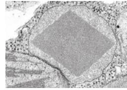
Peroxisomes are organelles that accumulate toxic peroxides, such as hydrogen peroxide (), that occur as by-products of some biochemical reactions. These peroxides are safely broken down inside the peroxisomes without mixing with other parts of the cell.
Peroxisomes are small organelles, about 1 m in diameter.
Glyoxysomes are similar to peroxisomes and are found only in plants. They are most abundant in young plants and are the locations where stored lipids are converted into carbohydrates for transport to growing cells. The direct conversion of fat to carbohydrate is unique to these plants.
Vacuoles occur in many eukaryotic cells but particularly those of plants, fungi, and protists. They arise from the ER or Golgi apparatus. Plant vacuoles (Figure 5.16) have several functions:
- Structure: In many plant cells, vacuoles take up more than 90% of the cell volume and grow as the cell grows. The presence of dissolved substances in the vacuole causes water to enter it from the cytosol, making the vacuole swell like a balloon. A mature plant cell does not swell when the vacuole fills with water, since it has a rigid cell wall. Instead, it stiffens from the increase in water pressure (called turgor), and this supports the plant body (see Figure 6.10).
- Reproduction: Vacuoles contain some of the pigments (especially blue and pink ones) in the petals and fruits of flowering plants. These pigments—the anthocyanins—are visual cues that help attract animals that assist in pollination or seed dispersal.
- Digestion: In some plants, vacuoles in seeds contain molecules that hydrolyze stored proteins into monomers. During seed germination, the monomers are used as food by the developing plant seedlings.
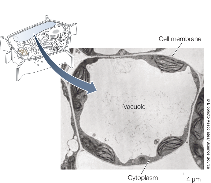
Figure 5.16 Vacuoles in Plant Cells Are Usually Large The large central vacuole in this cell is typical of some types of plant cells.
- Storage: In addition to storing water, vacuoles store toxic molecules and waste products. Because they are poisonous or distasteful, these stored materials deter some animals from eating the plants and may thus contribute to plant defense and survival. An example is tannins, a class of polyphenols that are made in many plants and may be familiar to you as the aftertaste in tea and red wine. Their bitter taste deters predators, and their dark color acts like animal melanin to protect the plant from ultraviolet light damage. Tannins are made in the chloroplast and end up in the vacuole. A newly discovered organelle, appropriately named the tannosome, contains tannins (Investigating Life: Discovering a New Organelle, the Tannosome).
So far we have discussed numerous membrane-enclosed organelles. Now we will turn to a group of cytoplasmic structures without membranes.
The cytoskeleton is important in cell structure and movement
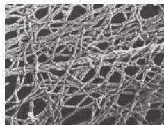
Cell shapes can sometimes change, and there appear to be rapid movements of structures inside cells. These phenomena involve a meshwork of rods within cells that can be seen in electron microscopy. Experimentation showed that this meshwork—called the cytoskeleton—fills several important roles:
- It supports the cell and maintains its shape.
- It holds cell organelles and other particles in position within the cell.
- It moves organelles and other particles around in the cell.
- It is involved with movements of the cytoplasm, called cytoplasmic streaming.
- It interacts with extracellular structures, helping anchor the cell in place.
There are three components of the eukaryotic cytoskeleton: microfilaments (smallest diameter), intermediate filaments, and microtubules (largest diameter). These filaments have very different functions (Figure 5.17).
Investigating LIFE Discovering a New Organelle, the Tannosome
Experiment
Original Paper: J.-M. Brillouet et al. 2013. The tannosome is an organelle forming condensed tannins in the chlorophyllous organs of Tracheophyta. Ann Bot 112: 1003–1014.
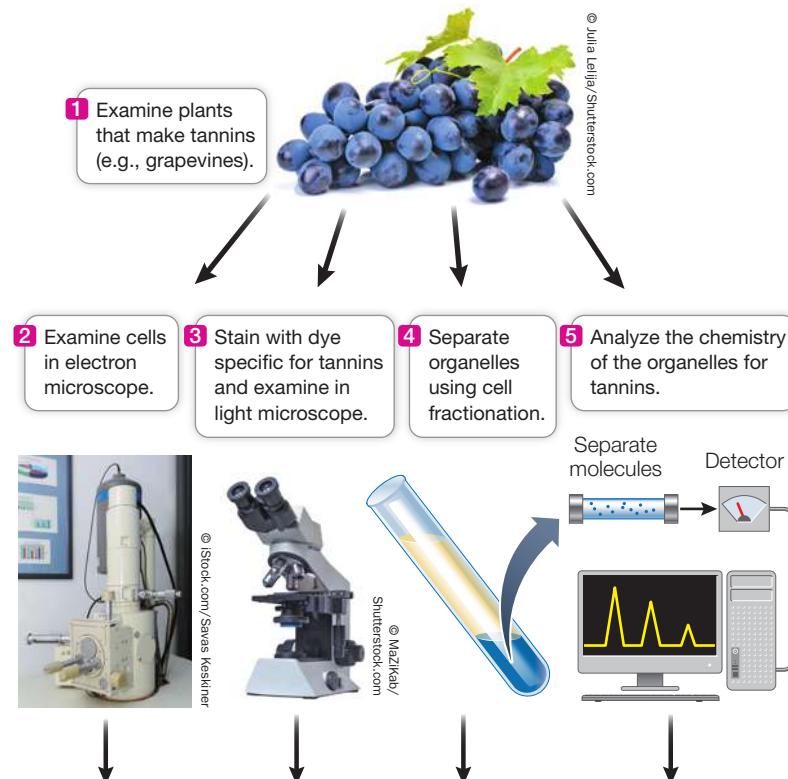
Through a series of investigations involving microscopy, cell fractionation, and chemical analysis, biologists discovered that plant tannins are compartmentalized in a heretofore undiscovered structure, the tannosome.
QUESTION Are pigments compartmentalized in all types of cells?
METHOD
RESULTS
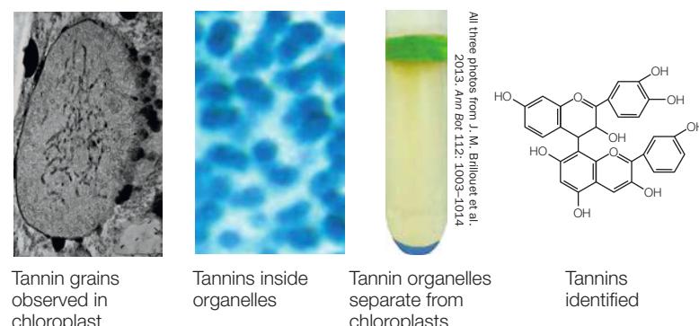
CONCLUSION Tannins are present in an organelle that begins in the chloroplast.
Work with the Data
The team of biologists and chemists (Brillouet et al. 2013) at the French National Institute for Agricultural Research, led by Geneviève Conejero, sought to investigate the cellular basis of tannin formation. It was well known from electron micrographs that these pigments accumulate in vacuoles, but how they got there was not established. The team used a combination of microscopy (light and electron; see Figure 5.4), cell fractionation (see Figure 5.8), and chemical analysis to show that tannins are formed in discrete vesicles, which the researchers named tannosomes.
QUESTIONS
- Plant organs rich in tannins were examined by electron microscopy. Representative photographs are shown in Figure A. What do these images indicate about the origin and final resting spot of tannins in the cell? Based on what you know of vesicle trafficking in the cell, how do you think the tannins get from the chloroplast to the vacuole?
- The scientists used specific dyes targeted to chlorophyll and tannins and examined slices of plant tissues by light microscopy. Figure B shows the results. What can you conclude about the subcellular origin of tannins, and how does your answer relate to your answer to Question 1?
- Cell fractionation was used to try to isolate tannin-containing structures separate from chloroplasts. In a very dense (72%), syrupy sucrose medium, fractionated cell contents showed two membrane-bound fractions (see the experiment). Both were examined by microscopy for tannin staining, and the contents were analyzed for chlorophyll and tannins. The results are shown in the table. What can you conclude about the tannin compartment?
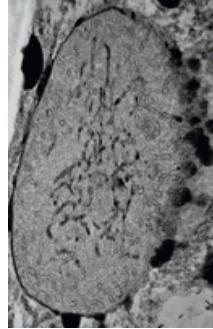
Figure A
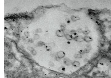
Vacuole with tannin vesicles Chloroplast with tannin granules
Both photos from J. M. Brillouet et al. 2013. Ann Bor 112: 1003
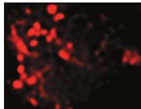
Figure B
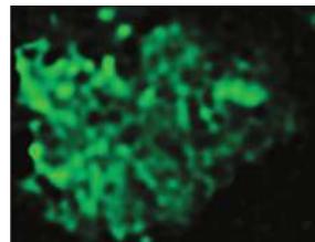
Staining of plant cell for chlorophyll Staining of plant cell for tannins
Both photos from J. M. Brillouet et al. 2013. Ann Bor 112: 1003
| Upper fraction | Lower fraction | |
|---|---|---|
| Staining for tannins | No | Yes |
| Staining for chlorophyll | Yes | Yes |
| Chemical analysis for tannins | No | Yes |
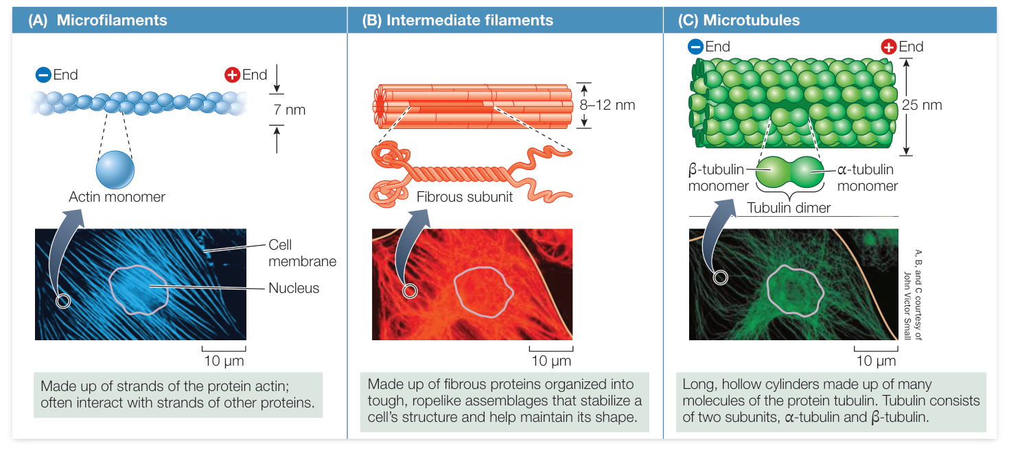
Figure 5.17 The Cytoskeleton Three highly visible and important structural components of the cytoskeleton are shown here in detail. The photographs are all of the same cell, treated with different fluorescent antibodies that detect microfilaments (A), intermediate filaments (B), or microtubules (C). These structures maintain and reinforce cell shape and contribute to cell movement. The position of the cell’s nucleus is near the center of the photos.
MICROFILAMENTS Microfilaments can exist as single filaments, in bundles, or in networks. They are about 7 nm in diameter and up to several micrometers long. Microfilaments have two major roles:
- They help the entire cell or parts of the cell move.
- They determine and stabilize cell shape.
Microfilaments are assembled from monomers of actin, a protein that exists in several forms and has many functions, especially in animals. The actin found in microfilaments (which are also known as actin filaments) has distinct ends, designated “plus” and “minus.” These ends permit actin monomers to interact with one another to form long, double-helical chains (see Figure 5.17A). Within cells, the polymerization of actin into microfilaments is reversible, and the microfilaments can disappear from cells by breaking down into monomers of free actin. Special actin-binding proteins mediate these processes.
Connect the Concepts In animal muscle cells, actin filaments are associated with another protein, the “motor protein” myosin, and the interactions of these two proteins account for the contraction of muscles, as described in Key Concept 46.1.
In non-muscle cells, actin filaments are associated with localized changes in cell shape. For example, microfilaments are involved in the flowing movement of the cytoplasm called cytoplasmic streaming, in amoeboid movement, and in the “pinching” contractions that divide an animal cell into two daughter cells. Microfilaments are also involved in the formation of cellular extensions called pseudopodia (pseudo, “false,” + podia, “feet”) that enable some cells to move. As you will see in Key Concept 40.3, cells of the immune system must move toward other cells during the immune response.
In some cell types, microfilaments form a meshwork just inside the cell membrane. Actin-binding proteins then cross-link the microfilaments to form a rigid network structure that supports the cell. For example, microfilaments support the tiny microvilli that line the human intestine, giving it a larger surface area through which to absorb nutrients (Figure 5.18).
INTERMEDIATE FILAMENTS There are at least 50 different kinds of intermediate filaments, many of them specific to a few cell types. They generally fall into six molecular classes (based on amino acid sequence) that share the same general structure. One of these classes consists of fibrous proteins of the keratin family, which also includes the proteins that make up hair and fingernails. Intermediate filaments are tough, ropelike protein structures 8–12 nm in diameter (see Figure 5.17B). They are more permanent than the other two types in that they do not continually form and re-form, as the microtubules and microfilaments do.
Intermediate filaments have two major structural functions:
- They anchor cell structures in place. In some cells, intermediate filaments radiate from the nuclear envelope and help maintain the positions of the nucleus and other organelles in the cell. The lamins of the nuclear lamina are intermediate filaments. Other kinds of intermediate filaments help hold in place the complex apparatus of microfilaments in the microvilli of intestinal cells (see Figure 5.18).
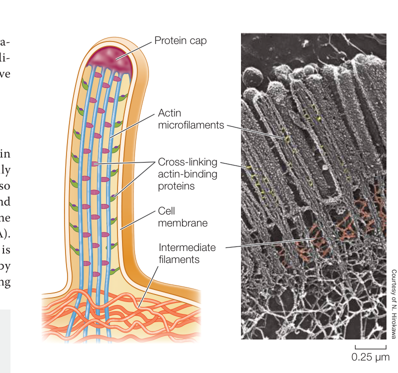
Figure 5.18 Microfilaments for Support Cells that line the intestine are folded into tiny projections called microvilli, which are supported by microfilaments. The microfilaments interact with intermediate filaments at the base of each microvillus.
- They resist tension. For example, they maintain rigidity in body surface tissues by stretching through the cytoplasm and connecting specialized membrane structures called desmosomes (see Figure 6.6).
MICROTUBULES The largest-diameter components of the cytoskeletal system, microtubules, are long, hollow, unbranched cylinders about 25 nm in diameter and up to several micrometers long. Microtubules have two roles in the cell:
- They form a rigid internal skeleton for some cells.
- They act as a framework along which motor proteins can move structures within the cell.
Microtubules are assembled from dimers—two similar molecules in a complex—of the protein tubulin, called -tubulin and -tubulin. Thirteen chains of tubulin doublets surround the central cavity of the microtubule (see Figures 5.17C and 5.19B).
As in microfilaments, the two ends of a microtubule are different: one is designated the “plus” end and the other the “minus” end. Tubulin dimers can be rapidly added or subtracted, mainly at the plus end, lengthening or shortening the microtubule.
Many microtubules appear to radiate from a region of the cell called the microtubule organizing center. Tubulin polymerization results in a rigid structure, and tubulin depolymerization leads to its collapse.
Microtubule ↔︎ tubulin doublets
The capacity to change length rapidly makes microtubules dynamic structures: they are readily adapted for new purposes in the cell. For example, by disassembly and reassembly, microtubules can move to new parts of the cell; or by disassembly, they can drag along substances attached to them.
In plants, microtubules act as a framework for the assembly of cellulose and help orient the cellulose fibers of the cell wall (see Figure 3.19). Electron micrographs of plants frequently show microtubules lying just inside the cell membranes of cells that are forming or extending their cell walls. If the orientation of these microtubules is altered experimentally, it leads to a similar change in the cell wall and a new shape for the cell.
Microtubules serve as tracks for motor proteins, specialized molecules that use cellular energy to change their shapes and move. Motor proteins bind to and move along the microtubules, carrying materials from one part of the cell to another. There are two main types of motor proteins:
- Kinesins move materials toward the “plus” end.
- Dyneins move materials toward the minus end.
The movement of organelles such as vesicles inside neurons occurs via kinesins; both kinesins and dyneins are involved in the movement of chromosomes during cell division; and as you will see next, dynein allows microtubule doublets to slide past one another in cilia and flagella.
CILIA AND FLAGELLA Microtubules and their associated proteins line the interior of certain movable appendages on eukaryotic cells: the cilia (Figure 5.19A) and flagella. Many cells have one or the other of these appendages, which form from projections of the cell membrane:
- Cilia are 0.25 µm in length. They occur by the hundreds of individual cells and move stiffly to either propel the cell (for example, in protists) or to move fluid over a stationary cell (as in the human respiratory system).
- Flagella are longer—100 to 200 µm—and occur singly or in pairs. They can push or pull a cell through its aqueous environment.
In cross section, a typical cilium or eukaryotic flagellum is surrounded by the cell membrane and contains a “9 + 2” array of microtubules. As Figure 5.19B shows, nine fused pairs of microtubules form an outer cylinder, and one pair of unfused microtubules runs up the center. Each doublet is connected to the center of the structure by a radial spoke. This structure is essential to the bending motion of both cilia and flagella. How does this bending occur?
The motion of cilia and flagella results from the sliding of the microtubule doublets past one another. This sliding is driven by the motor protein dynein, which, like other motor proteins, works by undergoing reversible shape changes that require chemical-bond energy. Dynein molecules bind between two neighboring microtubule pairs, and as the dynein molecules change shape, the pairs move past one another (Figure 5.20). Another protein, nexin, cross-links the pairs and appears to limit how far the doublets can slide. This causes the cilium or flagellum to bend.
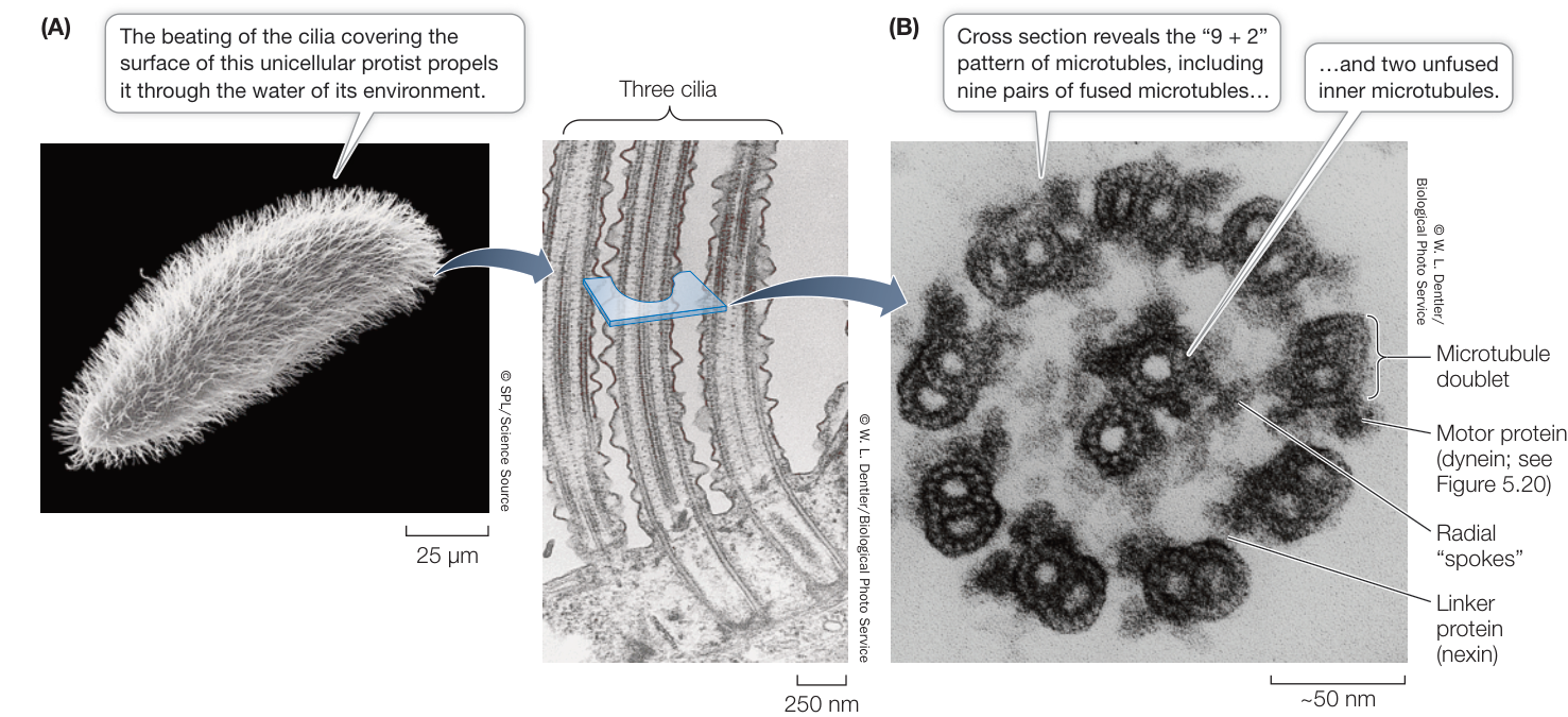
Figure 5.19 Cilia (A) This unicellular eukaryotic organism (a ciliated protist) can coordinate the beating of its cilia, allowing rapid movement. (B) A cross section of a single cilium shows the arrangement of micro-tubules and other proteins.
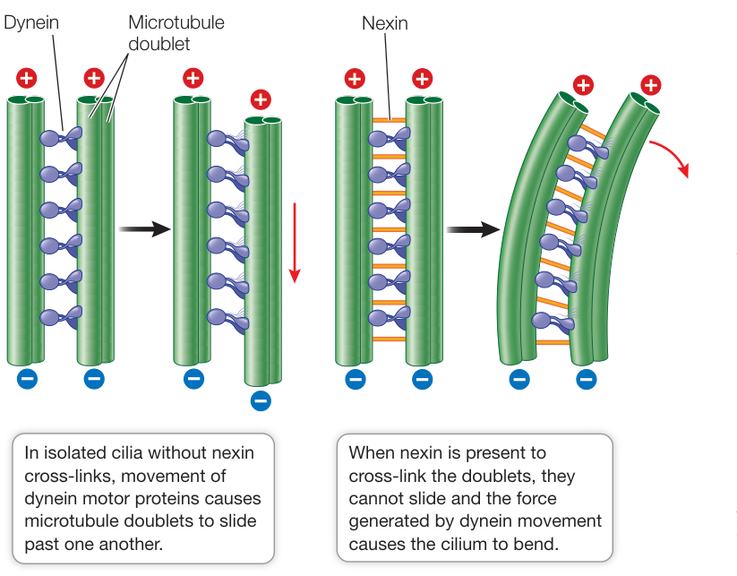
Figure 5.20 Motor Protein Moves Microtubules in Cilia and Flagella A motor protein, dynein, causes microtubule doublets to slide past one another. In a flagellum or cilium, anchorage of the microtubule doublets to one another results in bending. (After B. Alberts et al. 2008. The Molecular Biology of the Cell, 5th Edition. Garland Science: New York.) Q: In some people, the nexin links are absent and micro-tubule doublets are not attached to one another. What do you think the consequences of this are?
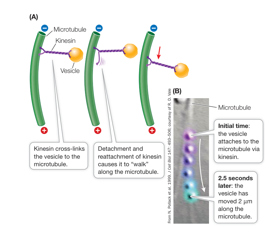
Figure 5.21 A Motor Protein Pulls Vesicles along Microtubules (A) Kinesin delivers vesicles to various parts of the cell by moving along microtubule “railroad tracks.” (B) A vesicle is pulled by kinesin along a microtubule in the protist Dictyostelium. The time sequence, in half-second intervals, is shown by the color changes from purple to blue.
Other motor proteins, including kinesin, carry protein-laden vesicles or other organelles from one part of the cell to another (Figure 5.21). These proteins bind to the organelle and “walk” it along a microtubule by a repeated series of shape changes. Recall that microtubules are directional, with a plus end where there is a net addition of tubulin and a minus end where there is removal. Cytoplasmic dynein (which has a different role than the one found in cilia and flagella) moves organelles attached to it toward the minus end, whereas kinesin moves them toward the plus end (see Figure 5.17). The ability of microtubules to carry vesicles is used by some fishes and amphibians to avoid predators. Motor proteins carrying pigment vesicles aggregate them in the center of the cell in response to environmental or nervous system stimuli. This makes the animal appear lighter. When the stimuli are removed, other motor proteins move the vesicles along microtubules to a more even distribution, restoring a darker hue.
Connect the Concepts Perhaps the most dramatic role of microtubules is in the process of chromosome movement during cell division. See Key Concept 11.3.
Biologists can manipulate living systems to establish cause and effect
How do we know that the elements of the cytoskeleton, such as microtubules and microfilaments, can perform all these functions? We can observe an individual structure under the microscope, and we can observe the functions of living cells that contain that structure. These observations may suggest that the structure carries out a particular function, but mere correlation does not show cause and effect. For example, light microscopy of living cells reveals that the cytoplasm is actively streaming around the cell, and that cytoplasm flows into an extended portion of an amoeboid cell during movement. The observed presence of cytoskeletal components suggests, but does not prove, their role in this process. Science seeks to show the specific links that relate one process, A, to a function, B. In cell biology, two approaches are often used to show that a structure or process A causes function B:
- Inhibition: Use a drug that inhibits A and see if B still occurs. If it does not, then A is probably a causative factor for B. Figure 5.22 shows an experiment with such a drug (an inhibitor) that demonstrates cause and effect in the case of the cytoskeleton and cell movement.
- Mutation: Examine a cell that lacks the gene (or genes) for A and see if B still occurs. If it does not, then A is probably a causative factor for B. Part Four of this book will describe many experiments using this genetic approach.
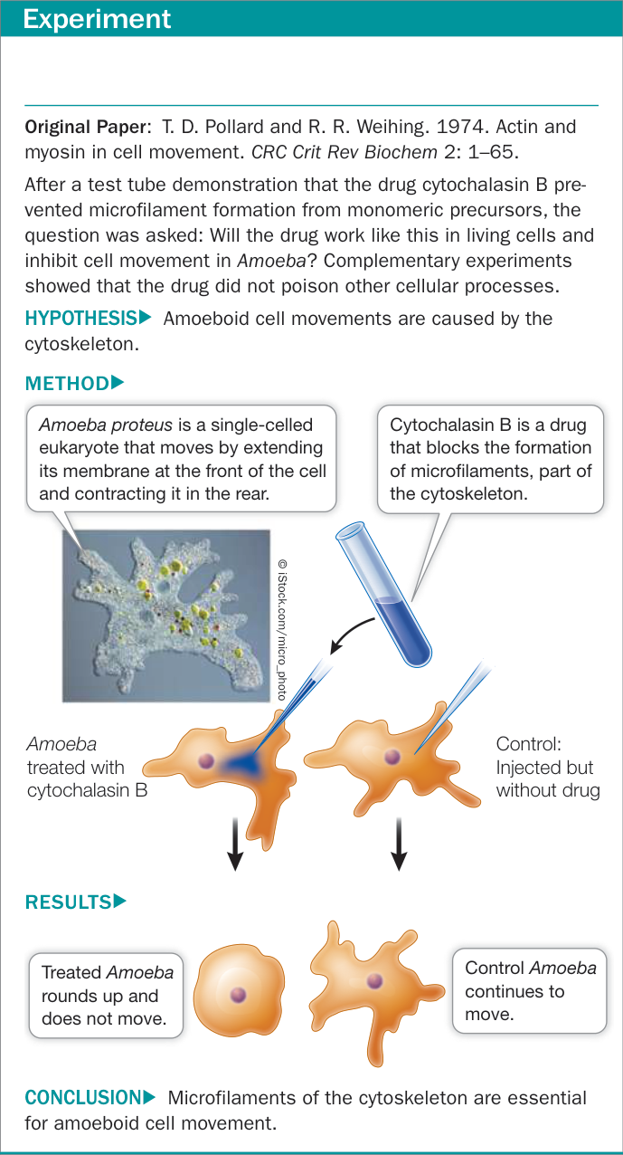
Figure 5.22A The Role of Microfilaments in Cell Movement— Showing Cause and Effect in Biology
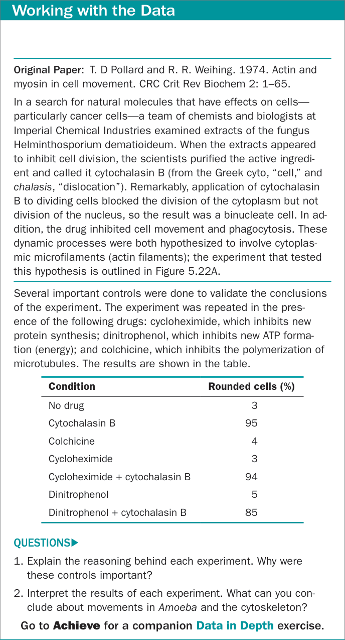
Figure 5.22B The Role of Microfilaments in Cell Movement— Showing Cause and Effect in Biology
Recap and Assess
The hallmark of eukaryotic cells is compartmentalization. Membrane-enclosed organelles process information, transform energy, form internal compartments for transporting proteins, and carry out intracellular digestion. An internal cytoskeleton plays several structural roles.
Using examples, explain why compartmentalization is important in eukaryotic cells.
Through how many membranes would a molecule have to pass in moving (a) from the interior (stroma) of a chloroplast to the interior (matrix) of a mitochondrion? (b) From the interior of a lysosome to the outside of a cell? (c) From one ribosome to another?
The drug vincristine is used to treat many cancers. It appears to work by causing microtubules to depolymerize. Vincristine use has many side effects, including loss of dividing cells and nerve problems. Explain why this might be so.
- The movements of newly synthesized proteins can be followed through cells using a “pulse–chase” experiment. During synthesis, proteins are tagged with a radioactive isotope (the “pulse”), then the cells are allowed to process the proteins for varying periods of time. The locations of the radioactive proteins are then determined by isolating cell organelles and quantifying their levels of radioactivity. What would be the expected pathway for (a) a lysosomal enzyme and (b) a protein that is released from the cell?
All cells interact with their environments. Many eukaryotic cells are parts of multicellular organisms and must closely coordinate their activities with other cells. The cell membrane plays a crucial role in these interactions, but other structures outside that membrane are involved as well.
Extracellular Materials Provide Structure
Learning Objectives
- 5.4.1 Explain structure–function relationships in cell walls.
- 5.4.2 Compare and contrast plant and animal cell extracellular matrices.
- 5.4.3 Analyze changes to cells involving extracellular components.
Although the cell membrane is the functional barrier between the inside and the outside of a cell, many structures are produced by cells and secreted to the outside of the cell membrane, where they play essential roles in protecting, supporting, or attaching cells to each other. Because they are outside the cell membrane, these structures are said to be extracellular. The peptidoglycan cell wall of bacteria is an example of an extracellular structure (see Figure 5.6), as is the chitinous matrix outside the cells of arthropods. In many eukaryotes, other extracellular structures, such as the cell walls of plants and the extracellular matrices found between the cells of animals, play similar roles. Each of these structures is made up of two main components: a prominent fibrous macromolecule and a gel-like medium in which the fibers are embedded.
The plant cell wall provides structure and protection
The plant cell wall is a semirigid structure outside the cell membrane (Figure 5.23). We will consider the structure and role of the cell wall in more detail in Chapter 32. For now, we note that it consists of cellulose fibers (see Figure 3.19) embedded in other complex polysaccharides and proteins. The plant cell wall has three major roles:
- It provides support for the cell and plant by remaining rigid. Yet it is flexible enough that it can allow the plant to bend in the wind, for example.
- It acts as a barrier to infection by fungi and other organisms that can cause plant diseases.
- It contributes to plant form by growing as the plant cells expand.
In some cells, such as those in a leaf, the cell wall is porous to allow the passage of molecules into and out of the cell. In other cells, such as those of the plant’s vascular system (which transports water and small molecules between organs), the wall is not porous.
Because of their thick cell walls, plant cells viewed under a light microscope appear to be entirely isolated from one another. But electron microscopy reveals that this is not the case. The cytoplasms of adjacent plant cells are connected by numerous cell membrane–lined channels called plasmodesmata, which are about 20–40 nm in diameter and extend through the cell walls (see Figure 5.9). Plasmodesmata permit the diffusion of water, ions, small molecules, RNA, and proteins between connected cells, allowing for the use of these substances far from their sites of synthesis.
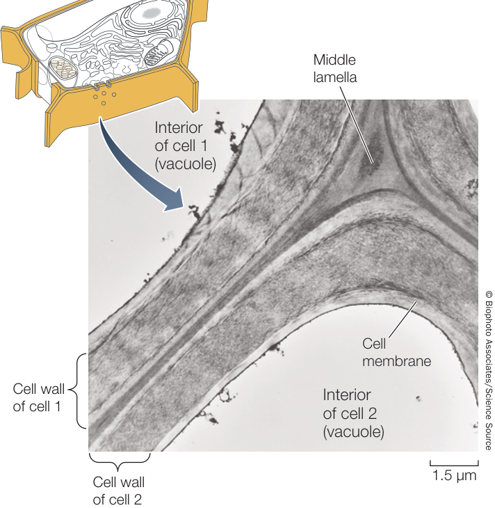
Figure 5.23 The Plant Cell Wall The semirigid cell wall provides support for plant cells. It is composed of cellulose fibrils embedded in a matrix of polysaccharides and proteins.
The extracellular matrix supports tissue functions in animals
Animal cells lack the semirigid wall that is characteristic of plant cells, but many animal cells are surrounded by, or in contact with, an extracellular matrix. This matrix is composed of three types of molecules: fibrous proteins such as collagen (the most abundant protein in mammals, constituting over 25% of the protein in the human body); a matrix of glycoproteins termed proteoglycans, consisting primarily of sugars; and a third group of proteins that link the fibrous proteins and the gel-like proteoglycan matrix together (Figure 5.24). These proteins and proteoglycans are secreted, along with other substances that are specific to certain body tissues, by cells that are present in or near the matrix.
The extracellular matrix has several functions:
- It holds cells together in tissues. In Chapter 6 we will describe how there is an intercellular “glue” that is involved in both cell recognition and adhesion.
- It contributes to the physical properties of cellular aggregates called tissues. Cartilage and bone are part of connective tissue. For example, the mineral component of bone is laid down on an organized extracellular matrix. This property can be used in reverse, to create organs in the laboratory. In tissue engineering, cells are removed from the extracellular matrix and the latter serves as a scaffold for the development of new organs with the correct architecture. This is a frontier in organ transplantation in humans whose organs fail.
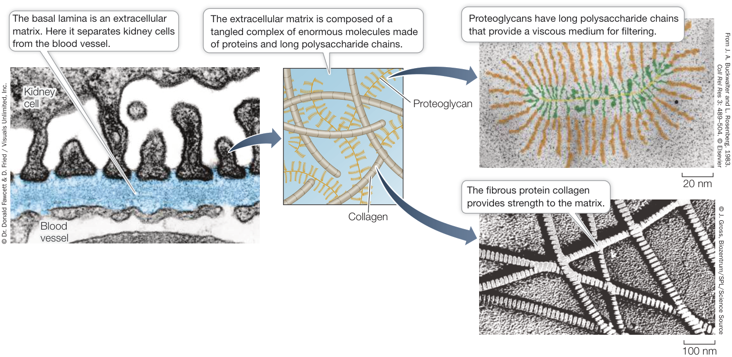
Figure 5.24 An Extracellular Matrix Cells in the kidney secrete a basal lamina, which is an extracellular matrix that separates the kidney cells from a nearby blood vessel. The basal lamina filters materials that pass between the kidney and the blood. Q: Why do some people take the dietary supplement chondroitin sulfate, which is a proteoglycan?
- It helps filter materials passing between different tissues. This is especially important in the kidney.
- It helps orient cell movements during embryonic development and during tissue repair.
- It plays a role in chemical signaling from one cell to another. Proteins connect the cell’s cell membrane to the extracellular matrix. These proteins (for example, integrin) span the cell membrane and are involved with transmitting signals to the interior of the cell. This allows communication between the extracellular matrix and the cytoplasm of the cell.
Recap and Assess
Extracellular structures are produced by cells and secreted outside the cell membrane. Most consist of a fibrous component in a gel-like medium.
- When a cancer spreads from the initial site to another location in the body, initial events are the detachment of the cancer cells from its surrounding extracellular matrix and digestion of the matrix as the cell migrates. How might these events occur?
- How does the structure of a cell wall contribute to its function?
- How could a biologist use chemical analysis to differentiate between a plant cell and an animal cell?
We have now discussed the structures and some functions of prokaryotic and eukaryotic cells. Both exemplify the cell theory, showing that cells are the basic units of life and of biological continuity. Much of the rest of this part of the book will deal with these two aspects of cells. There is abundant evidence that the simpler prokaryotic cells are more ancient than eukaryotic cells, and that the first cells were probably prokaryotic. We will now turn to the next step in cellular evolution, the origin of eukaryotic cells.
Eukaryotic Cells Evolved in Several Steps
Learning Objectives
- 5.5.1 Explain how the cell membrane could have been involved in the evolution of the endomembrane system in eukaryotes.
- 5.5.2 Relate endosymbiotic theory to observations of modern cells.
Life on Earth was entirely prokaryotic for about 1 billion years—from the time when prokaryotic cells first appeared until about 2.7 billion years ago, when evidence of eukaryotic cells appears in ancient rocks. The advent of compartmentalization—the hallmark of eukaryotes—was a major event in the history of life. It permitted many more biochemical functions to exist in the same cell than had previously been possible. Compared with a typical eukaryote, a single prokaryotic cell is often biochemically specialized, limited in the resources it can use and the functions it can perform.
What is the origin of compartmentalization? We will describe the evolution of eukaryotic organelles in more detail in Key Concept 24.2. Here we outline two major themes in this process.
Internal membranes and the nuclear envelope probably came from the cell membrane
We noted earlier in this chapter that some bacteria contain internal membranes. How could these arise? In electron micrographs, the internal membranes of prokaryotes often appear to be inward folds of the cell membrane. This observation has led to a theory that the endomembrane system and the cell nucleus originated by related processes (Figure 5.25A). The close relationship between the endoplasmic reticulum (ER) and the nuclear envelope in today’s eukaryotes is consistent with this theory.
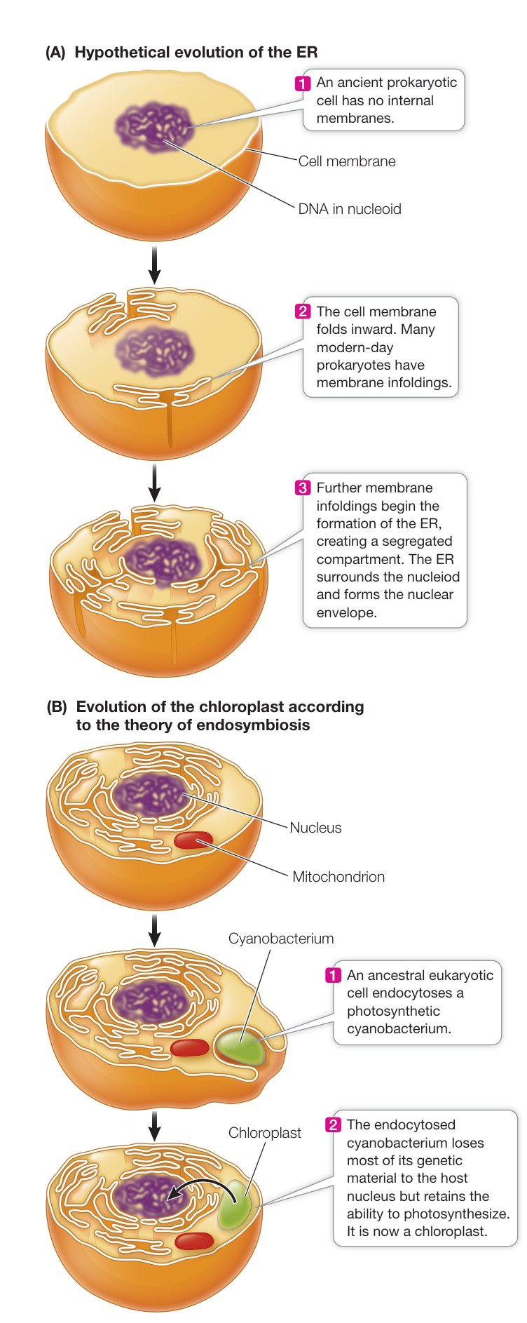
Figure 5.25 The Origins of Organelles (A) The endomembrane system and nuclear envelope may have been formed by infolding and then fusion of the cell membrane. (B) The endosymbiosis theory proposes that some organelles may be descended from prokaryotes that were engulfed by other, larger cells.
A bacterium with enclosed compartments would have several evolutionary advantages. Chemicals could be concentrated in particular regions of the cell, allowing chemical reactions to proceed more efficiently. A biochemical process could be segregated within an organelle with, for example, a different pH from the rest of the cell, creating more favorable conditions for that process. Finally, DNA transcription could be separated from translation, providing more opportunities for separate control of these steps in gene expression.
Some organelles arose by endosymbiosis
Symbiosis means “living together,” and often refers to two organisms that coexist, each one supplying something that the other needs. With the theory of endosymbiosis biologists have proposed that some organelles—the mitochondria and the plastids—arose not by an infolding of the cell membrane but by one cell ingesting (but not digesting) another cell, giving rise to a symbiotic relationship. Eventually, the ingested cell lost its autonomy and some of its functions. In addition, many of the ingested cell’s genes were transferred to the host’s DNA. Mitochondria and plastids in today’s eukaryotic cells are the remnants of these symbionts, retaining some specialized functions that benefit their host cells.
Consider the case of the plastid. About 2.5 billion years ago some prokaryotes (the cyanobacteria) developed photosynthesis. The emergence of these prokaryotes was a key event in the evolution of complex organisms because they increased the concentration in Earth’s atmosphere (see Key Concept 24.2).
According to the endosymbiosis theory, photosynthetic prokaryotes also provided the precursor of the modern-day plastid. Cells without cell walls can engulf relatively large particles by phagocytosis (see Figure 5.12). In some cases, such as that of phagocytes in the human immune system, the engulfed particle can be an entire cell, such as a bacterium. Plastids may have arisen by a similar event involving an ancestral eukaryote and a cyanobacterium (Figure 5.25B).
Recap and Assess
Eukaryotic cells arose long after prokaryotic cells. Some organelles may have evolved by infolding of the cell membrane, whereas others probably evolved by endosymbiosis.
- How could membrane infolding in a prokaryotic cell lead to the formation of the endomembrane system?
- When they are removed from a cell, organelles such as mitochondria no longer function as they did inside the cell. What aspect of endosymbiotic theory explains this observation?
In this chapter we presented an overview of the components of cells, with some ideas about their structures, functions, and origins. As you now embark on the study of major cellular processes, keep in mind that cellular components do not exist in isolation:
they are part of a dynamic, interacting system. In Chapter 6 you will see that the cell membrane is far from a passive barrier, but instead is a multifunctional system that connects the inside of the cell with its extracellular environment.
Investigating Life: QA Are pigments compartmentalized in all types of cells?
In this chapter you learned of several situations in which pigments are concentrated in discrete organelles: in humans, melanin is packaged in melanosomes in human skin cells; in plants, tannins are packaged in tannosomes, chlorophyll is packaged in thylakoids, other pigments are contained in chromoplasts, and anthocyanins that may attract pollinators and seed dispersers are packaged in the vacuoles in petals and fruits. You learned, too, of the remarkable ability of motor proteins to move pigment granules into the center of cells in fishes and amphibians, to lighten their skin color as a defensive mechanism.
Many biological traits and adaptations are linked to pigments in organelles. For example, the distribution of melanosomes is related to eye color, and genetically determined errors in the formation of melanosomes can lead to albinism and a lack of protection against ultraviolet light. In plants, the formation of tannosomes is associated with plant defenses (see Chapter 37). Understanding the pathway by which tannosomes are formed could have beneficial applications in agriculture. For example, tannins are important to the taste of red wine, and manipulating tannin concentrations can result in new flavors.
Future directions
The most common form of human cancer is skin cancer. Most skin cancers are easily treated, but a rare type, melanoma, can be lethal. As its name implies, melanoma occurs when melanocytes in the skin multiply excessively. This can happen if melanocytes are exposed to ultraviolet light from the Sun in excess of the ability of their melanin pigment to absorb it. When localized in the skin, melanoma is not harmful; it’s just a growth. But when the tumor cells spread to other organs in a process called metastasis, melanoma often is lethal. Carmit Levy at the Sackler School of Medicine in Israel has found that the tumor sends a signal, contained in melanosomes, to neighboring skin cells that changes them and allows the tumor to spread. An experimental drug that blocks melanosome release also inhibits metastasis in animal studies. The drug is under development for human use.
Visual Summary
© Dennis Kunkel Microscopy/Science Source
You should be able to relate each summary to the adjacent figures. If you go to this visual summary in Achieve, you can follow links to figures, animations, activities, and simulations that will help you consolidate the material.
Cells Are the Fundamental Units of Life
- The cell theory is the unifying theory of cell biology. All living things are composed of cells, and all cells come from preexisting cells.
- A cell is small and maintains a large surface area-to-volume ratio. This allows it to exchange adequate quantities of materials with its environment.
- Whereas certain biochemical processes, molecules, and structures are shared by all kinds of cells, there are two categories of organisms—prokaryotes and eukaryotes—that can be distinguished by characteristic cell structures.
Questions
- One cell type has twice the diameter of another. Yet the first cell type has a larger surface area-to-volume ratio. How can this occur?
- A cell has a diameter of 2 µm. Is the cell prokaryotic or eukaryotic? Explain.
Go to Activities 5.1 and 5.2
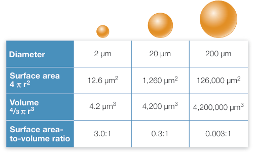
Figure 5.2
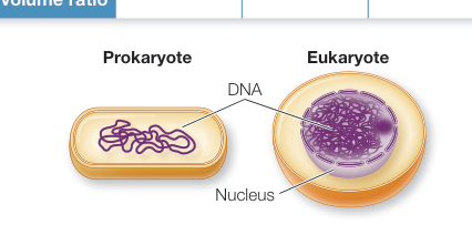
Figure 5.5
Prokaryotic Cells Are the Simplest Cells
- Prokaryotic cells have no internal compartments but have a nucleoid region containing DNA. The rest of the material inside the cell membrane, including the ribosomes, is called the cytoplasm. Some prokaryotes have additional protective structures, including a cell wall, an outer membrane, and a capsule.
- Some prokaryotes have folded internal membranes, and some have flagella or pili for motility or attachment.
Questions
- Penicillin and related antibiotics inhibit the synthesis of the bacterial cell wall. How do you think this kills bacteria?
- There are more prokaryotic cells than eukaryotic cells in the human body. Explain.
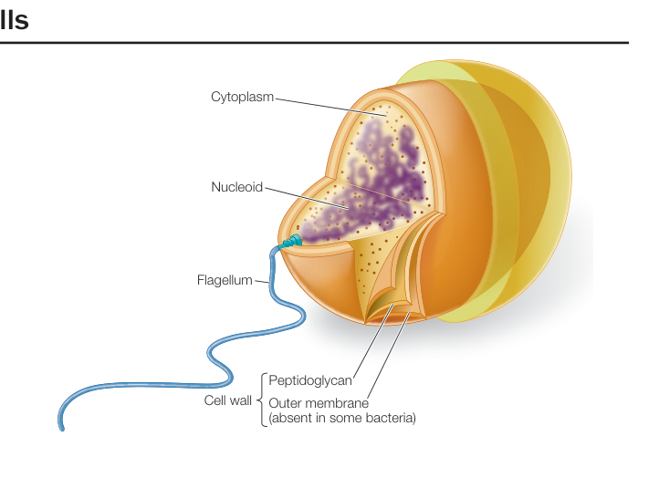
Figure 5.6
Eukaryotic Cells Contain Organelles
- Eukaryotic cells are larger than prokaryotic cells and contain many membrane-enclosed organelles that compartmentalize functions.
- The nucleus contains most of the cell’s DNA and participates in the control of protein synthesis, which occurs in the cytoplasm at ribosomes.
- The endomembrane system—consisting of the cell membrane, the nuclear envelope, the endoplasmic reticulum, the Golgi apparatus, and lysosomes—is a series of interrelated compartments enclosed by membranes. It segregates proteins and modifies them. Lysosomes contain many digestive enzymes.
- Mitochondria and chloroplasts are organelles that process energy.
- The microfilaments, intermediate filaments, and microtubules of the cytoskeleton provide the cell with shape, strength, and movement.
Questions
- What organelles or structures are unique to eukaryotic plant cells?
- A human genetic disease, Zellweger syndrome, arises when there is an absence of peroxisomes. What do you suppose are the biochemical consequences of this situation?
- Another human genetic disease, I-cell disease, arises from an inability of the Golgi apparatus to correctly modify certain proteins to target them to lysosomes. What would be the consequences?
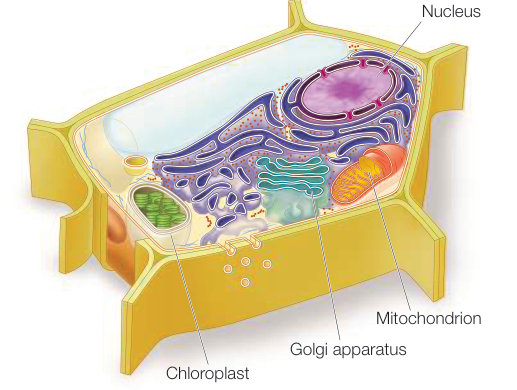
Figure 5.9
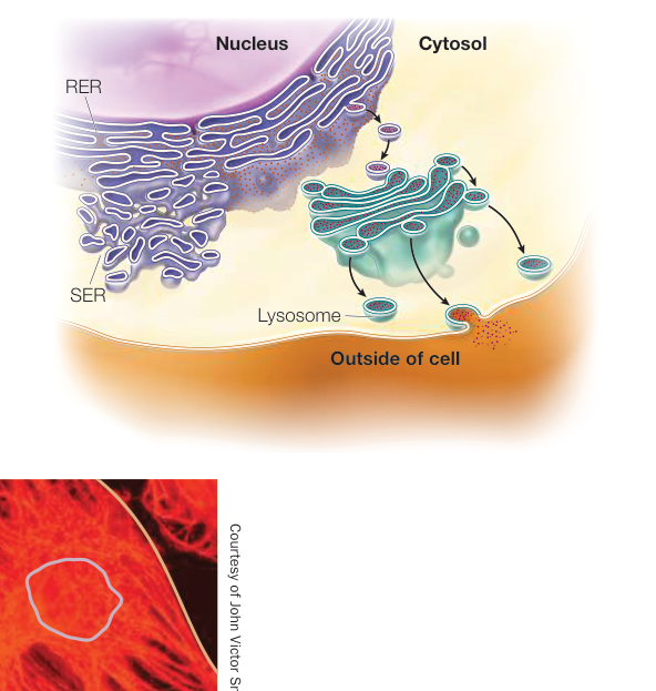
Figure 5.11
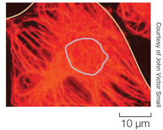
Figure 5.17
Extracellular Materials Provide Structure
- The plant cell wall consists principally of cellulose.
- In animal cells, the extracellular matrix consists of different kinds of proteins, including collagen and proteoglycans.
Questions
- Compare the plant cell wall with the animal extracellular matrix with respect to the fibrous and non-fibrous components.
- There are proteins and polysaccharides in both the plant cell wall and animal extracellular matrix. Where are these molecules made, and how do they get into the extracellular structures?
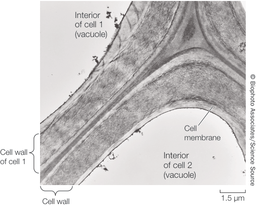
Figure 5.23
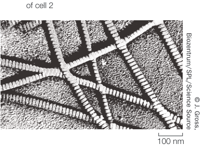
Figure 5.24
Eukaryotic Cells Evolved in Several Steps
- Infoldings of the cell membrane could have led to the formation of some membrane-enclosed organelles, such as the endomembrane system and the nucleus.
- The theory of endosymbiosis states that mitochondria and chloroplasts originated when larger cells engulfed, but did not digest, smaller cells.
Questions
- Could the lysosome have evolved by endosymbiosis? Explain.
- Could mitochondria have evolved nonsymbiotically? Explain.
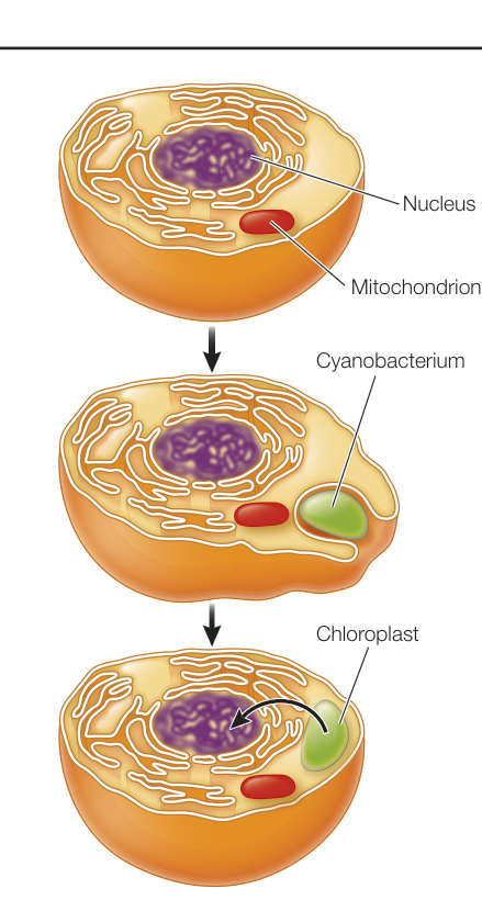
Figure 5.25