Chapter 7Cell Communication and Multicellularity
Courtesy of Todd Ahern
Key Concepts
- 7.1 Cells Detect a Variety of Signals
- 7.2 Receptors Bind Signals to Initiate a Cellular Response
- 7.3 Signal Transduction Spreads the Response to a Signal through the Cell
- 7.4 Cells Change in Response to Signals in Several Ways
- 7.5 Adjacent Cells in a Multicellular Organism Can Communicate Directly
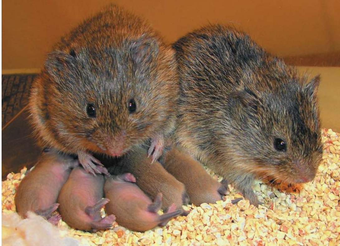
Voles Prairie voles display bonding behaviors after mating. These behaviors are mediated by peptides acting as intercellular signals.
Investigating Life: A Signal for Bonding
Prairie voles (Microtus ochrogaster) are small rodents that live in temperate climates, where they dig tunnels in fields. When a male prairie vole encounters a female, the female enters her reproductive cycle and mating often ensues. After mating the pair stays together, building a nest and raising their pups, born after 22 days gestation. The bond between the two voles is so strong that they stay together for life, producing a litter of pups about once a month for 2 to 3 years. Contrast this behavior with that of the montane vole (M. montanus), which is closely related to the prairie vole and lives in the hills not far away. In this species, mating is quick, and afterward the pair separates. The male looks for new mates and the female abandons her young soon after they are born.
The reason for these dramatic behavioral differences lies in the brains of these two species. When prairie voles mate, the brains of both males and females release specific peptides consisting of nine amino acids. In females, the peptide is oxytocin; in males, it is vasopressin. The peptides circulate in the bloodstream and reach all tissues in the body, but they bind to only a few cell types. These cells have surface proteins called receptors, to which the peptides specifically bind, like a key inserting into a lock.
The binding of peptide to receptor causes the receptor, which extends across the cell membrane, to change shape. Within the cytoplasm, this change sets off a series of events that ultimately result in changes in behavior. The receptors for oxytocin and vasopressin in prairie voles are most concentrated in the regions of the brain that are responsible for behaviors such as bonding and caring for young. In montane voles, there are far fewer receptors for these peptides, and as a result, fewer bonding and caring behaviors. Clearly, oxytocin and vasopressin are signals that induce these behaviors.
Intercellular signaling is a hallmark of multicellular organisms. A cell’s response to a signal molecule takes place in three sequential steps. First, the signal binds to a receptor in the cell, often embedded in the outside surface of the cell membrane. Second, signal binding conveys a message to the cell. Third, the cell changes its activity in response to the signal. In a multicellular organism, these steps lead to changes in that organism’s functioning, with signaling allowing different types of cells and tissues to work together.
QA Is oxytocin the “trust” signal in humans? (Find the answer on page 149.)
Cells Detect a Variety of Signals
Learning Objectives
- 7.1.1 Use the source and distribution mechanism of a signal to identify its type.
- 7.1.2 Give an example of how a particular type of signal (autocrine, juxtacrine, paracrine, or hormone) functions in an organism.
- 7.1.3 Explain how specificity is achieved in a signal transduction pathway.
- 7.1.4 Compare cells found in the same organism that can and cannot respond to a specific chemical signal.
The environment around an organism and cell varies both in space and time. All organisms, prokaryotic and eukaryotic, can detect and respond to a multitude of environmental variables, including light, temperature, sound, touch, magnetic fields, gravity, pH, and chemicals. For example, plants grow toward light, bacteria alter gene transcription when the temperature rises, barnacle larvae sense and then attach to solid surfaces, pigeons navigate using Earth’s magnetic field, and carrion beetles fly toward the odor of dead animals. While these responses involve the whole organism or particular organs, environmental variables are detected by individual cells, which undergo changes that lead to responses.
The presence of a signal does not always lead to a response; a cell does not respond to every signal, just as you are not paying close attention to every stimulus in your environment as you read this. To respond to a signal, a cell must have a specific receptor that can detect it and a way to use that information to influence cellular processes. A signal transduction pathway is a sequence of molecular events and chemical reactions that lead to a cell’s response to a signal. Signal transduction pathways vary greatly in their details, but every such pathway involves a signal, a receptor, and a response. In this key concept we provide a brief overview of signal transduction. We’ll consider receptors in Key Concept 7.2, and other aspects of signal transduction in Key Concepts 7.3 and 7.4.
Cells receive several types of signals
The environment is full of signals. For example, our sense organs allow us to respond to light (a physical signal), or odors and tastes (chemical signals). Bacteria and protists can respond to small chemical changes in their surroundings. Plants respond to light as a signal as well as an energy source, for example, by growing toward the source of light. A cell that is deep inside a large multicellular organism and far away from the exterior environment receives signals from neighboring cells and the surrounding extracellular fluids. In multicellular organisms, chemical signals are often made in one part of the body and arrive at target cells by local diffusion or by circulation in the blood or the plant vascular system. Chemical cell signals are usually present in tiny concentrations (as low as M) (see Key Concept 2.4 for an explanation of molar concentrations) and differ in their sources and mode of delivery (Figure 7.1):
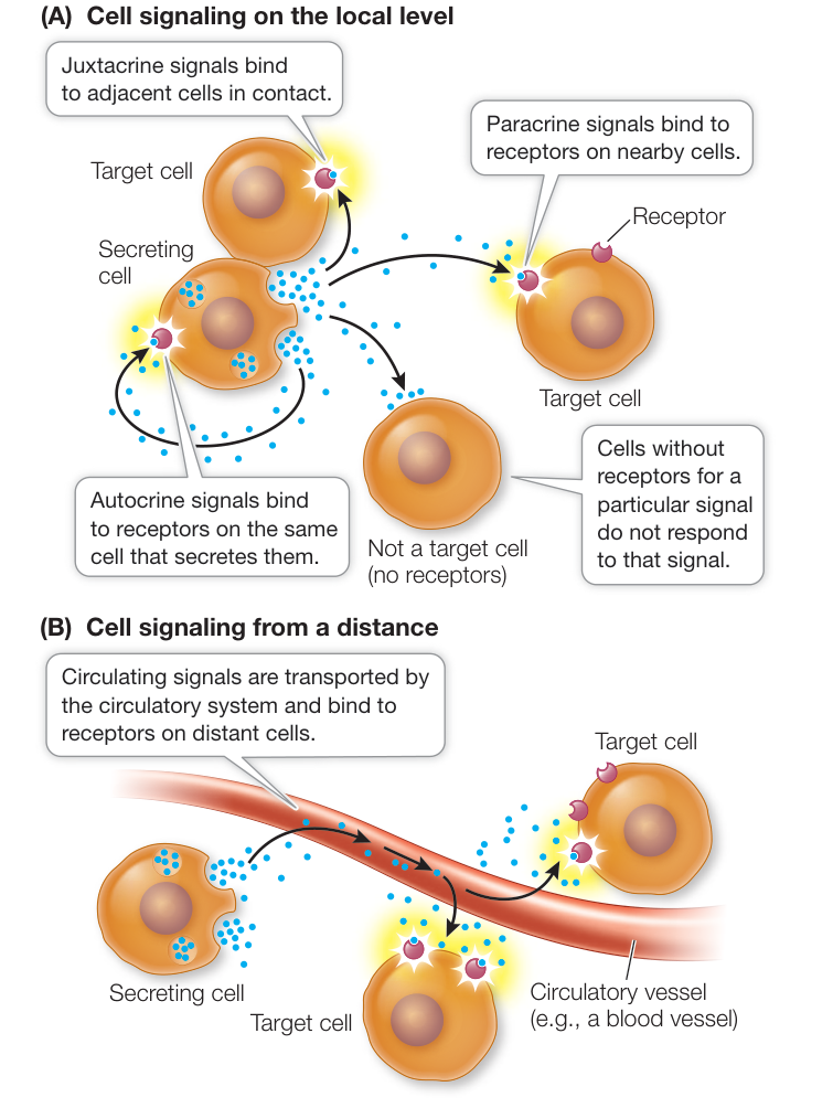
Figure 7.1 Chemical Signaling Systems (A) A signal molecule can diffuse to and act on the cell that produces it, an adjacent cell, or a nearby cell. (B) Chemical signals that act on distant cells must be transported by the organism’s circulatory system. Q: Based on the opening story, is oxytocin signaling autocrine, juxtacrine, or signaling at a distance? Activity 7.1 Chemical Signaling Systems
- Autocrine signals diffuse to and affect the cells that make them. For example, many tumor cells reproduce uncontrollably because they both make, and respond to, signals that stimulate cell division.
- Juxtacrine signals affect only cells right next to and in contact with the cell producing the signal. This type of signaling is especially common during development, when cells are in groups and changing to become specialized.
- Paracrine signals diffuse to and affect nearby cells. An example occurs in inflammation when the skin is cut. Signals from skin cells are sent to nearby blood cells to aid in healing (see Key Concept 40.2).
- Signals that travel through the circulatory systems of animals or the vascular systems of plants are generally called hormones.
A signal transduction pathway involves a signal, a receptor, and responses: Overview
As you saw in the opening story, the elements of a signal transduction pathway are a signal, a receptor, and a response (Figure 7.2). For the information from a signal to be transmitted to a cell, the target cell must be able to receive the signal and respond to it. This is the job of receptors. All cells may be exposed to a signal, but most cells in the organism may not be able to respond to it. Only cells with the appropriate receptors can respond. Signal–receptor interaction, which often but not always occurs at the cell membrane, sets off a series of events inside the cell. Usually the events of signal transduction amplify the signal’s effects. As Figure 7.2 shows, the ultimate responses of the cell (and organism) may be short-term or long-term. Think of the rapid “fight-or-flight” response that occurs when you see a bear in the woods a few feet away, compared with the bonding behaviors mediated by oxytocin in the prairie voles in the opening story.
In this chapter we consider signal transduction pathways in isolation from one another. In life, however, signal transduction pathways are often interconnected. A great deal of crosstalk—interactions between different signal transduction pathways—is common. For example, a single activated protein (receptor or enzyme) might activate enzymes or transcription factors in multiple pathways, leading to multiple responses to a single stimulus.
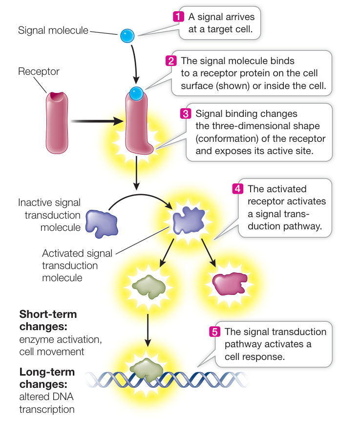
Figure 7.2 A Signal Transduction Pathway This general pathway is common to many cells and situations. The ultimate effects on the cell are either short-term or long-term molecular changes, or both.
Multiple signal transduction pathways might converge on a single transcription factor, allowing the transcription of a single gene to be adjusted in response to several different signals. Crosstalk can also result in the activation of one pathway and the inhibition of another. The phenomenon of crosstalk inside the cell is analogous to the “crosstalk” that occurs at the level of the whole body. For example, in your limbs you have opposing muscles. When you bend your elbow, you contract one set of muscles and relax the opposing muscles, so that your arm will bend. Because of crosstalk, biologists often refer to “signaling networks” rather than signal transduction pathways, reflecting the high degree of complexity in cellular signaling.
Recap and Assess
Cells are constantly exposed to molecular signals that can come from the external environment or, in a multicellular organism, from within the body. To respond to a signal, the cell must have a specific receptor that detects the signal and activates some type of cellular response. Signal transduction pathways involve regulation of enzymes and transcription factors, and crosstalk often occurs between pathways.
- Under what conditions would autocrine signaling be most advantageous for a cell?
- How are hormones delivered to target cell receptors?
- All cells have the potential to respond to all signals in an organism. What makes signaling specific?
- Some cells can detect pH levels in their surroundings. How might a receptor detect changes in pH?
The general features of signal transduction pathways described in this key concept will recur in more detail throughout the chapter. First, let’s consider more closely the nature of the receptors that bind signal molecules.
Receptors Bind Signals to Initiate a Cellular Response
Learning Objectives
- 7.2.1 Explain how and why different cells respond to different chemical signals.
- 7.2.2 Using equations, describe the chemical kinetics of receptor–ligand interactions.
- 7.2.3 Analyze dissociation constants as a measure of binding affinity.
- 7.2.4 Compare and contrast cell membrane receptors and intracellular receptors.
Any given cell in a multicellular organism is bombarded with many signals. However, it responds to only some of them, because no cell makes receptors for all signals. A receptor protein recognizes its signal very specifically, in much the same way that a membrane transport protein recognizes and binds to the substance it transports. This specificity ensures that only those cells that make a specific receptor will respond to a given signal.
Receptors that recognize chemical signals have specific binding sites
A ligand is a specific chemical signal molecule that fits into a three-dimensional site on its protein receptor (Figure 7.3A). Binding of the signaling ligand causes the receptor protein to change its three-dimensional shape, and that conformational change initiates a cellular response. The ligand does not contribute further to this response. In fact, the ligand is usually not changed; its role is purely to “knock on the door.”
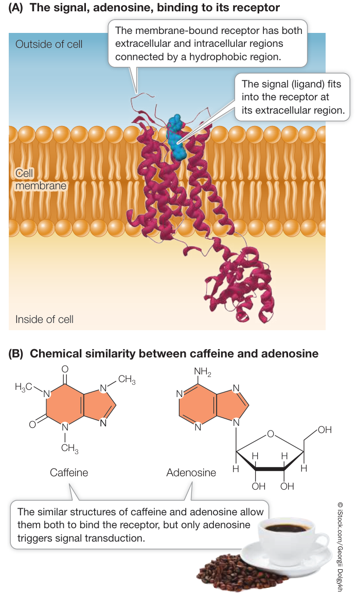
Figure 7.3 A Signal and Its Receptor (A) The adenosine 2A receptor occurs in the human brain, where it is involved in inhibiting active wakefulness. (B) Adenosine is the normal (endogenous) ligand for the receptor. Caffeine has a structure similar to that of adenosine and can act as an antagonist that binds the receptor and prevents its normal functioning. (A data from PDB 3EML. V. P. Jaakola et al. 2008. Science 322: 1211–1217.) Q: Is the binding of adenosine and caffeine covalent or noncovalent? Explain your answer.
The sensitivity of a cell to a signal is determined in part by the affinity of the cell’s receptors for the signal ligand—the likelihood that the receptor will bind to the ligand at any given ligand concentration. Receptors (R) bind to their ligands (L) according to chemistry’s law of mass action. This means that the binding is reversible:
For most ligand–receptor complexes (RL), binding is favored. Reversibility is important, however, because if the ligand were never released, the receptor would be continuously stimulated and the cell would never stop responding.
As with any reversible chemical reaction, the binding and dissociation processes each have a rate constant, here designated and :
A rate constant relates the rate of a reaction to the concentration(s) of the reactant(s):
where “[ ]” indicates the concentration of the substance inside the brackets. Binding of a receptor to a ligand is reversible, and when equilibrium is reached the rate of binding equals the rate of dissociation:
If this is rearranged, we get:
, the dissociation constant, is a measure of the affinity of the receptor for its ligand. The lower the , the higher the affinity of the ligand for its receptor. Some receptors have very low values, which allows them to bind their ligands at very low ligand concentrations; other receptors have higher values and need more ligand to be present to set off their signal transduction pathways.
An entire field of biology and medicine—called pharmacology—is devoted to the study of drugs. These molecules are usually thought of as synthetic, but some are natural substances, such as caffeine that come from plants used by traditional societies (see Figure 7.3B). Drugs function as ligands that bind specific receptors. In the discovery and design of new drugs, it is helpful to know the specific receptor that the drug will bind, because then it is possible to determine the value of its binding. This is one factor that can be taken into consideration when determining dosage levels. Of course, many drugs have side effects, and these are also dosage-dependent.
What happens when a ligand binds to a receptor? When we discussed small molecules binding to proteins in Key Concept 3.2, we described how proteins often change shape when bound. This is exactly what happens to receptors. The change in the receptor’s shape may expose a previously hidden group of amino acids on the protein that participate in a biochemical activity. This activity may be the binding of another molecule, such as another protein (e.g., a G protein, discussed in the next section), or a substrate for an enzyme.
Instead of the ligand, other chemicals that resemble it can bind to the receptor. Agonists are chemicals that set a receptor into signal transduction mode just as the ligand does. Antagonists are inhibitors that bind to the receptor and “freeze” it in place, preventing the real ligand from binding, but do not set off signal transduction.
Many substances that alter human behavior bind to specific receptors in the brain and prevent the binding of the receptors’ specific ligands. One example is caffeine, which is probably the world’s most widely consumed stimulant. In the brain, the nucleoside adenosine acts as a ligand that binds to a receptor on nerve cells, initiating a signal transduction pathway that reduces brain activity, especially feelings of active wakefulness. Because caffeine has a molecular structure similar to that of adenosine, it also binds to the adenosine receptor (Figure 7.3B) and acts as an antagonist, preventing adenosine binding and thereby allowing continued nerve cell activity and an active feeling.
Receptors can be classified by location and function
There are many kinds of chemical signals. Some ligands are hydrophobic (nonpolar) and can diffuse through membranes, whereas others cannot. Physical signals such as light also vary in their ability to penetrate cells and tissues. Correspondingly, a receptor can be classified by its location in the cell, which largely depends on the nature of its signal (Figure 7.4):
- Membrane receptors: Large or polar ligands cannot cross the lipid bilayer. Insulin, for example, is a protein hormone that cannot diffuse through the cell membrane; instead, it binds to a transmembrane receptor with an extracellular binding domain.
- Intracellular receptors: Small or nonpolar ligands can diffuse across the nonpolar phospholipid bilayer of the cell membrane and enter the cell. The hormone estrogen, for example, is a lipid-soluble steroid (see Figure 3.24) that can diffuse across the cell membrane; it binds to a receptor inside the cell.
In complex eukaryotes such as mammals and higher plants, there are three well-studied categories of cell membrane receptors that are grouped according to their functions: ion channels, protein kinase receptors, and G protein-coupled receptors.
ION CHANNELS As you saw in Key Concept 6.3, the cell membranes of many types of cells have gated ion channels that allow ions such as , , , or to enter or leave the cell. The gate-opening mechanism is an alteration in the three-dimensional shape of the channel protein upon interaction with a signal; thus these proteins function as receptors. An example is the acetylcholine receptor, a ligand-gated channel located in the cell membrane of skeletal muscle cells. Acetylcholine is a neurotransmitter—a chemical signal released from nerve cells (Figure 7.5). Opening of the channel allows , which is more concentrated outside the cell than inside it, to diffuse into the cell. This initiates a series of events that result in muscle contraction (see Figure 46.5).
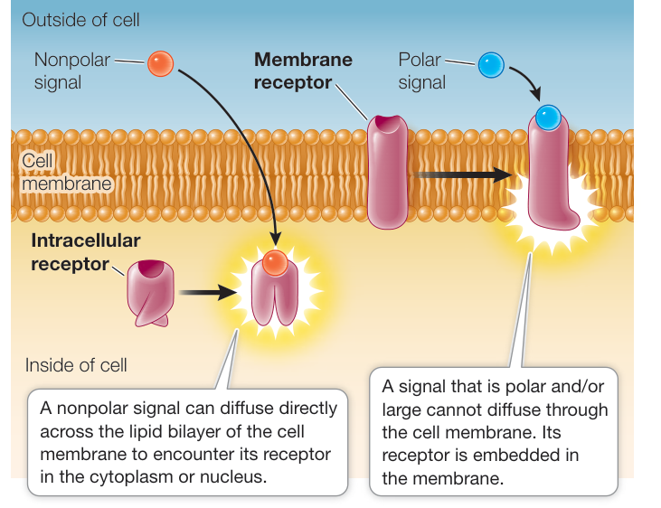
Figure 7.4 Two Locations for Receptors Receptors can be located inside the cell (in the cytoplasm or nucleus) or in the cell membrane. Q: Steroid hormones are nonpolar. Could a receptor for a steroid be located at the cell membrane?
Connect the Concepts Ion channels are key to the functioning of the nervous system. An example is the connection between nerve and muscle described in Key Concepts 43.3 and 46.1.
PROTEIN KINASE RECEPTORS Some eukaryotic receptor proteins, called protein kinases, catalyze the phosphorylation (addition of phosphate) of themselves or other proteins, thus changing their shapes and therefore their functions.
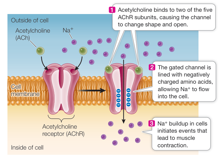
Figure 7.5 A Gated Ion Channel The acetylcholine receptor (AChR) is a ligand-gated ion channel for sodium ions ( ). It is made up of five polypeptide subunits. When acetylcholine molecules (ACh) bind to two of the subunits, the gate opens and flows into the cell. This channel helps regulate membrane polarity.
(altered shape and function)
Phosphorylation is a reaction that is especially important in biology. Of the estimated 20,500 genes that code for proteins in humans, more than 500 of them encode protein kinases. When you think of all the functions that make up a person, this is indeed an impressive number. There are three amino acids in proteins that can be phosphorylated (Figure 7.6A). The receptor for insulin is an example of a protein kinase receptor. Insulin is a protein hormone made by the pancreas. Its receptor has two copies each of two different polypeptide subunits called and (Figure 7.6B). When insulin binds to the receptor, the receptor becomes activated and is able to phosphorylate itself and certain cytoplasmic proteins that are appropriately called insulin-response substrates. These proteins then initiate many cellular responses, including the insertion of glucose transporters (see Figure 6.12A) into the cell membrane.
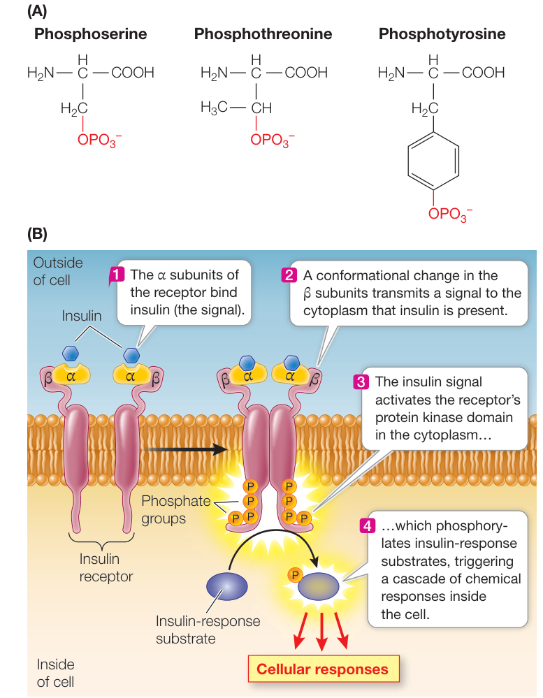
Figure 7.6 A Protein Kinase Receptor (A) The three amino acids that can be phosphorylated by protein kinase activity. (B) The hormone insulin binds to a receptor on the outside surface of the cell and initiates a response.
G PROTEIN-COUPLED RECEPTORS Another category of eukaryotic cell membrane receptors is the G protein-coupled receptors. These receptors have many roles, including light detection in the mammalian retina (photoreceptors), detection of odors (olfactory receptors), and regulation of mood and behavior (such as mating in mammals and even single-celled yeasts). The receptors that bind the hormones oxytocin and vasopressin, which affect mating behavior in voles (see the opening story), are G protein-coupled receptors. Humans have hundreds of G protein-coupled receptors.
Ligand binding on the extracellular side of the receptor changes the shape of its cytoplasmic region, exposing a site that binds to a mobile membrane protein called a G protein. The G protein is partially inserted into the lipid bilayer and partially exposed on the cytoplasmic surface of the membrane.
Many G proteins have three polypeptide subunits and can bind three different types of molecules (Figure 7.7A):
- The receptor
- GDP and GTP (guanosine diphosphate and triphosphate, respectively; these are nucleoside phosphates like ADP and ATP)
- An effector protein (see next paragraph)
When the G protein binds to an activated receptor protein, GDP is exchanged for GTP (Figure 7.7B). At the same time, the ligand is usually released from the extracellular side of the receptor. GTP binding causes a conformational change in the G protein. The GTP-bound subunit then separates from the rest of the G protein, diffusing in the plane of the phospholipid bilayer until it encounters an effector protein to which it can bind. An effector protein is just what its name implies: it causes an effect in the cell. The binding of the GTP-bearing G protein subunit activates the effector, thereby causing changes in cell function (Figure 7.7C).
After activation of the effector protein, the GTP bound to the G protein is hydrolyzed to GDP. The now inactive G protein subunit separates from the effector protein and diffuses in the membrane to bind to the other two G protein subunits. When the three components of the G protein are reassembled, the protein is capable of binding again to an activated receptor. After binding, the activated receptor exchanges the GDP on the G protein for a GTP, and the cycle begins again.
Intracellular receptors are located in the cytoplasm or the nucleus
Intracellular receptors are located inside the cell and respond to physical signals such as light (e.g., some photoreceptors in plants) or chemical signals that can diffuse across the cell membrane (e.g., steroid hormones in animals). Many intracellular receptors are transcription factors. Some are located in the cytoplasm until they are activated; after binding their ligands, these transcription factors move to the nucleus where they bind to DNA and alter the expression of specific genes. A typical example is the receptor for the steroid hormone cortisol. This receptor is normally bound to a chaperone protein that blocks it from entering the nucleus. Binding of the hormone causes the receptor to change its shape so that the chaperone is released
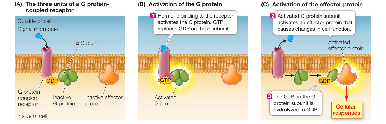
Figure 7.7 A G Protein-Coupled Receptor The G protein is an intermediary between the receptor and its effector. Q: How does ligand binding alter the subunit of the G protein, and what are two effects of this alteration?
Animation 7.1 A Signal Transduction Pathway
(Figure 7.8). This release allows the receptor to enter the nucleus, where it affects DNA transcription. Another group of intracellular receptors is always located in the nucleus, and their ligands must enter the nucleus before binding.
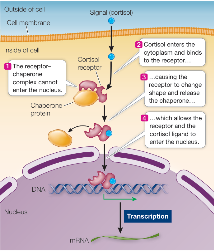
Figure 7.8 An Intracellular Receptor The receptor for cortisol is in the cytoplasm, bound to a chaperone protein that is released when cortisol binds to the receptor.
Recap and Assess
Receptors are proteins that bind, or are changed by, specific ligands or physical signals. The changed receptor initiates a response in the cell. These receptors are located in the cell membrane or inside the cell.
- What is the dissociation constant , and what does it tell us about the likelihood that a signal will produce a cellular response?
- What is the chemical nature and importance of specificity in the binding of a receptor to its particular ligand?
- What are the differences between receptors located in the cytoplasm and those at the cell membrane?
- An intact cell is treated to remove all proteins on the cell surface. Will the cell be able to detect any signals from other cells?
Now that we have discussed signals and receptors, let’s examine the characteristics of the molecules (transducers) that mediate the cellular response.
Signal Transduction Spreads the Response to a Signal through the Cell
Signal Transduction Spreads the Response to a Signal through the Cell
Learning Objectives
- 7.3.1 Describe how a protein cascade amplifies a signal.
- 7.3.2 Explain how the same second-messenger molecule can be used in many different signal transduction pathways.
- 7.3.3 Explain how signal transduction pathways are regulated in a cell.
As you have seen, there are different kinds of signals and receptors. Not surprisingly, the ways that signals are transduced, and the resulting cellular responses, also vary. Some signal transduction pathways are quite simple and direct, whereas others involve multiple steps. In addition, second messengers can diffuse throughout the cytoplasm and mediate further steps in pathways.
In many cases, a signal initiates a cascade (chain) of events, in which proteins interact with other proteins, which interact with still other proteins until the final responses are achieved. Through such a cascade, an initial signal can be both amplified and distributed to cause several different responses in the target cell. In this key concept we examine the kinds of molecules that transduce signals and look at several different signal transduction pathways.
The cell amplifies its response to ligand binding in a cascade
Scientists worked out the signal transduction pathway for one growth factor by studying a cell that went wrong. Many human bladder cancers contain an abnormal form of a protein called Ras (so named because a similar protein was previously isolated from a rat sarcoma tumor). Ras is a G protein, and as described in Key Concept 7.2, G proteins function as switches with “on” and “off” states. In the “off” state, G protein is bound to GDP, whereas in the “on” state the protein becomes bound to GTP. The abnormal form of the Ras protein under study was always active because it was permanently bound to GTP and thus caused continuous cell division (Figure 7.9).
Other types of cancer cells have abnormalities in different parts of the same signal transduction pathway. By comparing the defects in abnormal cells with the normal signaling process in non-cancer cells, biologists worked out the entire signaling pathway (Figure 7.10). Recall from Key Concept 7.2 that a G protein mediates a response after activation by the protein kinase receptor. In Figure 7.10, the activated G protein Ras initiates a series of additional events composing a signal transduction pathway known as a protein kinase cascade, wherein one protein kinase activates the next, and so on. In this pathway, the ligands stimulate cell division or mitosis, and so the ligands are called mitogens. The various kinases are called MAPKs—mitogen activated protein kinases. Protein kinase cascades are useful signal transducers for four reasons:
- At each step in the cascade of events, the signal is amplified, because each newly activated protein kinase is an enzyme that can catalyze the phosphorylation of many target proteins (see Figure 7.10, steps 5 and 6).
- The information from a signal that originally arrived at the cell membrane is communicated to the nucleus, where the expression of multiple genes is often modified.
- The multitude of steps provides some specificity to the process.
- Different target proteins at each step in the cascade can provide variation in the response.
Second messengers can amplify signals between receptors and target molecules
Often there is a small molecule intermediary between the activated receptor and the cascade of events that ensues. Earl Sutherland and his colleagues at Case Western Reserve University discovered one such molecule when they were investigating the activation of the liver enzyme glycogen phosphorylase by the hormone epinephrine. The hormone is released when an animal faces life-threatening conditions and needs energy fast for the fight-or-flight response (see Figure 39.3). Glycogen phosphorylase catalyzes the breakdown of glycogen stored in the liver so that the resulting glucose molecules can be released to the blood. The enzyme is present in the liver cell cytoplasm but is inactive unless the liver cells are exposed to epinephrine:
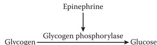
The researchers found that epinephrine bound to a receptor at the cell membrane but that enzyme activation occurred in the
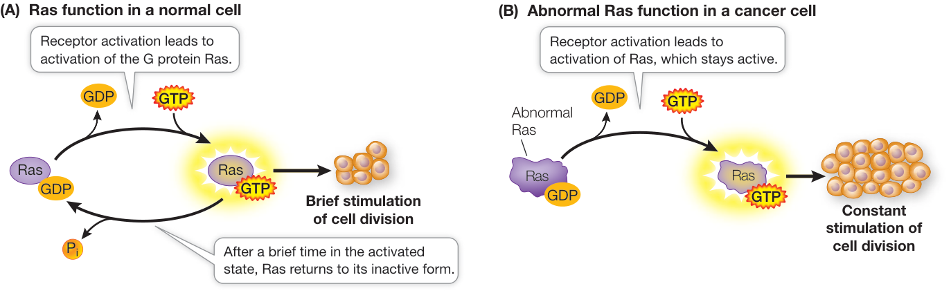
Figure 7.9 Signal Transduction and Cancer (A) Ras is a G protein that regulates cell division. (B) In some tumors, the Ras protein is permanently active, resulting in uncontrolled cell division.
Q: What would be the effect of a molecule that inhibits the interaction between GTP and Ras?
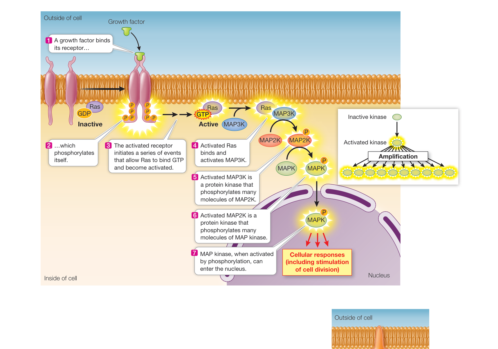
Figure 7.10 A Protein Kinase Cascade In a protein kinase cascade, a series of proteins are sequentially activated. Q: Sorafenib is a drug that was designed to inhibit MAP3K, which is very active in kidney cancer. How does this drug work on the protein kinase cascade?
cytoplasm. How was the information of ligand binding communicated to the enzyme in the cytoplasm? Sutherland and others found that a key was a chemical intermediary, a “second messenger” that transmits the epinephrine binding signal (epinephrine being the “first messenger”). The second messenger was identified as cyclic AMP (cAMP), which is produced from ATP by the enzyme adenylyl cyclase (Figure 7.11).
In contrast to the specificity of receptor binding, second messengers such as cAMP allow a cell to respond to a single event at the cell membrane with many events inside the cell. Thus second messengers amplify and distribute the signal—for example, binding of a single epinephrine molecule leads to the production of many molecules of cAMP, which then activate many enzyme targets by binding to them noncovalently. In the case of epinephrine and the liver cell, glycogen phosphorylase is just one of several enzymes that are activated.
Second messengers are often involved in crosstalk between different signaling pathways. Activation of the epinephrine receptor is not the only way for a cell to produce cAMP; and as noted, there are multiple targets of cAMP in the cell, and these targets are parts of other pathways.
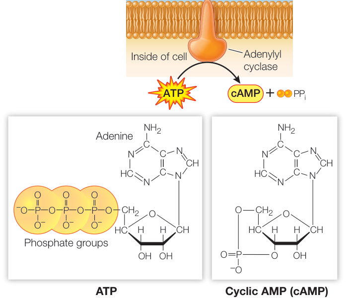
Figure 7.11 The Formation of Cyclic AMP The formation of cAMP from ATP is catalyzed by adenylyl cyclase, an enzyme that is activated by G proteins.

LIPID-DERIVED SECOND MESSENGERS In addition to their role as structural components of the cell membrane, phospholipids are also involved in signal transduction. When certain phospholipids are hydrolyzed into their component parts by enzymes called phospholipases, second messengers are formed. The best-studied examples of these second messengers come from hydrolysis of the phospholipid phosphatidyl inositol-bisphosphate (PIP2). Like all phospholipids, PIP2 has a hydrophobic portion embedded in the cell membrane: two fatty acid tails attached to a molecule of glycerol, which together form diacylglycerol, or DAG. The hydrophilic portion of PIP2 is inositol trisphosphate, or IP3, which projects into the cytoplasm.
Receptor binding activates phospholipase C, an enzyme that is also located in the membrane. This enzyme cleaves off the IP3 from PIP2, leaving the diacylglycerol (DAG) in the phospholipid bilayer:
In membrane Released to In membrane cytoplasm
IP3 and DAG are both second messengers; they have different modes of action that build on each other to activate protein kinase C (PKC). PKC refers to a family of protein kinases that can phosphorylate a wide variety of target proteins, leading to a multiplicity of cellular responses that vary depending on the tissue or cell type.
CALCIUM IONS Calcium ions (Ca2+) are scarce inside most cells, which have cytosolic Ca2+ concentrations of only about 0.1 micromolar (). Ca2+ concentrations outside cells and within the endoplasmic reticulum (ER) are usually much higher. Active transport proteins in the cell membrane and ER membrane maintain this concentration difference by pumping Ca2+ out of the cytosol. Opening and closing of gated ion channels allow Ca2+ to diffuse into the cell cytosol.
Connect the Concepts Review the functions of gated ion channels and membrane pumps in Key Concepts 6.3 and 6.4.
Many signals can cause calcium channels to open, including IP3. The entry of a sperm into an egg is a very important signal that causes a massive opening of calcium channels, resulting in numerous and dramatic changes that prepare the now-fertilized egg for cell division and development (Figure 7.12). Whatever the initial signal that causes calcium channels to open, their opening results in a dramatic increase in cytosolic Ca2+ concentration, which can increase up to 100-fold within a fraction of a second. This increase activates PKC. In addition, Ca2+ controls other ion channels and stimulates secretion by exocytosis in many cell types.
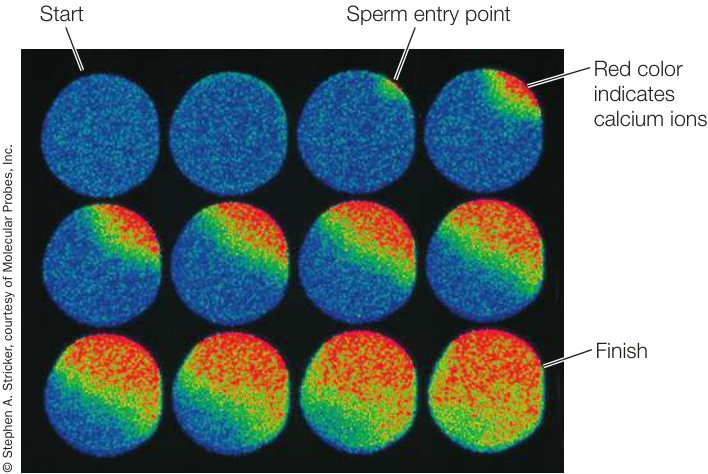
Figure 7.12 Calcium Ions as Second Messengers The concentration of Ca2+ can be measured using a dye that fluoresces when it binds the ion. Here, fertilization in a sea star egg causes a rush of Ca2+ from the environment into the cytoplasm. Areas of high Ca2+ concentration are indicated by the red color, and the events are photographed at 5-second intervals. Calcium signaling occurs in virtually all animal groups and triggers cell division in fertilized eggs, initiating the development of new individuals.
NITRIC OXIDE Most signaling molecules and second messengers are solutes that remain dissolved in either the aqueous or hydrophobic components of cells. It was a great surprise to find that a gas could also be active in signal transduction. Nitric oxide (NO) is a second messenger in the signal transduction pathway between the neurotransmitter acetylcholine (see Key Concept 7.2) and the relaxation of smooth muscle lining blood vessels, which allows more blood flow (Figure 7.13). In the body, NO is made from the amino acid arginine by the enzyme NO synthase. When the acetylcholine receptor on the surface of an endothelial cell is activated, IP3 is released from the membrane (via the pathway shown in Figure 7.13), causing a calcium channel in the ER membrane to open and a subsequent increase in cytosolic Ca2+. The Ca2+ then activates NO synthase to produce NO, which diffuses to the underlying smooth muscle cells, where it activates guanylyl cyclase (a close relative of adenylyl cyclase). This enzyme catalyzes the formation of the second messenger cyclic GMP (cGMP), which contributes to the relaxation of muscle cells.
The discovery of NO as a participant in signal transduction explained the action of nitroglycerin, a drug that has been used for more than a century to treat angina, the chest pain caused by insufficient blood flow to the heart. Nitroglycerin releases NO, which results in relaxation of the blood vessels and increased blood flow. The drug sildenafil (Viagra®) was developed to treat angina via the NO signal transduction pathway but was only modestly useful for that purpose. However, men taking it reported more pronounced penile erections. During sexual stimulation, NO acts as a signal, causing an increase in cGMP and a subsequent relaxation of the smooth muscles surrounding the arteries in the corpus cavernosum of the penis. As a result of this signal, the penis fills with blood, producing an erection.
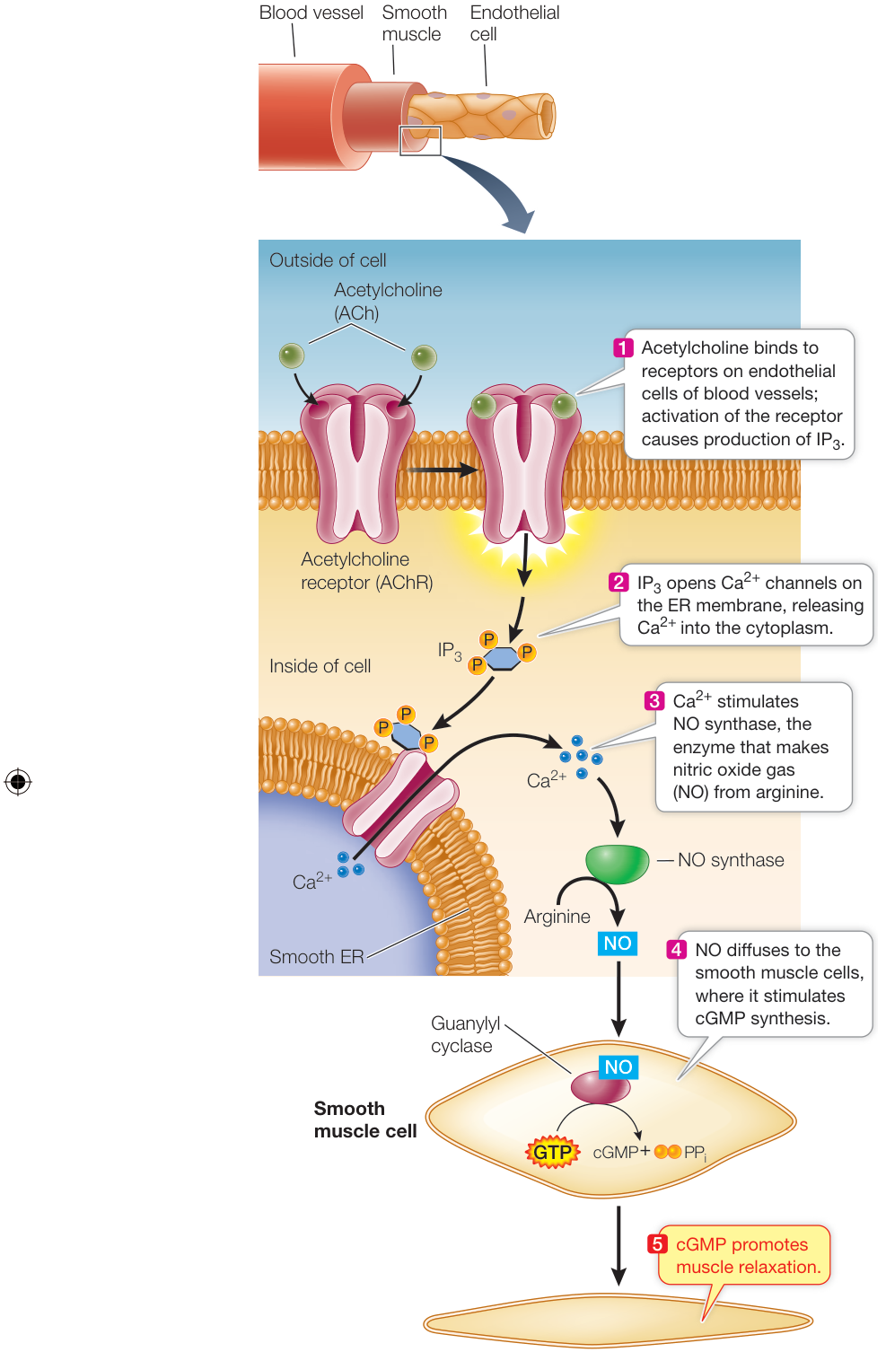
Figure 7.13 Nitric Oxide in Signal Transduction Nitric oxide (NO) is an unstable gas, which nevertheless serves as a mediator between a signal, acetylcholine (ACh), and its effect: the relaxation of smooth muscles. Q: Babies born prematurely (before 37 weeks gestation) can have pulmonary hypertension, whereby the blood vessels in the lungs are constricted and not enough blood reaches the lungs. These babies are given NO gas and this reduces hypertension. How does this work?
Signal transduction is highly regulated
Cells can regulate the activity of a molecule involved in signal transduction. The concentration of NO, which breaks down quickly, can be regulated only by how much of it is made. By contrast, membrane pumps and ion channels regulate the cytosolic concentration of , as you have seen. To regulate protein kinase cascades, G proteins, and cAMP, there are enzymes that inactivate the activated transducer (Figure 7.14).
The balance between the activities of enzymes that activate and inactivate transducers determines the ultimate cellular response to a signal. Cells can alter this balance in several ways:
- Synthesis or breakdown of the enzymes. For example, synthesis of adenylyl cyclase (which synthesizes cAMP) and breakdown of phosphodiesterase (which breaks down cAMP) will tilt the balance in favor of more cAMP in the cell.
- Activation or inhibition of the enzymes by other molecules. An example is that NO signaling from sildenafil inhibits the phosphodiesterase that normally breaks down cGMP.
Because cell signaling is so important in diseases such as cancer, a search is under way for new drugs that can modulate the activities of enzymes that participate in signal transduction pathways.
(A) Phosphatase inactivates a protein kinase
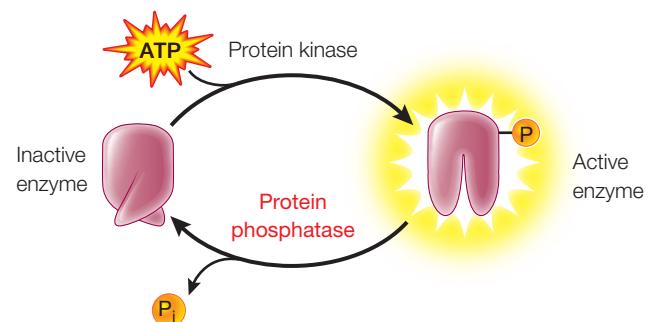
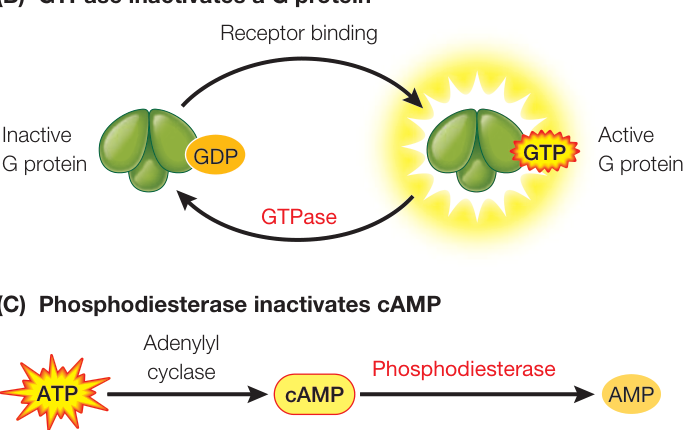
Figure 7.14 Regulation of Signal Transduction Some signals lead to the production of active transducers such as (A) protein kinases, (B) G proteins, and (C) cAMP. Other enzymes inactivate or remove these transducers.
Recap and Assess
Signal transduction is the series of steps between the binding of a signal to a receptor and the ultimate cellular response. A protein kinase cascade amplifies a signal through a series of protein phosphorylation reactions. In many cases, a second messenger amplifies and distributes the downstream effects of the signal. The activity of transducer molecules is regulated.
- How can an activated G protein such as Ras amplify a response to ligand binding?
- Cyclic AMP is a second messenger in many different responses. How can the same messenger act in different ways in different cells?
- How are signal transduction cascades regulated?
You have seen how the binding of a signal to its receptor initiates the response of a cell to the signal, and how signal transduction pathways amplify the signal and distribute its effects to numerous targets in the cell. In the next key concept we will look at the third step in the signal transduction process, the actual effects of the signal on cell function.
Cells Change in Response to Signals in Several Ways
Learning Objectives
- 7.4.1 Describe the signal transduction pathway stimulated by oxytocin.
- 7.4.2 Compare various mechanisms that cells use to respond to signals.
The effects of a signal on cell function take three primary forms: the opening of ion channels, changes in the activities of enzymes, or differential gene expression. These events set the cell on a path for further changes in form and function.
Ion channels respond to signals by opening or closing
You have seen that ion channels can function as receptors in cell signaling (see Figure 7.5). In Chapters 43 and 44 you will learn how signaling affects ion channels, particularly for in the nervous system, where stimulation of a receptor by light, sound, or touch can lead to channel opening. Inside some cells, channels in the ER membrane open in response to signal transduction to modulate muscle contraction.
Oxytocin, which we described in the opening of this chapter, combines the two ion channel responses. Oxytocin binds to a G protein-coupled receptor (see Figure 7.7), which is expressed in brain tissue as well as muscles involved in birth and lactation. Activation of the receptor results in signal transduction through the -DAG pathway, releasing into the cytoplasm via ion channels. In the brain, the effect of is to indirectly stimulate nerve cell activity by the opening of ion channels for , as you will learn in Chapter 43. To summarize:
Oxytocin Receptor G protein activation signal transduction channel opening channel opening
You saw in the opening of this chapter that the behavioral effect of oxytocin in voles involves bonding between them. Does this also happen in people? Neuroscientist Paul Zak at the Center for Neuroeconomics Studies thinks so. He has done experiments with human volunteers and shown that oxytocin signaling is important in trusting behavior (Investigating Life: Is Oxytocin a “Trust” Signal in Humans?). Recently, it was shown that people who have an inherited defect in the oxytocin receptor act in some ways like the montane voles that lack oxytocin receptors: they are less trusting.
Enzyme activities change in response to signals
Enzymes are often modified during signal transduction—either covalently or noncovalently. You have seen examples of both types of protein modifications earlier in this chapter. For example, addition of a phosphate group to an enzyme by a protein kinase is a covalent change; cAMP binding is noncovalent. Both types of modifications change the enzyme’s shape, activating or inhibiting its function. In the case of activation, the shape change exposes a previously inaccessible active site, and the target enzyme goes on to perform a new cellular role.
The G protein-mediated protein kinase cascade that is stimulated by epinephrine in liver cells results in the activation by cAMP of a key signaling molecule, protein kinase A. In turn, protein kinase A phosphorylates two other enzymes, with opposite effects (Figure 7.15):
- Inhibition: Glycogen synthase, which catalyzes the joining of glucose molecules to synthesize the energy-storing molecule glycogen, is inactivated when a phosphate group is added to it by protein kinase A. Thus the epinephrine signal prevents glucose from being stored in the form of glycogen (see Figure 7.15, step 1).
- Activation: Phosphorylase kinase is activated when a phosphate group is added to it. It is part of a protein kinase cascade that ultimately leads to the activation of glycogen phosphorylase, another key enzyme in glucose metabolism. This enzyme results in the liberation of glucose molecules from glycogen (see Figure 7.15, steps 2 and 3).
Connect the Concepts Enzyme regulation determines the rate of chemical transformations in cells, and thus cell functions. See Key Concept 8.5.
The amplification of the signal in this pathway is impressive; as detailed in Figure 7.15, each molecule of epinephrine that arrives at the cell membrane ultimately results in the release of 10,000 molecules of glucose into the bloodstream:
- 1 molecule of epinephrine bound to the membrane leads to
- 20 molecules of cAMP, which activate
- 20 molecules of protein kinase A, which activate
- 100 molecules of phosphorylase kinase, which activate
- 1,000 molecules of glycogen phosphorylase, which produce
- 10,000 molecules of glucose 1-phosphate, which produce
- 10,000 molecules of blood glucose
Investigating Life: Is Oxytocin a “Trust” Signal in Humans?
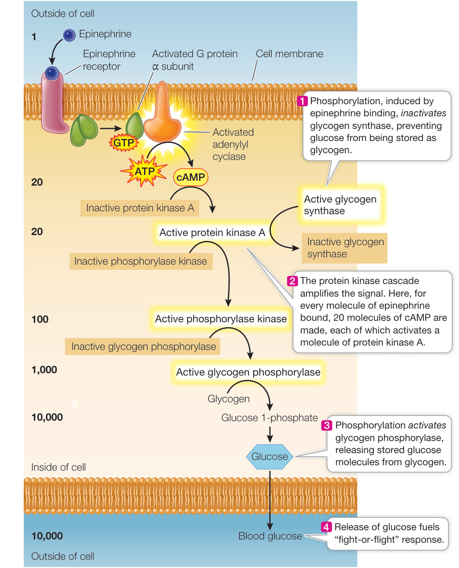
Figure 7.15 A Cascade of Reactions Leads to Altered Enzyme Activity Liver cells respond to epinephrine by activating G proteins, which in turn activate the synthesis of the second messenger cAMP. Cyclic AMP initiates a protein kinase cascade, greatly amplifying the epinephrine signal, as indicated by the bold, black numbers. The cascade both inhibits the conversion of glucose to glycogen and stimulates the release of previously stored glucose. Q: Theobromine, a molecule in cocoa beans that are used to make chocolate, is a phosphodiesterase inhibitor. Chocolate is poisonous to dogs, even at low doses, because dogs do not break down theobromine effectively. How does theobromine work as a poison?
Experiment
Original Paper: P Zak et al. 2005. Oxytocin is associated with human trustworthiness. Horm Behav 48: 522–527.
Cell signaling in the brain leads to changes in behavior. This experiment investigates whether the peptide oxytocin correlates with the development of trust between humans.
HYPOTHESIS The signal oxytocin is involved in trust between humans.
METHOD
RESULTS
| Experimental (donor intent) | Control (no intent) | |
|---|---|---|
| Amount given by donor | $5.52 | $5.63 |
| Amount returned by recipient | $6.96 | $3.53 |
| Oxytocin level in recipient (pg/mL) | 278 | 198 |
CONCLUSIONS
- People (recipients) who trust another person (donor, experimental) will be more trustworthy than people who do not have that feeling.
- Oxytocin levels are higher in people who are trustworthy.
Work with the Data
Original Paper: M. Kosfeld et al. 2005. Oxytocin increases trust in humans. Nature 435: 673–676.
Paul Zak used an economics experiment to investigate trust and found that when people felt that a stranger had trusted them with money, the recipients not only reciprocated by giving the donor some money back, but also had elevated levels of the intercellular signaling molecule oxytocin. As in many experiments, although these data were suggestive of a relationship, correlation does not necessarily mean causation. So Zak and his colleagues tried a direct experiment in which subjects were given oxytocin before the experiment, to determine if oxytocin induces greater trust.
QUESTIONS
- In the experiment, donors were divided into control and experimental groups and paired with recipients. All donors were given 12 money units and asked to donate to a paired recipient, whom they did not know or see. Donors knew that whatever amount they donated would be tripled in the recipient’s account (up to 36 units) and trusted that the recipient would generously give some money back to the donor. The experimental donor group was given nasal spray containing oxytocin just before the experiment; the control donor group received a nasal spray without oxytocin. The table shows the results.
| Average number of units (SD) sent to recipient | |
|---|---|
| Experimental group (with oxytocin spray) | 9.6 (2.8) |
| Control (no oxytocin spray) | 8.1 (3.1) |
What can you conclude from these data about oxytocin and trust in the donors? What statistical test would you use to show that the two averages are significantly different?
- The experiment described in Question 1 was repeated, but this time both donor groups were told exactly how much to give the recipient. The results are shown in the table.
| Average number of units (SD) sent to recipient | |
|---|---|
| Experimental group (with oxytocin spray) | 7.5 (3.3) |
| Control (no oxytocin spray) | 7.5 (3.4) |
What can you conclude about the effect of oxytocin in this case, and what does it indicate about the trust relationship?
Signals can initiate DNA transcription
You learned in Key Concept 4.1 that the genetic material, DNA, is transcribed into RNA, which is then translated into a protein whose amino acid sequence is specified by the original DNA sequence. Proteins are important in all cellular functions, so a key way to regulate specific functions in a cell is to regulate which proteins are made, and therefore which DNA sequences are transcribed.
Signal transduction plays an important role in determining which DNA sequences are transcribed. Common targets of signal transduction are proteins called transcription factors, which bind to specific DNA sequences in the cell nucleus and activate or inactivate transcription of the adjacent DNA regions. For example, the Ras signaling pathway (see Figure 7.10) ends in the nucleus. The final protein kinase in the Ras signaling cascade, MAPK, enters the nucleus and phosphorylates a protein that stimulates the expression of several genes involved in cell proliferation.
Connect the Concepts In this chapter you’ve learned about signaling pathways that occur in animal cells. Signal transduction pathways play equally important roles in other organisms, including plants, as we will explore extensively in Chapters 35 and 37.
Recap and Assess
Cells respond to signal transduction by opening membrane channels, activating or inactivating enzymes, or stimulating or inhibiting gene transcription.
- How does oxytocin signaling involve several signaling pathways?
- Which method(s) of cell response to signaling are rapid and which are slower? Explain.
We have described how signals from a cell’s environment can influence the cell. But the environment of a cell in a multicellular organism is more than the extracellular medium—it includes neighboring cells as well. In the next key concept we’ll see how specialized junctions between cells allow them to pass signals from one to another.
Adjacent Cells in a Multicellular Organism Can Communicate Directly
Learning Objectives
- 7.5.1 Explain the biological advantages of gap junctions in animals.
- 7.5.2 Design investigations to answer questions about intercellular communication.
- 7.5.3 Compare the structure and function of gap junctions and plasmodesmata.
The hallmark of multicellular organisms is their ability to have specialized functions in subsets of cells within their bodies. How do these cells communicate with one another so that they can work together for the good of the entire organism? As you learned in Key Concept 7.1, some intercellular signals travel through the circulatory system to reach their target cells. But cells also have more direct ways of communicating. Cells that are packed together within a tissue can communicate directly with their neighbors via specialized intercellular junctions: gap junctions in animals (see Figure 6.6) and plasmodesmata in plants (see Figure 5.9).
Animal cells communicate through gap junctions
Gap junctions are channels between adjacent cells that occur in many animals, occupying up to 25% of the area of the cell membrane (Figure 7.16A). Gap junctions traverse the narrow space between the cell membranes of two adjacent cells (the “gap”) by means of channel structures called connexons. The walls of a connexon are composed of six subunits of the integral membrane protein connexin. In adjacent cells, two connexons come together to form a gap junction that links the cytoplasms of the two cells. There may be hundreds of these channels between a cell and its neighbors. The channel pores are about 1.5 nanometers (nm) in diameter—too narrow for the passage of large molecules but adequate for passage of small molecules and ions.
Here is an example of gap junctions at work: In the lens of the mammalian eye, only the cells at the periphery are close enough to the blood supply for adequate diffusion of nutrients and wastes. But because lens cells are connected by large numbers of gap junctions, material can diffuse between them rapidly and efficiently. Hormones and second messengers can move through gap junctions. Sometimes just a few cells in a tissue have the receptor for a particular signal; in such cases, gap junctions allow a coordinated response to the signal by all the cells in the tissue.
Plant cells communicate through plasmodesmata
Instead of gap junctions, plants have plasmodesmata (singular plasmodesma), which are membrane-lined tunnels that traverse the thick cell walls separating plant cells from one another. A typical plant cell has several thousand plasmodesmata. Plasmodesmata differ from gap junctions in one fundamental way: unlike gap junctions, in which the wall of the channel is made of integral membrane proteins from the adjacent cell membranes, plasmodesmata are lined by the fused cell membranes themselves.
The diameter of a plasmodesma is about 6 nm, far larger than a gap junction channel. But the actual space available for diffusion is about the same—1.5 nm. Examination of the interior of the plasmodesma by transmission electron microscopy reveals that a tubule called the desmotubule, apparently derived from the endoplasmic reticulum, fills up most of the opening of the plasmodesma (Figure 7.16B). Typically, only small metabolites and ions can move between plant cells.
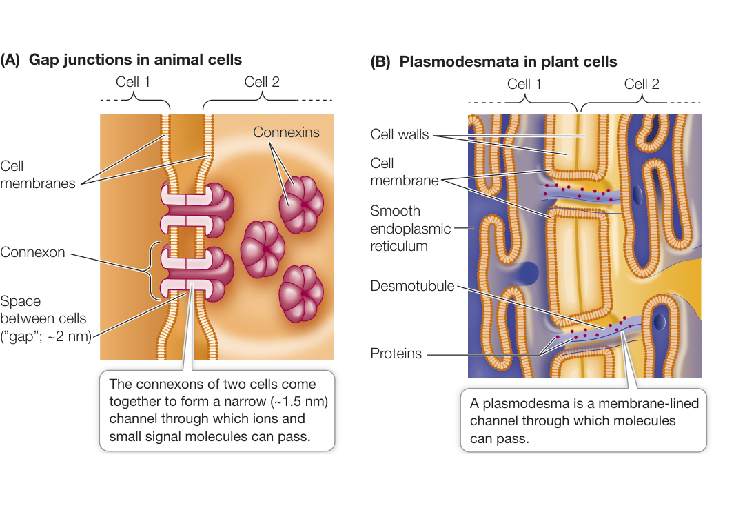
Figure 7.16 Communicating Junctions (A) An animal cell may contain hundreds of gap junctions connecting it to neighboring cells. The pores of gap junctions allow small molecules to pass from cell to cell, ensuring similar concentrations of important signaling molecules in adjacent cells so that the cells can coordinate their activities. (B) Plasmodesmata connect plant cells. The desmotubule, derived from the smooth endoplasmic reticulum, fills up most of the space inside a plasmodesma, leaving a tiny gap through which small metabolites and ions can pass. (A) An animal cell may contain hundreds of gap junctions connecting it to neighboring cells. The pores of gap junctions allow small molecules to pass from cell to cell, ensuring similar concentrations of important signaling molecules in adjacent cells so that the cells can coordinate their activities. (B) Plasmodesmata connect plant cells. The desmotubule, derived from the smooth endoplasmic reticulum, fills up most of the space inside a plasmodesma, leaving a tiny gap through which small metabolites and ions can pass.
Figure 7.16 Communicating Junctions
Figure 7.17 Multicellularity
The evolution of intercellular interactions in a multicellular organism can be inferred from these green algae.
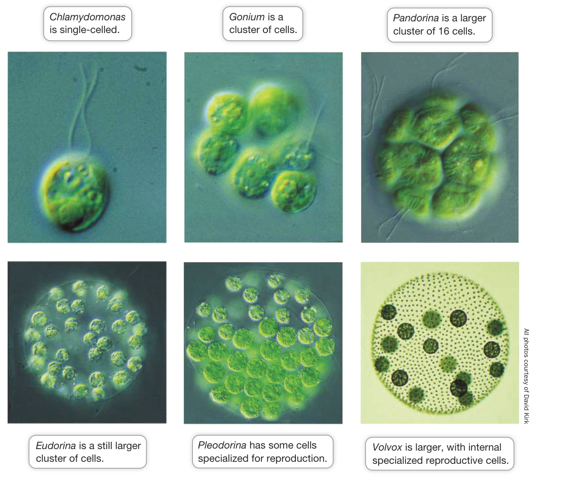
Figure 7.17 Multicellularity The evolution of intercellular interactions in a multicellular organism can be inferred from these green algae. Q: Why was the evolution of direct communication important for tissue formation? Q: Why was the evolution of direct communication important for tissue formation?
Plasmodesmata are vital in plants because their circulatory transport system, the vascular system, lacks the tiny vessels (capillaries) that many animals have for bringing gases and nutrients to every cell. For instance, simple diffusion from plant cell to cell across cell membranes is inadequate to account for the movement of a plant hormone from the site of production to the site of action. Instead, plants rely on more rapid diffusion through plasmodesmata to ensure that all cells of a tissue respond to a signal at the same time. There are cases in which larger molecules or particles can pass between cells via plasmodesmata. For example, some viruses can move through plasmodesmata by using “movement proteins” to assist their passage.
Modern organisms provide clues about the evolution of cell–cell interactions and multicellularity
- Multicellular organisms can indeed be multicellular—a human has
about 60 trillion eukaryotic cells and many more prokaryotic ones. But
it’s not just cells that make up a human or a rose plant—it is groups of
cells specialized into tissues and then groups of tissues in organs
(e.g., the nerve cells in the brain or the petals in a flower) that have
specific roles. As you will learn in Chapter 42, the embryonic
development of tissues and organs takes place in several steps:
- Migration of cells to form a group
- Organization of cells in the cluster
- Specialization of cells into tissues
- Connections between cells
Even though single-celled organisms continue to be highly successful on Earth, over time complex multicellular organisms evolved, along with their division of biological labor among specialized cells. The transition from single-celled to multicellular life took a long time. Indeed, while there is evidence that single-celled organisms arose about 500 million to a billion years after the formation of Earth (see Chapter 4), the first evidence of true multicellular organisms dates from more than a billion years later. Multicellularity probably arose several times.
Studying the evolutionary origin of multicellularity is a challenge because it happened so long ago. The closest unicellular relatives of most modern animals and plants probably existed hundreds of millions of years ago. The transition from single-celled to multicellular organisms may have occurred in several steps:
- Aggregation of cells into a cluster
- Intercellular communication within the cluster
- Specialization of some cells within the cluster
- Organization of specialized cells into groups (tissues)
Does this list look familiar? (See the first list in this section for the origin of organs in the embryo.)
A key event would have been the evolution of intercellular communication, which is necessary to coordinate the activities of different cells within a multicellular organism.
We can visualize how the evolution of multicellularity might have occurred by examining the Volvocine line of aquatic green algae (Chlorophyta). These plants range from single cells to complex multicellular organisms with differentiated cell clusters (Figure 7.17). Included in this range are a single-celled organism (Chlamydomonas); an organism that occurs in small cell clusters (Gonium); species with larger cell clusters (Pandorina and Eudorina); a colony of somatic and reproductive cells (Pleodorina);
and a larger, 1,000-celled alga with somatic and reproductive cells organized into separate tissues (Volvox).
Chlamydomonas is the single-celled member of this group. It has two cellular phases: a swimming phase, when the cells have flagella and move about, and a non-swimming phase, when the flagella are reabsorbed (disaggregated) and the cell undergoes cell division (reproduction). Compare this with Volvox: most of the cells of this multicellular, spherical organism are on the surface; the beating of their flagella gives the organism a rolling motion as it swims toward light, where it can perform photosynthesis. But some Volvox cells are larger and located inside the sphere. These cells are specialized for reproduction: they lose their flagella and then divide to form offspring.
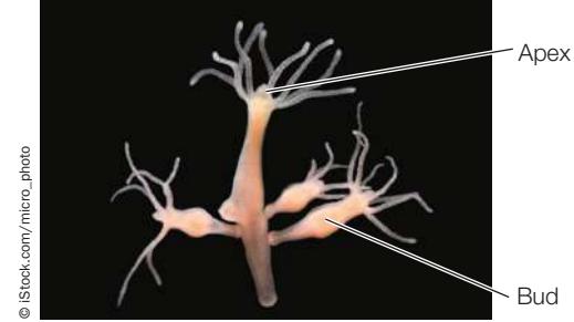
hypothesis that the inhibitory factor of Hydra passes through these junctions?
Investigating Life: QA Is oxytocin the “trust” signal in humans?
Paul Zak’s experiments showing the involvement of oxytocin in the trust involved in money transfers between people opened up a new field of study called neuroeconomics. In 1759 the philosopher Adam Smith wrote that social and economic interactions between people are driven by what he called “fellow feeling.” Now, 260 years later, the oxytocin studies validate this and have put it into the language of cell signaling. What other things besides economic generosity release oxytocin? Zak and others have shown that many pleasant activities, including sexual interactions and even massage and hugs, release oxytocin and induce positive behaviors as a result. Not surprisingly, oxytocin is available for purchase. A fragrance called “Liquid Trust” is marketed to sales people as well as those seeking love, and the book Make Love Like a Prairie Vole offers steps to a happier love life. In a somewhat more serious vein, oxytocin and other peptides that modulate behavior are being investigated as a treatment for autism, a disorder in which children can show alterations in trusting behaviors.
Future directions
In this chapter you have seen how a natural substance such as caffeine acts on receptors to alter cell function. As signal transduction pathways have been described for important processes such as the regulation of cell division, it has become possible to perturb intermediate steps in the pathway between signal and cell effects. In the signaling that stimulates cells to reproduce, two pathways are the focus of intense interest. One is at the cell membrane and involves protein kinase receptors and the ensuing signaling kinase pathway (see Figure 7.10). One or more steps along this pathway are often activated in cancer cells. For example, a kinase called Bcr-Abl is present and active in a type of leukemia and not in normal white blood cells. A drug has been developed that targets this kinase and results in a cure for the disease.
Recap and Assess
Cells can communicate with their neighbors through specialized cell junctions. In animals, these structures are gap junctions; in plants, they are plasmodesmata. The evolution of intercellular communication and tissue formation can be inferred from existing organisms, such as certain related green algae.
- List the characteristics of direct communication by cell junctions and chemical signaling between cells. What is the advantage of direct communication over chemical signaling?
- How does the Volvocine line of green algae show possible steps in the evolution of cell communication and tissue formation?
- The tiny invertebrate Hydra have an apical region with tentacles and a long, slender body. Hydra can reproduce asexually when cells on the body wall differentiate and form a bud, which then breaks off as a new organism. Buds form only at certain distances from the apex, leading to the idea that the apex releases a signal molecule that diffuses down the body and, at high concentrations (i.e., near the apex), inhibits bud formation. Hydra lack a circulatory system, so this inhibitor must diffuse from cell to cell. If you have an antibody that binds to connexons and plugs up the gap junctions, how would you test the


Courtesy of Todd Ahern
You should be able to relate each summary to the adjacent figures. If you go to this visual summary in Achieve, you can follow links to figures, animations, activities, and simulations that will help you consolidate the material.
Visual Summary
Cells Detect a Variety of Signals
- Cells receive many signals from the environment and from other cells.
- Autocrine signals affect the cells that make them; juxtacrine signals affect adjacent cells; paracrine signals diffuse to and affect nearby cells; and hormones are carried through the circulatory system of animals or the vascular system of plants.
- A signal transduction pathway involves the interaction of a signal molecule with a receptor; the transduction of the signal via a series of steps within the cell; and effects on the function of the cell.
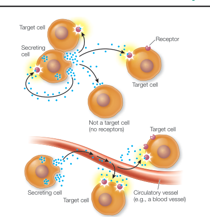
Figure 7.1
Questions
- Why don’t all cells respond to all cell signals?
- Are all signals chemical?
Receptors Bind Signals to Initiate a Cellular Response
- Binding of a signal ligand to its receptor obeys the chemical law of mass action. A key measurement of the strength of binding is the dissociation constant ().
- Depending on the nature of its signal or ligand, a receptor may be located in the cell membrane or inside the target cell.
- Receptors located in the cell membrane include ion channels, protein kinases, and G protein-coupled receptors.
- Intracellular receptors include steroid hormone receptors in animals. A lipid-soluble ligand such as a steroid hormone may enter the cytoplasm or the nucleus before binding.
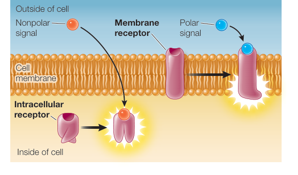
Figure 7.4
Questions
- What are the three protein domains of a typical membrane-bound receptor, and what are their respective functions?
- What is the general effect of activation of a receptor?
- Why do cytoplasmic receptors bind only nonpolar signals?
Signal Transduction Spreads the Response to a Signal through the Cell
- A protein kinase cascade amplifies the response to receptor binding.
- Second messengers between signal binding at the cell membrane and signal transduction in the cytoplasm include cyclic AMP (cAMP), inositol trisphosphate (IP3), diacylglycerol (DAG), nitric oxide, and calcium ions.
- Signal transduction can be regulated in several ways.
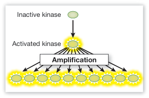
Figure 7.10
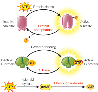
Figure 7.14
Questions
- In addition to binding to the adenosine receptor, caffeine is a phosphodiesterase inhibitor. What effect does this have on cell signaling?
- In addition to activating the MAPK pathway, Ras activates the calcium signaling pathway. What phenomenon is this an example of?
Cells Change in Response to Signals in Several Ways
- The cellular responses to signals may include the opening of ion channels, the alteration of enzyme activities, or changes in gene expression.
- Activated enzymes may activate other enzymes in a signal transduction pathway, leading to impressive amplification of a signal.
Questions
- What is the major difference between the activation of a protein kinase in a signal transduction cascade and the activation of an enzyme by cAMP?
- If there were not a multi-step signal transduction pathway, would there be an amplification of many molecules of glycogen phosphorylation?
Adjacent Cells in a Multicellular Organism Can Communicate Directly
- Many adjacent animal cells can communicate with one another directly through small pores in their cell membranes called gap junctions. Protein structures called connexions form thin channels between two adjacent cells through which small signal molecules and ions can pass.
- Plant cells are connected by somewhat larger pores called plasmodesmata, which traverse both cell membranes and cell walls. The desmotubule narrows the opening of the plasmodesma.
- The evolution of cell communication and tissue formation can be inferred from existing organisms, such as certain green algae.
Questions
- Most signaling molecules are polar and cannot readily diffuse through the cell membrane. When a signal binds to a cell, it is therefore difficult for a second messenger, other small molecule involved in signal transduction, or ions to pass on to an adjacent cell. What mechanism do animal and plant cells have for rapid movement of these substances from one cell to another?
- If you compared Chlamydomonas, Gonium, and Volvox with regard to intercellular signaling and plasmodesmata, what would you expect to find?
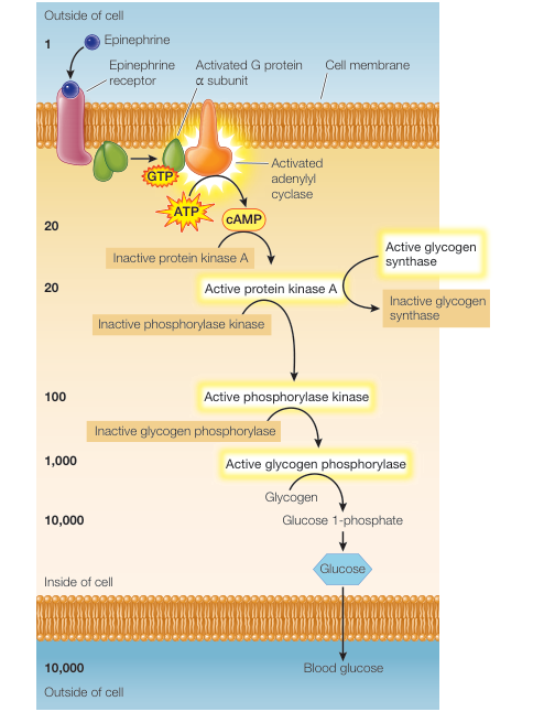
Figure 7.15
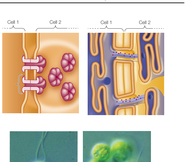
Figure 7.16
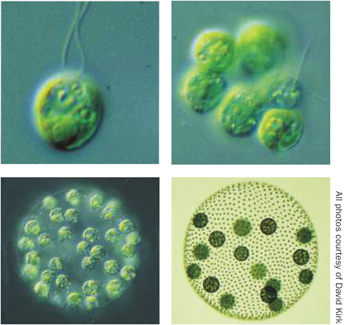
Figure 7.17