Chapter 9Pathways That Harvest Chemical Energy
Key Concepts
- 9.1 Cells Harvest Chemical Energy from Glucose Oxidation
- 9.2 In the Presence of Oxygen, Glucose Is Fully Oxidized
- 9.3 Oxidative Phosphorylation Forms ATP
- 9.4 In the Absence of Oxygen, Some Energy Is Harvested from Glucose
- 9.5 Metabolic Pathways Are Interrelated and Regulated
Mitochondria are the powerhouses of the cell.
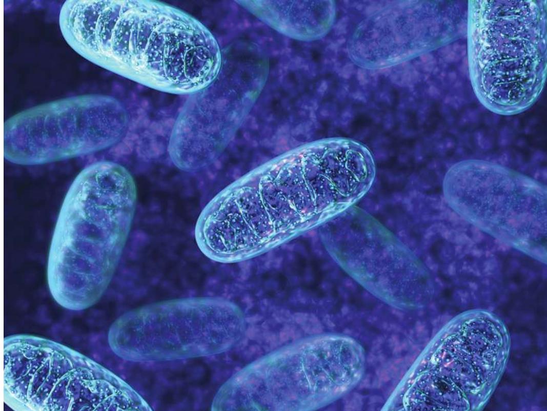
© 3d_mary/Shuterstock.com
Investigating Life: A Weighty Matter
With 40% of adults in the United States now described as obese, physicians have declared an “obesity epidemic.” Dramatic increases in diseases associated with obesity—among them diabetes, heart disease, and cancer—have understandably alarmed the medical establishment.
While nutritional science has shown that not all foods affect all people in the same ways, in many cases the significant excess body weight of obese people can be prevented or reduced if they eat less. It is a matter of energy: if we eat more energy-yielding molecules than we need to build our bodies and to fuel activities such as brain functions and physical activity, we will store the unneeded energy as fat. There is an evolutionary advantage in storing fat: the energy in the C—C and C—H bonds in fat can be used later when food is scarce. But excess fat has adverse consequences.
Not all fat (adipose) tissues are the same. White adipose tissue (sometimes referred to simply as “white fat”) is used primarily to store energy. Brown adipose tissue has high concentrations of mitochondria, which have iron-containing pigments. When energy-rich molecules in brown fat are catabolized, the stored energy is released not as chemical energy but as heat. The cells in brown fat make a protein called UCP1 (uncoupling protein 1) that inserts into the inner membranes of mitochondria, making them permeable to protons (). The general impermeability of these membranes to is key to coupling the catabolism of molecules such as fats to the release of their stored chemical-bond energy to make ATP. If the membranes become permeable to , this coupling is lost and the stored energy is released as heat.
Human infants are born with a great deal of brown fat in their back and shoulders. Because infants have a high surface area-to-volume ratio, they tend to lose a lot of heat. One way they keep warm is to produce heat in their brown fat tissues. As a child grows up, the brown fat content of the body is reduced. Adults have mostly white fat, which has less UCP1 and generates less heat when the fat is catabolized. Biologists recently found brown fat in adults, particularly in the upper chest and neck, but also scattered elsewhere in the body, mixed in with much more prevalent white fat. The role of this tissue is under intense study.
QA How are mitochondria related to obesity? (Find the answer on page 192.)
Cells Harvest Chemical Energy from Glucose Oxidation
Learning Objectives
- 9.1.1 Describe the principles that govern metabolic pathways in cells.
- 9.1.2 Identify the change in energy that occurs during an oxidation–reduction reaction.
- 9.1.3 Describe the roles of electron acceptors and donors in biological redox reactions.
- 9.1.4 Analyze biological redox reactions to identify the oxidizing and reducing agents involved.
Energy is stored in the chemical bonds of molecules, and it can be released and transformed by the metabolic pathways of living cells. A metabolic pathway consists of a coordinated series of biochemical reactions that convert molecules into other molecules. As you read this chapter, keep in mind five principles that govern metabolic pathways:
- A complex chemical transformation occurs in a series of separate reactions that form a metabolic pathway.
- Each reaction is catalyzed by a specific enzyme.
- Many metabolic pathways are similar in all organisms, from bacteria to humans.
- In eukaryotes, many metabolic pathways are compartmentalized, with certain reactions occurring inside specific organelles, or even specific regions of an organelle.
- Some key enzymes in each metabolic pathway can be inhibited or activated to alter the rate of the pathway.
Connect the Concepts The principles of energy transformations in living and nonliving systems are discussed in Key Concept 8.1.
Cells obtain energy from glucose
In cells, chemical energy released from bonds of molecules is used to make ATP (see Key Concept 8.2), which in turn drives endogenic reactions. Photosynthetic cells and organisms use energy from sunlight to synthesize their own energy-storing molecules, as we will describe in Chapter 10. While other carbohydrates, as well as lipids and proteins, can be used as energy sources, the most common chemical fuel in cells is the monosaccharide glucose. As you will see throughout this chapter, to release their energy to make ATP, the other molecules are converted into glucose or intermediate compounds that can enter into the various pathways of glucose metabolism.
As you saw in Key Concept 2.3, the familiar process of combustion (burning) is similar to the chemical processes that release energy in cells. If glucose is burned in a flame or is in a typical cell, it reacts with oxygen gas (), forming carbon dioxide and water and releasing energy in the form of heat. The balanced equation for the complete reaction is
This is an oxidation–reduction reaction (more on this shortly), in which glucose loses electrons (becomes oxidized) and oxygen gains them (becomes reduced). The large free energy change () is negative, indicating that the overall conversion is highly exergonic and can drive the endogenic formation of a lot of ATP from ADP and phosphate:
Three catabolic processes harvest the energy in the chemical bonds of glucose: glycolysis, cellular respiration, and fermentation (Figure 9.1). All three processes involve pathways made up of many distinct chemical reactions.
- Glycolysis begins glucose catabolism. Through a series of chemical rearrangements, glucose is converted to two molecules of the three-carbon product pyruvate, and a small amount of energy is captured in usable forms. Glycolysis is an anaerobic process because it does not require .
- Cellular respiration uses from the environment and thus is aerobic. Each pyruvate molecule is completely converted into three molecules of through a set of catabolic pathways including pyruvate oxidation, the citric acid cycle, and an electron transport chain. In the process, a great deal of the energy stored in the covalent bonds of pyruvate is captured to form ATP.
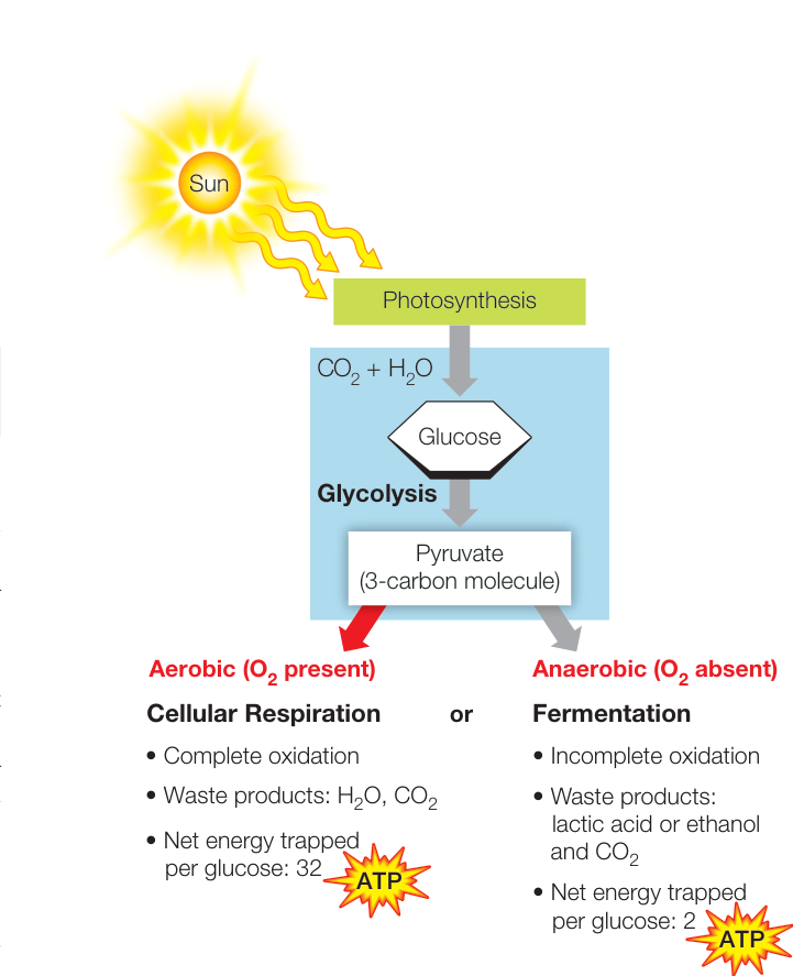
Figure 9.1 Energy for Life Many prokaryotes and all eukaryotes obtain their energy from the molecules ultimately produced by photosynthesis, which converts solar energy into chemical-bond energy. Often, organisms convert molecules into glucose, which they metabolize to trap energy in ATP.
- Fermentation does not involve (it is anaerobic). With the exception of many microorganisms, fermentation converts pyruvate into lactic acid or ethyl alcohol (ethanol), both of which are still relatively energy-rich molecules. Because the breakdown of glucose is incomplete, much less energy is released when glycolysis is coupled to fermentation than when it is coupled to cellular respiration.
Redox reactions transfer electrons and energy
The release of energy from a molecule and the transfer of that energy to another molecule often involve the transfer of electrons. A reaction in which one substance transfers one or more electrons to another substance is called an oxidation–reduction, or redox, reaction.
- Reduction is the gain of one or more electrons by an atom, ion, or molecule.
- Oxidation is the loss of one or more electrons.
Oxidation and reduction always occur together: as one chemical is oxidized, the electrons it loses are transferred to another chemical, reducing it. In a redox reaction, we call the reactant that becomes reduced an oxidizing agent and the one that becomes oxidized a reducing agent (Figure 9.2A). A familiar
(A) Complete transfer of electrons in a redox reaction
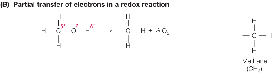
Figure 9.2 Oxidation and Reduction (A) Electron transfer may be complete, such that one or more electrons move from one molecule to another (blue line), resulting in the donor molecule becoming less negatively charged, and the recipient molecule more negatively charged. (B) Electron transfer can be partial, so that electrons are not lost or gained, but an atom’s share of electrons is changed (e.g., is more oxidized than ).
Q: In (B), which atom becomes oxidized and which becomes reduced?
example is the formation of salt (NaCl) (see Figure 2.10), whereby Na transfers an electron to Cl, forming and . In terms of oxidation–reduction,
- Na gives up an electron, and so becomes oxidized. It is a reducing agent.
- Cl gains an electron, and so becomes reduced. It is an oxidizing agent.
You will see many examples of complete electron transfer in this and subsequent chapters, for example, the electron transport chain (see Key Concept 9.3).
Not all redox reactions involve a complete transfer of electrons (Figure 9.2B). Thanks to electronegativity and resulting polarity, many coupled reactions involve a degree of sharing of electrons between two atoms. To greatly simplify, consider the oxidation of glucose described in the equation in the previous section. In glucose, there are bonds between carbon atoms. The electrons in the C—C bond are shared equally. But in , the bonds are , where the greater electronegativity of O results in a polar bond: electrons are shared unequally, spending more time near the O nucleus. Because there is movement of the shared electrons away from C, we can say that it has become oxidized. If the conversion of C—C to C=O is an oxidation, what is reduced? In an overall fashion, the oxidizing agent is oxygen. The electrons in the O=O bond are shared equally. But in , the O—H bond is polar, with the electrons spending more time near O (see Figure 2.9). Therefore O is reduced. In summary:
- Glucose is the reducing agent (electron donor) and becomes oxidized.
- is the oxidizing agent (electron acceptor) and becomes reduced.
Although oxidation and reduction are always defined in terms of electrons, it is often simpler to think in terms of the gain or loss of hydrogen atoms. The transfer of electrons is often associated with the transfer of hydrogen ions (a H atom contains ). So when a molecule loses hydrogen atoms, it becomes oxidized.
The more reduced a molecule is, the more energy it has stored in its covalent bonds (Figure 9.3). In a redox reaction, some energy is transferred from the reducing agent to the reduced product.
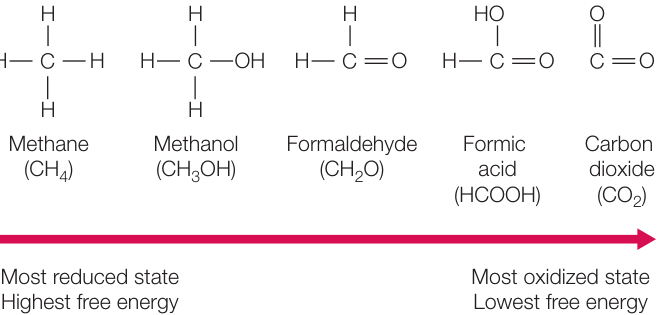
Figure 9.3 Oxidation, Reduction, and Energy The more oxidized a carbon atom in a molecule is, the less stored free energy it has.
The rest remains in the reducing agent or is lost to entropy. As you will see, some of the key reactions of glycolysis and cellular respiration are highly exergonic redox reactions.
The coenzyme NAD+ is a key electron carrier in redox reactions
The coenzyme nicotinamide adenine dinucleotide (NAD+) acts as an electron carrier in redox reactions (Figure 9.4A). NAD+ exists in two chemically distinct forms, one oxidized (NAD+) and the other reduced (NADH) (Figure 9.4B). Both forms participate in redox reactions. The reduction reaction is actually the transfer of a proton (the hydrogen ion, H+) and two electrons, which are released by the accompanying oxidation reaction.
The electrons do not remain with the coenzyme. Oxygen is highly electronegative and readily accepts electrons from NADH. The oxidation of NADH by O2 (which occurs in several steps)
is exergonic, with a standard free energy change at pH 7 () of -52.4 kcal/mol (-219 kJ/mol). Note that the oxidizing agent appears here as ” ” instead of “O.” This notation emphasizes that it is molecular oxygen, O2, that acts as the oxidizing agent.
Just as ATP can be thought of as a package of 7.3 kcal/mol (30.5 kJ/mol) of free energy, NADH can be thought of as a larger package of free energy (52.4 kcal/mol; see above). NAD+ is a common electron carrier in cells, but not the only one. Another carrier, flavin adenine dinucleotide (FAD), also transfers electrons during glucose metabolism.
Connect the Concepts Key Concept 8.4 describes the role of coenzymes, small molecules that assist in enzyme-catalyzed reactions. ADP acts as a coenzyme when it picks up energy released in an exergonic reaction and packages it to form ATP.
An overview: Harvesting energy from glucose
Both eukaryotic and prokaryotic cells can harvest energy from glucose using different combinations of the following metabolic pathways:
- Under aerobic conditions, when O2 is available as the final electron acceptor, four pathways operate (Figure 9.5A).
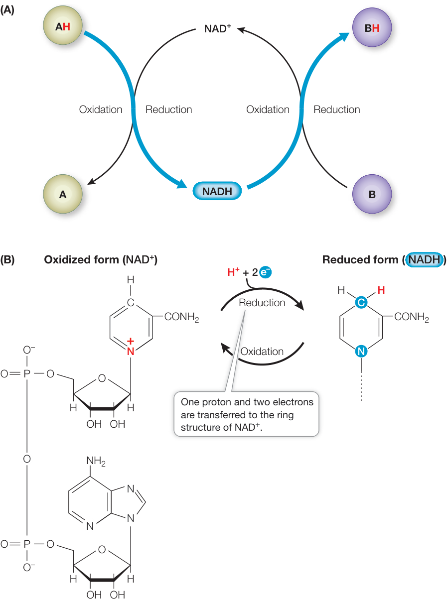
Figure 9.4 NAD+/NADH Is an Electron Carrier in Redox Reactions (A) NAD+ is an important electron acceptor in redox reactions, and thus its reduced form, NADH, is an important energy intermediary in cells. The blue lines show the flow of electrons. (B) Most of the NAD+ molecule (left) remains unchanged by the redox reaction.
Glycolysis is followed by the three pathways of cellular respiration: pyruvate oxidation, the citric acid cycle (also called the Krebs cycle or the tricarboxylic acid cycle), and electron transport/ATP synthesis (also called oxidative phosphorylation).
- In eukaryotes and many prokaryotes, pyruvate oxidation, the citric acid cycle, and oxidative phosphorylation do not function under anaerobic conditions. The pyruvate produced by glycolysis is further metabolized by fermentation
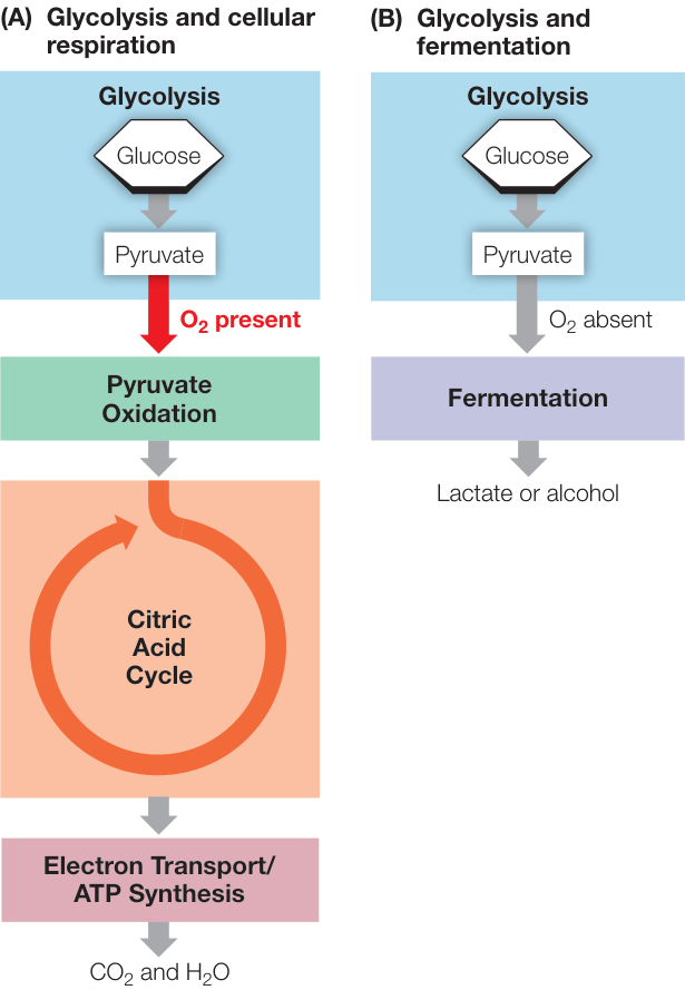
Figure 9.5 Energy-Yielding Metabolic Pathways Energy-yielding reactions can be grouped into five metabolic pathways: glycolysis, pyruvate oxidation, the citric acid cycle, the electron transport chain/ ATP synthesis, and fermentation. (A) The three lower pathways occur only in the presence of and are collectively referred to as cellular respiration. (B) When is unavailable, glycolysis is followed by fermentation.
(Figure 9.5B). Some prokaryotes, however, are able to harvest energy in pathways involving oxidative phosphorylation even in the absence of oxygen (anaerobic respiration; see Key Concept 9.3).
The five pathways shown in Figure 9.5 occur in different locations in the cell (Table 9.1).
Recap and Assess
The free energy released from the oxidation of glucose is trapped in the form of ATP. In many prokaryotes and all eukaryotes, five major pathways combine in different ways to produce ATP which supplies the energy for myriad other reactions in living cells.
- What principles govern metabolic pathways in cells?
- What are the roles of and with respect to electrons in a redox reaction?
- The following reaction occurs in the citric acid cycle:
Answer each of the following questions, and explain your answers:
- Is this reaction an oxidation or a reduction?
- Is the reaction exergonic or endergonic?
- What kind of coenzyme does this reaction require?
Now that you have an overview of the metabolic pathways that harvest energy from glucose, let’s take a closer look at the three pathways involved in aerobic glucose catabolism: glycolysis, pyruvate oxidation, and the citric acid cycle.
Table 9.1 Cellular Locations for Major Energy Pathways in Eukaryotes and Prokaryotes
In the Presence of Oxygen, Glucose Is Fully Oxidized
Learning Objectives
- 9.2.1 Describe the energy inputs and outputs of glycolysis.
- 9.2.2 Explain how pyruvate oxidation connects the transformations of glycolysis and the citric acid cycle.
- 9.2.3 Compare and contrast glycolysis and the citric acid cycle.
In the presence of O2 (aerobic), the pathways of glucose catabolism oxidize glucose completely to CO2 and H2O. Initially, the glycolysis reactions convert the six-carbon glucose molecule to two three-carbon pyruvate molecules (Figure 9.6). Pyruvate is then converted to CO2 in a second series of reactions beginning with pyruvate oxidation and followed by the citric acid cycle. In addition to generating CO2, the oxidation events are coupled with the reduction of electron carriers, mostly NAD+. We will begin our consideration of the catabolism of glucose with a closer look at glycolysis.
In the glycolysis pathway, glucose is partially oxidized
Glycolysis takes place in the cytoplasm and involves ten enzyme-catalyzed reactions. During glycolysis, some of the covalent bonds between carbon and hydrogen atoms in the glucose molecule are oxidized, releasing some of the stored energy. The products are two molecules of pyruvate (pyruvic acid), two molecules of ATP, and two molecules of NADH. Glycolysis can be divided into two stages: the initial energy-investing reactions that consume ATP, and the energy-harvesting reactions that produce ATP and NADH (see Figure 9.6).
To help you understand the process without getting into extensive detail, we focus on two consecutive reactions in this pathway (Figure 9.7). Steps 6 and 7 are examples of two types of reactions that occur repeatedly in glycolysis and in many other metabolic pathways: oxidation–reduction, wherein energy released by glucose oxidation is trapped via the reduction of NAD+ to NADH; and substrate-level phosphorylation, wherein energy released is sufficient to transfer a phosphate directly from the substrate to ADP, forming ATP.
The end product of glycolysis, pyruvate, is somewhat more oxidized than glucose. In the presence of O2, further oxidation can occur. In prokaryotes these subsequent reactions take place in the cytoplasm, but in eukaryotes they take place in the mitochondrial matrix.
To summarize:
- The initial steps of glycolysis use the energy of hydrolysis of two ATP molecules per glucose molecule.
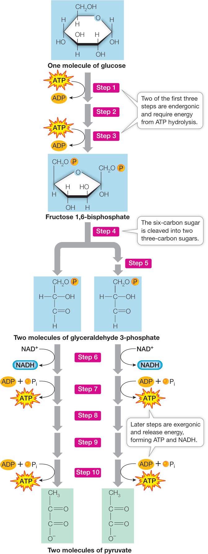
Figure 9.6 Glycolysis Converts Glucose into Pyruvate Glucose is converted to pyruvate in ten enzyme-catalyzed steps. Along the way, two ATP are used (Steps 1 and 3), two NAD+ are reduced to two NADH (Step 6), and four ATP are produced (Steps 7 and 10).
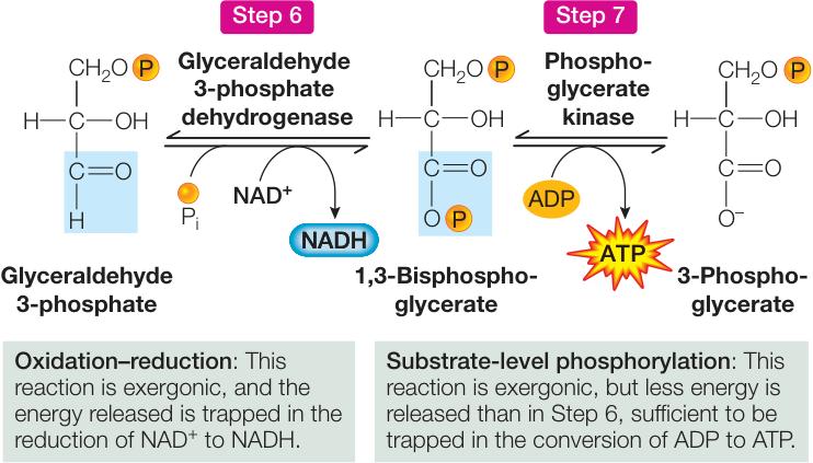
Figure 9.7 Glycolysis Close-Up Steps 6 and 7 in the glycolysis pathway (see Figure 9.6) illustrate the principles of molecular conversion and energy transfer. Q: Which steps in glycolysis involve energy transfer with a coenzyme or carrier?
- The remaining steps produce four ATP molecules per glucose molecule, so the net production of ATP is two molecules.
- Glycolysis produces two molecules of NADH.
If is present, glycolysis is followed by the three stages of cellular respiration: pyruvate oxidation, the citric acid cycle, and the electron transport chain/ATP synthesis.
Pyruvate oxidation links glycolysis and the citric acid cycle
In eukaryotes, pyruvate is transported into the mitochondrial matrix (see Figure 5.13), where the next step in the aerobic catabolism of glucose occurs. This step involves the oxidation of pyruvate to a two-carbon acetate molecule and (Figure 9.8A). The acetate is then bound to coenzyme A to form acetyl coenzyme A, or acetyl CoA. Acetyl CoA is used in various biochemical reactions as a carrier of the acetyl group (). Pyruvate is the link between glycolysis and further oxidative reactions (see Figure 9.5).
The formation of acetyl CoA is a multi-step reaction catalyzed by the pyruvate dehydrogenase complex (Figure 9.8B). The overall reaction is exergonic, in which one molecule of is reduced to NADH. However, the main role of acetyl CoA is to donate its acetyl group to the four-carbon compound oxaloacetate, forming the six-carbon molecule citrate. This initiates the citric acid cycle, one of life’s most important energy-harvesting pathways.
The citric acid cycle completes the oxidation of glucose to
Acetyl CoA is the starting point for the citric acid cycle. This pathway of eight reactions completely oxidizes the two-carbon acetyl group to two molecules of . The free energy released from these reactions is captured by GDP (guanosine diphosphate) and the electron carriers and FAD (Figure 9.9). (Remember from Key Concept 7.2 that GDP is a nucleoside diphosphate like ADP.) This is a cycle because the starting material, oxaloacetate, is regenerated in the last step and is ready to accept another acetate group from acetyl CoA. The citric acid cycle operates twice for each glucose molecule that enters glycolysis (once for each pyruvate that enters the mitochondrion).
Let’s focus on the final reaction of the cycle (see Step 8 in Figure 9.9), as an example of the kind of reaction that occurs (Figure 9.10). This oxidation reaction (see the carbon atom highlighted in blue) is exergonic, and the released energy is trapped by , forming NADH. With four such reactions (the produced in Step 6 is a reduced coenzyme similar to NADH), the citric acid cycle harvests a great deal of chemical energy from the oxidation of acetyl CoA.
To summarize:
- The inputs to the citric acid cycle are acetate (in the form of acetyl CoA), water, GDP, and the oxidized electron carriers and FAD.
- The outputs are carbon dioxide, reduced electron carriers ( and ), and a small amount of GTP. The energy in the terminal phosphate of GTP is transferred to ATP:
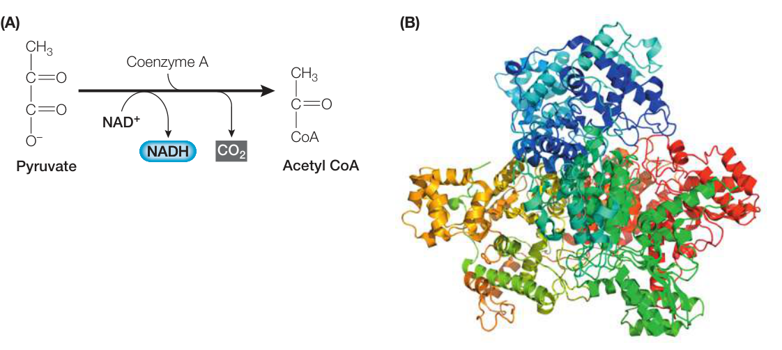
Figure 9.8 Pyruvate Oxidation The pyruvate dehydrogenase complex is actually three enzymes that carry out three intermediate steps in the process. There are over 100 polypeptide chain subunits. (B after Mcn2k10/CC BY-SA 3.0.) Q: Which atom or molecule becomes oxidized in this reaction and which becomes reduced?
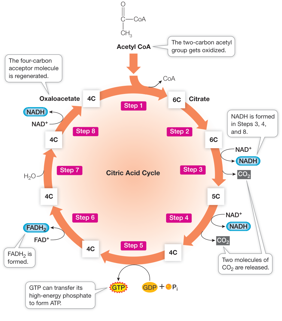
Figure 9.9 The Citric Acid Cycle The citric acid cycle has eight steps; in the last step, the starting material acceptor, oxaloacetate, is regenerated. Energy is released and captured by reducing NAD+ or FAD, or by producing GTP. “6C,” “5C,” and so on indicate the number of carbon atoms in each intermediate in the cycle.
Thus the citric acid cycle releases two carbons as CO2 and produces four reduced electron carrier molecules.
Overall, for each molecule of glucose that is oxidized, two molecules of pyruvate are produced during glycolysis, and after oxidation these feed two turns of the citric acid cycle. So the oxidation of one glucose molecule yields:
- Six CO2
- Ten NADH (two in glycolysis, two in pyruvate oxidation, and six in the citric acid cycle)
- Two FADH2
- Four ATP
Pyruvate oxidation and the citric acid cycle are regulated by the concentrations of starting materials
You have now seen that pyruvate, a three-carbon molecule, is completely oxidized to CO2 by pyruvate dehydrogenase and the citric acid cycle. For the cycle to continue, the starting molecules—acetyl CoA and oxidized electron carriers—must all be replenished.
The electron carriers are reduced during the citric acid cycle and in Step 6 of glycolysis (see Figure 9.6), and they must be reoxidized:
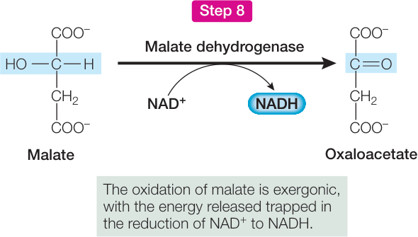
Figure 9.10 Citric Acid Cycle Close-Up Step 8 in the citric acid cycle (see Figure 9.9) illustrates the principles of molecular conversion and oxidation–reduction.
These oxidation reactions are coupled to reactions in which other molecules get reduced. When it is present, O2 is the molecule that eventually accepts these electrons, and it is reduced to form H2O (Figure 9.11).
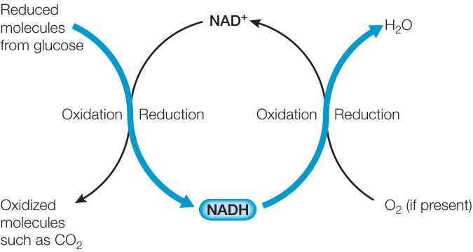
Figure 9.11 The Role of Oxygen: Overview Oxygen (O2) becomes reduced as NADH becomes oxidized, so that another molecule of glucose can be catabolized. Q: Can you predict what would happen in the absence of O2?
Recap and Assess
The oxidation of glucose in the presence of involves glycolysis, pyruvate oxidation, and the citric acid cycle. In glycolysis, glucose is converted to pyruvate with some chemical-bond energy capture. Pyruvate is oxidized first to acetyl CoA by pyruvate dehydrogenase, then completely to by the citric acid cycle, releasing energy that is captured in the form of reduced electron carriers.
- What is the net energy yield of glycolysis in terms of energy in ATP and reduced coenzymes invested and energy harvested?
- What role does pyruvate oxidation play in relation to the citric acid cycle?
- How is the citric acid cycle a “cycle” but glycolysis is not?
Pyruvate oxidation and the citric acid cycle cannot continue operating unless is available to receive electrons during the reoxidation of reduced electron carriers. However, these electrons are not passed directly to , as you will learn next.
Oxidative Phosphorylation Forms ATP
Oxidative Phosphorylation Forms ATP
Learning Objectives
- 9.3.1 Describe how the proton-motive force is established by the electron carriers and enzymes of the respiratory chain.
- 9.3.2 Analyze experimental results that relate to electron transport and chemiosmosis.
- 9.3.3 Predict results from experiments designed to explore aspects of electron transport and chemiosmosis.
In the presence of , oxidative phosphorylation is a process of ATP synthesis resulting from the reoxidation of electron carriers in the presence of . In this key concept we describe oxidative phosphorylation as it occurs in mitochondria in eukaryotes, but the same process occurs in prokaryotes (see Table 9.1).
There are two steps in oxidative phosphorylation
Two components can be distinguished in the process of oxidative phosphorylation:
- Electron transport. The electrons from NADH and pass through the respiratory chain, a series of membrane-associated electron carriers. The flow of electrons along this pathway results in the active transport of protons out of the mitochondrial matrix and across the inner mitochondrial membrane, creating a proton concentration gradient.
- Chemiosmosis. The protons diffuse back into the mitochondrial matrix through a channel protein, ATP synthase, which couples this diffusion to the synthesis of ATP. As we mentioned in the chapter opening, the inner mitochondrial membrane is normally impermeable to protons, so the only way for them to follow their concentration gradient is through the channel.
Before we proceed with the details of these pathways, let’s consider an important question: Why should the respiratory chain be such a complex process? Why don’t cells use the following single step?
The answer is that this reaction would simply release too much energy to be efficiently trapped to make ATP. Oxidizing NADH to is extremely exergonic—doing it in one step would be like setting off a stick of dynamite in the cell. There is no biochemical way to harvest that burst of energy efficiently and put it to physiological use (that is, no single metabolic reaction is so endergonic as to consume a significant fraction of that energy in a single step). To control the release of energy during the oxidation of glucose, cells have evolved a lengthy respiratory chain: a series of reactions, each of which releases a small amount of energy, one step at a time.
The respiratory chain transfers electrons and protons, and releases energy
The respiratory chain (an electron transport chain) is located in the inner mitochondrial membrane. Because of the extensive folding of the membrane, there is more room for the proteins involved in the chain than there would be in a membrane with less surface area. There are several interacting components, including large integral proteins, a small peripheral protein, and a small lipid molecule. Figure 9.12 shows a plot that describes the free energy released as electrons are passed between the carriers.
- Four large protein complexes (I, II, III, and IV) contain electron carriers and associated enzymes. In eukaryotes they are integral proteins of the inner mitochondrial membrane, and three are transmembrane proteins.
- Cytochrome c is a small peripheral protein that lies in the intermembrane space. It is loosely attached to the outer surface of the inner mitochondrial membrane.
- Ubiquinone (often referred to as coenzyme Q10; abbreviated Q) is a small, nonpolar, lipid molecule that moves freely within the hydrophobic interior of the phospholipid bilayer of the inner mitochondrial membrane.
As illustrated in Figure 9.12, NADH passes electrons to protein complex I (called NADH-Q reductase), which in turn passes the electrons to Q. This electron transfer is accompanied by a large drop in free energy. Complex II (succinate dehydrogenase) passes electrons to Q from , which was generated in Step 6 of the citric acid cycle (see Figure 9.9). These electrons enter the chain later than those from NADH and will ultimately produce less ATP.
Complex III (cytochrome c reductase) receives electrons from Q and passes them to cytochrome c. Complex IV (cytochrome c oxidase) receives electrons from cytochrome c and passes them to oxygen. Finally the reduction of oxygen to occurs:
Notice that four protons () are also consumed in this reaction. This contributes to the proton concentration gradient across the inner mitochondrial membrane by reducing the proton concentration in the mitochondrial matrix.
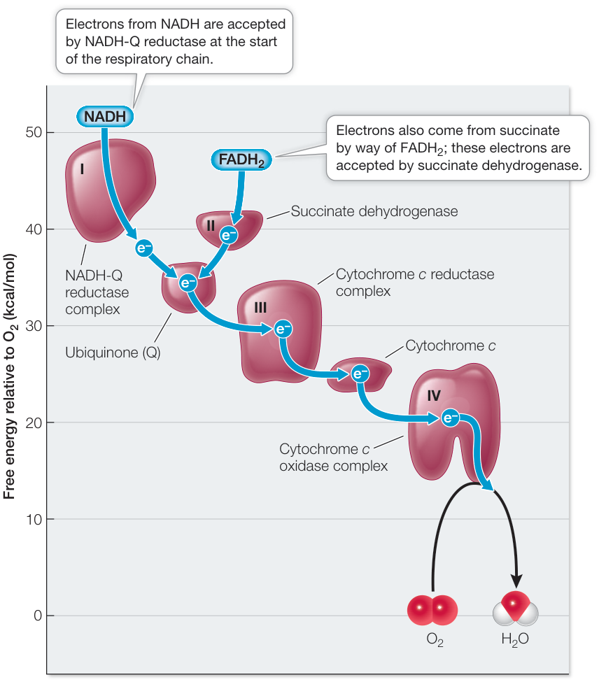
Figure 9.12 The Oxidation of NADH and FADH2 in the Respiratory Chain Electrons from NADH and FADH2 are passed along the respiratory chain, a series of protein complexes in the inner mitochondrial membrane containing electron carriers and enzymes. The carriers gain free energy when they become reduced and release free energy when they are oxidized. This illustration shows the standard free energy changes along the respiratory chain.
ATP is made through chemiosmosis
During electron transport, protons are also actively transported across the membrane: electron transport within each of the three transmembrane complexes (I, III, and IV) results in the transfer of protons from the matrix to the intermembrane space (Figure 9.13). Since the lipid bilayer does not allow charged H+ to diffuse across it, transfer of H+ across it through the electron transport chain sets up a gradient, with the concentration of H+ in the intermembrane space higher than in the matrix. In addition, because H+ carries a charge, there is more positive charge in the intermembrane space. About 20 H+ are transported into the intermembrane space as four electrons are carried through the respiratory chain to O2. These two gradients—of concentration and charge—set up a proton-motive force which is a key factor in energy metabolism in cells: The gradient of H+ across the inner membrane is a source of potential energy.
This energy is tapped for use by the cell by the membrane channel protein ATP synthase, which allows the H+ to diffuse back into the matrix down its concentration gradient. In the process, potential energy is captured and used for the formation of ATP. The coupling of the proton-motive force and ATP synthesis is called the chemiosmotic mechanism—or chemiosmosis—and is found in all respiring cells.
To summarize, the energy originally contained in glucose and other fuel molecules is ultimately captured in the cellular energy currency, ATP. For each pair of electrons passed along the chain from NADH to oxygen, about 2.5 molecules of ATP are formed. FADH2 oxidation produces about 1.5 ATP molecules because it enters the electron transport chain at a later step than NADH (see Figure 9.13).
ATP synthesis is a reversible reaction, and ATP synthase can also act as an ATPase, hydrolyzing ATP to ADP and Pi:
If the reaction goes to the right, free energy is released. In the mitochondrion, it is used to transfer H+ out of the mitochondrial matrix—not the usual mode of operation. If the reaction goes to the left, it uses the free energy from H+ diffusion into the matrix to make ATP. What makes it prefer ATP synthesis? There are two answers to this question:
- ATP leaves the mitochondrial matrix for use elsewhere in the cell as soon as it is made, keeping the ATP concentration in the matrix low, and driving the reaction toward the left.
- The H+ gradient is maintained by electron and proton transport.
Every day a person hydrolyzes about 1025 ATP molecules to ADP. This amounts to 9 kg, a significant fraction of the person’s entire body weight! The vast majority of this ADP is “recycled”—converted back to ATP—using free energy from the oxidation of glucose.
Experiments demonstrate chemiosmosis
Chemiosmosis has been tested in the lab. Let’s look at two lines of evidence; first, a direct demonstration that a H+ gradient can drive ATP synthesis, and second, disruption of the coupling of electron transport to ATP synthesis.
DIRECT DEMONSTRATION OF CHEMIOSMOSIS The key experiment that demonstrated that a proton (H+) gradient across a membrane could drive ATP synthesis was first performed using chloroplasts, the organelles in plants that convert the energy in sunlight into chemical energy (photosynthesis; see Chapter 10)
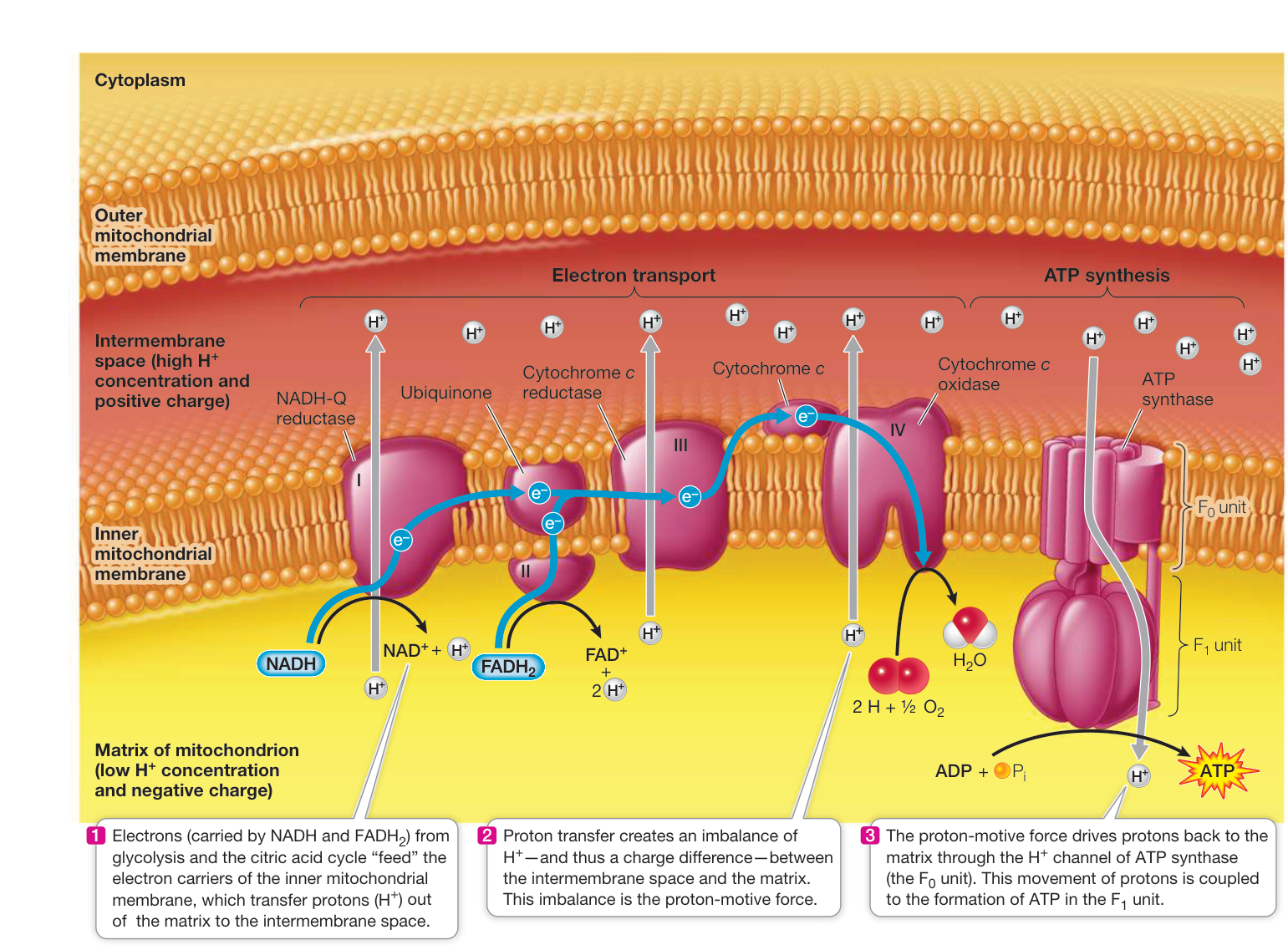
Figure 9.13 The Respiratory Chain and ATP Synthase Produce ATP by a Chemiosmotic Mechanism As electrons pass through the transmembrane protein complexes in the respiratory chain, protons are transferred from the mitochondrial matrix into the intermembrane space. As the protons return to the matrix through ATP synthase, ATP is formed.
(Figure 9.14). Soon thereafter, the same mechanism was shown to work in mitochondria.
UNCOUPLING ELECTRON TRANSPORT FROM ATP PRODUCTION As you have seen, the coupling of electron transport (which generates the proton gradient) with chemiosmosis is vital for the capture of free energy in the form of ATP. Uncoupling protein 1 (UCP1), which is found in the mitochondria of brown fat cells (described in the chapter opening), demonstrates the importance of this coupling. By disrupting the gradient, UCP1 allows the energy released during electron transport to be in the form of heat, rather than chemical-bond energy trapped in ATP.
The relationship between UCP1 in brown fat and body weight has been investigated experimentally in a study of a genetic strain of mice that—unlike normal mice—do not get fat as they age (people tend to put on weight as they age, too). As Investigating Life: Mitochondria, Genetics, and Obesity reveals, it appears that the leaner mice make more UCP1 and oxidize (burn off) more fat than normal mice do. Might this observation point to ways to control weight gain in people? In a related study of obesity conducted in the 1930s, a chemical, dinitrophenol, was shown to uncouple oxidation and phosphorylation, just as UCP1 (which had not been discovered yet) does. Physicians got the idea that giving obese people the uncoupler would cause them to oxidize their fat. It worked, but since dinitrophenol acted on all mitochondria everywhere in the body, and not just in fat cells, severe side effects occurred as ATP production all over the body became critically low—in some cases too low for survival. So the idea of an artificial uncoupler for weight loss was dropped. However, stimulation of the natural uncoupler in brown fat is under study for obesity treatment.
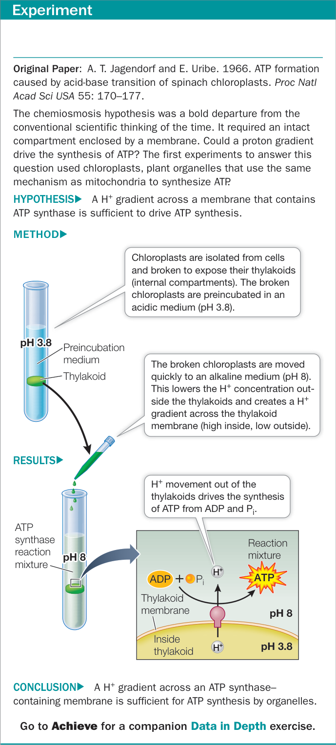
Figure 9.14 An Experiment Demonstrates the Chemiosmotic Mechanism
HOW ATP SYNTHASE WORKS: A MOLECULAR MOTOR The structure and mechanism of ATP synthase, illustrated in Figure
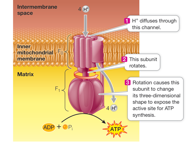
Figure 9.15 How ATP Is Made Mitochondrial ATP synthase is a rotary motor.
Media Clip 9.1 ATP Synthase in Motion Life12e.com/mc9.1
9.15, are shared by living organisms as diverse as bacteria and humans. ATP synthase is a molecular motor composed of two parts: the unit, a transmembrane region that is the channel; and the unit, which contains the active sites for ATP synthesis. consists of six subunits (three each of two polypeptide chains), arranged like the segments of an orange around a central shaft-like polypeptide that interacts with the membrane-embedded . Electron transport sets up an gradient across the membrane. This gradient has potential energy, and when diffuses through the channel, the potential energy is converted to kinetic energy, causing the central polypeptide to rotate. The energy is transmitted to the catalytic subunits of , resulting in ATP synthesis. These molecular motors make ATP at rates up to 100 molecules per second.
Some microorganisms use non- electron acceptors
A general way to describe the last reaction in electron transport is:
Part of the amazing success of bacteria and archaea is that they have evolved biochemical pathways that allow them to exist in environments where is scarce or absent. As you will see in the next key concept, for most animals and plants, the anaerobic (no ) catabolism of glucose generally yields much less energy than aerobic catabolism. However, many bacteria and archaea exploit their environments to use alternative electron acceptors. This allows them to complete the electron transport chain and produce ATP even in the absence of . Table 9.2 summarizes some of these pathways, which are referred to as anaerobic respiration. Note that some of these microbes use ions as electron acceptors while others use small molecules.
Investigating Life: Mitochondria, Genetics, and Obesity
Experiment
Original Paper: X. L. Ma et al. 2011. Ablations of ghrelin and ghrelin receptor exhibit differential metabolic phenotypes and thermogenic capacity during aging. PLOS ONE 6: e16391.
As people (and mice) get older, they tend to accumulate fat. In the course of investigating a strain of mice genetically unable to make the receptor for a hormone called ghrelin that is involved in controlling appetite, Yuxiang Sun and her team at Baylor College of Medicine compared these mice with normal mice as they aged. They were surprised to find that the genetically changed mice did not put on as much weight as their normal counterparts. The researchers discovered that the genetic mutation in these mice caused uncoupling of oxidative phosphorylation and burning of body fat. Their experiment investigated whether levels of mitochondrial uncoupling protein 1 (UCP1) were different in the genetically changed mice than in normal mice.
HYPOTHESIS Mice that make more uncoupling protein (UCP1) burn more body fat.
METHOD
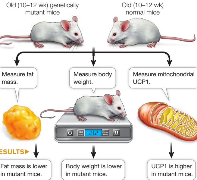
CONCLUSION Increased UCP1 correlates with less fat and a lower body weight.
Work with the Data
In an effort to determine whether weight gain in normal mice might be due to inactivity or to eating too much (two of the well-known causes of weight gain), as opposed to a biochemical abnormality (such as unusual UCP1 levels), Sun and her team examined young (3–4 months old) and older (10–12 months old) mice of both strains. Error bars represent standard error of the mean. (Figures A–C after X. L. Ma et al. 2011. PLOS ONE 6: e16391. © 2011 Ma et al.; CC BY 4.0.)
QUESTIONS
- The researchers measured weight and body composition in terms of fat and lean body tissues. The results are shown in Figure A. Did the mutant mice and normal mice gain weight as they aged?
- The researchers wondered whether the weight changes noted in Figure A were due to changes in eating patterns or exercising. So they measured how much food the mice ate per day and measured their movements over time in a special chamber. The results are shown in Figure B. What can you conclude about the role of food intake and exercise on the weight differences between the two strains of mice?
- The uncoupling protein UCP1 found in the inner mitochondrial membrane of brown fat cells uncouples mitochondrial electron transport (oxidation) and ATP production (phosphorylation), so that instead of being trapped as chemical energy in the formation of ATP the energy released by oxidation is released as heat. Sun and her team measured UCP1 levels in brown fat mitochondria of the two strains of mice. The results are shown in Figure C. What can you conclude about the role of UCP1 in the weight differences between the two strains of mice?
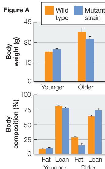
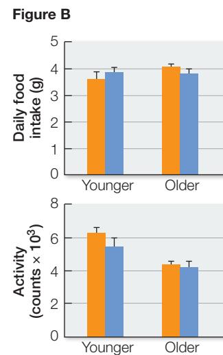
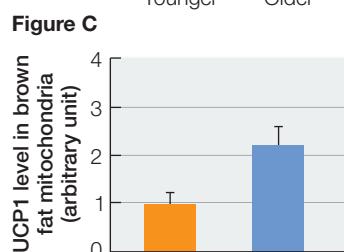
Table 9.2 Electron Acceptors Used in the Respiratory Chain of Anaerobic Microbes
| Terminal electron acceptor | Product formed | Organism |
|---|---|---|
| Desulfovibrio desulfuricans | ||
| Geobacter metallireducens | ||
| Escherichia coli | ||
| Methanosarcina barkeri | ||
| Clostridium aceticum | ||
| Fumarate | Succinate | Wolinella succinogenes |
Connect the Concepts The diversity of microbial metabolic pathways allows them to adapt to life in many environments. See Key Concept 24.2.
Recap and Assess
The oxidation of reduced electron carriers in the respiratory chain drives the active transport of protons across the inner mitochondrial membrane, generating a proton-motive force. Diffusion of protons down their electrochemical gradient through ATP synthase is coupled to the synthesis of ATP. Some bacteria and archaea can respire using alternative electron acceptors instead of .
- How are protons transported from the mitochondrial matrix to the intermembrane space during electron transport?
- How does the experiment described in Figure 9.14 demonstrate the chemiosmotic mechanism?
- Trace the sequence of changes in redox reactions that occur in mammalian tissue when the oxygen supply is cut off. The first change is that all of the cytochrome c becomes reduced, because electrons can still flow from cytochrome c, but there is no oxygen to accept electrons from cytochrome c oxidase. What happens after this?
- The drug antimycin A blocks electron transport in mitochondria and chloroplasts. Explain what would happen if the experiment in Figure 9.14 were repeated in the presence of this drug.
Oxidative phosphorylation captures a great deal of energy in ATP. But it does not occur if is absent. We will turn now to the metabolism of glucose in anaerobic conditions.
In the Absence of Oxygen, Some Energy Is Harvested from Glucose
In the Absence of Oxygen, Some Energy Is Harvested from Glucose
Learning Objectives
9.4.1 Describe the biochemical pathways of fermentation. 9.4.2 Explain why fermentation is necessary in anaerobic conditions.
In eukaryotes, in the absence of (anaerobic conditions), a small amount of ATP can be produced by glycolysis and fermentation. Like glycolysis, fermentation pathways occur in the cytoplasm. There are many different types of fermentation, but they all operate to regenerate so that the -requiring reactions of glycolysis can continue. The two best-understood fermentation pathways are found in a wide variety of organisms, including eukaryotes:
- Lactic acid fermentation, the end product of which is lactic acid (lactate)
- Alcoholic fermentation, the end product of which is ethyl alcohol (ethanol)
Fermentation pathways allow glucose to be partially oxidized in the absence of oxygen
In lactic acid fermentation, pyruvate serves as the electron acceptor and lactate is the product (Figure 9.16A). This process takes place in many microorganisms and complex organisms, including higher plants and vertebrates. A notable example of lactic acid fermentation occurs in vertebrate muscle tissue. Usually, vertebrates get their energy for muscle contraction aerobically, with the circulatory system supplying to muscles. In small vertebrates, this is almost always adequate: for example, birds can fly long distances without resting. But in larger vertebrates such as humans, the circulatory system is not up to the task of delivering enough when the need is great, such as during intense activity. At this point, the muscle cells break down glycogen (a stored polysaccharide; see Figure 3.19) and undergo lactic acid fermentation.
Lactate buildup becomes a problem after prolonged periods of intense exercise because it is associated with an increase in the concentration in the cell, lowering the pH. This affects cellular activities, which return to normal upon resting. Lactate dehydrogenase, the enzyme that catalyzes the fermentation reaction, works in both directions. That is, when is available it can catalyze the oxidation of lactate to form pyruvate, which is then catabolized to with concomitant energy release to form ATP. When lactate levels are decreased, muscle activity can resume.
Alcoholic fermentation takes place in certain yeasts (eukaryotic microbes) and some plant cells under anaerobic conditions. This process requires two enzymes, pyruvate decarboxylase and alcohol dehydrogenase, which metabolize pyruvate to ethanol (Figure 9.16B). As with lactic acid fermentation, the reactions are essentially reversible. For thousands of years, humans have used anaerobic fermentation by yeast cells to produce alcoholic beverages. The bubbles in beer and champagne are from the action of pyruvate decarboxylase. The cells use sugars from plant sources (glucose from grapes or maltose from barley) to produce the end product, ethanol, in wine and beer.
By recycling , fermentation allows glycolysis to continue, thus producing small amounts of ATP through substrate-level phosphorylation. The net yield of two ATPs per glucose molecule is much lower than the energy yield from cellular respiration. For this reason, most organisms existing in anaerobic environments are small microbes that grow relatively slowly.
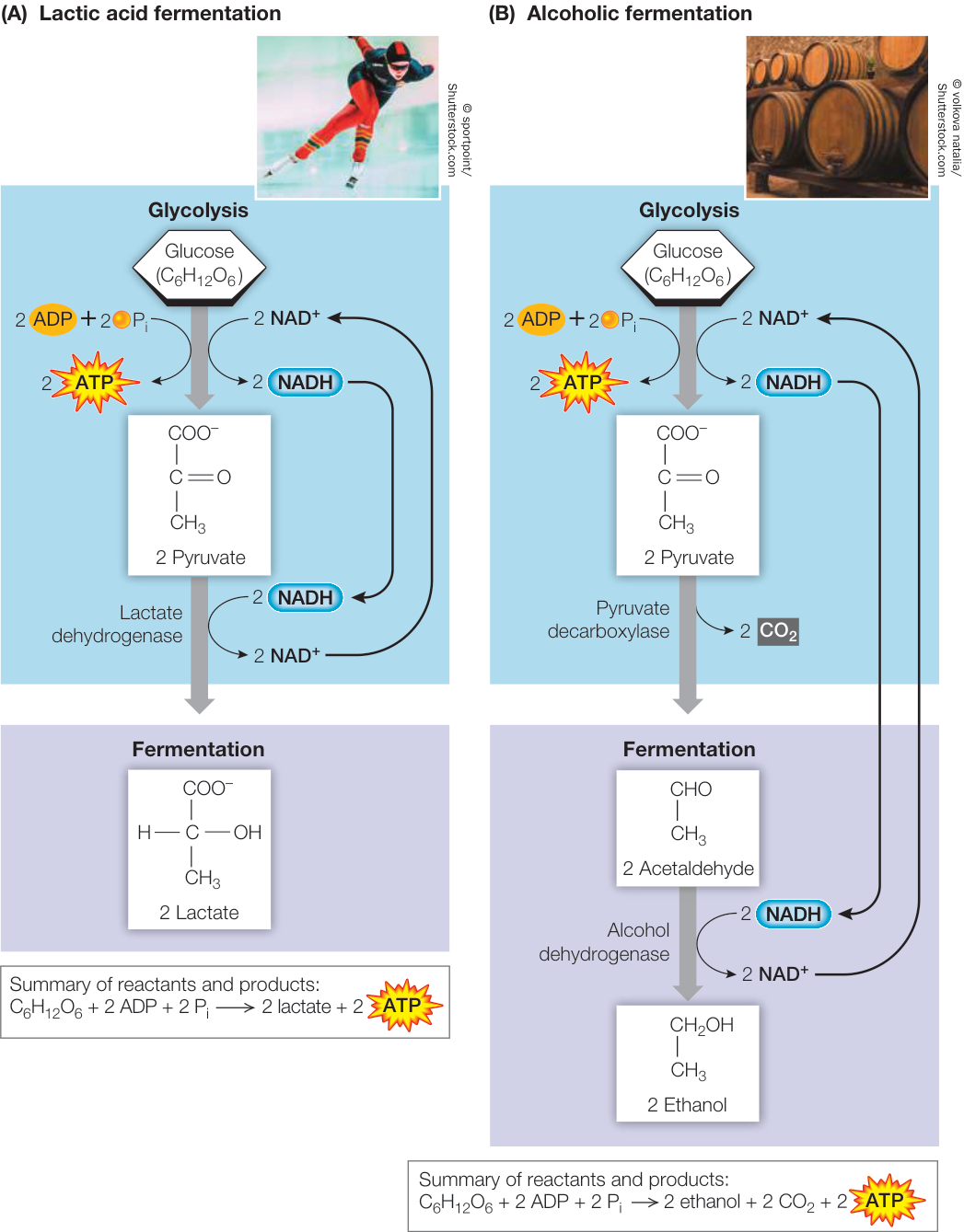
Figure 9.16 Fermentation Glycolysis produces pyruvate, ATP, and NADH from glucose. (A) Lactic acid fermentation uses NADH as a reducing agent to reduce pyruvate to lactate, thus regenerating NAD+ to keep glycolysis operating. (B) In alcoholic fermentation, pyruvate from glycolysis is converted into acetaldehyde, and CO2 is released. NADH from glycolysis is used to reduce acetaldehyde to ethanol, thus regenerating NAD+ to keep glycolysis operating. Q: Where do the bubbles in champagne and beer come from? Figure 9.16 Fermentation Glycolysis produces pyruvate, ATP, and NADH from glucose. (A) Lactic acid fermentation uses NADH as a reducing agent to reduce pyruvate to lactate, thus regenerating NAD+ to keep glycolysis operating. (B) In alcoholic fermentation, pyruvate from glycolysis is converted into acetaldehyde, and CO2 is released. NADH from glycolysis is used to reduce acetaldehyde to ethanol, thus regenerating NAD+ to keep glycolysis operating. Q: Where do the bubbles in champagne and beer come from?
Cellular respiration yields much more energy than fermentation
The total net energy yield from glycolysis plus fermentation is two molecules of ATP per molecule of glucose oxidized. The maximum yield of ATP that can be harvested from a molecule of glucose through glycolysis followed by cellular respiration is much greater—about 32 molecules of ATP (Figure 9.17). (Review Figures 9.6, 9.7, 9.9, and 9.13 to see where all the ATP molecules come from.)
Why do the metabolic pathways that operate in aerobic environments produce so much more ATP? Glycolysis and fermentation only partially oxidize glucose. Much more energy remains in the end products of fermentation (lactic acid and ethanol)
than in CO2, the end product of cellular respiration. In cellular respiration, carriers (mostly NAD+) are reduced during pyruvate oxidation and the citric acid cycle. Then the reduced carriers are oxidized by the respiratory chain, with the accompanying production of ATP by chemiosmosis (about 2.5 ATP for each NADH and 1.5 ATP for each FADH2). In an aerobic environment, a cell or organism capable of aerobic metabolism will have the advantage over one that is limited to fermentation, in terms of its ability to harvest chemical energy. Two key events in the evolution of multicellular organisms were the rise in atmospheric O2 levels (see Key Concept 24.2) and the development of metabolic pathways to use that O2.
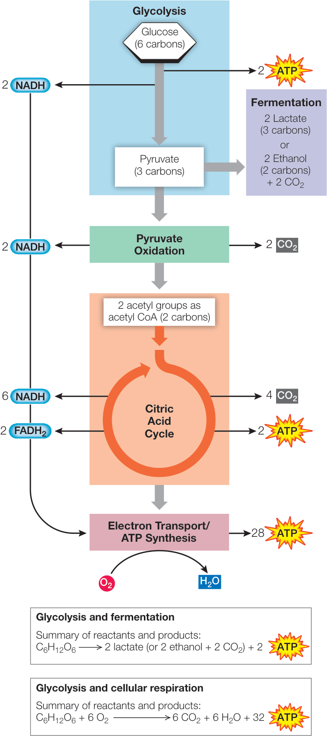
Figure 9.17 Cellular Respiration Yields More Energy Than Fermentation Electron carriers are reduced in pyruvate oxidation and the citric acid cycle, then oxidized by the respiratory chain. These reactions produce ATP via chemiosmosis.
Recap and Assess
In the absence of , fermentation pathways use NADH formed by glycolysis to reduce pyruvate and regenerate . The energy yield of glycolysis coupled to fermentation is low because glucose is only partially oxidized. When is present, the electron carriers of cellular respiration allow for the full oxidation of glucose, so the energy yield from glucose is much higher.
- Why is replenishing required to continue glycolysis in the absence of ?
- How does fermentation replenish ?
Now that you’ve seen how cells harvest energy, let’s see how that energy moves through other metabolic pathways in the cell.
Metabolic Pathways Are Interrelated and Regulated
Learning Objectives
- 9.5.1 Describe catabolic and anabolic pathways that link carbohydrate, lipid, and protein metabolism.
- 9.5.2 Trace the path of an atom as it moves through various metabolic pathways within a cell.
- 9.5.3 Explain how allosteric changes in an enzyme can be used to regulate flow through a metabolic pathway.
Glycolysis and the pathways of cellular respiration do not operate in isolation. Rather, there is an interchange of molecules into and out of these pathways, to and from the metabolic pathways for the synthesis and breakdown of amino acids, nucleotides, fatty acids, and other building blocks of life (see Figure 8.14). Carbon skeletons (i.e., the carbon backbones of organic molecules) can enter the catabolic pathways and be broken down to release their energy, or they can enter anabolic pathways to be used in the formation of the macromolecules that are the major constituents of the cell. These relationships are summarized in Figure 9.18. In this key concept we will explore how pathways are interrelated by the sharing of intermediate molecules, and we will see how pathways are regulated by the inhibitors of key enzymes.
Catabolism and anabolism are linked
A hamburger or veggie burger on a bun contains three major sources of carbon skeletons: carbohydrates, mostly in the form of starch (a polysaccharide); lipids, mostly as triglycerides (three fatty acids attached to glycerol); and proteins (polymers of amino acids). Look at Figure 9.18 to see how each of these three types of macromolecules can be hydrolyzed and used in catabolism or anabolism.
CATABOLIC INTERCONVERSIONS Polysaccharides, lipids, and proteins can all be broken down to provide energy:
- Polysaccharides are hydrolyzed to glucose. Glucose then passes through glycolysis and cellular respiration, where its energy is captured in ATP.
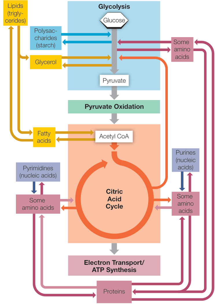
Figure 9.18 Relationships among the Major Metabolic Pathways of the Cell Note the central positions of glycolysis and the citric acid cycle in this network of metabolic pathways. Also note that many of the pathways can operate essentially in reverse. Q: Can DNA be an energy source? Explain. Why do you think it is not usually employed in this way?
- Lipids are broken down into their constituents, glycerol and fatty acids. Glycerol is converted into dihydroxyacetone phosphate (DHAP), an intermediate in glycolysis. Fatty acids are highly reduced molecules that are converted to acetyl CoA inside the mitochondrion by a series of oxidation enzymes, in a process known as -oxidation. For example, the -oxidation of a 16-carbon () fatty acid occurs in several steps:
Repeat 6 times 8 acetyl CoA
The acetyl CoA can then enter the citric acid cycle and be catabolized to .
- Proteins are hydrolyzed to their amino acid building blocks. The 20 different amino acids feed into glycolysis or the citric acid cycle at different points determined by their structures. For example, the amino acid glutamate is converted into -ketoglutarate, an intermediate in the citric acid cycle (the five-carbon molecule in Figure 9.9):
Note that one of the products of this reaction is ammonia (), which is toxic to cells. If there is too much , the equilibrium point of the reaction will shift to the left, and the concentration of -ketoglutarate, a citric acid cycle intermediate, will be reduced. The cycle can begin to shut down, with drastic effects on energy provision. Therefore excess must be removed, and this is done by making urea: , a waste product excreted in urine.
ANABOLIC INTERCONVERSIONS Many catabolic pathways can operate essentially in reverse, with some modifications. Glycolytic and citric acid cycle intermediates, instead of being oxidized to form , can be reduced and used to form glucose in a process called gluconeogenesis (which means “new formation of glucose”). Likewise, acetyl CoA can be used to form fatty acids. The most common fatty acids have even numbers of carbons: 14, 16, or 18. These are formed by the addition of two-carbon acetyl CoA “units” one at a time until the appropriate chain length is reached. Acetyl CoA is also a building block for various pigments, plant growth substances, rubber, steroid hormones, and other molecules.
Some intermediates in the citric acid cycle are reactants in pathways that synthesize important components of nucleic acids. For example, -ketoglutarate is a starting point for purines, and oxaloacetate for pyrimidines. In addition, -ketoglutarate is a starting point for the synthesis of chlorophyll (used in photosynthesis; see Chapter 10) and the amino acid glutamate (used in protein synthesis).
Catabolism and anabolism are integrated
A carbon atom from a protein in your burger can end up in DNA, fat, or , among other fates. How does the organism “decide” which metabolic pathways to follow, in which cells? With all of the possible interconversions, you might expect that cellular concentrations of various biochemical molecules would vary widely. Remarkably, the levels of these substances in what is called the metabolic pool—the sum total of all the biochemical molecules in a cell—are quite constant. Organisms regulate the enzymes in various cells in order to maintain a steady state between catabolism and anabolism.
Connect the Concepts Control of metabolic pathways often rests with regulation of enzymes already present. Review the mechanisms of enzyme regulation in Key Concept 8.5.
The regulation of interconnecting biochemical pathways is a problem of systems biology, which seeks to understand how biochemical pathways interact (see Key Concept 8.5). It is a bit like trying to predict traffic patterns in a city: if an accident blocks traffic on a major road, drivers take alternate routes, where the traffic volume consequently changes.
Several mechanisms can be used to regulate the rate of each step in a biochemical pathway:
- Changing the amount of active enzyme: The cell can increase the expression of a gene encoding an enzyme.
- Changing enzyme activity by covalent modifications: Adding phosphate groups by protein kinase can alter the activity of an enzyme.
- Feedback inhibition: Allosteric changes in an enzyme due to binding of a product in a pathway can cause the entire pathway to shut down.
- Substrate availability: If the substrate of a particular enzyme is used up by another pathway, the first enzyme can no longer function and the pathway shuts down.
Consider what happens to the starch in your burger bun. In the digestive system, starch is hydrolyzed to glucose, which enters the blood for distribution to the rest of the body. But before the glucose is distributed, a regulatory check must be made: If there is already enough glucose in the blood to supply the body’s needs, the excess glucose is converted into glycogen and stored in the liver and muscles. If not enough glucose is supplied by food, glycogen is broken down, or other molecules are used to make glucose by gluconeogenesis. The end result is that the level of glucose in the blood is remarkably constant. How does the body accomplish this?
Glycolysis and the citric acid cycle are subject to allosteric regulation (see Key Concept 8.5) of key enzymes involved. In a metabolic pathway, a high concentration of the final product can inhibit the action of an enzyme that catalyzes an earlier reaction (see Figure 8.20). Furthermore, an excess of the product of one pathway can activate an enzyme in another pathway, speeding up its reactions and diverting raw materials away from synthesis of the first product. These negative and positive feedback mechanisms are used at many points in the energy-harvesting pathways and are summarized in Figure 9.19.
- The main control point in glycolysis is the glycolytic enzyme phosphofructokinase, which catalyzes Step 3 in Figure 9.6. This enzyme is allosterically inhibited by ATP or citrate, and activated by ADP or AMP. Under anaerobic conditions, fermentation yields a relatively small amount of ATP, and phosphofructokinase operates at a high rate. However, when conditions are aerobic, respiration makes 16 times more ATP
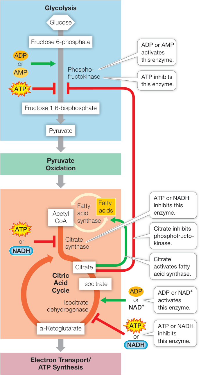
Figure 9.19 Allosteric Regulation of Glycolysis and the Citric Acid Cycle Allosteric regulation controls glycolysis and the citric acid cycle at crucial early steps, increasing their efficiency and preventing the excessive buildup of intermediates.
- than fermentation does, and the abundant ATP allosterically inhibits phosphofructokinase. So glycolysis slows down.
- The main control point in the citric acid cycle is the enzyme isocitrate dehydrogenase (which catalyzes Step 3 in Figure 9.9). This enzyme is activated by increases in substrate concentrations (ADP, NAD+, and isocitrate) and is inhibited by products of the citric acid cycle: ATP and NADH. If too much ATP or NADH accumulates, the reaction is slowed and the citric acid cycle shuts down.
- Another control point involves acetyl CoA. If the level of ATP is high and the citric acid cycle shuts down, the accumulation of citrate activates fatty acid synthase, diverting acetyl CoA to the synthesis of fatty acids for storage (contributing to a person’s accumulation of fat). These fatty acids may be metabolized later to produce more acetyl CoA.
Recap and Assess
Glucose can be made from intermediates in glycolysis and the citric acid cycle by gluconeogenesis. The metabolic pathways for the production and breakdown of lipids and amino acids are linked to those of glucose metabolism. Reaction products regulate key enzymes in the various pathways.
- Give examples of the pathways relating to glycolysis and the citric acid cycle for catabolism of a lipid and the anabolism of a protein.
- How does phosphofructokinase serve as a control point for glycolysis?
- You eat a burger that contains polysaccharides, proteins, and lipids. How could carbon atoms in the proteins’ amino acids and the glucose in the polysaccharides end up as fats?
Investigating Life: QA How are mitochondria related to obesity?
Recall from the opening of this chapter that brown fat is adipose tissue with a high concentration of mitochondria, which have iron-containing pigments. The uncoupling protein UCP1, found in the inner membranes of mitochondria in brown fat cells, uncouples mitochondrial electron transport (oxidation) and ATP production (phosphorylation), so that instead of being trapped as chemical-bond energy in the formation of ATP, the energy released by oxidation is released as heat.
Hibernating animals take advantage of the heat emitted by the catabolism of energy-rich molecules in brown fat, especially when aroused. In hibernating animals, brown fat and UCP1 increase during the winter. It turns out that adult humans also experience a seasonal variation in brown fat, having more of it in the cooler months. Other factors such as diet and exercise being equal, increased fat catabolism occurs in the presence of UCP1 (as demonstrated by the experiment in Investigating Life: Mitochondria, Genetics, and Obesity).
Genetic studies confirm the role of UCP1 in fat breakdown as well. For example, some people have a genetically determined variant of UCP1 that has lower uncoupling activity than normal UCP1. As people with the variant get older, they accumulate more body fat than their peers with normal UCP1 do.
Future directions
Activating brown fat tissue may help in treating obesity. When brown fat is activated by cold temperature in animals, there is an increase in mitochondrial uncoupling, but also an increase in fatty acid breakdown ( -oxidation) and glucose uptake into cells to provide energy from a nonfat source (see Figure 9.18). This is accompanied by significant weight loss. Metabolic effects are seen throughout the body, especially in liver and muscle tissue. To get the message from brown fat cells to other tissues, the fat cells produce a protein called adiponectin. Amazingly, this protein is elevated in centenarians, many of whom are lean. In some centenarians, their genetic offspring also have elevated adiponectin, so the activation of brown fat may be inherited. Several drugs are being evaluated for their potential to activate brown fat.
© 3d_man/Shutterstock.com
You should be able to relate each summary to the adjacent figures. If you go to this visual summary in Achieve, you can follow links to figures, animations, activities, and simulations that will help you consolidate the material.
Visual Summary
Cells Harvest Chemical Energy from Glucose Oxidation
- The pathways of glucose catabolism are glycolysis, pyruvate oxidation, the citric acid cycle, and electron transport/ATP synthesis. Under anaerobic conditions, fermentation occurs.
- As glucose is oxidized, the electrons it loses are transferred or are part of a more electronegative bond in another material, which is thereby reduced.
- The coenzyme is a key electron carrier in biological oxidation–reduction, or redox, reactions. It exists in two forms, one oxidized () and the other reduced ().
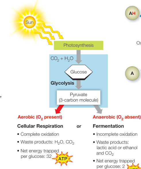
Figure 9.1
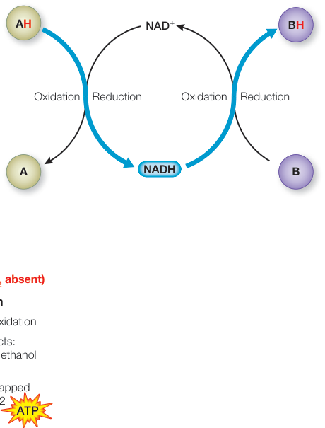
Figure 9.4 Questions
- When glucose is oxidized, energy is trapped by ADP to make ATP. What is the source in nature of the energy in glucose?
- Which is higher in free energy, or , and why?
In the Presence of Oxygen, Glucose Is Fully Oxidized
- Glycolysis consists of ten enzyme-catalyzed reactions that occur in the cell cytoplasm. The end products are two three-carbon molecules of pyruvate, two molecules of ATP, and two molecules of . Energy is trapped in ATP and .
- After pyruvate oxidation, the citric acid cycle fully oxidizes the glucose catabolites to .
Question
- Why does glycolysis involve multiple steps, each catalyzed by a single enzyme?
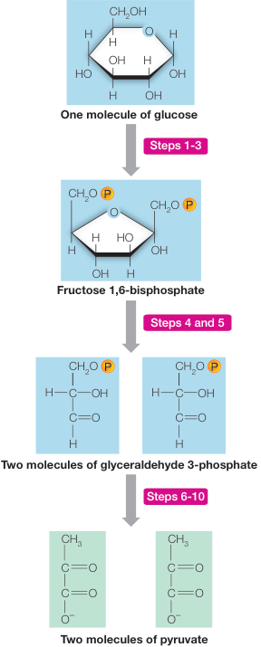
Figure 9.6
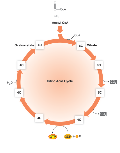
Figure 9.9
Oxidative Phosphorylation Forms ATP
- Oxidation of electron carriers in the presence of releases energy that can be used to form ATP in a process called oxidative phosphorylation.
- The NADH and produced in glycolysis, pyruvate oxidation, and the citric acid cycle are oxidized by the respiratory chain, regenerating and . Oxygen () is the final acceptor of electrons and protons, forming water ().
- The respiratory chain transfers protons across the inner mitochondrial membrane, creating the proton-motive force that drives the synthesis of ATP from ADP by chemiosmosis.
Questions
- Rotenone is an insecticide isolated from the roots of certain tropical plants. It inhibits NADH-Q reductase, which catalyzes the oxidation of NADH in the electron transport chain. How does rotenone kill cells?
- What is the source of the electrons in the electron transport chain?
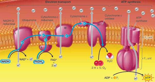
Figure 9.13
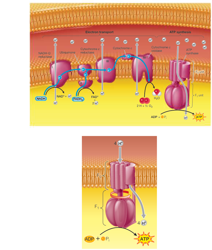
Figure 9.15
In the Absence of Oxygen, Some Energy Is Harvested from Glucose
- In the absence of , glycolysis is followed by fermentation. In the process, is regenerated from NADH so that glycolysis can continue, thus generating a small amount of ATP.
- For each molecule of glucose used, glycolysis plus fermentation yields two molecules of ATP. In contrast, glycolysis operating with pyruvate oxidation, the citric acid cycle, and oxidative phosphorylation yields up to 32 molecules of ATP per molecule of glucose.
Questions
- Why does the production of wine occur in sealed barrels or tanks?
- In an initial attempt to run a marathon without training, a woman gets tired after a mile, with the pH in her muscle cells reduced. After rest, the pH is raised and her muscle function resumes. How do these events occur?
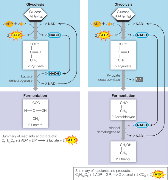
Figure 9.16
Metabolic Pathways Are Interrelated and Regulated
- The catabolic pathways for the breakdown of carbohydrates, fats, and proteins feed into the energy-harvesting metabolic pathways.
- Anabolic pathways use intermediate components of the energy-harvesting pathways to synthesize fats, amino acids, and glucose.
- The rates of glycolysis and the citric acid cycle are controlled by allosteric regulation and by the diversion of excess acetyl CoA into fatty acid synthesis.
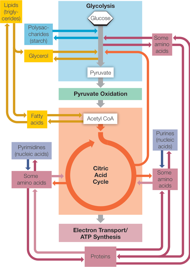
Figure 9.18
Questions
- Before a race, some runners eat peanut butter, which contains abundant protein. How does protein provide energy?
- Most biochemical pathways can operate essentially in both directions, and also can operate with increased or decreased rates. How does this regulation occur? Give an example.