Chapter 13DNA and Its Role in Heredity
Key Concepts
13.1 Experiments Revealed the Function of DNA as Genetic Material
13.2 DNA Has a Structure That Suits Its Function
13.3 DNA Is Replicated Semiconservatively
13.4 Errors in DNA Can Be Repaired
13.5 The Polymerase Chain Reaction Amplifies DNA
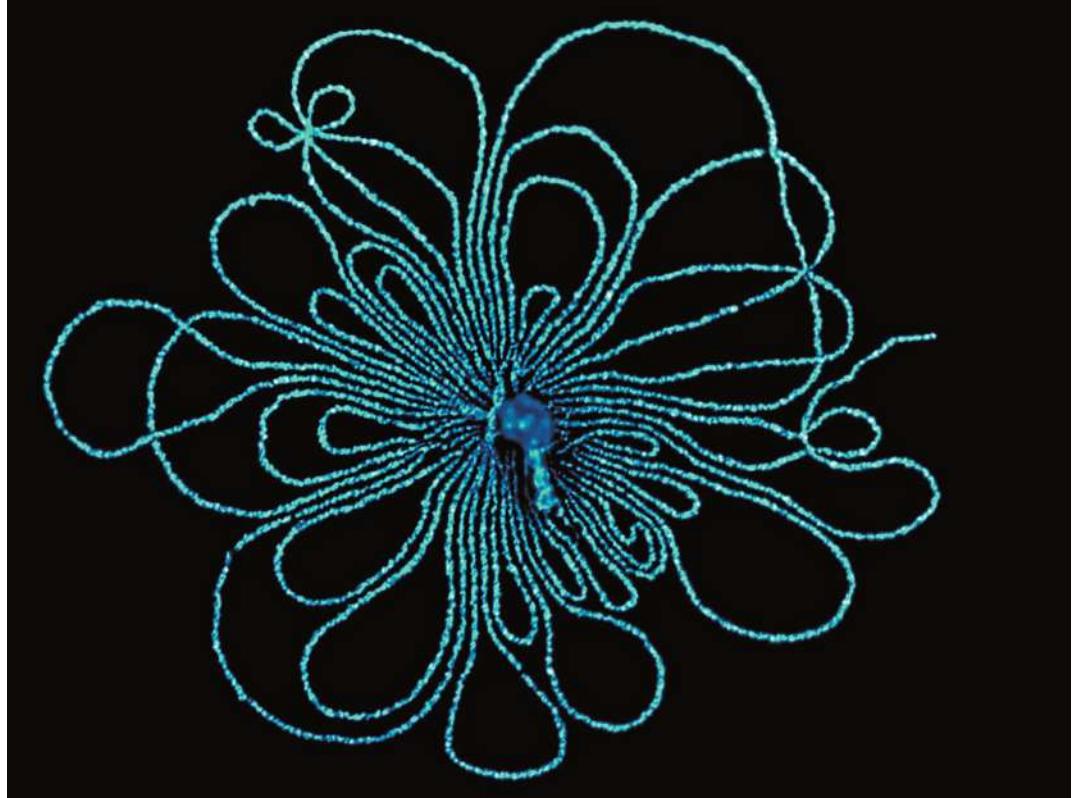
A tiny virus, at center, burst, revealing its long thread of DNA.
Biology Place/Science Source
Investigating Life: Targeting DNA Replication in Cancer Therapy
Testicular cancer occurs with rapid cell divisions of germ cells and often spreads to other organs. It is the most common cancer in young men and is one of the few tumors of adults that is highly curable, due to a drug called cisplatin.
Dr. Barnett Rosenberg, a scientist at Michigan State University, was curious about how electric fields might affect cells. He put bacteria into a growth medium with platinum electrodes connected to a battery. The result was striking: the bacteria stopped dividing. To generalize the effect of electromagnetism on cells, Rosenberg tried the experiment again, this time using copper and zinc electrodes. This time the bacteria kept dividing. Only platinum electrodes inhibited cell division.
In light of the data, Rosenberg revised his hypothesis to propose that something leaked out of the platinum electrodes into the medium, and that this “something” blocked cell division. He confirmed his hypothesis by treating bacteria with the medium in which the platinum electrodes had been inserted; the bacteria did not divide. Realizing that cancer cells have uncontrolled cell division, he duplicated his experiments with tumor cells in a laboratory dish. His success at stopping tumor cell growth led to the isolation and development of cisplatin. The drug was so successful with testicular cancer that it has also been used with some success on other tumors. How does it work?
Cisplatin works by making the replication of DNA impossible. As you learned in Chapter 11, an essential event for cell division is the complete and precise duplication of the genetic material, DNA. The two strands of DNA unwind and separate, each strand acting as a template for the synthesis of a new strand. Strand separation is possible because the two strands are held together by weak forces, including hydrogen bonds. Cisplatin forms covalent bonds with nucleotides on opposite strands of the DNA, irreversibly cross-linking the two strands together. As a result, the DNA strands cannot separate for replication or expression. With such severe damage to its DNA, the cell then undergoes programmed cell death.

Dr. P. Marazzi/Science Source
QA What do we need to know about DNA replication to describe the mechanism of a drug that blocks it? (Find the answer on page 294.)
Experiments Revealed the Function of DNA as Genetic Material
Experiments Revealed the Function of DNA as Genetic Material
Learning Objectives
13.1.1 Describe how Griffith showed that a dead S strain could transform a living R strain of pneumococcal bacteria.
13.1.2 Explain how Avery’s experiments using enzymes that break down different macromolecules indicated that DNA was the transforming agent in Griffith’s experiment.
13.1.3 Explain why Hershey and Chase used radioactive sulfur and phosphorus and not nitrogen or carbon in their bacteriophage experiments to show that DNA, not protein, was the genetic material.
By the early twentieth century, geneticists had associated the presence of genes with chromosomes. Research began to focus on exactly which chemical component of chromosomes composed the genetic material.
Circumstantial evidence indicates that the genetic material is DNA
Scientists knew that chromosomes were made up of DNA and proteins. The development of a new dye that could bind specifically to DNA provided circumstantial evidence that DNA was the genetic material:
- DNA was in the right place. DNA was confirmed to be an important component of the nucleus and the chromosomes, which were known to carry genes.
- DNA was present in the right amounts. The amount of DNA in somatic cells (body cells not specialized for reproduction) was twice that in reproductive cells (eggs and sperm)—as might be expected for diploid and haploid cells, respectively.
- DNA varied among species. When cells from different species were stained with the dye and their color intensity measured, each species appeared to have its own specific amount of nuclear DNA.
You should already realize that circumstantial evidence is not a scientific demonstration of cause and effect. After all, proteins are also present in cell nuclei. Science relies on experiments to test hypotheses. The convincing demonstration that DNA is the genetic material came from two sets of experiments, one with bacteria and the other with viruses.
DNA from one type of bacterium genetically transforms another type
In the 1920s, English physician Frederick Griffith was studying the bacterium Streptococcus pneumoniae, or pneumococcus, one of the agents that cause pneumonia in humans (Figure 13.1). He was trying to develop a vaccine against this devastating illness (antibiotics had not yet been discovered). Griffith was working with two strains of pneumococcus:
- Cells of the S strain produced colonies that looked smooth (S). Covered by a polysaccharide capsule, these cells were protected from attack by a host’s immune system. When S cells were injected into mice, they reproduced and caused pneumonia, which killed the mice (the strain was virulent).
- Cells of the R strain produced colonies that looked rough (R), lacked the protective capsule, and were not virulent, so the mice survived infection.
When Griffith injected mice with heat-killed S-type pneumococcus cells, the cells did not produce infection—dead bacteria don’t kill mice. However, when he injected other mice with a mixture of living R-type cells and heat-killed S-type cells, to his astonishment, the mice died of pneumonia. When he examined blood from these mice, he found it full of living bacteria with characteristics of the virulent S strain! Griffith concluded that in the presence of the dead S-type pneumococcus cells, some of the living R-type cells had been transformed into virulent S cells, which then killed the mice. The fact that these S-type cells reproduced to make more S-type cells showed that the change from R-type to S-type was genetic.
Did this transformation of the bacteria need to occur in a mouse’s body? No. The same transformation could be achieved in a test tube by mixing living R cells with heat-killed S cells, or even with a cell-free extract of the heat-killed S-type cells. (A cell-free extract contains all the contents of ruptured cells, but no intact cells.) These results demonstrated that some substance from the dead S pneumococcus cells could cause a heritable change in the living cells.
Oswald Avery and his colleagues at what is now The Rockefeller University identified the substance causing bacterial transformation in two ways:
- Eliminating other possibilities. Cell-free extracts containing the transforming substance were treated with enzymes that destroyed candidates for the genetic material, such as proteins, RNA, and DNA. When the treated samples were tested, the ones treated with RNase and protease (which destroy RNA and proteins, respectively) were still able to transform R-type bacteria into the S-type. But the transforming activity was lost in the extract treated with DNase (which destroys DNA) (Figure 13.2).
- Positive experiment. The researchers isolated virtually pure DNA from a cell-free extract containing the transforming substance. The DNA alone caused bacterial transformation.
We now know that the gene for the enzyme that catalyzes the synthesis of the polysaccharide capsule, which makes the bacterial colony look “smooth,” was transferred into the R cells during transformation (recall Key Concept 12.6).
Viral infection experiments confirmed that DNA is the genetic material
Even with the bacterial transformation experiments, many biologists were still not convinced that DNA is the genetic material. One problem was that DNA, being made up of only four nucleotides (see Key Concept 4.1), seemed too uniform a substance to be able to confer instructions for all the functions and variety of life. The possibility remained that proteins, with all their chemical and structural diversity (20 amino acids), fulfilled that role. Experiments with a virus were designed to distinguish between these alternatives.
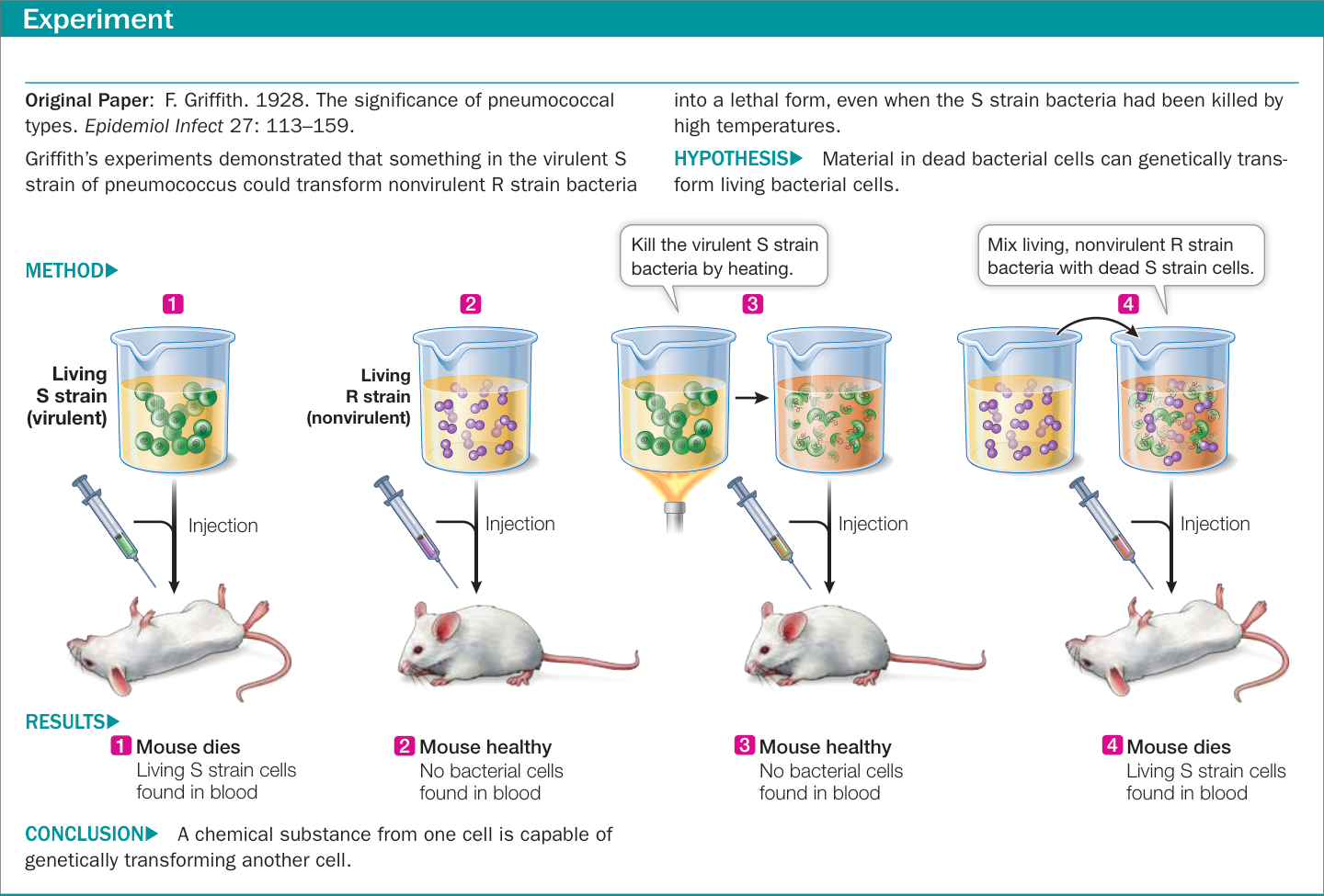
Figure 13.1 Genetic Transformation
Alfred Hershey and Martha Chase of the Cold Spring Harbor Laboratory in New York studied bacteriophage T2 (phage T2), which infects the bacterium Escherichia coli. Phage T2 consists of a DNA core packed inside a protein coat (Figure 13.3). When it attacks a bacterium, part (but not all) of the virus enters the bacterial cell. About 20 minutes later, the cell bursts, releasing dozens of virus particles that are copies of the infecting virus particle. Clearly the virus is somehow able to hijack the host cell’s molecular machinery and convert it into a viral replication machine. Hershey and Chase set out to determine what part of the virus—DNA or protein—enters the host cell to bring about this genetic change. To trace the two components of the virus over its life cycle, the scientists labeled each component with a specific radioisotope:
Proteins were labeled with radioactive sulfur. Proteins contain some sulfur (in the amino acids cysteine and methionine), but DNA does not. Sulfur has a radioactive isotope, 35S. Hershey and Chase grew bacteriophage T2 in a bacterial culture in the presence of 35S, so the proteins of the resulting viruses were labeled with (contained) the radioisotope.
DNA was labeled with radioactive phosphorus. DNA contains a lot of phosphorus (in the deoxyribose–phosphate backbone; see Figure 4.5), whereas proteins contain little or none. Phosphorus also has a radioisotope, 32P. The researchers grew another batch of T2 in a bacterial culture in the presence of 32P, thus labeling the viral DNA with 32P.
Hershey and Chase used these radioactively labeled viruses in their experiments (Figure 13.4). In one experiment, they allowed 32P-labeled bacteriophage to infect bacteria; in the other, the bacteria were infected with 35S-labeled bacteriophage. After a few minutes they agitated each mixture vigorously in a kitchen blender, stripping away the parts of the viruses that had not penetrated the bacteria, without bursting the bacteria. Then they separated the bacteria from the rest of the material (the remains of the viruses) from each experiment individually in a centrifuge. The result was that the bacterial cells in the centrifuge pellet contained most of the 32P (and thus the viral DNA), and the supernatant fluid with the viral remains contained most of the 35S (and thus the viral protein). These results indicated that it was the DNA that had been transferred into the bacteria, and that DNA was the molecule responsible for redirecting the genetic program of the bacterial cell.
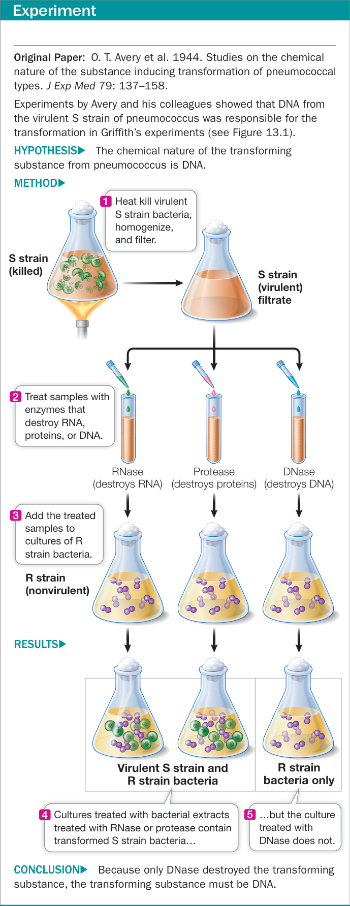
Figure 13.2 Genetic Transformation by DNA Figure 13.2 Genetic Transformation by DNA
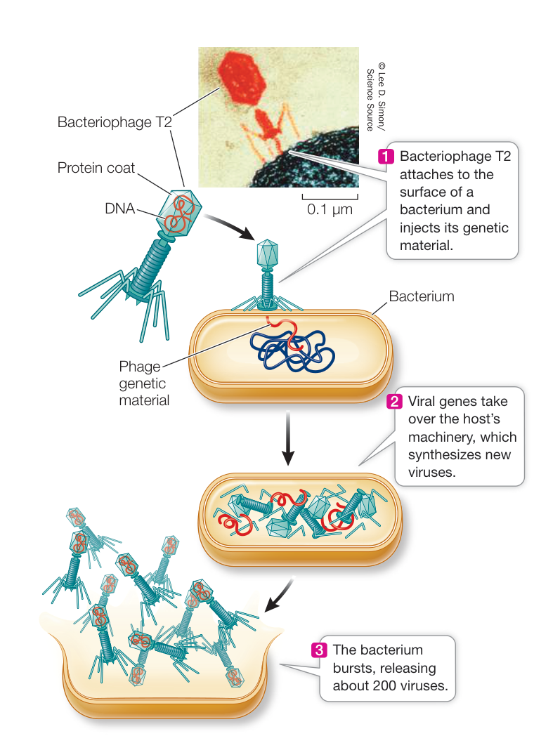
Figure 13.3 Bacteriophage T2: Reproduction Cycle Bacteriophage T2 is parasitic on E. coli, dependent on the bacterium to produce new viruses. The external structures of bacteriophage T2 consist entirely of protein, and the DNA is contained within the protein coat. When the virus infects an E. coli cell, its genetic material is injected into the host bacterium. Figure 13.3 Bacteriophage T2: Reproduction Cycle Bacteriophage T2 is parasitic on E. coli, dependent on the bacterium to produce new viruses. The external structures of bacteriophage T2 consist entirely of protein, and the DNA is contained within the protein coat. When the virus infects an E. coli cell, its genetic material is injected into the host bacterium.
Eukaryotic cells can also be genetically transformed by DNA
The transformation of eukaryotic cells by DNA is often called transfection. Transfection can be demonstrated using a genetic marker, a gene whose presence in the recipient cells confers an observable phenotype. When transforming both prokaryotes and eukaryotes, researchers often use a genetically determined marker that can be easily selected—for example, antibiotic resistance or a nutritional requirement—and that permits the growth of transformed cells but not of nontransformed cells on a selective medium (for example, one that contains an antibiotic or that lacks a nutrient). A common marker in mammalian transfection experiments is a gene that confers resistance to the antibiotic neomycin, which kills nontransformed mammalian cells. Transfection is achieved by various methods, including chemical treatments that allow the DNA to be taken up by the cells. Any cell can be transfected, even an egg cell. In the later case, a transgenic organism can result, which has every cell genetically transformed. More detail on transforming eukaryotic cells is described in Chapter 18.
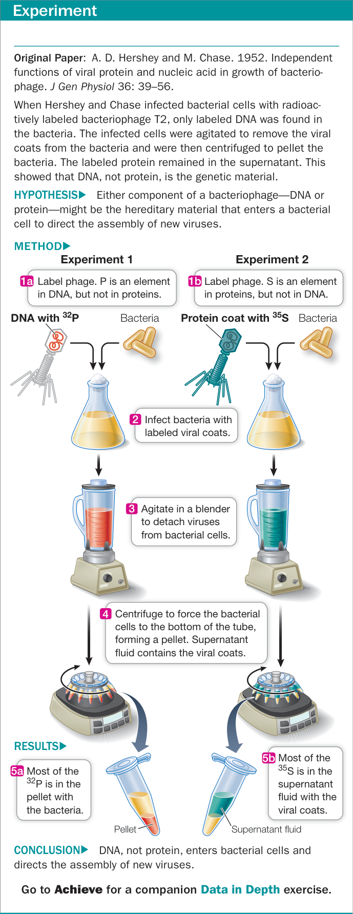
Figure 13.4 The Hershey–Chase Experiment
Recap and Assess
Experiments on bacteria and on viruses demonstrated that DNA is the genetic material. DNA from one genetic strain of bacteria was able to genetically transform another strain into the donor strain. Viral DNA was shown to be injected into a host cell and to genetically change that cell into a virus factory.
- At the time of Griffith’s experiments in the 1920s, what circumstantial evidence suggested to scientists that DNA might be the genetic material?
- How did the experiments of Avery and his colleagues rule out protein as the genetic material?
- What attributes of bacteriophage T2 were key to the Hershey–Chase experiments demonstrating that DNA, rather than protein, is the genetic material?
The transformation and viral infection experiments convinced biologists that the genetic material is DNA. It had been known for several decades that chemically, DNA is a polymer of nucleotides. Next, scientists turned to DNA’s precise three-dimensional structure.
DNA Has a Structure That Suits Its Function
Learning Objectives
- 13.2.1 List the information that Watson and Crick used to develop their model of DNA structure.
- 13.2.2 Describe the structure of DNA and how it is consistent with Chargaff’s rules.
- 13.2.3 Write down the sequence of the complementary strand of DNA if given the sequence of the other strand, indicating polarity.
- 13.2.4 Relate structural aspects of the DNA molecule to its biological functions.
Once it was known that DNA was the genetic material, scientists turned their attention to determining its structure. In doing so, they hoped to answer two questions: (1) how is DNA replicated between cell divisions, and (2) how does DNA direct the synthesis of specific proteins? DNA’s structure was deciphered only after many types of experimental evidence were considered together in a theoretical framework.
Watson and Crick used chemical and physical evidence to deduce the structure of DNA
Data on the chemical makeup of DNA, coupled with X-ray diffraction data on the physical structure of the molecule, were used to solve DNA’s structure.
CHEMICAL EVIDENCE FROM BASE COMPOSITION Biochemists knew that DNA was a polymer of nucleotides. Each nucleotide consists of a molecule of the sugar deoxyribose, a phosphate group, and a nitrogen-containing base (see Figure 4.1). The only differences among the four nucleotides of DNA are their nitrogenous bases: the purines adenine (A) and guanine (G), and the pyrimidines cytosine (C) and thymine (T).
In the early 1950s, biochemist Erwin Chargaff and his colleagues at Columbia University reported that DNA from many different species exhibits certain regularities. This led to the following rule: in any DNA sample, the amount of adenine equals the amount of thymine (), and the amount of guanine equals the amount of cytosine (). This finding means that the total abundance of purines equals the total abundance of pyrimidines: . While Chargaff and colleagues found that this rule held for every organism they examined, they noted that the relative abundances of versus vary among organisms. In human DNA, and each account for 27% of the nitrogenous bases present, while and each account for 23%. Put another way, in human DNA there is a ratio of 54% to 46% .
PHYSICAL EVIDENCE FROM X-RAY DIFFRACTION Some chemical substances, when they are isolated and purified, can be made to form crystals. The positions of atoms in a crystallized substance can be inferred from the diffraction pattern of X-rays passing through the substance. In the early 1950s the New Zealand-born biophysicist Maurice Wilkins discovered a way to make highly ordered fibers of DNA that were suitable for X-ray diffraction studies. Images of his samples were made by Rosalind Franklin of Kings College, London (Figure 13.5) and were instrumental in deciphering the structure of DNA. Franklin’s images suggested that DNA was a double (two-stranded) helix with ten nucleotides in each full turn, that each full turn was 3.4 nanometers (nm) in length, that the molecule had a constant diameter of 2 nm, and that phosphates were most likely on the outside of the helix.
WATSON AND CRICK’S MODEL If you have taken chemistry courses, you may be familiar with model building, whereby balls (atoms) and sticks (bonds) are used to put together molecules based on known physical and chemical properties and bond angles. A physicist, Francis Crick, and a geneticist, James D. Watson
(Figure 13.6A), who were then at the Cavendish Laboratory at the University of Cambridge, used model building to solve the structure of DNA. They used the physical and chemical evidence we just described:
- To be consistent with Franklin’s X-ray diffraction images, Watson and Crick’s model had the nucleotide bases on the interior of the two strands, with a sugar–phosphate “backbone” on the outside. The backbone consisted of nucleotides attached via the deoxyribose sugar of one nucleotide and the phosphate of the next nucleotide. In addition, the two DNA strands ran in opposite directions, that is, they were antiparallel, meaning that the ribose of one strand was across from a phosphate of the other strand at each end of the molecule. The two strands would not fit together unless they were in an antiparallel configuration.
- To satisfy Chargaff’s rule ( and ), Watson and Crick’s model always paired a double-ring purine ( or ) on one strand with a single-ring pyrimidine ( or ) on the opposite strand. These complementary base pairs ( - and - ) have the same width down the double helix, a uniformity that was also confirmed by X-ray diffraction.
Crick and Watson built their model of DNA in late February 1953. This structure explained the known chemical properties of DNA, and it opened the door to understanding its biological functions.
Five key features define DNA structure
Five features summarize the molecular architecture of the DNA molecule (Figure 13.6B):
- DNA is a double-stranded helix, with a sugar–phosphate backbone on the outside and base pairs lined up on the inside.
- DNA is usually a right-handed helix. If you curl the fingers of your right hand and point your thumb upward, the curve of the helix follows the direction of your fingers, and it winds upward in the direction of your thumb. (See Figure 3.9, showing right-versus left-handed helices.)
- DNA is antiparallel (the two strands run in opposite directions).
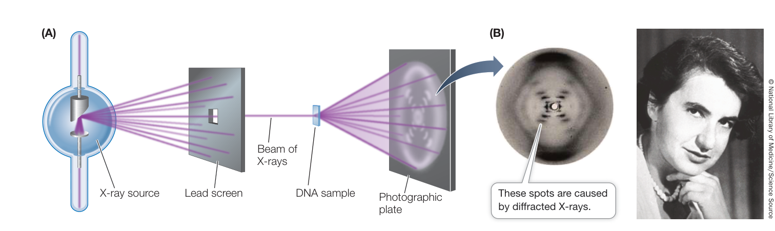
Figure 13.5 X-Ray Diffraction Helped Reveal the Structure of DNA (A) The positions of atoms in a crystallized chemical substance can be inferred by the pattern of diffraction of X-rays passed through it. The pattern of DNA is both highly regular and repetitive. (B) Rosalind Franklin’s diffraction and her “photograph 51” (shown) helped scientists visualize the helical structure of the DNA molecule.
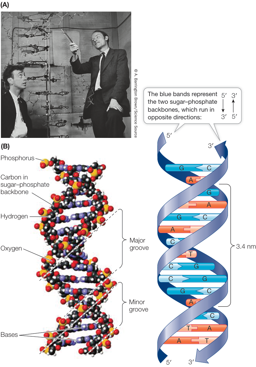
Figure 13.6 DNA Is a Double Helix (A) James Watson (left) and Francis Crick (right) proposed that the DNA molecule has a double-helical structure. (B) Biochemists can now pinpoint the position of every atom in a DNA molecule. To see that the essential features of the original Watson–Crick model have been verified, follow with your eyes the double-helical chains of sugar–phosphate groups and note the horizontal rungs of the bases. (B data from S. Arnott and D. W. Hukins. 1972. Biochem Biophys Res Commun 47: 1504–1509.) Q: In DNA, where do the following chemical forces occur: hydrogen bonds, covalent bonds, and van der Waals forces?
- DNA strands are held together by complementary base pairing, which involves hydrogen bonding between the bases on opposite strands. Base pairs are either an A purine with a T pyrimidine or a G purine with a C pyrimidine.
- DNA has major and minor grooves in which the outer edges of the nitrogenous bases are exposed.
THE HELIX The sugar–phosphate backbones of the polynucleotide chains form a coil around the outside of the helix, and the nitrogenous bases point toward the center. In addition to the covalent bonds occurring between the atoms present in each of the strands, hydrogen bonds and van der Waals forces play important roles in the structure of DNA:
- Hydrogen bonds between complementary base pairs hold the two strands of the DNA helix together. Adenine (A) pairs with thymine (T) by forming two hydrogen bonds, and guanine (G) pairs with cytosine (C) by forming three hydrogen bonds (see Figure 13.7B).
- van der Waals forces occur between adjacent bases on the same strand. When the base rings come near one another, they tend to stack like poker chips because of these weak attractions.
ANTIPARALLEL STRANDS The backbone of each DNA strand contains repeating units of the five-carbon monosaccharide deoxyribose attached to a phosphate group (Figure 13.7A). The number followed by a prime (‘) designates the position of a carbon atom in this sugar molecule. In a nucleotide, the phosphate () is attached to the 5’ carbon of the deoxyribose. In the sugar–phosphate backbone of DNA, each 5’ phosphate group is connected to the 3’ carbon of the next deoxyribose, linking successive sugars together.
Thus the two ends of a polynucleotide chain differ. At one end of a chain is a free (not connected to another nucleotide) 5’ phosphate group; this is called the 5’ end. At the other end is a free 3’ hydroxyl group (); this is called the 3’ end. In a DNA double helix, the 5’ end of one strand is paired with the 3’ end of the other strand, and vice versa. In other words, if you drew an arrow for each strand running from 5’ to 3’, the arrows would point in opposite directions (Figure 13.7B; see also Figure 4.5A).
BASE EXPOSURE IN THE GROOVES Look back at Figure 13.6B and note the major and minor grooves in the helix. These grooves exist because the backbones of the two strands are closer together on one side of the double helix (forming the minor groove) than on the other side (forming the major groove). Figure 13.8 shows a “top-down” view of the four possible configurations of the flat, hydrogen-bonded base pairs in the major and minor grooves. The exposed outer edges of the base pairs are accessible for additional hydrogen bonding. Note that the arrangements of unpaired atoms and groups differ in A-T and G-C pairs. Thus the surfaces of the A-T and C-G base pairs are chemically distinct, allowing other molecules such as proteins to recognize specific base pair sequences and bind to them, which is crucial for DNA function. The binding of proteins to specific base pair sequences
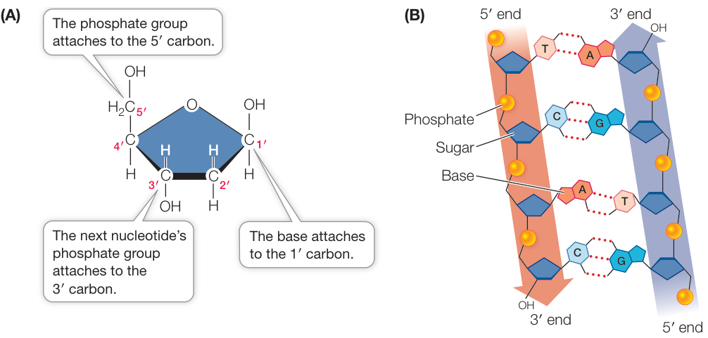
Figure 13.7 Each DNA Strand Consists of a Sugar–Phosphate Backbone (A) The deoxyribose sugar has five carbons, numbered 1’ to 5’. The 5’ carbon is attached to a phosphate in a nucleotide. In a DNA molecule, each nucleotide in a strand has its 3’ carbon attached to the 5’ phosphate of the next nucleotide. (B) Each sugar–phosphate backbone has a free 5’ phosphate at one end and a free 3’ hydroxyl group at the other end, giving the backbone directionality. The two backbones run in opposite directions such that the 5’ end of one strand is opposite the 3’ end of the other strand, and vice versa.
is the key to protein–DNA interactions, which are necessary for the replication and expression of the genetic information in DNA.
The double-helical structure of DNA is essential to its function
The genetic material performs four important functions, and the DNA structure proposed by Watson and Crick was elegantly suited to three of them.
- The genetic material stores an organism’s genetic information. With its millions of nucleotides, the base sequence of a DNA molecule can encode and store an enormous amount of information. Variations in DNA sequences can account for species and individual differences. DNA fits this role nicely.
- The genetic material is susceptible to mutations (permanent changes) in the information it encodes. For DNA, mutations might be simple changes in the linear sequence of base pairs.
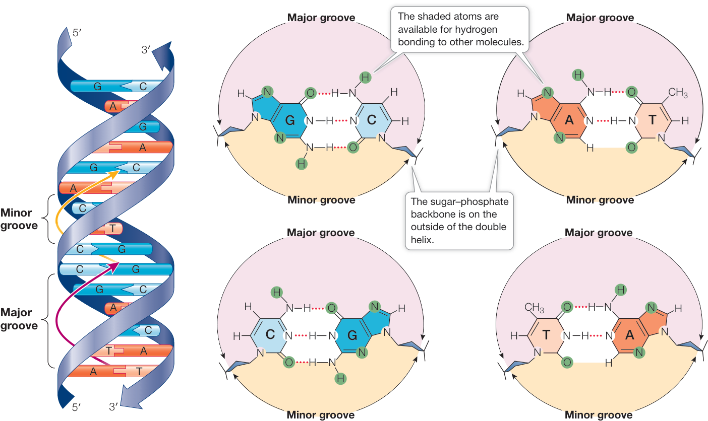
Figure 13.8 Base Pairs in DNA Can Interact with Other Molecules These diagrams show the four possible configurations of base pairs within the double helix. Atoms shaded in green are available for hydrogen bonding with other molecules, such as proteins.
- The genetic material is precisely replicated in the cell division cycle. Replication could be accomplished using complementary base pairing, A with T and G with C. In the original publication of their findings in 1953, Watson and Crick copy pointed out, “It has not escaped our notice that the specific pairing we have postulated immediately suggests a possible copying mechanism for the genetic material.”
- The genetic material (the coded information in DNA) is expressed as the phenotype. This function is not obvious in the structure of DNA. However, as we will see Chapter 14, the nucleotide sequence of DNA is copied into RNA, which uses the coded information to specify a linear sequence of amino acids—a protein. The folded forms of proteins determine many of the phenotypes of an organism.
Recap and Assess
DNA is a right-handed, double helix made up of two antiparallel polynucleotide chains. The two chains are held together by hydrogen bonds between the nucleotide bases, which exhibit complementary pairing: A with T, and G with C. Chemical groups on the bases that are exposed in the grooves of the helix are available for hydrogen bonding with other molecules, such as proteins.
- Describe the evidence that Watson and Crick used to come up with the double helix model for DNA.
- Speculate as to how the double-stranded structure of DNA relates to its functions.
- A key to the function of DNA in directing information in the cell is the binding of other molecules such as proteins to the double helix at specific base sequences. Recalling the forces involved in protein interactions (see Figure 3.13) and the chemical groupings exposed in base pairs (see Figure 13.8), how do specific interactions between proteins and DNA occur?
Once the structure of DNA was understood, it was possible to investigate how DNA replicates itself. Next we examine the experiments that taught us how this elegant process works.
DNA Is Replicated Semiconservatively
Learning Objectives
- 13.3.1 Draw the results obtained in the Meselson–Stahl experiment that supported semiconservative replication, and explain what would have been found for conservative and dispersive replication.
- 13.3.2 Identify the function of each of the proteins involved in DNA replication.
- 13.3.3 Explain the end-replication problem and how it is solved by telomerase.
The mechanism of DNA replication that Watson and Crick proposed was soon confirmed. First, researchers showed that DNA could be replicated in a test tube containing simple substrates and an enzyme. A subsequent study showed that each of the two strands of the double helix can serve as a template for a new strand of DNA.
Test tube synthesis of DNA needed the following substances:
- The deoxyribonucleoside triphosphates dATP, dCTP, dGTP, and dTTP. These are the monomers from which the DNA polymers are synthesized.
- DNA molecules of a particular sequence that serve as templates to direct the sequence of nucleotides in the new molecules.
- A DNA polymerase enzyme to catalyze the polymerization reaction.
- Salts and a pH buffer, to create an appropriate chemical environment for the DNA polymerase.
The fact that DNA could be synthesized in a test tube confirmed that a DNA molecule contains the information needed for its own replication. The next challenge was to determine which of three possible replication patterns occurs during DNA replication:
- Semiconservative replication, in which each parent strand serves as a template for a new strand, and the two new DNA molecules each have one original and one new strand (Figure 13.9A)
- Conservative replication, in which the original double helix serves as a template for, but does not contribute to, a new double helix (Figure 13.9B)
- Dispersive replication, in which fragments of the original DNA molecule serve as templates for assembling two new molecules, each containing original and new parts, perhaps at random (Figure 13.9C)
Watson and Crick’s original paper suggested that DNA replication was semiconservative, but the test tube demonstration just described did not provide a basis for choosing among these three models.

Animation 13.2 DNA Replication and Polymerization
An elegant experiment demonstrated that DNA replication is semiconservative
In 1958 Matthew Meselson and Franklin Stahl at the California Institute of Technology convinced the scientific community that DNA is reproduced by semiconservative replication. They used density labeling to distinguish between parent strands of DNA and newly copied strands. Historians of science have called this one of the “most beautiful experiments” in biology. For that reason alone, as well as its results, it’s worth taking a close look at it.
The key to the experiment was the use of a “heavy” isotope of nitrogen. Heavy nitrogen (15N) is a rare, nonradioactive isotope that makes molecules containing it denser than chemically identical molecules containing the common isotope 14N. Two cultures of the bacterium E. coli were grown for many generations, one in a medium containing 15N and the other in a medium containing 14N. When DNA extracts from the two cultures were combined and centrifuged in a solution of cesium chloride, which forms a density gradient under centrifugation, two separate bands of DNA formed in the centrifugation tube. The DNA from the culture was heavier than the DNA from the culture, so it formed a band at a different position in the density gradient. Figure A in the Work with the Data portion of Investigating Life: The Meselson–Stahl Experiment shows a photo of the two bands.
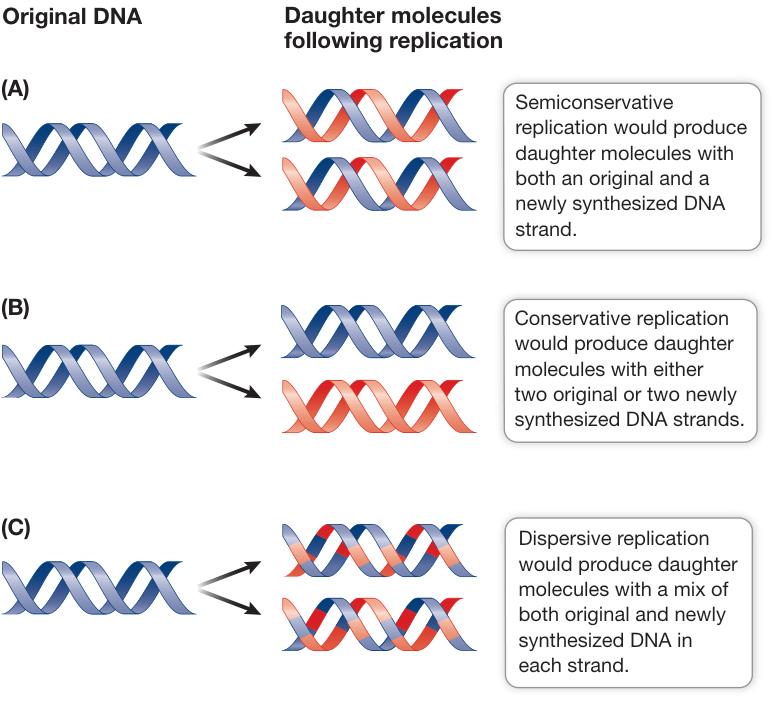
Figure 13.9 Three Models for DNA Replication In each model, the original DNA is shown in blue and the newly synthesized DNA in red.
Next, Meselson and Stahl grew another E. coli culture in medium, then transferred the bacteria to normal medium and allowed them to continue growing. The cells replicated their DNA and divided every 20 minutes. Meselson and Stahl collected some of the bacteria at time intervals and extracted DNA from the samples. You can follow their results for the first two generations in Investigating Life: The Meselson–Stahl Experiment. The results can be explained only by the semiconservative model of DNA replication. The crucial observations demonstrating this model were that all the DNA at the end of the first generation was of intermediate density, while at the end of the second generation there were two discrete bands: one of intermediate and one of light DNA. If the conservative model had been true, there would have been no intermediate density DNA. If the dispersive model were correct, then all of the replicated DNA would have been intermediate for the first few generations, with the single intermediate band becoming progressively lighter.
There are three steps in DNA replication
Semiconservative DNA replication in the cell involves several different enzymes and other proteins. It takes place in three general steps:
- Initiation: The DNA double helix is unwound to separate the two template strands and make them available for new base pairing. Each of the strands is then primed with RNA primers, as described in the next section, in preparation for template-dependent DNA synthesis.
- Elongation: DNA synthesis occurs from the RNA primers with the addition of nucleotides that form complementary base pairs with template DNA. Incoming nucleotides are covalently linked together by phosphodiester bonds, forming a polymer whose base sequence is complementary to the bases in the template strand.
- Termination: DNA synthesis ends when all of the DNA regions have been replicated.
The nucleotides that make up DNA are deoxyribonucleoside monophosphates (dNMPs) because they each contain deoxyribose and one phosphate group (see Figure 4.1). The four monomers that are used during DNA elongation are the deoxyribonucleoside triphosphates dATP, dTTP, dCTP, and dGTP, collectively referred to as dNTPs. They are triphosphates because each has three phosphate groups. The three phosphate groups are attached to the 5’ carbon on the deoxyribose sugar (as shown in the incoming nucleoside triphosphate in Figure 13.10).
Note that during DNA replication nucleotides are added to the growing new strand at the 3’ end—the end at which the DNA strand has a free hydroxyl () group on the 3’ carbon of its terminal deoxyribose. In the formation of the phosphodiester bond (a transesterification reaction), two of the phosphate groups on an incoming dNTP are removed (as pyrophosphate ), and the remaining phosphate is bonded to the 3’ carbon on the terminal deoxyribose (see Figure 4.2). Just as energy is released when ATP is hydrolyzed to AMP (with subsequent hydrolysis of to two phosphates), energy is released when dNTP releases to form dNMP, and this energy is used to drive the transesterification reaction.
DNA polymerases add nucleotides to the growing chain
DNA replication begins with the binding of a large protein complex (the pre-replication complex) to a specific site on the DNA molecule. This complex contains several different proteins, including the enzyme DNA polymerase, which catalyzes the addition of nucleotides as the new DNA chain grows. All chromosomes have at least one region called the origin of replication (ori), to which the pre-replication complex binds. Binding occurs when proteins in the complex recognize specific DNA sequences within the ori.
ORIGINS OF REPLICATION The single circular chromosome of the bacterium E. coli has base pairs (bp) of DNA. The 245-bp ori sequence is at a particular location on the chromosome. Once the pre-replication complex binds to it, initiation begins with the local unwinding of the DNA and then replication proceeds in
▶ Investigating LIFE The Meselson-Stahl Experiment
Experiment
Original Paper: M. Meselson and F. Stahl. 1958. The replication of DNA in Escherichia coli. Proc Natl Acad Sci USA 44: 671–682.
A centrifuge was used to separate DNA molecules labeled with isotopes of different densities. This experiment revealed a pattern that supports the semiconservative model of DNA replication.
HYPOTHESIS DNA replicates semiconservatively.
METHOD
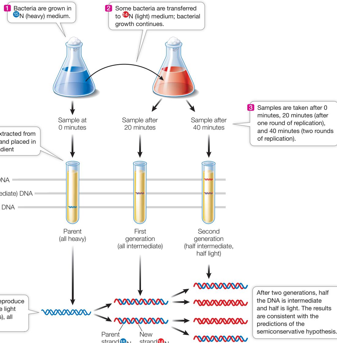
CONCLUSION This pattern could only have been observed if each DNA molecule contains a template strand from the parental DNA; thus DNA replication is semiconservative.
Work with the Data
The Meselson–Stahl experiment has been called one of the “most beautiful experiments” in biology because of its essential simplicity. Meselson and Stahl used density gradients to examine how DNA molecules replicate, as illustrated in the experimental results shown in Figure A. The peaks on the drawing are proportional to the amount of DNA.
Figure B shows results of the experiment after each of four generations. Each sample contained the same number of bacteria, so the total amount of DNA in each panel was the same. (Both photos from M. Meselson and F. Stahl. 1958. Proc Natl Acad Sci USA 44: 671–682.)
QUESTIONS►
- Use the heights of the peaks to estimate the percent of total DNA that was heavy, intermediate, and light at each generation. Create a table summarizing these calculations and discuss whether they support the authors’ conclusions.
- What would the data look like if the bacteria had been allowed to divide for three more generations?
- If Meselson and Stahl had done their experiment starting with light DNA and then added for succeeding generations, what would the bands look like?
- What would the data look like if conservative replication were the correct model? What would the data look like if dispersive replication were correct?
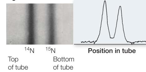
Figure A
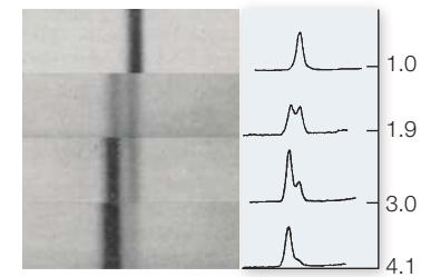
Figure B
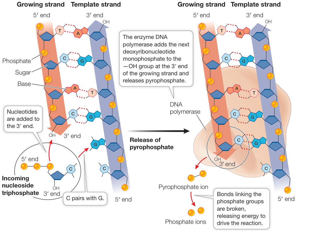
Figure 13.10 Each New DNA Strand Grows from Its 5’ End to Its 3’ End The DNA strand at the right (blue) is the template for the synthesis of the complementary strand that is growing at the left (orange). Here dCTP (circled) is about to react with the growing strand to add dCMP to the new strand, releasing pyrophosphate.
both directions around the circle, forming two replication forks (Figure 13.11A). The replication rate in E. coli is approximately 1,000 bp per second, so it takes about 40 minutes to fully replicate the chromosome (with two replication forks). Rapidly dividing E. coli cells divide every 20 minutes. In these cells, new rounds of replication begin at the ori of each new chromosome before the first chromosome has fully replicated. In this way the cells can divide more frequently than the time needed to finish replicating the original chromosome!
Eukaryotic chromosomes are typically much longer than those of prokaryotes—up to 1 billion bp—and are linear, not circular. If replication occurred from a single ori with two forks growing away from each other, it would take weeks to fully replicate a chromosome. So eukaryotic chromosomes have multiple origins of replication, scattered at intervals of 10,000 to 40,000 bp (Figure 13.11B).
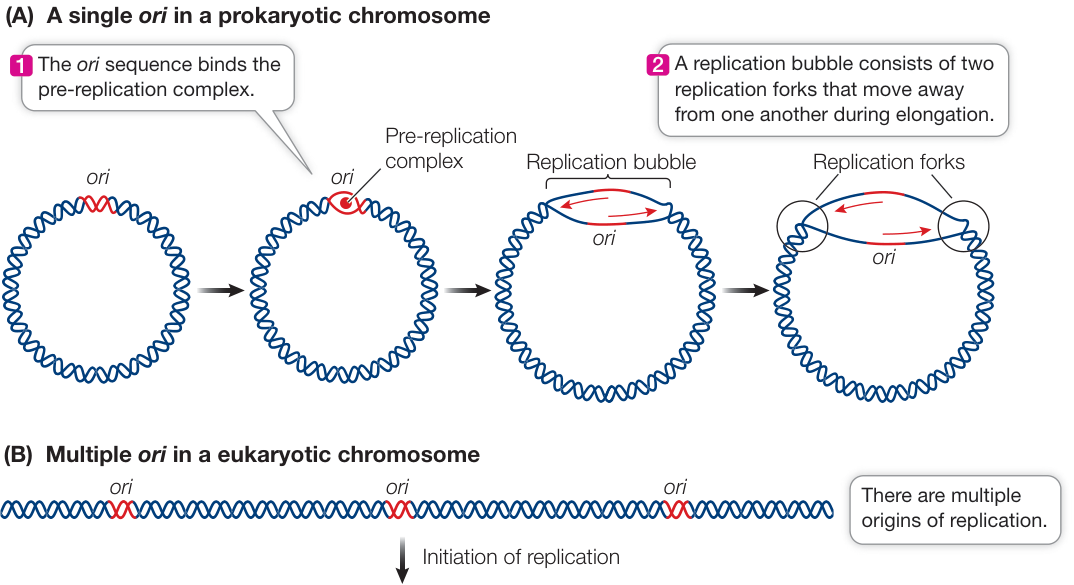
Figure 13.11 The Origin of DNA Replication (A) Prokaryotic chromosomes usually have a single origin where DNA replication starts and proceeds in both directions. (B) The usually much larger eukaryotic chromosome typically has multiple replication origins.
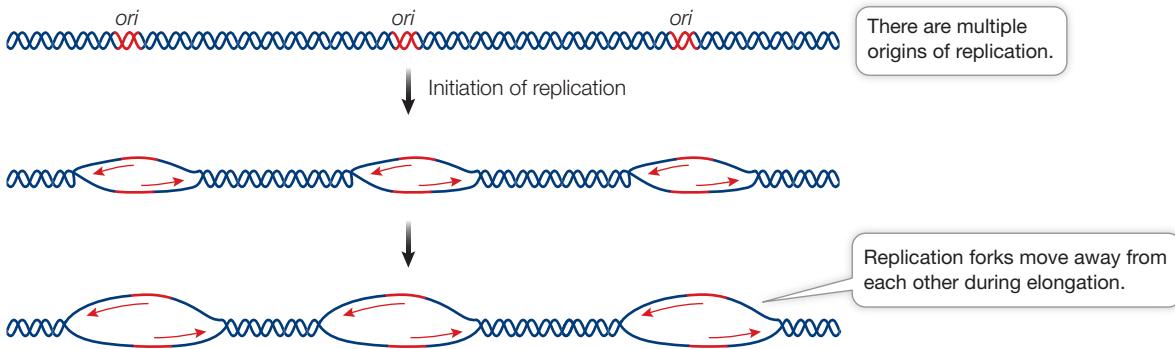
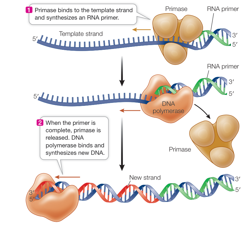
Figure 13.12 DNA Synthesis Requires a Primer DNA polymerases require a primer—a “starter” strand of RNA (or in some situations DNA; not shown) to which they can add new nucleotides.
DNA REPLICATION BEGINS WITH A PRIMER A DNA polymerase elongates a polynucleotide strand by covalently linking new nucleotides to a preexisting strand. However, it cannot start this process without a short “starter” strand, called a primer. In most organisms this primer is a short single strand of RNA (Figure 13.12), but in some organisms it is DNA. The RNA primer is complementary to the DNA template and is synthesized one nucleotide at a time by an enzyme called a primase. Once the primase has added a primer, the elongation phase of replication begins, with the DNA polymerase adding nucleotides to the 3’ end of the primer and continuing until replication of that section of DNA has been completed. The DNA polymerase continues to synthesize DNA until it runs out of template. Then the RNA primer is degraded, DNA is added in its place (by a different DNA polymerase), and the resulting DNA fragments are connected by the action of other enzymes. When DNA replication is complete, each new strand consists only of DNA.
DNA POLYMERASES ARE LARGE DNA polymerases are much larger than their substrates (the dNTPs) and the template DNA, which is very thin. Molecular models of the enzyme–substrate–template complex from bacteria show that the enzyme is shaped like an open right hand with a palm, a thumb, and fingers (Figure 13.13). Within the “palm” is the active site of the enzyme, which brings together each dNTP substrate and the template. The “finger” regions have precise shapes that can recognize the different shapes of the four nucleotide bases. They bind to the bases by hydrogen bonding and rotate inward. Most cells contain more than one kind of DNA polymerase, but only one of them is responsible for chromosomal DNA replication. The others are involved in primer removal and DNA repair. Fifteen DNA polymerases have been identified in humans, whereas the bacterium E. coli has five.
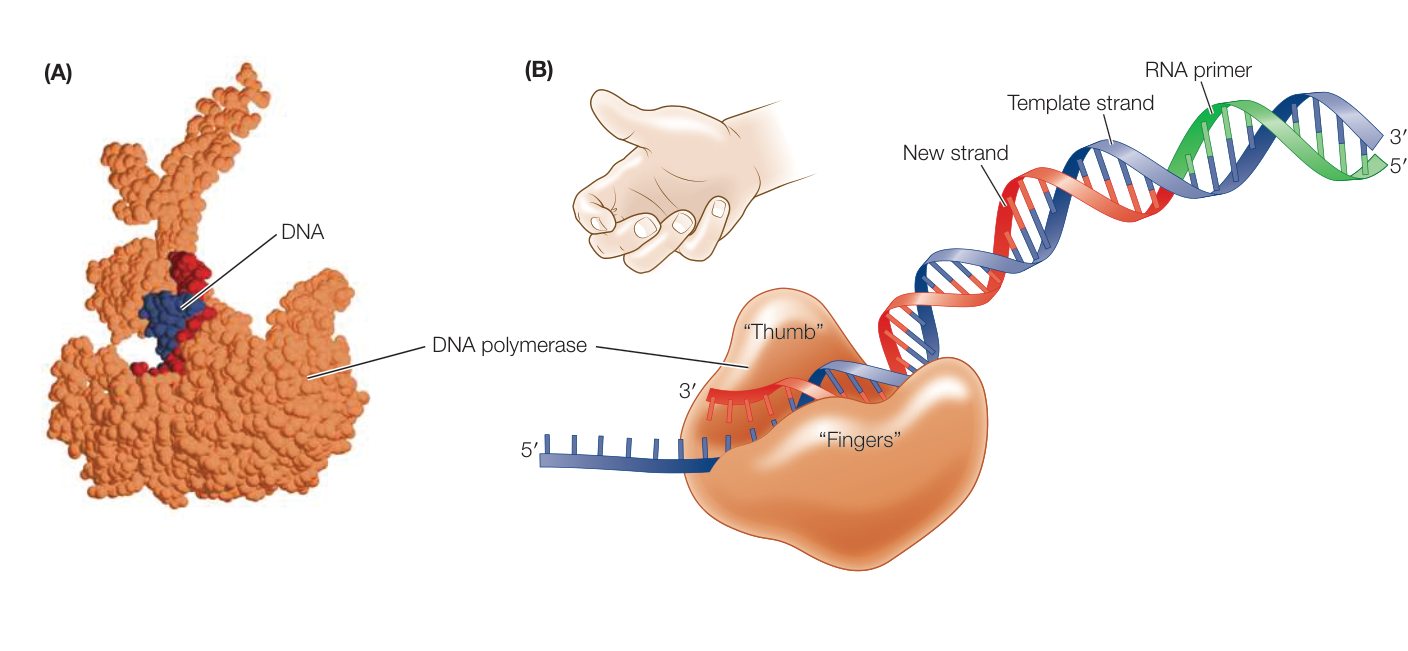
Figure 13.13 DNA Polymerase Binds to the Template Strand (A) The DNA polymerase enzyme (brown) is much larger than the DNA molecule (red and blue). (B) DNA polymerase is shaped like a hand, and in this side-on view, its “fingers” can be seen curling around the DNA. These “fingers” can recognize the distinctive shapes of the four bases. (A data from PDB 1SKW. Y. Li et al. 2004. Nat Struct Mol Biol 11: 784–790.)
Many other proteins assist with DNA polymerization
Various other proteins play roles in other replication tasks; some of these are shown in Figure 13.14. The first event at the origin of replication is the localized unwinding and separation (denaturation) of the DNA strands. As we discussed in Key Concept 13.2, the two strands are held together by hydrogen bonds. An enzyme called DNA helicase uses energy from ATP hydrolysis to unwind and separate the strands, and single-stranded binding proteins bind to the unwound strands to keep them from reassociating into a double helix. This process makes each of the two template strands available for complementary base pairing.
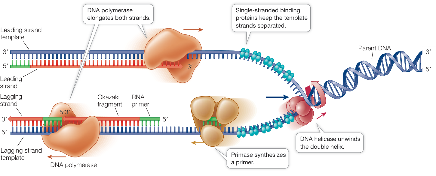
Figure 13.14 Many Proteins Collaborate in the Pre-Replication Complex Several proteins in addition to DNA polymerase are involved in DNA replication. The two molecules of DNA polymerase shown here are actually part of the same complex.
Q: In bidirectional replication from a single origin point, with two replication forks moving in opposite directions, will the same DNA strands be leading and lagging on both forks?
Activity 13.1 The Pre-Replication Complex
The two DNA strands grow differently at the replication fork
The DNA at the replication fork—the site where DNA unwinds to expose the bases so that they can act as templates—opens up like a zipper in one direction. Study Figure 13.15 and try to imagine what is happening over a short period of time. Remember that the two DNA strands are antiparallel; that is, the 3’ end of one strand is paired with the 5’ end of the other.
- One newly replicating strand (the leading strand) is oriented so that it can grow continuously at its 3’ end as the fork opens up.
- The other new strand (the lagging strand) is oriented so that as the fork opens up, its exposed 3’ end gets farther and farther away from the fork, and an unreplicated gap is formed. This gap would get bigger and bigger if there were not a special mechanism to overcome this problem.
Synthesis of the lagging strand requires the synthesis of relatively small, discontinuous stretches of DNA (100–200 nucleotides in eukaryotes; 1,000–2,000 nucleotides in prokaryotes). These discontinuous stretches are synthesized just as the leading strand is, by the addition of new nucleotides one at a time to the 3’ end of the new strand, but the synthesis of this new strand moves in the direction opposite to that in which the replication fork is moving. These stretches of new DNA are called Okazaki fragments (after their discoverer, the Japanese biochemist Reiji Okazaki). While the leading strand grows continuously “forward,” the lagging strand grows in shorter, “backward” stretches with gaps between them.
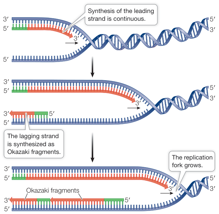
Figure 13.15 The Two New Strands Form in Different Ways As the parent DNA unwinds, both new strands are synthesized in the 5’-to-3’ direction, although their template strands are antiparallel. The leading strand grows continuously forward, but the lagging strand grows in short discontinuous stretches called Okazaki fragments. Eukaryotic Okazaki fragments are hundreds of nucleotides long, with gaps between them.
A single primer is needed for synthesis of the leading strand, but each Okazaki fragment requires its own primer to be synthesized by the primase. In bacteria, priming is followed by DNA polymerase III synthesizing an Okazaki fragment by adding nucleotides to the primer until it reaches the primer of the previous fragment (Figure 13.16). At this point, DNA polymerase I replaces DNA polymerase III, and then removes the old primer and replaces it with DNA. Left behind is a tiny nick—the final phosphodiester bond between the adjacent Okazaki fragments is missing. The enzyme DNA ligase catalyzes the formation of that bond, linking the fragments and making the lagging strand whole.
DNA replication involves remarkable teamwork among various proteins that act on the DNA strands. Let’s review the proteins involved in DNA replication in the order of their activity at the replication fork:
- DNA helicase unwinds the double helix and separates the two strands.
- Single-stranded binding proteins bind to separated strands and prevent them from re-forming the double helix.
- DNA primase makes RNA primers.
- DNA polymerase links new nucleotides to form the new DNA strands and removes the primers.
- DNA ligase connects Okazaki fragments made by DNA polymerase to one another.
Working together, these proteins make new DNA at a rate in excess of 1,000 base pairs per second.
A SLIDING CLAMP INCREASES THE RATE OF DNA REPLICATION How do DNA polymerases work so fast? We saw in Key Concept 8.3 that an enzyme catalyzes a chemical reaction:
Substrate binds to enzyme → one product is formed → enzyme is released → cycle repeats
DNA replication would not proceed as rapidly as it does if it went through such a cycle for each nucleotide. Instead, DNA polymerases are processive—that is, they catalyze the formation of many phosphodiester bonds each time they bind to a DNA molecule:
Substrates bind to enzyme → many products are formed → enzyme is released → cycle repeats
The DNA polymerase–DNA complex is stabilized by a sliding DNA clamp, which has multiple identical subunits assembled into a doughnut shape (Figure 13.17). The doughnut’s “hole” is just large enough to encircle the DNA double helix, along with a thin layer of water molecules for lubrication. The clamp binds to the DNA polymerase–DNA complex, keeping the enzyme and the
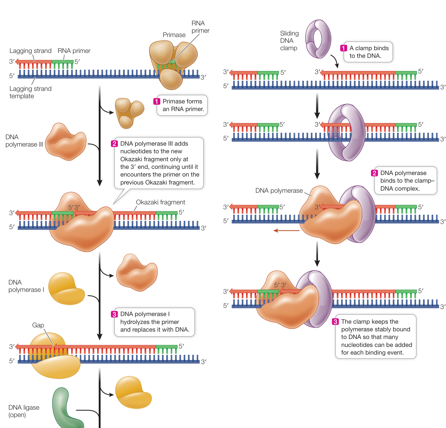
Figure 13.17 A Sliding DNA Clamp Increases the Efficiency of DNA Polymerization The clamp increases the efficiency of polymerization by keeping the enzyme bound to the substrate, so the enzyme does not have to repeatedly bind to template and substrate.
DNA associated tightly with each other. If the clamp is absent, DNA polymerase dissociates from DNA after forming 20 to 100 phosphodiester bonds. With the clamp, it can polymerize up to 50,000 nucleotides before it detaches.
DNA IS THREADED THROUGH A REPLICATION COMPLEX From our discussion of the various enzymes involved in DNA replication, it is natural to think that the enzymes involved at different stages each move along the DNA templates separately. But this is not so. Instead, the enzymes used on both the leading and lagging strands are together, forming a large replication complex. Commonly in eukaryotes, the replication complexes seem to be stationary in the nucleus. It is the DNA that moves, essentially moving into the complex as a double stranded molecule and emerging as two double-stranded molecules.
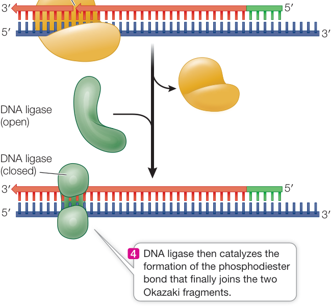
Figure 13.16 The Lagging Strand Story In bacteria, DNA polymerase I and DNA ligase cooperate with DNA polymerase III to complete the complex task of synthesizing the lagging strand.
Telomeres are not fully replicated and are prone to undergoing repair
As we discuss in Key Concept 13.4, DNA may be damaged by radiation or chemicals. When this happens, DNA repair mechanisms are activated, and breaks exposing free 3’ and 5’ groups in DNA are rejoined via a combination of DNA synthesis and DNA ligase activity. So the ends of chromosomes are a potential problem: the DNA repair system might recognize the ends as breaks, and join two chromosomes together. This would create havoc with genomic integrity.
In many eukaryotes, there are highly repetitive sequences at the ends of chromosomes called telomeres. In humans, the telomere sequence is TTAGGG-3’, and it is repeated about 2,500 times at each chromosome end. These repeats bind special proteins that prevent the DNA repair system from recognizing the chromosome ends as breaks. In addition, the repeats may form loops that have a similar protective role.
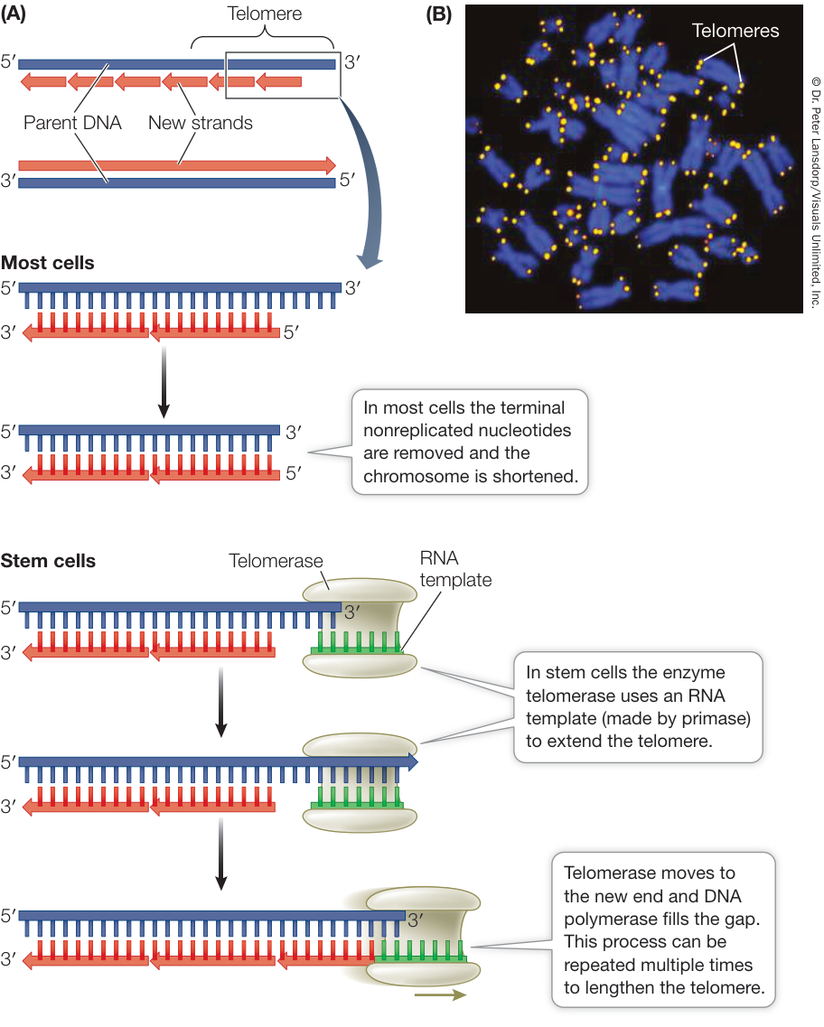
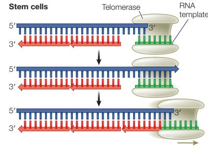
Figure 13.18 Telomeres and Telomerase (A) In most cells, the chromosome shortens with each replication because the nonreplicated DNA at the 3’ end of the template DNA is removed. However, in stem cells (including gamete-forming stem cells) telomerase uses an RNA template to extend the telomere and prevent chromosome shortening. (B) Bright fluorescent staining marks the telomeric regions on these blue-stained human chromosomes. Q: Why do the cells that form gametes express telomerase?
But there is another problem with chromosome ends. As you have seen, replication of the lagging strand occurs by the addition of Okazaki fragments to RNA primers. When the terminal RNA primer is removed on the lagging strand at the end of a chromosome, no DNA can be synthesized to replace it because there is no 3’ end to extend. In most cells, the short piece of single-stranded DNA at each end of the chromosome is removed. Thus the chromosome becomes slightly shorter with each cell division, an issue termed the “end-replication problem” (Figure 13.18).
Each human daughter chromosome can lose 50 to 200 base pairs of telomeric DNA from the end of what was the lagging strand after a round of DNA replication and cell division. After many cell divisions, the genes near the ends of the chromosomes can be lost, and the cell dies. This phenomenon explains, in part, why many cell lineages do not last the entire lifetime of the organism: their telomeres are lost. Continuously dividing cells, such as stem cells in somatic tissues and those producing gametes, have a special mechanism for maintaining their telomeric DNA. An enzyme called telomerase catalyzes the addition of any lost telomeric sequences in these cells (see Figure 13.18). Telomerase contains an RNA sequence that acts as a template for the synthesis of the telomeric DNA repeat sequence.
Telomerase is expressed in more than 90% of human cancers, and may be an important factor in the ability of cancer cells to divide continuously. Since most normal cells do not have this ability, telomerase is an attractive target for drugs designed to attack tumors specifically. There is also interest in telomerase and aging. When cultured human cells are transformed with a telomerase gene that is expressed at high levels, their telomeres do not shorten. Instead of living 20 to 30 cell generations and then dying, the cells become immortal. It remains to be seen how this finding relates to the aging of a whole organism.
Recap and Assess
Meselson and Stahl showed that DNA replication is semiconservative: each parent DNA strand serves as a template for a new strand. Several proteins, most notably DNA polymerase, are involved in replication. New DNA is polymerized in one direction only. Since the two strands are antiparallel, one strand is made continuously and the other is synthesized in short Okazaki fragments that are eventually joined by DNA ligase, after primers are removed by DNA polymerase I.
- How did the Meselson–Stahl experiment differentiate among the three models for DNA replication?
- Name five proteins needed for DNA replication. What are their roles?
- Why are there special sequences at the ends of chromosomes, and how are they replicated?
- The drug dideoxycytidine, used to treat certain viral infections, is a nucleotide made with 2’,3’-dideoxyribose. This sugar lacks —OH groups at both the 2’ and the 3’ positions. Explain why dideoxycytidine stops the synthesis of DNA when it is added to the growing DNA chain.
The complex process of DNA replication is amazingly accurate, but it is not perfect. What happens when things go wrong?
Errors in DNA Can Be Repaired
Learning Objectives
- 13.4.1 Identify all the possible mismatches that can occur in a DNA molecule.
- 13.4.2 Explain how proofreading and mismatch repair act to reduce the rate of DNA replication errors.
- 13.4.3 Describe how excision repair acts on damaged nucleotides.
DNA must be accurately replicated and maintained. This is essential for the proper functioning of every cell, whether a prokaryote or a cell in a complex, multicellular organism. Yet the replication of DNA is not perfectly accurate, and DNA is subject to change by chemicals and other environmental agents. In the face of these threats, how has life gone on for so long?
Repair mechanisms preserve DNA
DNA polymerases make mistakes in assembling polynucleotide strands—typically about 1 wrong base is inserted per 100,000 replicated. While this may seem insignificant, consider the fact that every human cell has about 6 billion base pairs, so polymerases make about 60,000 mistakes (incorrect incorporations) in new strands every time a human cell divides. Such incorporation errors cause mismatches, in which the two bases across from each other in a DNA molecule are not complementary (for example, A across from C is a mismatch). It gets worse: bases can change spontaneously as well, and outside agents such as radiation can damage them. Mismatches and damaged bases can lead to mutations—permanent changes in the DNA sequence. Fortunately, all cells, including our own, have mechanisms for correcting mismatches and for repairing DNA containing damaged nucleotides. These mechanisms include the following:
- A proofreading mechanism corrects errors in replication as DNA polymerase makes them.
- A mismatch repair mechanism scans DNA immediately after it has been replicated and corrects any base-pairing mismatches.
- An excision repair mechanism removes damaged bases and replaces them with functional bases.
Most DNA polymerases perform a proofreading function each time they introduce a new nucleotide into a growing DNA strand (Figure 13.19A). When a DNA polymerase recognizes a mispairing of bases, it removes the improperly introduced nucleotide and tries again. (Other proteins in the replication complex also play roles in proofreading.) Proofreading catches 99% or more mismatches incorporated by the DNA polymerase, which lowers the overall error rate to about 1 error in every nucleotides replicated.
After the DNA has been replicated, a second set of proteins surveys the newly replicated molecule and looks for mismatched base pairs that were missed in proofreading (Figure 13.19B). For example, this mismatch repair mechanism might detect an A-C base pair instead of an A-T pair. The repair system keeps track of the template versus newly synthesized strand and can recognize which of the two bases in the A-C pair is the wrong one. It then makes the appropriate repair. If the correct pair should be A-T, the repair system replaces C with T, restoring an A-T pair. Alternatively, if it recognizes that the correct pair should be C-G, it replaces A with G. After mismatch repair, the overall error rate of DNA replication falls to 1 error in nucleotides replicated. When mismatch repair fails, more DNA mismatches escape repair and lead to mutations that alter DNA sequences. One form of colon cancer arises in part from a failure of mismatch repair.
DNA molecules can also be damaged during the life of a cell (e.g., when it is in G1 phase). High-energy radiation, chemicals from the environment, and spontaneous chemical changes can damage DNA. For example, when adjacent thymines on the same DNA strand absorb ultraviolet light (at about 260 nm), they form a covalent bond between the bases, making a thymine dimer. These dimers interfere with base pairing during replication, leading to random bases being inserted. This is the primary cause of skin cancer in humans. Excision repair mechanisms recognize damaged nucleotides and remove them (Figure 13.19C).
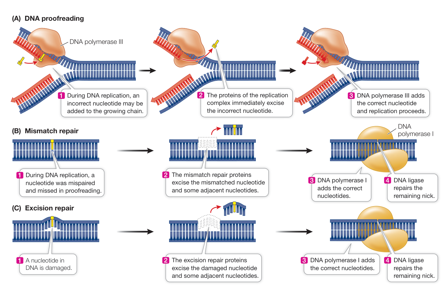
Figure 13.19 DNA Repair Mechanisms During replication, DNA polymerase III can detect and remove a mismatch when it is introduced (A), or mismatch repair can do so later (B), reducing the rate of errors in the replicated DNA. (C) Excision repair detects and repairs damage to existing DNA molecules.
Recap and Assess
DNA polymerases are not perfectly accurate. Although DNA polymerases make incorporation errors, proofreading and mismatch repair reduce the overall error rate of replication. In addition, excision repair mechanisms recognize and repair nucleotides altered or damaged by environmental factors.
- From what you know about the role of DNA in the cell, what would be the genetic consequences of an unrepaired DNA mismatch in a G1 phase cell that then entered the cell cycle to give rise to gametes from meiosis?
- One form of colon cancer is caused by a mutation in a gene encoding a protein involved in mismatch repair, so that it does not function correctly. Do you think this mutation is the one that leads to unregulated cell cycling? If not, what kind of gene might be affected? Recall cell cycle control genes involved in cancer formation (see Figure 11.24).
Understanding how DNA is replicated and repaired has allowed scientists to develop techniques for studying genes. We look next at one of the most widely used of these techniques.
The Polymerase Chain Reaction Amplifies DNA
Learning Objectives
13.5.1 Explain the purpose for each of the three steps in the PCR process.
13.5.2 Explain how PCR is targeted to particular DNA sequences.
The principles underlying DNA replication in cells have been used to develop an important laboratory technique that has been vital in analyzing genes and genomes. This technique allows researchers to make multiple copies of short DNA sequences.
The polymerase chain reaction can make multiple copies of a DNA sequence
In order to study DNA and perform genetic manipulations in the lab, it is often necessary to make multiple copies of a particular DNA sequence. While there are several methods for making multiple copies of a DNA sequence (see Chapter 18), one method, the polymerase chain reaction (PCR), relies on characteristics of DNA polymerases. The PCR is performed in a small tube that contains:
- A sample of double-stranded DNA from a biological sample, which contains the template sequence
- Two short, artificially synthesized primers that are complementary to the ends of the template sequence to be amplified
- The four dNTPs (dATP, dTTP, dCTP, and dGTP)
- A DNA polymerase that can tolerate high temperatures without losing function
- Salts and a buffer to maintain a near-neutral pH
PCR amplification is a cyclic process in which a sequence of steps is repeated over and over (Figure 13.20):
- The first step involves heating the reaction mixture to near boiling point, to separate (denature) the two strands of the DNA template.
- The reaction is then cooled to allow the primers to bind to their complementary sequences in the template strands.
- Next, the reaction is warmed to an optimum temperature for the DNA polymerase to catalyze the production of the complementary new strands.
A single cycle takes a few minutes to produce two copies of the target DNA sequence, leaving the new DNA in the double-stranded state. Repeating the cycle many times leads to an exponential increase in the number of copies of the DNA sequence.
The PCR technique requires that the base sequences at the 3’ end of each strand of the target DNA sequence be known, so that complementary primers, usually 15 to 30 bases long, can be made in the laboratory. Because of the uniqueness of DNA sequences, a pair of primers this length will usually bind to only a single region of DNA in an organism’s genome. This specificity, despite the incredible diversity of DNA sequences, is a key to the power of PCR.
One initial problem with PCR was its temperature requirements. To denature DNA, it must be heated to more than 90°C—a temperature that destroys most DNA polymerases. When the PCR technique was first being developed, new enzyme had to be added after denaturation in each cycle, which made the technique impractical for widespread use. This problem was solved by nature: in the hot springs at Yellowstone National Park, as well as in other high-temperature locations, there lives a bacterium called, appropriately, Thermus aquaticus (“hot water”). The means by which this organism survives temperatures of up to 95°C was investigated by Thomas Brock and his colleagues at the University of Wisconsin, Madison. They discovered that T. aquaticus has metabolic machinery that is heat-resistant, including a DNA polymerase that does not denature at these high temperatures.
Scientists pondering the problem of copying DNA by PCR read Brock’s basic research articles and got a clever idea: why not use
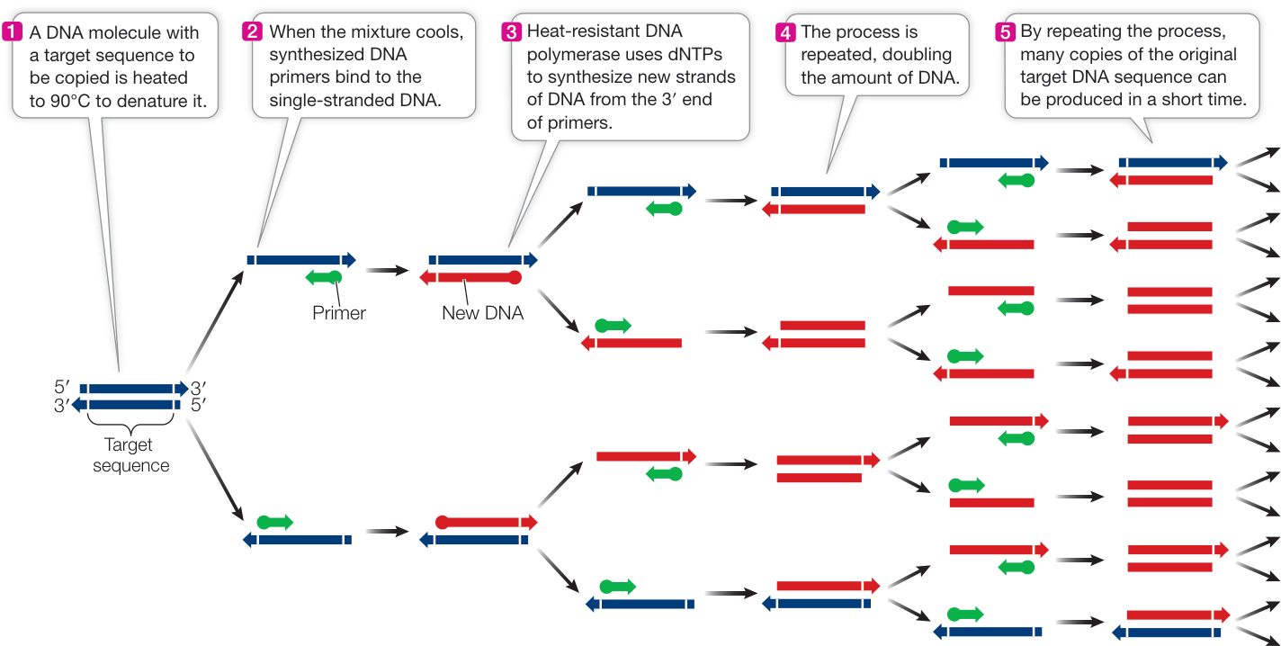
Figure 13.20 The Polymerase Chain Reaction The steps in this cyclic process are repeated many times to produce multiple identical copies of a DNA fragment. This makes enough DNA for chemical analysis and genetic manipulations.
Q: How do PCR primers relate to DNA replication primers?
Simulation 13.1 Polymerase Chain Reaction
T. aquaticus DNA polymerase in the PCR technique? It could withstand the 90°C denaturation temperature and would not have to be added during each cycle. The idea worked, and it earned biochemist Kary Mullis a Nobel prize. PCR has had an enormous impact on genetic research. Some of its most striking applications will be described in Chapters 15–18. These applications range from amplifying DNA in order to identify an individual person or organism, to detection of diseases.
Recap and Assess
Knowledge of the mechanisms of DNA replication led to the development of the PCR technique for making multiple copies of DNA sequences.
- What is the role of primers in PCR?
- One way to quickly identify an organism in the field is to use PCR: if there is amplification of a particular sequence, then the organism is identified. What is the basis for this test?
▶ Investigating LIFE
QA What do we need to know about DNA replication to describe the mechanism of a drug that blocks it?
Recall that the opening story in this chapter described the discovery that platinum electrodes inhibit cell division, which lead researchers to wonder if they could invent a drug containing platinum that might inhibit the uncontrolled growth of cancer cells. The drug that Dr. Barnett Rosenberg developed, cisplatin, was remarkably efficient at halting certain kinds of tumor growth. Understanding how cisplatin works came with an understanding of the chemistry of DNA and the mechanism by which it replicates.
Investigating Life: The Meselson–Stahl Experiment discusses the experiment that revealed that DNA replicates semiconservatively—that each strand of DNA serves as a template for a new strand. In the presence of cisplatin, DNA cannot denature to allow replication, suggesting that cisplatin interferes with strand separation. How is separation prevented, and what does it have to do with platinum?
Cisplatin contains a platinum atom bonded to two amino groups and two chlorine atoms (Figure 13.21A). In Rosenberg’s experiments, this compound was formed when the platinum electrode reacted with salts in the surrounding solution. The bonds between the platinum atom and the chlorine atoms are weak, and the latter can be displaced by electron-rich substances (you may know from chemistry that these are called nucleophiles). In DNA, one of the nitrogen atoms of guanine (Figure 13.21B) displaces one of the chlorines, forming a strong covalent bond. If there is a nearby guanine on the opposite DNA strand, the other chlorine of the cisplatin molecule can be displaced as well, so that the cisplatin is effectively bonded to both of the DNA strands. In short, the DNA strands become cross-linked and cannot separate as needed for DNA replication (Figure 13.21C). Without replication, the cell cannot divide, and it undergoes apoptosis. The type of DNA lesion caused by cisplatin cannot be repaired by any of the cell’s usual DNA repair mechanisms.
Future directions
Eukaryotic chromosomes have long strands of DNA, each with up to tens of millions of base pairs. During S phase of the cell cycle, each chromosome must be replicated completely and only once. Since replication is bidirectional from an origin, and since S phase typically is not long enough to replicate an entire chromosome, there must be many starting points for replication. Understanding how these origins are recognized is a major challenge for biologists. A recent discovery may provide a clue as to how a DNA pre-replication complex arrives at each replication origin. While most RNA molecules transcribed from DNA leave their site of synthesis, some do not. These short RNAs, complementary to their DNA template, turn back instead and bind to the DNA by base pairing. Indeed, this rapid binding displaces the nontemplate strand of DNA, so it forms a loop. These loops of DNA cannot pair with their complementary strand, because that strand at that location is bound up with RNA. Emerging evidence suggests that the “R-loops,” with their exposed DNA bases, act to recognize binding of the proteins of the DNA pre-replication complex.
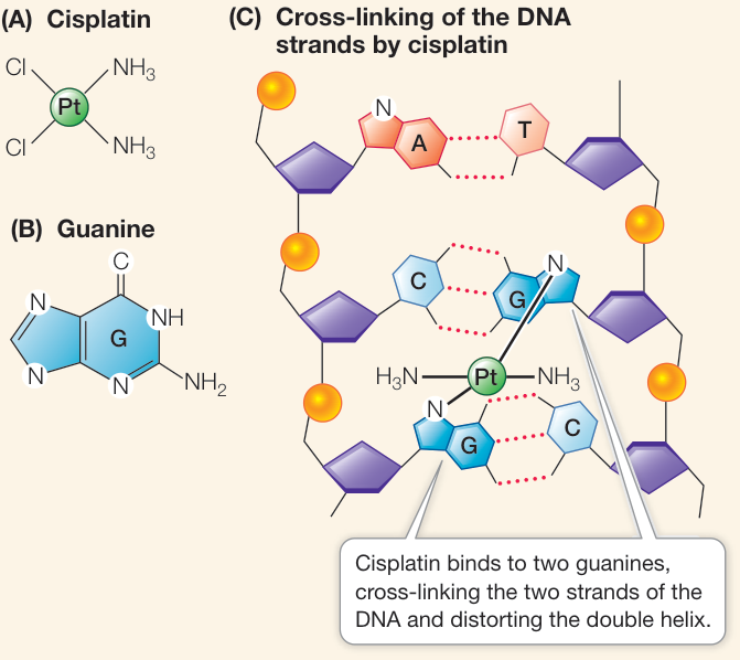
Figure 13.21 Cisplatin: A Small but Lethal Molecule
© Biology Pics/Science Source
You should be able to relate each summary to the adjacent figures. If you go to this visual summary in Achieve, you can follow links to figures, animations, activities, and simulations that will help you consolidate the material.
Visual Summary
Experiments Revealed the Function of DNA as Genetic Material
- Bacterial transformation requires DNA.
- Bacteriophages inject DNA, not protein, into bacterial hosts.
Questions
- How is a mixture of a dead S strain and a living R strain of pneumococcal bacteria able to kill a mouse?
- Why did Hershey and Chase use sulfur and phosphorus to label proteins and DNA in bacteriophage?
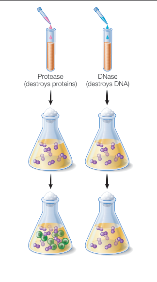
Figure 13.2
DNA Has a Structure That Suits Its Function
- Chargaff showed that the amount of adenine (A) in DNA = the amount of thymine (T), and that the amount of guanine (G) = the amount of cytosine (C).
- Franklin determined many of the characteristics of DNA structure from her X-ray images.
- Watson and Crick proposed that DNA was a double-stranded molecule with each strand composed of a sugar–phosphate backbone with hydrogen bonding between complementary base pairs.
Questions
- A sample of DNA from a rabbit consists of 24% adenine nucleotides. What are the frequencies of the other nucleotides?
- One strand of a double-stranded DNA molecule has the sequence 5’ ACGGTTACC 3’. What is the sequence and polarity of the other strand?
- How many different sequences are possible for a 10-base-pair DNA molecule?
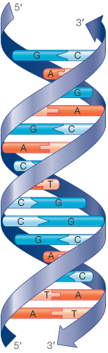
Figure 13.6
DNA Is Replicated Semiconservatively
- Meselson and Stahl used density labeling to show that DNA undergoes semiconservative replication. Each parent strand serves as a template for the synthesis of a new strand.
- DNA polymerases, which require primers, catalyze the addition of nucleotides to the 3’ end of each new strand.
- Replication begins at the origins of replication (ori) on both strands and proceeds in the 5’-to-3’ direction, forming replication forks, which have a leading strand and a lagging strand.
- Telomerase can extend telomeres, overcoming the chromosome shortening that occurs each time DNA is replicated.
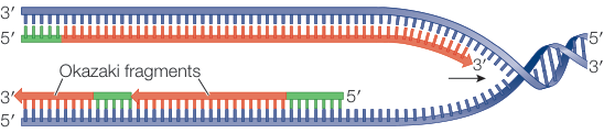
Figure 13.15
Questions
- If replication was actually conservative, what results would Meselson and Stahl have obtained after one round of replication on light medium?
- A replication fork is moving from left to right. If the top template strand is 5’-to-3’ in the left-to-right direction, which strand is the leading and which is the lagging?
- Many cancer cells express telomerase. Why does that make them more able to divide indefinitely?
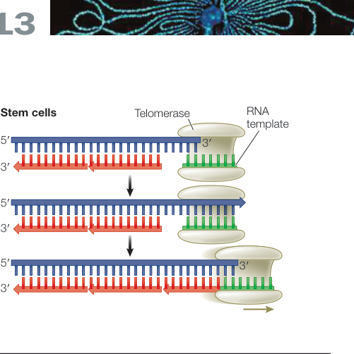
Figure 13.18
Errors in DNA Can Be Repaired
- DNA polymerases occasionally incorporate the incorrect base in the new strand, which causes mismatches.
- Mismatches can be repaired by proofreading or mismatch repair.
- DNA damage is often repaired by excision repair.
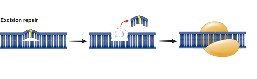
Figure 13.19
Questions
- An adenosine nucleotide is on the template strand of a DNA molecule. What are the possible mismatches that could be introduced by a DNA polymerase as it synthesizes the new strand?
- How can the DNA replication error rate be lower than the DNA polymerase incorporation error rate?
The Polymerase Chain Reaction Amplifies DNA
- The polymerase chain reaction (PCR) allows amplification of a particular region of DNA.
Questions
- What are the three steps of PCR?
- Why is a heat-resistant DNA polymerase important for PCR?
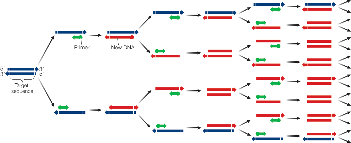
Figure 13.20