Chapter 15Gene Mutation and Molecular Medicine
Key Concepts
15.1 Mutations Are Heritable Changes in DNA
15.2 Mutations in Humans Can Lead to Diseases
15.3 Mutations Can Be Detected and Analyzed
15.4 Genetic Screening Is Used to Detect Diseases
15.5 Genetic Diseases Can Be Treated
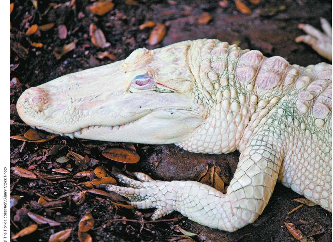
Albinism in this alligator is due to a mutation in both alleles of a single gene.
© The Florida collection/Alany Stock Photo
Investigating Life: The Angelina Jolie Effect
The actress Angelina Jolie surprised many people by announcing that she had both breasts removed upon learning that she carries a mutation in the “breast cancer gene.” Her family history led to her being tested for the mutation. Her mother and grandmother both died of ovarian cancer and her aunt died of breast cancer. Ms. Jolie learned that a mutation in a gene called BRCA1 increases the risk of breast and ovarian cancer. A woman with a harmful mutation in one allele of her BRCA1 gene has an 80% chance of getting breast cancer by age 60, far in excess of the 8% likelihood for women without a mutation. Removal of breast tissue lowers this risk to 10%.
Ms. Jolie’s announcement was widely praised by health-care professionals. Women at risk for carrying a BRCA1 mutation have increasingly elected to be tested and, when found to be carriers, more often have preventative surgery.
The BRCA1 mutation test is the culmination of research in the molecular genetics of cancer. As we discussed in Chapter 11, cancer can arise through mutations of either oncogenes (resulting in overstimulation of the cell cycle) or tumor suppressor genes (which would otherwise function to slow down the cell cycle). BRCA1 is a tumor suppressor gene involved in DNA repair. Women who are at high risk for breast cancer inherit a mutated copy of this gene, so that they have one wild-type allele and one mutated allele in every cell. If a certain mutation occurs in the wild-type allele in a cell then no functional protein can be made. BRCA1 is not expressed in all tissues but in breast cells it is particularly important, and so the loss of functional BRCA1 protein results in an increase in mutation rate in these cells. A higher mutation rate leads to accumulation of mutations in other genes, some of which may lead to improper activation of the cell cycle, resulting in uncontrolled cell division and other adverse effects of a growing tumor.
The precise description of mutations in genetically caused diseases exemplifies the emerging field of molecular medicine. Our ability to identify mutations is leading to more precise testing, diagnoses, and treatments for the diseases they cause.
Q&A What is the breast cancer gene, and what are the issues with DNA testing for cancer? (Find the answer on page 347.)
© Photo Brand Stock Photo
Mutations Are Heritable Changes in DNA
Learning Objectives
- 15.1.1 Use a defined DNA sequence to distinguish among a transition, transversion, single base-pair insertion mutation, and single base-pair deletion mutation.
- 15.1.2 Using a normal and a mutant mRNA sequence, recognize silent, missense, nonsense, loss-of-stop, and frame-shift mutations.
- 15.1.3 Give examples of how a mutation in a noncoding region could cause a change in phenotype.
- 15.1.4 Describe what happens in the following chromosomal rearrangements: deletion, duplication, inversion, and translocation.
- 15.1.5 Compare and contrast spontaneous and induced mutations, and give examples of each.
- 15.1.6 Explain why some C-G base pairs in DNA are hot spots for mutation.
In Chapter 12 we defined mutations as stable, inherited changes in genes, and we saw that different alleles may produce different phenotypes (short pea plants versus tall, for example). In Chapters 13 and 14 we described the chemical nature of genes as DNA sequences, and how they are expressed as phenotypes (in particular, proteins). To reiterate, we can now define a mutation as a change in the nucleotide sequence of DNA that can be passed on from one cell, or organism, to another.
Mutations have different phenotypic effects
In organisms that have specialized cell lineages that give rise to gametes, a distinction is made between mutations that can be passed on to offspring and those that can’t:
- Somatic mutations occur in somatic (non-gamete) cells. These mutations are passed on to the daughter cells during mitosis, and to the daughters of those cells in turn, but are not passed on to sexually produced offspring. For example, a mutation in a single human skin cell could result in a patch of skin cells that all have the same mutation, but it would not be passed on to the person’s children.
Figure 15.1 Mutation and Protein Function
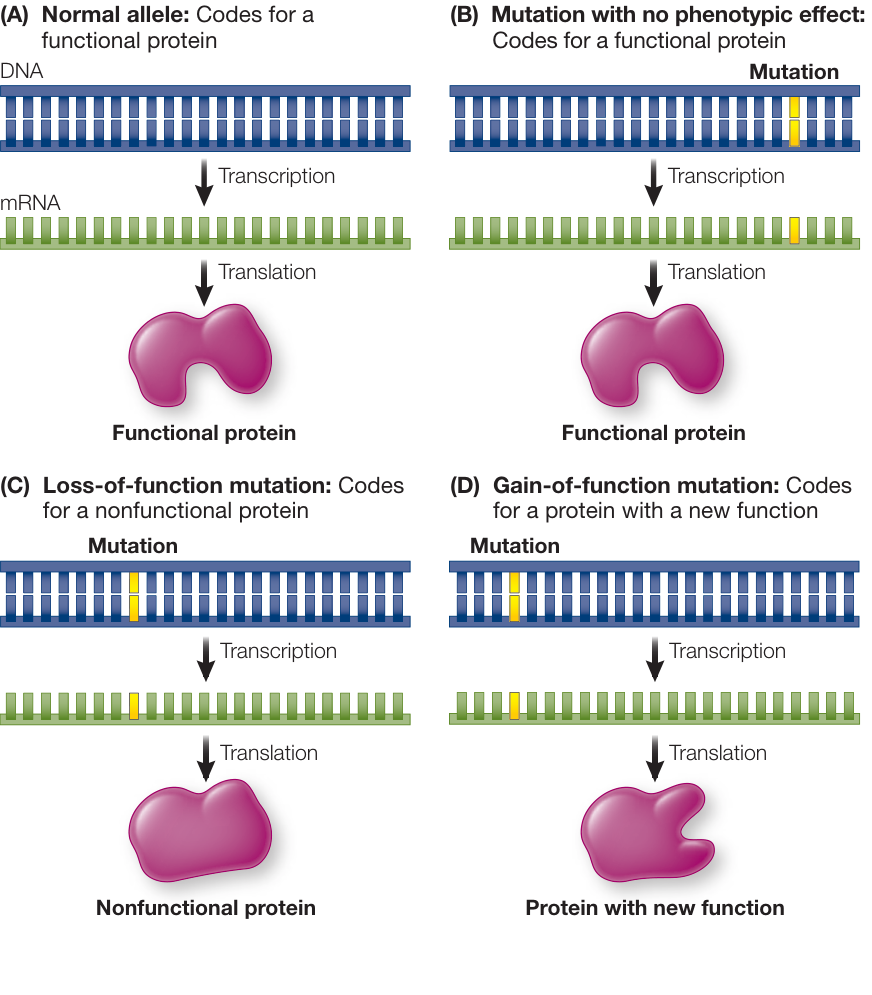
Figure 15.1 Mutation and Protein Function Mutations may or may not affect the protein phenotype. Q: For a protein-coding gene, which of the three types of mutations will be most common, and why? Simulation 15.1 Mutations and Function Mutations may or may not affect the protein phenotype. Q: For a protein-coding gene, which of the three types of mutations will be most common, and why?
- Germ line mutations occur in the cells of the germ line—the specialized cells that give rise to gametes. A gamete with the mutation passes it on to a new organism at fertilization. The new organism will have the mutation in every cell of its body and will be able to pass the mutation on to its offspring. The BRCA1 mutation that Angelina Jolie, her aunt, and her mother inherited from Angelina’s grandmother is a germ line mutation.
Does every mutation have a phenotypic effect? Not necessarily (Figure 15.1). Many mutations occur in a nonfunctional region of a DNA molecule, such that no gene product is affected. Alternatively, a mutation may occur in a gene but cause either no change in the gene product or a change that does not alter function (see Figure 15.1B). These cases result in no phenotypic change. For those mutations that do alter phenotype, we distinguish two main classes:
- A loss-of-function mutation may cause a gene to not be expressed at all, or the gene may be expressed but produces a dysfunctional RNA or a protein that can no longer play its cellular role, such as its catalytic function if it is an enzyme (see Figure 15.1C). Loss-of-function mutations almost always show recessive inheritance in diploid organisms, because the presence of one wild-type allele will usually result in sufficient functional RNA or protein for the cell. For example, recall from Key Concept 12.1 that the familiar wrinkled seed allele in pea plants, originally studied by Mendel, is due to a recessive loss-of-function mutation in the SBE1 (starch branching enzyme) gene. The protein made by this gene catalyzes the branching of starch as seeds develop. In the homozygous mutant, the SBE1 protein is not functional, and that leads to osmotic changes, causing the wrinkled appearance. Complete loss-of-function mutations are often referred to as null alleles.
- A gain-of-function mutation leads to a gene product, usually a protein, with an altered function (see Figure 15.1D). A gain-of-function mutation usually shows dominant inheritance, because the presence of the wild-type allele does not prevent the mutant allele from functioning. This type of mutation is common in cancer. For example, there are mutations in oncogenes that result in proteins that constantly stimulate cell division.
Some mutations have effects on phenotype only under certain conditions. For example, a conditional mutation affects phenotype only under restrictive conditions and is not detectable under what are called permissive conditions. In Key Concept 14.1 we discussed arginine-requiring mutant strains of Neurospora. Such mutants grow normally when arginine is supplied to the medium but are unable to grow when arginine is not present in the medium, so their phenotype depends on the growth medium (see Figure 14.2). Some conditional mutations are temperature-sensitive, resulting in proteins with reduced stability at high temperatures. For example, the point restriction phenotype in rabbits and Siamese cats (see Figure 12.15) is due to a temperature-sensitive (conditional, loss-of-function) mutation in a coat color gene. At normal body temperature, the protein encoded by the gene is unstable and nonfunctional, so the animal’s fur is not pigmented. However, in the body’s cooler extremities, the protein is functional and dark fur results.
The effects of mutations on phenotype can be reversed, such that the phenotype reverts to wild type. Reversion mutations result when a gene is mutated a second time so that the DNA reverts to its original sequence or to a different sequence that results in the non-mutant phenotype.
Now let’s look more closely at the DNA level to see how various kinds of mutations play out.
Point mutations are changes in single base pairs
A point mutation is the addition (insertion) or subtraction (deletion) of a single base pair, or the substitution of one base pair for another. There are two kinds of base substitutions (Figure 15.2). A transition occurs when the purine nucleotide in a base pair is replaced by the other purine (A replaces G or vice versa) and the pyrimidine is replaced by the other pyrimidine (T replaces C or vice versa). A transversion occurs when the purine nucleotide in a base pair is replaced by a pyrimidine (A or G replaces C or T) and the pyrimidine nucleotide is replaced by a purine (C or T replaces A or G).
What are the possible effects of a point mutation that occurs in the protein-coding region of a gene, such that the mature mRNA has a single nucleotide change? A silent mutation has no effect on the amino acid sequence of a protein, but missense, nonsense, loss-of-stop, and frame-shift mutations do result in changes in the protein, some of them drastic (Figure 15.3). Note that substitutions alter only a single codon in the mRNA, while insertions and deletions generally alter many codons.
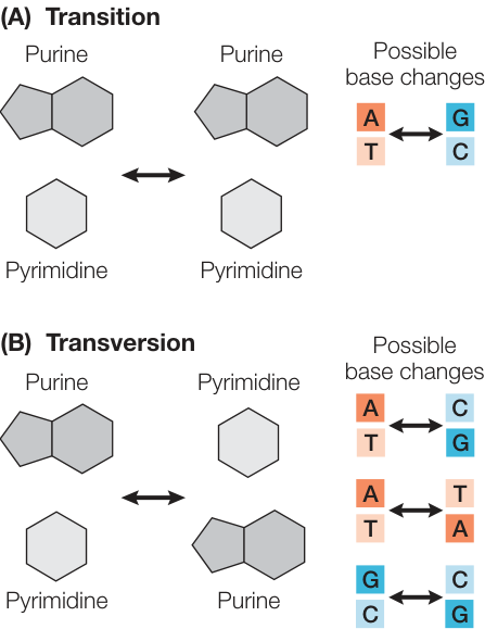
Figure 15.2 Two Types of Base Pair Substitutions A substitution mutation replaces one base pair with another. (A) In a transition, the purine in a base pair is replaced by the other purine, and the pyrimidine is replaced by the other pyrimidine. (B) In a transversion, the purine is replaced by a pyrimidine, and the pyrimidine is replaced by a purine.
SILENT MUTATIONS Silent mutations are DNA substitutions that result in a change of an mRNA sense codon (one that encodes an amino acid) into another sense codon that encodes the same amino acid (see Figure 15.3B). A change in a codon does not necessarily mean a change in the amino acid that gets translated, because of the redundancy of the genetic code (see Figure 14.11).
MISSENSE MUTATIONS Missense mutations are DNA substitutions that change an mRNA sense codon into another sense codon that encodes a different amino acid, such that one amino acid substitutes for another in the encoded polypeptide (see Figure 15.3C). An example is the mutation that causes sickle-cell disease, a serious heritable blood disorder. The disease occurs in people who carry two copies of the sickle allele of the gene for -globin—a subunit of hemoglobin, the protein in human blood that carries oxygen. The sickle allele differs from the wild-type allele by one base pair, resulting in a polypeptide that differs by one amino acid from the wild-type protein. Individuals who are homozygous for this recessive allele have defective, sickle-shaped red blood cells (Figure 15.4).
Connect the Concepts Many base substitution mutations result in changes in protein structure due to amino acid substitutions. Review the relationships between amino acid sequence and protein three-dimensional structure in Key Concept 3.2.
A missense mutation may result in a defective protein, but in many cases has no effect on the protein’s function. For example, a hydrophilic amino acid may be substituted for another hydrophilic amino acid, so that the tertiary (and quaternary) structure of the protein is unchanged. In some cases a missense mutation reduces the functional efficiency of a protein rather than completely inactivating it. Therefore individuals homozygous for a missense mutation in a protein essential for life may survive if enough of the protein’s function is retained.
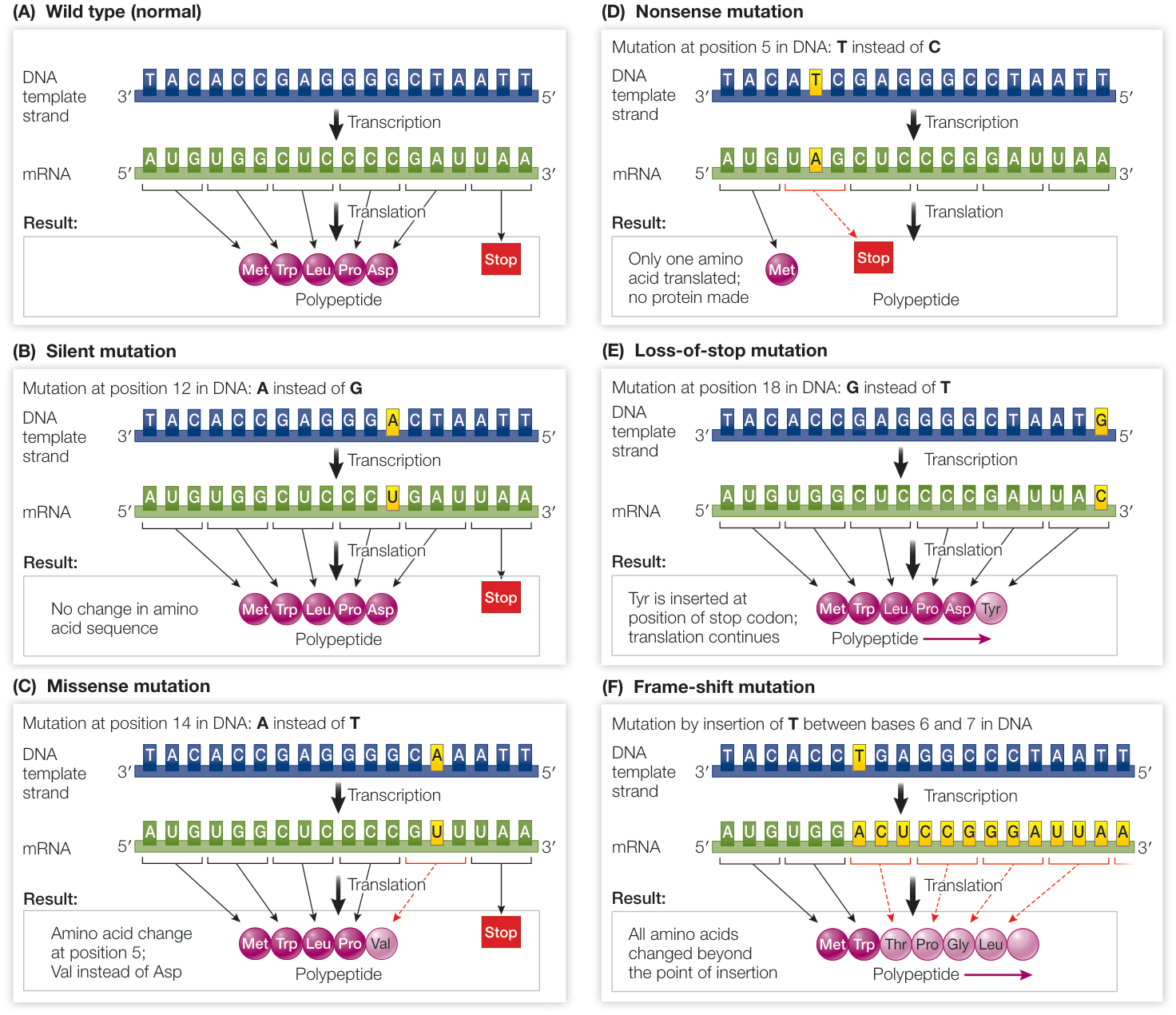
Figure 15.3 Point Mutations When they occur in the coding region of a protein, single base pair changes, which cause single nucleotide changes in the mRNA, can cause silent, missense, nonsense, loss-of-stop, or frame-shift mutations.
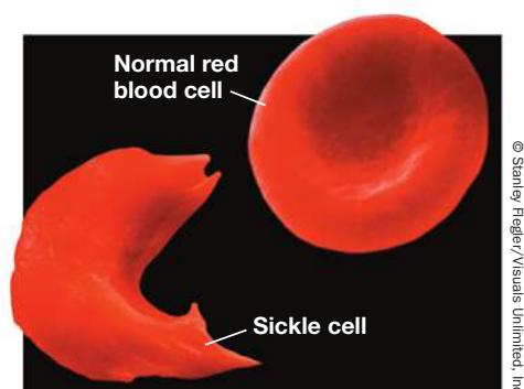
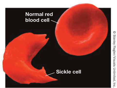
Figure 15.4 Sickle and Normal Red Blood Cell The sickle cell anemia allele is a missense mutation that causes red blood cells to sickle when homozygous.
In some cases, a gain-of-function missense mutation occurs, such that the mutated polypeptide has a novel function not exhibited by the wild-type polypeptide. An example is a mutation in the human P53 gene, which codes for a tumor suppressor—a protein that inhibits the cell cycle (see Key Concept 11.7). Certain mutations of the P53 gene cause this protein to both no longer inhibit cell division (an example of loss-of-function) and to promote cell division and prevent programmed cell death, which represent gain of function.
NONSENSE MUTATIONS A nonsense mutation involves a base pair substitution that causes an mRNA sense codon to become a stop codon (see Figure 15.3D). A nonsense mutation results in a shortened protein, since translation does not proceed beyond the point where the mutation occurred. For example, a common mutation causing thalassemia (another blood disorder affecting hemoglobin) in Mediterranean populations is a nonsense mutation that drastically shortens the -globin subunit. Shortened proteins usually are not functional; however, if the nonsense mutation occurs near the end of the gene, it may have little to no effect on function.
LOSS-OF-STOP MUTATIONS A loss-of-stop mutation involves a base pair substitution that results in a change from an mRNA stop codon to a sense codon, causing additional amino acids to be added to the end of the polypeptide. For example, a substitution that converts UAA (stop) to UAC (tyrosine) allows the ribosome to continue translating (see Figure 15.3E). The effect of a loss-of-stop mutation depends on how many amino acids are added to the end of the polypeptide and how important that part of the polypeptide is to function.
FRAME-SHIFT MUTATIONS Not all point mutations are base substitutions. A base pair may be inserted into, or deleted from, a sequence of DNA. Such changes in coding sequences are known as frame-shift mutations because they alter the mRNA reading frame (that is, consecutive triplets) in which the codons are read during translation (see Figure 15.3F). Think again of codons as three-letter words, each corresponding to a particular amino acid. Translation begins at a particular starting point (the AUG start codon) and proceeds three bases at a time—that is, codon by codon. If a nucleotide is inserted or deleted from a codon, then the three-letter “words” are shifted as translation proceeds beyond that point, and the result is a completely different amino acid sequence. Given this massive impact on polypeptide sequence, it is not surprising that frame-shift mutations almost always lead to the production of nonfunctional proteins.
MUTATIONS OUTSIDE THE CODING REGION As we discuss in Chapter 17, in genomes such as ours, much of the DNA does not encode protein information. What happens if a point mutation occurs outside a protein-coding region, and thus does not alter a codon? In many such cases there is no effect on phenotype. For example, most of the positions in an intron are not required for intron splicing, and so most mutations in intron-coding regions do not alter phenotype. Similarly, intergenic regions—that is, regions that are found between genes—have many positions that have no function; mutations at these sequences have no effect on phenotype. However, while generally rare, mutations outside of coding regions may have significant effects. For example:
- Mutations in promoters or in other regulatory sequences may alter the rate of transcription of the gene.
- Mutations in DNA that alter RNA splicing sites may lead to incorrectly spliced mRNA and synthesis of an abnormal polypeptide.
Chromosomal rearrangements are extensive changes in the genetic material
Mutations can alter larger sequences of DNA than just a single base pair. In fact, some mutations are so large that they are visible at the chromosome level. You have already met one kind of chromosomal mutation, a change in chromosome number (see Key Concept 11.5). In aneuploidy an individual has an abnormal number of one or a few chromosomes. In polyploidy, an individual has more than two entire sets of chromosomes. In addition to undergoing changes in number, chromosomes (or more accurately, whole DNA molecules) can break and rejoin, grossly disrupting the sequence of genetic information. There are four types of such chromosomal rearrangements: deletions, duplications, inversions, and translocations. These mutations can be caused by double-strand breakage of chromosomes resulting from mutagens or by errors during crossing over.
- A deletion occurs by the removal of part of the genetic material and can happen if a chromosome breaks at two points and then rejoins, leaving out the DNA between the breaks (Figure 15.5A).
- A duplication can be produced at the same time as a deletion and can occur if homologous chromosomes break at different positions and then reconnect to the wrong partners (Figure 15.5B). Inappropriate alignment of homologs during prophase I followed by crossing over also causes this rearrangement. One of the two chromosomes ends up with a deleted segment, and the other has two copies (a duplication) of the same segment.
- An inversion can also result from the breaking and rejoining of a chromosome and can occur if a segment of DNA becomes “flipped,” so that it runs in the opposite direction from its original orientation (Figure 15.5C).
- A translocation results when a segment of a chromosome breaks off and becomes attached to a different chromosome. As we mentioned in Key Concept 11.5, a translocation of a large segment of chromosome 21 is one cause of Down syndrome. Translocations often involve reciprocal exchanges of chromosome segments, as in Figure 15.5D.
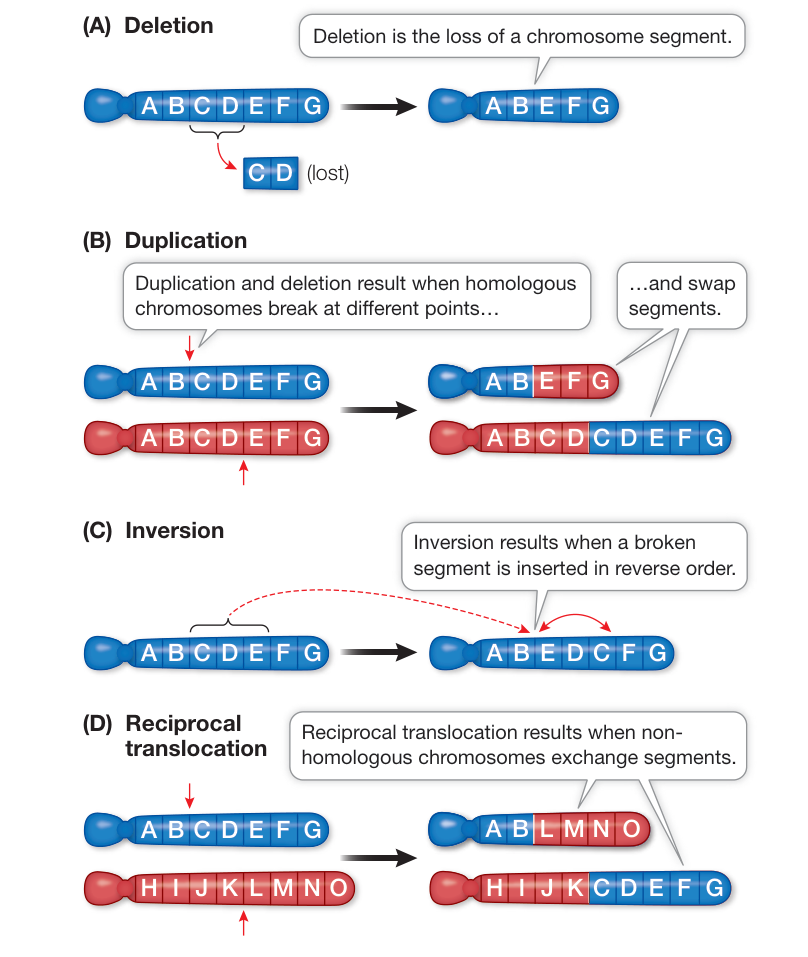
Figure 15.5 Chromosomal Rearrangements Chromosomes may experience double-strand breaks or may exhibit incorrect crossing over during meiosis, resulting in the incorrect joining of chromosomal regions. This can result in deletions (A, B), duplications (B), inversions (C), or reciprocal translocations (D). Note that the letters on these illustrations represent large segments of the chromosomes. Because chromosomes contain regions of noncoding DNA, each segment may include anywhere from zero to hundreds or thousands of genes. Q: How are chromosomal mutations detected? Hint: See Figure 11.20.
Retroviruses and transposons can cause loss-of-function mutations or duplications
Key Concept 14.2 described how certain viruses called retroviruses can insert their genetic material into the host cell’s genome. If such an insertion occurs within a gene, it usually causes a loss-of-function mutation in that gene. In many cases, the viral DNA remains in the host genome and is passed on from one generation to the next. When this happens the virus is called an endogenous retrovirus. Endogenous retroviruses are common—in fact, they make up 5% to 8% of the human genome.
Another form of DNA, called a transposon or transposable element, can also insert itself into the genome, causing insertion mutations. As you will see in Chapter 17, transposons are widespread in both prokaryotic and eukaryotic genomes. A transposon is a DNA sequence of a few hundred to a few thousand base pairs that can move from one position in the genome to another. It usually carries genes that encode the enzymes needed for this movement. Some transposons remove themselves from their positions in the genome and then insert themselves into other sites (the “cut and paste” mode of transposition). These transposons do not always excise cleanly, but leave behind short sequences of a few base pairs that become permanent mutations in the affected genes. Other transposons first replicate themselves, and then the new copies are inserted into new sites in the genome (the “copy and paste” mode). A sequence of genomic DNA is sometimes carried along with the transposon DNA when it moves, and gene duplication occurs. As you’ll see, gene duplication plays an important role in evolution.
Mutations can be spontaneous or induced
It is useful to distinguish between mutations that are spontaneous or induced, based on their causes. Spontaneous mutations are permanent changes in the genetic material that occur without any outside influence. The movement of transposons is an example of spontaneous mutation. Spontaneous mutations can also occur because cellular processes are imperfect, and may occur by several mechanisms (Figure 15.6):
- Mistakes during replication. As we discussed previously (in Key Concept 13.4), DNA polymerases sometimes incorporate the incorrect nucleotide into the growing strand, causing a mismatch. Incorporation errors occur rarely and are often caught by the proofreading function of the replication complex. However, they are more likely to occur and escape proofreading if the template base has undergone a spontaneous chemical rearrangement. Each base can exist in two different forms (called tautomers), one of which is common and one rare (see Figure 15.6A). When a base temporarily forms its rare tautomer, it can pair with the wrong base. For example, C normally pairs with G, but if C takes on the form of its rare tautomer at the time of DNA replication, it pairs with (and DNA polymerase will insert) an A. The result of such an incorporation error is a mismatch, which can be repaired to the correct base pair (see Figure 13.19). However, if the mismatch is repaired incorrectly (see Figure 15.6C), or if it is replicated before it is repaired (see Figure 15.6D), then a point mutation results. In this example, the A-C mismatch results in the original G-C base pair being replaced with an A-T base pair.
- A chemical reaction may alter the structure and base-pairing properties of a DNA base. For example, a spontaneous deamination reaction can result in loss of the amino group () in cytosine, converting it to a uracil base (Figure 15.6B). If this occurs in a DNA molecule, the result is a mismatch that is usually repaired, because uracil should not be present in DNA and is preferentially removed during repair. However, since the repair mechanism is not perfect, the mismatch will sometimes remain when a cell undergoes replication. In these cases, DNA polymerase will add an A (which base-pairs with U on the template DNA) instead of a G (which normally pairs with C). This A-U mismatch will be repaired, or will undergo replication to give an A-T base pair, which is a point mutation: , or equivalently, .
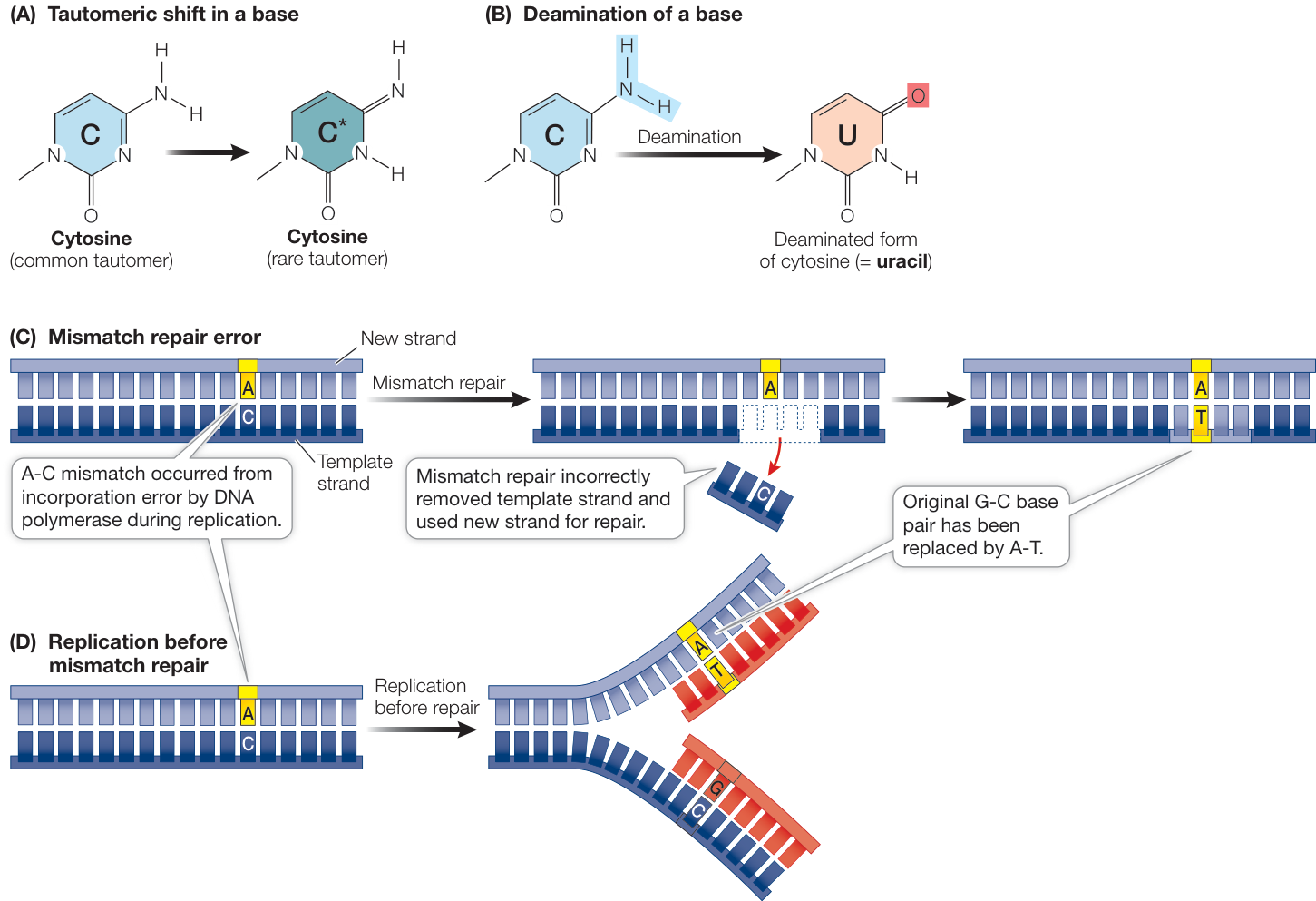
Figure 15.6 Spontaneous Mutations Spontaneous mismatches, if not repaired, will lead to permanent point mutations in the DNA sequence following replication or incorrect repair. (A) All four nitrogenous bases in DNA exist in both a prevalent (common) form and a rare form. When a base spontaneously forms its rare tautomer, it can pair with a different base, causing a mismatch during replication. (B) Some bases can undergo other spontaneous chemical changes. In this example, spontaneous deamination results in loss of the amino group ( ) in cytosine, converting it to uracil, to give an A-G mismatch. (C) In some cases, mismatch repair incorrectly removes the template strand instead of the new strand, resulting in a base-pair substitution. (D) In other cases, the mismatch may not be repaired before the next round of replication, again resulting in a base-pair substitution.
- Meiotic errors. Nondisjunction—the failure of homologous chromosomes to separate during meiosis—can occur, leading to one too many chromosomes or one too few (see Figure 11.19). Random chromosome breakage and rejoining, or crossing over between nonhomologous chromosomal regions, can produce deletions, duplications, inversions, or translocations.
Induced mutations occur when some agent from outside the cell—a mutagen—causes a change in DNA that leads to a mutation. As we mentioned earlier, retroviruses can function as mutagens. In addition, certain chemicals and radiation can cause mutations:
Some chemicals can alter nucleotide bases. For example, nitrous acid () and similar molecules can react with cytosine and convert it to uracil by deamination (see Figure 15.6B). This alteration has the same result as spontaneous deamination: instead of inserting a G, DNA polymerase inserts an A.
Some chemicals add groups to the bases. For instance, benzopyrene, a component of cigarette smoke, adds a large chemical group to guanine, making it unavailable for base pairing. When DNA polymerase reaches such a modified guanine, replication stops. In some cases, replication is restarted by the incorporation of any one of the four bases at random into the new strand. Three-fourths of the time the inserted base is not cytosine, and a mismatch results, which may then lead to a mutation at the next round of replication.
Radiation damages the genetic material. Radiation can damage DNA in two ways. First, ionizing radiation (including X-rays, gamma rays, and radiation from unstable isotopes) produces highly reactive chemicals called free radicals. Free radicals can change bases in DNA to forms that are not recognized by DNA polymerase. Ionizing radiation can also break the sugar–phosphate backbone of DNA, causing chromosomal rearrangements. Second, ultraviolet radiation (from the sun or a tanning lamp) is absorbed by thymine, causing it to form covalent bonds with adjacent thymine bases. Such thymine dimers, if unrepaired, block DNA replication, and the cell may respond by synthesizing a new strand that has a random sequence across from the block, increasing the likelihood of a mismatch and then a mutation.
Mutagens can be natural or artificial
Many people associate mutagens with materials made by humans, but many mutagenic substances are found in nature. An example of a naturally occurring mutagen is aflatoxin, which is made by the mold Aspergillus. When mammals ingest the mold, the aflatoxin is converted by the smooth endoplasmic reticulum (SER) in liver cells into a product that, like benzopyrene from cigarette smoke, binds to guanine; this also leads to mutations. Plants (and to a lesser extent animals) make thousands of small molecules with a variety of functions, some of which are mutagenic and potentially carcinogenic. Examples of human-made mutagens are nitrites, which are used to preserve meats. Once in mammals, nitrites get converted by the SER to nitrosamines, which are strongly mutagenic because they cause deamination of cytosine (see Figure 15.6B).
Radiation can also be human-made or natural. Some of the isotopes made in nuclear reactors and nuclear bomb explosions are certainly harmful. For example, extensive studies have shown increased mutations in the survivors of the atomic bombs dropped on Japan in 1945. As previously mentioned, natural ultraviolet radiation in sunlight also causes mutations.
By now, you may be getting worried about all the ways your DNA can be mutated. But for us, and for most organisms, DNA repair mechanisms have evolved so that most mismatches or damaged nucleotides in DNA do not get passed on to either the daughter cells or the next generation (see Figure 13.19). A large number of DNA damage occurs in the human genome under normal circumstances: among the diploid genome’s 6.2 billion base pairs, there are tens of thousands of DNA-damaging events per cell per day. Of these, the vast majority affect only one strand and are repaired using the undamaged strand to give the correct DNA sequence, so that only a very small number become mutations.
Some base pairs are more vulnerable than others to mutation
In certain regions of DNA, many of the cytosine residues have methyl groups added at their 5 positions, forming 5-methylcytosine. Methylation plays an important role in gene regulation (discussed in Key Concept 16.4). DNA sequencing has revealed that mutation “hot spots” are often located where cytosines have been methylated. Figure 15.6 shows unmethylated cytosine, which can lose its amino group, either spontaneously or because of a chemical mutagen, to form uracil (see Figure 15.6B). This type of error is usually detected by the cell and repaired, because uracil is recognized as inappropriate for DNA. However, when 5-methylcytosine (methylated cytosine) loses its amino group, the product is thymine, a natural base for DNA. The DNA repair mechanism cannot distinguish which base, T or G, in the mismatch is incorrect and so half the time will repair the mismatch incorrectly (from T-G to T-A). In the absence of repair, the mismatch will segregate at replication and again result in a mutation in one of the daughter cells (Figure 15.7). Note that the reason that methylated C-G base pairs are hot spots for mutation does not imply that they deaminate at higher frequency than unmethylated C-G base pairs; instead, the resulting U-G mismatch from an unmethylated C deamination is more often correctly repaired because the repair machinery recognizes U as an incorrect base.
Mutations have both benefits and costs
As we will see in Part Six of this book, mutations are the raw material of evolution: they provide the genetic diversity that makes natural selection possible. This diversity can be beneficial in two ways. First, a mutation may benefit the organism immediately. Second, a mutation may have no immediate selective advantage to the organism, but it may cause a phenotypic change in the organism’s offspring. The result will be a genetic variant in the population that may become advantageous if the environment changes in a later generation; the initially unselected mutation might thus be selected for under the new conditions.
We noted that gene duplication can arise through either chromosomal rearrangements or through the movements of transposons. Gene duplication is an important source of genetic variation. In a duplicated pair of genes, one gene may continue to play its original role in the organism, while the other may acquire a gain-of-function mutation that produces a new phenotype. As with any other mutation, this may be of immediate benefit for the organism, or it may provide a later generation with a selective advantage.
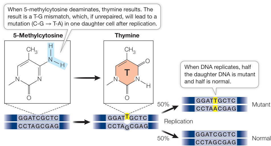
Figure 15.7 5-Methylcytosine in DNA Is a “Hot Spot” for Mutations If cytosine has been methylated to 5-methylcytosine, deamination results in a T-G mismatch that, if repaired incorrectly (by replacing the G nucleotide) or, if replicated (shown), will result in a C-G base pair being replaced by a T-A pair.
By contrast, mutations in genes whose products are needed for normal cellular processes are often harmful, especially if they occur in germ line cells that produce eggs or sperm and thus are present in every cell in the body. In some cases, offspring can inherit harmful recessive alleles from both parents and have the homozygous condition. In their extreme form, such mutations produce phenotypes that are lethal, killing the organism during early development. Likewise, mutations in somatic cells can be harmful, as in the case of cancer. Think about what would happen if an oncogene that stimulates cell division in a colon cell had a gain-of-function mutation during development. The result would be the same as inheriting the mutant allele: a higher likelihood of developing colon cancer.
What can we do about mutation? While spontaneous mutagenesis is not in our control, we can certainly try to avoid mutagenic substances and radiation. Not surprisingly, many things that cause cancer (carcinogens) are also mutagens. A good example is benzopyrene (discussed earlier), which is found in coal tar, car exhaust fumes, charbroiled foods, and cigarette smoke. A major public-policy goal is to reduce the effects of both human-made and natural mutagens on human health. An example is the Montreal Protocol, an international environmental agreement signed and adhered to by all members of the United Nations. It bans chlorofluorocarbons and other substances that cause depletion of the ozone layer in the upper atmosphere of Earth. The thinning of the ozone layer can result in increased ultraviolet radiation reaching Earth’s surface, which is implicated in somatic mutations that lead to skin cancer.
Recap and Assess
Mutations are permanent changes in the nucleotide sequence of DNA. Mutations can vary in magnitude from changes in single base pairs (point mutations) to extensive rearrangements of chromosomes or changes in the number of chromosomes. When mutations occur in somatic cells, they will be passed on to daughter cells; when they occur in germ line cells, they may be passed on to offspring.
- Suppose a small polypeptide consists of five amino acids: methionine-threonine-glutamine-tryptophan-phenylalanine. If a point mutation occurred in the DNA that encoded the third mRNA codon (the one encoding glutamine), what would be the effects on the polypeptide sequence if the mutation was a silent, missense, nonsense, or frame-shift mutation?
- What distinguishes the various kinds of chromosomal rearrangements: deletions, duplications, inversions, and translocations?
- What are the differences between spontaneous and induced mutation? Give an example of each.
- Why are some C-G base pairs prone to mutation?
You have seen that there are many different ways in which DNA can be altered, in terms of both the types of changes and the mechanisms by which they occur. We turn now to the ways in which mutations can cause disease.
Mutations in Humans Can Lead to Diseases
Learning Objectives
- 15.2.1 Give an example of an abnormal protein in humans that results from a genetic mutation and causes a disease.
- 15.2.2 Explain why point mutations, large deletions, and chromosomal abnormalities can all cause genetic disorders.
- 15.2.3 Describe what is meant by “expanding triplet repeats” in genetic disorders.
- 15.2.4 Justify the claim that many diseases arise from complex interactions between genetic and environmental factors.
The biochemistry that relates genotype (DNA) and phenotype (proteins) has been most completely described for model organisms, such as the prokaryote E. coli and the eukaryotes yeast and Drosophila. While the details vary, there is great similarity in the fundamental processes among these forms of life. These similarities have permitted the application of knowledge and methods discovered using these model organisms to the study of human biochemical genetics. Our focus in this chapter is mutations that affect human phenotypes, leading to diseases.
Disease-causing mutations may make proteins dysfunctional
Genetic mutations are often expressed phenotypically as proteins that differ from normal (wild-type) proteins. Abnormalities in enzymes, receptor proteins, transport proteins, structural proteins, and most of the other functional classes of proteins have all been implicated in genetic diseases.
LOSS OF ENZYME FUNCTION In 1934, the urine of two intellectually disabled young siblings was found to contain phenylpyruvic acid, an unusual by-product of the metabolism of the amino acid phenylalanine. It took two decades for scientists to trace the complex clinical phenotype of the disease that afflicted these children, called phenylketonuria (PKU), back to its molecular cause. The disease results from an abnormality in a single enzyme, phenylalanine hydroxylase (PAH), which catalyzes the conversion of dietary phenylalanine to tyrosine (Figure 15.8). This enzyme is not active in the livers of PKU patients, leading to excesses of phenylalanine and phenylpyruvic acid in the blood. Since then, the nucleotide sequence of the PAH gene has been compared between healthy people and those with PKU, and more than 400 different disease-causing mutations have been found. The most common one is a missense mutation that results in tryptophan instead of arginine at position 408 in the polypeptide chain (Table 15.1). As is often the case with loss-of-function mutations, the mutant alleles are recessive, because one functional allele is all that is needed to produce enough functional PAH to prevent the disease.
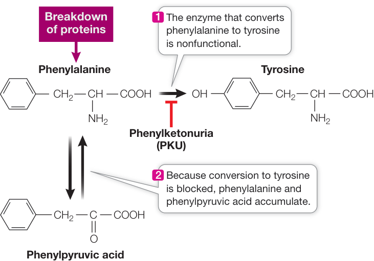
Figure 15.8 One Mutation Inactivates One Enzyme Phenylketonuria is caused by an abnormality in a specific enzyme that metabolizes the amino acid phenylalanine. Knowing the molecular causes of such single-gene, single-enzyme metabolic diseases can aid researchers in developing screening tests as well as treatments.
Hundreds of human genetic diseases that result from enzyme abnormalities have been discovered, some of which lead to intellectual disability and premature death. Most of these diseases are rare; PKU, for example, shows up in 1 out of every 10,000 newborns. As you by now realize, for any given gene there may be numerous alleles. Some alleles encode proteins that function normally, whereas others produce proteins that cause disease. Let’s take a look at a gene whose alleles illustrate both of these circumstances—the gene for one of the polypeptide chains in hemoglobin.
ABNORMAL HEMOGLOBIN As mentioned in Key Concept 15.1, sickle-cell disease is caused by a recessive, missense mutation. This blood disorder most often afflicts people whose ancestors came from the tropics or from the Mediterranean region.
Recall that human hemoglobin is composed of four globin subunits—two -chains and two -chains—as well as the pigment heme (see Figure 3.12). In the sickle-cell disease allele, one of the 146 amino acids in the -globin polypeptide chain is abnormal: at position 6, glutamic acid is replaced by valine. This replacement changes the charge of the protein (glutamic acid is negatively charged and valine is uncharged), causing it to form long, needlelike aggregates in the red blood cells. The phenotypic result is sickle-shaped red blood cells and an impaired ability of the blood to carry oxygen. The sickled cells tend to block narrow blood capillaries, resulting in tissue damage and eventually death by organ failure.
Because hemoglobin is easy to isolate and study, its variations in the human population have been extensively documented (Figure 15.9). Hundreds of alleles caused by single amino acid alterations in -globin, all due to mutations, have been reported. For example, at the same position that is mutated in sickle-cell disease (resulting in hemoglobin S), glutamic acid may be replaced by lysine, causing hemoglobin C disease. In this case, the resulting anemia is usually not severe. Many alleles that result in alterations of the amino acid sequence of hemoglobin do not affect its function. In fact, about 5% of us carry at least one missense point mutation in a -globin allele.
Some of the more common examples of inherited diseases caused by specific protein defects are listed in Table 15.2. These mutations can be dominant, codominant, or recessive, and some are sex-linked.
Disease-causing mutations may involve any number of base pairs
Disease-causing mutations may involve a single base pair, a long stretch of DNA, multiple segments of DNA, or even entire chromosomes (as we saw for Down syndrome in Key Concept 11.5).
POINT MUTATIONS Sickle-cell anemia is just one of many diseases caused by a point mutation. In some cases (sickle-cell anemia, for example), everyone with the disease has the same genetic mutation. In other cases, different loss-of-function point mutations in one gene can lead to the same disease. Think about it: the three-dimensional structure of an enzyme protein depends on its primary structure, so any change in the amino acid sequence of a protein has the potential to affect its structure, and consequently its function. For example, cystic fibrosis is a recessive disorder for which over 2,000 different causal mutant alleles have been identified.
Table 15.1 Two Common Mutations That Cause Phenylketonuria
| Codon 408 (20% of PKU cases) | Codon 280 (2% of PKU cases) | |||
|---|---|---|---|---|
| Normal | Mutant | Normal | Mutant | |
| Length of PAH protein | 452 amino acids | 452 amino acids | 452 amino acids | 452 amino acids |
| DNA at codon | 5’…CGG…3’ | 5’…TGG…3’ | 5’…GAA…3’ | 5’…AAA…3’ |
| 3’…GCC…5’ | 3’…ACC…5’ | 3’…CTT…5’ | 3’…TTT…5’ | |
| mRNA at codon | …CGG… | …UGG… | …GAA… | …AAA… |
| Amino acid at codon | Arginine | Tryptophan | Glutamic acid | Lysine |
| Active PAH enzyme? | Yes | No | Yes | No |
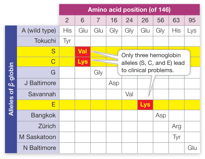
Figure 15.9 Hemoglobin Polymorphism Each of these mutant alleles codes for a protein with a single amino acid change in the 146-amino acid chain of -globin. Only three of the hundreds of known variants of -globin, highlighted in yellow, are known to lead to clinical abnormalities. “S” is the sickle-cell anemia allele.
LARGE DELETIONS Larger mutations may involve many base pairs of DNA. For example, deletions in the X chromosome that include the gene for the protein dystrophin result in Duchenne muscular dystrophy. Dystrophin is important in organizing the structure of muscles, and people who have only the abnormal form have severe muscle weakness. Sometimes only part of the dystrophin gene is missing, leading to an incomplete but partly functional protein and a mild form of the disease. In other cases, deletions span the entire sequence of the gene, so that the protein is missing entirely, resulting in a severe form of the disease. In yet other cases, deletions involve millions of base pairs and cover not only the dystrophin gene but adjacent genes as well; the result may be several diseases in the same person.
CHROMOSOMAL ABNORMALITIES Chromosomal abnormalities also cause human diseases. Such abnormalities result from the gain or loss of complete chromosomes (aneuploidy) (see Figure 11.19), or from the gain or loss of chromosomal segments (see Figure 15.5). About 1 newborn in 200 has a chromosomal abnormality.
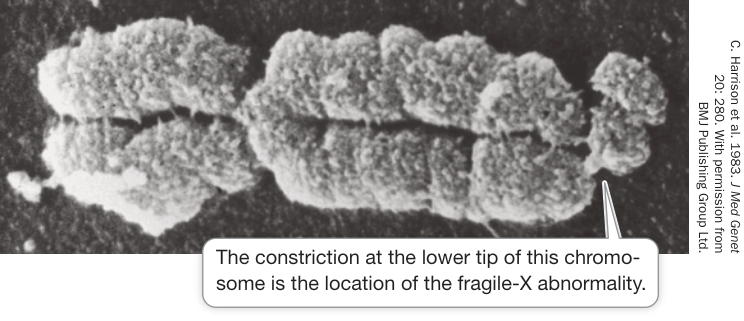
Figure 15.10 A Fragile-X Chromosome at Metaphase The chromosomal abnormality associated with fragile-X syndrome shows up under the microscope as a constriction in the chromosome. This occurs during preparation of the chromosome for microscopy.
This may be inherited from a parent who also has the abnormality, or it may result from an error in meiosis during the formation of gametes in one of the parents. One example is fragile-X syndrome, which is a constriction in the tip of the X chromosome that can result in intellectual disability (Figure 15.10). About 1 male in 3,000 and 1 female in 7,000 are affected. Although the basic pattern of inheritance is that of an X-linked recessive trait, there are departures from this pattern. Not all people with the fragile-X chromosomal abnormality are intellectually disabled, as we will see.
Expanding triplet repeats demonstrate the fragility of some human genes
About one-fifth of all males who have the fragile-X chromosomal abnormality are phenotypically normal, as are most of their daughters. But many of those daughters’ sons are intellectually disabled. In a family in which fragile-X syndrome appears, later generations tend to show earlier onset and more severe symptoms of the disease. It is almost as if the abnormal allele itself is changing—and getting worse. And that’s exactly what is happening.
The gene associated with fragile-X syndrome (FMR1) contains a repeated triplet, CGG, at a certain point in the promoter region (Figure 15.11). In normal people, this triplet is repeated 6 to 54 times (the average is 29). In intellectually disabled people with fragile-X syndrome, the CGG sequence is repeated 200 to 2,000 times.
Males carrying a moderate number of repeats (55–199) show no symptoms and are called premutated. These repeats become more numerous as the daughters of these men pass the chromosome on
Table 15.2 Some Human Genetic Diseases
| Disease name | Inheritance pattern; births frequency | Gene mutated; protein product | Clinical phenotype |
|---|---|---|---|
| Marfan syndrome | Autosomal dominant; 1 in 5,000 | FBN1 ; fibrillin-1 | Weakened connective tissue, aortic aneurism, vision problems |
| Familial hypercholesterolemia | Autosomal codominant; 1 in 500 heterozygous | LDLR ; low-density lipoprotein receptor | High blood cholesterol, heart disease |
| Cystic fibrosis | Autosomal recessive; 1 in 4,000 | CFTR ; chloride ion channel in membrane | Immune, digestive, and respiratory illness |
| Duchenne muscular dystrophy | Sex-linked recessive; 1 in 3,500 males | DMD ; the muscle membrane protein dystrophin | Muscle weakness |
| Hemophilia A | Sex-linked recessive; 1 in 5,000 males | HEMA ; factor VIII blood clotting protein | Inability to clot blood after injury, hemorrhage |
Sources: H. Dietz. 2001. In GeneReviews®. www.ncbi.nlm.nih.gov/books/NBK1335/. Updated 2017 Oct 12; S. Singh and V. Bittner. 2015. Curr Atheroscler Rep 17: 3; https://omim.org/entry/219700; https://omim.org/entry/310200; https://rarediseases.org/rare-diseases/duchenne-muscular-dystrophy/; https://omim.org/entry/306700.
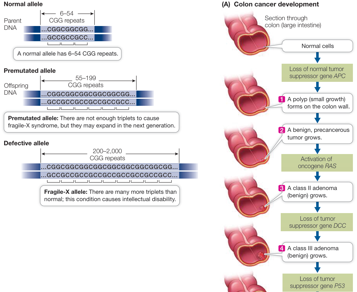
Figure 15.11 The CGG Repeats in the FMR1 Gene Expand with Each Generation The genetic defect in fragile-X syndrome is caused by 200 or more repeats of the CGG triplet. Q: Suppose an individual homozygous for 20 CGG repeats had a crossover during meiosis between homologous chromatids involving repeat number 8 in one chromatid and repeat number 12 in the other. What would the resulting repeat numbers be in each chromatid from this incorrect alignment during crossing over?
to their children. With 200 or more repeats, increased methylation of the cytosines in the CGG triplets is likely, which inhibits transcription of the FMR1 gene (see Key Concept 16.4). The normal role of the protein product of this gene is to bind to mRNAs involved in neuron function and to regulate their translation at the ribosome. When the FMR1 protein is not made in adequate amounts, these mRNAs are not properly translated, and nerve cells die. Their loss often results in intellectual disability. The methylated cytosines bind proteins that cause the chromosome constriction and fragile appearance.
This phenomenon of expanding triplet repeats has been found in more than a dozen other diseases, such as myotonic dystrophy (involving repeated CTG triplets) and Huntington’s disease (in which CAG is repeated). Such repeats, which may be found within a protein-coding region or outside it, appear to be present in many other genes without causing harm. How the repeats expand is not completely understood. One hypothesis is that DNA polymerase may slip after synthesizing a repeat and then fall back to copy it again; another is that incorrect alignment during crossing over in meiosis increases the copy number.
Cancer often involves somatic mutations
Many chromosomal and point mutations have been described in cancer cells. Such mutations affect oncogenes, whose products stimulate cell division, or tumor suppressor genes, whose products inhibit cell division (see Key Concept 11.7). More than two gene mutations are usually needed for full-blown cancer.
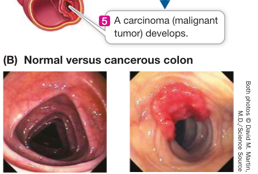
Figure 15.12 Multiple Somatic Mutations Transform a Normal Colon Epithelial Cell into a Cancer Cell (A) At least four genes must be mutated in a single cell to produce metastatic colon cancer. (B) These images from a screening test reveal a normal colon (left) and colon cancer (right).
Because colon cancer progresses to full malignancy slowly, it has been possible to identify the gene mutations that lead to each stage. Figure 15.12 outlines the “molecular biography” of this form of cancer. At least three tumor suppressor genes and one oncogene must be mutated in sequence in a cell in the colon lining for cancer to develop.
Although the occurrence of all of these events in a single cell might seem unlikely, remember that the colon lining has millions of cells, that these cells arise from stem cells that are constantly dividing, and that these changes take place over many years of exposure to natural and synthetic substances in foods, some of which act as mutagens.
Many diseases are caused by multiple genes and environment
The example of cancer illustrates how many common phenotypes, including ones that cause disease, are multifactorial; that is, they are caused by the interactions of many genes and proteins with one or more factors in the environment. When studying genetics, we tend to call individuals either normal (wild type) or abnormal (mutant); however, in reality every individual contains thousands or millions of genetic variations that arose through mutations. Our susceptibility to disease is often determined by complex interactions between these genotypes and factors in the environment, such as the foods we eat or the pathogens we encounter. For example, a complex set of genotypes determines who among us can eat a high-fat diet and not experience a heart attack, or who will succumb to disease when exposed to infectious bacteria. Estimates suggest that up to 60% of all people are affected by diseases that are genetically influenced. Identifying these genetic influences is another major task of molecular medicine and human genome sequencing.
Recap and Assess
Many genetic mutations result in loss of protein function and cause a mutant phenotype (a genetic disease). Disease-causing mutations range from point mutations to chromosomal abnormalities. In expanding triplet repeat disease alleles are altered as they are passed down, such that repeat number can increase and give rise to a disease phenotype. Some genetic diseases are influenced by multiple genes and the environment.
- Alkaptonuria is an inborn error of metabolism, caused by defects in an enzyme in the pathway that breaks down tyrosine (see Key Concept 14.1). Humans who are homozygous for one of these mutations make nonfunctional enzyme and accumulate the enzyme’s substrate, homogenistic acid, which causes their disease symptoms. In 1996, researchers in Spain cloned and sequenced the gene for the enzyme and characterized several mutant alleles. Here is the wild-type coding strand sequence for part of the gene, with the amino acid sequence of the protein below:
Here is the sequence for one of the mutant alleles:
- Use the genetic code table (see Figure 14.11) to determine the amino acid sequence produced by the mutant allele. What type of mutation is this: silent, nonsense, missense, loss-of-stop, or frame-shift?
- Why is this mutation likely to affect the function of the enzyme? (Hint: see Key Concept 3.2, especially Table 3.2.)
- Not all women with a mutation in the BRCA1 gene get early-onset breast cancer. Explain.
- Why do some members of a family show symptoms of fragile-X syndrome and others do not?
In this key concept we described the ways in which mutations can lead to human disease. Let’s turn now to the ways that biologists detect mutations in DNA.
Mutations Can Be Detected and Analyzed
Learning Objectives
- 15.3.1 Describe how restriction enzymes cut DNA.
- 15.3.2 Explain how gel electrophoresis separates DNA fragments based on their size.
- 15.3.3 Define DNA fingerprinting and explain how it works.
- 15.3.4 Describe how linkage to a genetic marker can be used to identify a disease-causing gene.
A challenge for biologists studying mutations is to precisely describe the DNA changes that lead to specific protein changes—an area of research called molecular genetics. Of course, the most direct and comprehensive way to analyze DNA is to determine its sequence of bases. DNA sequencing technologies are continually improving, and the entire genomes of many organisms have now been sequenced completely. Furthermore, the genomes of closely related organisms have been compared in order to identify mutations. We will discuss sequencing technology in Chapter 17. We are approaching the era where DNA sequencing will be the method of choice in searching for mutations. Whole genome DNA sequencing is not yet, however, in routine use in medicine. In this key concept we look at some of the techniques that are used in combination with DNA sequencing to study DNA, and to identify mutations that cause disease.
Cleavage of DNA by restriction enzymes can be used to rapidly detect mutations
All organisms, including bacteria, must have ways of dealing with their enemies. As we saw in Key Concept 13.1, bacteria can be attacked by viruses called bacteriophages. These viruses inject their genetic material into the host cell and turn it into a virus-producing factory, eventually killing the cell. Some bacteria defend themselves against such invasions by producing restriction enzymes (also known as restriction endonucleases), which cut double-stranded DNA molecules—such as those injected by bacteriophages—into smaller, noninfectious fragments (Figure 15.13). These enzymes break the bonds of the DNA backbone between the 3’ hydroxyl group of one nucleotide and the 5’ phosphate group of the next nucleotide. This cutting process is called restriction digestion.
There are many such restriction enzymes, each of which cleaves DNA at a specific sequence of bases called a recognition sequence or a restriction site. Most restriction sites are four to six base pairs long. Because each sequence of bases has a unique structure (see Key Concept 13.2), it can be specifically recognized by a particular restriction enzyme. Cells protect themselves from being digested by their own enzymes by either not having the particular restriction site in their genome, or by modifying their DNA, often with methyl groups, to prevent cutting by the restriction enzymes.
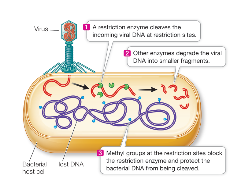
Figure 15.13 Bacteria Fight Invading Viruses by Making Restriction Enzymes
Restriction enzymes can be isolated from the cells that make them and used as biochemical reagents in the laboratory to give information about the nucleotide sequences of DNA molecules from other organisms. If DNA from any organism is incubated in a test tube with a restriction enzyme (along with buffers and salts that help the enzyme function), that DNA will be cut wherever the restriction site occurs. A specific sequence of bases defines each restriction site. For example, the enzyme EcoRI (named after its source strain of the bacterium E. coli) cuts DNA only where it encounters a specific six-base-pair sequence in the DNA double helix (Figure 15.14A). Note that the restriction site is palindromic, like the word “radar.” This means that both strands have the same sequence when they are read from their 5’ (or their 3’) ends. The EcoRI enzyme has two identical active sites on its two subunits, which cleave the two strands simultaneously between the G and the A of each strand, resulting in a “staggered” cut that leaves identical 5’ overhangs (Figure 15.14B).
The EcoRI recognition sequence occurs, on average, about once in every 4,000 base pairs in a typical prokaryotic genome, or about once per four prokaryotic genes. So EcoRI can chop a large piece of DNA into smaller pieces containing, on average, just a few genes.
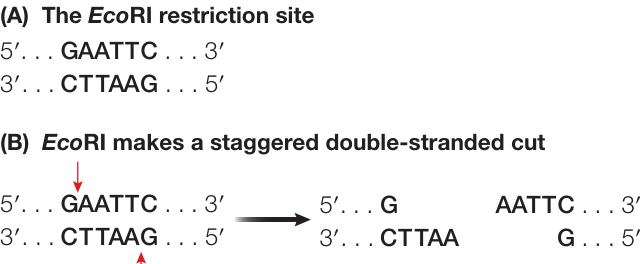
Figure 15.14 Restriction Enzymes Cut DNA at Specific Sequences (A) The restriction enzyme EcoRI recognizes a specific six-base-pair sequence in the DNA double helix. (B) When it makes a double-strand staggered cut, EcoRI generates identical 5’ overhangs (AATT).
Using EcoRI in the laboratory to cut small genomes, such as those of viruses that have tens of thousands of base pairs, may result in just a few fragments. For a huge eukaryotic chromosome with tens or hundreds of millions of base pairs, a very large number of fragments will be created.
Of course, “on average” does not mean that the enzyme cuts all stretches of DNA at regular intervals. For example, the EcoRI recognition sequence does not occur even once in the 40,000 base pairs of the bacteriophage T7 genome—a fact that is crucial to the survival of this virus, since its host is E. coli. Fortunately for E. coli, the EcoRI recognition sequence does appear in the DNA of other bacteriophages.
Gel electrophoresis separates DNA fragments
Restriction enzyme digestion is used to manipulate DNA in the laboratory so that mutations can be identified and analyzed. After a laboratory sample of DNA has been cut with one or more restriction enzymes, the DNA is in fragments, which must be separated to identify (map) where the cuts were made. Because the recognition sequence does not occur at regular intervals, the fragments are not all the same size, and these size differences can be used to separate the fragments from one another. Separating the fragments is necessary to determine the number and molecular sizes (in base pairs) of the fragments produced, or to identify and purify an individual fragment for further analysis or for use in an experiment.
Gel electrophoresis is a common and convenient technique for separating or purifying DNA fragments. Samples containing the fragments are placed in wells at one end of a semisolid gel (usually made of agarose or polyacrylamide), and an electric field is applied to the gel (Figure 15.15). Because of its phosphate groups, DNA is negatively charged at neutral pH; therefore, because opposite charges attract, the DNA fragments move through the gel toward the positive end of the field. Because the spaces between the polymers of the gel are small, small DNA molecules can move through the gel faster than larger ones. Thus DNA fragments of different sizes separate from one another, forming bands that can be detected with a dye. This provides three types of information:
- The number of fragments. The number of fragments produced by digestion of a DNA sample with a given restriction enzyme depends on how many times that enzyme’s recognition sequence occurs in the sample. Thus gel electrophoresis can provide some information about the presence of specific DNA sequences (the restriction sites) in the DNA sample.
- The sizes of the fragments. DNA fragments of known size (size markers) are often placed in one well of the gel to provide a standard for comparison. The size markers are used to determine the sizes of the DNA fragments in samples in the other wells. By comparing the fragment sizes obtained with two or more restriction enzymes, the locations of their recognition sites relative to one another can be worked out (mapped).
- The relative abundance of a fragment. In many experiments the investigator is interested in how much DNA is present. The relative intensity of a band produced by a specific fragment can indicate the amount of that fragment.
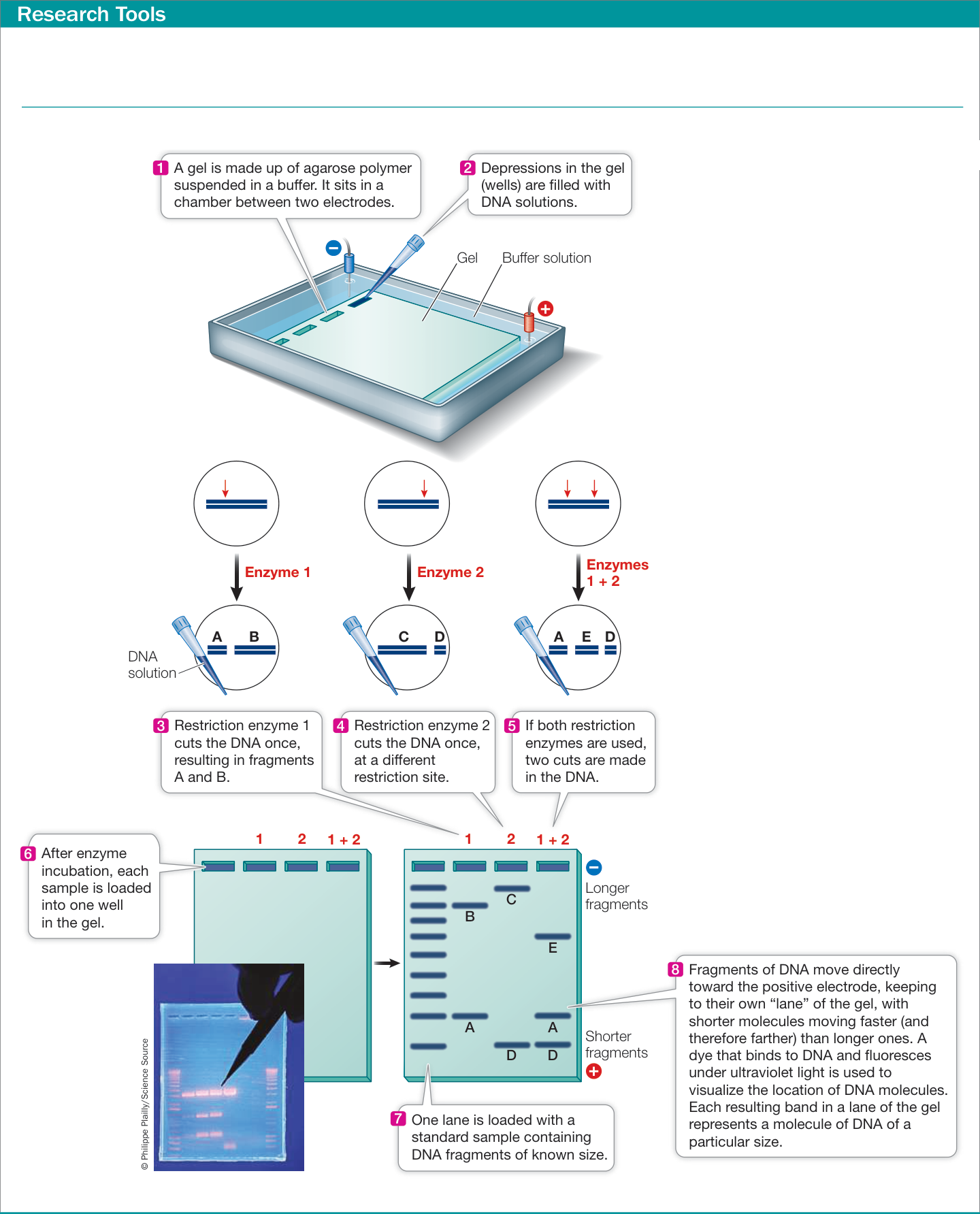
Figure 15.15 Separating Fragments of DNA by Gel Electrophoresis A mixture of DNA fragments is placed in a gel, and an electric field is applied across the gel. The negatively charged DNA moves toward the positive end of the field, with smaller molecules moving faster than larger ones. After minutes to hours for separation, the electric power is shut off and the separated fragments can be analyzed.
DNA fingerprinting combines PCR with restriction analysis and electrophoresis
The methods we have just described are used in DNA fingerprinting, which identifies individuals based on differences in their DNA sequences. DNA fingerprinting works best with sequences that are highly polymorphic—that is, sequences that have multiple alleles (because of many mutations during the evolution of the organism) and are therefore likely to be different in different individuals. Two types of polymorphisms are especially informative:
Single nucleotide polymorphisms (SNPs; pronounced “snips”) are nucleotide base pairs at which point mutations have become sufficiently frequent in a population to be readily detected. A polymorphic base pair is referred to as a SNP locus, or simply SNP, and the different base-pairs are termed SNP alleles. SNPs are numerous within genomes and have been mapped for many organisms. If one parent is homozygous for the A-T allele at a particular SNP locus in the genome, and the other parent is homozygous for G-C, the offspring will be heterozygous: one chromosome will have the A-T allele and the other will have the G-C allele. A SNP is often referred to with reference to only one of the two DNA strands—in this example in which one allele is A-T and the other G-C, the SNP is referred to as an A/G SNP or, equivalently, a T/C SNP. If a SNP occurs in a restriction enzyme recognition site, such that one variant is recognized by the enzyme and the other isn’t, then individuals can be distinguished from one another very easily using the polymerase chain reaction (PCR; see Key Concept 13.5). A fragment containing the polymorphic sequence is amplified by PCR from samples of total DNA isolated from each individual. The fragments are then cut with the restriction enzyme and analyzed by gel electrophoresis.
Short tandem repeats (STRs) are short, repetitive DNA sequences that occur side by side (in tandem) on the chromosomes, usually in the noncoding regions. These repeat patterns, which contain one to five base pairs, are also inherited. For example, at a particular locus on chromosome 15 there may be an STR of “AGG.” An individual may inherit an allele with six copies of the repeat (AGGAGGAGGAGGAGGAGG) from her mother and an allele with two copies (AGGAGG) from her father. Again, PCR is used to amplify DNA fragments containing these repeated sequences, and then the amplified fragments, which have different sizes because of the different numbers of the repeats, are distinguished by gel electrophoresis (Figure 15.16A).
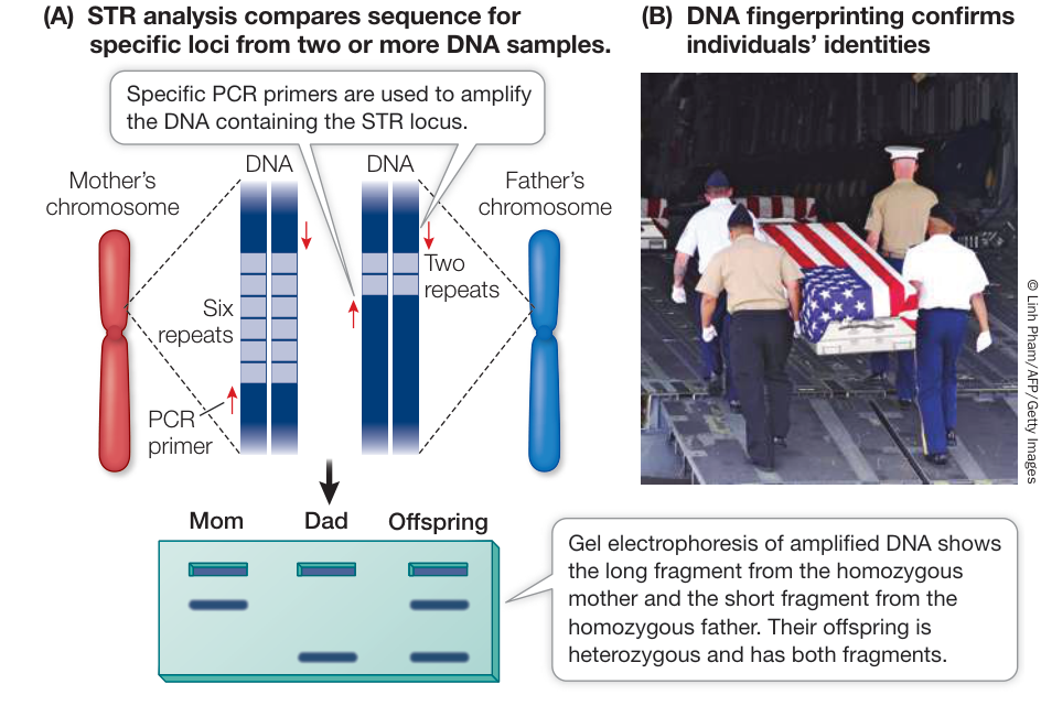
Figure 15.16 DNA Fingerprinting with Short Tandem Repeats (A) A particular STR locus can be analyzed to determine the number of repeat sequences that were inherited by an individual from each biological parent. The two alleles can be identified in an electrophoresis gel on the basis of their sizes. When several STR loci are analyzed, the pattern can constitute a definitive identification of an individual. (B) Remains of soldiers are positively identified by comparing their STR genotypes to relatives. Q: Should everyone have their DNA fingerprinted at birth to create a “genetic ID” for use in screening for genetic diseases? What would be the advantages and disadvantages of this?
Connect the Concepts As described in Key Concept 13.5, PCR is a technique used to make multiple copies of a DNA sequence—that is, to amplify it. The process essentially automates DNA replication; over many cycles the amount of DNA can be increased exponentially.
The method of DNA fingerprinting used most commonly today relies on STR analysis. The Federal Bureau of Investigation in the United States currently uses 20 STR loci in its Combined DNA Index System (CODIS) database (Table 15.3). An analysis of these loci in your DNA would reveal your particular DNA fingerprint. Looking at Table 15.3, you might inherit:
- From your mother: allele 72 from chromosome 4; allele 23 from chromosome 7; allele 14 from chromosome 11; and allele 12 from chromosome 18
Table 15.3 Four of the Genetic Loci Used for Identification in the CODIS Database
| Human chromosome | Locus name | Repeated sequence | Number of alleles |
|---|---|---|---|
| 4 | FGA | CTTT | 98 |
| 7 | D7S820 | GATA | 32 |
| 11 | TH01 | AATG | 21 |
| 18 | D18S51 | AGAA | 55 |
Source: https://strbase.nist.gov/fbicore.htm
- From your father: allele 56 from chromosome 4; allele 22 from chromosome 7; allele 16 from chromosome 11; and allele 12 from chromosome 18
Note that in this case you are heterozygous for the alleles on three of the chromosomes and homozygous for the allele on chromosome 18. With the large number of alleles at each locus and 20 loci, the probability of two people sharing the same alleles across all the loci is very small. For example, if all 20 STR loci had 20 equally frequent alleles, the probability of two unrelated individuals showing an exact match would be about , which is less than 1 in . So a DNA sample from a crime scene can be used to uniquely identify the individual who left the sample at the scene. This implies that a DNA sample can be used to uniquely identify an individual. This ability has been useful in criminal cases both to identify perpetrators and to exonerate previously convicted individuals, sometimes many years after their conviction. In addition, STR loci are often used to identify human remains. In these cases, identification involves comparing the STR fingerprint of the unknown individual to possible relatives, such as parents or siblings, who are expected to carry the same alleles (Figure 15.16B)
DNA analysis with genetic markers such as SNPs and STRs has applications throughout all areas of biological research. For example, these markers are used to analyze the organization of genomes, to identify species or individuals within species, to compare species or organisms to see how closely related they are, and to analyze particular genes and the phenotypes associated with them. In the remainder of this chapter we focus on the use of these markers and other technologies that are used to study and treat genetic diseases.
DNA analysis can be used to identify mutations that lead to disease
With diseases such as PKU and sickle-cell anemia, you have seen that the clinical phenotypes of inherited diseases could be traced to individual proteins, and that the genes could then be identified. For example, in sickle-cell anemia, the protein abnormality in hemoglobin was described first (a single amino acid change), and then the gene for -globin was isolated and the DNA mutation was pinpointed:
Clinical phenotype protein phenotype gene
With the advent of new ways to identify DNA variations, a new pattern of human genetic analysis emerged. In these cases, the clinical phenotype is first related to a DNA variation, and then the gene and protein involved are identified. For cystic fibrosis (see Table 15.2), a mutant version of the gene CFTR was isolated first, and then the protein was characterized:
Clinical phenotype gene protein phenotype
Whichever approach is used, final identification of the protein(s) involved in a disease is important in designing specific therapies.
Genetic markers can be used to find disease-causing genes
Identifying a mutant gene requires finding a location on the chromosome that is closely linked to the gene of interest, a process called linkage analysis. The reference locations for gene isolation are genetic markers. Genetic markers such as STRs and SNPs can be used as landmarks to find a gene of interest, if the gene also has multiple alleles (for example, normal and disease-causing alleles). The key to this method is the well-established observation that if two genes are located near each other on the same chromosome, they are usually passed on together from parent to offspring (see Key Concept 12.4). The same holds true for any pair of DNA genetic markers. In the case of linkage analysis, the idea is to find markers that are progressively closer to the gene of interest.
As you have seen, SNPs and STRs are widespread in eukaryotic genomes. There is roughly one SNP for every 1,330 base pairs in the human genome, and any regions of the genome also contain repetitive DNA sequences such as those found in STRs. SNPs and STRs can be analyzed using the PCR techniques mentioned earlier. SNPs can also be detected using sophisticated chemical methods such as mass spectrometry.
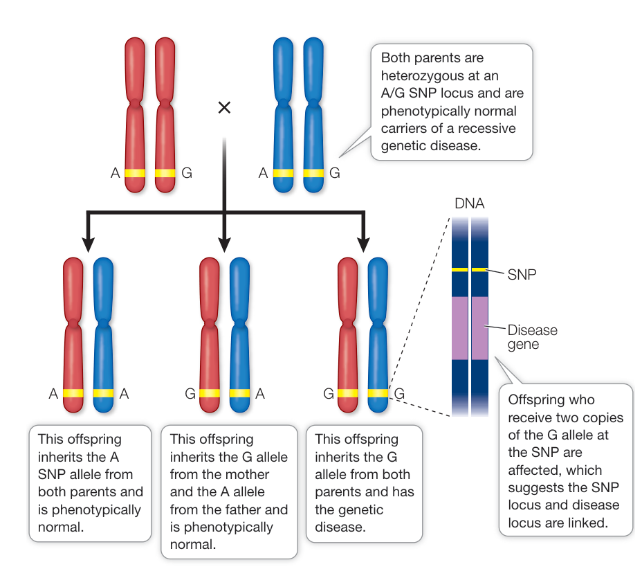
Figure 15.17 DNA Linkage Analysis Linkage of an SNP to a disease-causing allele in many families narrows down the location of the defective gene, making its isolation and identification possible. In the example shown here, the disease locus is linked to an A/G SNP, with the disease-causing recessive allele linked to the G (or G-C base pair) SNP. Q: In this example the G allele at this A/G SNP is associated with the disease-causing allele. Do you think this will also be the case in other families?
To narrow down the location of a gene, a scientist must find a genetic marker that is always inherited with the gene. To do this, family medical histories are taken and pedigrees are constructed. If a genetic marker and a genetic disease are inherited together in many families, then they must be near each other on the same chromosome (Figure 15.17). This situation recalls the conclusion reached in the classic studies of inheritance undertaken by Thomas Hunt Morgan and discussed in Key Concept 12.4: two genes do not always assort independently. Alleles at loci that are “linked” on the same chromosome will be more often inherited together—especially when the two loci are close to one another on the chromosome.
Linkage narrows down the position of a gene on a chromosome to a few hundred thousand base pairs. Once a linked DNA region is identified, many methods are available to identify the actual gene responsible for a genetic disease. The complete sequence of the region can be searched for candidate genes, using information available from databases of genome sequences. With luck a scientist can make an educated guess, based on biochemical or physiological information about the disease, along with information about the functions of candidate genes, as to which gene is responsible for the disease. The identification of DNA polymorphisms within candidate genes that correlate with the presence or absence of disease can also help narrow down the search. A variety of techniques, such as analyzing mRNA levels of candidate genes in diseased and healthy individuals, are used to confirm that the correct gene has been identified.
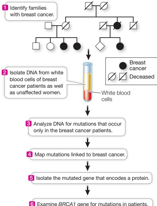
The isolation of the BRCA1 gene that is involved in breast cancer (described in the chapter opening) offers a good illustration of molecular techniques used to identify genes associated with disease, and is described in Investigating Life: How Was the BRCA1 Gene Identified? Analysis of the BRCA1 mutations came after Mary-Claire King of the University of California at Berkeley
▶ Investigating LIFE How Was the BRCA1 Gene Identified?
Experiment
Original Paper: Y. Miki et al. 1994. A strong candidate for the breast and ovarian cancer susceptibility gene BRCA1. Science 266: 66–71.
DNA linkage analysis of families led to the isolation of BRCA1, a gene that can lead to breast cancer.
HYPOTHESIS A gene mutated in hereditary breast cancer can be isolated.
METHOD
RESULTS
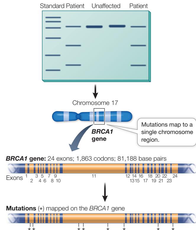
CONCLUSION The BRCA1 gene is mutated in people with hereditary breast cancer.
Investigating LIFE Work with the Data follows on the next page.
Work with the Data
About one in ten cases of breast cancer is hereditary. That is, the cancer arises from a germ line mutation (meaning it occurs in all cells of the body) that is expressed in breast cells as cancer. The other nine in ten cases come about from somatic mutations in breast cells. The identification of a gene involved in hereditary breast cancer was made by DNA analysis. First, the location of the gene was narrowed down by analysis of SNPs in families in which several women had breast cancer (see the experiment). Then a team led by Mark Skolnick at the University of Utah and the company Myriad Genetics sequenced the DNA in the candidate region and looked for a sequence that had a promoter, transcription terminator, start and stop codons, and sequences at the ends of introns—in short, the sequences that mark a protein-coding gene. Once they found the candidate gene, they subjected it to a series of tests to see if it was the one associated with breast cancer.
QUESTIONS►
- Hereditary (as opposed to somatic) breast cancer is often suspected when the cancer develops relatively early in life. Hereditary cancers tend to arise earlier because they involve a germ line mutation present in all cells at birth, whereas a cancer due to a spontaneous somatic mutation in the DNA of a breast cell can happen any time (statistically, later). Skolnick’s team examined the timing of breast cancer cases in families of patients who had different mutations in BRCA1. The results are shown in Table A. What conclusions can you draw concerning whether cancer was hereditary in these families?
Table A
| Family | Total cases | Cases before age 50 |
|---|---|---|
| A | 31 | 20 |
| B | 22 | 14 |
| C | 10 | 7 |
- DNA sequencing evaluated BRCA1 in the cancer patients and their families, as well as in controls who did not have breast cancer themselves or in their family. The results are shown in Table B. Were the mutations in BRCA1 present only in the breast cancer patients? What do you think the effects of the mutations were in each of the three families?
Table B
| Family | Codon number in BRCA1 | Change in patients | Change in controls? |
|---|---|---|---|
| A | 1313 | C → T | None (0/170) |
| B | 1775 | T → G | None (0/120) |
| C | 24 | Deletion 11 bp | None (0/180) |
- Human tissues from people without breast cancer were examined by nucleic acid hybridization (see Figure 14.7) for the level of BRCA1 mRNA. The results are shown in Table C. What can you conclude about the kinds of cancer that might be caused by mutations in BRCA1?
Table C
| Tissue | mRNA detected? |
|---|---|
| Breast | Yes |
| Colon | No |
| Intestine | No |
| Ovary | Yes |
| Prostate | No |
| Spleen | No |
| Testis | Yes |
| Thymus | Yes |
worked with breast cancer patients and their families to identify SNPs closely linked to the breast cancer phenotype.
Recap and Assess
DNA molecules can be cut into fragments by restriction digestion and then sorted by gel electrophoresis. PCR is used to amplify sequences of interest from complex samples. These techniques are used in DNA fingerprinting to analyze DNA polymorphisms for the purpose of identifying individuals. Genes involved in disease can be identified by linkage analysis with markers, especially SNPs and STRs, to identify candidates, followed by confirmation using protein and expression differences between healthy and diseased individuals.
A DNA molecule is 100,000 base pairs in length. Assuming all four base pairs are equally frequent and the sequence is random, how many six-base-pair EcoRI restriction sites do you expect to be present?
Cystic fibrosis is an autosomal recessive disease in which thick mucus is produced in the lungs and airways. The gene responsible for this disease encodes a protein composed of 1,480 amino acids. In most patients with cystic fibrosis, the protein has 1,479 amino acids: a phenylalanine is missing at position 508. A baby is born with cystic fibrosis. He has an older brother who is not affected. How would you test the DNA of the older brother to determine whether he is a carrier for cystic fibrosis?
How can a gene mutation that causes a disease be mapped and detected before its protein product is known?
The determination of the precise molecular phenotypes and genotypes of various human genetic diseases has made it possible to diagnose these diseases even before symptoms appear. Let’s take a detailed look at some of these genetic screening techniques.
Genetic Screening Is Used to Detect Diseases
Learning Objectives
15.4.1 Compare and contrast phenotypic and genotypic genetic screening.
15.4.2 Explain how DNA hybridization is used for genetic screening.
Genetic screening is the use of a test to identify people who have, are predisposed to, or are carriers of a genetic disease. It can be done at many times of life and used for many purposes:
- Prenatal screening can be used to identify an embryo or fetus with a disease so that medical intervention can be applied or decisions can be made about whether or not to continue the pregnancy.
- Newborn babies can be screened so that proper medical intervention can be initiated quickly for those babies who need it.
- Asymptomatic people who have relatives with a genetic disease can be screened to determine whether they are carriers of the disease-associated allele or are likely to develop the disease themselves.
Genetic screening can be done at the level of either the phenotype or the genotype.
Genetic screening can be done by examining the phenotype
Genetic screening can involve examining a protein or other chemical that is relevant to a phenotype associated with a particular disease. Perhaps the best example is the test for phenylketonuria (PKU), which has made it possible to identify the disease in newborns, so that treatment can be started immediately. It is very likely that you were screened as a newborn for PKU.
Newborn babies with PKU have a normal phenotype because phenylalanine is not yet elevated in their blood; before birth, excess phenylalanine in the fetal blood diffuses across the placenta and into the mother’s circulatory system. Since the mother is almost always heterozygous, and therefore has adequate phenylalanine hydroxylase activity, her body metabolizes the excess phenylalanine from her fetus. After birth, however, phenylalanine begins to accumulate in the baby’s blood. After a few days, the phenylalanine level may be ten times higher than normal. Within days, the developing brain is damaged, and untreated children with PKU become severely intellectually disabled. But if detected early, PKU can be treated with a special diet low in phenylalanine to avoid the brain damage that would otherwise result. Thus early detection is imperative.
Routine newborn screening for PKU and other diseases began in 1963 with the development of a simple, rapid test for the presence of excess phenylalanine in blood serum (Figure 15.18). This method uses drops of blood from newborn babies and can be automated so that a screening laboratory can process many samples in a day. Newborn babies’ blood is now screened for up to 35 genetic diseases. Some are relatively common, such as congenital hypothyroidism, which occurs about once in 4,000 births and causes reduced growth and intellectual disability because of low levels of thyroid hormone. With early intervention, many of these infants can be successfully treated. So it is not surprising that newborn screening is required in many countries, including the United States and Canada. You were probably screened for several genetic diseases in the first days of your life. Ask your parents which ones!
DNA testing is the most accurate way to detect genetic disease
The level of phenylalanine in the blood is an indirect measure of phenylalanine hydroxylase activity in the liver. But how can we screen for genetic diseases that are not detectable by blood tests? What if blood is difficult to obtain, as it is in a fetus? How are genetic abnormalities in heterozygotes, who express the normal protein at some level, identified?
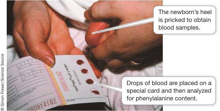
Figure 15.18 Phenotypic Screening of Newborns for Phenylketonuria A blood test is used to screen newborns for phenylketonuria. Small samples of blood are taken from a newborn’s heel. The samples are placed in a machine that measures the phenylalanine concentration in the blood. Early detection means that the symptoms of the condition can be prevented by putting the baby on a therapeutic diet.
Connect the Concepts Genetic testing is a rapidly changing field, with tests being done at different stages of prenatal development. See Key Concept 42.2 for an overview of the stages of human development, to place the timing of genetic tests in perspective.
DNA testing is the direct analysis of DNA for a mutation, and it offers the most direct and accurate way of detecting an abnormal allele. Now that the mutations responsible for many human diseases have been identified, any cell in the body can be examined at any time of life for mutations. The amplification power of PCR means that only one or a few cells are needed for testing. These methods work best for diseases caused by only one or a few different mutations.
Consider, for example, two parents who are both heterozygous for the cystic fibrosis allele but who want to have a healthy child. If treated with the appropriate hormones, the mother can be induced to “superovulate,” releasing several eggs. Eggs can be harvested and artificially inseminated. The fertilized eggs can be allowed to divide to the eight-cell stage. If one of these eight embryonic cells is removed, it can be tested for the presence of the cystic fibrosis allele. If the test is negative, the remaining seven-cell embryo can be implanted in the mother’s womb, where it will develop normally.
Such preimplantation screening is performed only rarely. More typical are analyses of fetal cells after normal fertilization and implantation in the womb. Fetal cells or even DNA that has leaked out of dead cells can be analyzed at about the tenth week of pregnancy by a procedure termed chorionic villus sampling, or during the thirteenth to seventeenth weeks by amniocentesis. In either case, only a few fetal cells are necessary to perform DNA testing. Recently, very sensitive methods were developed so that DNA testing can be done with the few fetal cells that are released into the mother’s blood. A 10-milliliter blood sample from a pregnant woman has enough fetal cells for the analysis of many disorders, including Down syndrome and cystic fibrosis. This relatively noninvasive procedure could replace amniocentesis and chorionic villus sampling—which both carry a slight risk of causing a miscarriage—in the near future.
DNA testing can also be performed with newborns. The blood samples used for screening for PKU and other disorders contain enough of the baby’s blood cells to permit DNA analysis using PCR-based techniques. DNA analysis is now being used to screen for sickle-cell disease and cystic fibrosis; similar tests for other diseases will surely follow.
Among the numerous methods available for testing for a mutant sequence of DNA is DNA hybridization, and in particular allele-specific oligonucleotide hybridization. As you saw in Figure 14.7, nucleic acid hybridization can be used to detect the presence of a specific DNA sequence, such as a sequence containing a particular mutation. Samples of DNA are collected from people who may or may not carry the mutation, and PCR is used to amplify the region of DNA where the mutation may occur. Short synthetic DNA strands called oligonucleotide probes are hybridized with the denatured PCR products. The probe is labeled in some way (e.g., with radioactivity or a fluorescent dye) so that hybridization can be readily detected. This technique is illustrated in Figure 15.19 using sickle-cell anemia as an example.
Detection of a mutation by DNA screening can be used for diagnosis of a genetic disease, so that appropriate treatment can begin. In addition, DNA screening provides a person with important information about his or her genome.
Recap and Assess
Genetic screening can be used to identify people who have, are predisposed to, or are carriers of genetic diseases. Screening can be done at the phenotype level by identifying an abnormal protein such as an enzyme with altered activity. It can also be done at the genotype level by direct testing of DNA.
- What is the advantage of screening for genetic mutations by allele-specific oligonucleotide hybridization relative to screening phenotype differences in enzyme activity?
- Explain how allele-specific oligonucleotide hybridization could be used to perform genetic screening for sickle-cell anemia.
Ongoing research has resulted in the development of increasingly accurate diagnostic tests and a better understanding of various genetic diseases at the molecular level. This knowledge is now being applied to the development of new treatments for genetic diseases. In the next key concept we survey various approaches to treatment, including modifications of the mutant phenotype and gene therapy, in which the normal version of a mutant gene is supplied.
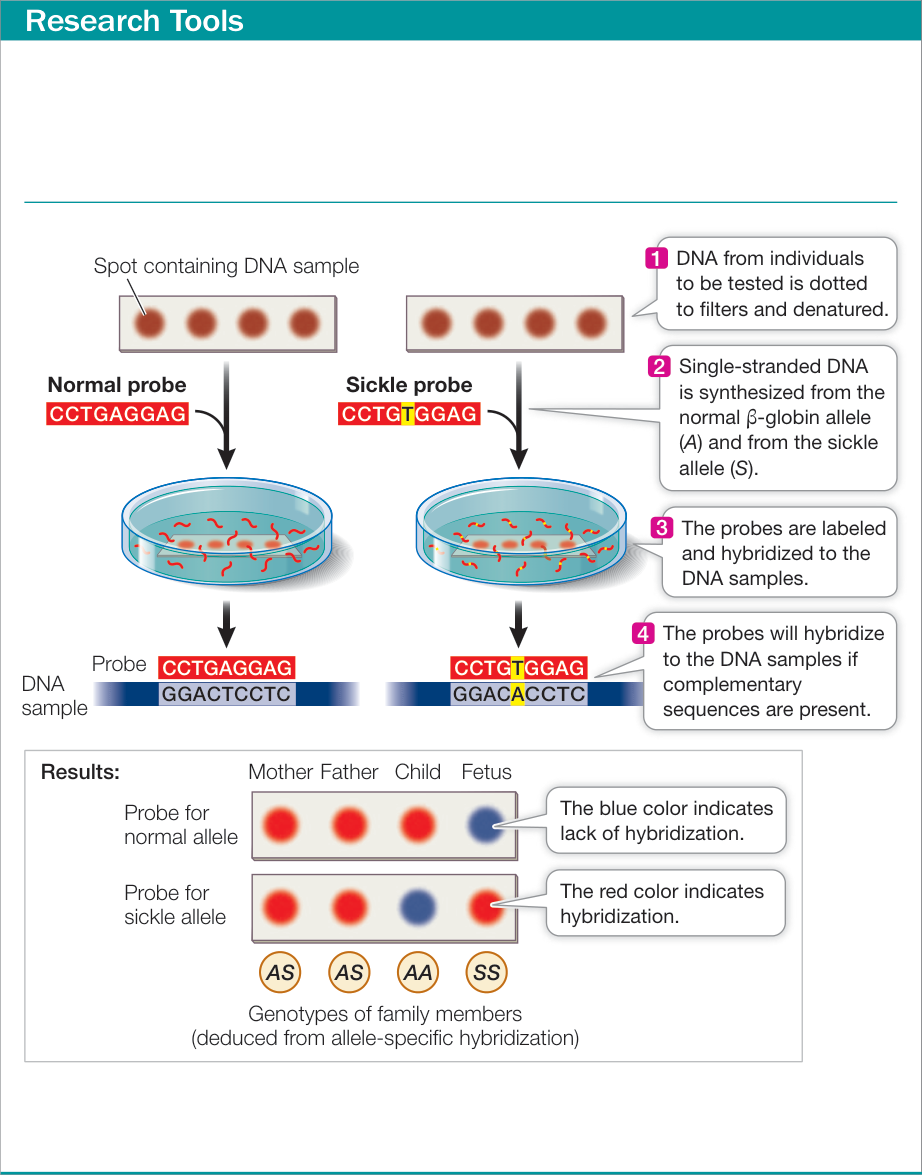
Figure 15.19 DNA Testing by Allele-Specific Oligonucleotide Hybridization Testing of this family reveals that the two biological parents are heterozygous carriers of the sickle allele. The first child has inherited two normal alleles and is neither affected by the disease nor a carrier. The unborn fetus has inherited two mutant alleles and will be affected by sickle cell disease.
Genetic Diseases Can Be Treated
Learning Objectives
- 15.5.1 Explain how a genetic disease can be treated by modifying the disease phenotype.
- 15.5.2 Define gene therapy and compare ex vivo and in vivo approaches to gene therapy.
Most treatments for genetic diseases simply try to alleviate the patient’s symptoms. But to effectively treat these diseases—whether they affect all cells, as in inherited disorders such as PKU, or only somatic cells, as in cancer—physicians must be able to diagnose the disease accurately, understand how the disease works at the molecular level, and intervene early, before the disease ravages or kills the individual. There are two main approaches to treating genetic diseases:
- Modifying the disease phenotype
- Replacing the defective gene (modifying the genotype)
Genetic diseases can be treated by modifying the phenotype
Altering the phenotype of a genetic disease so that it no longer harms an individual is commonly done in one of three ways: by restricting the substrate of a deficient enzyme, by inhibiting a harmful metabolic reaction, or by supplying a missing protein product (Figure 15.20).
RESTRICTING THE SUBSTRATE Restricting the substrate of a deficient enzyme is the approach taken when a newborn is diagnosed with PKU. In this case, the deficient enzyme is phenylalanine hydroxylase, and the substrate is phenylalanine (see Figure 15.8). The infant’s inability to break down phenylalanine in food leads to a buildup of the substrate, which causes the clinical symptoms. So the infant is immediately put on a special diet that contains only enough phenylalanine for immediate use. Lofenalac®, a milk-based product that is low in phenylalanine, is fed to these infants just like formula. Later, certain fruits, vegetables, cereals, and noodles low in phenylalanine can be added to the diet. Meat, fish, eggs, dairy products, and bread, which contain high amounts of phenylalanine, must be avoided, especially during childhood, when brain development is most rapid. The artificial sweetener aspartame must also be avoided because it is made of two amino acids, one of which is phenylalanine.
People with PKU are generally advised to stay on a low-phenylalanine diet for life. Although maintaining these dietary restrictions may be difficult, it is effective. Numerous follow-up studies since newborn screening was initiated have shown that people with PKU who stay on the diet are no different from the rest of the population in terms of mental ability. This is an impressive achievement in public health, given the severity of intellectual disability in untreated patients.
METABOLIC INHIBITORS In Key Concept 11.7 we described how drugs that are inhibitors of various cell cycle processes are used to treat cancer. Drugs are also used to treat the symptoms of many genetic diseases. As biologists have gained insight into the molecular characteristics of these diseases and the specific proteins involved, a more specific approach to treatment is taking shape. Targeted therapies are being developed, especially for cancer, and some have resulted in life-saving interventions. This is a major area of applied research.
SUPPLYING THE MISSING PROTEIN An obvious way to treat a disease caused by the lack of a functional protein is to supply that protein. This approach is used to treat hemophilia A, a disease in which blood factor VIII is missing and blood clotting is impaired (see Table 15.2). In the past, the missing protein was obtained from blood. Sometimes, however, blood carries contaminants, such as viruses (e.g., HIV) or other pathogens that could harm the recipient. Now human clotting proteins are produced by recombinant DNA technology (see Chapter 18), making it possible to provide the protein in a much purer form.
Unfortunately, the phenotypes of many diseases caused by genetic mutations are very complex. In these cases, simple interventions like those we have just described do not work. Indeed, a recent survey of 351 diseases caused by single-gene mutations showed that current therapies increased patients’ life spans by an average of only 15%.
Gene therapy offers the hope of specific treatments
If a cell lacks an allele that encodes a functional product, an optimal treatment would be to provide a functional allele. The objective of gene therapy is to add a new gene that will be expressed in appropriate cells in a patient. What cells should be targeted? There are two approaches:
- Germ line gene therapy: The new gene is inserted into a gamete (usually an egg) or the fertilized egg. In this case, all cells of the adult will carry the new gene. Ethical considerations preclude the use of this method in humans.
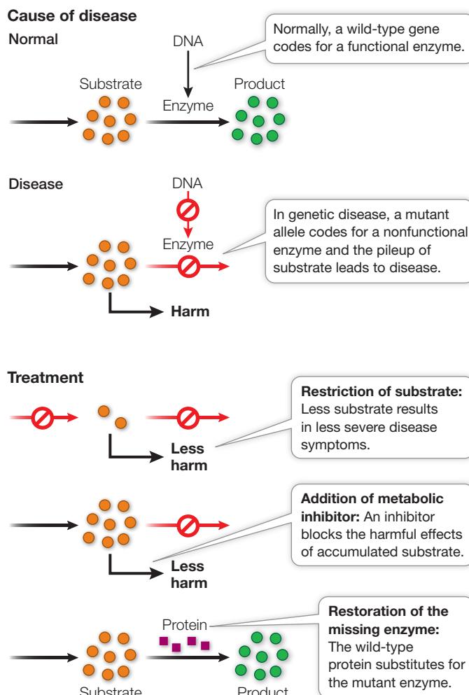
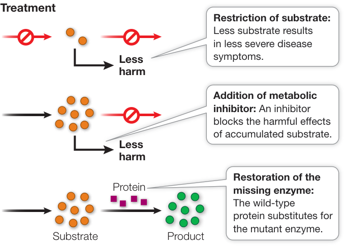
Figure 15.20 Strategies for Treating Genetic Diseases
2. Somatic cell gene therapy: The new gene is inserted into somatic cells involved in the disease. This method is being tried for numerous diseases, ranging from inherited genetic disorders to cancer.
There are two approaches to somatic cell gene therapy:
- 1. Ex vivo gene therapy: Target cells are removed from the patient, given the new gene, and then reinserted into the patient. This approach is being used, for example, for diseases caused by defects in genes that are expressed in white blood cells.
- 2. In vivo gene therapy: The gene is inserted directly into a patient, targeted to the appropriate cells. An example is a treatment for lung cancer in which a solution with a therapeutic gene is squirted onto a tumor.
Armed with knowledge of how genes are expressed (see Chapter 14) and regulated (see Chapter 16), physicians can design a therapeutic gene that contains not only a normal protein-coding sequence but also other sequences—such as an appropriate promoter—required for the gene’s expression in targeted cells.
A major challenge has been getting the therapeutic gene into cells. Uptake of DNA into eukaryotic cells is a rare event, and once the DNA is inside a cell, its entry into the nucleus and expression are rarer still. One solution to these problems is to insert the gene into a carrier virus (a viral vector) that can infect human cells but has been altered genetically to prevent viral replication. An example is the DNA virus called adeno-associated virus, which has been widely used in human gene therapy clinical trials. This virus has a small genome that can be spliced into a human gene; infects most human cells, including nondividing cells such as neurons; is harmless to humans; does not provoke rejection by the immune system; and enters the cell nucleus, where its DNA with the new gene can be expressed.
Adeno-associated virus has been used successfully in human gene therapy targeting Parkinson’s disease (Figure 15.21). Parkinson’s disease is a neurological condition that affects about 1 in 200 persons in their 60s, and 1 in 25 persons in their 80s. Its symptoms include muscle stiffness and shaking and—in about half of the patients—dementia. Patients with Parkinson’s disease produce inadequate amounts of the neurotransmitter -aminobutyric acid (GABA) in a particular part of the brain due to degeneration of the neurons that normally produce GABA. Low GABA levels result in poor coordination of movement. Raising GABA levels would thus be a reasonable approach to treatment. Recognizing that synthesis of GABA is catalyzed by the enzyme glutamate decarboxylase, a team led by Dr. Andrew Feigin at the Feinstein Institute in New York packaged a glutamate decarboxylase gene into adeno-associated virus that was then injected into the affected part of the brain. After six months, the patients who received the virus-encapsulated gene had increased levels of GABA and significant improvement in their disease symptoms. Other diseases being treated with gene therapy using this virus include cystic fibrosis, hemophilia, and muscular dystrophy. While this looks exciting on paper, bear in mind that this type of therapy is very rare, at the frontier of medicine. It may be quite a while before we can go see our local gene therapist for a cure.
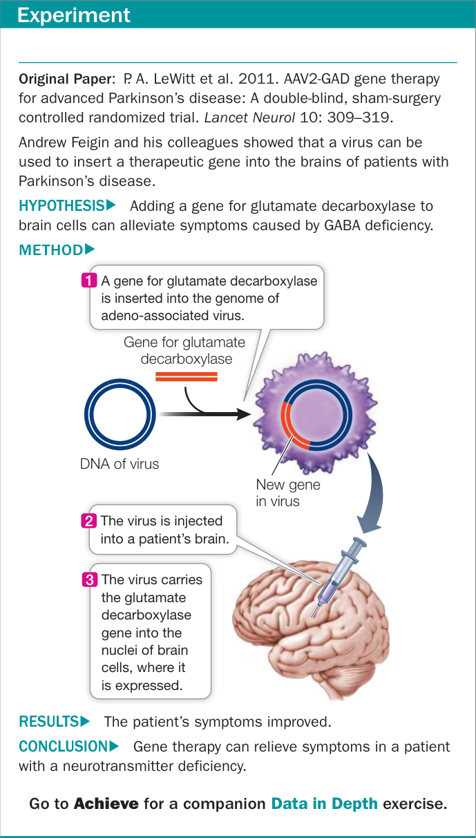
Figure 15.21 Gene Therapy
Recap and Assess
Treatment of a human genetic disease may involve an attempt to modify the abnormal phenotype by restricting the substrate of a deficient enzyme, inhibiting a harmful metabolic reaction, or supplying a missing protein. By contrast, gene therapy aims to address a genetic defect by inserting a normal allele into a patient’s cells.
- 1. How do metabolic inhibitors function in treating genetic diseases such as cancer?
- 2. How does in vivo gene therapy work? Can you give an example?
- 3. In the past, it was common for people with phenylketonuria (PKU) who were placed on a low-phenylalanine diet after birth to be allowed to return to a normal diet during their teenage years. Although the levels of phenylalanine in their blood were high, their brains were thought to be beyond the stage when they could be harmed. If a woman with PKU becomes pregnant, however, a problem arises. Typically, the fetus is heterozygous but is unable, at early stages of development, to metabolize the high levels of phenylalanine that arrive from the mother’s blood.
- Why is the fetus likely to be heterozygous?
- What do you think would happen to the fetus during this “maternal PKU” situation?
- What would be your advice to a woman with PKU who wants to have a child?
In this chapter you have learned about mutations, focusing on DNA changes that affect phenotypes through specific protein products. But there is much more to molecular genetics than the sequences of genes and proteins. Determining which genes will be expressed when and where is a major function of the genome. In Chapter 16 we will turn to gene regulation.
Investigating Life
QA What is the breast cancer gene, and what are the issues with DNA testing for cancer?
Angelina Jolie’s public announcement that she had elected to have her breasts removed in light of her cancer gene mutations attracted worldwide interest. In fact, the phrase “mutation” had never been so popular on internet search engines! Requests for BRCA1 testing from worried women have increased dramatically. Two issues have arisen: who should be tested, and who should perform the test? Because breast cancer is relatively common (women have about a 10% lifetime chance of having it), having several relatives who have had the disease is not unusual. Medical geneticists have come up with several criteria for defining when BRCA1 DNA analysis is warranted. These include having several close relatives who had or have the disease, with early onset (typically before age 50) and the presence of multiple tumors.
Even when a test shows no BRCA1 mutations in a woman with a family history of breast or ovarian cancer, we cannot say that breast or ovarian cancer may not arise. It turns out that BRCA1 mutations account for only about one in five cases of hereditary breast cancer. Other genes are involved; we just don’t know what they are, so we can’t analyze their DNA. Overall, hereditary breast cancer (a germ line mutation) accounts for only 10% of breast cancers. The other 90% arise from somatic mutation of breast cells.
As to who does the test, the BRCA1 gene was isolated by a private company that patented the DNA sequence. Until recently, that company was the only lab that could do BRCA1 analysis, and not surprisingly, the price was high. In 2013 the U.S. Supreme Court invalidated the patent, and now more labs can do it, and more cheaply.
Future directions
Are there genetic tests for other hereditary cancers? About 10% of all cancers are caused by inheritance of mutated oncogenes or tumor suppressor genes (as opposed to mutations that occur after birth). As with breast cancer, a person who inherits a cancer-causing mutation has a much higher chance of developing cancer than one who does not have that mutation. As whole genome DNA sequencing is developed (see Chapter 18), mutations involved with other types of hereditary cancers are being identified, and screening of relatives with a propensity for cancer is on the horizon.

© The Florida collection/Alamy Stock Photo
Visual Summary

You should be able to relate each summary to the adjacent figures. If you go to this visual summary in Achieve, you can follow links to figures, animations, activities, and simulations that will help you consolidate the material.
Mutations Are Heritable Changes in DNA
- ■ Point mutations can be transitions, transversions, or single base-pair insertions or deletions.
- ■ Point mutations in codons cause silent, missense, nonsense, loss-of-stop, and frame-shift mutations.
- ■ Point mutations in noncoding regions can have effects through changes in expression or splicing.
- ■ Large chromosomal rearrangements include deletions, duplications, inversions, and translocations.
- ■ Mutations are more likely in the presence of mutagens and can occur at hot spots.
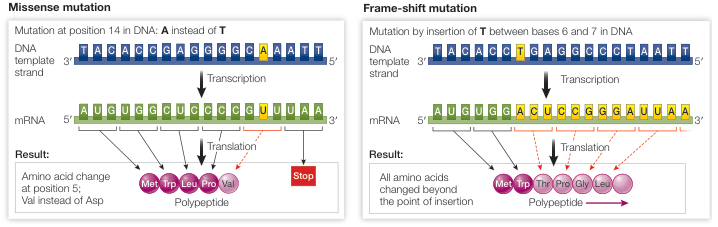
Figure 15.3
Questions
- For each of the following point mutations in a protein-coding gene, indicate whether the mutation is a transition or transversion and whether it is a silent, missense, nonsense, or loss-of-stop mutation. Mutation 1: UCA → ACA Mutation 2: UCC → UCU Mutation 3: UGA → UGG Mutation 4: UGG → UGA Mutation 5: GGC → GGG
- A cell culture is started from an organism possessing only two different chromosomes. Chromosome 1 contains eight regions, A—B—C—D—E—F—G—H, and chromosome 2 contains three regions, X—Y—Z. Over time, cells arise that carry different chromosomal rearrangements. Identify the type of chromosomal rearrangement for the following three cells: Cell 1: A—B—C—F—E—D—G—H and X—Y—Z; cell 2: X—D—E—F—G—H and A—B—C—Y—Z; cell 3: A—B—C—F—G—H and X—Y—Z.
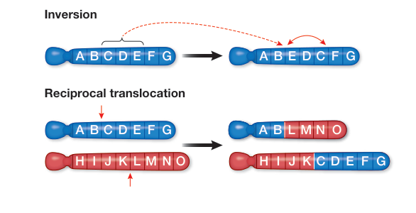
Figure 15.5
Mutations in Humans Can Lead to Diseases
- Many genetic diseases are caused by point mutations that alter only a single amino acid and render a protein nonfunctional.
- Some genetic diseases involve large deletions or chromosomal abnormalities.
- Multifactorial diseases arise from complex interactions between multiple genes and the environment.
Questions
- Explain why some missense mutations cause genetic disease whereas others do not.
- Genotyping of breast cancer tumors in women indicates that some women have two mutated alleles of the BRCA1 gene, even though there is no history of hereditary breast cancer in their families. How does this observation relate to BRCA1 being a cause of higher rates of breast cancer in some families?
- In expanding triplet repeat diseases, the incidence of the genetic disorder can increase over generations within a family. Why might this be the case?
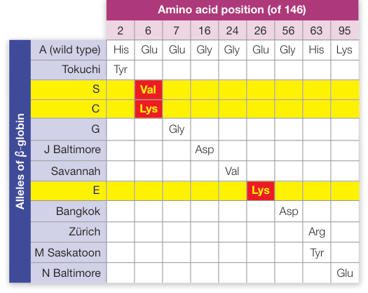
Figure 15.9
Mutations Can Be Detected and Analyzed
Go to Activity 15.1 and Animation 15.1
- Restriction enzymes are used to cut DNA into small fragments.
- Highly variable loci consisting of short tandem repeats (STRs) are used for DNA fingerprinting.
- A linked marker allows a disease-causing gene to be identified.

Figure 15.14 Diagram illustrating the process of detecting a mutation in DNA using a fingerprint marker. The initial state is . A downward arrow points to the mutation at the position, which is consistent with the linked marker. The final state is . A downward arrow points to the mutation at the position, which is consistent with the short tandem repeat. The final state is . The marker is a single blue bar at the position.
Questions
- PCR is used to amplify a 10,000-base-pair linear sequence of DNA
from an individual. When digested with EcoRI, the PCR product
gives two DNA molecules: one 3,000 and one 7,000 base pairs in length.
- How many restriction sites are on the original amplified DNA molecule?
- The same PCR is performed on a sample from another individual and the 10,000-base-pair sequence subjected to EcoRI digestion. After digestion, a single 10,000-base-pair molecule is obtained. How many restriction sites are on the original DNA molecule that was amplified from this individual?
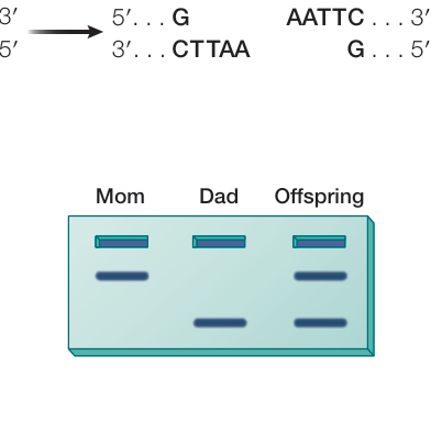
Figure 15.16
- The same PCR is performed on a sample from a third individual, and upon EcoRI digestion three different-sized pieces of DNA are obtained: 10,000, 7,000 and 3,000 base pairs. Given your answers to a and b, explain this result.
- DNA fingerprinting is often used in paternity cases. A man is trying to determine whether he is the father of a particular child. The genotypes for three STR loci are shown in the table. For each STR, the number of repeats in each allele is shown. Could the man be the father of the child?
| Locus | Mother | Child | Man |
|---|---|---|---|
| FGA | 28, 42 | 28, 40 | 31, 40 |
| TPOX | 9, 12 | 12, 15 | 13, 15 |
| VWA | 16, 22 | 16, 16 | 17, 18 |
Genetic Screening Is Used to Detect Diseases
- ■ Genetic screening for disorders can involve examination of a phenotype.
- ■ Genetic screening for disorders can involve examination of a genotype using DNA testing.
Questions
- What are the advantages of phenotypic versus genotypic screening for individuals who are heterozygous for a genetic disease?
- Genotypic screening sometimes involves hybridization of an individual’s DNA to allele-specific DNA sequences. DNA corresponding to both mutant and wild-type DNA is used. Why use both, rather than use just the mutant DNA sequence?
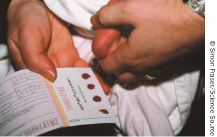
Figure 15.18
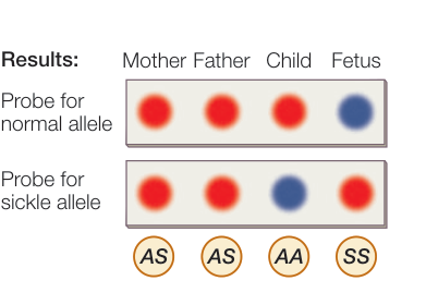
Figure 15.19
Genetic Diseases Can Be Treated
- ■ Some genetic disease can be treated by restricting a substrate, inhibiting an enzyme, or supplying a missing protein.
- ■ The goal of gene therapy is to alter an individual’s genotype in those cells where the expression of the mutant allele is causing problems.
Questions
- Type I diabetes is caused by an inability to make insulin. How might such a disorder be treated? Would the same strategy work for type II diabetes (insulin insensitive)?
- More genetic disorders of the bone marrow have been successfully treated using gene therapy than have disorders of other organs such as the lungs or liver. Why might that be the case?
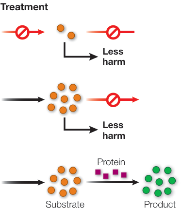
Figure 15.20
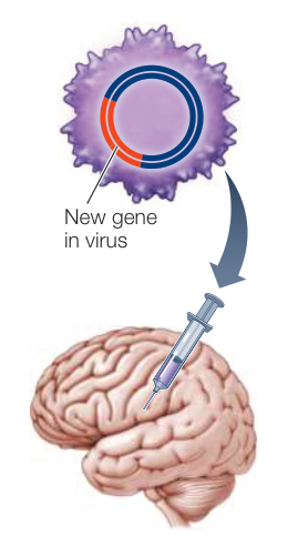
Figure 15.21