Chapter 16Regulation of Gene Expression
Key Concepts
16.1 Prokaryotic Gene Expression Is Regulated in Operons
16.2 Eukaryotic Gene Expression Is Regulated by Transcription Factors
16.3 Viruses Regulate Their Gene Expression during the Reproductive Cycle
16.4 Epigenetic Changes Regulate Gene Expression
16.5 Eukaryotic Gene Expression Can Be Regulated after Transcription
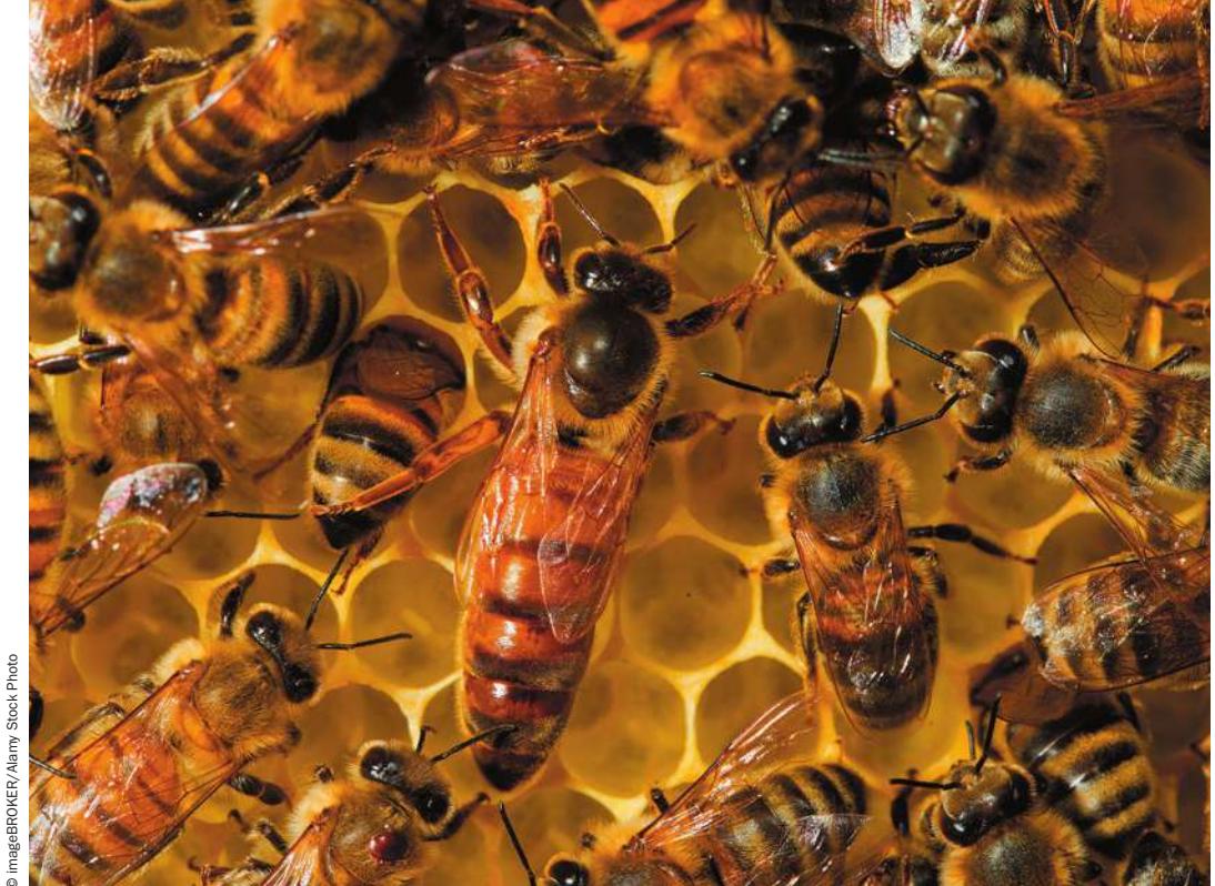
Worker bees care for a queen Gene expression differences underlie the differences in behavior of worker and queen bees.
© imageBROKER/Alany Stock Photo
Investigating Life: Gene Expression and Behavior
Any teenager or parent of a teenager will tell you that those years can be rough. But some young people have a rougher time than others. Teens whose mothers were extremely stressed during pregnancy often have more behavioral problems than those whose mothers had calmer pregnancies. A recent study led by psychologist Thomas Elbert and evolutionary biologist Axel Meyer at the University of Konstanz in Germany suggests an underlying genetic cause for these behavioral differences. The study examined a gene for the glucocorticoid receptor, which is involved in regulating hormonal responses to stress. The researchers found that teenagers whose mothers had suffered physical abuse during pregnancy had higher rates of cytosine methylation in the promoter of this gene than did teenagers whose mothers had not suffered such abuse.
A major control point for gene expression is the promoter, a sequence of DNA adjacent to the coding region of a gene where proteins bind and control the rate of transcription. The ability of these proteins to bind to the promoter is affected by the level of DNA methylation in the promoter. As we mentioned in Key Concept 15.1, in certain regions of DNA many of the cytosine residues have methyl groups added at their 5 positions, forming 5-methylcytosine. If a gene promoter has a high degree of methylation, some proteins that promote transcription can’t bind to it. Instead, other proteins bind to the methylated DNA and prevent expression of the gene. DNA methylation plays an important role in gene regulation and is a normal part of development. But the level of methylation can change over time and can vary among individuals, as was the case for the teenagers in Elbert and Meyer’s study. Their finding is interesting because it shows a correlation between maternal stress in humans and DNA methylation in their offspring.
Such studies have spawned the new field of behavioral epigenetics. Epigenetics is the study of heritable changes in gene expression that do not involve changes in the DNA sequence. These changes may underlie behaviors in other animals as well. A fascinating example is the well-known behavior of a queen bee, who “lords it over” her genetically identical worker sisters. Genes in the brains of these two types of bees have very different patterns of expression, and these in turn are caused by epigenetic changes.
QA Can epigenetic changes be manipulated? (Find the answer on page 372.)

© Oleksii Sergielev/Alany Stock Photo
Prokaryotic Gene Expression Is Regulated in Operons
Learning Objectives
- 16.1.1 Describe the conditions that cause high expression of genes of the lac and trp operons.
- 16.1.2 Describe the roles of structural or regulatory DNA sequences in the lac and trp operons.
- 16.1.3 Determine how mutations in structural or regulatory genes of the lac or trp operons would alter the phenotype of a cell.
- 16.1.4 Explain how sigma factors and promoter sequences are involved in regulating classes of genes in prokaryotes.
Prokaryotes conserve energy and resources by making certain proteins only when they are needed. The protein content of a bacterium can change rapidly when conditions warrant. Based on what you learned about gene expression in Chapter 14, you might suggest several ways in which a prokaryotic cell could shut off the supply of an unneeded protein. The cell could:
- decrease the rate of transcription of mRNA for that protein;
- hydrolyze the mRNA after it is made, preventing translation;
- prevent translation of the mRNA at the ribosome;
- hydrolyze the protein after it is made; or
- temporarily inhibit the function of the protein in the cell.
Whichever mechanism is used, it must be both responsive to environmental signals and efficient. The earlier the cell intervenes in the process of protein synthesis, the less energy it wastes making an unneeded protein. Selective blocking of transcription is far more efficient than transcribing the gene, translating the message, and then degrading or inhibiting the protein. While all five mechanisms for regulating protein levels are found in nature, prokaryotes generally use the most efficient one: transcriptional regulation.
Gene expression begins at the promoter (see Key Concept 14.3), where RNA polymerase binds to initiate transcription. Note, however, that in a given cell at a given point in time, not all promoters are active. This observation suggests that gene transcription must be selective. The “decision” regarding which genes to activate involves two types of regulatory proteins that bind to DNA: repressor proteins and activator proteins. In both cases, these proteins bind to DNA to regulate the gene (Figure 16.1):
- In negative regulation, binding of a repressor protein prevents transcription.
- In positive regulation, an activator protein binds DNA to stimulate transcription.
Connect the Concepts A key DNA sequence involved in regulation is the promoter, where transcription begins and various regulatory proteins bind. Learn details about proteins at the promoter in Key Concept 14.3.
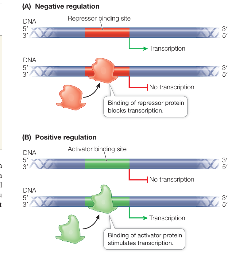
Figure 16.1 Positive and Negative Regulation Proteins regulate gene expression by binding to DNA and preventing or allowing RNA polymerase to bind DNA at the promoter region to control transcription of the gene. Q: Could a gene be under both positive and negative regulation?
and viruses. We’ll focus first on a regulatory system for the use of the sugar lactose.
Regulating gene transcription conserves energy
As a normal inhabitant of the human intestine, E. coli must be able to adjust to sudden changes in its chemical environment. Its host may present it with one foodstuff one hour (e.g., glucose in fruit) and another the next (e.g., lactose in milk). Such changes in nutrients present the bacterium with a metabolic challenge. Glucose is the easiest sugar for the bacterium to metabolize, and is its preferred energy source. Lactose is a -galactoside—a disaccharide containing galactose -linked to glucose (see Key Concept 3.3). Three proteins are upregulated by E. coli in response to an increase in lactose:
- -Galactoside permease is a carrier protein in the bacterial cell membrane that transports the sugar into the cell.
- -Galactosidase is an enzyme that hydrolyses lactose to glucose and galactose.
- -Galactoside transacetylase transfers acetyl groups from acetyl CoA to certain -galactosides. Its role in the metabolism of lactose is not clear.
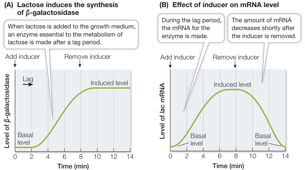
Figure 16.2 Lactose Induces the Expression of a Gene for an Enzyme (A) When the inducer lactose is added to the growth medium for the bacterium E. coli, the synthesis of β-galactosidase begins only after an initial lag period. (B) There is a lag period because the mRNA for β-galactosidase has to be made before the protein can be made. The amount of mRNA decreases rapidly after the lactose is removed, indicating that transcription is no longer occurring. These changes in mRNA levels indicate that the mechanism of induction by lactose is transcriptional regulation. Q: Why doesn’t the level of β-galactosidase drop following removal of the inducer, like the level of mRNA does? CHAPTER 16 Regulation of Gene Expression Figure 16.2 Lactose Induces the Expression of a Gene for an Enzyme (A) When the inducer lactose is added to the growth medium for the bacterium E. coli, the synthesis of -galactosidase begins only after an initial lag period. (B) There is a lag period because the mRNA for -galactosidase has to be made before the protein can be made. The amount of mRNA decreases rapidly after the lactose is removed, indicating that transcription is no longer occurring. These changes in mRNA levels indicate that the mechanism of induction by lactose is transcriptional regulation. Q: Why doesn’t the level of -galactosidase drop following removal of the inducer, like the level of mRNA does?
three proteins are extremely low—the cell does not waste energy and materials making the unneeded enzymes. But if the environment changes such that lactose is the predominant sugar available and very little glucose is present, the bacterium promptly begins making all three enzymes after a short lag period. While few molecules of -galactosidase (and the other two enzymes) are present in an E. coli cell in the presence of glucose, in the absence of glucose the addition of lactose can induce the synthesis of about 1,500 times more molecules of -galactosidase per cell (Figure 16.2A)!
What’s behind this dramatic increase? An important clue comes from measuring the amount of mRNA for -galactosidase. The mRNA level increases during the lag period after lactose is added to the medium, and this mRNA is translated into protein (Figure 16.2B). Moreover, the high mRNA level depends on the presence of lactose, because if the lactose is removed, the mRNA level goes down. The response of the bacterial cell to lactose is clearly at the level of transcription.
Compounds such as lactose that stimulate the synthesis of a protein are called inducers. The proteins that are produced are called inducible proteins, whereas proteins that are made all the time at a constant rate are called constitutive proteins.
We have now seen two basic ways of regulating the rate of a metabolic pathway. In Key Concept 8.5 we described the allosteric regulation of enzyme activity, which allows the rapid fine-tuning of metabolism. Regulation of protein synthesis—that is, regulation of the concentration of enzymes—is slower but results in greater savings of energy and resources. Protein synthesis is a highly endergonic process, since assembling mRNA, charging tRNA, and moving the ribosomes along mRNA all require the hydrolysis of nucleoside triphosphates such as ATP. Figure 16.3 compares these two modes of regulation.
Operons are units of transcriptional regulation in prokaryotes
The genes in E. coli that encode the three enzymes for using lactose are structural genes; structural genes specify the primary structures (the amino acid sequences) of protein molecules that act as enzymes or cytoskeletal proteins. The three genes are adjacent to one another on the E. coli chromosome. This arrangement is no coincidence: the genes share a single promoter, and their DNA is transcribed into a single, continuous molecule of mRNA. Because this particular mRNA governs the synthesis of all three lactose-metabolizing enzymes, either all or none of these enzymes are made, depending on whether their common message—their mRNA—is present in the cell.
A cluster of genes with a single promoter is called an operon, and the operon that encodes the three lactose-metabolizing enzymes in E. coli is called the lac operon. The lac operon promoter can be very efficient (the maximum rate of mRNA synthesis can be high), but mRNA synthesis is very low when the enzymes are not needed. In addition to having a promoter, an operon has other regulatory sequences (DNA sequences to which the protein products of regulatory genes bind) that are not transcribed. A typical operon consists of a promoter, an operator, and two or more structural genes (Figure 16.4). The operator is a short stretch of DNA that lies between the promoter and the structural genes. It can bind very tightly with regulatory proteins that either activate or repress transcription.
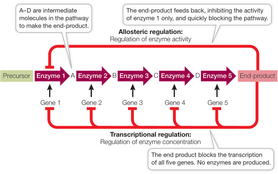
Figure 16.3 Two Ways to Regulate a Metabolic Pathway Feedback from the end-product of a metabolic pathway can block enzyme activity (allosteric regulation), or it can stop the transcription of genes that code for the enzymes in the pathway (transcriptional regulation).
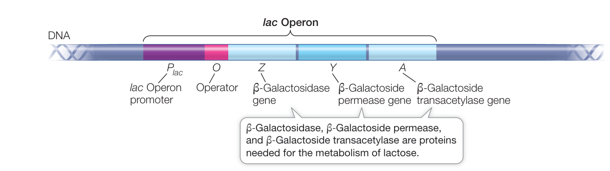
Figure 16.4 The lac Operon of E. coli The lac operon of E. coli is a segment of DNA that includes a promoter, an operator, and the three structural genes that code for lactose-metabolizing enzymes.
There are numerous mechanisms to control the transcription of operons; we describe three examples:
- An inducible operon regulated by a repressor protein
- A repressible operon regulated by a repressor protein
- An operon regulated by an activator protein
Operator-repressor interactions control transcription in the lac and trp operons
The lac operon contains a promoter, to which RNA polymerase binds to initiate transcription, and an operator, to which a repressor protein can bind. The gene that encodes this repressor is located near the lac operon on the E. coli chromosome. When the repressor is bound, transcription of the operon is blocked. This example of negative regulation was elegantly worked out by Nobel Prize winners François Jacob and Jacques Monod.
The repressor protein has two binding sites: one that binds to the operator DNA and the other that binds to the carbohydrate inducer. The environmental signal that induces the lac operon (for example, in the human digestive tract) is lactose, but the actual inducer is allolactose, a molecule that forms from lactose once it enters the cell. In the absence of the inducer, the repressor protein fits into the major groove of the operator DNA and recognizes and binds to a specific nucleotide base sequence. This prevents the binding of RNA polymerase to the promoter, and the operon is not transcribed (Figure 16.5A).
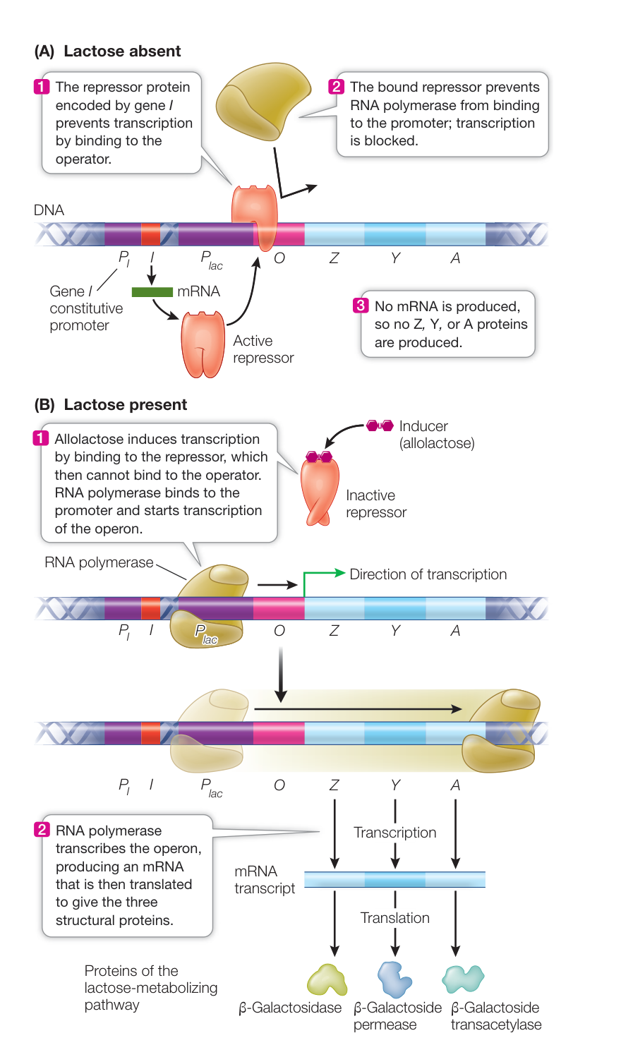
Figure 16.5 The lac Operon: An Inducible System (A) When lactose is absent, the synthesis of enzymes for its metabolism is inhibited. (B) Lactose leads to synthesis of the enzymes in the lactose-metabolizing pathway. The actual inducer is allolactose, which binds to the repressor protein and prevents its binding to the operator. Q: If part B of this figure were not labeled “Lactose present,” how could you determine that lactose is present?
When the inducer is present, it binds to the repressor and changes the shape of the repressor. This change in three-dimensional structure (conformation) prevents the repressor from binding to the operator. As a result, RNA polymerase can bind to the promoter and start transcribing the structural genes of the lac operon (Figure 16.5B).
You can see from this example that a key to transcriptional control of gene expression is the presence of regulatory sequences that do not code for proteins but are binding sites for regulatory proteins and other proteins involved in transcription.
In contrast to the inducible system of the lac operon, other operons in E. coli are repressible; that is, they are usually expressed but can be repressed under specific conditions. In such a system, the repressor is not normally bound to the operator. But if another molecule called a co-repressor binds to the repressor, the repressor changes shape and binds to the operator, thereby inhibiting transcription. An example is the trp operon, whose five structural genes catalyze the synthesis of the amino acid tryptophan. When tryptophan is absent, the repressor does not bind to the operator and the structural genes are transcribed and translated (Figure 16.6A). However, if tryptophan is present in the cell, it is advantageous to stop making the enzymes for tryptophan synthesis. To do this, the repressor binds to tryptophan (the co-repressor) and then binds to the operator in the trp operon, shutting off transcription (Figure 16.6B).
Let’s pause to summarize the differences between these two types of operons:
- In inducible systems, the substrate of a metabolic pathway (the inducer) interacts with a regulatory protein (the repressor), rendering the repressor incapable of binding to the operator and thus allowing transcription.
- In repressible systems, the product of a metabolic pathway (the co-repressor) binds to a regulatory protein (the repressor), which is then able to bind to the operator and block transcription.
Usually, inducible systems control catabolic pathways (which are turned on only when the substrate is available), whereas repressible systems control anabolic pathways (which are turned on until the concentration of the product becomes excessive). In both systems, the regulatory protein is a repressor that functions by binding to the operator. Next we consider an example of positive control involving an activator.
Connect the Concepts As discussed in Key Concept 8.1, there are two kinds of metabolism: Catabolic pathways break down complex molecules into simpler ones, releasing energy formerly stored in the chemical bonds. Anabolic pathways link simple molecules to form more complex molecules, a process that requires an input of energy.
Protein synthesis can be controlled by increasing promoter efficiency
In negative control, transcription is decreased in the presence of a repressor protein. E. coli can also use positive control to increase transcription through the presence of an activator protein. For an example we return to the lac operon, where the relative levels of glucose and lactose determine the amount of transcription. We have seen that in the presence of lactose the lac repressor is unable to bind to the lac operator to repress transcription (see Figure 16.5B). But glucose is the preferred source of energy for the cell, so if glucose and lactose levels are both high, the lac operon is still not transcribed efficiently. This is because efficient transcription of the lac operon requires binding of an activator protein to its promoter.
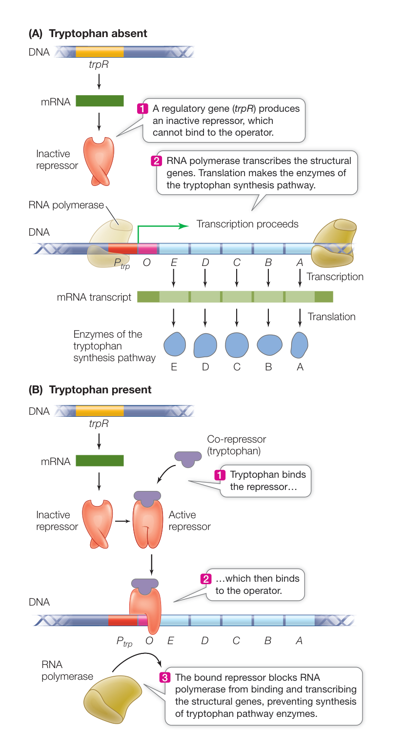
Figure 16.6 The trp Operon The tryptophan (trp) operon has a promoter, an operator, and five structural genes that encode the enzymes of the tryptophan synthesis pathway. A repressor protein, encoded by the trpR gene, produces an inactive repressor that becomes active when it binds to tryptophan and then is able to bind to the operator. The operon is thus transcribed when tryptophan is absent (A) but not when tryptophan is present (B).
Low levels of glucose in the cell set off a signaling pathway that leads to increased levels of the second messenger cyclic AMP (cAMP) (see Key Concept 7.3). Cyclic AMP binds to an activator protein called cAMP receptor protein (CRP), producing a conformational change in CRP that allows it to bind to the lac promoter. CRP is an activator of transcription, because its binding results in more efficient binding of RNA polymerase to the promoter, and thus increased transcription of the structural genes (Figure 16.7). In the presence of abundant glucose, cAMP levels are low, CRP does not bind to the promoter, and the efficiency of transcription of the lac operon is reduced. This is an example of catabolite repression, a system of gene regulation in which the presence of the preferred energy source represses other catabolic pathways. The mechanisms controlling positive and negative regulation of the lac operon are summarized in Table 16.1.
RNA polymerases can be directed to particular classes of promoters
Thus far we have described a promoter as a specific DNA sequence located upstream of a transcription initiation site. The promoter binds RNA polymerase so that the enzyme can then catalyze the synthesis of RNA from a gene-coding region of DNA. The promoter also orients the polymerase so that it transcribes the correct one of the two DNA strands for that gene. Not all promoters are identical, but they all have similar sequences by which they are recognized by the RNA polymerase and other proteins. Prokaryotic promoters generally have two sites for these recognition sequences, which begin 10 and 35 base pairs upstream of the transcription start site (the –10 element and the –35 element). Different classes of promoters have different recognition sequences at these two sites. The largest class consists of promoters for “housekeeping genes,” which are all the genes that are normally expressed in actively growing cells. In these genes, the –10 element is 5’-TATAAT-3’, and the –35 element is 5’-TTGACAT-3’ (N stands for any nucleotide):
5’-NNNTTGACATNNNNNNNNNNNNNNNNNNNNTATAATNNNNN*NNNNNNNNNNNN-3’ 3’-NNNAACTGTANNNNNNNNNNNNNNNNNNNNATATTANNNN*NNNNNNNNNNNN-5’ -35 Element -10 Element Transcription start site
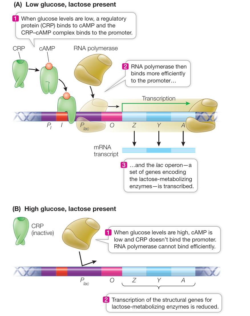
Figure 16.7 Catabolite Repression Regulates the lac Operon (A) The promoter for the lac operon binds RNA polymerase more efficiently in the presence of cAMP, as occurs when glucose levels are low. (B) High glucose levels thus lead to low levels of transcription of the lac operon.
Table 16.1 Positive and Negative Regulation in the lac Operon
| Glucose | cAMP levels | CRP regulator | Lactose | lac Repressor | RNA polymerase binding? | Transcription of lac genes? | Lactose used by cells? |
|---|---|---|---|---|---|---|---|
| Present | Low | Not bound to promoter | Absent | Bound to operator | Absent | No | No |
| Present | Low | Not bound to promoter | Present | Not bound to operator | Present, not efficient | Low level | No |
| Absent | High | Bound to promoter | Absent | Bound to operator | Absent | No | No |
| Absent | High | Bound to promoter | Present | Not bound to operator | Present, very efficient | High level | Yes |
Other classes of genes have different recognition sequences at their and elements. Why do DNA recognition sequences differ in different classes of promoters? After all, they are all binding the same protein, RNA polymerase. The answer lies in the fact that these DNA sequences bind not just RNA polymerase but other proteins as well. And it is those other proteins that control how well RNA polymerase binds to certain promoter sequences, thereby giving the system the ability to regulate classes of genes together.
Sigma factors are the proteins in prokaryotic cells that bind to RNA polymerase and direct it to specific classes of promoters. The RNA polymerase must be bound to a sigma factor before it can recognize a promoter and begin transcription. For example, the sigma-70 factor is active most of the time and binds to the recognition sequences of housekeeping genes; other sigma factors are activated only under specific conditions and bind to other promoter sequences. For example, when E. coli cells experience conditions such as DNA damage or osmotic stress, the sigma-38 factor is activated, and it directs RNA polymerase to the promoters of various genes that are expressed under stress conditions. E. coli has seven sigma factors; this number varies in other prokaryotes.
Regulation of proteins directing RNA polymerase to certain promoters is not unusual. In fact, you’ll see in the next key concept that it is also common in eukaryotes.
Recap and Assess
Gene expression in prokaryotes is most commonly regulated through control of transcription. An operon consists of two or more adjacent structural genes and a single set of regulatory sequences (promoter and operator) that control their transcription. Operons can be regulated by both negative and positive controls. Sigma factors control the expression of specific classes of prokaryotic genes that share recognition sequences in their promoters.
- A prokaryotic cell can metabolize sugar “X,” using the enzyme “Xase.” When there is a low concentration of X in the environment, there is low activity of Xase in the cells; but when the X concentration is high, Xase activity is also high. What five mechanisms could the cell use to reduce the activity of Xase in the absence of X?
- Assuming that glucose is absent, compare the lac operon promoter in the presence versus absence of lactose in terms of what proteins can and cannot bind.
- How do sigma factors and recognition sequences act to affect the expression of classes of genes?
- The repressor protein that acts on the lac operon of E. coli is encoded by a regulatory gene. Suppose that a mutation occurs in the regulatory gene that renders it unable to bind to DNA. What would the effect of such a mutation be on the regulation of the lac operon?
Studies of bacteria have provided a basic understanding of mechanisms that regulate gene expression and of the roles of regulatory proteins in both positive and negative regulation. You’ll see these same types of mechanisms again as we now turn to the transcriptional control of gene expression in eukaryotes.
Eukaryotic Gene Expression Is Regulated by Transcription Factors
Learning Objectives
- 16.2.1 Distinguish between general and specific transcription factors.
- 16.2.2 Describe the function of mediator.
- 16.2.3 State the similarities and differences between enhancers and silencers.
- 16.2.4 Contrast coordinated regulation in prokaryotes and eukaryotes.
- 16.2.5 Give an example of a structural motif that is present in some transcription factors that allows them to recognize and bind specific regions of DNA.
For cell function in single-celled eukaryotes as well as the normal development of a multicellular organism from fertilized egg to adult, certain proteins must be made at just the right times and in just the right cells; these proteins must not be made at other times in other cells. Here are two examples from humans:
- In human pancreatic exocrine cells, the digestive enzyme procarboxypeptidase A makes up 7.6% of all the protein in the cell; in other cell types it is usually undetectable.
- In human breast duct cells, alpha-lactalbumin, a protein in breast milk, is made only late in pregnancy and during lactation. Alpha-lactalbumin is not made in any other cell types or at any other times.
Clearly the expression of eukaryotic genes is regulated.
As in prokaryotes, gene expression in eukaryotes can be regulated at several different points in the process of transcribing and translating the gene into a protein (Figure 16.8). In this key concept we describe the mechanisms that result in the selective transcription of specific genes. The mechanisms for regulating gene expression in eukaryotes have similar themes as in prokaryotes. Both types of cells use DNA-protein interactions and negative and positive regulation. However, there are many differences, some of them dictated by the presence in eukaryotes of a nucleus, which physically separates transcription and translation (Table 16.2). In the following discussion we restrict our attention to the initiation of transcription for protein-coding genes, which are transcribed by RNA polymerase II, one of the three RNA polymerases found in eukaryotes.
General transcription factors act at eukaryotic promoters
As in prokaryotes, a promoter in a eukaryotic gene is a sequence of DNA near the 5’ end of the coding region, where RNA polymerase binds and initiates transcription. Although eukaryotic promoters are more diverse than those of prokaryotes, many protein-coding genes contain a nucleotide sequence similar to the element in prokaryotic promoters. This element is usually located close to the transcription start site and is called the TATA box because it is rich in A-T base pairs. When present, the TATA box is the site where DNA begins to denature so that the template strand can be exposed. In addition to having a TATA box, eukaryotic promoters typically include multiple regulatory sequences that are recognized and bound by transcription factors: regulatory proteins that help control transcription.
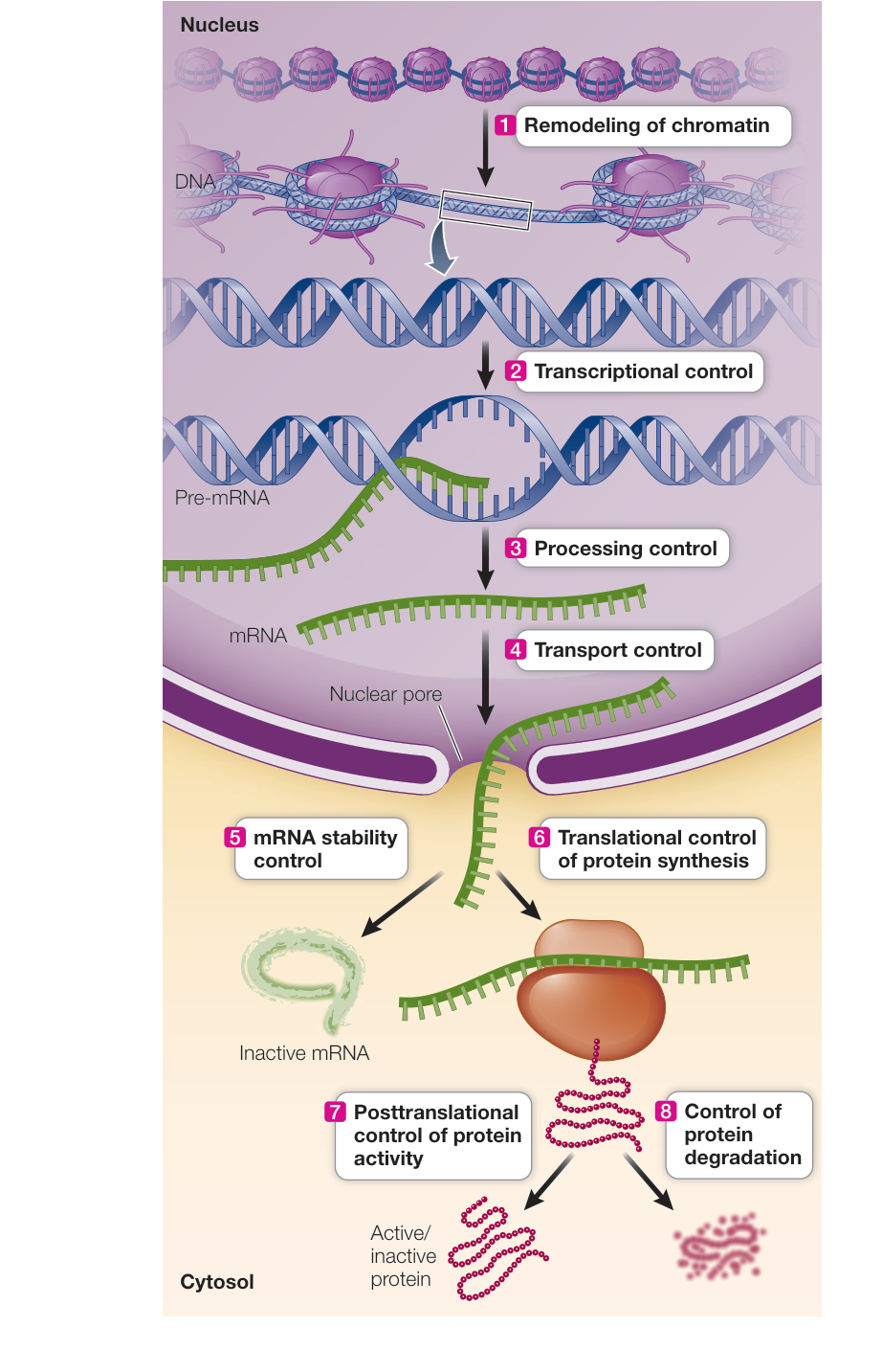
Figure 16.8 Potential Points for the Regulation of Gene Expression in Eukaryotes Gene expression can be regulated before transcription (1), during transcription (2), after transcription but before translation (3, 4, 5), at translation (6), or after translation (7, 8). Q: In prokaryotes, transcription and translation are often coupled in time and space, but in eukaryotes they are separated. What are the advantages of the nucleus as a compartment?
Like the prokaryotic RNA polymerase, eukaryotic RNA polymerase II cannot simply bind to the promoter and initiate transcription. Rather, it does so only after various general transcription factors have assembled on the chromosome to form the basal transcription apparatus (Figure 16.9). General transcription factors are proteins that bind to most promoters and are distinct from specific transcription factors that act only at certain promoters or classes of promoters. First, the protein complex called TFIID (“TF” stands for transcription factor) binds to the TATA box. Binding of TFIID changes both its own shape and that of the DNA, presenting a new surface that attracts the binding of other general transcription factors to form a basal transcription apparatus, which is the minimal set of proteins needed to initiate transcription. RNA polymerase II binds only after several other proteins have bound to this apparatus.
Each general transcription factor has a role in gene expression:
- TFIIB binds both RNA polymerase II and TFIID and helps identify the transcription initiation site.
- TFIIF prevents nonspecific binding of the basal transcription apparatus to DNA and helps recruit RNA polymerase II; it is similar in function to a bacterial sigma factor.
- TFIIE binds to the promoter and stabilizes the denaturation of the DNA.
- TFIIH opens up the DNA for transcription.
Some regulatory DNA sequences, such as the TATA box, are common to the promoters of many eukaryotic genes and are
Table 16.2 Transcription in Prokaryotes and Eukaryotes
| Prokaryotes | Eukaryotes | |
|---|---|---|
| Locations of functionally related genes | Often clustered in operons with one promoter | Often distant from one another with separate promoters |
| RNA polymerases | One | Three (RNA pol I, II, and III) I transcribes rRNA II transcribes mRNA III transcribes tRNA and small RNAs |
| Regulatory sequences besides promoter | Few | Many |
| Initiation of transcription | Binding of RNA polymerase to promoter | Binding of general and specific transcription factors, and of RNA polymerase |
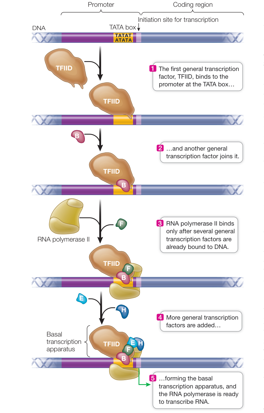
Figure 16.9 The Initiation of Transcription in Eukaryotes Apart from TFIID, which binds to the TATA box, each general transcription factor in this basal transcription apparatus has binding sites only for the other proteins in the apparatus and does not bind directly to DNA. B, E, F, and H are general transcription factors.
recognized by general transcription factors that are found in all the cells of an organism. Other regulatory sequences are present in only a few genes and are recognized by specific transcription factors. These factors may be found only in certain types of cells or at certain stages of the cell cycle, or they may be activated by signaling pathways in response to cellular or environmental signals (see Chapter 7). Once the basal transcription apparatus forms, transcription can begin. However, other transcription factors also play a role in regulating transcription.
Specific proteins can recognize and bind to DNA sequences and regulate transcription
Some regulatory DNA sequences are termed enhancers: they bind transcription factors that either activate transcription or increase the rate of transcription. Other regulatory elements are silencers: they bind factors that repress transcription. Binding of transcription factors to enhancers and silencers exemplifies positive and negative regulation respectively (see Figure 16.1). In eukaryotic microbes such as yeast, regulatory elements are almost always very close to the transcriptional start site. Similarly, most of the regulatory elements needed for correct expression of many vertebrate genes can be found within a few hundred base pairs of the transcription start site. For example, the mouse albumin gene promoter contains all the information needed for liver cell–specific expression within 170 base pairs upstream of the transcription start site. However, in many genes, regulatory elements may be located thousands or tens of thousands of base pairs away, and they may affect the expression of several nearby genes. Specific transcription factors bind to their regulatory elements and then bind to a protein complex called mediator, which facilitates the interaction with the basal transcription apparatus. In order for specific transcription factors to interact through mediator with the basal transcription apparatus, the DNA bends, bringing distant regulatory sequences into close proximity to the promoter (Figure 16.10).
The combination of transcription factors binding to a gene determines the rate of transcription. For example, the immature red blood cells in bone marrow make large amounts of -globin. At least 13 different transcription factors are involved in regulating transcription of the -globin gene in these cells. Not all of these factors are present or active in other cells, such as the immature white blood cells produced by the same bone marrow. As a result, the -globin gene is not transcribed in those cells. So although the same genes are present in all cells, the fate of the cell is determined by which of its genes are expressed. How do transcription factors recognize specific DNA sequences?
Specific protein–DNA interactions underlie binding
As we have seen, transcription factors with specific DNA-binding domains are involved in the activation and inactivation of specific genes. There are several common structural themes in the protein domains that bind to DNA in both eukaryotes and prokaryotes. These themes, or structural motifs, consist of different combinations of structural elements (protein conformations) and may include special components such as zinc. One common structural motif is the helix-turn-helix, in which two helices are connected via a non-helical turn. The interior-facing “recognition” helix interacts with the bases inside the DNA. The exterior-facing helix sits on the sugar–phosphate backbone, ensuring that the interior helix is presented to the bases in the correct configuration (Figure 16.11).
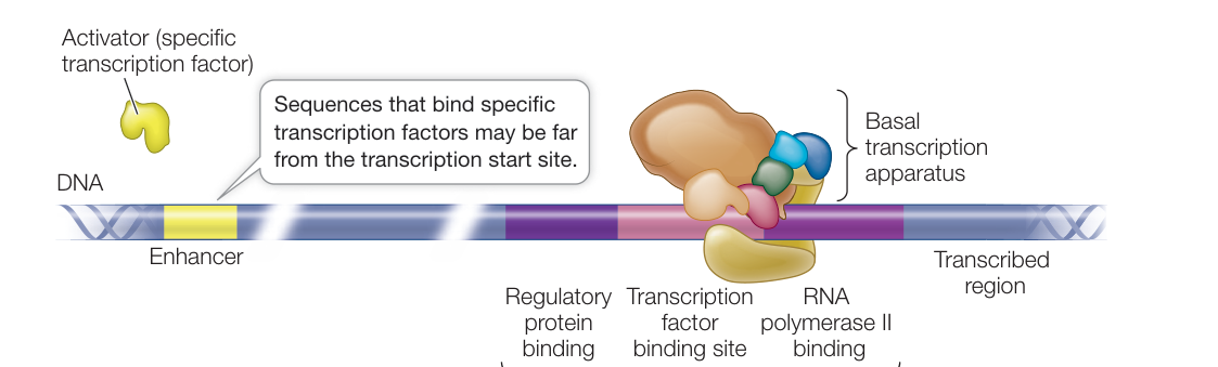
Figure 16.10 Transcription Factors and Transcription Initiation The actions of many proteins determine whether and where RNA polymerase II will transcribe DNA. This example shows binding of two specific transcription factor activators, one at the regulatory promoter and one at an enhancer element.
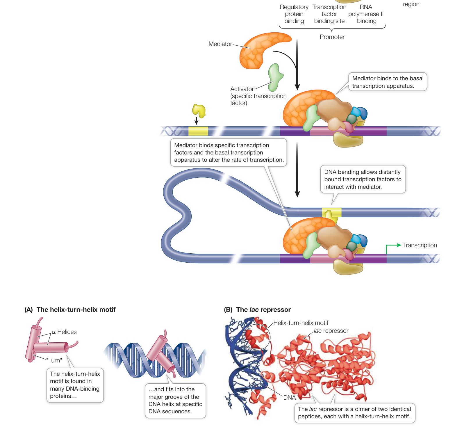
Figure 16.11 The Helix-Turn-Helix DNA-Binding Motif (A) The helix-turn-helix is one of several structural motifs seen in protein domains that bind to DNA. Such motifs facilitate recognition of, and binding to, specific DNA sequences. (B) The lac repressor binds to its operator using helix-turn-helix motifs. Data from PBD 2PE5. R. Daber et al. 2007. J Mol Biol 370: 609–619.
Connect the Concepts The structure and chemistry of DNA are key to its recognition by proteins. How the shapes and chemical structures of proteins allow them to bind noncovalently to other molecules is covered in Key Concept 3.2.
How does a protein recognize a sequence in DNA? As you learned in Key Concept 3.2, the complementary bases in DNA not only form hydrogen bonds with each other, but also can form additional hydrogen bonds with proteins, particularly at points exposed in the major and minor grooves. In this way, an intact DNA double helix can be recognized by a protein motif whose structure:
- fits into the major or minor groove;
- has amino acids that can project into the interior of the double helix; and
- has amino acids that can form hydrogen bonds with the interior bases.
Transcription factors underlie cell differentiation
During the development of a complex organism from fertilized egg to adult, cells become more and more differentiated (specialized). Since all differentiated cells contain the entire genome, their specific characteristics must arise from differential gene expression. Changes in gene expression result from the activation (and inactivation) of various transcription factors.
One well-studied example of cell differentiation is the conversion of undifferentiated muscle precursor cells into cells destined to form muscle (Figure 16.12). A key event in the commitment of these cells to become muscle is that they stop dividing. Indeed, during development, cell division and cell differentiation are often mutually exclusive. Cell signaling activates the gene for a transcription factor called MyoD (for myoblast-determining gene); this in turn activates the gene for p21, which is an inhibitor of the cyclin-dependent kinases (CDKs) that normally stimulate the cell cycle at G1 phase (see Figure 11.6). Expression of the p21 gene causes the cell cycle to stop, and other transcription factors then enter the picture so that differentiation can proceed. Interestingly, MyoD is also activated in the stem cells that are present in adult muscle, indicating a role for this transcription factor in the repair of muscle tissue as it gets damaged and worn out.
Genes such as myoD that direct the most fundamental decisions in development (often by regulating other genes on other chromosomes) usually encode transcription factors. In some cases, a single transcription factor can cause a cell to differentiate in a certain way. In others, complex interactions between genes and proteins determine a sequence of transcriptional events that leads to differentiation.
The expression of sets of genes can be coordinately regulated by transcription factors
How do eukaryotic cells coordinate the regulation of several genes whose transcription must be turned on at the same time? Prokaryotes solve this problem by arranging multiple genes in an operon that is controlled by a single promoter, and by using sigma factors to recognize particular classes of promoters. Most eukaryotic genes have their own separate promoters, and genes that are coordinately regulated may be far apart. In these cases, the expression of genes can be coordinated if they share regulatory sequences that bind the same transcription factors.
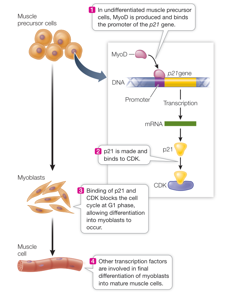
Figure 16.12 Transcription and Differentiation in the Formation of Muscle Cells Production of the transcription factor MyoD is important in muscle cell differentiation.
Shared regulatory sequences enable organisms to respond to stress—plants, for example, use shared regulatory sequences to respond to drought. Under conditions of drought stress, a plant must simultaneously synthesize several proteins whose genes are scattered throughout the genome. To coordinate expression of the stress response, each of the associated genes has a specific regulatory sequence near its promoter called the stress response element (SRE). A transcription factor binds to this element and stimulates mRNA synthesis (Figure 16.13). The stress response proteins not only help the plant conserve water, but also protect the plant against excess salt in the soil and freezing. This finding has considerable importance for agriculture because crops are often grown under less than optimal conditions or are affected by weather.
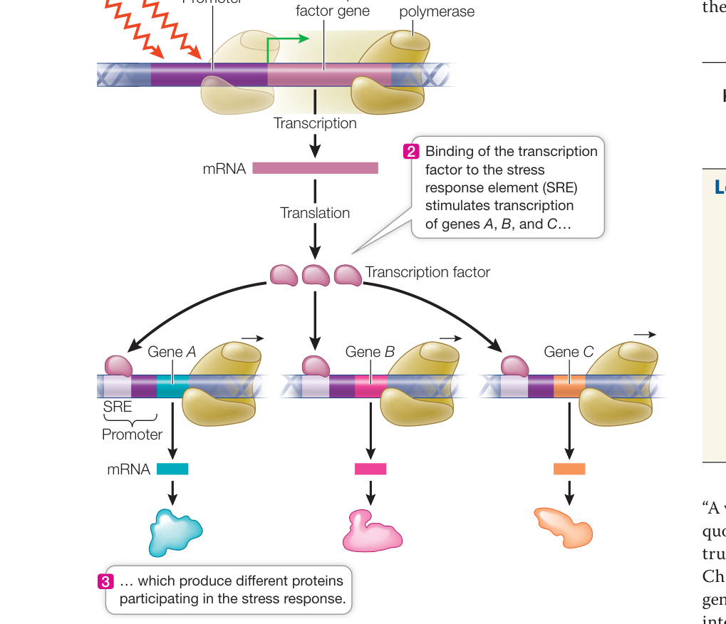
Figure 16.13 Coordinating Gene Expression A single environmental signal, such as drought stress, causes the synthesis of a transcription factor that acts on many genes.
Recap and Assess
Eukaryotes can increase or decrease transcription in various ways to help regulate gene expression. Several general transcription factors must bind to a eukaryotic promoter before RNA polymerase II will bind to it. Once the basal transcription apparatus forms, RNA polymerase II can begin transcription. Other, specific transcription factors bind to regulatory DNA sequences and interact with the basal transcription apparatus through mediator to control the level of gene expression. The regulation of gene expression determines cell differentiation, explaining why cells with the same DNA content can have different phenotypes.
- How do specific and general transcription factors regulate the rate of gene transcription?
- How do transcription factors recognize specific DNA sequences?
- How can more than one gene show the same regulation in a eukaryote? How does this differ from coordinated gene regulation in prokaryotes?
We have seen how prokaryotes and eukaryotes regulate the transcription of their genes and operons. Next we will see how viruses can hijack prokaryotic and eukaryotic transcription mechanisms in order to complete their life cycles.
Viruses Regulate Their Gene Expression during the Reproductive Cycle
Learning Objectives
- 16.3.1 Distinguish between the lytic and lysogenic cycles of a virus.
- 16.3.2 Explain the control of early versus late gene transcription during viral infection.
- 16.3.3 Give an example of how host transcription is turned off during viral infection.
- 16.3.4 Explain how expression of some HIV genes is regulated during the elongation stage of transcription.
- 16.3.5 Detail the steps at which different anti-HIV drugs work.
“A virus is a piece of bad news wrapped in protein.” This quote from immunologist Sir Peter Medawar is certainly true for the cells that viruses infect. As we described in Chapter 13, a bacterial virus (bacteriophage) injects its genetic material into a host bacterium and turns that cell into a virus factory (see Figure 13.3). Other viruses enter cells intact and then shed their coats and take over the cell’s replication machinery. Viral life cycles can be very efficient. An example is the poliovirus: a single poliovirus infecting a single mammalian cell can produce more than 100,000 new virus particles!
Understanding the reproductive cycle of a virus makes it possible to design therapeutic agents to fight infections by the virus. Viruses are small infective agents that make copies of themselves inside cellular organisms and that cannot reproduce outside host cells. Most virus particles, called virions, consist of only two or three components: the genetic material made up of DNA or RNA, a protein coat that protects the genetic material, and in some cases, an envelope of lipids that surrounds the protein coat. As we will see in this key concept, viral genomes include sequences that encode regulatory proteins. These proteins hijack the host cells’ transcriptional machinery, allowing the viruses to complete their reproductive cycles.
Viruses undertake two kinds of reproductive cycles
After a virus infects a cell, typically it takes over the cell’s molecular genetic machinery and begins the production of new virus. But in some cases there is an alternate series of events, in which the viral genome becomes integrated into the host genome.
LYTIC CYCLE The Hershey–Chase experiment (see Figure 13.4) involved a typical lytic viral reproductive cycle, so named because soon after infection, the host cell bursts (lyses), releasing progeny viruses. In this cycle, the viral genome takes over the host’s genetic machinery for its own reproduction immediately after infection. In the case of some bacteriophages, the process is extremely rapid—within 15 minutes, new phage particles appear in the bacterial cell. Ten minutes later, the “game is over,” and these particles are released from the lysed cell. What happened?
At the molecular level, the reproductive cycle of a typical lytic virus has two stages: early and late, as illustrated in Figure 16.14. Follow along in the text and Figure 16.14 and you’ll see examples of both stimulation and inhibition of host and viral gene expression:
The viral genome contains a promoter that is recognized by a host RNA polymerase that is bound to the housekeeping sigma factor, sigma-70. As a result, in the early stage (1–2 min after phage DNA entry), viral genes that lie adjacent to this promoter are transcribed.
These early genes often encode proteins that shut down host transcription. In some bacteriophages, one of these genes encodes a protein that binds to host sigma factors, especially sigma-70, inactivating them. In addition, proteins from the early genes stimulate viral genome replication and transcription of viral late genes. Three minutes after DNA entry, viral nuclease enzymes digest the host’s chromosome, providing nucleotides for the synthesis of viral genomes.
In the late stage, viral late genes are transcribed; they encode the proteins that make up the capsid (the outer shell of the virus) and other protein components of the virus and enzymes that lyse the host cell to release the new virions. This begins 9 minutes after DNA entry and 6 minutes before the first new phage particles appear.
The entire process—from binding and infection to release of new phage—takes about 30 minutes. During this period, the sequence of transcriptional events is carefully controlled to produce complete, infective virions.
LYSOGENIC CYCLE Some viruses have evolved a more complex reproductive cycle that includes a process called lysogeny, which postpones the lytic cycle. In lysogeny, the viral DNA becomes integrated into the host DNA and is not expressed. A bacteriophage genome that is inserted into the host genome and is not currently producing progeny phage is termed a prophage (Figure 16.15). As the host cell divides, the viral DNA gets replicated as part of the host chromosome. The prophage can remain inactive within the bacterial genome for thousands of generations, producing many copies of the original viral DNA, but no progeny bacteriophage.
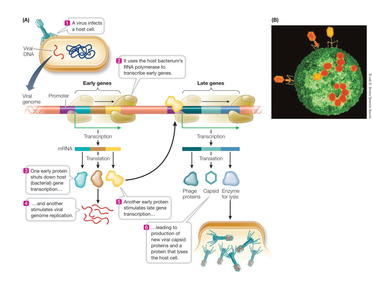
Figure 16.14 The Lytic Cycle: A Strategy for Viral Reproduction (A) In a host cell infected with a virus, the viral genome uses its early genes to shut down host transcription while it replicates itself. Once the viral genome is replicated, its late genes produce capsid proteins that package the genome and other proteins that lyse the host cell. (B) Bacteriophages have attached to this E. coli cell, and the reproductive cycle is underway, producing new phage particles. The cell is viewed in transverse section. Q: How does the virus co-express early genes and then co-express late genes?
However, under certain conditions, such as if the host cell is not growing well, the virus “cuts its losses.” It switches to a lytic cycle, in which the prophage excises itself from the host chromosome and reproduces, producing progeny phage that lyse the cell. Lysogeny allows a virus to make copies of its genome without harming its host by inserting its DNA into the host chromosome, where it sits as a silent passenger until conditions are right for lysis.
Eukaryotic viruses can have complex life cycles
Eukaryotes are susceptible to infection by various kinds of viruses (see Key Concept 24.4), which can be broadly categorized based on whether their genome is single-or double-stranded DNA or RNA and how they replicate.
- DNA viruses. Some viruses contain double-stranded DNA genomes. Others have single-stranded DNA, and a complementary strand is made after the viral genome enters the host cell. Like some bacteriophages, DNA viruses that infect eukaryotes are capable of undergoing both lytic and lysogenic life cycles. Examples include the herpes viruses and papillomaviruses (which cause warts).
- RNA viruses. Most RNA virus genomes are single-stranded, though some are double-stranded. The RNA is translated by the host’s machinery to produce viral proteins, some of which are involved in replication of the RNA genome. The influenza virus has an RNA genome.
- Retroviruses. As we described in Key Concept 14.2, a retrovirus is an RNA virus that carries a gene for reverse transcriptase, a protein that synthesizes DNA from an RNA template. The retrovirus uses this protein to make a DNA copy of its genome, which then becomes integrated into the host genome. The integrated DNA acts as a template for both mRNA and new viral genomes. HIV is a retrovirus that infects cells of the immune system and causes acquired immune deficiency syndrome (AIDS; see Key Concept 40.6).
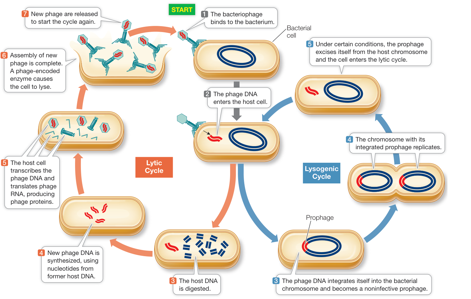
Figure 16.15 Some Bacteriophages Have Both Lytic and Lysogenic Cycles In the lytic cycle, infection of a bacterium by viral DNA leads directly to multiplication of the virus and lysis of the host cell. In the lysogenic cycle, an inactive prophage is integrated into the host DNA, where it is replicated during the bacterial life cycle.
Connect the Concepts Viral diversity is discussed in Key Concept 25.4, which explains why the genomes of some viruses consist of single-stranded RNA and how RNA retroviruses reproduce themselves by reverse transcription.
HIV gene regulation occurs at the level of transcription elongation
As we have discussed so far, many instances of gene regulation occur at the level of transcription initiation, involving both activator and repressor proteins that bind to the promoters of genes. However, studies of HIV and other viruses have revealed that transcription can also be controlled at the elongation stage.
HIV is an enveloped virus; it is enclosed within a phospholipid membrane derived from its host cells (a specific type of immune system cell) (Figure 16.16). During infection, proteins in this membrane interact with proteins on the host cell surface, and the viral envelope fuses with the host cell membrane. After the virus enters the cell, its capsid is broken down. The viral reverse transcriptase then uses the virus’s RNA template to produce a complementary DNA (cDNA) strand, while at the same time degrading the viral RNA. The enzyme then makes a complementary copy of the cDNA, and the resulting double-stranded DNA is inserted into the host’s chromosome by an enzyme appropriately named integrase. The integrated DNA is referred to as the provirus. Both the reverse transcriptase and the integrase are carried inside the HIV virion, along with other proteins needed at the very early stages of infection.
The HIV provirus resides permanently in the host chromosome and can remain in a latent (inactive) state for many years.
During this time transcription of the viral DNA is initiated, but host cell proteins prevent the RNA from elongating, and transcription is terminated prematurely. Under some circumstances, such as when the host immune cell is activated, the level of transcription initiation increases, and some viral RNA is made. One of these viral genes encodes a protein called tat (transactivator of transcription), which binds to a stem-and-loop structure at the 5’ end of the viral RNA. As a result of tat binding, the production of full-length viral RNA is dramatically increased, and the rest of the viral reproductive cycle is able to proceed. It was only after the discovery of this mechanism in HIV and similar viruses that researchers found that many eukaryotic genes are regulated at the level of transcription elongation.
Almost every step in the reproductive cycle of HIV is, in principle, a potential target for drugs to treat AIDS. The classes of anti-HIV drugs currently in use include:
- Inhibitors of virus binding to target cells (at step 1 in Figure 16.16)
- Inhibitors of internalization of virus following binding (at step 1)
- Reverse transcriptase inhibitors that block viral DNA synthesis from RNA (at step 2)
- Integrase inhibitors that block the incorporation of viral DNA into the host chromosome (at step 3)
- Protease inhibitors that block the posttranslational processing of viral proteins (at step 5)
Combinations of drugs from these classes have been spectacularly successful in treating HIV infection.
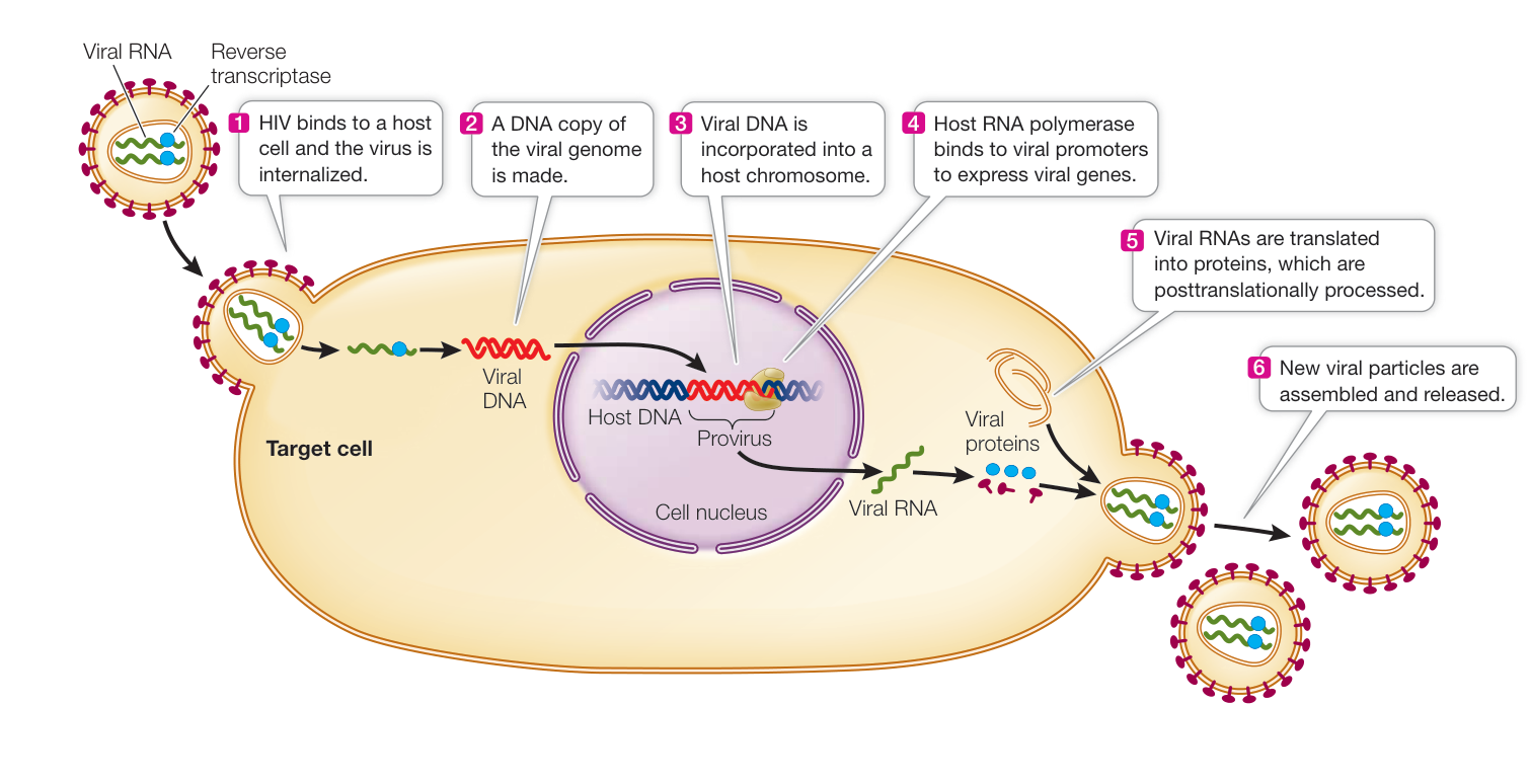
Figure 16.16 The Reproductive Cycle of HIV This retrovirus enters a host cell via fusion of its envelope with the host’s cell membrane. Reverse transcription of retroviral RNA then produces a DNA provirus—a molecule of complementary DNA that inserts itself into the host’s genome.
Recap and Assess
A virus consists of one or more nucleic acid molecules, a few proteins, and in some cases, a lipid envelope. Viruses require host cells to reproduce. Viral life cycles can include lytic and lysogenic stages. In the lytic life cycle, the cellular machinery is hijacked to synthesize new virions. In the lysogenic life cycle, the viral genome enters the host genome and is inactive until conditions become appropriate to become lytic. Studies of HIV revealed a new mechanism for gene regulation: the regulation of transcription elongation.
- The sequence of the promoter for the early gene operon in a bacteriophage that infects E. coli is similar to the sequence of a housekeeping bacterial gene promoter. Why is this the case?
- In some bacteriophages, one protein encoded by an early gene is a sigma factor. What does this viral sigma factor likely bind?
- If the function of the protease that cuts HIV proteins was specifically blocked by a drug, what would be the effect on the HIV reproductive cycle?
So far we have discussed mechanisms that cells and viruses use to control gene transcription. These mechanisms usually involve the interaction of regulatory proteins with specific DNA sequences. However, as we discuss next, there are other mechanisms for controlling gene expression that do not depend on specific DNA sequences.
Epigenetic Changes Regulate Gene Expression
Learning Objectives
- 16.4.1 Define what is meant by CpG islands and describe their role in gene expression.
- 16.4.2 Explain how acetylation of histone proteins changes the structure of chromatin and affects the rate of transcription.
- 16.4.3 Draw and/or describe a methylated DNA molecule and follow it through replication, showing how inheritance of DNA methylation occurs.
- 16.4.4 Give an example that supports the claim that environmental effects have a role in epigenetic changes.
- 16.4.5 Describe how and where X chromosome inactivation occurs and explain why it is believed to occur.
In the mid-twentieth century, the developmental biologist Conrad Hal Waddington coined the term “epigenetics” and defined it as “that branch of biology which studies the causal interactions between genes and their products which bring the phenotype into being.” Today epigenetics is defined more specifically, referring to the study of changes in gene expression not caused by changes in the DNA sequence.
Epigenetic changes are reversible but sometimes are stable and heritable. You saw an example of an epigenetic change in the opening story of this chapter, which described how genes inherited from mothers who had particularly stressful pregnancies were expressed at different levels than they were in children whose mothers had not suffered during pregnancy. In that example, stress—an environmental factor—prompted a higher degree of DNA methylation
(addition of a methyl group) in the promoter, reducing expression of a gene associated with behavior in teenagers. Here we examine more closely how epigenetic effects are caused either by DNA methylation or by alterations in chromosomal proteins.
DNA methylation occurs at the promoter and silences transcription
For those organisms in which DNA methylation occurs, from 1% to 5% of cytosine residues in the organism’s DNA are chemically modified by the addition of a methyl group () to the 5-position carbon of the base, to form 5-methylcytosine (Figure 16.17A). This covalent addition is catalyzed by the enzyme DNA methyltransferase and, in mammals, usually occurs in C residues that are adjacent to G residues. DNA regions rich in these doublets are called CpG islands and are especially abundant in promoters.
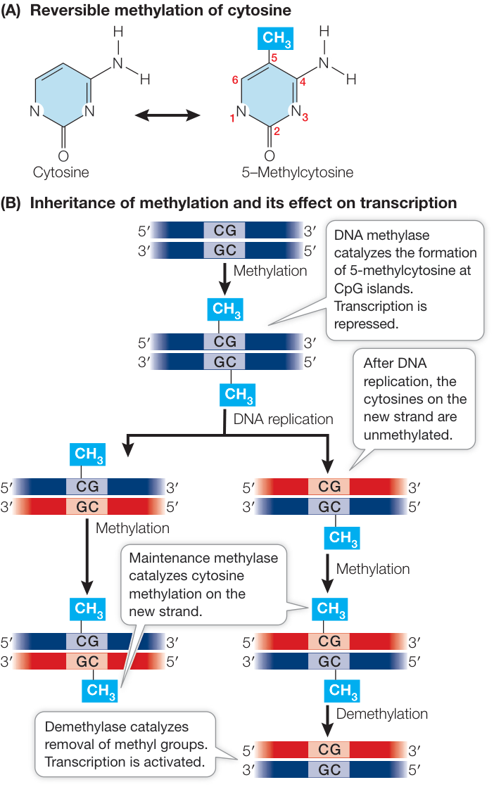
Figure 16.17 DNA Methylation: An Epigenetic Change (A) The reversible formation of 5-methylcytosine in DNA. (B) DNA methylation can alter the rate of transcription: methylation of cytosine residues generally inhibits transcription, whereas demethylation generally activates transcription. A methylated region can be stably inherited when a maintenance methylase methylates the new strand following replication. Q: Referring back to Figure 15.7, do you expect cytosines that are in CpG sites to have higher mutation rates than cytosines that are adjacent to one of the other bases?
This covalent change in DNA is heritable: when DNA is replicated, a maintenance methylase catalyzes the formation of 5-methylcytosine in the new DNA strand. However, the pattern of cytosine methylation can also be altered, because methylation is reversible: a third enzyme, appropriately called demethylase, catalyzes the removal of the methyl group from cytosine (Figure 16.17B).
What is the effect of DNA methylation? During replication and transcription, 5-methylcytosine behaves just like plain cytosine: it base-pairs with guanine. But extra methyl groups in a promoter attract proteins that bind methylated DNA. These proteins are generally involved in the repression of gene transcription; thus heavily methylated genes tend to be inactive. Note that a gene that has CpG islands in its promoter region is not necessarily heavily methylated. Instead, it may be unmethylated in some tissues, or in some environments, and methylated in other tissues and environments. Methylation is thus a way to regulate gene expression across tissues and environments. This form of regulation is epigenetic because it affects gene expression without altering the DNA sequence.
DNA methylation is important in development from egg to embryo. For example, when a mammalian sperm enters an egg, many genes in first the male and then the female genome become demethylated. Thus many genes that are usually inactive are expressed during early development. As the embryo develops and its cells become more specialized, genes whose products are not needed in particular cell types become methylated. These methylated genes are “silenced”; their transcription is repressed. However, unusual or abnormal events can sometimes turn silent genes back on.
For example, DNA methylation may play roles in the genesis of some cancers. In cancer cells, oncogenes get activated and promote cell division, and tumor suppressor genes (which normally inhibit cell division) are turned off (see Chapter 11). This misregulation can occur when the promoters of oncogenes become demethylated while those of tumor suppressor genes become methylated, as is the case in colorectal cancer (see Figure 15.12).
Histone protein modifications affect transcription
Another mechanism for epigenetic gene regulation is the alteration of chromatin structure, or chromatin remodeling. As we saw in Chapter 11, DNA is packaged with histone proteins into nucleosomes (see Figure 11.8), which can make DNA physically inaccessible to RNA polymerase and the rest of the transcription apparatus. Each histone protein has a “tail” of approximately 20 amino acids at its N terminus that sticks out of the compact structure and contains certain positively charged amino acids (notably lysine). Ordinarily there is strong ionic bonding between the positively charged histone proteins and DNA, which is negatively charged because of its phosphate groups. However, enzymes called histone acetyltransferases can add acetyl groups to these positively charged amino acids, thus changing their charges (Figure 16.18A).
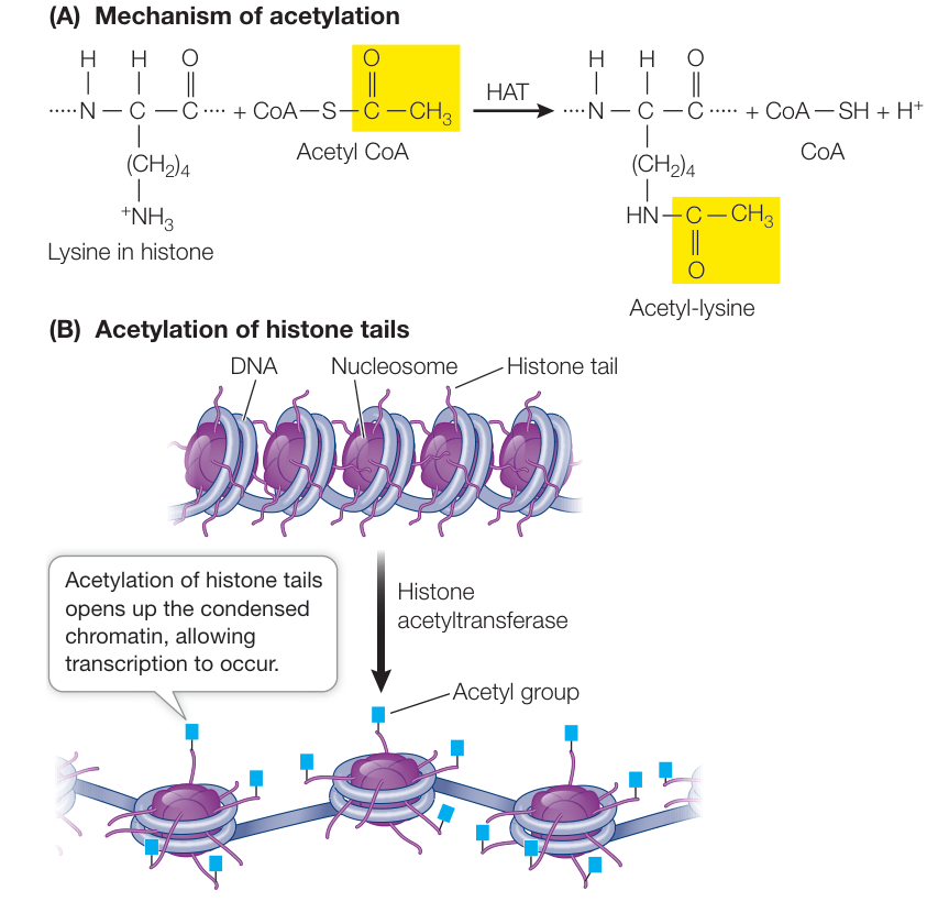
Figure 16.18 Epigenetic Remodeling of Chromatin for Transcription Initiation of transcription requires that nucleosomes change their structure, becoming less compact. (A) Histone acetyl transferase (HAT) adds an acetyl group to lysine residues. (B) Histone acetylation neutralizes the charge of positively charged lysines on histone proteins, weakening their binding to DNA, and making the DNA more accessible to bind general transcription factors (see Figure 16.9).
Reducing the positive charges of the histone tails reduces the affinity of the histones for DNA, opening up the compact nucleosome (Figure 16.18B). Additional chromatin remodeling proteins can bind to the loosened nucleosome–DNA complex, opening up the DNA for gene expression. Histone acetyltransferases can thus activate transcription.
Another kind of chromatin remodeling protein, histone deacetylase, can remove the acetyl groups from histones and thereby repress transcription. Histone deacetylases are targets for drug development to treat some forms of cancer. As noted earlier, certain genes block cell division in normal specialized tissues. In some cancers these genes are less active than in normal cells, and the histones near them show excessive levels of deacetylation. Theoretically, a drug acting as a histone deacetylase inhibitor could tip the balance toward acetylation, and this might activate genes that normally inhibit cell division.
Acetylation isn’t the only type of histone modification that can affect gene activation and repression. For example, histone methylation (not to be confused with DNA methylation) is associated with gene inactivation, and histone phosphorylation also affects gene expression, the specific effects depending on which amino acids are modified. All of these effects are reversible, so the activity of a eukaryotic gene may be determined by very complex patterns of histone modification.
Epigenetic changes can be induced by the environment
Female honey bees all have the same genetic makeup. When they are in the immature stage called a larva, however, one female in the hive eats a protein-rich substance called royal jelly that dramatically alters the expression of many genes. The queen grows much larger than her peers, stays in the hive, and is tended to by the other bees. Above all, the queen lives up to several years and is fertile, laying the eggs that will produce more bees.
The multitudes of her female compatriot larvae express a different set of genes, becoming workers who build the honeycomb, forage for food, and tend to the queen for their short lives of a few weeks. All of these differences ultimately come from the environment, specifically the royal jelly diet. Recent investigations have shown that the differences in gene expression between these very different yet genetically identical bees are due to differences in DNA methylation (Investigating Life: Gene Expression and Behavior).
Investigating Life: Gene Expression and Behavior
Experiment
Original Paper: R. Kucharski et al. 2008. Nutritional control of reproductive status in honeybees via DNA methylation. Science 319: 1817–1830.
Most female honey bee larvae grow up to be workers; just a few eat royal jelly and become queens. To understand differences in gene expression in queen bees versus in their sisters with identical genomes, Ryszard Maleszka and colleagues at Australian National University developed an experiment to test for possible epigenetic effects.
The experiment involved injecting female honey bee larvae with a substance that inhibits expression of the enzyme DNA cytosine-5-methyltransferase (DNMT)—the enzyme that catalyzes the formation of 5-methylcytosine from cytosine in DNA. In a technique called RNAi, the scientists injected female larvae with a small RNA complementary to the mRNA for DNMT to specifically reduce mRNA levels of the DNMT gene.
HYPOTHESIS DNA methylation underlies the developmental and behavioral differences between queen and worker honey bees.
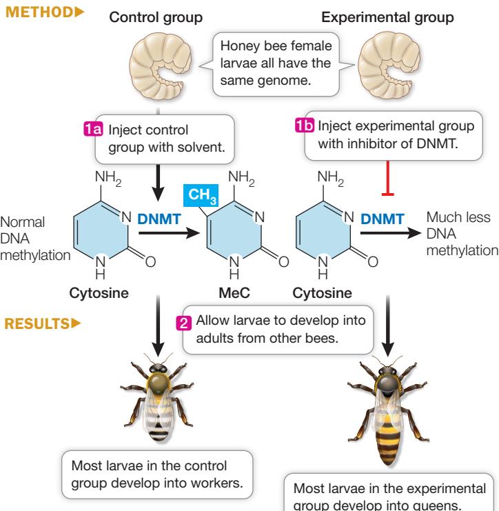
CONCLUSION Decreasing DNA methylation mimics the effects of royal jelly, causing larvae destined to become worker bees to instead become queens.
Work with the Data
QUESTIONS►
- After injection with either an inhibitor of DNMT or a control, the level of DNMT mRNA was measured in the heads of the larvae and compared with the level of a control mRNA that is always expressed at a high level. The results are shown in Table A.
Table A
| Time (h) | DNMT mRNA level compared with control (%) |
|---|---|
| 23 | 105 |
| 48 | 41 |
Why was mRNA measured in the heads of the larvae?
What can you conclude about the effectiveness of the inhibition of DNMT expression?
The DNA of a gene that is normally expressed in the brain (head) of the larvae was sequenced and the percentage of 5-methylcytosine measured to determine the extent of cytosine methylation in CpG regions. Table B shows the results. What can you conclude about the effect of DNMT inhibition?
Table B
| Condition | Percent 5-methylcytosine |
|---|---|
| Control | 79 |
| DNMT expression inhibited | 63 |
- Larvae that had been injected with the inhibitor for DNMT gene expression or a control were allowed to develop into adults. The phenotypes of the adults were evaluated, and categorized as workers, queens, or queen-like adults (intermediate). The data are shown in Table C. What do the data show about the effect of the inhibition of DNA methylation on the development of queens? Does the amount of DNA methylation strictly predict the extent of change in queen production?
Table C
| Condition | Number of workers | Number of queen-like adults | Number of queens |
|---|---|---|---|
| Control | 238 | 73 | 0 |
| Inhibited | 74 | 0 | 188 |
Although they are reversible, many epigenetic changes such as DNA methylation and histone modification can permanently alter gene expression patterns in a cell. In a germ line cell that forms gametes, it might be possible for epigenetic changes to be passed on to the next generation. But what determines these epigenetic changes? A clue comes from a recent study of monozygotic (identical) twins. Monozygotic twins come from a single fertilized egg that divides to produce two separate cells; each of these goes on to develop into a separate individual. Monozygotic twins thus have identical genomes. But are they identical in their epigenomes? A comparison of DNA in hundreds of such twin pairs shows that in tissues of 3-year-olds, the DNA methylation patterns are virtually the same. But by age 50, by which time the twins have usually been living apart in different environments for decades, the patterns are quite different. This indicates that the environment plays an important role in epigenetic modifications, and thus in the regulation of genes that these modifications affect. Note, however, that epigenetic differences in somatic tissues do not mean there will also be differences in the germ line. In addition, in mammals, histone modifications and DNA methylation are mostly removed during gamete formation, meaning that epigenetic changes are reset, making epigenetic inheritance across generations unlikely in mammals.
Media Clip 16.1 The Surprising Epigenetics of Identical Twins Life12e.com/mc16.1
Global chromosome changes involve DNA methylation
Like single genes, large regions of chromosomes or even entire chromosomes can have distinct patterns of DNA methylation. Under a microscope, two kinds of chromatin can be distinguished in the stained interphase nucleus: euchromatin and heterochromatin. The euchromatin appears diffuse and stains lightly; it contains the DNA that is transcribed into mRNA. Heterochromatin is condensed and stains darkly; any genes it contains are generally not transcribed (Figure 16.19).
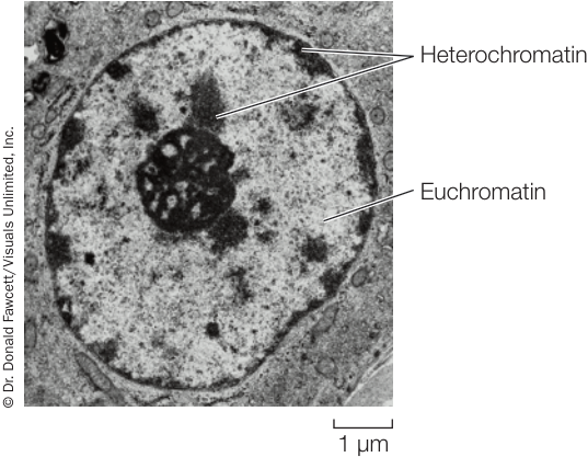
Figure 16.19 Chromatin in a Nucleus Highly condensed heterochromatin is darkly stained, while less condensed euchromatin is lightly stained.
One of the most dramatic examples of heterochromatin is the inactive X chromosome in the nuclei of female mammals. A normal female mammal has two X chromosomes; a normal male has an X and a Y (see Key Concept 12.4). The X and Y chromosomes probably arose from a pair of autosomes (non-sex chromosomes) at least 180 million years ago. Over time, mutations caused the Y chromosome to gradually lose most of the genes it once shared with the X. As a result, females and males differ greatly in the “dosage” of X-linked genes. Each female cell has two copies of each gene on the X chromosome and therefore has the potential to produce twice as much of each protein product as a male with one X. Nevertheless, for over 75% of the genes on the X chromosome, transcription is essentially identical in males and females. How does this happen?
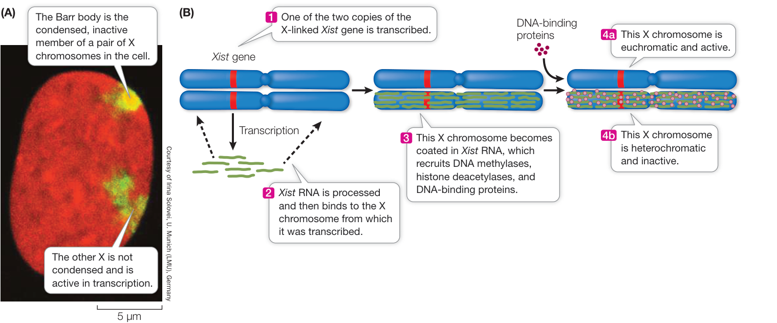
Figure 16.20 X Chromosome Inactivation (A) A Barr body and an active X chromosome in the nucleus of a human female cell. The X chromosomes are stained with a yellow-green fluorescent dye; the other chromosomes are stained with a red fluorescent dye. (B) A model for X chromosome inactivation.
During early embryonic development, one of the X chromosomes in each cell of a female is largely inactivated with regard to transcription. The same X chromosome remains inactive in all of that cell’s descendants. In a given embryonic cell, the “choice” of which X in the pair to inactivate is random. Recall that one X in a female comes from her biological father and one from her biological mother. Thus in one embryonic cell the paternal X might be the one remaining transcriptionally active, but in a neighboring cell the maternal X might be active.
The inactivated X chromosome is identifiable within the nucleus because it is very compact, even during interphase. Typically, a nuclear structure called a Barr body (after its discoverer, Murray Barr) can be seen in human female cells under the light microscope (Figure 16.20A). This clump of heterochromatin, which is not present in XY males, is the inactivated X chromosome, and it consists of heavily methylated DNA. A normal XX female will have one Barr body in every somatic cell, whereas a rare XXX female will have two, and an XXXX female will have three. Interestingly, males that are XXY will have one Barr body, indicating that the process of inactivating X chromosomes when there is more than one in a nucleus works similarly in both sexes. These observations suggest that the interphase cells of each person, regardless of sex and of X chromosome copy number, have a single active X chromosome, and thus the same dosage of expressed X chromosome genes.
Condensation of the inactive X chromosome makes its DNA sequences physically unavailable to the transcriptional machinery. Most of the genes of the inactive X are heavily methylated. However, one gene, Xist (for X inactivation-specific transcript), is only lightly methylated and is transcriptionally active. On the active X chromosome, Xist is heavily methylated and not transcribed. The RNA transcribed from Xist binds to the X chromosome from which it is transcribed, and this binding leads to inactivation along the chromosome (Figure 16.20B).
Recap and Assess
Epigenetics describes stable changes in gene expression that do not involve changes in DNA sequences. These changes involve modifications of DNA (cytosine methylation) or of histone proteins bound to DNA and can be passed down through mitosis from a parent cell to daughter cells. Epigenetic changes can be affected by the environment. Large stretches of DNA can be epigenetically modified, leading to inactivation of many genes.
- How do histone modifications affect transcription?
- Give an example of evidence that epigenetic modifications affect behavior.
- How does X chromosome inactivation occur, and why is it believed to occur?
- In colorectal cancer, certain tumor suppressor genes are not active, and this results in uncontrolled cell division. Two possible explanations for the inactive genes are (a) mutations in the promoter or coding regions, resulting in reduced transcription or inactive proteins, respectively, and (b) epigenetic silencing at the promoters of the genes, resulting in reduced transcription. How would you investigate these two possibilities?
Gene expression involves transcription and then translation. So far we have described how gene expression is regulated at the transcriptional level. But as Figure 16.8 shows, there are many points at which regulation can occur after the initial gene transcript is made.
Eukaryotic Gene Expression Can Be Regulated after Transcription
Learning Objectives
- 16.5.1 Explain how the number of proteins produced by a cell can be greater than the number of protein-coding genes.
- 16.5.2 List three ways in which the relative amount of mRNA and its encoded protein can differ in a cell.
- 16.5.3 Describe how miRNAs and siRNAs reduce protein levels.
- 16.5.4 Predict whether a protein that is resistant to ubiquitination will tend to persist longer than a protein that is rapidly ubiquinated.
- 16.5.5 Describe the function of the proteasome in a cell.
Eukaryotic gene expression can be regulated both in the nucleus prior to mRNA export, and after the mRNA leaves the nucleus. This can happen in several ways.
RNA splicing can result in different mRNAs being made from the same gene
Most primary mRNA transcripts contain several introns (see Figure 14.8). Before the RNA is exported from the nucleus, a splicing mechanism recognizes the boundaries between exons and introns and converts pre-mRNA, which has the introns, into mature mRNA, which does not. For many genes, alternative splicing can occur, whereby some exons are spliced out along with the introns (Figure 16.21). This mechanism generates a family of different proteins, with different functions, from a single gene. Recent surveys show that over 90% of all human genes are alternatively spliced. Alternative splicing may be a key to the differences in levels of complexity among organisms. For example, although humans and chimpanzees have similar-sized genomes, there is more alternative splicing in the human brain than in the chimpanzee brain.
Alternative RNA splicing is controlled both by regulatory elements in the RNA sequence that bind specific proteins (similar to regulatory sequences in DNA) and by secondary RNA structures that form by hybridization between nucleotides in the single-stranded RNA molecule.
Small RNAs are important regulators of gene expression
As you’ll see in Chapter 17, less than 5% of the genome in most plants and animals codes for proteins. Some of the genome encodes ribosomal RNA and transfer RNAs, but until recently biologists thought that the rest of the genome was not transcribed; some even called it “junk.” Recent investigations, however, have shown that some of these noncoding regions are transcribed. The mature RNAs produced from these regions are often very small and therefore difficult to detect. In both prokaryotes and eukaryotes, these small RNA molecules are called microRNA (miRNA).
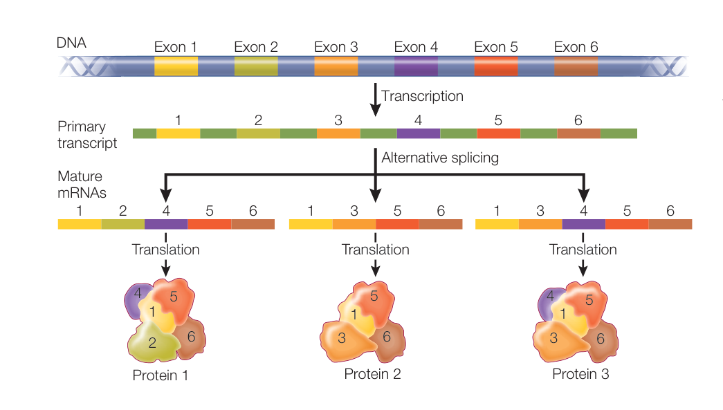
Figure 16.21 Alternative Splicing Results in Different Mature mRNAs and Proteins Pre-mRNA can be spliced differently in different tissues, resulting in different proteins. Q: A protein-coding gene in a eukaryote has four introns. Assuming the first and last exons must remain in the mature mRNA, how many different proteins could be made by alternative splicing of the pre-mRNA from this gene?
There are more than 2,000 miRNAs encoded in the human genome. MicroRNAs are transcribed as longer precursors that fold into double-stranded RNA molecules and are then processed through a series of steps into single-stranded miRNAs. A mature miRNA is about 22 bases long. A protein complex called the RNA-induced silencing complex, or RISC, guides the miRNA to its target mRNA, to which it binds by base pairing and inhibits translation (Figure 16.22A). Each 22-bp miRNA often has dozens of mRNA targets because the base pairing between the miRNA and the target mRNA doesn’t have to be perfect.
In addition to miRNAs, there is a similarly acting class of molecules called small interfering RNAs (siRNAs), which use the same RNA-induced silencing complex. Small interfering RNAs often arise from viral infections, when two complementary strands of a viral genome are transcribed. Large double-stranded RNAs are formed, and as with miRNAs, these are converted into shorter single-stranded sequences; these bind to the target RNA and cause its degradation (Figure 16.22B). Small interfering RNAs are also derived from transposon sequences, which are widespread in eukaryotic genomes (see Key Concept 15.1). Therefore it is likely that gene silencing involving siRNAs evolved as a defense mechanism to prevent the translation of viral and transposon sequences. MicroRNAs and siRNAs are similar molecules that are processed by the same cellular enzymes. A major difference between them is that:
- miRNAs are synthesized from DNA sequences separate from their target, whereas
- siRNAs are targeted to their sequence of origin.
The remarkable conservation of the miRNA and siRNA gene-silencing mechanism across diverse organisms indicates that it is evolutionarily ancient and biologically important.
Translation of mRNA can be regulated by proteins
Is the amount of a protein in a cell determined only by the amount of its mRNA? The answer is no. For example, a survey of genes
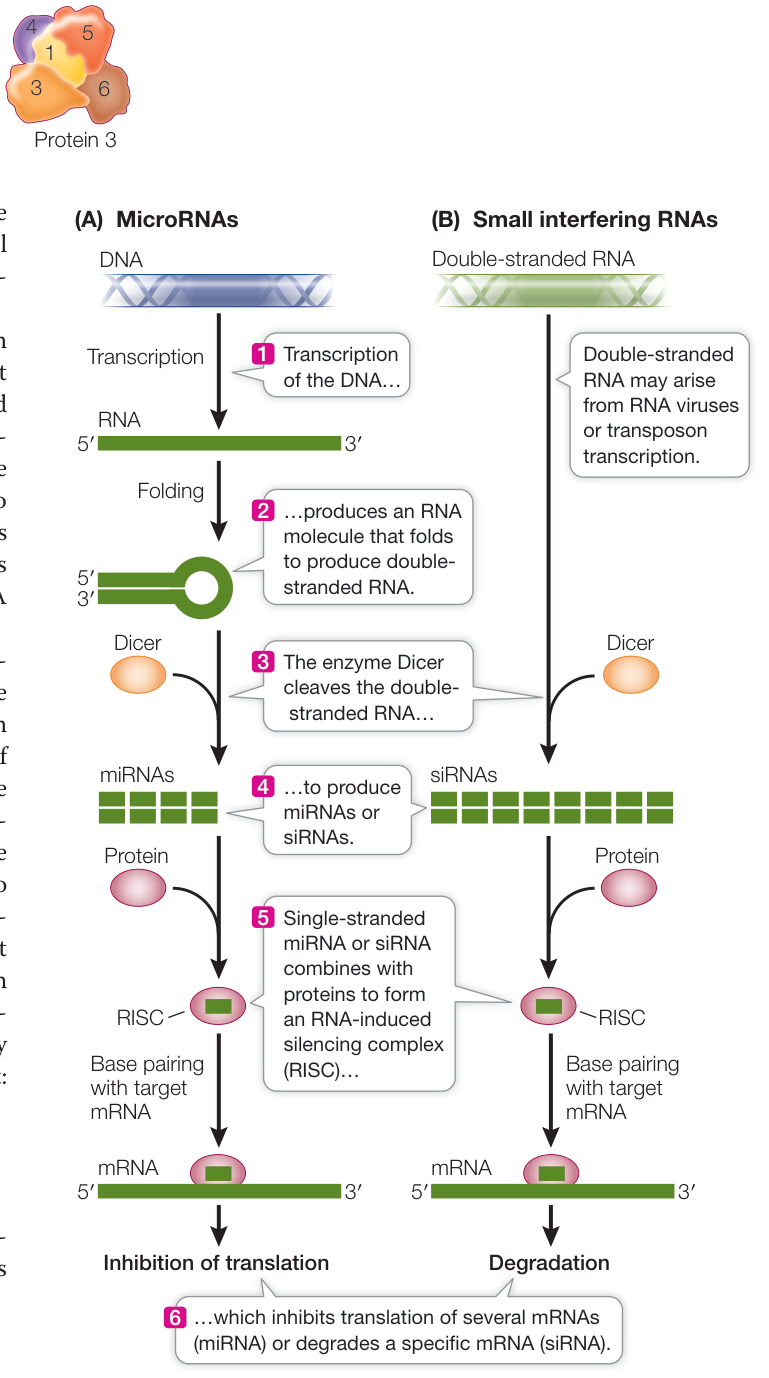
Figure 16.22 mRNA Inhibition by RNAs MicroRNAs (A) and small interfering RNAs (B) can inhibit translation by binding to target mRNAs. Figure 16.22 mRNA Inhibition by RNAs MicroRNAs (A) and small interfering RNAs (B) can inhibit translation by binding to target mRNAs.
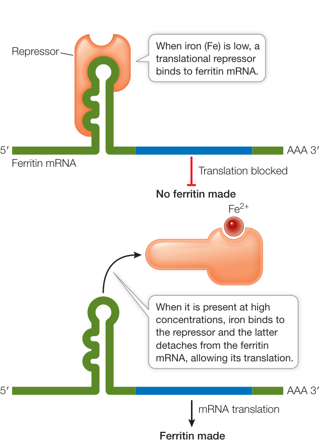
Figure 16.23 A Translational Repressor Can Repress Translation Binding of a protein to a target mRNA can inhibit its translation.
and their expression in yeast cells showed that for about one-third of the genes, there was a clear correlation between mRNA and protein: more mRNA led to more protein. But for two-thirds of the genes, there was no apparent relationship between the two: sometimes there was lots of mRNA and little or no protein, or lots of protein and little mRNA. Lots of protein when there is little mRNA can be explained by relatively short-lived mRNAs encoding long-lived proteins. But how to explain little protein when there is a lot of mRNA? The concentrations of these proteins must be determined by factors acting after the mRNA is made. Cells have two major ways to control the amount of a protein after transcription:
- They can regulate translation of the protein’s mRNA.
- They can regulate how long a newly synthesized protein persists in the cell (protein longevity).
REGULATION OF TRANSLATION There are a variety of ways in which the translation of mRNA can be regulated. One way, as we saw in the previous section, is to inhibit translation using the miRNA or siRNA pathway. A second way involves modification of the guanosine triphosphate (GTP) cap on the 5’ end of the mRNA (see Key Concept 14.4). An mRNA that is capped with an unmodified GTP molecule is not translated. For example, stored mRNAs in the egg cells of the tobacco hornworm moth are capped with unmodified GTP molecules and are not translated. After the egg is fertilized, however, the caps are modified, allowing the mRNA to be translated to produce the proteins needed for early embryonic development.
In another system, repressor proteins directly block translation. For example, in mammalian cells the protein ferritin binds free iron ions (). When iron is present in excess, ferritin synthesis rises dramatically, but the amount of ferritin mRNA remains constant, indicating that the increase in ferritin synthesis is due to an increased rate of mRNA translation. Indeed, when the iron level in the cell is low, a translational repressor protein binds to the 5’ noncoding region of ferritin mRNA and prevents its translation by blocking its attachment to a ribosome. When the iron level rises, some of the excess ions bind to the repressor and alter its three-dimensional structure, causing the repressor to detach from the mRNA and allowing translation to proceed (Figure 16.23). The binding site for the translational repressor on mRNA is a stem-and-loop region with sufficient three-dimensional structure for recognition by a protein.
REGULATION OF PROTEIN LONGEVITY The protein content of a cell at any given time is a function of both protein synthesis and protein degradation. Certain proteins can be targeted for destruction in a chain of events that begins when an enzyme attaches a 76-amino acid protein called ubiquitin (so named because it is ubiquitous, or widespread) to a lysine residue of the protein to be destroyed. Other ubiquitins then attach to the primary one, forming a polyubiquitin chain. The protein–polyubiquitin complex then binds to a huge protein complex called a proteasome (Figure 16.24). Upon entering the proteasome, the polyubiquitin is removed and ATP energy is used to unfold the target protein. Three different proteases then digest the protein into small peptides and amino acids.
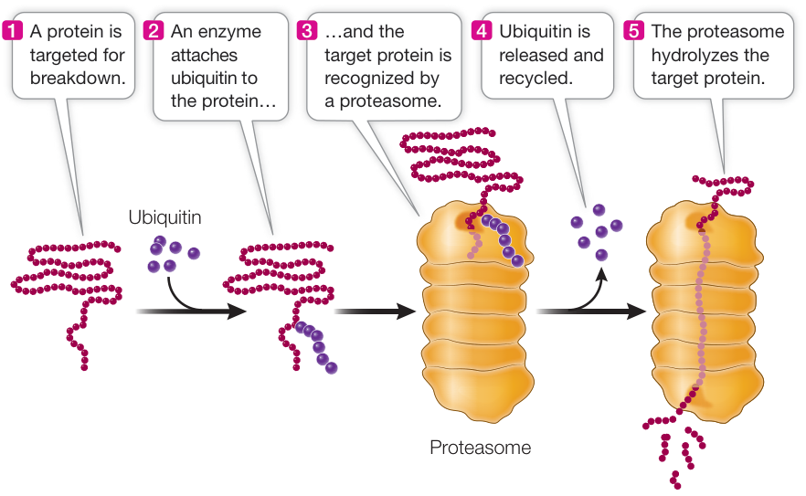
Figure 16.24 A Proteasome Breaks Down Proteins Proteins targeted for degradation are bound to ubiquitin, which then binds the targeted protein to a proteasome. The proteasome is a complex structure where proteins are digested by several powerful proteases.
You may recall from Key Concept 11.2 that cyclins are proteins that regulate the activities of key enzymes at specific points in the cell cycle. Cyclins must be broken down at just the right time, and this is done by attaching ubiquitin to them and degrading them in the proteasomes. Viruses can hijack this system. For example, some strains of the human papillomavirus (HPV) add ubiquitin to the p53 and retinoblastoma proteins, targeting them for proteasomal degradation. These proteins normally inhibit the cell cycle, so the result of this HPV activity is unregulated cell division (cancer).
Recap and Assess
One of the most important means of posttranscriptional regulation is alternative RNA splicing, which allows more than one protein to be made from a single gene. The stability of mRNA in the cytoplasm can also be regulated. MicroRNAs, siRNAs, mRNA modifications, and translational repressors can prevent mRNA translation. Proteins in the cell can be targeted for breakdown by ubiquitin and then hydrolyzed in proteasomes.
- How do miRNAs and siRNAs regulate gene expression?
- How does the three-dimensional structure of mRNA contribute to the regulation of its expression?
- What is the role of the proteasome?
- Before the human genome was sequenced, most scientists thought it contained 80,000 to 150,000 protein-coding genes. The actual sequence revealed only about 20,500. How can mRNA processing explain this discrepancy?
- Pharmaceutical companies are interested in developing miRNA drugs. How might they work in cancer? (Hint: See Figure 11.23, about oncogene and tumor suppressor proteins.)

Activity 16.2 Concept Matching: Gene Expression
Investigating Life: QA Can epigenetic changes be manipulated?
In the opening story you saw how epigenetic changes in cytosine methylation of a gene encoding a receptor resulted in behavioral changes in people’s response to stress. In Investigating Life: Gene Expression and Behavior, you learned that epigenetic changes underlie the differences between worker and queen bees. Both examples involve changes in DNA that were triggered by the environment and resulted in altered gene expression.
As in bees, nutrients in the diets of mammals (including humans), such as folic acid and SAM-e (S-adenosyl methionine), contain methyl groups that can participate in reactions that modify DNA. Experiments with mice have shown that feeding young animals a diet enriched with these nutrients causes changes in epigenetic patterns and gene expression that remain throughout life. A new field called nutrient genomics (could you have guessed the name?) has developed to investigate the possibility of altering gene expression in mammals by diet.
Future directions
In addition to investigating the possibility of altering DNA methylation by diet, researchers have developed specific drugs that target the enzymes involved in DNA methylation. In cancer, gene expression can be altered in many ways, such as by mutation (as you saw in Chapter 11). Cancer cells can alter gene expression through epigenetic mechanisms as well. For example, in some colon cancers, the expression of an important tumor suppressor gene involved in DNA repair is silenced by extensive DNA methylation at the promoter, resulting in uncontrolled cell division. The nucleotide analog 5’-azacytidine blocks DNA methyltransferase and can be employed to combat some cancers, by reducing DNA methylation and halting uncontrolled cell division. Other drugs are being used to block histone modifications that in the tumor cell result in gene expression changes.
Visual Summary
© imageBROKER/Alamy Stock Photo
You should be able to relate each summary to the adjacent figures. If you go to this visual summary in Achieve, you can follow links to figures, animations, activities, and simulations that will help you consolidate the material.
Prokaryotic Gene Expression Is Regulated in Operons
- ■ Positive regulation occurs when binding of an activator to DNA increases transcription. Negative regulation occurs when binding of a repressor to DNA decreases transcription.
- ■ Expression of the lac operon requires high lactose (repressor not bound to the operator) and low glucose (cAMP–CRP bound to the promoter).
- ■ The lac operon repressor is bound to its operator when lactose is absent; the trp operon repressor is bound to its operator when tryptophan is present.
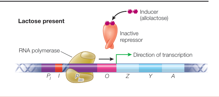
Figure 16.5
- ■ Sigma factors bind to RNA polymerase and direct it to specific classes of prokaryotic promoters.
Questions
- Suppose a mutation occurs in the lac operon repressor gene so that the repressor can no longer bind its inducer (allolactose) but it can still bind DNA. What effect will this have on regulation of the lac operon?
- Suppose a mutation occurs in the trp operon repressor gene such that the repressor can no longer bind its co-repressor (tryptophan) but it can still bind DNA. What effect will this have on regulation of the trp operon?
- What would the effect of a mutation in sigma-70 that rendered it non-functional be on an E. coli cell?
Eukaryotic Gene Expression Is Regulated by Transcription Factors
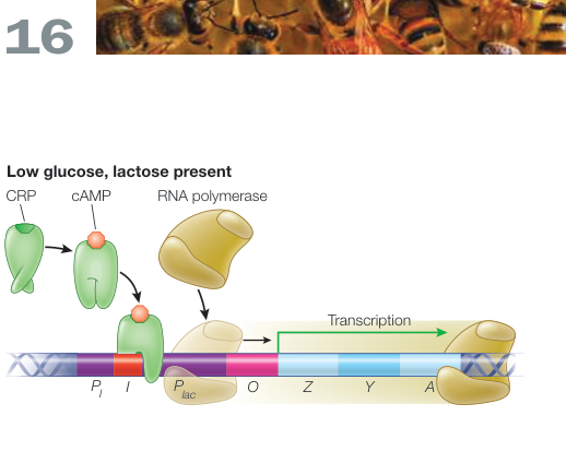
Figure 16.7 - ■ Protein transcription factors bind to silencer and enhancer sequences on DNA to regulate transcription in eukaryotes. - ■ General transcription factors are required for initiation of transcription at all eukaryotic protein-coding promoters. - ■ Specific transcription factors differ among genes and interact with the basal transcription apparatus through mediator. - ■ Transcription factors possess particular structural motifs that allow them to bind to particular DNA sequences. - ■ Transcription factors are responsible for differences in gene expression in cells that contain the same genome, and thus underlie cell differentiation.
Questions
- What effects on gene expression and cell viability would occur if a general versus a specific transcription factor were knocked out (rendered nonfunctional)?
- What effects on gene expression and cell viability would occur if a binding site for a general versus a specific transcription factor were knocked out (rendered unable to bind a transcription factor)?
- What would be the effect of knocking out mediator in a cell?
- How might a researcher prevent muscle precursor cells from differentiating into myoblasts (see Figure 16.12)?
Viruses Regulate Their Gene Expression during the Reproductive Cycle
- ■ Bacteriophages are viruses that infect bacteria and can have lytic or lysogenic life cycles.
- ■ Eukaryotic viruses include DNA, RNA, and retroviruses.
- ■ Retroviruses such as HIV make a DNA copy of their genome that integrates into the host genome, and then transcription leads to production of new virus.
Questions
- A bacterial strain that has been growing for many hours in a closed tube suddenly succumbs to bacteriophage infection. What do you think happened?
- How might an early gene product turn off transcription of its bacterial host genes?
- HIV may spend many years in the host genome before producing virions. Transcription initiation occurs during these years, but no viral proteins are made. Why not?
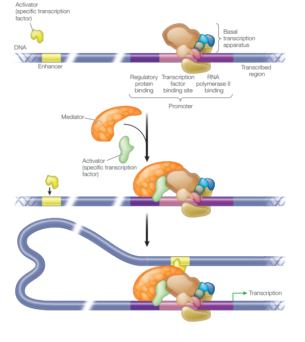
Figure 16.10
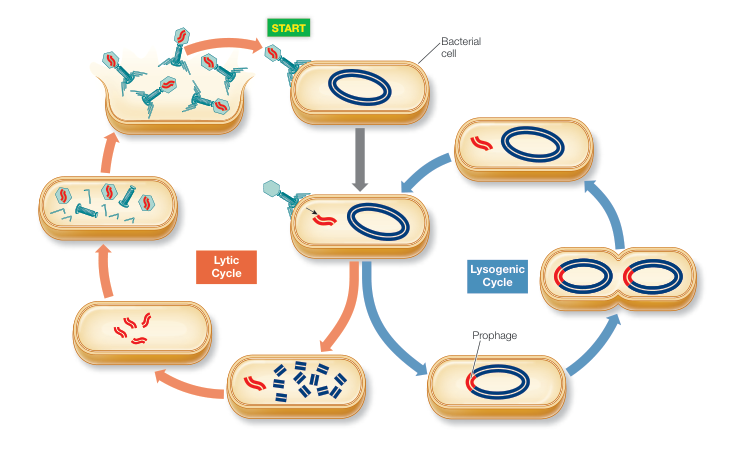
Figure 16.15
Epigenetic Changes Regulate Gene Expression
- ■ In many species, DNA methylation occurs at CpG islands, shutting down transcription.
- ■ Histone deacetylation increases histone affinity for DNA, which reduces transcription.
- ■ Epigenetic changes can be passed on through mitosis.
- ■ X inactivation involves production of an Xist RNA, which coats the chromosome from which it was transcribed, causing it to become heterochromatic.
Questions
- Some cytosine bases are never methylated. Why?
- Explain why CpG methylation sites facilitate inheritance of methylation across cell division.
- What would be the effect of deleting the Xist gene in a typical (XY) male mammal? In a typical (XX) female mammal?
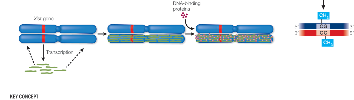
Figure 16.20
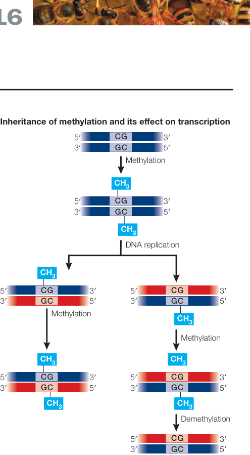
Figure 16.17
Eukaryotic Gene Expression Can Be Regulated after Transcription
- ■ Alternative splicing allows many different proteins to be produced from the same gene.
- ■ mRNA levels can be uncorrelated with protein levels due to differences in stability and translational control of expression.
- ■ The microRNA (miRNA) pathway inhibits translation; the similar small interfering RNA (siRNA) pathway degrades mRNAs.
- ■ Protein stability is controlled by ubiquitination.
Questions
- A gene encodes a four-exon pre-mRNA. Assuming that the first and last exons remain in the mature mRNA, how many different proteins can be produced by alternative splicing of the pre-mRNA?
- Why might the abundance of a protein in a cell be quite low compared with its mRNA level?
- A protein-coding gene experiences a missense mutation that changes a lysine to an arginine, both of which have positively charged side groups. This change substantially increases the stability of the protein. Why might this be the case?
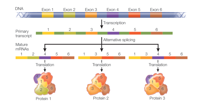
Figure 16.21