Chapter 30Protostome Animals
Key Concepts
- 30.1 Protostomes Account for More Than Half of All Described Species
- 30.2 Many Lophotrochozoans Have Ciliated Feeding Structures or Life Stages
- 30.3 Ecdysozoans Grow by Shedding Their Cuticles
- 30.4 Arthropods Are the Most Abundant and Diverse Group of Animals
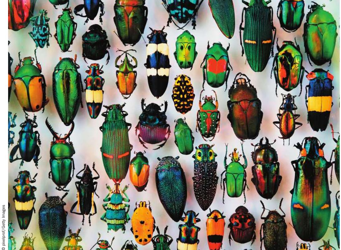
The majority of described species of life are protostomes, largely because of the enormous diversity of insects. Beetles alone account for almost one-fourth of all described species.
© pixelprof/Getty Images
Investigating Life: Exploring the Diversity of Life on Earth
Of the 1.8 million species of life that have been discovered and named by biologists, a large majority are protostomes. One group of protostomes, the insects, accounts for more than 1 million of these species, or more than half of all known species of living organisms. Although these numbers may seem incredibly large, they represent a relatively small fraction of the total protostome diversity that is thought to exist on Earth.
As recently as the 1980s, many biologists thought that about half of existing insect species had been described, but today they think that the number of described insect species may be a much smaller fraction of the total number of living species. Why did they change their minds?
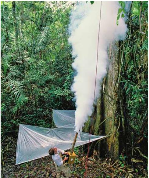
A simple but important field study suggested that the number of existing insect species had been significantly underestimated. Knowing that the insects of tropical rainforests—the most species-rich habitat on Earth—were poorly known, entomologist Terry Erwin made a comprehensive sample of one group of insects, the beetles, in the canopies of a single species of tropical forest tree, Luehea seemannii, in Panama. Erwin fogged the canopies of large L. seemannii trees with an insecticide and collected the insects that fell from the trees in collection nets.
He collected about 1,200 species of beetles—many of them previously undescribed—from this one species of tree.
Erwin then used a set of assumptions to estimate the total number of insect species in tropical rainforests. His assumptions included estimates of the number of species of trees in these forests; the proportion of beetles that specialize on a specific species of host tree; the relative proportion of beetles to other insect groups; and the proportion of beetles that live in trees versus those that live in leaf litter on the ground. From this and similar studies, Erwin estimated that there may be 30 million or more species of insects on Earth. Although recent tests of Erwin’s assumptions suggest that 30 million was an overestimate, it is clear that the vast majority of insect species remain to be discovered.
Erwin’s pioneering study highlighted the fact that we live on a poorly known planet, most of whose species have yet to be named and described. Much of that undiscovered diversity occurs among several groups of protostomes.
Q&A Which groups of protostomes are thought to contain the most undiscovered species? (Find the answer on page 692.)
Protostomes Account for More Than Half of All Described Species
Learning Objectives
- 30.1.1 Contrast the advantages of having a hard body covering versus a soft body in different habitats.
- 30.1.2 Associate major evolutionary innovations among the protostomes with life in aquatic versus terrestrial environments.
You may recall that the embryos of diploblastic animals (the ctenophores, placozoans, and cnidarians, which we discussed in Chapter 29) have two cell layers: an outer ectoderm and an inner endoderm (see Key Concept 29.1). Sometime after the origin of the diploblastic animals, a third embryonic cell layer evolved: the mesoderm, which lies between the ectoderm and the endoderm. Mesoderm is found in the two major triploblastic animal clades, the protostomes and the deuterostomes. If we were to judge solely on the basis of numbers, both of species and of individuals, the protostomes would emerge as by far the more successful of the two groups.
As noted in Key Concept 29.1, the name “protostome” means “mouth first.” In protostomes, the embryonic blastopore becomes the mouth as the animal develops. In contrast, in deuterostomes (“mouth second”), the blastopore becomes the anal opening of the gut. The protostomes are extremely varied, but they are all bilaterally symmetrical animals whose bodies exhibit two major derived traits:
- An anterior brain that surrounds the entrance to the digestive tract
- A ventral nervous system consisting of paired or fused longitudinal nerve cords
Other aspects of protostome body organization differ widely from group to group (Table 30.1). Before gene sequences were available for phylogenetic analysis, biologists considered the structure of the body cavity to be a critical feature in animal classification. But the results of genetic analyses have shown that body cavity forms have undergone considerable convergence in the course of protostome evolution. Although the common ancestor of the protostomes had a coelom, subsequent modifications of the coelom distinguish many protostome lineages. In some lineages (such as the flatworms and entoprocts), the coelom has been lost (that is, these groups reverted to an acoelomate state). Some lineages are characterized by a pseudocoel, a body cavity lined with mesoderm in which the internal organs are suspended (see Figure 29.6B). In two of the most prominent protostome clades, the coelom has been highly modified:
- Arthropods lost the ancestral condition of the coelom over the course of evolution. Their internal body cavity has become a hemocoel, or “blood chamber,” in which fluid from an open circulatory system bathes the internal organs before returning to blood vessels.
Table 30.1 Anatomical Characteristics of Some Major Protostome Groups
| Group | Body cavity | Digestive tract | Circulatory system |
|---|---|---|---|
| ARROW WORMS | Coelom | Complete | None |
| LOPHOTROCHOZOANS | |||
| Bryozoans | Coelom | Complete | None |
| Entoprocts | None | Complete | None |
| Flatworms | None | Blind gut | None |
| Rotifers | Pseudocoel | Complete | None |
| Gastrotrichs | Pseudocoel | Complete | None |
| Ribbon worms | Coelom | Complete | Closed |
| Brachiopods | Coelom | Complete in most | Open |
| Phoronids | Coelom | Complete | Closed |
| Annelids | Coelom | Complete | Closed or open |
| Mollusks | Reduced coelom | Complete | Open except in cephalopods |
| ECDYSOZOANS | |||
| Nematodes | Pseudocoel | Complete | None |
| Horsehair worms | Pseudocoel | Greatly reduced | None |
| Arthropods | Hemocoel | Complete | Open |
- Most mollusks have an open circulatory system with some of the attributes of the hemocoel, but they retain vestiges of an enclosed coelom around their major organs.
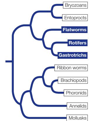
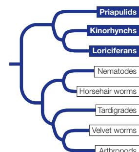
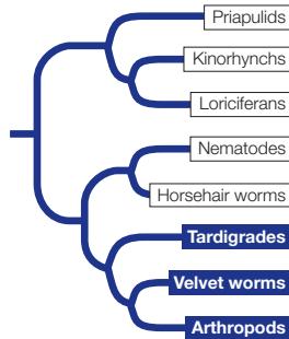
The protostomes can be divided into two major clades—the lophotrochozoans and the ecdysozoans—largely on the basis of DNA sequence analysis (Figure 30.1).
Cilia-bearing lophophores and trochophores evolved among the lophotrochozoans
Lophotrochozoans derive their name from two different ciliated features: a feeding structure known as a lophophore and a free-living larval form known as a trochophore. Neither the lophophore nor the trochophore is universal to all lophotrochozoans, however.
Several distantly related groups of lophotrochozoans (including bryozoans, entoprocts, brachiopods, and phoronids) have a lophophore, a circular or U-shaped ring of ciliated, hollow tentacles around the mouth (Figure 30.2). This complex organ is used for both food collection and gas exchange. Biologists once grouped taxa that have lophophores together as “lophophorates,” but it is now clear that they are not one another’s closest relatives. The lophophore appears to have evolved independently at least twice, or else it is an ancestral feature of lophotrochozoans and has been lost in many groups. Nearly all animals with a lophophore are sessile as adults. They use the tentacles and cilia of the lophophore to capture small floating organisms from the water. Other sessile lophotrochozoans have less well-developed tentacles that they use for the same purpose.
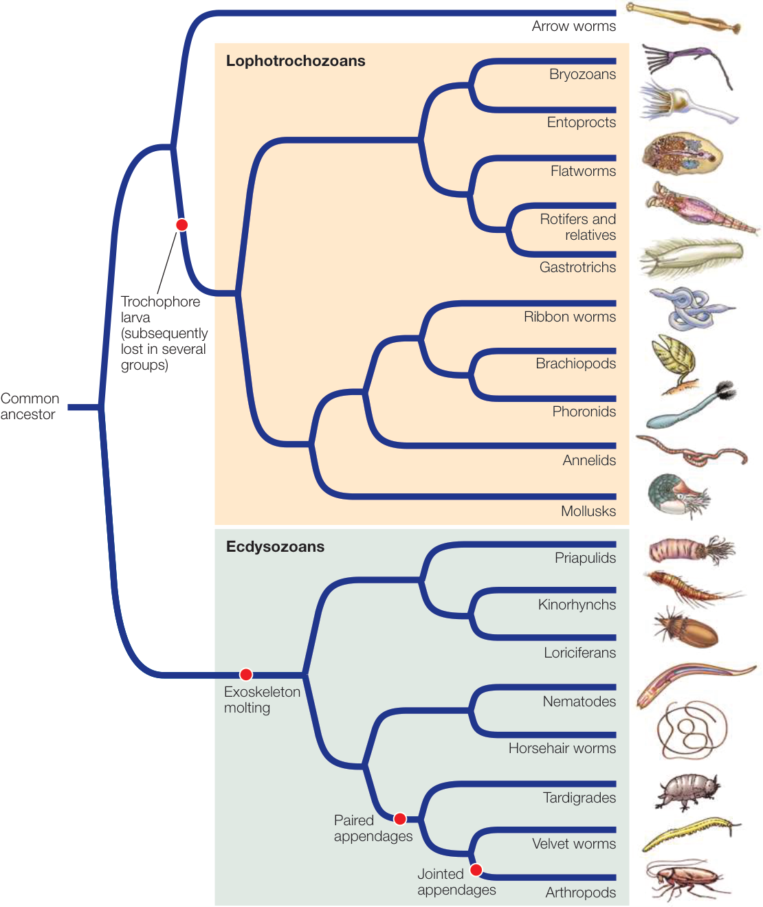
Figure 30.1 Phylogenetic Tree of Protostomes Two major lineages, the lophotrochozoans and the ecdysozoans, dominate the protostome tree. Some small groups are not included in this tree. The phylogenetic relationships shown here are supported mainly by genomic sequence data. Although genomic studies are contributing greatly to our knowledge of animal phylogeny, most species of protostomes have yet to be studied in detail. Activity 30.2 Protostome Classification
Some lophotrochozoans, especially in their larval form, use cilia for locomotion. The larval form known as a trochophore moves by beating a band of cilia (see Figure 29.11A). This movement of cilia also brings plankton closer to the larva, where it can capture and ingest them (its cilia are therefore similar in function to the cilia of the lophophore). Trochophore larvae are found among many of the major groups of lophotrochozoans, including the mollusks, annelids, ribbon worms, entoprocts, and bryozoans. This larval form was probably present in the common ancestor of lophotrochozoans but has been subsequently lost in several lineages.
Some lophotrochozoans (including flatworms, ribbon worms, annelids, and mollusks) exhibit spiral cleavage in early development, in which layers of new cells spiral obliquely around the axis of the animal–vegetal pole of the early embryo. Some biologists group these taxa together as “spiralians,” although phylogenetic analyses of gene sequences do not support monophyly of taxa with spiralian cleavage. Nonetheless, spiral cleavage may have been present in the lophotrochozoan ancestor and subsequently lost in several descendant lineages.
Connect the Concepts As described in Key Concept 42.2, cleavage is the sequence of early cell divisions that transforms the diploid zygote into a mass of undifferentiated cells that will develop into the embryo. Different patterns of cleavage result in differential distributions of nutrients and cytoplasmic determinants within the egg cytoplasm.
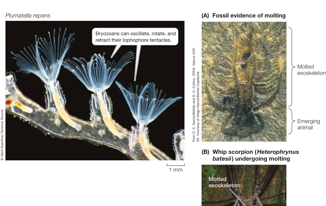
Figure 30.2 Bryozoans Use the Lophophore to Feed The extended lophophore dominates the anatomy of the colonial bryozoans. This species inhabits fresh water, although most bryozoans are marine.
Many lineages of lophotrochozoans have a wormlike body form, which means that they are bilaterally symmetrical, legless, soft-bodied, and at least several times longer than they are wide. A wormlike body form enables animals to burrow efficiently through marine sediment or soil. However, as you will see in Key Concept 30.2, the mollusks—the most familiar of the lophotrochozoans to many people—have a very different body organization.
Ecdysozoans must shed their cuticles
Ecdysozoans have an external covering, or cuticle, that is secreted by the underlying epidermis (the outermost cell layer). The cuticle provides these animals with both protection and support. Once formed, however, the cuticle cannot grow. How, then, can ecdysozoans increase in size? They do so by shedding, or molting, the cuticle and replacing it with a new, larger one.
A fossil Cambrian arthropod preserved in the process of molting shows that molting evolved more than 500 million years ago (Figure 30.3A). An increasingly rich array of molecular and genetic evidence, including a set of Hox genes shared by all ecdysozoans, suggests they have a single common ancestor. Thus molting of a cuticle is a trait that may have evolved only once during animal evolution.
Before an ecdysozoan molts, a new cuticle is already forming underneath the old one. Once the old cuticle is shed, the new one expands and hardens. Until it has hardened, though, the animal is vulnerable to its enemies, both because its outer surface is easy to penetrate and because an individual with a soft cuticle can move only slowly or not at all (Figure 30.3B).
The cuticles of some ecdysozoans, mainly arthropods, function as external skeletons, or exoskeletons. These exoskeletons are thickened by layers of protein and a strong, waterproof polysaccharide called chitin. An animal with a rigid, chitin-reinforced exoskeleton can neither move in a wormlike manner nor use cilia for locomotion. A hard exoskeleton also impedes the passage of oxygen and nutrients into the animal, presenting new challenges in other areas besides growth. Thus new mechanisms of locomotion and gas exchange evolved in those ecdysozoans with hard exoskeletons.
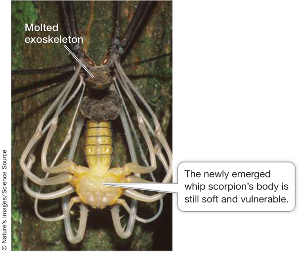
Figure 30.3 Molting: Past and Present (A) This 500-million-year-old fossil from the Cambrian, an individual of a long-extinct arthropod species captured in the process of molting, shows that the molting process is an evolutionarily ancient trait. (B) This tail-less whip scorpion has just emerged from its discarded exoskeleton. It will be highly vulnerable until its new cuticle has hardened.
To move rapidly, an animal with a rigid exoskeleton must have body extensions that can be manipulated by muscles. Such appendages evolved in the late Precambrian, leading to the arthropod (“jointed foot”) clade. Arthropod appendages exist in an amazing variety of forms. They serve many functions, including walking and swimming, gas exchange, food capture and manipulation, copulation, and sensory perception. Arthropods grasp food with their mouth and associated appendages and digest it internally. Their muscles are attached to the inside of the exoskeleton. Each segment has muscles that operate that segment and the appendages attached to it (Figure 30.4).
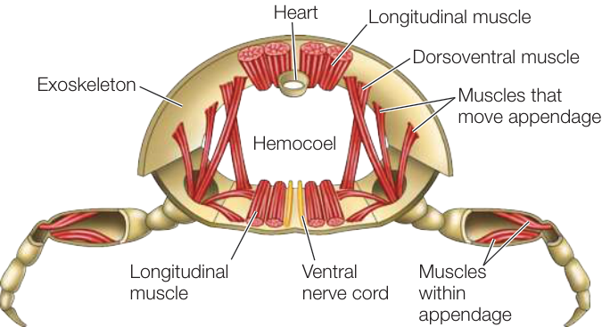
Figure 30.4 Arthropod Skeletons Are Rigid and Jointed This cross section through a thoracic segment of a generalized arthropod illustrates the arthropod body plan, which is characterized by a rigid exoskeleton with jointed appendages.
The arthropod exoskeleton has had a profound influence on the evolution of these animals. Encasement within a rigid body covering provides support for walking on dry land, and the waterproofing provided by chitin keeps the animal from dehydrating in dry air. Thus aquatic arthropods were, in short, excellent candidates to invade terrestrial environments. As you will see, they did so several times.
Arrow worms retain some ancestral developmental features
Nearly all triploblastic animal groups can be readily classified as either protostomes or deuterostomes, but the evolutionary relationships of one small group, the arrow worms, were debated for many years. The blastopore of arrow worms develops into an anus (as in deuterostomes), but they have ventral nerve cords and spiral cleavage (as in many protostomes). Recent studies of gene sequences, however, clearly identify arrow worms as protostomes. There is still some question as to whether they are the closest relatives of the lophotrochozoans (as shown in Figure 30.1) or possibly the sister group of all other protostomes.
The arrow worm body is divided into three compartments: head, trunk, and tail (Figure 30.5). The body is transparent or translucent. Most arrow worms swim in the open sea. A few species live on the seafloor. Their abundance as fossils indicates that they were common more than 500 million years ago. The 180 or so known living species of arrow worms are small enough—ranging from 3 millimeters to 12 centimeters in length—that their gas exchange and waste excretion requirements are met by diffusion through the body surface. They lack a circulatory system; wastes and nutrients are moved around the body in the coelomic fluid, which is propelled by cilia that line the coelom.
Arrow worms are hermaphroditic; that is, each individual produces both male and female gametes. In most species, eggs are fertilized internally following elaborate courtship between two individuals, although some species can self-fertilize. Miniature adults hatch directly from the eggs; these animals have no distinct larval stage.
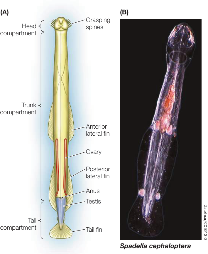
Figure 30.5 An Arrow Worm Arrow worms have a three-part body organization. Their fins and grasping spines are adaptations for a predatory lifestyle. Individuals are hermaphroditic, producing both eggs in an ovary and sperm in a testis.
Arrow worms are stabilized in the water by means of one or two pairs of lateral fins and a tail fin. They are major predators of planktonic organisms in the open ocean, ranging in size from small protists to young fishes as large as the arrow worms themselves. An arrow worm typically lies motionless in the water until water movement signals the approach of prey. The arrow worm then darts forward and uses the stiff spines adjacent to its mouth to grasp its prey.
Recap and Assess
The shared derived traits of protostomes include a blastopore that develops into a mouth (except in arrow worms), an anterior brain, and a ventral nervous system. Several lophotrochozoan groups are characterized by a filter-feeding ciliated structure known as a lophophore or by cilia-bearing larvae known as trochophores. Ecdysozoans, which have a body covering known as a cuticle, must molt periodically in order to grow.
- How does an animal’s body covering influence the way it exchanges gases, feeds, and moves?
- What features make arthropods well adapted for colonizing terrestrial environments?
In the next key concept we continue our survey of the protostomes with a more detailed look at the major groups of lophotrochozoans and the diverse body forms that are found among them.
Many Lophotrochozoans Have Ciliated Feeding Structures or Life Stages
Learning Objectives
- 30.2.1 Associate major morphological features of protostomes with the environment and life history of the respective organisms.
- 30.2.2 Provide examples demonstrating how modifications of existing structures can lead to increases in species diversity.
Lophotrochozoans come in a variety of sizes and shapes, ranging from relatively simple animals with a blind gut (that is, a gut with only one opening) and no internal transport system to animals with a complete gut (having separate entrance and exit openings) and a complex internal transport system. They include some species-rich groups, such as flatworms, annelids, and mollusks. Several of these groups have wormlike bodies, but the lophotrochozoans encompass a wide variety of morphologies, including a few groups with external shells. Some lophotrochozoan groups have only recently been discovered by biologists.
Most bryozoans and entoprocts live in colonies
Most of the 5,500 known species of bryozoans (“moss animals”) and 170 known species of entoprocts (meaning “anus inside”) are colonial animals that live in a “house” made of material secreted by the external body wall. The colonial species are sessile, but the few solitary species can slowly move around in their environment. Almost all bryozoans and entoprocts are marine, although a few species occur in fresh or brackish water.
A bryozoan colony consists of many small (1–2 mm) individuals connected by strands of tissue along which nutrients can be moved. Bryozoan colonies can grow to contain more than 2 million individuals, all stemming from the asexual reproduction of the colony’s founder. Rocks in coastal regions in many parts of the world are covered with luxuriant growths of bryozoans. Some bryozoans create miniature reefs in shallow waters. In some species, the individual colony members are differentially specialized for feeding, reproduction, defense, or support. Individual bryozoans in a colony are able to oscillate their lophophore to increase contact with prey. They can also retract it into their “house” (see Figure 30.2).
Bryozoans can also reproduce sexually. Sperm are released into the water, which carries the sperm to other individuals. Eggs are fertilized internally; developing embryos are brooded before they exit as larvae to seek suitable sites for attachment to the substrate. Entoprocts can also reproduce asexually. Some species of entoprocts release unfertilized eggs into the water for fertilization, whereas other species brood their developing young as bryozoans do.
Bryozoans and entoprocts differ in the placement of the anus. In bryozoans the anus is located outside the ring of tentacles that make up the lophophore, whereas the anus of entoprocts is located in the center of this ring. The lophophores of the two groups also function differently: food particles are carried from the tips to the bases of the tentacles in bryozoans, but from the bases to the tips of the tentacles in entoprocts. Entoprocts lack a coelom, whereas bryozoans have a three-part coelom.
Flatworms, rotifers, and gastrotrichs are structurally diverse relatives
Flatworms, rotifers, gastrotrichs, and their close relatives are a structurally diverse group of organisms whose relationships to one another have been hypothesized only recently. Recent genomic studies show that this monophyletic lophotrochozoan group includes both acoelomate subgroups (e.g., the flatworms) and pseudocoelomate subgroups (e.g., the rotifers and gastrotrichs), and yet the closest relatives of this group—the bryozoans and entoprocts—are coelomate and acoelomate, respectively. Thus this group provides an example of the evolutionary convergence in body cavity form that we described in Key Concept 30.1.
FLATWORMS Flatworms lack specialized organs for transporting oxygen to their internal tissues. In the absence of a gas transport system, each cell must be near a body surface, a requirement met by the dorsoventrally flattened body form that gives these animals their common name. The digestive tract of a free-living flatworm consists of a mouth opening into a blind gut. The gut is often highly branched, forming intricate patterns that increase the surface area available for the absorption of nutrients. Some small free-living flatworms are cephalized, with a head bearing chemoreceptor organs, two simple eyes, and a tiny brain composed of anterior thickenings of the longitudinal nerve cords. Free-living flatworms glide over surfaces on a layer of mucus, powered by broad bands of cilia (Figure 30.6A).
Although many flatworms are free-living, most flatworm species are parasites. Of the parasitic species, most are endoparasites. There are also flatworms that feed externally on animal tissues (living or dead), and some graze on plants. A likely evolutionary transition was from feeding on dead organisms to feeding on the body surfaces of dying hosts to invading and consuming parts of healthy hosts.
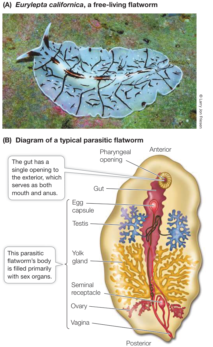
Figure 30.6 Flatworms Include Both Parasites and Free-Living Forms (A) Some flatworm species, such as this Pacific marine flatworm, are free-living. (B) The fluke diagrammed here lives endoparasitically in the gut of sea urchins and is typical of endoparasitic flatworms. Because their hosts provide all the nutrition they need, these internal parasites do not require elaborate feeding or digestive organs and can devote most of their body to reproduction.
Most of the 30,000 known species of living flatworms are tapeworms and flukes; members of these two groups are endoparasites, particularly of vertebrates (Figure 30.6B). Because they absorb digested food from the digestive tracts of their hosts, many endoparasitic flatworms lack digestive tracts of their own. Some cause serious human diseases, such as schistosomiasis, which is common in parts of Asia, Africa, and South America. The species that causes this devastating disease has a complex life cycle involving both freshwater snails and mammals as hosts. Members of another flatworm group, the monogeneans, are ectoparasites of fishes and other aquatic vertebrates. The turbellarians include most of the free-living species.
ROTIFERS Most species of rotifers are tiny (50–500 µm long)—smaller than some ciliate protists—but they have specialized internal organs (Figure 30.7A,B). A complete gut passes from an anterior mouth to a posterior anus; the body cavity is a pseudocoel that functions as a hydrostatic skeleton. Rotifers typically propel themselves through the water by means of rapidly beating cilia rather than by muscular contraction.
The most distinctive organ of rotifers is a conspicuous ciliated organ called the corona, which surmounts the head of many species. Coordinated beating of the cilia sweeps particles of organic matter from the water into the animal’s mouth and down to a complicated structure called the mastax, in which food is ground into small pieces. By contracting muscles around the pseudocoel, a few rotifer species that prey on protists and small animals can protrude the mastax through the mouth and seize small objects with it.
Most of the known species of rotifers live in fresh water. Some species rest on the surfaces of mosses or lichens in a desiccated, inactive state until it rains. When rain falls, they absorb water and become mobile, feeding in the films of water that temporarily cover the plants. Most rotifers live no longer than a few weeks.
Both males and females are found in some species of rotifers, but only females are known among the bdelloid rotifers (the b in “bdelloid” is silent). Biologists have concluded that the bdelloid rotifers may have existed for tens of millions of years without regular sexual reproduction. Lack of genetic recombination generally leads to the buildup of deleterious mutations, so long-term asexual reproduction typically leads to extinction. Recent studies, however, have indicated that bdelloid rotifers may avoid this problem by picking up fragments of genes from their environment during the desiccation–rehydration cycle, which allows genetic recombination among individuals in the absence of direct sexual exchange.
Connect the Concepts The consequences of asexual reproduction, and the reasons that it commonly leads to a buildup of deleterious mutations, are discussed in Key Concept 21.3.
A few highly reduced lineages appear to have descended from the free-living rotifers. The spiny-headed worms are parasites with complex life cycles, often parasitizing several animal hosts (Figure 30.7C). The jaw worms are tiny marine organisms that glide between sand grains in shallow marine environments. Although spiny-headed worms and jaw worms are structurally quite distinct, molecular analyses have revealed that both groups are essentially highly modified rotifers.
GASTROTRICHS The 800 known species of gastrotrichs (also called “hairy-backs”) are abundant, tiny (0.05–3 mm) animals that live in marine sediments, in fresh waters, and in the water films that surround grains of soil. Their transparent bodies have a flat ventral surface that is covered with cilia (Figure 30.7D). Most species are simultaneous hermaphrodites, with both male and female reproductive organs, although the male organs have been greatly reduced or lost in some species that reproduce asexually.
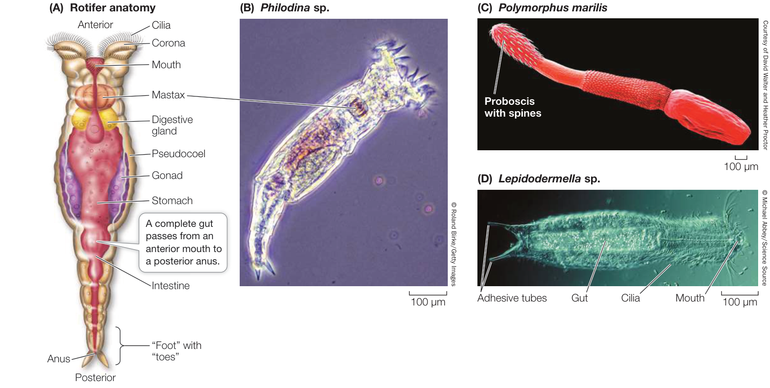
Figure 30.7 Rotifers and Gastrotrichs (A) The rotifer diagrammed here reflects the general structure of many rotifers. (B) A micrograph reveals the internal complexity of the microscopic rotifers. (C) Spiny-headed worms are parasitic rotifer relatives. The spines of the proboscis anchor the animal to the organs of its host. (D) Gastrotrichs superficially resemble rotifers but have flattened ventral surfaces covered with cilia, as flatworms do.
Ribbon worms have a long, protrusible feeding organ
Ribbon worms (or proboscis worms) have simple nervous and excretory systems similar to those of flatworms. Unlike flatworms, however, they have a closed circulatory system and a complete digestive tract with a mouth at one end and an anus at the other. Small ribbon worms move slowly by beating their cilia. Larger ones employ waves of muscle contraction to move over the surface of sediments or to burrow into them.
Within the body of nearly all of the 1,200 known species of ribbon worms is a fluid-filled cavity called the rhynchocoel, within which lies a hollow, muscular proboscis. The proboscis, which is the worm’s feeding organ, may extend much of the length of the body. Contraction of the muscles surrounding the rhynchocoel causes the proboscis to evert explosively through an anterior pore (Figure 30.8A). The proboscis may be armed with sharp stylets that pierce prey and discharge paralysis-causing toxins into the wound.
Media Clip 30.4 Explosive Extrusion of Ribbon Worm Proboscis Life12e.com/mc30.4
Most ribbon worm species are marine, although there are species that live in fresh water or on land. Most species are less than 20 centimeters long, but individuals of some species reach 20 meters or more. Some genera feature species that are conspicuous and brightly colored (Figure 30.8B). Recent molecular analyses suggest that ribbon worms may be most closely related to the brachiopods and phoronids.
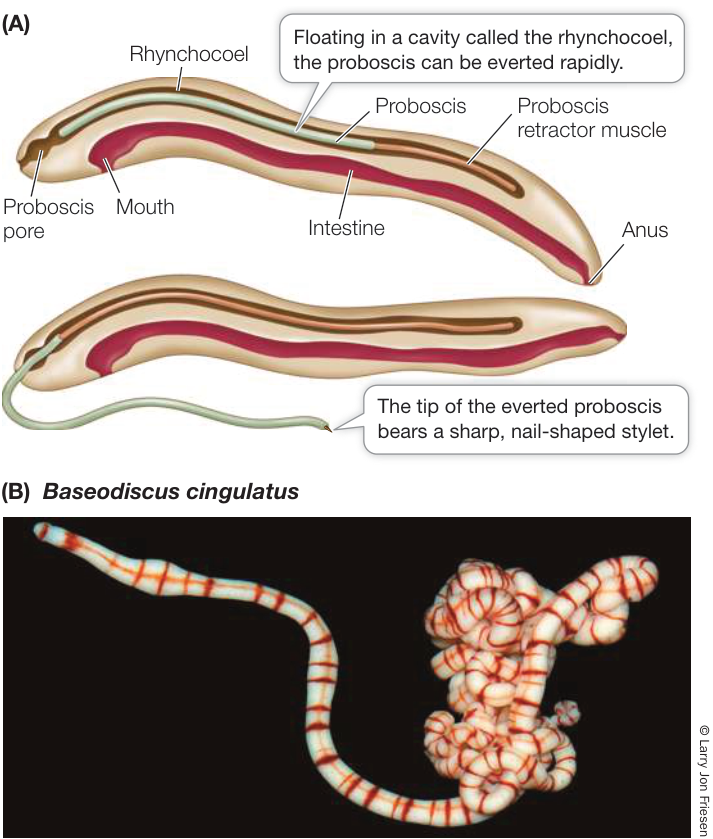
Figure 30.8 Ribbon Worms (A) The proboscis is the ribbon worm’s feeding organ. (B) Although most ribbon worms are small, some marine species such as this one can reach several meters in length.
Brachiopods and phoronids use lophophores to extract food from the water
Recall that the bryozoans and entoprocts use a lophophore to feed. Brachiopods and phoronids also feed using a lophophore, but this structure may have evolved separately in these groups. Although neither the brachiopods nor the phoronids are represented by many living species, the brachiopods (which have hard external shells and thus leave an excellent fossil record) are known to have been much more abundant in the past.
BRACHIOPODS Brachiopods (lampshells) are solitary marine animals. They have a rigid shell that is divided into two parts connected by a ligament (Figure 30.9). The two halves can be pulled shut to protect the soft body. Brachiopods superficially resemble bivalve mollusks, but shells have evolved independently in the two groups. The two halves of the brachiopod shell are dorsal and ventral, rather than lateral as in bivalves. The lophophore is located within the shell. The beating of cilia on the lophophore draws water into the slightly opened shell. Food is trapped in the lophophore and directed to a ridge, along which it is transferred to the mouth.
Most brachiopods are 4 to 6 centimeters long. They live attached to a solid substrate or embedded in soft sediments. Most species are attached by means of a short, flexible stalk that holds the animal above the substrate. Gases are exchanged across body surfaces, especially the tentacles of the lophophore. Most brachiopods release their gametes into the water, where they are fertilized. The larvae remain among the plankton for only a few days before they settle and develop into adults.
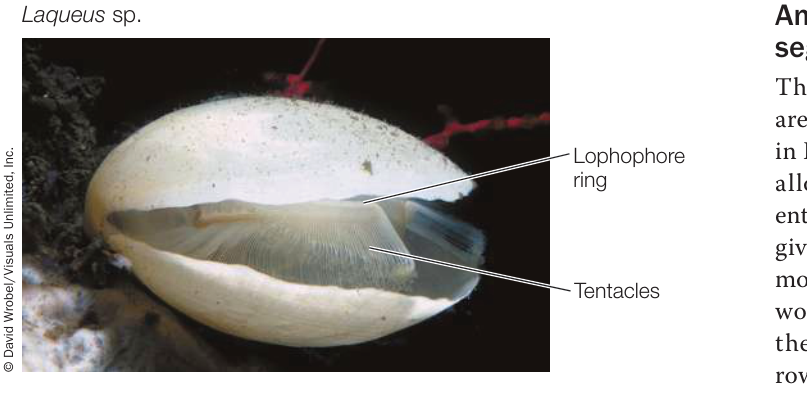
Figure 30.9 A Brachiopod’s Lophophore The lophophore of this North Pacific brachiopod can be seen between the valves of its shell.
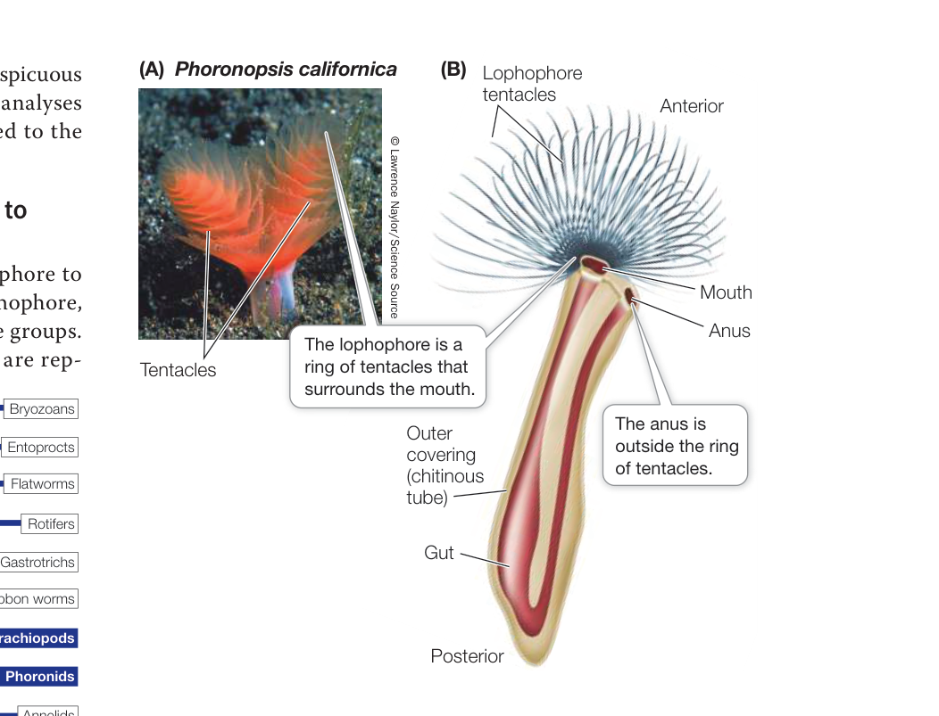
Figure 30.10 Phoronids (A) The tentacles of this phoronid’s lophophores form spirals. (B) The phoronid gut is U-shaped, as seen in this generalized diagram.
Brachiopods reached their peak abundance and diversity in Paleozoic and Mesozoic times. More than 26,000 fossil species have been described. Only about 450 species are known to survive, but they remain common in some marine environments.
PHORONIDS The ten known species of phoronids are small (5–25 cm long), sessile worms that live in muddy or sandy sediments or attached to rocky substrates. Phoronids are found in marine waters from the intertidal zone to about 400 meters deep. They secrete tubes made of chitin, within which they live, and have a U-shaped gut with the anus located outside the lophophore (Figure 30.10). Their cilia drive water into the top of the lophophore, and the water exits through the narrow spaces between the tentacles. Suspended food particles are caught and transported to the mouth by ciliary action. Some species release eggs into the water, where they are fertilized, but other species produce large eggs that are fertilized internally and retained in the parent’s body, where they are brooded until they hatch.
Annelids have segmented bodies
The wormlike bodies of annelids are clearly segmented. As described in Key Concept 29.1, segmentation allows an animal to move different parts of its body independently, giving it much better control of its movement. The earliest segmented worms, preserved as fossils from the middle Cambrian, were burrowing marine annelids.
In most large annelids, the coelom in each segment is isolated from those in other segments (Figure 30.11). A separate nerve center called a ganglion (plural ganglia) controls each segment; nerve cords that connect the ganglia coordinate their functioning. Most annelids lack a rigid external protective covering; instead they have a thin, permeable body wall that serves as a general surface for gas exchange. Most annelids are thus restricted to moist environments because they lose body water rapidly in dry air. The approximately 19,000 described species live in marine, freshwater, and moist terrestrial environments.
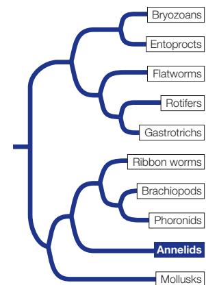
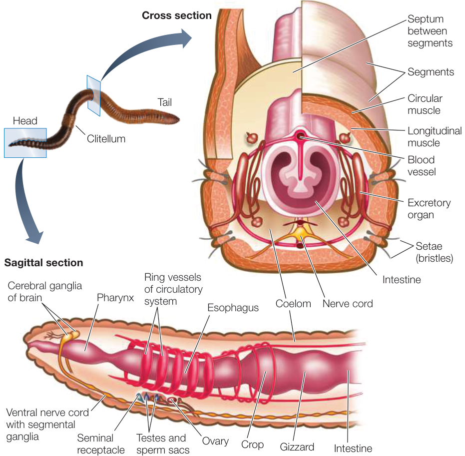
Figure 30.11 Annelids Have Many Body Segments The segmented structure of annelids such as this earthworm is apparent both externally and internally. Many organs are repeated serially.
POLYCHAETES More than half of all annelid species are commonly known as polychaetes (“many hairs”), although this is a descriptive term rather than the name of a single clade. Recent molecular studies indicate that polychaetes are paraphyletic with respect to the remaining annelids. Most polychaetes are marine, and many live in burrows in soft sediments. Most of them have one or more pairs of eyes and one or more pairs of tentacles, with which they capture prey or filter food from the surrounding water, at the anterior end of the body (Figure 30.12A; see also Figure 29.7A). In some species, the body wall of most segments extends laterally as a series of thin outgrowths called parapodia. The parapodia function in gas exchange, and some species use them to move. Stiff bristles called setae protrude from each parapodium, forming temporary contact with the substrate and preventing the animal from slipping backward when its muscles contract.
Members of one polychaete clade, the pogonophorans, secrete tubes made of chitin and other substances, in which they live (Figure 30.12B). Pogonophorans have lost their digestive tract (they have no mouth or gut). So how do they obtain nutrition? Part of the answer is that pogonophorans can take up dissolved organic matter directly from the sediments in which they live or from the surrounding water. Much of their nutrition, however, is provided by endosymbiotic bacteria that the pogonophorans house in a specialized organ known as the trophosome. These bacteria oxidize hydrogen sulfide and other sulfur-containing compounds, fixing carbon from methane in the process. Up-take of the hydrogen sulfide, methane, and oxygen used by the bacteria is facilitated by hemoglobin in the pogonophorans’ tentacles. It is this hemoglobin that gives the tentacles their red coloration.
Pogonophorans were not discovered until early in the twentieth century, when the first species were discovered on the seafloor at depths of up to a few hundred meters. In recent decades, deep-sea explorers have found them living many thousands of meters below the ocean surface. In these deep oceanic sediments, they may reach densities of many thousands per square meter. About 160 species have been described. The largest and most remarkable pogonophorans are 2 meters or more in length and live near deep-sea hydrothermal vents—volcanic openings in the seafloor through which hot, sulfide-rich water pours. The methane and hydrogen sulfide from these vents provide the raw materials for carbon fixation by the pogonophorans’ endosymbiotic bacteria.
CLITELLATES The approximately 3,000 described species of clitellates, which form a well-supported clade within the annelids, are found in freshwater, marine, and terrestrial environments. The clitellates appear to be phylogenetically nested among various groups of polychaetes, although the exact relationships are not yet clear. There are two major groups of clitellates, the oligochaetes and the leeches.
Oligochaetes (“few hairs”) have no parapodia, eyes, or anterior tentacles, and they have only four pairs of setae bundles per segment. Earthworms—the most familiar oligochaetes—burrow in and ingest soil, from which they extract food particles. All oligochaetes are hermaphroditic. Sperm are exchanged simultaneously between two copulating individuals (Figure 30.12C). Eggs and sperm are deposited in a cocoon outside the adult’s body. Fertilization occurs within the cocoon after it is shed, and when development is complete, miniature worms emerge and immediately begin independent life.
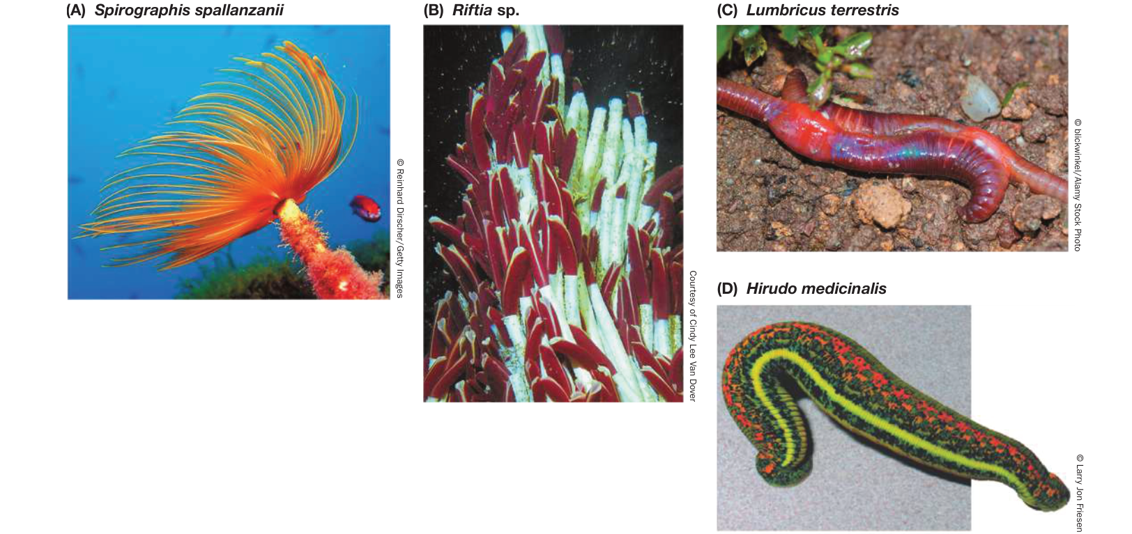
Figure 30.12 Diversity among the Annelids (A) Fan worms, or “feather duster worms,” are sessile marine polychaetes that grow in masses, filtering food from the water with their tentacles. This individual has been removed from its chitinous tube. (B) Pogonophorans live around hydrothermal vents deep in the ocean. Their tentacles can be seen protruding from their chitinous tubes. (C) Earthworms, like all oligochaetes, are hermaphroditic; when they copulate, each individual donates and receives sperm. (D) The medicinal leech has been a tool of physicians and healers for centuries. Even today, leeches have uses in clinical practice.
Leeches, like oligochaetes, lack parapodia and tentacles. The coelom of leeches is not divided into compartments; the coelomic space is largely filled with undifferentiated tissue. Groups of segments at each end of the body are modified to form suckers, which serve as temporary anchors that aid the leech in its movement. With its posterior sucker attached to a substrate, the leech extends its body by contracting its circular muscles. The anterior sucker is then attached, the posterior one detached, and the leech shortens itself by contracting its longitudinal muscles. Leeches live in freshwater or terrestrial habitats.
Most leeches are ectoparasites that feed by making an incision in a host, from which blood flows. A leech can ingest so much blood in a single feeding that its body may enlarge severalfold. The leech secretes an anticoagulant into the wound that keeps the host’s blood flowing. For centuries, medical practitioners employed leeches to draw blood to treat diseases they believed were caused by an excess of blood or by “bad blood.” Although most leeching practices (such as inserting a leech in a person’s throat to alleviate swollen tonsils) have been abandoned, Hirudo medicinalis (the medicinal leech; Figure 30.12D) is used today to reduce fluid pressure and prevent blood clotting in damaged tissues, to eliminate pools of coagulated blood, and to prevent scarring. The anticoagulants of certain other leech species also contain anesthetics and blood vessel dilators and are being studied for possible medical uses.
Media Clip 30.5 Leeches Feeding on Blood Life12e.com/mc30.5
Mollusks have undergone a dramatic evolutionary radiation
Mollusks are the most diverse group of lophotrochozoans, both in numbers of species and in the environments they occupy. Although the major groups of mollusks differ dramatically in morphology, they all share the same three major body components: a foot, a visceral mass, and a mantle (Figure 30.13A).
- The molluscan foot is a large, muscular structure that originally was both an organ of locomotion and a support for the internal organs. In squid and octopuses, the foot has been modified to form arms and tentacles borne on a head with complex sensory organs. In other groups, such as clams, the foot is a burrowing organ. In some groups the foot is greatly reduced.
- The heart and the digestive, excretory, and reproductive organs are concentrated in a centralized, internal visceral mass.
- The mantle is a fold of tissue that covers the organs of the visceral mass. The mantle secretes the hard, calcareous shell that is typical of many mollusks.
In most mollusks, the mantle extends beyond the visceral mass to form a mantle cavity. Within this cavity lie gills that are used for gas exchange. When cilia on the gills beat, they create a current of water. The tissue of the gills, which is highly vascularized (contains many blood vessels), takes up oxygen from the water and releases carbon dioxide. Many mollusk species use their gills as filter-feeding devices, whereas others feed using a rasping structure known as a radula to scrape algae from rocks. In some mollusks, such as the marine cone snails, the radula has been modified into a drill or poison dart.
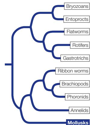
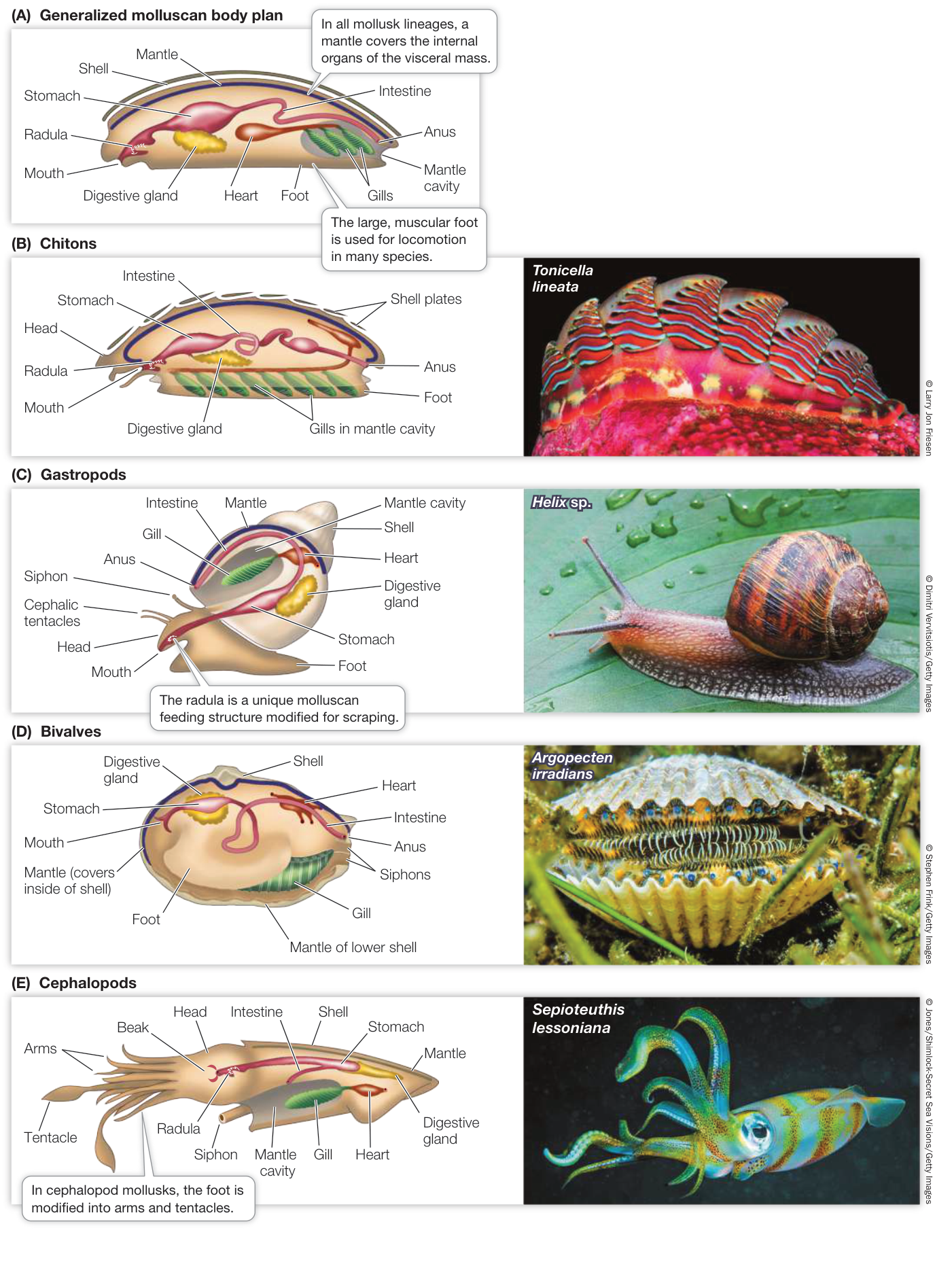
Figure 30.13 Organization and Diversity of Molluscan Bodies (A) The major molluscan groups display different variations on a body plan that includes three major components: a foot, a visceral mass of internal organs, and a mantle. In many species, the mantle secretes a calcareous shell. (B) Chitons have eight overlapping calcareous plates surrounded by a girdle. (C) Most gastropods have a single dorsal shell, into which they can retreat for protection. (D) Bivalves get their name from their two hinged shells, which can be tightly closed. (E) Cephalopods are active predators; they use their arms and tentacles to capture prey. This squid has an internal shell but no external shell. Figure 30.13 Organization and Diversity of Molluscan Bodies (A) The major molluscan groups display different variations on a body plan that includes three major components: a foot, a visceral mass of internal organs, and a mantle. In many species, the mantle secretes a calcareous shell. (B) Chitons have eight overlapping calcareous plates surrounded by a girdle. (C) Most gastropods have a single dorsal shell, into which they can retreat for protection. (D) Bivalves get their name from their two hinged shells, which can be tightly closed. (E) Cephalopods are active predators; they use their arms and tentacles to capture prey. This squid has an internal shell but no external shell.
In all mollusks except cephalopods, the blood vessels do not form a closed system. Blood and other fluids empty into a large, fluid-filled hemocoel, through which fluids move and deliver oxygen to the internal organs. Eventually the fluids reenter the blood vessels and are moved by a heart.
Monoplacophorans were the most abundant mollusks during the Cambrian, 500 million years ago, but only a few species survive today. In monoplacophorans, in contrast to all other living mollusks, the gas exchange organs, muscles, and excretory pores are repeated over the length of the body.
The four major clades of living mollusks are the chitons, gastropods, bivalves, and cephalopods. Each of these groups is readily identifiable and distinct, even though they share variations on a common body plan.
CHITONS Eight overlapping calcareous plates, surrounded by a structure known as the girdle, protect the internal organs and muscular foot of chitons (Figure 30.13B). The chiton body is bilaterally symmetrical, and the internal organs, particularly the digestive and nervous systems, are relatively simple. Most chitons are marine omnivores that scrape algae, bryozoans, and other organisms from rocks with a sharp radula. An adult chiton spends most of its life clinging tightly to rock surfaces with its large, muscular, mucus-covered foot. It moves slowly by means of rippling waves of muscular contraction in the foot. Fertilization in most chitons takes place in the water, but in a few species fertilization is internal and embryos are brooded within the body. There are approximately 1,000 living species of chitons known.
GASTROPODS Gastropods (Figure 30.13C) are the most species-rich and widely distributed mollusks, with about 85,000 known living species. Snails, whelks, limpets, slugs, nudibranchs (sea slugs), and abalones are all gastropods. Most species move by gliding on the muscular foot, but in a few species—the sea butterflies and heteropods—the foot is a swimming organ with which the animal moves through open ocean waters.
Marine nudibranchs and terrestrial slugs are gastropods that have lost their protective shell over the course of evolution (Figure 30.14). Without a shell, these groups rely on other forms of protection from predation. The coloration of many nudibranchs is aposematic, meaning that it serves to warn potential predators of toxicity. Other nudibranch species and most terrestrial slugs exhibit camouflaged coloration.
Shelled gastropods have one-piece shells. The only mollusks that live in terrestrial environments—land snails and slugs—are gastropods. In these terrestrial species, the mantle tissue is modified into a highly vascularized lung.
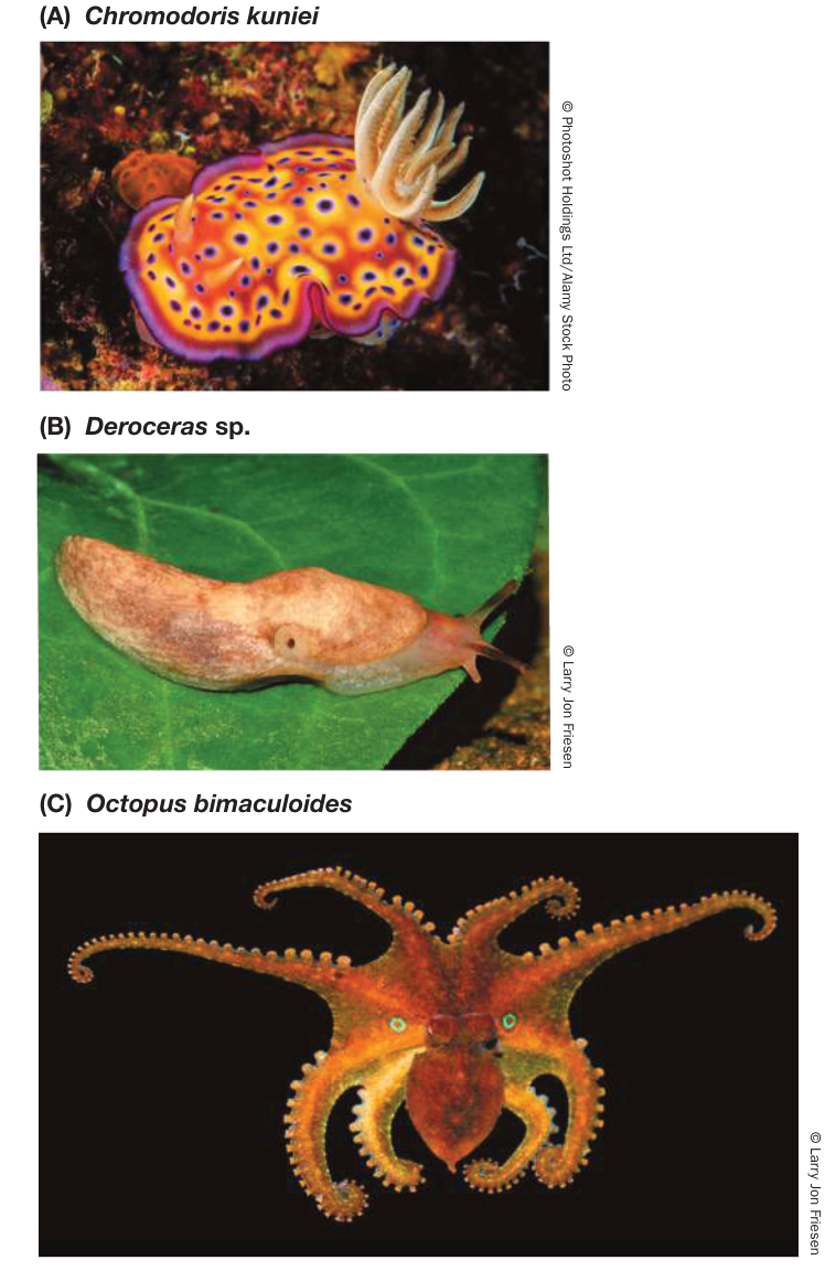
Figure 30.14 Mollusks in Some Groups Have Lost Their Shells (A) Nudibranchs (“naked gills”), also called sea slugs, are shell-less gastropods. This species is brightly colored, alerting potential predators of its toxicity. (B) Slugs are terrestrial, shell-less gastropods that feed on decomposing vegetation on the damp forest floor. (C) Octopuses have neither an external nor an internal shell, which allows these cephalopods to squeeze through tight spaces.
BIVALVES Clams, oysters, scallops, and mussels are all familiar bivalves. The approximately 30,000 known species are found in both marine and freshwater environments. Bivalves have a hinged, two-part shell that extends over the sides of the body as well as the top (Figure 30.13D). Many clams use the foot to burrow into mud and sand. Bivalves feed by taking in water through an opening called an incurrent siphon and filtering food from the water with their large gills, which are also the main sites of gas exchange.
Water and gametes exit through the excurrent siphon. Fertilization takes place in open water in most species.
CEPHALOPODS The cephalopods—squid, cuttlefishes, octopuses, and nautiluses—first appeared near the beginning of the Cambrian. By the Ordovician a variety of types were present. Today there are about 800 described living species. In these mollusks the excurrent siphon is modified to allow the animal to control the volume of the mantle cavity and thereby bring in or expel water (Figure 30.13E). The modification of the mantle into a device for forcibly ejecting water from the cavity through the siphon enables these animals to move rapidly through the water by “jet propulsion.” With their greatly enhanced mobility, cephalopods became the major predators in the open waters of the Devonian oceans. They remain important marine predators today.
As is typical of active, rapidly moving predators, cephalopods have a head with complex sensory organs—most notably eyes that are comparable to those of vertebrates in their ability to resolve images. The head is closely associated with a large, branched foot that bears the arms and/or tentacles and a siphon. Arms are distinguished by the presence of suckers along most of their length. Tentacles, in contrast, have suckers only near the tips or lack suckers altogether. Octopuses typically have eight arms and no tentacles, whereas squid and cuttlefishes have eight arms plus two tentacles. Cephalopods use their arms and tentacles to capture and subdue prey; octopuses also use their arms to move over the substrate. The large, muscular mantle provides a solid external supporting structure. The gills hang in the mantle cavity.
Many early cephalopods had an external chambered shell divided by partitions. The only surviving cephalopods with such shells are the nautiluses (genus Nautilus). The chambers inside nautilus shells are connected by a strand of tissue that runs through ducts in the partitions. Blood in this tissue carries water from the chambers and gases into the chambers, thus providing buoyancy. Most cephalopods retain an internal shell that functions for internal support and, in some species, is also chambered and buoyant. Octopuses have completely lost their shells, which allows them to compress their bodies through very small openings.
Recap and Assess
Lophotrochozoans include animals with diverse body types. Worm-like forms include some flatworms, ribbon worms, phoronids, and annelids. There has been convergent evolution of lophophores (in bryozoans and entoprocts versus brachiopods and phoronids) and of external two-part shell coverings (in brachiopods versus bivalve mollusks).
- How do flatworms survive without an internal transport system?
- Why are most annelids restricted to moist environments?
- Mollusks have evolved a great diversity of body plans. Provide some examples that demonstrate how the basic body organization of mollusks has been modified to yield this diversity.
The second of the two major protostome clades, the ecdysozoans, contains the vast majority of Earth’s animal species. What evolutionary innovations led to this massive diversity?
Ecdysozoans Grow by Shedding Their Cuticles
Learning Objectives
- 30.3.1 Compare cuticle thickness in the different ecdysozoans and explain the consequences of such variation.
- 30.3.2 Explain why the nematode Caenorhabditis elegans is a widely used model system in laboratory research.
- 30.3.3 Explain why nematode diversity is important to global ecosystems.
- 30.3.4 Provide examples of nematodes that are human parasites.
Many ecdysozoans are wormlike in form, although others—the arthropods, velvet worms (onychophorans), and tardigrades—have limbs. In this key concept we look at the two clades of wormlike ecdysozoans: the priapulids, kinorhynchs, and loriciferans in one clade and the nematodes and horsehair worms in the other. Key Concept 30.4 is devoted to the most diverse ecdysozoans—the arthropods and their relatives—and the many forms their appendages take.
As we noted in Key Concept 30.1, ecdysozoans can be identified by the presence of a body covering known as a cuticle, which provides these animals with both protection and support. The name of the group, Ecdysozoa, is derived from the Greek ecdysis (“to get out of”) plus zoa (“animals”). Periodic shedding of the cuticle is necessary so that ecdysozoans can grow. In many ecdysozoans that have wormlike bodies, the cuticle is relatively thin and flexible; it offers the animal some protection but provides only modest body support. A thin cuticle allows the exchange of gases, minerals, and water across the body surface, but it restricts the animal to moist habitats. Many species of ecdysozoans with thin cuticles live in marine sediments from which they obtain food, either by ingesting sediments and extracting organic material from them or by capturing larger prey using a toothed pharynx (a muscular organ at the anterior end of the digestive tract). Some freshwater species absorb nutrients directly through their thin cuticles, as do parasitic species that live within their hosts (endoparasites). Many wormlike ecdysozoans are predators, eating protists and small animals.
Several marine ecdysozoan groups have relatively few species
Members of three species-poor groups of wormlike marine ecdysozoans—the priapulids, kinorhynchs, and loriciferans—have relatively thin cuticles that are molted periodically as the animals grow to full size. Embryos of a fossil species related to these ecdysozoans have been discovered in sediments laid down in China about 500 million years ago. This remarkable discovery shows that the ancestors of these animals developed directly from an egg to the adult form, as most of their modern descendants do.
The 20 known species of priapulids are cylindrical, unsegmented, wormlike animals with a three-part body plan consisting of a proboscis, trunk, and caudal appendage (“tail”). It should be clear from their appearance why they were named after the Greek fertility god Priapus (Figure 30.15A). Priapulids range in length from 0.5 millimeters to 20 centimeters. They live in burrows in fine marine sediments and prey on soft-bodied invertebrates such as polychaetes, which they capture with a toothed, muscular pharynx that they evert through the mouth and then withdraw into the body together with the grasped prey. Fertilization is external, and most species have a larval form that also lives in the mud.
About 180 species of kinorhynchs have been described. They live in marine sands and muds and are virtually microscopic; no kinorhynchs are longer than 1 millimeter. Their bodies are divided into 13 segments, each covered with a separate cuticular plate (Figure 30.15B). These plates are periodically molted during growth. Kinorhynchs feed by ingesting sediments through a retractable proboscis (the group name means “movable snout”). They then digest the organic material found in the sediment, which may include living algae as well as dead matter. Kinorhynchs have no distinct larval stage; fertilized eggs develop directly into juveniles, which emerge from their egg cases with 11 of the 13 body segments already formed.
Loriciferans are also minute animals less than 1 millimeter long. They were not discovered until 1983. About 100 living species are known to exist, although only about 30 of these have been formally described to date. The body is divided into a head, neck, thorax, and abdomen and is covered by six plates, from which the loriciferans get their name (Latin lorica, “corset”). The plates around the base of the neck bear anterior-directed spines of unknown function (Figure 30.15C). Loriciferans live in coarse marine sediments. Little is known about what they eat, but some species apparently eat bacteria.
Nematodes and their relatives are abundant and diverse
Nematodes (roundworms) have a thick, multilayered cuticle that gives their unsegmented body its shape (Figure 30.16). As a nematode grows, it sheds its cuticle four times. Nematodes exchange oxygen and nutrients with their environment through both the cuticle and the gut wall, which is only one cell layer thick. Materials are moved through the gut by rhythmic contraction of a highly muscular pharynx. Nematodes move by contracting their longitudinal muscles.
Nematodes are probably the most abundant and universally distributed of all animal groups. Many nematodes are microscopic; the largest known nematode, which reaches a length of 9 meters, is a parasite in the placentas of sperm whales (Physeter macrocephalus). About 25,000 species have been described, but the actual number of living species may be more than 1 million. Countless nematodes live as scavengers in the upper layers of the soil, on the bottoms of lakes and streams, and in marine sediments. The topsoil of rich farmland may contain contain over 20 billion nematodes per hectare. A single rotting apple may contain as many as 90,000 individuals.
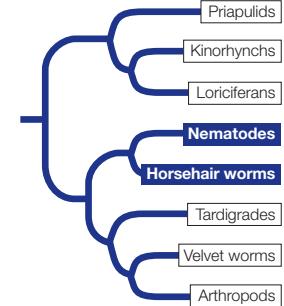
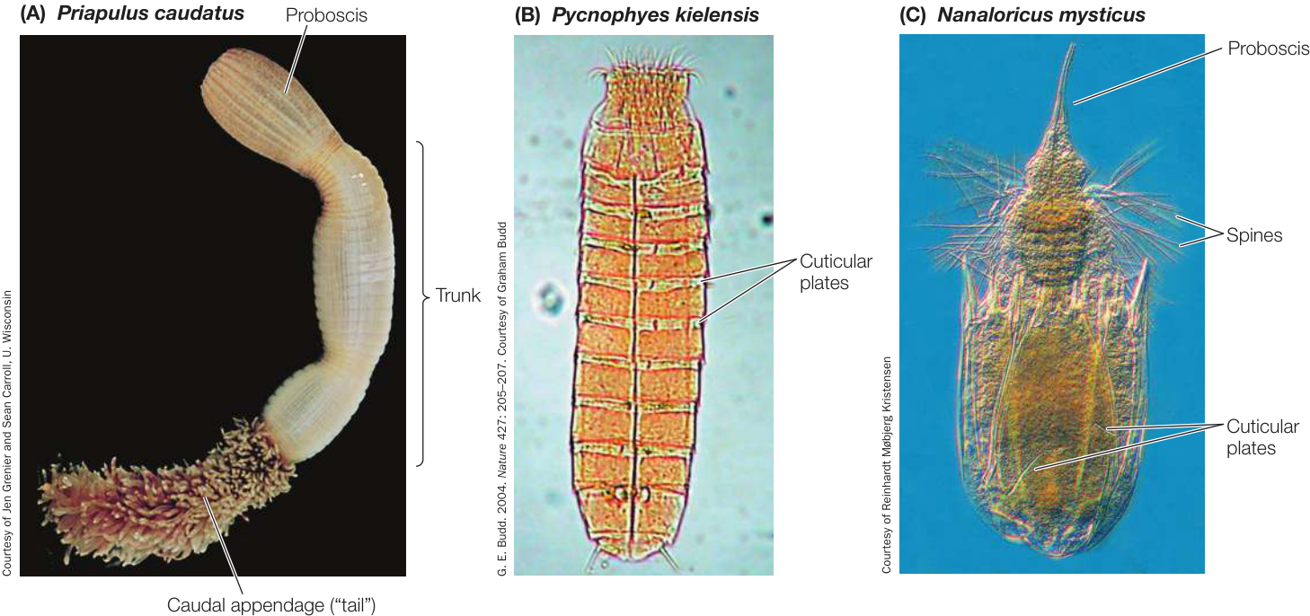
Figure 30.15 Wormlike Marine Ecdysozoans Members of three ecdysozoan groups are marine bottom-dwellers. (A) Most priapulid species live in burrows on the ocean floor, extending the proboscis to feed. (B) Kinorhynchs are virtually microscopic. The cuticular plates that cover their bodies are molted periodically. (C) Six cuticular plates form a “corset” around the minute loriciferan body.
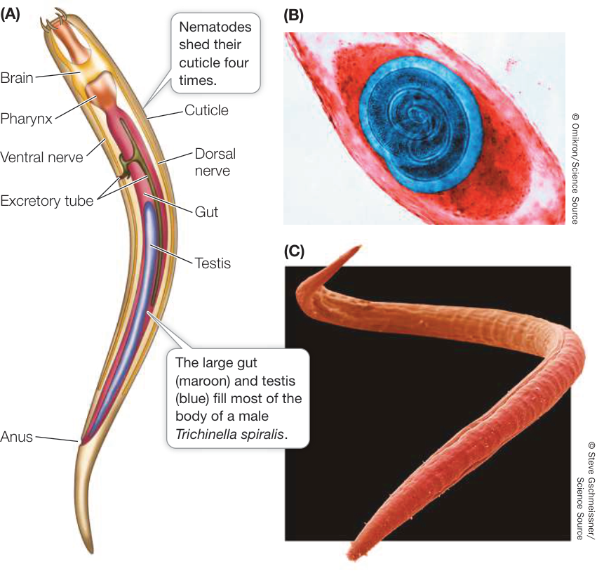
Figure 30.16 Nematodes (A) The body plan of Trichinella spiralis, which causes trichinosis, is typical of parasitic nematodes. (B) This polarized light micrograph shows a cyst of T. spiralis in the muscle tissue of a host. (C) This free-living nematode lives in freshwater environments.
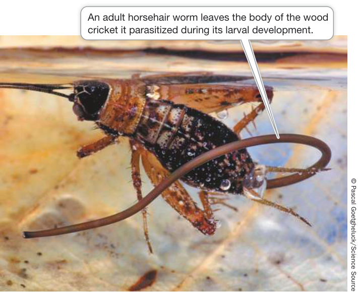
Figure 30.17 Horsehair Worm Larvae Are Parasitic The larva of this horsehair worm (Paragordius tricuspidatus) can manipulate its host’s behavior. The hatching worm causes the cricket to jump into water, where the worm emerges from the insect’s body to continue its life cycle as a free-living adult. The cricket, having delivered its parasitic burden, drowns. 1. 1. How and why does cuticle thickness vary among the ecdysozoans? 2. 2. Describe at least three ways in which nematodes have significant impacts on humans.
One soil-inhabiting nematode, Caenorhabditis elegans, serves as a model organism in the laboratories of geneticists and developmental biologists. It is ideal for such research because it is easy to cultivate, matures in 3 days, and has a fixed number of body cells. Its genome has been completely sequenced.
Many nematodes are predators, feeding on protists and small animals (including other round-worms). Most significant to humans, however, are the many species that parasitize plants and animals. The nematodes that parasitize humans (causing serious diseases such as trichinosis and elephantiasis), domestic animals, and economically important plants have been studied intensively in an effort to find ways of controlling them.
The structure of parasitic nematodes is similar to that of free-living species, but the life cycles of many parasitic species have special stages that facilitate the transfer of individuals among hosts. Trichinella spiralis, the species that causes the human disease trichinosis, has a relatively simple life cycle. A person may become infected by eating the flesh of an animal (usually a pig) that has Trichinella larvae encysted in its muscles (see Figure 30.16B). The larvae are activated in the person’s digestive tract, emerge from their cysts, and attach to the intestinal wall, where they feed. Later they bore through the intestinal wall and are carried in the bloodstream to muscles, where they form new cysts. If present in great numbers, these cysts can cause severe pain or death.
About 350 species of the unsegmented horsehair worms have been described. As their name implies, these animals are extremely thin in diameter; horsehair worms range from a few millimeters up to 1 meter in length. Most adult worms live in fresh water, among leaf litter and algal mats near the edges of streams and ponds. A few species live in damp soil.
Horsehair worm larvae are endoparasites of freshwater crayfishes and of terrestrial and aquatic insects (Figure 30.17). An adult horsehair worm has no mouth, and its gut is greatly reduced and probably nonfunctional. Some species may feed only as larvae, absorbing nutrients from their hosts across the body wall. But other species continue to shed their cuticles and grow after they have left their hosts, suggesting that adult worms may also absorb nutrients from their environment.
Recap and Assess
Priapulids, kinorhynchs, and loriciferans are relatively small, poorly known groups of wormlike marine ecdysozoans. Nematodes and horsehair worms have unsegmented wormlike bodies. Nematodes are among the most abundant and widely distributed animal groups.
We turn next to the animals that not only dominate the ecdysozoan clade but also constitute the most diverse group of animals on Earth.
Arthropods Are the Most Abundant and Diverse Group of Animals
Arthropods Are the Most Abundant and Diverse Group of Animals
Learning Objectives
- 30.4.1 Describe morphological innovations of arthropods and their effects on arthropod diversity.
- 30.4.2 Distinguish among the two different forms of metamorphosis in arthropods.
- 30.4.3 Explain how the evolutionary innovations among arthropods are related to the habitats in which they live.
Arthropods and their relatives are ecdysozoans with paired appendages. Arthropods are the most diverse group of animals in numbers of species (more than 1.2 million have been described, and many more remain to be discovered). Furthermore, the number of individual arthropods alive at any one time is estimated to be about , or 1 billion billion. Among the animals, only the nematodes are thought to exist in greater numbers.
Several key features have contributed to the success of the arthropods. As you have seen, their muscles are attached to the inside of their rigid exoskeletons. Their bodies are segmented, and each segment has muscles that operate that segment and the jointed appendages attached to it (see Figure 30.4). Jointed appendages permit complex movements, and different appendages are specialized for different functions. Encasement of the body within a rigid exoskeleton provides the animal with support for walking in the water or on dry land and provides some protection against predators. The waterproofing provided by chitin keeps the animal from dehydrating in dry air.
The four major arthropod groups living today are all species-rich: chelicerates (including the arachnids—spiders, scorpions, mites, and their relatives), myriapods (millipedes and centipedes), crustaceans (including shrimps, crabs, and barnacles), and hexapods (insects and their wingless relatives). The latter three groups are together known as mandibulates.
The jointed appendages of arthropods gave the clade its name, from the Greek words arthron, “joint,” and podos, “foot” or “limb.” Arthropods evolved from ancestors with simple, unjointed appendages. The exact forms of those ancestors are unknown, but some arthropod relatives with segmented bodies and unjointed appendages survive today. Before we describe the modern arthropods, we discuss those arthropod relatives, as well as an early clade that went extinct but left an important fossil record.
Arthropod relatives have fleshy, unjointed appendages
The two living groups most closely related to the arthropods provide us with clues about the likely appearance of ancestral arthropod appendages. Tardigrades (water bears) have fleshy, unjointed legs and use their fluid-filled body cavity as a hydrostatic skeleton (Figure 30.18A). Tardigrades are tiny (0.5–1.5 mm long) and lack both a circulatory system and gas exchange organs. The 1,200 known extant species live in marine sands and on temporary water films on plants. When these films dry out, the animals also lose water and shrink to small, barrel-shaped objects that can survive for at least a decade in a dormant state. Tardigrades have been found at densities as high as 2 million per square meter of moss.
Until fairly recently, biologists debated whether the velvet worms (onychophorans) were more closely related to annelids or to arthropods, but molecular evidence clearly links them to the latter. Indeed, with their soft, fleshy, unjointed, claw-bearing legs and elongated bodies, velvet worms may be similar in appearance to the ancestors of arthropods (Figure 30.18B). The 180 known species of velvet worms live in leaf litter in humid tropical environments. They have soft, segmented bodies that are covered by a thin, flexible cuticle that contains chitin. Like the tardigrades, velvet worms use their fluid-filled cavity as a hydrostatic skeleton. Fertilization is internal, and the large, yolky eggs are brooded within the body of the female.
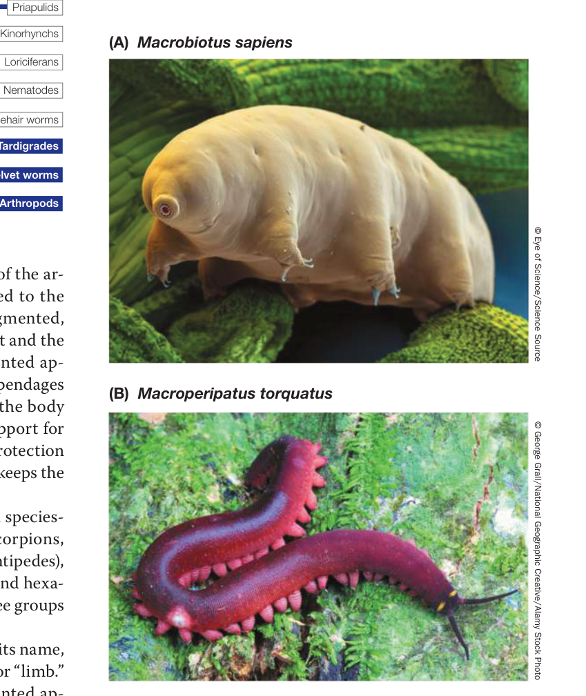
Figure 30.18 Arthropod Relatives with Unjointed Appendages (A) Tardigrades (water bears) can be abundant on the wet surfaces of mosses and plants and in temporary pools of water. (B) Velvet worms have unjointed legs and use the body cavity as a hydrostatic skeleton. They are sometimes referred to as “living fossils,” meaning they are an ancient group that has changed very little over millennia.
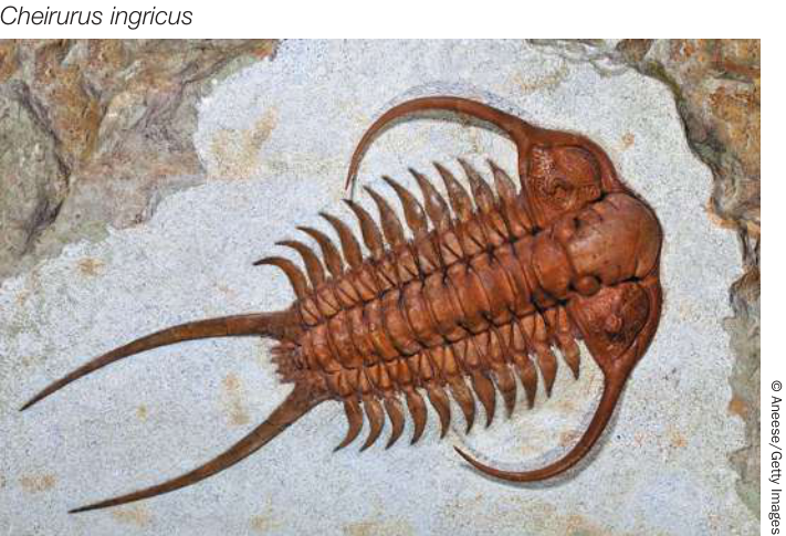
Figure 30.19 A Trilobite Fossil The relatively simple, repetitive segments of the now-extinct trilobites are illustrated by a fossil trilobite from the shallow seas of the Ordovician, some 450 million years ago.
Jointed appendages appeared in the trilobites
The trilobites flourished in Cambrian and Ordovician seas, but they disappeared in the great Permian extinction at the close of the Paleozoic (251 million years ago). Because they had heavy exoskeletons that readily fossilized, they left behind an abundant record of their existence (Figure 30.19). About 10,000 species have been described.
The trilobites are the earliest known arthropods to have had jointed appendages. The body segmentation and appendages of trilobites followed a relatively simple, repetitive plan, but some of their appendages were modified for different functions. This specialization of appendages is a theme in the continuing evolution of the arthropods.
Chelicerates have pointed, nonchewing mouthparts
In the chelicerates, the head bears two pairs of pointed appendages modified to form mouthparts, called chelicerae, that are used to grasp (rather than chew) prey. Chelicerates typically have a two-part body plan, with anterior segments fused to form a cephalothorax, and rear segments fused to form an abdomen. In some groups, such as mites and ticks, there is no clear distinction between these two body parts. Most chelicerates have four pairs of walking legs. The 114,000 described species are grouped into three major clades: sea spiders (pycnogonids), horseshoe crabs, and arachnids.
The sea spiders, make up a poorly known group of about 1,000 marine species (Figure 30.20A). Most are small, with leg spans less than 1 centimeter, but some deep-sea species have leg spans up to 60 centimeters. A few sea spiders eat algae, but most are carnivorous, eating a variety of small invertebrates.
There are only four living species of horseshoe crabs, but many close relatives are known from fossils. Horseshoe crabs, which have changed very little morphologically over their long history, have a large horseshoe-shaped covering over most of the body. They are common in shallow waters along the eastern coast of North America and the southern and eastern coasts of Asia, where they scavenge and prey on bottom-dwelling animals. Periodically they crawl into the intertidal zone in large numbers to mate and lay eggs (Figure 30.20B).
Arachnids are abundant in terrestrial environments. Most arachnids have a simple life cycle in which miniature adults hatch from internally fertilized eggs and begin independent lives almost immediately. Some arachnids retain their eggs during development and give birth to live young.
The most species-rich and abundant arachnids are the spiders, scorpions, harvestmen, mites, and ticks (Figure 30.21). More than 60,000 described species of mites and ticks live in soil, leaf litter, mosses, and lichens, under bark, and as parasites of plants and animals. Mites are vectors for wheat and rye mosaic viruses; they cause mange in domestic animals and skin irritation in humans.
Spiders, of which about 50,000 species have been described, are important terrestrial predators with hollow chelicerae, which they use to inject venom into their prey. Some have excellent vision that enables them to chase and seize their prey. Others spin elaborate webs made of protein threads in which they snare prey. The threads are produced by modified abdominal appendages connected to internal glands that secrete the proteins, which solidify on contact with air. The webs of different groups of spiders are strikingly varied, and this variation enables the spiders to position their snares in many different environments for many different types of prey.
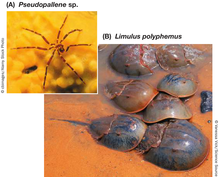
Figure 30.20 Two Small Chelicerate Groups (A) Although they are not spiders, it is easy to see how sea spiders got their common name. (B) A spawning aggregation of horseshoe crabs. Horseshoe crabs, like the velvet worms (see Figure 30.18B), are an example of “living fossils.”
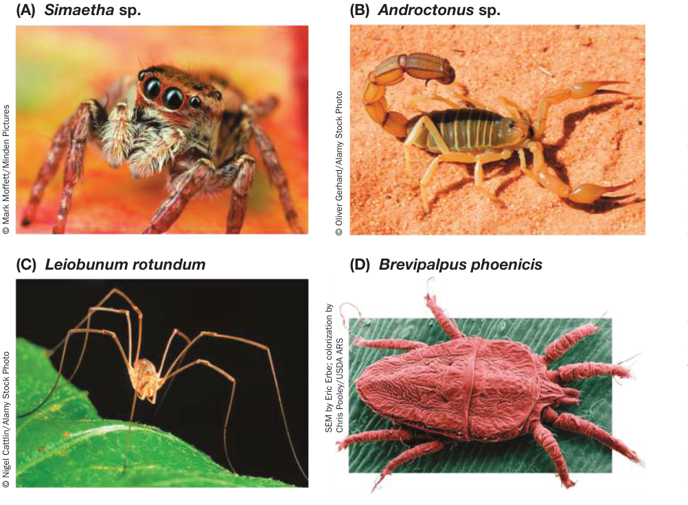
Figure 30.21 Arachnid Diversity (A) Jumping spiders are active, visually oriented, diurnal predators of insects. (B) Scorpions are nocturnal predators of small animals. (C) Harvestmen, also called daddy longlegs, are scavengers. (D) Mites include many free-living species as well as blood-sucking ectoparasites.
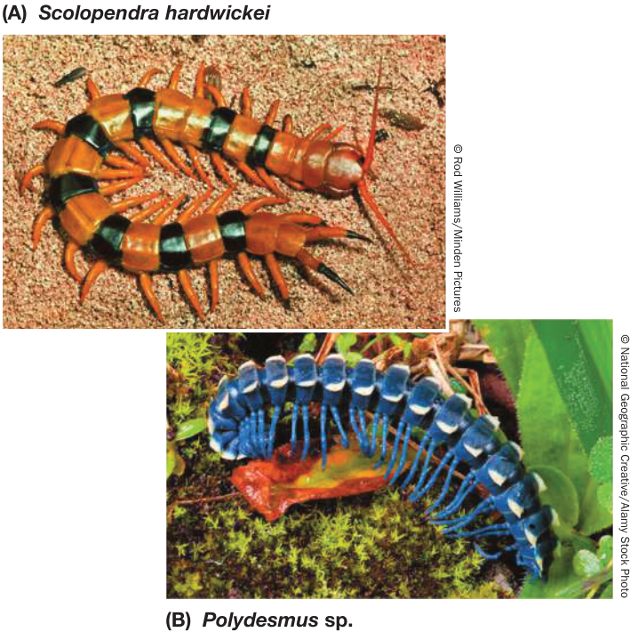
Figure 30.22 Myriapods (A) Centipedes have modified appendages that function as poisonous fangs for capturing active prey. They have one pair of legs per segment. (B) Millipedes are scavengers and plant eaters; they have smaller jaws and legs than centipedes do, and they have two pairs of legs per segment.
Mandibles and antennae characterize the remaining arthropod groups
The remaining three arthropod groups—the myriapods, crustaceans, and hexapods—have mouthparts that are mandibles, rather than chelicerae, so they are collectively called mandibulates. Mandibles can be used for chewing as well as for biting and holding food. Another distinctive characteristic of the mandibulates is the presence of sensory antennae on the head.
MYRIAPODS The myriapods comprise the centipedes, millipedes, and their close relatives. Centipedes and millipedes have a well-formed head that bears the mandibles and antennae characteristic of mandibulates. Their distinguishing feature is a long, flexible, segmented trunk that bears many pairs of legs. Centipedes, which have one pair of legs per segment (Figure 30.22A), prey on insects and other small animals. In millipedes, two adjacent segments are fused so that each fused segment has two pairs of legs (Figure 30.22B). Millipedes scavenge and eat plants. More than 3,000 species of centipedes and 9,000 species of millipedes have been described; many more species probably remain unknown. Although most myriapods are less than a few centimeters long, some tropical species are ten times that size.
CRUSTACEANS Crustaceans are the dominant marine arthropods today, and they are also common in fresh water and some terrestrial environments. The most familiar crustaceans are the shrimps, lobsters, crayfishes, and crabs (all decapods; Figure 30.23A) and the sow bugs (isopods; Figure 30.23B). Additional species-rich groups include the amphipods, ostracods, branchiopods (Figure 30.23C), and copepods (Figure 30.23D), all of which are found in freshwater and marine environments.
Barnacles are unusual crustaceans that are sessile as adults (Figure 30.23E). Adult barnacles look more like mollusks than like other crustaceans, but as the zoologist Louis Agassiz remarked more than a century ago, a barnacle is “nothing more than a little shrimp-like animal, standing on its head in a limestone house and kicking food into its mouth.”
Most of the 67,000 described species of crustaceans have a body that is divided into three regions: head, thorax, and abdomen (Figure 30.24). The segments of the head are fused together, and the head bears five pairs of appendages. Each of the multiple thoracic and abdominal segments usually bears one pair of appendages. The appendages on different parts of the body are specialized for different functions, such as gas exchange, chewing, capturing food, sensing, walking, and swimming. In many species, a fold of the exoskeleton, the carapace, extends dorsally and laterally back from the head to cover and protect some of the other segments.
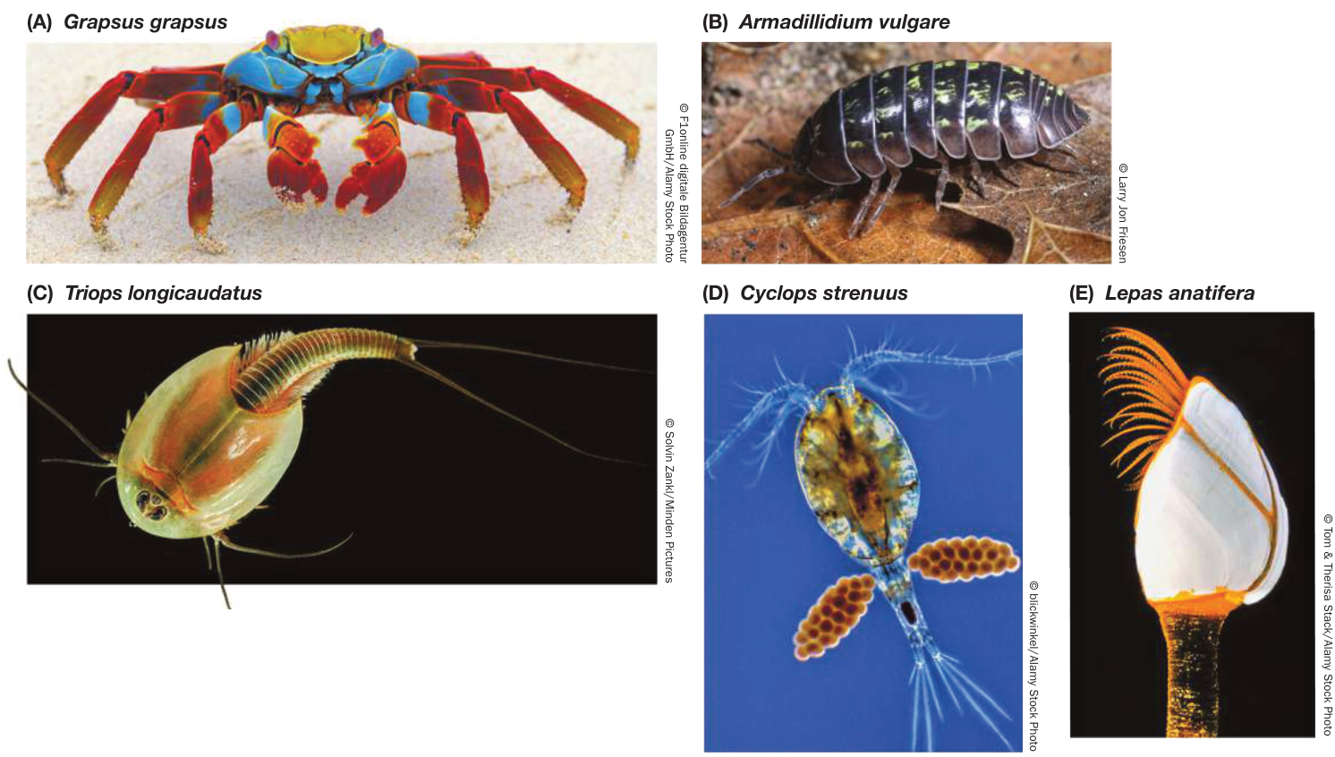
Figure 30.23 Crustacean Diversity (A) This decapod crustacean is a “Sally Lightfoot” crab from the Galápagos. (B) This pillbug, a terrestrial isopod, can roll into a tight ball when threatened. (C) Tadpole shrimp are branchiopods, not to be confused with the decapod crustaceans commonly called shrimp, or with the brachiopods (see Figure 30.9). (D) Minute copepods such as this one are an important link in aquatic food chains. (E) Barnacles are sessile as adults, attaching to a substrate by their muscular stalks and feeding with retractable feeding appendages (the delicate, tentacle-like structures seen here).
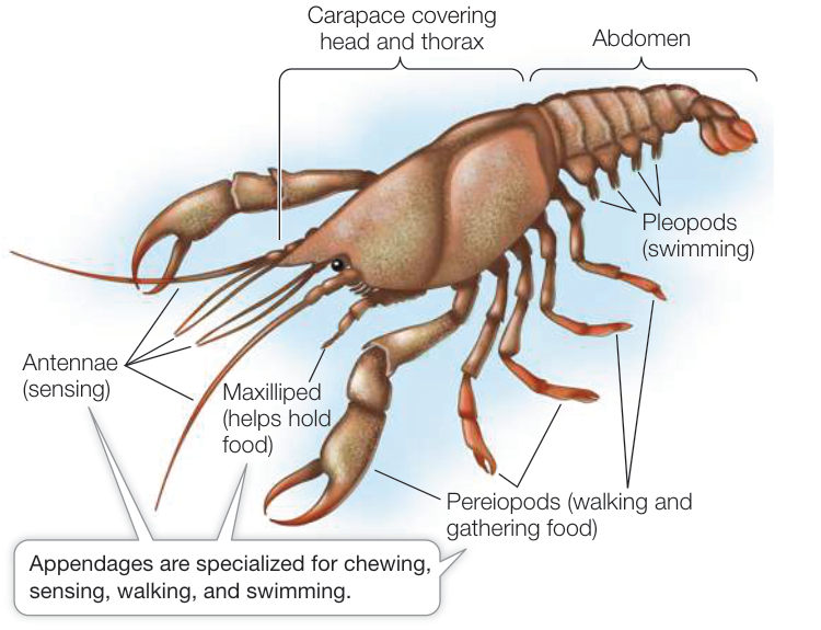
Figure 30.24 Crustacean Body Plan The bodies of crustaceans are divided into three regions: the head, thorax, and abdomen. Each body region bears specialized appendages. A shell-like carapace covers the head and thorax. Q: In what ways is the crustacean body plan similar to and different from the insect body plan shown in Figure 30.26?
The fertilized eggs of most crustacean species are attached to the outside of the female’s body, where they remain during their early development (see Figure 30.23D). At hatching, the young of some species are released as larvae; those of other species are released as juveniles that are similar in form to the adults. Still other species release eggs into the water or attach them to an object in the environment.
More than half of all described species are insects
During the Devonian, more than 400 million years ago, some mandibulates colonized terrestrial environments. Of the several groups (including some crustacean isopods and decapods) that successfully colonized the land, none is more prominent today than the six-legged hexapods, which include the insects and their wingless relatives.
As you saw at the opening of this chapter, more than 1 million of the 1.8 million described living species on Earth are insects, but that is a small fraction of the number of species of insects that are thought to exist on Earth. One of Terry Erwin’s experiments is shown in more detail in Investigating Life: How Many Unknown Species? This and similar experiments show that we have much to learn about the biodiversity of our own planet.
Investigating Life: How Many Unknown Species?
Experiment
Original Papers: T. L. Erwin. 1988. In Biodiversity, E. O. Wilson (ed.), pp. 123–129. National Academy Press: Washington, D.C.
T. L. Erwin. 1997. In Biodiversity II, M. L. Reaka-Kudla et al. (eds.), pp. 27–40. Joseph Henry Press: Washington, D.C.
Entomologist Terry Erwin decided to estimate the number of undescribed species of insects. He devised and executed the following experiment in the forests of Panama.
QUESTION How many species of insects exist on Earth?
METHOD
© Mark Morfetz/Winden Pictures
- Use insecticide fog to collect all beetles from Luehea seemannii trees.
- Sort beetles into species based on morphological differences.
- Calculate the number of host-specific species (those restricted to L. seemannii) and the number of generalists.
- Estimate the number of tree species per hectare of Panamanian forest.
- Calculate the percent of tree-dwelling versus ground-dwelling beetles.
- Calculate the percent of beetles among all insect species.
- Use the calculations above to extrapolate the total number of insect species on Earth.
RESULTS Erwin estimated that there are more than 30 million insect species on Earth (only about 1 million of which have been described).
CONCLUSION Insect diversity is far greater than indicated by the known number of species, and the number of undescribed insect species far exceeds the total number of described species on Earth.
Work with the Data
The data in the table were used by entomologist Terry Erwin to estimate the undescribed diversity of insects. Review the design of Erwin’s experiment in the accompanying box and in the opening story of this chapter. Then use Erwin’s data to answer the questions.
| Approximate number of beetle species collected from Luehea seemannii trees | 1,200 |
|---|---|
| Estimated number of host-specific beetle species in this sample | 163 |
| Number of species of canopy trees per hectare of forest | 70 |
| Percent of beetle species living in canopy (as opposed to ground-dwelling species) | 75% |
| Percent of beetles among all insect species | 40% |
Source: T. L. Erwin. 1982. The Coleopterists Bulletin 36: 74–75.
QUESTIONS
- From the data in the table, estimate the number of insect species in an average hectare of Panamanian forest. Assume that the data for beetles on L. seemannii are representative of the other tree species, and that all the species of beetles that are not host-specific were collected in the original sample. Remember to sum your estimates of the number of (a) host-specific beetle species in the forest canopy; (b) non-host-specific beetle species in the forest canopy; (c) beetle species on the forest floor; and (d) species of all insects other than beetles.
- There are about 50,000 species of tropical forest trees. Assume that the data for beetles on L. seemannii are representative of other species of tropical trees and calculate the number of host-specific beetle species found on these trees. Add an estimated 1 million species of non-host-specific beetles that are expected across different species of trees (including those in temperate regions). Estimate the number of ground-dwelling beetle species based on the percent used in Question 1. Now use this information to estimate the number of insect species on Earth, based on the percent of beetles among all insect species.
The wingless relatives of the insects—the springtails, two-pronged bristletails, and proturans—are probably the most similar of living forms to insect ancestors (Figure 30.25). These hexapods have a simple life cycle: they hatch from eggs as miniature adults. They differ from insects in having internal mouthparts. Springtails can be extremely abundant (up to 200,000 per m2) in soil, leaf litter, and on vegetation and are the most abundant hexapods in the world in terms of number of individuals (as opposed to number of species).
Like crustaceans, insects have a body with three regions: head, thorax, and abdomen. They have a single pair of antennae on the head and three pairs of legs attached to the thorax. In most groups of insects, the thorax also bears two pairs of wings. Unlike other arthropods, insects have no appendages growing from their abdominal segments (Figure 30.26). Insects are distinguished from springtails and other hexapods by their external mouthparts and by antennae that contain a motion-sensitive receptor called Johnston’s organ. In addition, insects have a derived mechanism for gas exchange in air: a system of air sacs and tubular channels called tracheae (singular trachea) that extend from external openings called spiracles inward to tissues throughout the body (see Figure 47.4).
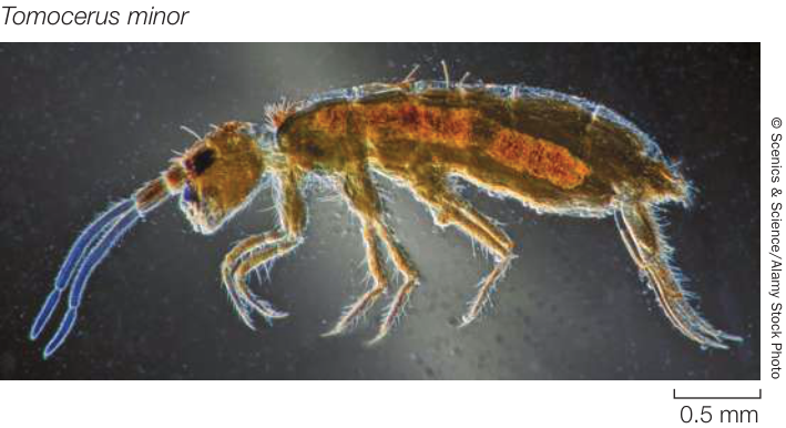
Figure 30.25 Wingless Hexapods The wingless hexapods, such as this springtail, have a simple life cycle. They hatch looking like miniature adults, then grow by successive molts of the cuticle.
Table 30.2 lists the major insect groups. Two groups—the jumping bristletails and silverfishes—are wingless and have simple life cycles, like the springtails and other non-insect hexapods. The remaining groups are all pterygote insects. Pterygotes have two pairs of wings, except in some groups in which one or both pairs of wings have been secondarily lost. These secondarily wingless groups include the parasitic lice and fleas, some beetles, and the worker individuals of many ant species.
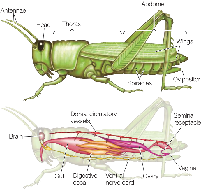
Figure 30.26 Insect Body Plan Like those of crustaceans, the bodies of insects are divided into three regions: head, thorax, and abdomen. In insects, however, the thorax bears three pairs of legs and, in most groups, two pairs of wings. Unlike other arthropods, insects have no appendages growing from their abdominal segments.
Hatchling pterygotes do not look like adults; they undergo substantial changes at each molt. The immature stages of insects between molts are called instars. A substantial change that occurs between one developmental stage and another is called metamorphosis. If the changes between stages are gradual, an insect is
Table 30.2 The Major Insect Groupsa
| Group | Approximate number of described living species |
|---|---|
| Jumping bristletails (Archaeognatha) | 560 |
| Silverfishes (Thysanura) | 580 |
| PTERYGOTE (WINGED) INSECTS (PTERYGOTA) | |
| Mayflies (Ephemeroptera) | 3,250 |
| Dragonflies and damselflies (Odonata) | 6,000 |
| NEOPTERANS (NEOPTERA)b | |
| Ice-crawlers (Grylloblattodea) | 35 |
| Gladiators (Mantophasmatodea) | 15 |
| Stoneflies (Plecoptera) | 3,800 |
| Webspinners (Embioptera) | 500 |
| Angel insects (Zoraptera) | 40 |
| Earwigs (Dermaptera) | 2,000 |
| Grasshoppers and crickets (Orthoptera) | 24,000 |
| Stick insects (Phasmida) | 3,000 |
| Cockroaches (Blattodea) | 4,500 |
| Termites (Isoptera) | 3,000 |
| Mantids (Mantodea) | 2,500 |
| Booklice and barklice (Psocoptera) | 5,800 |
| Thrips (Thysanoptera) | 6,000 |
| Lice (Phthiraptera) | 5,000 |
| True bugs, cicadas, aphids, leafhoppers (Hemiptera) | 105,000 |
| HOLOMETABOLOUS NEOPTERANS (HOLOMETABOLA)c | |
| Ants, bees, wasps (Hymenoptera) | 150,000 |
| Beetles (Coleoptera) | 400,000 |
| Twisted-wing parasites (Strepsiptera) | 650 |
| Lacewings, ant lions, mantidflies (Neuropterida) | 6,000 |
| Dobsonflies, alderflies, fishflies (Megaloptera) | 350 |
| Snakeflies (Raphidioptera) | 250 |
| Scorpionflies (Mecoptera) | 800 |
| Fleas (Siphonaptera) | 2,000 |
| True flies (Diptera) | 125,000 |
| Caddisflies (Trichoptera) | 12,000 |
| Butterflies and moths (Lepidoptera) | 180,000 |
aThe hexapod relatives of insects include the springtails (Collembola; 3,000 spp.), two-pronged bristletails (Diplura; 600 spp.), and proturans (Protura; 10 spp.). All are wingless and have internal mouthparts.
bNeopteran insects can tuck their wings close to their body.
cHolometabolous insects are neopterans that undergo complete metamorphosis.
said to have incomplete metamorphosis. If the change between at least some stages is dramatic, an insect is said to have complete metamorphosis (see Figure 29.10). In many insects with complete metamorphosis, different stages are specialized for different functions and use different food sources. In many species the larvae are specialized for feeding and growing, whereas the adults are specialized for reproduction and dispersal.
The adults of most flying insects have two pairs of stiff, membranous wings attached to the thorax. True flies, however, have one pair of wings and a pair of stabilizers called halteres (which are derived from the second pair of wings). In winged beetles, one pair of wings—the forewings—forms heavy, hardened wing covers.
Two groups of pterygotes, the mayflies and dragonflies (Figure 30.27A), cannot fold their wings against their body. This is the ancestral condition for pterygote insects, and the mayflies and dragonflies are not closely related to one another. Members of both groups have predatory or herbivorous aquatic larvae that transform into flying adults after they crawl out of the water. Dragonflies (and their relatives the damselflies) are active predators as adults. In contrast, adult mayflies lack functional digestive tracts. Adult mayflies live only about a day, just long enough to mate and lay eggs.
All other pterygote insects—the neopterans—can tuck their wings out of the way upon landing and crawl into crevices and other tight places. Some neopteran groups undergo incomplete metamorphosis, so hatchlings of these insects are sufficiently similar in form to adults to be recognizable. Examples include the grasshoppers (Figure 30.27B), roaches, mantids, stick insects, termites, stoneflies, earwigs, thrips, true bugs (Figure 30.27C), aphids, cicadas, and leafhoppers. These groups acquire adult organ systems, such as wings and compound eyes, gradually through several juvenile instars.
More than 80% of all insects belong to a subgroup of the neopterans called the holometabolous insects (see Table 30.2), which undergo complete metamorphosis (see Figure 30.27D-H). The many species of beetles account for almost half of this group (Figure 30.27D). Also included are lacewings and their relatives; caddisflies (Figure 30.27E); butterflies and moths (Figure 30.27F); true flies (Figure 30.27G); and bees, wasps, and ants, some species of which display unique and highly specialized social behaviors (Figure 30.27H).
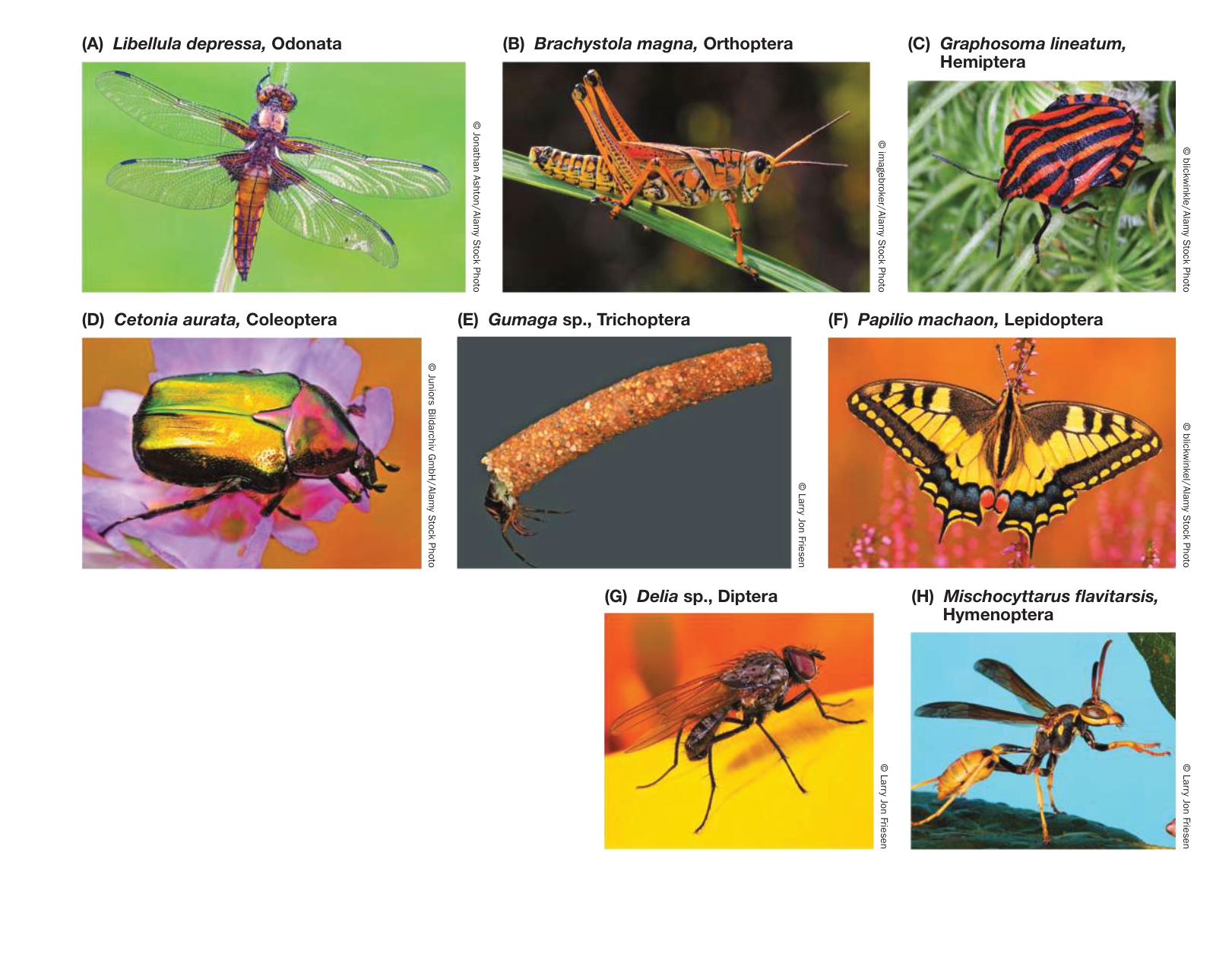
Figure 30.27 Diverse Pterygotes (A) Unlike most flying insects, a dragonfly cannot fold its wings over its back. (B) Orthopteran insects such as grasshoppers undergo several larval molts (instars), but the juveniles resemble small adults (incomplete metamorphosis). (C) Hemipterans are “true” bugs. (D–H) Holometabolous insects undergo complete metamorphosis. (D) Coleoptera is the largest insect group; beetles such as this rose chafer account for more than half of all holometabolous species. (E) A larval caddisfly emerges from its case constructed from silk and sand. (F) The swallowtail butterfly is found in most parts of North America. (G) Blowflies are among the “true” flies, the Dipterans. Adult blowflies feed on pollen or nectar but lay their eggs on carrion, upon which the larvae feed. (H) This western paper wasp is a hymenopteran, a group in which most members display social behaviors.
Molecular data suggest that insects began to diversify over 400 million years ago, soon after the appearance of the first land plants. These early hexapods evolved in a terrestrial environment that lacked any other similar organisms, which in part accounts for their remarkable success. But the success of the insects is also due to their wings. Pterygote insects were the first animals in evolutionary history to achieve the ability to fly. Homologous genes control the development of insect wings and crustacean appendages, suggesting that the insect wing evolved from a dorsal branch of a crustacean-like limb (Figure 30.28). The dorsal limb branch of crustaceans is used for gas exchange. Thus the insect wing probably evolved from a gill-like structure that had a gas exchange function.
Flight opened up many new lifestyles and feeding opportunities that only the insects could exploit, such as pollination of (and coevolution with) flowering plants. Flight is almost certainly one of the reasons for the remarkable numbers of both insect species and individual insects, and for their unparalleled evolutionary success.
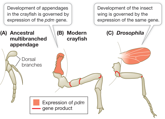
Figure 30.28 The Origin of Insect Wings? Insect wings may be derived from an ancestral appendage similar to that of modern crustaceans. (A) A diagram of the ancestral, multibranched arthropod limb. The uppermost dorsal branch may have been used for gas exchange. (B, C) The pdm gene, a Hox gene, is expressed throughout the dorsal limb segment of the thoracic limb of a crayfish (B) and in the wings of Drosophila (C). Q: If the hypothesized origin of insect wings is correct, then what was a possible function of the structure that would become a wing in flying insects?
Recap and Assess
All arthropods have segmented bodies. Muscles in each segment operate that segment and the appendages attached to it. Jointed, specialized appendages permit complex patterns of movement, including, in insects, the ability to fly. With flight, insects took advantage of new feeding and lifestyle opportunities, which contributed to the unparalleled evolutionary success of this group.
- What features have contributed to making arthropods among the most abundant animals on Earth, both in number of species and in number of individuals?
- Describe the difference between incomplete and complete metamorphosis.
- There are more described and named species of insects than of all other species on Earth combined. However, only a very few insect species live in marine environments, and those species are restricted to the intertidal zone or the ocean surface. What factors may have contributed to the insects’ lack of success in the oceans?
Key Features of Protostome Evolution
The protostomes encompass a staggering number of different body forms and lifestyles. The following aspects of protostome evolution have contributed to this enormous diversity:
- The evolution of segmentation permitted some groups of protostomes to move different parts of the body independently of one another. Species in some groups gradually evolved the ability to move rapidly over and through the substrate, through water, and through air.
- Complex life cycles with dramatic changes in form between one stage and another allow individuals of different stages to specialize on different resources.
- Parasitism has evolved repeatedly, and many protostome groups parasitize plants and animals.
- The evolution of diverse feeding structures allowed protostomes to specialize on many different food sources. Specialization on food sources undoubtedly contributed to reproductive isolation and further diversification.
- Predation was a major selection pressure favoring the development of hard external body coverings (exoskeletons and shells). Such coverings evolved independently in many lophotrochozoan and ecdysozoan groups. In addition to providing protection, these coverings became key elements in the development of new systems of locomotion.
- Better locomotion permitted prey to escape from predators, but also allowed predators to pursue their prey more effectively. Thus the evolution of animals has been, and continues to be, a complex “arms race” among predators and prey.
Animation 30.1 An Overview of the Protostomes
Many major evolutionary trends among the protostomes are shared by the deuterostomes, which include the chordates, the group to which humans belong. We will turn to the deuterostomes in the next chapter.
Investigating Life
QA Which groups of protostomes are thought to contain the most undiscovered species?
It is perhaps easier to list the groups of protostomes for which a nearly complete inventory of living species has been done than to list all the groups for which many new species remain to be described. Among the insects, the best-studied group, in terms of species, is the butterflies, which are widely collected and studied. There are still many species of other lepidopterans (such as moths), however, remaining to be discovered. As we explored in Investigating Life: How Many Unknown Species?, some other major insect groups, such as beetles, contain many undescribed species.
New species discovery and description rates remain high for almost all other major groups of protostomes. Second to the insects, and perhaps even rivaling the insects in undiscovered diversity, are the nematodes. Although known nematode diversity is only about one-fortieth of known insect diversity (in terms of number of described species), the taxonomy of nematodes has been much more poorly studied than that of insects. Some biologists think there are likely to be species-specific parasitic nematodes specializing on most other species of multicellular organisms. If so, then there may be as many species of nematodes as there are of plants, fungi, and other animals combined.
Most of the other diverse groups of protostomes also contain many as yet undetected species, judging from the rate of new species descriptions. In particular, flatworms (especially the parasitic flukes and tapeworms), marine annelids, mollusks, crustaceans, myriapods, and chelicerates all contain large numbers of undescribed species.
Future directions
Biologists have now placed almost all known species into a comprehensive tree of life (see Appendix A). This achievement allows the development of new technology for automated species discovery and identification. Once we can identify an organism, it is then possible to connect it to all the known information about that species. It should soon be possible to construct a hand-held device that will isolate DNA from any sample, and then to sequence a series of target genes that can be used to place an unknown sample within the tree of life. This process results in either identification of a known species, or placement of an undescribed species with its closest relatives in the tree. This technology is expected to revolutionize the study of biodiversity and its applications.
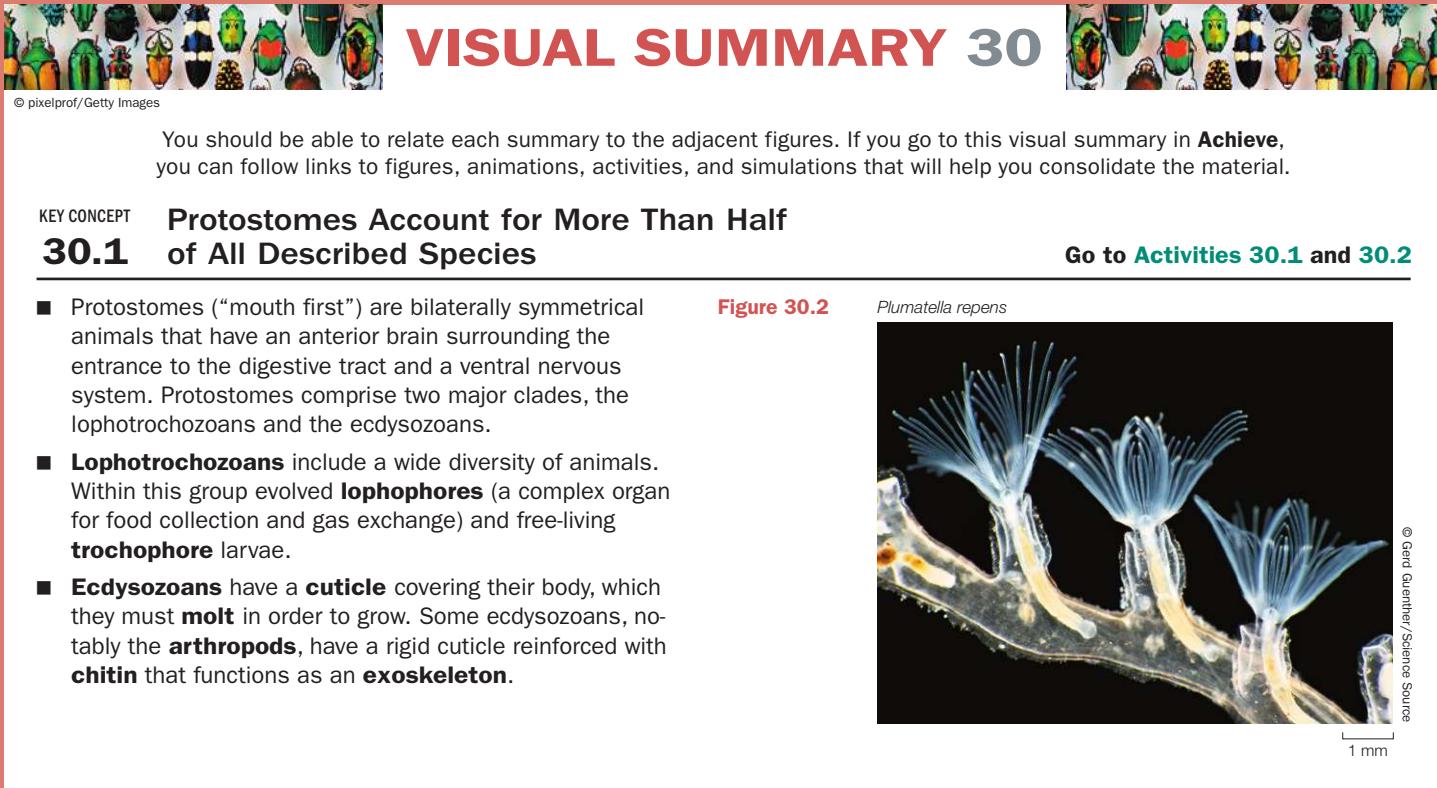
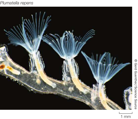
Figure 30.2
Questions
- What developmental trait distinguishes most protostomes from other animals?
- Give examples of familiar lophotrochoans and ecdysozoans.
- What are some lophotrochozoans and ecdysozoans that are commonly consumed by many humans?
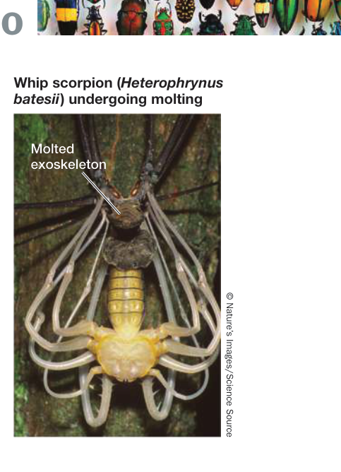
Figure 30.3
Visual Summary
Many Lophotrochozoans Have Ciliated Feeding Structures or Life Stages
- ■ Lophotrochozoans range from animals with a blind gut and no internal transport system to animals with a complete digestive tract and a complex internal transport system.
- ■ Most species of bryozoans and entoprocts live in colonies produced through asexual reproduction.
- ■ Ribbon worms feed using a long, protrusible proboscis.
- ■ The shelled brachiopods and worm-like phoronids use a lophophore to feed; this lophophore may have evolved independently of the lophophore in bryozoans and entoprocts.
- ■ The most species-rich groups of lophotrochozoans are the flatworms, annelids, and mollusks.
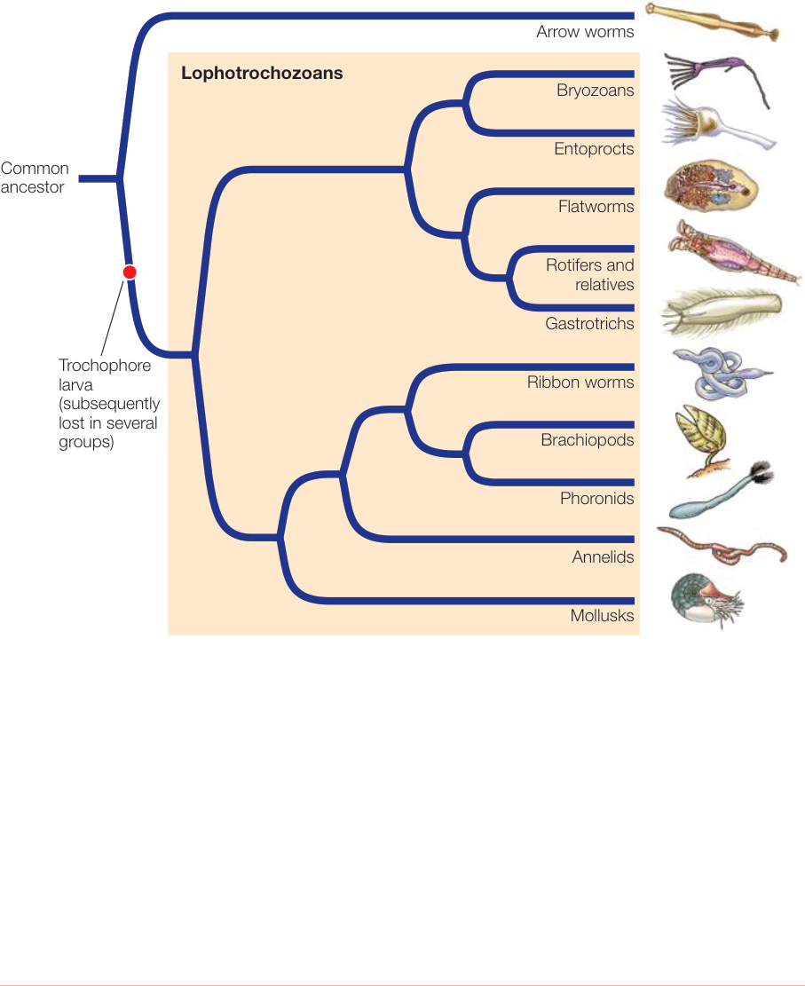
Figure 30.1 (top)
Questions
- If the lophophores of brachiopods, phoronids, bryozoans, and entoprocts did not evolve independently in these groups, what is an alternative explanation for its presence in these relatively distantly related lophotrochozoans?
- Paired, hinged shells that completely enclose the body evolved independently in which two major groups of lophotrochozoans?
Ecdysozoans Grow by Shedding Their Cuticles
- ■ Ecdysozoans include the most diverse groups of animals, including nematodes and arthropods.
- ■ Several species-poor groups of wormlike marine ecdysozoans—priapulids, kinorhynchs, and loriciferans—have thin cuticles.
- ■ Horsehair worms are extremely thin; many are endoparasites as larvae.
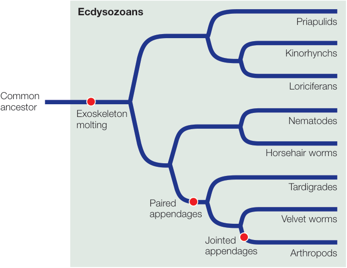
Figure 30.1 (bottom)
Questions
- What are the primary functions of the cuticle in ecdysozoans?
- What attributes make the nematode Caenorhabditis elegans ideal for use as a model organism in the laboratories of many geneticists and developmental biologists?
Arthropods Are the Most Abundant and Diverse Group of Animals
- ■ Arthropods are the dominant animals on Earth in number of described species, and among the most abundant in number of individuals.
- ■ Encasement within a rigid exoskeleton provides arthropods with support for walking as well as some protection from predators. The waterproofing provided by chitin keeps arthropods from dehydrating in dry air.
- ■ Jointed appendages permit complex movements. Each arthropod segment has muscles attached to the inside of the exoskeleton that operate that segment and the appendages attached to it.
- ■ Crustaceans are the dominant marine arthropods and are also found in many freshwater and some terrestrial environments. Their segmented bodies are divided into three regions (head, thorax, and abdomen) with different, specialized appendages in each region.
- ■ Hexapods—insects and their relatives—are the dominant terrestrial arthropods. They have the same three body regions as crustaceans, but no appendages form in their abdominal segments. Wings and the ability to fly first evolved among the insects, allowing them to exploit new lifestyles.
Questions
- What are the other major groups of living arthropods, besides the crustaceans and hexapods?
- What was the likely original function of the organ from which insect wings evolved?
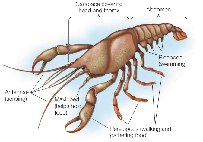
Figure 30.24
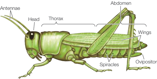
Figure 30.26