Chapter 32The Plant Body
PART SEVEN
Flowering Plants: Form and Function
© Misokhili/Gatty Images
Key Concepts
32.1 The Plant Body Is Organized in a Distinctive Way
32.2 Plant Organs Are Made Up of Cells Organized into Three Types of Tissues
32.3 Meristems Build a Continuously Growing Plant
32.4 Domestication Has Altered Plant Form
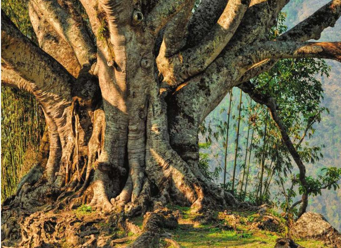
Plants such as this old tree have roots, stems, and leaves that perform the functions of the organism.
Investigating Life: Bread of the Tropics
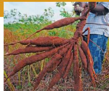
To North Americans and Europeans, it is tapioca. To Central and South Americans, it is yuca. To Africans, it is manioc or bananku. The roots of the cassava plant, Manihot esculenta, are important in the diets of more than 800 million people. Cassava is grown mostly by family farmers for their own consumption. The root of the plant is a store of starch, which can be hydrolyzed and used by the plant as the stems and leaves grow. Other crop plants such as rice and wheat apportion about 35% of the total carbon fixed in photosynthesis into their storage organs (the grains), whereas cassava apportions an astounding 80% into the root, as starch. For humans, this is a convenient and concentrated source of food energy. Indeed, cassava has been nicknamed the “bread of the tropics” because, just as wheat bread is the major starchy food in the Western world, cassava supplies the starch in tropical areas.
Most cassava plants are clones. The plants have wide adaptability and grow well in dry soils, in both hot and cool climates. Typically, the farmer breaks off some pieces of stem and plants them. Because most plant cells are totipotent (see Key Concept 18.2), some of the stem cells redifferentiate and form roots, while others become growing shoots. A whole new plant develops, and the roots are ready to eat 6 months to 2 years later.
There are countless recipes for preparing cassava as food, but it is nutritionally incomplete. Although rich in carbohydrates, cassava is a relatively poor source of protein, a requirement for the human diet. Eating cassava presents other difficulties. Cassava roots and leaves contain cyanogenic compounds, which are converted to cyanide by enzymes released when the cells are broken open. Cyanide is highly toxic and potentially lethal because it blocks electron transport in the mitochondria. Therefore it is essential to soak, cook, or ferment cassava, to break down the cyanogenic compounds and eliminate the cyanide, before it can be eaten safely. In all probability, the plant uses cyanide production as a protection against predators that eat it.
We open with the topic of cassava because it offers a preview of a wide range of studies embraced by the discipline of plant physiology. Plant physiology is a broad subject, covering photosynthesis, transport, plant nutrition, regulation of growth and development, reproduction, and the interactions between plants and their environments.
© Plus Utomi Etepi/APP/Gatty Images
QA How might plant biologists improve the cassava plant for human use? (Find the answer on page 745.)
The Plant Body Is Organized in a Distinctive Way
Learning Objectives
32.1.1 Describe the overall structure of a flowering plant in terms of its major systems and axes.
32.1.2 Identify ways in which meristem-based growth overcomes some of the problems posed by scarce resources and an inability to move.
32.1.3 List criteria that distinguish monocots and eudicots.
32.1.4 Explain the process by which a zygote develops into an embryo.
Despite their obvious differences in size and form, all vascular plants have essentially the same simple structural organization. This chapter describes the basic architecture of the largest group of vascular plants, the angiosperms (flowering plants), and shows how so much diversity can literally grow out of such a simple, basic form. There are more than 300,000 angiosperm species. A major distinguishing feature of angiosperms is flowers, which consist of modified leaves and stems and carry the organs for sexual reproduction. We will examine the structures and functions of flowers in detail in Chapter 37.
Vegetative (nonsexual) plant organs are organized into two systems (Figure 32.1):
- The root system anchors the plant in place, absorbs water and dissolved minerals, and stores the products of photosynthesis from the shoot system. The extreme branching of plant roots and their high surface area-to-volume ratios allow them to absorb water and mineral nutrients from the soil efficiently.
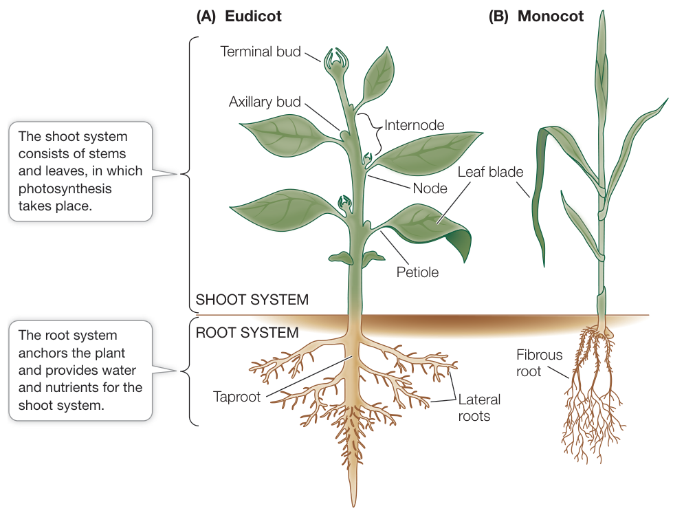
Figure 32.1 Vegetative Plant Organs and Systems The basic plant body plan, showing the root and shoot systems and the principal vegetative organs, of (A) a eudicot and (B) a monocot.
- The shoot system consists of the stems, leaves, and flowers. Broadly speaking, the leaves are the chief organs of photosynthesis. The stems hold and display the leaves to the Sun and provide connections for the transport of materials between roots and leaves. Axillary buds form in the angle (axil) where a leaf meets the stem. A bud is an undeveloped shoot that can develop further. If an axillary bud becomes active, it can develop into a new branch, which is an extension of the shoot system.
Most angiosperms are either monocots or eudicots
As you saw in Key Concept 27.3, most angiosperms belong to one of two major clades. Monocots are generally narrow-leaved flowering plants such as grasses, lilies, and orchids. Eudicots are broad-leaved flowering plants such as soybeans, roses, sunflowers, and maples. These two clades, which account for 97% of flowering plant species, are named for the number of cotyledons, leaf-like organs, that are present in a mature seed. However, monocots (with one cotyledon) and eudicots (with two cotyledons) differ in several basic characteristics, as shown in Figure 32.2. We’ll provide more detailed comparisons between these clades throughout the chapter.
Meristems generate the plant body
Plants harvest energy from sunlight and collect water and mineral nutrients from the atmosphere and the soil. Because these resources are sometimes limited, plants must collect them from large areas, both above and below the ground. Plants are further challenged by their inability to walk; a plant cannot, for example, relocate from a dry, shady location to one that is wet and sunny. The plant body plan responds to these challenges in two ways:
- Stems, leaves, and roots enable a plant anchored to one spot to capture scarce resources effectively, both above and below the ground.
- Plants can grow throughout their lifetimes, enabling them to respond to environmental cues. A plant can redirect its growth to exploit opportunities in its immediate environment; for example, it can extend its roots toward a water supply.
The ability of plants to grow new body parts throughout their lifetime distinguishes plants from most animals. Most animals stop growing when they reach the adult state, a growth pattern referred to as determinate. In contrast, the open-ended growth of plants is indeterminate. The number of branches they will produce depends on the environment; it is not specified when a seedling starts growing. Indeterminate growth of the shoot and root systems is made possible by meristems. Meristems are localized regions of undifferentiated cells that are the sources of all new growth in the adult plant. Even before seed germination, the plant embryo has two apical meristems: a shoot apical meristem near the end of the embryonic shoot, and a root apical meristem at the end of the embryonic root.
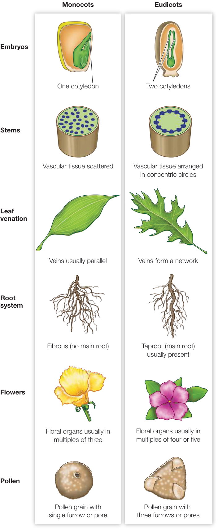
Figure 32.2 Comparing the Two Major Angiosperm Clades There are several structural differences between monocots and eudicots.
Meristematic cells are small and closely packed. The cells that perpetuate the meristems, called initials, are comparable to animal stem cells (discussed in Key Concept 18.2). When the initials divide, some of the daughter cells develop into new initials, and some differentiate into more specialized cells.
Two types of meristems contribute to the growth and development of the adult plant (Figure 32.3):
- The apical meristems in the root and shoot orchestrate primary growth (the growth of shoots and roots), ultimately giving rise to every cell in the primary plant body, directly or through the formation of additional meristems.
- Lateral meristems orchestrate growth in thickness and facilitate branching of roots or shoots. Two lateral meristems (also called secondary meristems), vascular cambium and cork cambium, contribute to the secondary plant body (the bark and wood).
Apical–basal polarity and radial symmetry are characteristics of the plant body
Two basic patterns are established early in plant embryogenesis (embryo formation) (Figure 32.4). The apical–basal axis, the ends of which are defined by the shoot and root meristems, is the plant’s central axis. The radial axis is the concentric arrangement of the tissue systems (discussed in Key Concept 32.2). We will focus here on how these axes develop in a model eudicot, Arabidopsis thaliana, that has been studied extensively by plant physiologists and geneticists. You can follow the steps that occur during A. thaliana embryogenesis in Figure 32.5.
The first step in the formation of an A. thaliana embryo is a mitotic division of the zygote that gives rise to two daughter cells, an apical daughter cell and a basal daughter cell (see Figure 32.5, Step 1). An asymmetrical plane of cell division results in an uneven distribution of cytoplasm between these two cells, and this determines their different fates. Signals in the smaller, apical daughter cell induce it to produce the embryo proper, while the larger basal daughter cell produces the suspensor, which forms a stalk under the embryo (see Figure 32.5, Step 2). This division not only establishes the apical–basal axis of the new plant but also determines its polarity (which end is the tip, or apex, and which is the base). The end of the embryo attached to the suspensor forms the root. A long, thin suspensor and a more spherical or globular embryo are distinguishable after just four mitotic divisions. The suspensor soon ceases to elongate.
In eudicots such as A. thaliana, the initially globular embryo develops into the characteristic heart stage as the cotyledons
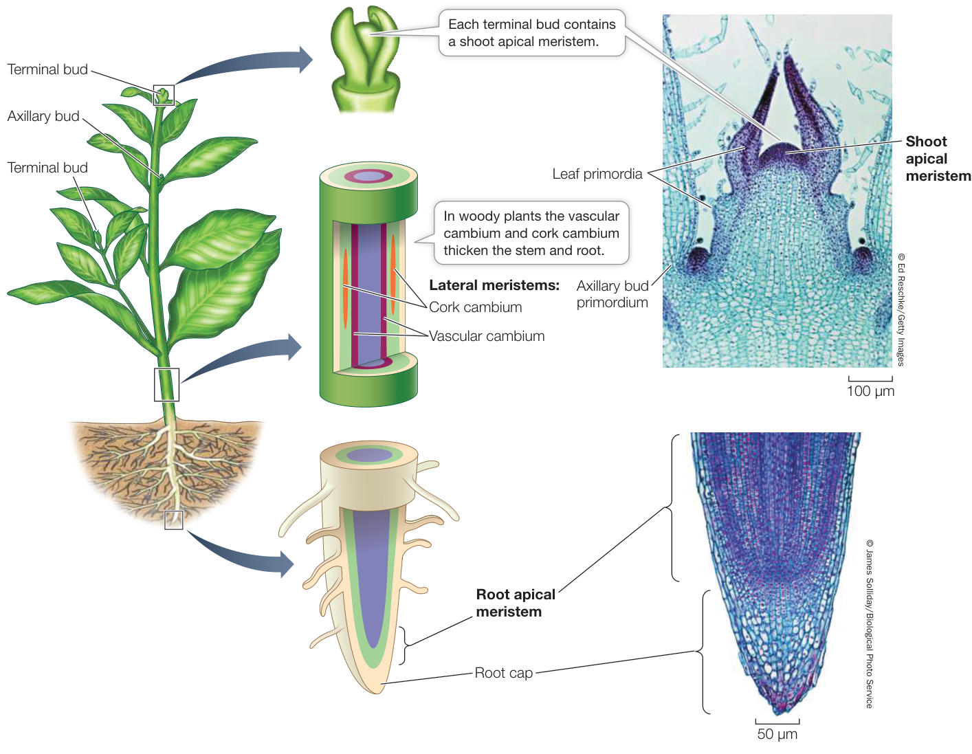
Figure 32.3 Apical and Lateral Meristems Root and shoot apical meristems produce the primary plant body, lengthening it; lateral meristems produce the secondary plant body, thickening it.
Media Clip 32.1 Rapid Growth of Brambles Life12e.com/mc32.1
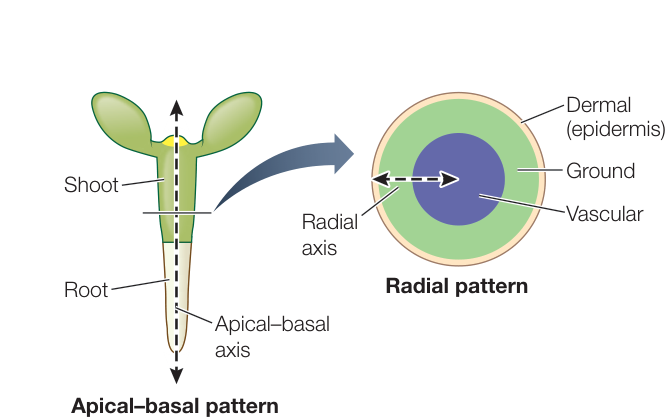
Figure 32.4 Two Patterns for Plant Morphogenesis The apical-basal pattern is the arrangement of cells and tissues along a main axis from root to shoot. The radial pattern determines the concentric arrangement of tissues as organs grow in thickness.
start to grow (see Figure 32.5, Step 3). Further elongation of the cotyledons and of the main axis of the embryo gives rise to the torpedo stage, during which some of the internal tissues begin to differentiate (see Figure 32.5, Step 4). Between the cotyledons is the shoot apical meristem; at the other end of the axis is the root apical meristem. Each of these meristems contains undifferentiated cells that will continue to divide to give rise to the organs that will develop during the life of the plant.
As shown in Step 2 of Figure 32.5, the plant embryo is first a sphere and later a cylinder. The root and stem retain this cylindrical shape throughout the plant’s life. You can see this most easily in the trunk (mature stem) of a tree. By the end of embryogenesis, the radial symmetry of the plant has been established (see Figure 32.4). The embryonic plant contains three tissue types, arranged concentrically, which will give rise to the tissues of the adult plant body.
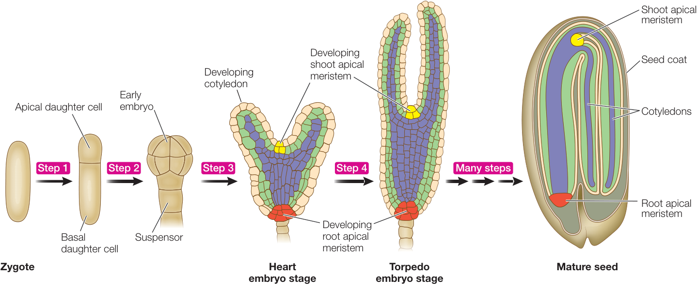
Figure 32.5 Plant Embryogenesis The basic body plan of the model eudicot Arabidopsis thaliana is established in several steps. By the heart stage, three tissue types—dermal (tan), ground (light green), and vascular (blue)—are established. These tissue types are shown in the mature seed, which develops after many more stages.
Q: How do you think the differences between the apical and basal daughter cells are established?
Recap and Assess
The vegetative plant body consists of a root system and a shoot system. Most angiosperms are either monocots or eudicots, which differ in several basic ways. Plants are distinct from most animals in that they grow from meristems, a feature that makes plant growth indeterminate and allows plants to adapt their bodies to the changing environment. The tissues of the plant body develop along two axes: the apical–basal axis (which establishes a polar pattern from root to shoot) and the radial axis (which establishes a concentric pattern of vascular, ground, and dermal tissue systems).
- How do plants explore their environment for resources even though they are rooted in place?
- Describe one primary way in which plant development differs from animal development.
- Describe the initial step during embryogenesis that leads to the apical–basal pattern.
- What would you expect a mutant strain of A. thaliana that lacks apical–basal polarity in the embryo to look like when it matures?
In Key Concept 32.2 we will look more closely at the types of cells that plants contain and the tissue systems that are formed from them.
Plant Organs Are Made Up of Cells Organized into Three Types of Tissues
Learning Objectives
- 32.2.1 Explain how primary and secondary cell walls differ in terms of their location, structure, and when they are produced.
- 32.2.2 Compare and contrast parenchyma, collenchyma, and sclerenchyma cells.
- 32.2.3 Describe the three tissue systems in terms of their origin and location in the plant.
- 32.2.4 Describe the water-conducting elements in plants, and compare these elements in gymnosperms and angiosperms.
By the end of embryogenesis, both the apical–basal and radial axes have been established. Unlike complex animals that can have dozens of different tissues (humans, for example, have three kinds of muscle tissue alone), plants have just three major tissue systems, each of which has specialized cells.
Unlike animals, plant cells have walls made out of cellulose (a polysaccharide). This wall is interrupted by membrane-lined cytoplasmic channels called plasmodesmata (see Key Concept 5.4). As plants have neither an internal skeleton nor a hard exo-skeleton, their cell walls serve the important structural function of holding the plant upright. However, this rigid extracellular matrix makes it impossible for cells to move from place to place, as they do in animal development. Furthermore, overly strong walls can prevent cell elongation.
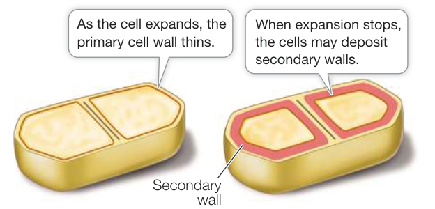
Figure 32.6 Primary and Secondary Cell Walls
The wall of a growing plant cell is called the primary cell wall. When cell expansion stops, some types of plant cells deposit one or more additional cellulosic layers to form a thick, rigid secondary cell wall that is internal to the primary cell wall (Figure 32.6).
Secondary cell walls cannot expand. Instead they provide the mechanical support that allows some plants to produce large stems. The secondary wall contains layers of ordered cellulose microfibrils embedded in a remarkable substance called lignin. Lignin is a major component of wood; it is a complex, carbon-containing polymer.
This matrix is strong, waterproof, and resistant to digestion by animals. After cellulose, lignin is the most abundant biological polymer on Earth, accounting for 20%–35% of the dry weight of wood.
Three types of plant cells have distinct cell wall properties
Each species must strike a compositional balance between thin cell walls, which permit growth, provide flexibility, and conserve resources, and thicker cell walls that provide more mechanical support. Different plants take different approaches to this challenge, involving three types of cells: parenchyma, collenchyma, and sclerenchyma (Figure 32.7).
- Parenchyma cells are the most abundant cells in plants. They have large vacuoles and relatively thin walls (see Figure 32.7A). They perform a wide variety of functions, including photosynthesis (in the shoot), transport, synthesis and secretion of metabolites, and storage of protein in seeds and of starch in roots. Because their walls are thin, the firmness of parenchyma cells depends on their water content: they are firm when they are turgid (full of water), somewhat like a water balloon. Parenchyma cells have only primary cell walls, meaning that cell wall material is added only while the cell is expanding. Many retain the capacity to divide, as when a wound results in cell proliferation.
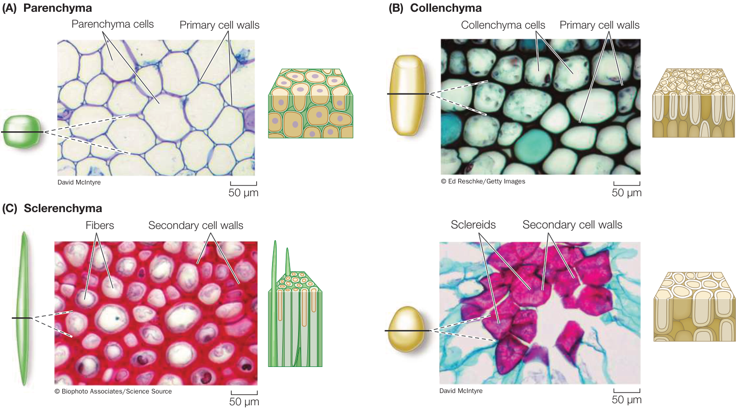
Figure 32.7 Plant Cell Types (A) Parenchyma cells in the petiole of Coleus. Note the thin, uniform cell walls. (B) Collenchyma cells make up the outer cell layers of this spinach leaf vein. Their walls are thick at the corners of the cells and thin elsewhere. (C) Two types of sclerenchyma cells. Left: Fibers in a sunflower stem (Helianthus). The thick secondary walls are stained red. Right: Sclereids. The extremely thick secondary walls of sclereids are laid down in layers. They provide support and a hard texture to structures such as nuts and seeds.
- Collenchyma cells are firm enough to provide support for growing tissues such as stems, yet flexible enough to permit growth. They are generally elongated, and their primary walls are characteristically thicker at the corners of the cells due to the deposition of pectins (see Figure 32.7B). When a plant becomes dry, cells lose their turgor and wilting occurs in portions of the plant (generally the growing tips) that are composed of parenchyma and collenchyma. Collenchyma cells provide support to leaf petioles, nonwoody stems, and growing organs. The familiar “strings” in celery stalks consist primarily of collenchyma. Like parenchyma cells, collenchyma cells have only primary cell walls.
- Sclerenchyma cells have very thick walls reinforced with lignin. They form a primary cell wall when they are elongating, and then, after cell elongation stops, they add additional layers of material to the wall, forming a secondary cell wall (see Figure 32.7C). Most sclerenchyma cells then undergo programmed cell death (apoptosis; see Key Concept 11.6), but their strong cell walls remain and provide support for the plant even when the cells are not turgid. Sclerenchyma cells come in several types. Fibers are often organized into bundles and provide relatively rigid support to wood, bark, and other parts of the plant. Sclereids may pack together densely, as in a nut’s shell or in some seed coats. Isolated clumps of sclereids, called stone cells, occur in pears and some other fruits and give them their characteristic gritty texture.
These three cell types are distributed across the three tissue types described below.
Plants are composed of three main tissues
Plant cells are organized into three tissue types: dermal, ground, and vascular (Figure 32.8). These tissues first arise in the embryo and persist throughout the life of the plant. In the shoot meristem, cell division frequently takes place such that the newly generated cells sit side by side. Over time, this pattern of cell division generates three distinct layers within the meristem (Figure 32.9). The outermost layer produces the dermal layer. Layer two produces ground tissue. Vascular tissue arises from the third layer.
The dermal tissue forms the outer covering of the plant. The aboveground epidermal cells secrete a protective extracellular layer called a cuticle. The cuticle is made up of cutin (a polymer composed of long chains of fatty acids), a complex mixture of waxes, and cell wall polysaccharides. The cuticle limits water loss, reflects potentially damaging solar radiation, and serves as a barrier against pathogens. Some epidermal cells differentiate to form:
- Stomatal guard cells, which form stomata (pores) for gas exchange in leaves
- Trichomes, or leaf hairs, which provide protection against insects and damaging solar radiation, as well as secrete oils (such as those produced by mints)
- Root hairs, which greatly increase root surface area, thus providing more surface for the uptake of water and mineral nutrients
- The stems and roots of woody plants develop a dermal tissue called periderm (discussed in Key Concept 32.3).
The ground tissue comprises most of the tissue between the dermal tissue and the vascular tissue and thus makes up most of the plant body. Cells in this tissue may play important roles in photosynthesis or storage of carbohydrates, fats, or in succulent plants, water.
The vascular tissue is the plant’s plumbing, or transport system. Its presence is the distinguishing feature of vascular plants. Its two constituent tissues, the xylem and phloem, distribute materials throughout the plant. The xylem distributes water and
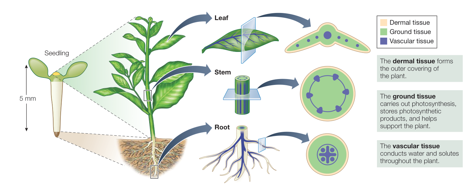
Figure 32.8 Three Tissue Types Extend throughout the Plant Body The arrangement shown here is typical of eudicots, but the three tissue types are continuous in the bodies of all vascular plants.
Q: What is a major difference between cells in the ground tissue in the leaf compared with the root?
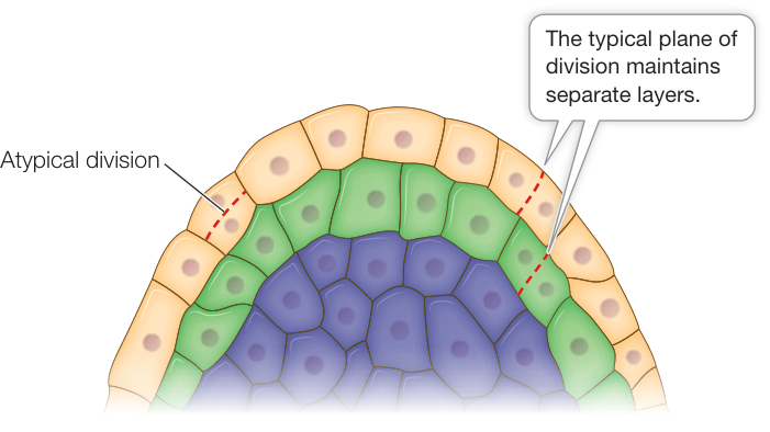
Figure 32.9 The Shoot Apical Meristem Is Composed of Three Layers of Cells Cells in each layer typically divide in a plane that maintains the separate layers. Occasional atypical divisions in other planes can result in cells originally of one layer “invading” the next layer, as shown.
mineral ions taken up by the roots to all the cells of the stems and leaves. The phloem performs a variety of functions, including transport, support, and storage. It plays a particularly critical role in supplying the energy-rich molecules and chemical building blocks needed by all living cells of the plant body. The phloem transports carbohydrates away from the sites of production, which are called sources (primarily leaves), and delivers them to sites of utilization or storage, called sinks. Sinks include growing tissues, storage organs, and developing flowers.
Let’s take a closer look at the structures of the diverse cell types that make up these vascular tissues. In Chapter 33 you’ll see how they transport water and materials throughout the plant body.
XYLEM TISSUE Xylem tissue contains conducting cells called tracheary elements, which have secondary cell walls and undergo apoptosis before assuming their function of transporting water and dissolved minerals. There are two types of tracheary elements: tracheids and vessel elements. The spindle-shaped tracheids (Figure 32.10A) are evolutionarily more ancient than vessel elements and are the major cell type in the wood of gymnosperms (major groups of gymnosperms are covered in Key Concept 27.2). When tracheids die, their internal components disintegrate and pits remain between the cells. Pits are cavities in secondary walls, arranged such that pits of adjacent cells are situated across from each other. Water and minerals move rapidly from one cell to another because these substances need to cross only one thin extracellular barrier, the primary cell wall.
Flowering plants have evolved a water-conducting system made up of vessels that are formed from individual cells called vessel elements. These cells are laid down end-to-end. Like tracheids, vessel elements have pits in their cell walls, but their pits are generally larger in diameter than those of tracheids. Before they undergo apoptosis, the end walls of vessel elements partially break down, forming a continuous hollow tube that functions as an open pipeline for water conduction (Figure 32.10B). In the course of angiosperm evolution, vessel elements have become shorter and wider, and their end walls have become less oblique and less obstructed. These adaptations have presumably increased the efficiency of water transport through the vessels. The xylem of many angiosperms includes both tracheids and vessels.
PHLOEM TISSUE The transport cells of the phloem, unlike those of the mature xylem, are living cells. In flowering plants the characteristic cells of the phloem are called sieve tube elements (Figure 32.10C). Like vessel elements, these cells meet end-to-end. They form long sieve tubes, which transport carbohydrates and many other materials from their sources (usually leaves) to tissues that consume or store them (for example, roots).
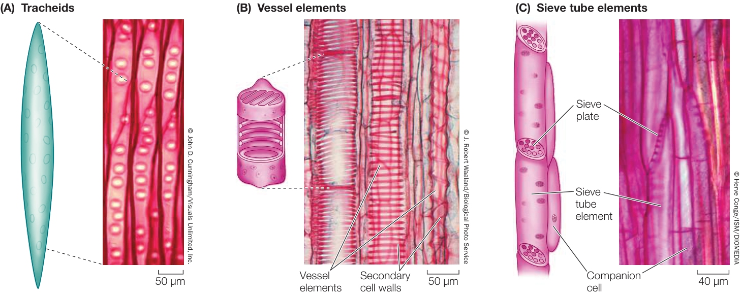
Figure 32.10 Vascular Tissue Cell Types (A, B) Tracheary elements: (A) Tracheids in pinewood. The thick secondary walls are stained dark red. (B) Vessel elements in the stem of a squash. The secondary walls are stained red; note the different patterns of thickening, including rings and spirals. (C) Sieve tube elements and companion cells in the stem of a cucumber.
Whereas the end walls of vessel elements break down to facilitate flow through the vessels, the end walls of sieve tube elements form tunnels through which carbohydrates and other needed substances flow. The end walls of sieve tube elements are called sieve plates. Although the sieve tube elements remain alive, some of their components, including the nucleus, ribosomes, and vacuole, break down during development. The sieve tube elements are, however, closely connected via plasmodesmata to companion cells—specialized parenchyma cells that retain all their organelles and function as “life support systems” for the sieve tube elements.
Recap and Assess
The plant body is composed of cells that can be divided into three classes based on the properties of their cell walls: the thin-walled parenchyma cells; collenchyma cells, which provide more rigid support without preventing growth; and sclerenchyma cells, whose secondary cell walls can support plants even when the cells are not turgid. The three concentric tissue types in the plant embryo—dermal, ground, and vascular—give rise to the tissues and organs of the adult plant. These tissues have unique combinations of specialized cells that carry out the various functions necessary for plant life.
- Create a table comparing the three cell types in terms of their structure and function.
- What structural differences make tissues consisting of collenchyma cells more flexible than those consisting primarily of sclerenchyma cells?
- Describe the differences between tracheids and vessel elements.
In Key Concept 32.3 we will see how the tissues are established by the meristems and how they contribute to the overall structure of the plant.
Meristems Build a Continuously Growing Plant
Learning Objectives
32.3.1 Explain the function of meristems in plant growth, including the types of cells and organs that are produced by the root and shoot apical meristems.
32.3.2 Draw a longitudinal section of the developing root, from the tip upward, indicating the names and activities of the zones.
32.3.3 Explain how the vascular cambium increases the width of a woody plant.
Plants and animals develop and function differently. While animals use their mobility to look for food, plants are sessile (rooted in one place) and instead grow toward scarce resources, both aboveground (shoots toward sunlight) and belowground (roots toward water and minerals). This open-ended growth can occur in either of two ways: Primary growth is characterized by cell division followed by cell enlargement. Primary growth results in the proliferation and lengthening of shoots and roots. All seed plants have a primary plant body, which consists of all the nonwoody parts of the plant. Many herbaceous plants consist entirely of a primary plant body. Secondary growth increases plant thickness. Woody plants, such as trees and shrubs, have a secondary plant body consisting of wood and bark. As the tissues of the secondary plant body are laid down, the stems and roots thicken.
Indeterminate primary growth originates in apical meristems
Because apical meristems can perpetuate themselves indefinitely, a stem or root can continue to lengthen and grow indefinitely; in other words, growth of the plant body as a whole is indeterminate. All plant organs arise ultimately from cell divisions in apical meristems, followed by cell expansion and differentiation. Several types of apical meristems play roles in organ formation:
- Shoot apical meristems supply the cells for new leaves and stems. The shoot apical meristem also retains some cells as initials, which allows the meristem to be maintained. In addition to the main stem of the plant, each branch has its own shoot apical meristem. Shoot apical meristems are also called vegetative meristems because they give rise to vegetative tissues (leaves, stems, and roots).
- When the plant is ready to flower, one or more of its shoot apical meristems are transformed into inflorescence meristems, and these in turn develop floral meristems. (Chapter 36 is dedicated to the reproduction of flowering plants; inflorescence and floral meristems are discussed in Key Concept 36.2.)
- Root apical meristems supply the cells that extend roots, enabling the plant to penetrate and explore the soil for water and minerals. Like the shoot apical meristem, the root apical meristem retains some cells as initials. Each type of root (i.e., the taproot, a lateral root, or an adventitious root; see below) has its own meristem.
Because meristems can continue to produce new organs throughout the lifetime of the plant, the plant body is much more variable in form than the typical animal body, which produces each organ only once.
Let’s look more closely at how the root apical meristem produces the root system.
The root apical meristem gives rise to the root cap and the primary root
The root apical meristem produces all the cells that contribute to the growth and development of the root (Figure 32.11). Some of the daughter cells from the apical (tip) end of the root apical meristem contribute to a root cap, which protects the delicate growing region of the root as it pushes through the soil. The root cap secretes a mucopolysaccharide (slime) that acts as a lubricant. Even so, the cells of the root cap are often damaged or scraped away and must therefore be replaced constantly. The root cap is also the structure that detects the pull of gravity and thus controls the downward growth of roots.
In the middle of the root apical meristem is a quiescent center center, in which cell divisions are rare. The quiescent center cells
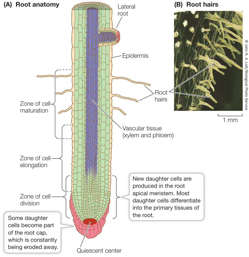
Figure 32.11 Tissues and Regions of the Root Tip (A) Extensive cell division creates the complex structure of the root. (B) Root hairs, seen with a scanning electron microscope.
The root apical meristem produces cells that make up the root tissues
While the longitudinal axis of a root can be described in terms of the zones it contains, the radial axis can be described in terms of the differing cell types that make up the different portions of the root. In flowering plants, the cells that make up the root are similar in that they contain the three tissue types described above, however, the differing arrangement of tissues in the roots of eudicots and monocots is one of the ways in which the two clades are distinguished (Figure 32.12). Some commonalities in the cell types that comprise the radial structure are as follows:
- The epidermis is the outer layer of cells that is adapted for protection of the root and absorption of mineral ions and water. Many of the epidermal cells produce long, delicate root hairs, which vastly increase the surface area of the root (see Figure 32.11B). Root hairs grow out among the may be in G1 phase of the cell cycle for 10 days. The quiescent center influences the cells around it such that they retain the character of initial cells rather than differentiating. The daughter cells produced above the quiescent center (that is, on the shootward side away from the root cap) form the main body of the root.
Because plant cells do not move past one another, the distance between a cell and the quiescent center is related to the length of time since the cell was created. This leads to several distinct zones in the root. The meristem is close to the root tip and includes both the initial cells that surround the quiescent center and the zone of cell division. Just shootward of this is the zone of cell elongation, where the newly formed cells elongate and thus push the root tip farther into the soil. Shootward of that zone is the zone of cell differentiation (also called the maturation zone), where the cells take on their mature forms.
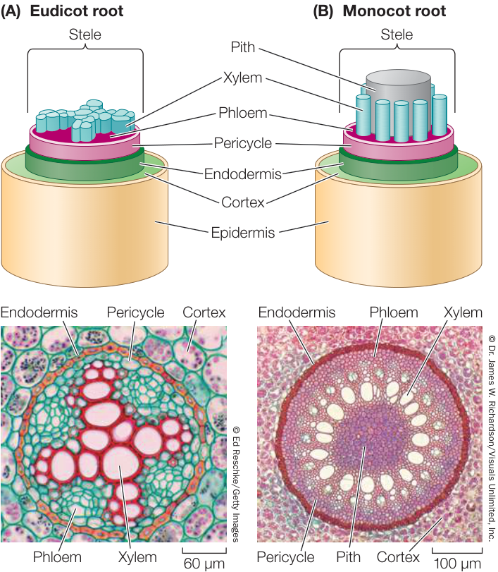
Figure 32.12 Products of the Root Meristem The arrangement of tissues in the stele differs in the roots of eudicots (A) and monocots (B). The photomicrographs show cross sections of the stele of a representative eudicot (a buttercup, Ranunculus) and a representative monocot (corn, Zea mays), showing the arrangement of the primary root tissues.
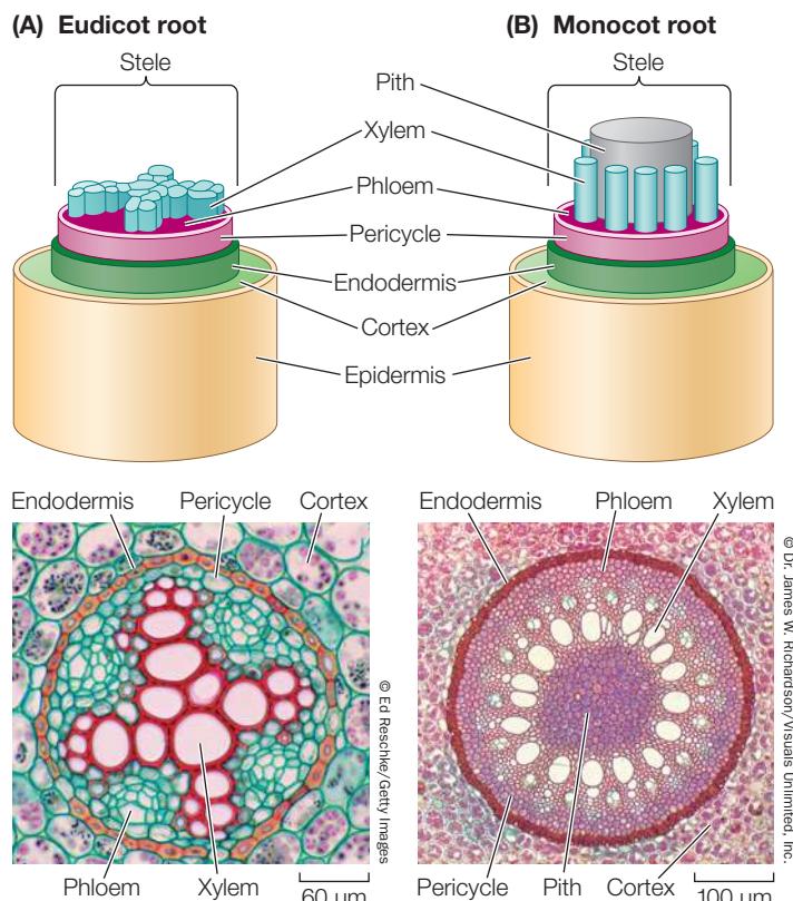
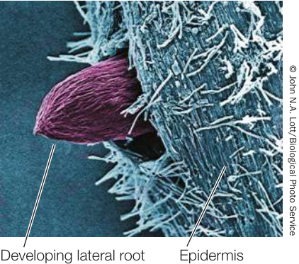
Figure 32.13 Lateral Root Anatomy Scanning electron micrograph of the tip of a lateral root in a willow tree. Cells in the pericycle divide and the products differentiate, forming the tissues of a lateral root.
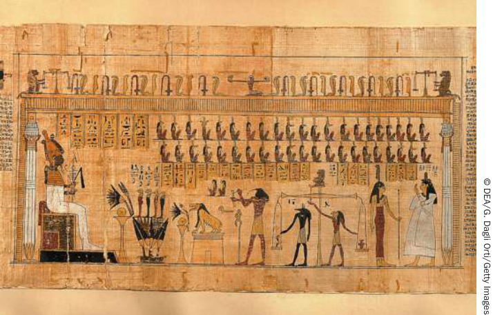
Figure 32.14 Egyptian Papyrus Depicting the Weighing of Souls from the Book of the Dead
soil particles, probing nooks and crannies and taking up water and minerals.
- The cortex is a region of ground tissue produced internal to the epidermis. The cells of the multilayered cortex have various functions, including storage.
- The endodermis is the innermost layer of the cortex. Unlike other cortical cells, the endodermal cells contain a water-proof substance called suberin in their primary cell walls. The suberin forms a cylindrical ring around the inside of the endodermis, which allows the endodermal cells to control the movement of water and dissolved mineral ions into and out of the vascular tissue system.
- Interior to the endodermis is the vascular cylinder, or stele. The stele consists of the pericycle, xylem tissue, and phloem tissue. The pericycle consists of one or more layers of cells with flexible developmental potential. It has three important functions: (1) It is the tissue within which lateral roots arise (Figure 32.13). (2) It can contribute to secondary growth by giving rise to lateral meristems that thicken the root. (3) Its cells contain membrane transport proteins that export nutrient ions into the cells of the xylem.
At the very center of the root of a eudicot lies the xylem. Seen in cross section, it typically has the shape of a star with a variable number of points (see Figure 32.12A). Between the points are bundles of phloem. In monocots, a region of parenchyma cells called the pith typically lies in the center of the root, surrounded by xylem and phloem (see Figure 32.12B). Pith, which often stores carbohydrate reserves, is also found in the stems of both eudicots and monocots. Strips of pith of the stem of the papyrus plant Cyperus papyrus can be glued together and used to make a paperlike substance called by its species name. Papyrus dates from earlier than 2500 BCE in Egypt, and was the writing surface of choice until about 300 CE, when parchment from animal skins replaced it (Figure 32.14).
The root system anchors the plant and takes up water and dissolved minerals
Water and minerals enter most plants through the root system, which is located in the soil. Because light does not penetrate the soil, roots typically lack the capacity for photosynthesis. Although hidden from view, the root system is often larger than the visible shoot system. For example, the root system of a 4-month-old winter rye plant (Secale cereale) was found to be 130 times longer in total than the shoot system, with almost 13 million branches that had a cumulative length of more than 500 kilometers!
Angiosperm root systems develop from the embryonic root, called the radicle. From this common starting point, the root systems of monocots and eudicots develop differently. Following seed germination, the radicle of most eudicots develops as a primary root called the taproot, which extends downward by tip growth and outward by initiating lateral roots. The taproot and the lateral roots form a taproot system, which can take a variety of forms. For example, the taproot itself often functions as a nutrient storage organ, as in carrots (Daucus carota), sugar beets (Beta vulgaris), and sweet potato (Ipomoea batatas) (Figure 32.15A).
In contrast, the primary root of monocots (and some eudicots) is short-lived. Because they originate from the stem at ground level or just below, the roots of a typical monocot are called adventitious (“arriving from outside”) roots, and they form a fibrous root system composed of numerous thin roots that are all roughly equal in length (Figure 32.15B). Many fibrous root systems have large surface areas for the absorption of water and minerals. A fibrous root system clings to soil very well. The fibrous root systems of grasses, for example, may protect steep hillsides where runoff from rain would otherwise cause erosion.
In some plants—corn, banyan, and pandanus trees, for example—adventitious roots grow down from above the ground and function as props to help support the shoot system (Figure 32.15C). These prop roots have evolved for different reasons in different species. For example, some monocots may develop prop roots because they are unable to support aboveground growth through the thickening of their stems. Pandanus trees often grow near coastal beaches, where their prop roots help provide support in very sandy soils. Banyans begin life as epiphytes (plants that grow on other plants) and then develop woody prop roots, which enable them to grow into huge trees.
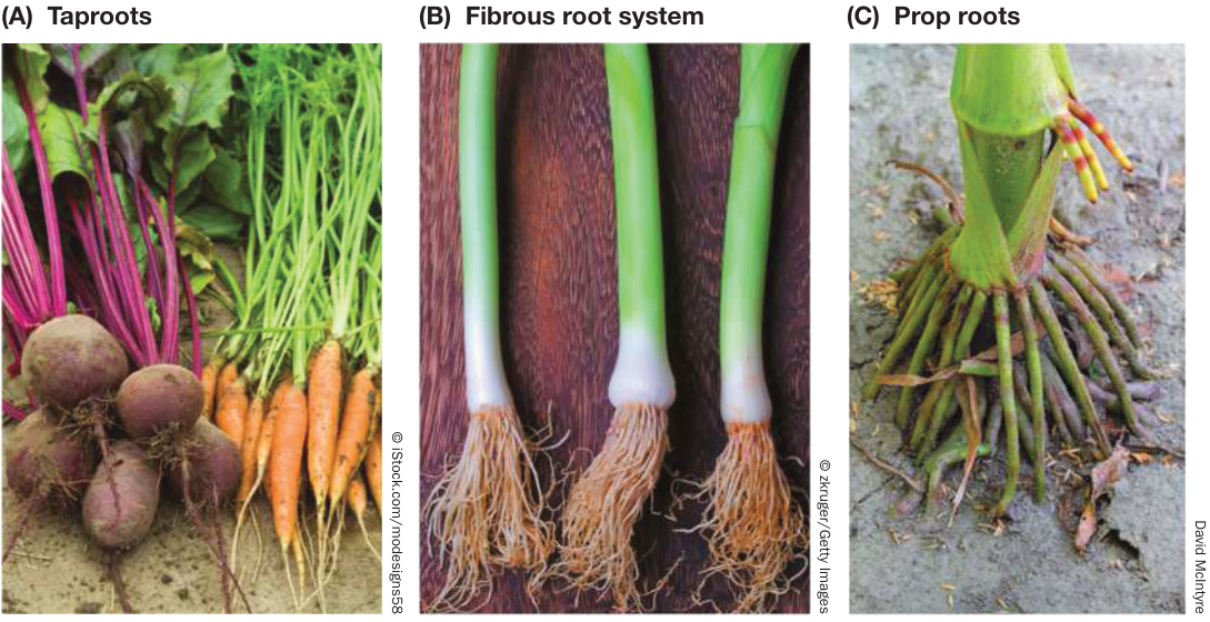
Figure 32.15 Root Systems of Eudicots and Monocots (A) The taproot systems of eudicots, such as carrots and sugar beets, contrast with (B) the fibrous root system of a leek and (C) the adventitious prop roots of corn.
The products of the shoot apical meristem form the shoot system
Now let’s turn to the shoot, which comprises the stem and leaves. Shoots are composed of repeating modules called phytomers (Figure 32.16). A phytomer in the shoot system consists of a node with its attached leaf or leaves, the internode below the node, and axillary buds, each of which forms in the angle between a leaf and the stem. Shoots grow by adding new phytomers. The new phytomers originate from cells in the shoot apical meristem, which is present at the apex of the main stem, and from meristems that are located at the tip of each branch.
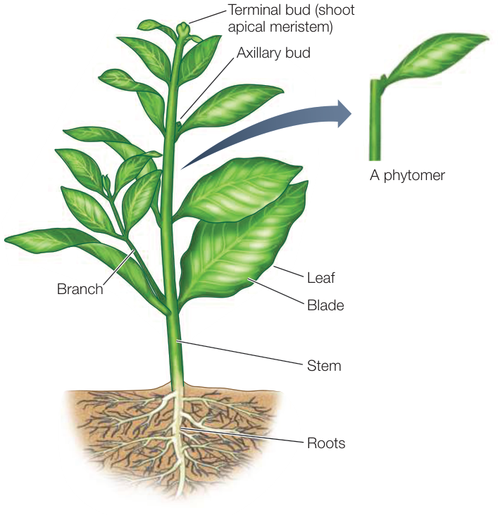
Figure 32.16 The Shoot Is Built of Repeating Units Called Phytomers Each phytomer consists of an internode (stem), leaf, and axillary bud.
As the shoot grows and extends, bulges called leaf primordia develop on the sides of the shoot apical meristem at regular intervals (see Figure 32.3). Each primordium can develop into a mature leaf. In addition, axillary buds form at the bases of the leaf primordia. These have the potential to grow out and form a new shoot (branch) with a meristem at its tip. The sites where leaf primordia form become nodes on the developing stem. The regions between the nodes (the internodes) lengthen initially via cell division and later by cell elongation. The growing stem has no protective structure analogous to the root cap, but the leaf primordia can act as a protective covering for the shoot apical meristem.
The vascular systems of stems differ from those of roots. In a root, the vascular tissue lies deep in the interior, with the xylem at or near the center (see Figure 32.12). The vascular tissue of a young stem, however, is divided into discrete vascular bundles (Figure 32.17). Each vascular bundle contains both xylem and phloem. In eudicots the vascular bundles generally form a ring, whereas in monocots they are scattered throughout the stem.
In addition to the vascular tissues, the stem contains other important storage and supportive tissues. In eudicots the pith lies inside the ring of vascular bundles and also extends between them, forming regions called pith rays. To the outside lies the cortex, which may contain supportive collenchyma cells with thickened walls. The pith and cortex constitute the ground tissue system of the stem. The outermost cell layer of the young stem is the epidermis.
The stem supports leaves and flowers but can have other roles
The main role of stems is to elevate and support the photosynthetic organs (leaves) as well as the reproductive organs (flowers). But in some plants the stem does other things for the plant. For instance, the potato tuber that we eat comes from the ground but is not a root; it’s an underground stem that acts as a storage organ for the plant. The “eyes” of a potato are depressions containing axillary buds—in other words, a sprouting potato is just a branching stem (Figure 32.18A). Many desert plants have enlarged, water-retaining stems (Figure 32.18B). The runners of strawberry plants are horizontal stems that develop adventitious roots some distance from the main stem (Figure 32.18C); if the links between the rooted portions are broken, independent plants can develop on each side of the break—a form of vegetative (asexual) reproduction (see Key Concept 36.3).
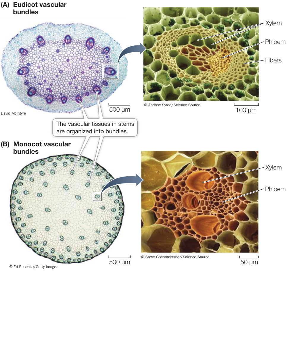
Figure 32.17 Vascular Bundles in Stems (A) In herbaceous eudicot stems, the vascular bundles are arranged in a cylinder, with pith in the center and the cortex outside the cylinder. (B) A scattered arrangement of vascular bundles is typical of monocot stems. Activity 32.3 Eudicot Stem Activity 32.4 Monocot Stem
Leaves are determinate organs produced by shoot apical meristems
For most of its life, a plant produces leaves from apical meristems. Leaves originate from the edges of the apical meristem as initial cells that differentiate into leaf primordia. Leaves differ from stems in two important ways:
- Unlike the growth of the stem, which is indeterminate, the growth of a leaf is determinate.
- Whereas the tissues of the stem are arranged in a radial pattern, the leaf, as a flat organ, has a distinct top side and bottom side.
Connect the Concepts The leaf is elegantly structured for the reactions of photosynthesis. For more detail on photosynthesis and the role of leaf tissues, see Key Concepts 10.1 and 10.4.
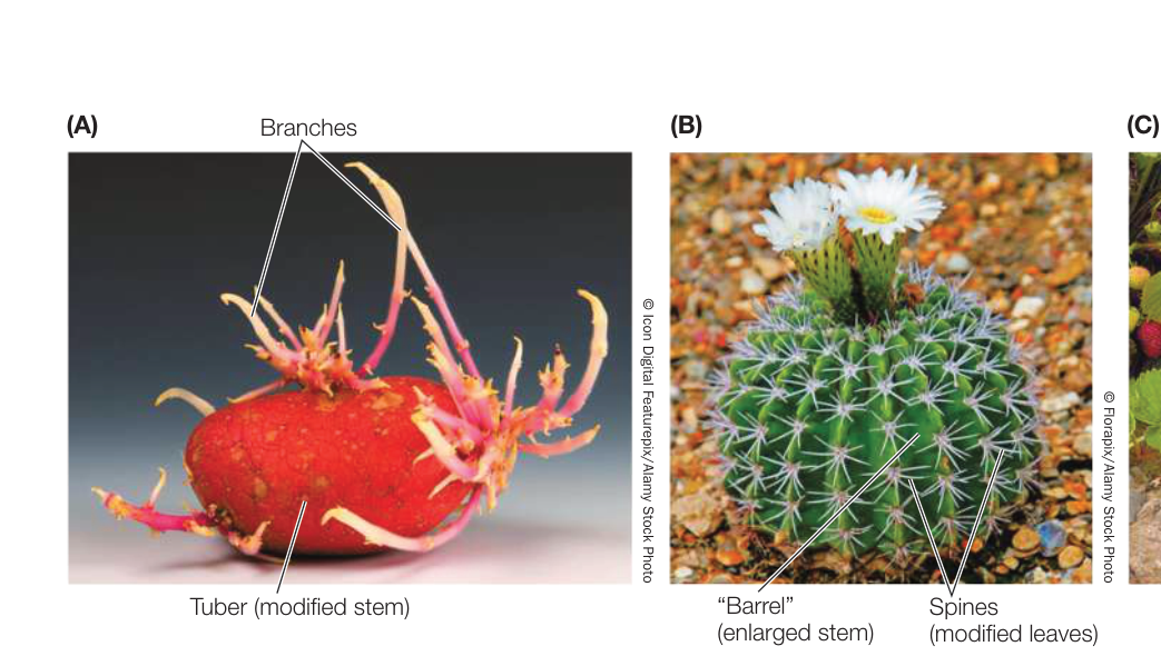
Figure 32.18 Modified Stems (A) A potato is a modified stem called a tuber; the sprouts that grow from its “eyes” are shoots, not roots. (B) The stem of this barrel cactus is enlarged to store water. Its highly modified leaves serve as thorny spines. Most of this plant’s photosynthesis occurs in the stem. (C) The runners of strawberry are horizontal stems that produce roots and shoots at intervals. Rooted portions of the plant can live independently if the runner is cut.
Leaf anatomy is beautifully adapted to carry out photosynthesis, and to support that process by exchanging the gases and with the environment, while at the same time limiting evaporative water loss (Figure 32.19). Its extensive vascular system supplies the leaf with water and mineral nutrients and exports the products of photosynthesis to the rest of the plant. Figure 32.19A shows a typical eudicot leaf in three dimensions. The photosynthetic parenchyma tissue in leaves is the mesophyll (which means “middle of the leaf”). Most eudicot leaves have two zones of mesophyll: an upper layer of elongated cells called the palisade mesophyll, and a lower layer of irregularly shaped cells called the spongy mesophyll. Within the spongy mesophyll is a great deal of air space through which can diffuse to photosynthesizing cells.
Vascular tissue branches extensively throughout the leaf, forming a network of veins (see Figure 32.19B). Veins extend to within a few cell diameters of all the cells of the leaf by the xylem, ensuring that the mesophyll cells are well supplied with water and minerals. The products of photosynthesis are loaded into the veins for export to the rest of the plant. In addition to photosynthetic products, leaves make defensive molecules such as the ones in cassava that you learned about in the chapter opener. Investigating Life: Understanding the Synthesis and Transport of Cyanogenic Glycosides describes how it was determined that cassava leaves produce these chemicals.
The epidermis covers the entire surface of the leaf. The epidermal cells secrete a waxy cuticle that is impermeable to water. Although this impermeability prevents excessive water loss, it also poses a problem: while the epidermis keeps water in the leaf, it also keeps out —the other raw material of photosynthesis.
The problem of balancing water retention and availability is solved by guard cells, a system we’ll examine in detail in Key Concept 33.3. Stomatal guard cells are modified epidermal cells that can change their shape, thereby opening or closing pores called stomata (singular stoma). The stomata serve as passageways between the environment and the leaf’s interior (see Figure 32.19C). When the stomata are open, can enter and can leave, but water can also be lost.
Many stems and roots undergo secondary growth
As you have seen, the roots and stems of some eudicots develop a secondary plant body, the tissues of which include wood and bark. These tissues are derived by secondary growth from the two lateral meristems:
- The vascular cambium is a cylindrical layer of tissue consisting predominantly of elongated cells that divide frequently. It supplies the cells of the secondary xylem and secondary phloem, which eventually become wood and bark, respectively.
- The cork cambium produces mainly waxy-walled protective cells. It supplies some of the cells that become bark.
In cold parts of the world, each year deciduous trees lose their leaves and have bare branches and twigs over the winter. The apical meristems of the twigs are enclosed in buds protected by bud scales. When the buds begin to grow in spring, the scales fall away, leaving scars that show where the bud was. These scars
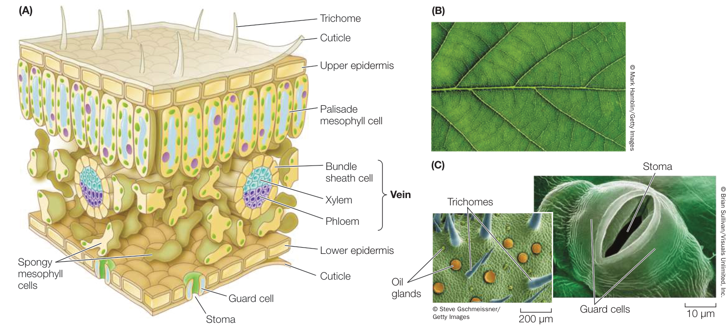
Figure 32.19 The Eudicot Leaf (A) This three-dimensional diagram shows a section of a eudicot leaf. (B) The network of fine veins in this maple leaf carries water to the mesophyll cells and carries photosynthetic products away from them. (C) Carbon dioxide enters the leaf through stomata like this one on the epidermis of a eudicot leaf. Trichomes are epidermal hairs that can break up airflow, reducing water loss from the leaf. In some species, including the marjoram shown here, modified epidermal hairs serve as oil glands.
Investigating Life: Understanding the Synthesis and Transport of Cyanogenic Glycosides
Experiment
Original Paper: K. Jørgensen et al. 2005. Cassava plants with a depleted cyanogenic glycoside content in leaves and tubers. Distribution of cyanogenic glycosides, their site of synthesis and transport and blockage of biosynthesis by RNA interference technology. Plant Physiol 139: 363–374.
As described in the chapter opener, cassava is the most important root crop in the world. Its use as food is challenging, however, because the plant produces molecules that can be transformed into toxic cyanide when the tissues are disrupted. These cyanogenic glycosides (CGs) are important in defending the plant against animals that eat it, including humans. For the millions of people who eat cassava, careful processing is needed to avoid poisoning. Unfortunately, the processing methods lower the overall nutritional value of the food. Understanding how CGs are made and end up in the fleshy root is an important step in possibly using genetics to produce a crop that does not make CGs. At the Royal Veterinary and Agricultural University in Copenhagen, a team led by Birger Møller investigated the synthesis and transport of CGs in cassava plants.
HYPOTHESIS Molecules that produce cyanide in the cassava are made in the leaves and transported to the underground root.
METHOD
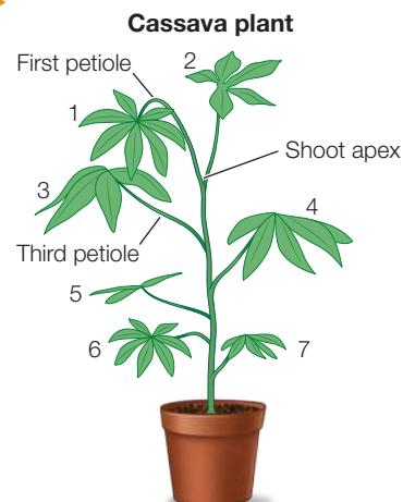
1 Leave stem intact and measure CG in plant shoot regions.
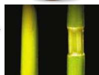
2 Remove phloem from stem below fifth leaf and then measure CG in plant parts.
RESULTS
| Region | Relative CG concentration | Region | Relative CG concentration |
|---|---|---|---|
| Shoot apex | Moderate | Fifth leaf | High |
| First petiole | High | Stem above cut | Moderate |
| Third leaf | Low | Stem below cut | Very Low |
| Third petiole | Low |
CONCLUSION Cyanogenic glycosides are made in leaves and are transported through petioles to the stem, from which they are transported to the root.
Work with the Data
QUESTIONS
- Cyanogenic glycosides in plant organs were measured in an intact stem of a cassava plant and in one in which the phloem had been cut below the fifth leaf. The results are shown in the graphs. What can you conclude about the source of CGs?
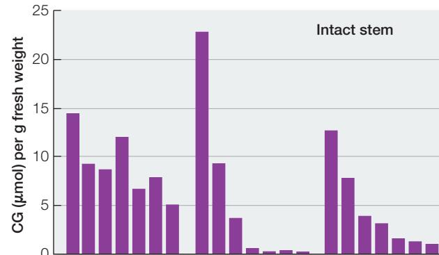
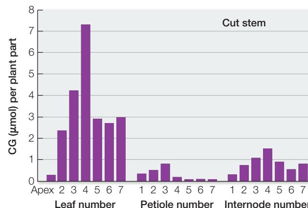
- When a stem is girdled, it is cut carefully so that the epidermal, cortical, and outer vascular tissues (phloem) are removed. Using intact plants, the biologists girdled the stem of a cassava plant. After 2 days, they measured CGs in various regions on the stem. The results for CG levels after girdling between the fifth and sixth nodes are shown in the table. What can you conclude about the source and transport of CGs? Why was the level in the sixth leaf (below the girdle) so high?
| Region | CG level () |
|---|---|
| Fifth leaf | 59 |
| Stem above cut | 15 |
| Stem below cut | 0.2 |
| Sixth leaf | 55 |
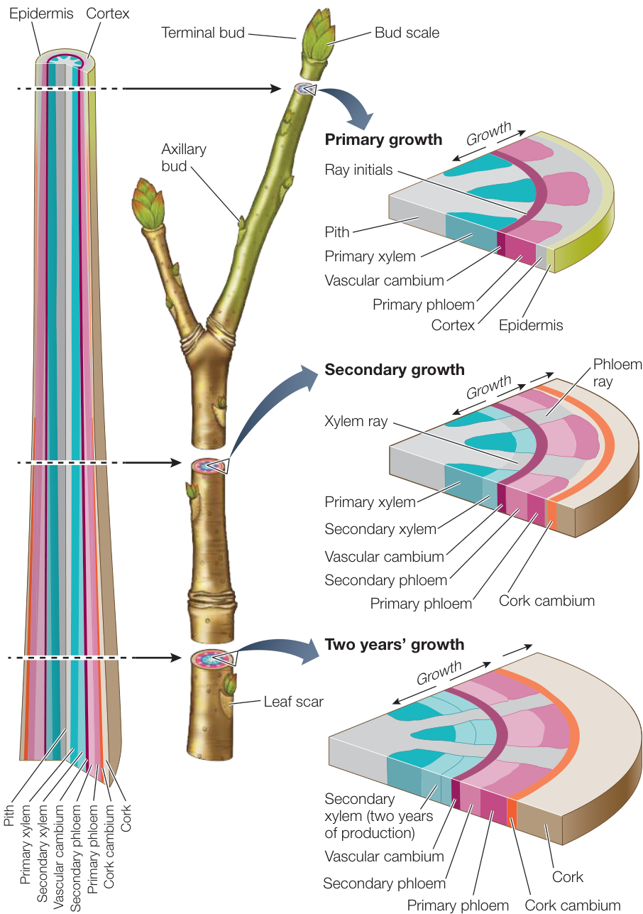
Figure 32.20 A Woody Twig Has Both Primary and Secondary Growth The apical meristems in this dormant twig will produce primary growth in spring. Lateral meristems are responsible for secondary growth. When the vascular cambium divides parallel to the outside of the stem, it forms a new ring of cells. New layers of cells on the interior side of the vascular cambium add to the xylem, while those exterior to the vascular cambium add to the phloem.
allow us to identify the parts of the twig from each year’s growth. The dormant twig shown in Figure 32.20 is the product of both primary and secondary growth. The buds consist entirely of primary tissues.
The vascular cambium is initially a single layer of cells lying between the primary xylem and the primary phloem within the vascular bundles. The root or stem increases in diameter when the cells of the vascular cambium divide, producing secondary xylem cells toward the inside of the root or stem and producing secondary phloem cells toward the outside. In the stem, cells in the pith rays between the vascular bundles also divide, forming a continuous cylinder of vascular cambium running the length of the stem. This cylinder, in turn, gives rise to complete cylinders of secondary xylem (the wood) and secondary phloem, which contributes to the bark. It also produces vascular rays for lateral transport, a structure not found in primary xylem and phloem. Therefore the vascular cambium produces vessel elements, tracheids, parenchyma cells, and supportive fibers in the secondary xylem; and sieve tube elements, companion cells, fibers, and parenchyma cells in the secondary phloem.
Animation 32.1 Secondary Growth: The Vascular Cambium
As secondary growth of stems or roots continues, the expanding vascular tissue stretches and breaks the epidermis and the outer layers of the cortex, which ultimately flake away. Before these dermal tissues are broken away, cells lying near the surface of the secondary phloem begin to divide, forming a cork cambium. This meristematic tissue produces layers of cork, a protective tissue composed of cells with thick walls waterproofed with suberin. The cork soon becomes the outermost tissue of the stem or root (see Figure 32.20). Without the activity of the cork cambium, the sloughing off of the outer primary tissues would expose the plant to potential damage, such as excessive water loss or invasion by microorganisms. Sometimes the cork cambium produces cells toward the inside as well as the outside; these cells constitute a tissue known as the phelloderm.
The cork cambium, cork, and phelloderm constitute the secondary dermal tissue called periderm. As the vascular cambium continues to produce secondary vascular tissue, these corky layers are lost, but the continuous formation of new cork cambia in the underlying secondary phloem gives rise to new corky layers. The periderm and the secondary phloem—that is, all the tissues external to the vascular cambium—constitute the bark.
When periderm forms on stems or roots, the underlying tissues still need to release and take up for cellular respiration. Spongy regions in the periderm called lenticels can allow such gas exchange (Figure 32.21).
You may be curious about cork, which is used as stoppers in wine bottles and in flasks in the chemistry lab. Cork is harvested from the cork oak tree (Quercus suber) (Figure 32.22), which is grown for that purpose mainly in Portugal. The tree lives more than 100 years, and cork is first harvested when the tree is 25 years old. The outer bark is removed first, carefully exposing the cork layers, which are then removed without damaging the living tissues. The cork cambium then produces more cork, so a tree can be harvested every 10 years.
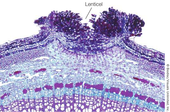
Figure 32.21 Lenticels Allow Gas Exchange through the Periderm The region of periderm that appears to be broken open is a lenticel in a year-old elderberry (Sambucus) twig,
Cross sections of most tree trunks (stems) in temperate-zone forests show annual rings of wood (Figure 32.23), which result from seasonal environmental conditions. In spring, when water is relatively plentiful, the tracheids or vessel elements produced by the vascular cambium tend to be large in diameter and relatively thin-walled. Such wood is well adapted for transporting water and minerals. As water becomes less available during the summer, narrower cells with thicker walls are produced, making this summer wood darker and perhaps more dense than the wood formed in spring. Thus each growing season is usually recorded in a tree trunk by a clearly visible annual ring. Trees in the moist tropics do not undergo seasonal growth, so they do not lay down such obvious regular rings. Variations in temperature or water supply can lead to the formation of more than one ring in a single year, but most commonly a single annual ring is formed. Next time you are in the woods and see a fallen tree, try to count the annual rings to gauge the age of the tree.
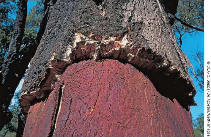
Figure 32.22 Cork Grows from the Cork Cambium of the Cork Oak (Quercus suber)
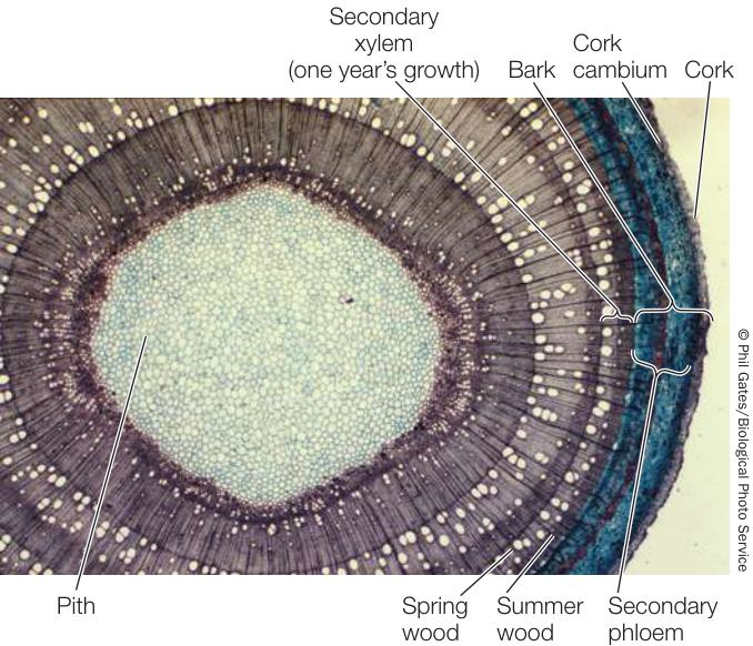
Figure 32.23 Annual Rings Rings of secondary xylem are the most noticeable feature of this cross section from a tree trunk
Only eudicots and other non-monocot angiosperms, along with many gymnosperms, have a vascular cambium and a cork cambium and thus undergo secondary growth. The few monocots that form thickened stems—palms, for example—do so without secondary growth. Palms have a very wide apical meristem that produces a wide stem, and dead leaf bases add to the diameter of the stem. All monocots grow in essentially this way, as do other angiosperms that lack secondary growth.
Recap and Assess
Meristems are localized regions of cell division that are the sources of all new organs in the adult plant. Initial cells within the root apical meristem divide to form the root cap and the cells of the primary root. The cells do not move past one another so the zones of cell division, elongation and maturation in the primary root are located at increasing distances from the initial cells. Root branching can occur when new meristems arise from pericycle cells within the root. The shoot apical meristem gives rise to the cells that comprise the shoot system, including the branches which arise from meristems that form the axils of leaves. The vascular cambium increases plant thickness and forms wood and bark in many eudicots.
- How can an apical meristem be maintained for years while continuing to form leaves?
- Identify the meristems of the plant that are responsible for primary growth and indicate how tissues and organs are formed from them.
- What cells are derived from the root apical meristem, and what is the general process of root growth?
- Compare and contrast the vascular cambium and the shoot apical meristem. What aspects of the vascular cambium define it as a meristem?
The building of the plant body by meristems allows a plant to respond to its environment by redirecting its growth. Thus individual plants of the same species can vary greatly in form. What underlies this variation, and how have we humans used it to our advantage?
Domestication Has Altered Plant Form
Learning Objectives
- 32.4.1 Explain how the simple, repeating units that make up a plant can be modified to generate a wide variety of plant architectures.
- 32.4.2 Explain why the seeds from wild relatives of crop plants are valuable.
You have seen in this chapter that a simple, modular plant body plan underlies the remarkable diversity of the flowering plants that cover our planet. Differences in plant form among species are not surprising, given the high levels of genetic diversity among plant species. However, members of the same species can also show remarkable diversity in form. From a genetic perspective, this suggests that minor differences in gene content or gene regulation can underlie dramatic differences in plant form.
It is hard to believe that modern corn was domesticated from the wild grass teosinte, which still grows in the hills of Mexico (Figure 32.24). One of the most conspicuous differences is that teosinte, like other wild grasses, is highly branched, whereas domesticated corn has a single shoot. This morphological difference is due in large part to the activity of a single gene called TEOSINTE BRANCHED 1 (TB1). The protein product of TB1 regulates the growth of axillary buds (see Figure 32.1). The allele of tb1 in domesticated corn represses branching, whereas the allele in teosinte permits branching.
Even harder to believe is that a single species, Brassica oleracea (wild mustard), is the ancestor of many familiar and morphologically diverse crops, including kale, broccoli, brussels sprouts, and cabbage.
Connect the Concepts The diversity of plants derived from wild mustard illustrated in Figure 19.4 demonstrates how artificial selection—the purposeful selection of specific phenotypes by humans—can produce rapid evolutionary change.
Each of these familiar vegetable crops has the same basic body plan. Starting with morphologically diverse populations of the wild ancestor, humans selected and planted the seeds of variants with traits they found desirable. For example, brussels sprout plants were selected for their enlarged axillary buds, cabbage plants for their enlarged terminal buds and short internodes, and broccoli and cauliflower plants for their large clusters of flower buds. Such selection is really an act of genetic engineering because in selecting plants that have some particular trait one is choosing those plants that have a collection of genes that allow the trait to be expressed or formed. Many generations of such artificial selection produced the crops that fill the produce section of supermarkets and the stands of farmers’ markets.
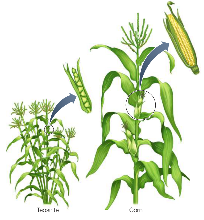
Figure 32.24 Modern Corn Was Domesticated from the Wild Grass Teosinte Each teosinte plant has multiple branches. Beginning more than 8,000 years ago in Mexico, farmers favored plants with minimal branching. Reducing the number of branches results in fewer ears per plant but allows each ear to grow larger and produce more seeds.
Just as they were for ancient farmers, the genomes of plants are priceless resources today. The genetic variation in crop plants and their wild relatives includes DNA sequences that are adapted to different conditions than those found in current farm fields, often including resistance to a wider range of pests. These genomes can be used to improve our crop plants or adapt them to changing conditions. The improvement of crop plants is a work in progress that is being carried out in plant breeding programs worldwide. These programs are now more important than ever. Increased human activity is dramatically changing our planet and leading to the extinction of more and more plant species. For this reason, various organizations around the world have developed seed banks, where seeds of diverse species, and variants within species, are stored. As the climate continues to change, new varieties of old crops and new crops that are better adapted to a warmer climate with more extreme fluctuations in water availability will be needed.
Recap and Assess
Crop domestication involves artificial selection of certain desirable traits found in wild plant populations. By understanding the basic body plan of plants, one can more easily understand the morphological relationship between a crop plant and its wild relatives.
- Why are the seeds from wild relatives of crop plants valuable?
▶ Investigating LIFE
Q&A Future directions How might plant biologists improve the cassava plant for human use?
Many people depend on cassava roots for food. Unfortunately, the roots must undergo processing to remove sources of toxic cyanide before they are eaten. As you saw in Investigating Life: Understanding the Synthesis and Transport of Cyanogenic Glycosides, cyanide-forming molecules are formed in leaves and then transported to the roots through the vascular system. Molecular biologists have described the details of organ-specific synthesis of the machinery responsible for a major cyanogenic molecule. RNA interference (see Key Concept 18.4) has been used to block cassava leaves from making the toxic precursor, and as a result its concentration in the edible roots goes down by 99% compared with untreated plants. This is a promising way to reduce the requirements for processing of the crop.
Future directions
An international group of plant biologists and agricultural scientists has been set up to stimulate research on cassava. The Bio-Cassava Plus Consortium has not only found ways to improve the nutritional quality of cassava, but has also sought ways to improve the plant’s ability to grow in drought conditions. Cassava has been crossed with a treelike relative, Manihot glaziovii, and the resulting plants produce roots that are not only fleshy and edible but that grow deep into the soil, where they can tap into water supplies far below the surface in dry climates. The hope is that this new crop variant will improve conditions for people in drought-prone places such as sub-Saharan Africa, where tens of millions of people routinely experience famine.
Visual Summary
© MisoKnitt/Getty Images
You should be able to relate each summary to the adjacent figures. If you go to this visual summary in Achieve, you can follow links to figures, animations, activities, and simulations that will help you consolidate the material.
The Plant Body Is Organized in a Distinctive Way
- ■ Angiosperms (flowering plants) are grouped in two major clades: monocots and eudicots.
- ■ Plant growth is indeterminate: the number of organs depends on the environment.
- ■ Groups of cells called meristems give rise to the post-embryonic plant body.
- ■ The apical–basal and radial axes are established during embryo-genesis.
Questions
- How does the meristematic nature of plant growth minimize the negative effects of being anchored in one place?
- Iris plants generally have narrow leaves with parallel veins and flowers that contain six petal-like organs: an outer set of three organs that hang downward, and an inner set of three organs that are more vertically oriented. Are irises monocots or eudicots?
- Draw a picture of a eudicot embryo at the heart stage. Do the cotyledons begin to form before or after the shoot apical meristem is mature?
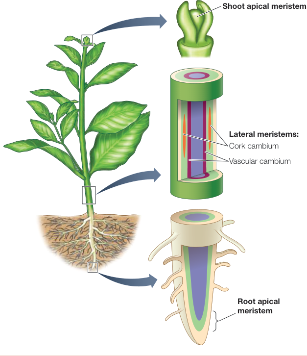
Figure 32.3
Plant Organs Are Made Up of Cells Organized into Three Types of Tissues
- Three types of plant cells can be classified based primarily on the properties of their cell walls: parenchyma, collenchyma, and sclerenchyma.
- The planes of division within the shoot apical meristem give rise to separate layers of cells that form the tissues.
- Plant structures are built from three types of tissue: dermal, ground, and vascular tissue.
Questions
- The cells that make up a plant stem are produced in the shoot apical meristem by cell division that results in small cells. Many mature sclerenchyma cells are quite long, yet their walls are reinforced in a way that does not permit them to expand. Explain how both of these statements can be true.
- Why are the growing tips of a plant more likely to wilt than more mature parts of a plant? Your answer should reference specific types of cells.
- In which tissue can cutin and trichomes be found?
- How do the conducting cells of xylem differ in gymnosperms and angiosperms?
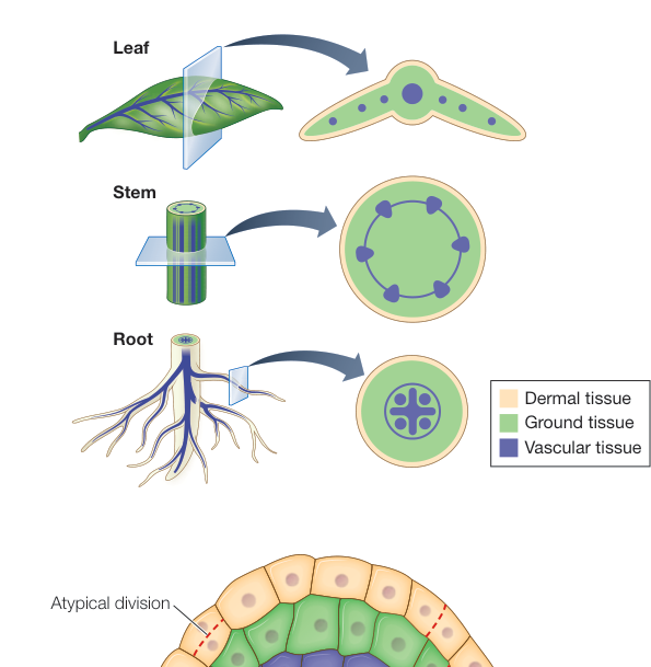
Figure 32.8
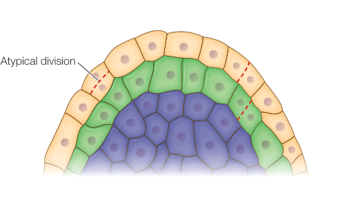
Figure 32.9
Meristems Build a Continuously Growing Plant
- The shoot apical meristem gives rise to the leaves and stem. Branches form indirectly from axillary buds.
- The root apical meristem gives rise to the root, which forms lateral roots through division of internal cells.
- Tree stems grow thicker through the action of the vascular cambium, which can divide to create a new ring of cells. One ring remains vascular cambium; the other ring becomes secondary xylem (if produced toward the interior) or secondary phloem (if produced toward the exterior).
Questions
- How does the existence of meristems contribute to the indeterminate nature of plant growth?
- In which longitudinal zone of the root are the initial cells located?
- In what tissue are the palisade mesophyll cells (a type of parenchyma cell) in a leaf located?
- In what ways are the activities of the shoot and root apical meristems similar to that of the vascular cambium? In what ways are they different?
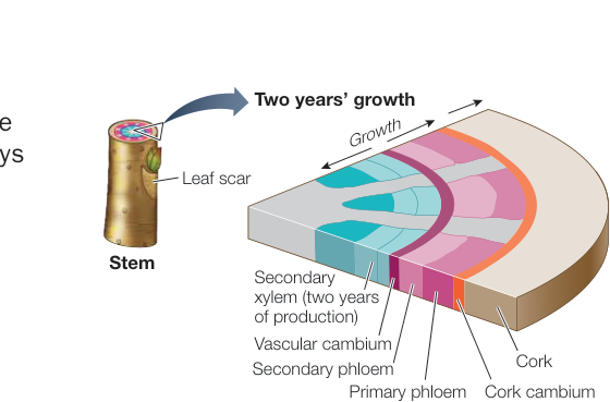
Figure 32.20
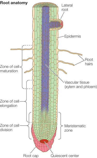
Figure 32.11
Domestication Has Altered Plant Form
- ■ The plant body plan is simple, yet it can be changed dramatically by minor differences in genes, as evidenced by the natural diversity of wild plants.
- ■ Crop domestication involves artificial selection of certain desirable traits found in wild populations.
Questions
- Changes in the proportions of different plant organs generate great diversity in plant architecture. What anatomical part of the plant are brussels sprouts and garlic cloves?
- How can you tell that the anatomical part of an onion that people generally eat is not a root?
- Why is it that selecting plants with desirable traits can be considered a form of genetic engineering?
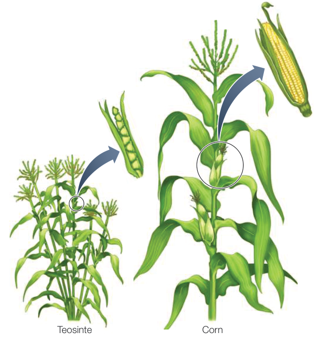
Figure 32.24