Chapter 41Animal Reproduction
© Tui De Roy/Minden Pictures
Key Concepts
41.1 Asexual Reproduction Is Efficient but Limits Genetic Variability
41.2 Sexual Reproduction Involves the Union of Haploid Egg and Sperm
41.3 Male Sex Organs Produce and May Deliver Sperm
41.4 Female Sex Organs Produce Eggs and May Nurture Embryos
41.5 Fertility Can Be Controlled
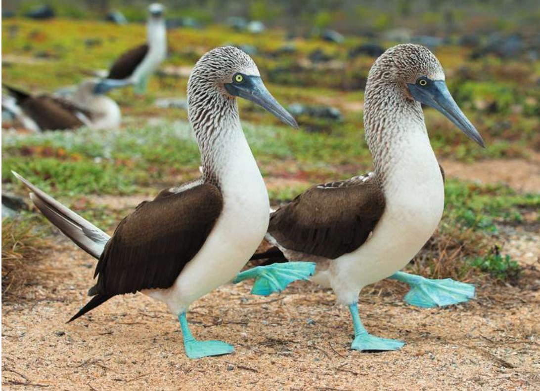
This Blue-footed Booby (Sula nebouxii) pair is in a courtship dance.
Investigating Life: No Time to Waste
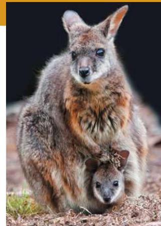
Female tammar wallabies (Macropus eugenii) become sexually receptive and mate soon after they give birth. The resulting embryo can be born any time from a month to a year later. Humans have a set 9-month pregnancy, so this highly variable duration of pregnancy seems unusual and leads to questions of “why” and “how.” Knowing more about the development of the young wallaby, called a joey, helps. The joey is very immature when born. It crawls into its mother’s pouch (the marsupium) where it suckles and grows for up to 10 months when it is weaned.
Answering the “why” question is not difficult. Reproductive success is the currency of evolution. Reproductive success depends on how many offspring a female can produce and on the chances that those offspring survive, reproduce, and pass on their genes to the following generation. Mating soon after giving birth (as the wallaby does) can maximize the number of offspring a female produces, but it does not necessarily maximize the survival and success of those offspring. Survival of offspring in many species depends on the ability of the mother to provide maternal care. Mammalian mothers produce milk to nourish their young, and that has an enormous energy cost. Therefore the females of many mammalian species do not become fertile and sexually receptive again until they have weaned their young. But this is not true for the tammar wallaby.
The large energy investment that a tammar wallaby mother makes to successfully rear a single offspring means that giving birth while still having one joey in the pouch lowers the chances of having either of those young survive. The adaptation of this species is developmental arrest, or diapause. The fertilized egg begins to develop into an embryo, but at an early stage of development its cell divisions cease, its metabolism decreases, and it remains in the uterus without implanting in the uterine lining as long as the previous offspring is suckling. If that offspring dies or is weaned, the quiescent embryo in the uterus is activated, implants in the uterine lining, and continues its development. Having an embryo ready to go when conditions are right is a way of maximizing reproductive success, but the “how” question remains.
© Danita Delimont/Getty Images
QA What signal controls the breaking of diapause in the tammar wallaby? (Find the answer on page 936.)
Asexual Reproduction Is Efficient but Limits Genetic Variability
Learning Objectives
- 41.1.1 Describe circumstances that make asexual reproduction advantageous or disadvantageous.
- 41.1.2 Describe the characteristics of the different types of asexual reproduction.
- 41.1.3 Explain conditions that would favor parthenogenesis over other forms of asexual reproduction.
When we think of reproduction, we think of sex, but many species can reproduce asexually—the offspring are exact genetic copies of the parents. Most of the animal species that reproduce asexually are invertebrates living in rather constant environments. However, examples of asexual reproduction do occur in vertebrates. Reproduction by female sharks living in aquariums with no males has been reported. The natural habitat of these fishes is the open ocean where potential mates might not be readily available. Asexual reproduction is efficient because no time or energy is wasted on mating and every member of the population can convert resources into offspring.
You can demonstrate to yourself the efficiency of asexual reproduction with a thought exercise. Start with two females, one reproducing sexually and one asexually. Assume that each can have two offspring, but in the case of the sexually reproducing female, one will be a male. Also assume that in each subsequent generation all of the females (mothers and daughters) will have two offspring each. In the first generation, the asexually reproducing female will have two daughters. Thus in the next generation there will be three asexually reproducing females that can have two female offspring each. In contrast, the sexually reproducing female will have only one female daughter, so in the next generation there will be only two females reproducing, having two offspring each—only one of which will be female. Run this thought exercise through three or four cycles and compare the numbers of offspring produced.
Asexual reproduction limits genetic diversity
A disadvantage of asexual reproduction is that the offspring are genetically identical to one another and to their parents. Thus asexual reproduction does not generate genetic diversity. Genetic diversity is the raw material that enables natural selection to shape adaptations in response to environmental change. When environmental changes occur, lack of genetic diversity can be disadvantageous to a population. Asexual reproduction is, however, a good way to preserve a successful genotype in a stable environment. Aphids are an example of a species that has it both ways (Figure 41.1). In the spring, the aphids that hatch on rose bushes from overwintering eggs are all females. Each of those aphids reproduces asexually to produce many more females—all genetically identical. This is a time when there is plenty of food and the environment is stable. When the conditions change due to drought or season, the females still reproduce asexually, but some of the offspring develop into males. The ensuing sexual reproduction introduces genetic variation into the eggs that will remain dormant until favorable conditions return.
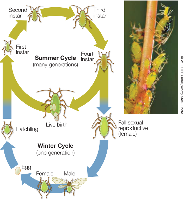
Figure 41.1 Aphids Are Sexual and Asexual When environmental conditions are favorable, such as in a rose garden in the summer, aphids make the most of resources by rapidly reproducing asexually. They are actually making identical copies of themselves. But when environmental conditions are unsure, such as in the fall, aphids reproduce sexually and thereby increase their genetic diversity in the face of changing, unpredictable conditions.
Three common modes of asexual reproduction are budding, regeneration, and parthenogenesis.
Budding and regeneration produce new individuals by mitosis
Some simple multicellular animals produce offspring by budding. New individuals form as outgrowths or buds from the bodies of older animals. A bud grows by mitotic cell division of somatic cells that dedifferentiate. The cells of the bud differentiate into the cell types of the new individual before the bud breaks away from the parent (Figure 41.2A). The bud is genetically identical to the parent, and it may grow as large as the parent before it becomes independent.
Regeneration is usually thought of as the replacement of damaged tissues or lost limbs, but in some cases pieces of an organism can regenerate complete individuals. Echinoderms, for example, have remarkable abilities to regenerate. If sea stars are cut into pieces, each piece that includes an arm and a portion of the central disc can grow into a new animal (Figure 41.2B). In the early 1900s oyster fishermen in Narragansett Bay between Rhode Island and Massachusetts tried to eliminate the sea stars that were preying on their oysters. Whenever they encountered sea stars, they cut them up with their knives and threw them back into the water. As a result, the sea star population increased explosively. A variant of regeneration called fission involves the animal splitting in half, with each half growing into a whole individual. An example is seen in sea anemones.
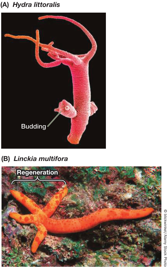
Figure 41.2 Two Forms of Asexual Reproduction (A) Budding: a new individual forms as an outgrowth from an adult hydra. (B) Regeneration: a single severed arm and a piece of the central disc of a mature sea star can regenerate into an entire animal.
Regeneration can occur when an animal is broken by an outside force such as wave action in the intertidal zone. In some cases, breakage occurs in the absence of external forces. Some species of segmented marine worms develop segments with rudimentary heads bearing sensory organs. The segments then break apart and each one forms a new worm.
Parthenogenesis is the development of unfertilized eggs
Not all eggs must be fertilized to develop. A common mode of asexual reproduction in arthropods (including aphids) is the development of offspring from unfertilized eggs. This phenomenon, called parthenogenesis, also occurs in some species of fishes, amphibians, and reptiles. Most species that reproduce parthenogenetically also engage in sexual reproduction or at least sexual behavior at other times. In some species, parthenogenesis is part of the mechanism that determines sex. In honey bees (as well as in most ants and wasps), males develop from unfertilized eggs and are haploid, whereas females develop from fertilized eggs and are diploid.
Parthenogenetic reproduction in some species requires sexual behavior even though sperm are not involved and eggs are not fertilized. David Crews and his students at the University of Texas extensively investigated one such case, that of parthenogenetic reproduction in a species of whiptail lizard. There are no males of this species. Females can act as males, engaging in all aspects of courtship display and mating, although no sperm are produced or transferred (Figure 41.3). Whether a specific female acts as a female or as a male depends on cyclical hormonal states. When her estrogen levels are high, she acts as a female. When her progesterone levels peak, she acts as a male. The stimulation resulting from the sexual activity triggers the release of eggs from the ovaries of the acting female.
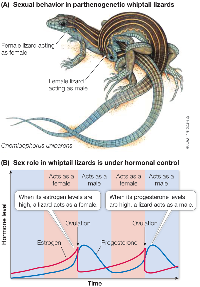
Figure 41.3 Asexual Reproduction May Require Sexual Behavior (A) Parthenogenetic whiptail lizards are all females, but they take turns acting the male role in reproductive behavior. The stimulation from sexual behavior is necessary for ovulation to occur. (B) The cycling of ovarian hormones determines the role in individual whiptail plays. (B after D. Crews. 1988. Psychobiology 16: 321–334.) Q: What selective advantages could accrue to a bisexual species becoming unisexual? What disadvantages?
Recap and Assess
Many species of animals, especially invertebrates, can reproduce asexually through budding, regeneration, or parthenogenesis. Asexual reproduction is an efficient use of resources but does not produce genetic diversity.
- What is similar about budding and regeneration but not parthenogenesis?
- Is global climate change likely to have a more negative impact on a species that reproduces asexually or one that reproduces sexually, and why?
- The Komodo dragon is a very large, sexually breeding species of lizard that lives on the Indonesian island of Komodo. A young Komodo dragon named Flora was raised alone for many years in London Zoo. One year after reaching sexual maturity, she laid a clutch of eggs that produced several baby dragons, all males. How did Flora reproduce? What advantage might this ability have for an island-dwelling species?
Asexual reproduction is an efficient way to use resources. Since most animals reproduce sexually, however, the genetic diversity produced by sexual reproduction must confer a tremendous advantage.
Sexual Reproduction Involves the Union of Haploid Egg and Sperm
Sexual Reproduction Involves the Union of Haploid Egg and Sperm
Learning Objectives
- 41.2.1 Discuss the similarities and differences between oogenesis and spermatogenesis.
- 41.2.2 Explain the connection between sex chromosome asymmetry and spermatocytes remaining in cytoplasmic contact.
- 41.2.3 Describe the interactions between sperm and eggs in sea urchins.
- 41.2.4 Summarize the ways that aquatic animals enhance the likelihood that sperm and eggs of the same species will meet.
- 41.2.5 Identify and describe conditions that favor the evolution of hermaphroditism.
Given the inefficiency of sexual reproduction, its prevalence is surprising. In most sexually reproducing species, the sex ratio is 1:1 and therefore half the population (males) do not produce offspring. Mating behaviors involve costs and risks. Costs include time and energy spent finding, attracting, and competing for a mate, as well as the “opportunity costs” of detracting from other activities such as feeding and caring for existing offspring. Risks include increased exposure to predation and the potential for physical damage. Despite these disadvantages, most eukaryotic organisms reproduce sexually. Thus it would seem that genetic diversity is an evolutionary advantage that can overwhelm the cost of sex (see Key Concepts 19.2 and 19.4).
Sexual reproduction requires the joining of two haploid sex cells to form a diploid individual. These haploid cells, or gametes, are produced through gametogenesis, a process that involves meiotic cell divisions. Two events in meiosis contribute to genetic diversity: crossing over between homologous chromosomes and the independent assortment of chromosomes (see Key Concepts 11.5 and 12.1). Mating behavior itself also contributes to genetic diversity. The genetic variation among the gametes of an individual and the genetic variation between any two parents produce an enormous potential for genetic variation between any two offspring of a sexually reproducing pair of individuals.
Sexual reproduction in animals consists of three steps:
- Gametogenesis: making gametes
- Spawning or mating: bringing gametes together
- Fertilization: fusing gametes
The process of gametogenesis is similar across sexually reproducing animal species. Processes of fertilization are also quite similar in widely different species. Therefore, while our discussion of gametogenesis will focus generally on mammals, and our discussion of fertilization will feature sea urchins, the facts would not be dramatically different were we to consider many other animal groups. Adaptations for spawning and mating, in contrast, show incredible anatomical, physiological, and behavioral diversity across species.
Gametogenesis produces eggs and sperm
Gametogenesis occurs in the gonads: male testes (singular testis) and female ovaries (singular ovary). The tiny male gametes, sperm, move by beating their flagella. The larger female gametes, eggs or ova (singular ovum), are nonmotile.
Gametes are produced from germ cells, which have their origin in early cell divisions of the embryo and remain distinct from all the other cells of the body (the somatic cells). Germ cells are sequestered in the body of the embryo until its gonads begin to form. The germ cells then migrate to the developing gonads, take up residence, and proliferate by mitosis, producing spermatogonia (singular spermatogonium) in males and oogonia (singular oogonium) in females (Figure 41.4). Spermatogonia and oogonia are diploid, multiply by mitosis, and are stem cells. They are self-regenerating, and they produce progeny—spermatocytes and oocytes—that enter the next stage of gametogenesis.
In that next stage of gametogenesis, meiotic cell division reduces the chromosomes to the haploid number (see Key Concept 11.5). The progeny of the spermatogonia and oogonia that enter meiosis are primary spermatocytes and primary oocytes. The steps of meiosis are similar in males and females, but there are important sex differences in gametogenesis.
SPERMATOGENESIS The initial proliferation of male germ cells into spermatogonia proceeds by mitosis in the embryo. Throughout the male life span, spermatogonia continue to divide by mitosis, with one daughter cell retaining the self-regenerating capacity of a spermatogonium and the other daughter cell becoming a primary spermatocyte. As illustrated in Figure 41.4A, primary spermatocytes undergo the first meiotic division—the reduction division—to form two haploid secondary spermatocytes. The second meiotic division produces four haploid spermatids for each primary spermatocyte that enters meiosis. In mammals, the progeny of primary spermatocytes remain connected by cytoplasmic bridges after each division.
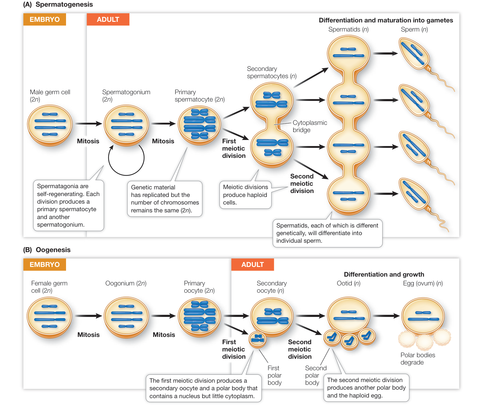
Figure 41.4 Gametogenesis Male and female germ cells proliferate by mitosis and produce diploid spermatogonia and oogonia that mature into primary spermatocytes and oocytes before entering meiosis. (A) Spermatogonia continue to divide by mitosis in adults, producing a steady supply of spermatocytes that divide meiotically to produce haploid spermatids, which differentiate into sperm. In many species, the progeny of spermatocytes remain in contact through cytoplasmic bridges until the sperm mature. (B) In mammals, oogonia cease division in the embryo, and primary oocytes remain arrested in prophase I of meiosis until they are ovulated and fertilized. Each oocyte will produce one haploid ootid, which matures into an egg.
Mammalian spermatocytes remain in cytoplasmic contact throughout their development because of the asymmetry of sex chromosomes in males. Half the secondary spermatocytes receive an X chromosome, the other half a Y chromosome. The Y chromosome contains fewer genes than the X chromosome, and some of the products of genes found only on the X chromosome are essential for spermatocyte development. By remaining in cytoplasmic contact, all four spermatocytes can share the gene products of the X chromosomes, although only half of them have an X chromosome.
A spermatid bears little resemblance to a mature sperm. Through further differentiation the spermatid becomes compact, streamlined, and develops a flagellum to become motile. We discuss the production of human sperm in Key Concept 41.3.
OOGENESIS Oogonia, like spermatogonia, proliferate through mitosis (see Figure 41.4B). The resulting primary oocytes immediately enter prophase of the first meiotic division. In many species, including humans, the oocyte experiences developmental arrest at this point and may remain in that state for days, months, or years. In the human female, this period of arrest is at least 10 years (i.e., until puberty), and some primary oocytes remain in prophase I for more than 50 years (i.e., until menopause). In contrast, spermatogenesis continues, uninterrupted, to completion once the primary spermatocyte has differentiated.
During the prolonged prophase I, or shortly before it ends, the primary oocyte grows larger through increased production of ribosomes, RNA, cytoplasmic organelles, and energy stores. At this point the primary oocyte acquires all the energy, raw materials, and RNA that the egg will need to survive its first cell divisions after fertilization. In fact, the nutrients in the egg must maintain the embryo until it is either nourished by the maternal circulatory system or can feed on its own.
When a primary oocyte resumes meiosis, its nucleus completes the first meiotic division near the surface of the cell. The daughter cells of this division receive grossly unequal shares of cytoplasm. This asymmetry represents another major difference from spermatogenesis, in which cytoplasm is apportioned equally. The daughter cell that receives almost all the cytoplasm becomes the secondary oocyte, and the one that receives almost none forms the first polar body (see Figure 41.4B).
The second meiotic division—that of the large secondary oocyte—is also accompanied by asymmetrical division of the cytoplasm. One daughter cell forms the large, haploid ootid that eventually differentiates into a mature egg, and the other forms the second polar body. In most species the polar bodies rapidly degenerate, so the end result of oogenesis is only one mature egg for each primary oocyte that enters meiosis. However, that egg is a large, well-provisioned cell. In a few species the polar bodies have evolved specialized functions. In some species that reproduce parthenogenetically, a polar body can fuse with the egg to restore the diploid number of chromosomes. In some insect species the polar bodies form organs with specialized functions. An example is scale insects, in which cells derived from the polar bodies form a container for endosymbiotic bacteria that the insects require for life.
A second period of arrested development occurs after the first meiotic division forms the secondary oocyte. The egg may be expelled from the ovary in this condition. In many species, including humans, the second meiotic division is not completed until the egg is fertilized by a sperm.
Fertilization is the union of sperm and egg
The union of the haploid sperm and the haploid egg (fertilization) creates a single diploid cell, a zygote that will develop into an embryo. Fertilization does more than just restore the full genetic complement of the animal. The processes associated with fertilization help the egg and sperm get together, prevent the union of the sperm and egg of different species, and guarantee that only one sperm will enter and activate the egg. Fertilization involves a complex series of events:
- The sperm and the egg chemically recognize each other.
- The sperm is activated, enabling it to gain access to the cell membrane of the egg.
- The cell membrane of the egg fuses with the cell membrane of a single sperm.
- The egg blocks entry of additional sperm.
- The egg is metabolically activated and stimulated to start development.
- The egg and sperm nuclei fuse to create the diploid nucleus of the zygote.
SPECIFICITY IN SPERM–EGG INTERACTIONS Specific recognition molecules mediate interactions between sperm and eggs. These molecules ensure that the activities of sperm are directed toward eggs and not other cells, and they help prevent eggs from being fertilized by sperm from the wrong species. The latter function is particularly important in aquatic species that release eggs and sperm into the surrounding water, where the eggs can readily be exposed to sperm of other species. The sea urchin is a good example of such a species, and sea urchin fertilization has been well studied.
Sea urchin eggs release chemical attractants that increase the motility of sperm and cause them to swim toward the egg. These chemical attractants are species-specific. For example, eggs of one species of sea urchin release a specific peptide consisting of 14 amino acids. This peptide binds to receptors present on sperm of the same species. The sperm respond by increasing their mitochondrial respiration and motility. Before exposure to the peptide, the sperm swim in tight little circles, but after binding to the peptide, they swim energetically up the concentration gradient of the peptide until they reach the egg that is releasing it.
When a sperm reaches an egg, it must get through two protective layers before it can fuse with the egg cell membrane. The eggs of sea urchins are covered with a jelly coat that surrounds a proteinaceous vitelline envelope (Figure 41.5A). The success of a sperm’s assault on these protective layers depends on a membrane-enclosed structure at the front of the sperm head called an acrosome.
The acrosome contains enzymes and other proteins. When a sperm makes contact with an egg of its own species, substances in the jelly coat trigger an acrosomal reaction. This reaction begins with the breakdown of the cell membrane covering the sperm head and the underlying acrosomal membrane (Figure 41.5B). The acrosomal enzymes are released and digest a hole through the jelly coat.
The acrosomal reaction also triggers polymerization of actin in the sperm head, and an acrosomal process extends out of the front of the sperm head. This acrosomal process is coated with species-specific recognition molecules called bindin, and there are bindin receptors on the vitelline envelope of the egg. The interaction of bindin with their receptors enables the sperm to contact the egg cell membrane. That contact results in fusion of the sperm and egg cell membranes and the formation of a fertilization cone that engulfs the sperm head, bringing it into the egg cytoplasm. The sperm mitochondria, which largely constitute the midpiece of the sperm, are also drawn into the egg cytoplasm, but they degrade and disappear; this means that the mitochondria and mitochondrial genes of the new urchin are derived only from the egg.
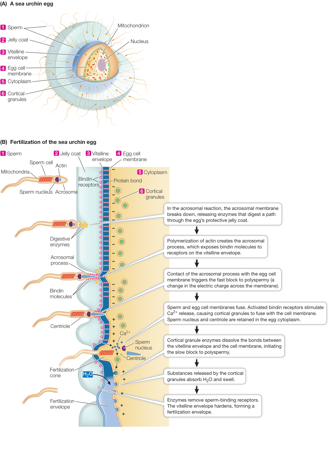
Figure 41.5 Fertilization of the Sea Urchin Egg (A) Sea urchin eggs are protected by a jelly layer and a proteinaceous vitelline envelope. Sperm must penetrate both to reach the egg cell membrane. Many sperm attach to the vitelline envelope, but only the first one to reach the egg cell membrane achieves fertilization. The numbers match structures with the events shown in (B). (B) The acrosomal reaction allows a sea urchin sperm to recognize an egg of the same species and pass through its protective layers. Enzymes from the egg’s cortical granules trigger the slow block to polyspermy. (After D. Epel. 1977. Sci Am 237: 128–138. Drawing by Carol Donner.) Animation 41.1 Fertilization in a Sea Urchin Egg
Connect the Concepts Because mitochondrial genes are inherited only from the mother, traits controlled by those genes are not inherited in a Mendelian fashion (see Key Concept 12.5), and they enable establishment of matrilineal phylogenies (see Key Concept 20.2).
In animals that use internal fertilization, mating behaviors help guarantee species specificity, but egg–sperm recognition mechanisms still exist. The mammalian egg is encased by the cumulus, a loose assemblage of maternal cells in a gelatinous matrix (Figure 41.6). Beneath the cumulus is a glycoprotein envelope called the zona pellucida, which is functionally similar to the vitelline envelope of sea urchin eggs. Mammalian sperm become activated when they are deposited in the female reproductive tract, and they are then capable of an acrosomal reaction should they encounter an egg. An activated sperm can penetrate the cumulus and interact with the zona pellucida.
Unlike the jelly coat of sea urchin eggs, the cumulus of mammalian eggs does not trigger the acrosomal reaction. When sperm make contact with the zona pellucida, a species-specific glycoprotein binds to recognition molecules on the head of the sperm. This binding triggers the acrosomal reaction, releasing acrosomal enzymes that digest a path through the zona pellucida. When the sperm head reaches the egg cell membrane, other proteins facilitate its adhesion to and fusion with the egg cell membrane.
BLOCKS TO POLYSPERMY The fusion of the sperm and egg cell membranes and the entry of the sperm into the egg initiate a programmed sequence of events. The first responses to sperm entry are blocks to polyspermy: mechanisms that prevent more than one sperm from entering the egg. Survival of the embryo is unlikely if more than one sperm enters the egg. The sperm contributes a haploid set of chromosomes, so an extra set would disrupt the mitotic division of the egg and subsequent cells. In addition, in most mammals the sperm contributes a centriole to the fertilized egg, and the centriole forms the centrosome, which is critical for organizing the mitotic spindle. Having two centrosomes would disrupt mitosis of the fertilized egg.
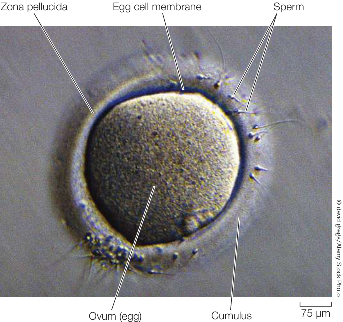
Figure 41.6 Barriers to Sperm This human egg, like other mammalian eggs, is surrounded by the cumulus and zona pellucida. Sperm must penetrate both to fertilize the egg. Only one sperm will penetrate the zona pellucida and fuse with the egg cell membrane. Q: What would be the consequences of two sperm fertilizing the egg?
Connect the Concepts The roles of the centriole and the centrosome in mitosis are discussed in Key Concept 11.3.
Blocks to polyspermy have been studied extensively in sea urchin eggs because they can be fertilized in a dish of seawater under a microscope. Within seconds after the sperm membrane contacts the egg membrane, an influx of sodium ions changes the electric charge difference across the egg cell membrane. This fast block to polyspermy prevents the fusion of any other sperm with the egg cell membrane, but it is transient. The change in membrane electric charge lasts only about a minute, but that is enough time to allow a slower block to sperm entry to develop.
The slow block to polyspermy involves converting the vitelline envelope to a physical barrier that sperm cannot penetrate. Before fertilization, the vitelline envelope is bonded to the egg cell membrane. Just under the cell membrane are vesicles called cortical granules (see Figure 41.5) that contain enzymes and other proteins.
The sea urchin egg, like all animal cells, sequesters calcium in its endoplasmic reticulum. Sperm entry into the egg stimulates the release of calcium ions from the endoplasmic reticulum and into the egg cytosol. This increase in cytosolic calcium causes the egg’s cortical granules to fuse with the cell membrane and release their contents. Cortical granule enzymes break the bonds between the vitelline envelope and the cell membrane, and other proteins released from the cortical granules attract water into that space. As a result, the vitelline envelope rises to form a fertilization envelope. Cortical granule enzymes also degrade sperm-binding molecules on the surface of the fertilization envelope and cause it to harden. Thus additional sperm cannot contact the egg cell membrane.
In mammalian eggs, sperm entry does not cause a rapid change in membrane potential, but it does trigger a release of calcium from the endoplasmic reticulum. As in the sea urchin egg, increased calcium causes the cortical granules to fuse with the egg cell membrane. A fertilization envelope does not form around the mammalian egg, but the cortical granule enzymes destroy the sperm-binding molecules in the zona pellucida. The rise in cytosolic calcium also signals the egg to complete meiosis. The stage is set for the first cell division.
Spawning and mating behaviors get eggs and sperm together
As you have just seen, sexual reproduction requires the production of haploid gametes (gametogenesis) and the joining together of those gametes to form a diploid zygote (fertilization). Spawning and mating behaviors get eggs and sperm close enough together that fertilization can occur. Fertilization can occur externally or internally.
EXTERNAL FERTILIZATION In an aquatic environment, animals can bring their gametes together by simply releasing them into the water. This practice, called spawning, results in external fertilization. Many aquatic animals are not very mobile, but they produce huge numbers of gametes that can travel far from the point of release. A female oyster, for example, releases millions of eggs when she spawns, and the number of sperm produced by a male oyster is astronomical.
Numbers alone, however, do not guarantee that gametes will meet. The reproductive activities of the males and females of a population must be synchronized, since released gametes have a limited life span. Seasonal breeders may use day length, changes in temperature, or changes in weather to time the production and release of their gametes. Mutual stimulation is also important. Release of gametes into the water by one individual can stimulate others to spawn.
Behavior can play an important role in bringing gametes together even when fertilization is external. Some species travel great distances to congregate with potential mates and release their gametes at the same time in a suitable environment. Salmon are an extreme example. They hatch and develop in freshwater streams and then migrate to the ocean. They remain as oceanic fishes for years. When they are mature and ready to spawn, they travel hundreds of miles back to and then up the same stream to the region where they hatched. Males and females expend great amounts of energy to swim up the streams to the spawning grounds, where they pair up, prepare a depression in the streambed gravel, and together release their sperm and eggs. As the gametes drift down into the gravel, fertilization occurs.
INTERNAL FERTILIZATION Terrestrial animals cannot simply release their gametes into the environment. Sperm can move only through liquid, and delicate gametes released into air would dry out and die. Most terrestrial animals avoid these problems by internal fertilization, the release of sperm into the female reproductive tract. Some aquatic animals also practice internal fertilization, but it is ubiquitous in terrestrial animals.
Animals have evolved an astonishing diversity of behavioral and anatomical adaptations for internal fertilization. The reproductive organs together are the animal’s genitalia. Gametogenesis occurs in the gonads, which are the primary sex organs. All additional components of an animal’s genitalia are called accessory sex organs. The internal accessory sex organs include a variety of glands, tubules, ducts, and other structures. External accessory sex organs are referred to as the external genitalia. In males of many species, the external genitalia includes a penis that enables the male to deposit sperm in the female’s vagina, the first segment of her reproductive tract.
Copulation is the physical joining of male and female accessory sex organs. Most male insects copulate and transfer sperm to the female’s vagina through a penis. The external genitalia of insects often have species-specific shapes that match in a lock-and-key fashion to ensure a tight, secure fit between the mating pair during the prolonged period of sperm transfer. In some insect species in which females mate with more than one male, the male external genitalia includes elaborate structures that can scoop sperm deposited by other males out of the female’s reproductive tract, allowing a male to replace that sperm with his own.
Transfer of sperm in internal fertilization can also be indirect. Males of some invertebrate species (e.g., mites and scorpions) and a few vertebrates (e.g., salamanders) deposit spermatophores—packets of sperm protected from desiccation—in the environment. When a female mite encounters a spermatophore from a potential mate, she straddles it and opens a pair of plates in her abdomen so that the tip of the spermatophore enters her reproductive tract and allows the sperm to enter.
Male squid and spiders play a more active role in spermatophore transfer. The male spider secretes a drop containing sperm onto a bit of web, then uses a special structure on his foreleg to pick up the sperm-containing web and insert it through the female’s genital opening. Some male squid use one specialized tentacle to pick up a spermatophore and insert it into the female’s genital opening.
Some individuals can function as both male and female
In most species, gametes are produced by individuals that are either male or female. Species that have separate male and female members are referred to as dioecious (Greek, “two houses”). In some species, however, a single individual may produce both sperm and eggs. Such species are monoecious (“one house”), or hermaphroditic.
Almost all invertebrate groups contain some hermaphroditic species. An earthworm is an example of a simultaneous hermaphrodite, meaning an individual is both male and female at the same time. When two earthworms mate, they exchange sperm, and as a result the eggs of each are fertilized (see Figure 30.12C). Some vertebrates are sequential hermaphrodites, meaning an individual may function as a male or a female at different times in its life. An example is the anemonefishes, or clownfishes, species that live in small groups within large sea anemones (Figure 41.7). All anemone fishes are born male. The largest fish in a group becomes a functional female. If that fish is removed from the group, the next-largest male becomes a female. The second-largest fish in the group is the only male in breeding condition. The reverse situation exists in some species in which a large dominant male controls a harem of females. If the male is removed, the largest female becomes the male in the group.
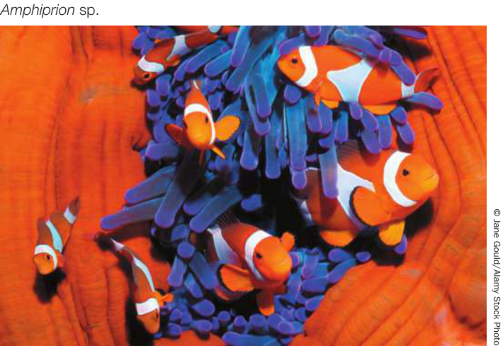
Figure 41.7 When Size Determines Sex Anemonefishes (also known as clownfishes) live in groups of about a dozen centered on a single sea anemone. All anemonefishes are born male; the largest fish in the group becomes a functional female. Thus a fish may function as a male and as a female at different times in its life.
What is the evolutionary advantage of hermaphroditism? Some simultaneous hermaphrodites, such as parasitic tapeworms, have a low probability of meeting a potential mate—an individual tapeworm may be the only one in its host. Tapeworms can fertilize their own eggs, but most simultaneous hermaphrodites must mate with another individual. Because every simultaneous hermaphrodite is both male and female, however, the probability of encountering a possible mate doubles. In some sequential hermaphrodites, siblings are all male or all female at the same time, thus reducing the incidence of inbreeding.
The evolution of vertebrate reproductive systems parallels the move to land
The earliest vertebrates evolved in aquatic environments. The closest living relatives of those earliest vertebrates are modern-day fishes. They remain exclusively aquatic, and most practice external fertilization. The most primitive fishes, the lampreys and hagfishes, simply release their gametes into the environment. In most fishes, however, mating behaviors bring females and males into close proximity at the time of gamete release. In sharks and rays, fins have evolved into claspers that hold the male and female together and enable sperm to be transferred directly into the female reproductive tract.
Amphibians were the first vertebrates to live in terrestrial environments. They dealt with the challenge of a dry environment by returning to water to reproduce, as most amphibians still do today.
Reptiles were the first vertebrate group to solve the problem of reproduction in the terrestrial environment (Figure 41.8). Their solution was the amniote egg (see Figure 31.19A). A hard or leathery shell protects the embryo and impedes water loss while allowing the diffusion of oxygen into the egg and carbon dioxide out of the egg. The eggshell creates an obvious problem for fertilization. Sperm cannot penetrate the shell, so they must reach the egg before the shell forms. Hence internal fertilization and the evolution of accessory sex organs were necessary for the evolution of the amniote egg.
Male snakes and lizards have paired hemipenes (singular hemipenis) that can be engorged with blood and thereby extruded from the male’s body. Only one hemipenis is inserted into the female’s reproductive tract. It is usually rough or spiny at the end to achieve a secure hold while sperm are transferred down a groove on its surface. Retractor muscles pull the hemipenis back into the male’s body when mating is completed. Some evolutionarily ancient bird species have erectile penises that channel sperm along a groove into the female’s reproductive tract. Birds with more recent evolutionary origins, however, do not have erectile penises; instead, the male and female simply bring their genital openings (cloacae; singular cloaca) close together to transfer sperm. Usually this involves the male standing on the female’s back (see Figure 41.8B).
All mammals practice internal fertilization. Then all except the prototherian mammals retain the developing embryo for some time in the female reproductive tract. Prototherian mammals (the monotremes; see Figure 31.26) lay eggs. The other mammals (the therians) vary enormously as to the developmental stage of their offspring at the time of birth. As you saw at the beginning of the chapter, the tammar wallaby is extremely immature at birth. By comparison, a horse foal can stand up soon after birth and is able to run within hours.
Animals with internal fertilization are distinguished by where the embryo develops
Two patterns of care and nurture of the embryo have evolved in animals: oviparity (egg laying) and viviparity (live bearing).
Oviparous animals lay eggs in the environment, and their embryos develop outside the mother’s body. Oviparous terrestrial animals such as insects and reptiles—including birds—protect their eggs from desiccation with waterproof membranes or shells. Oviparity is possible because eggs are stocked with abundant nutrients to supply the needs of the embryo. Some oviparous animals engage in various forms of parental behavior to protect their eggs, but until the eggs hatch, the embryos depend entirely on the nutrients stored in the egg.
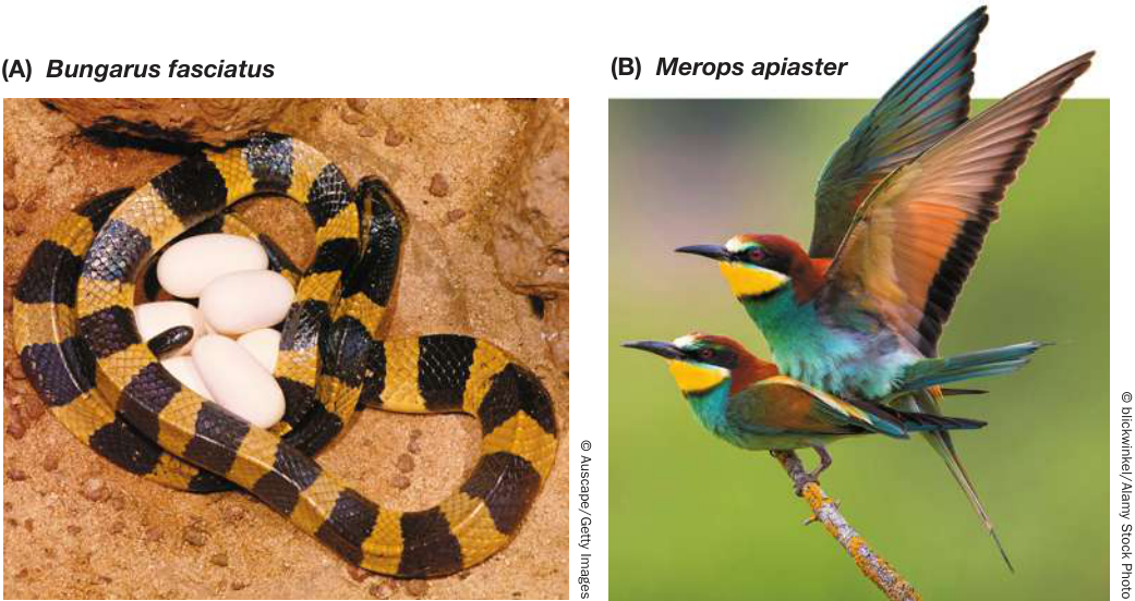
Figure 41.8 The Shelled Egg The terrestrial environment is desiccating. Therefore the shelled egg was a major evolutionary step that allowed reptiles to reproduce in the terrestrial environment. (A) A female banded krait has deposited her eggs in the sand. (B) The shelled egg requires that sperm meet egg before the shell forms. Therefore reptiles must practice internal fertilization, as these European Bee-eaters are doing.
Viviparous animals retain the embryo within the mother’s body during its early developmental stages. The embryo is nourished and its wastes removed through exchanges with the mother’s tissues. Viviparity occurs in many vertebrate groups, and with the exception of the prototherians, it is universal in mammals. Therian mammals have a specialized portion of the female reproductive tract, the uterus or womb, that contains the embryo until it is born. The early embryo arriving in the uterus from the fallopian tube implants into the lining of the uterus. Implantation involves interactions between the tissues of the embryo and the uterus to form a placenta. Intimate contact in the placenta between the blood of the embryo and the blood of the mother facilitates the exchange of nutrients and wastes.
In most non-mammalian viviparous animals, such as garter snakes and the well-known aquarium fish the guppy, fertilized eggs are retained in the mother’s body until they hatch. These embryos still receive nutrition from stores in the egg, so this reproductive adaptation is called ovoviviparity.
Recap and Assess
Sexual reproduction involves gametogenesis, mating, and fertilization. Fertilization can be external or internal and involves mechanisms for ensuring that only one sperm from the right species enters the egg. Oviparous female animals lay eggs in the environment, and their embryos develop outside the mother’s body. Viviparous female animals retain the developing embryo in the uterus until it is born.
- Crossing over is the reciprocal exchange of segments in homologous chromatids. Why does it occur more frequently in human oocytes than in human spermatocytes?
- If a mutation in a mammal eliminated the maintenance of cytoplasmic bridges between spermatids, would you expect all offspring sired by that mammal to be male or female? Explain your answer.
- In animals that spawn, what mechanisms ensure that an egg is fertilized by sperm of the same species, and by only one sperm?
- What are three situations in which hermaphroditism would be advantageous?
Now that we have covered some of the general aspects of gametogenesis and fertilization and have briefly discussed the great diversity of mating systems, we next consider the human reproductive systems in detail.
41.3 Male Sex Organs Produce and May Deliver Sperm
Learning Objectives
- 41.3.1 Identify the different components of semen, where they are produced, and their functions.
- 41.3.2 Describe the anatomical and functional relationships between Leydig cells and Sertoli cells.
- 41.3.3 Explain the roles of NO and cGMP in the male sexual response.
- 41.3.4 Summarize the hormonal changes that occur during puberty in males and how they lead to the development of male secondary sexual characteristics.
This key concept describes the structures and functions of the male reproductive system in mammals, using humans as the prime example. You will also learn about the hormonal regulation of the male reproductive system. The discussion covers:
- The primary sex organs, testes, that produce sperm and serve endocrine functions.
- The accessory sex organs, including the ducts through which the sperm pass, the glands that produce and deliver secretions into those ducts, and the external genitalia.
- Secondary sexual characteristics that result in male appearance but are not directly involved in reproduction.
Semen is the product of the male reproductive system
Semen contains sperm and a mixture of fluids and molecules (seminal fluid) that support the sperm and facilitate fertilization. Sperm make up less than 5% of the volume of the semen. Sperm are produced in the testes, the paired male gonads, and the seminal fluid is produced by accessory glands.
Figure 41.9 illustrates the human male reproductive organs. The testes of most mammals are located outside the body cavity in a pouch of skin called the scrotum. Why should the testes be located outside the body cavity? The optimal temperature for spermatogenesis in most mammals is slightly lower than the normal body temperature. The scrotum keeps the testes at this optimal temperature. Muscles in the scrotum contract in a cold environment, bringing the testes closer to the warmth of the body; in a hot environment the muscles relax, cooling the testes by suspending them farther from the body.
Spermatogenesis takes place within the seminiferous tubules tightly coiled in each testis (Figure 41.10A). Between the seminiferous tubules are clusters of Leydig cells that produce testosterone (Figure 41.10B). Spermatogonia reside in the outer regions of the seminiferous tubules, just under the basement membrane. Moving inward toward the lumen of the tubule are germ cells in successive stages of spermatogenesis (Figure 41.10C). The germ cells are intimately associated with Sertoli cells, which provide nutrients for the developing sperm and secrete several things, including hormones and growth factors that are necessary for spermatogenesis (which is why Sertoli cells are sometimes called “nurse cells”). Tight junctions (see Key Concept 6.2) between Sertoli cells create a blood–sperm barrier that prevents many harmful substances that can be circulating in the blood from getting to the developing sperm.
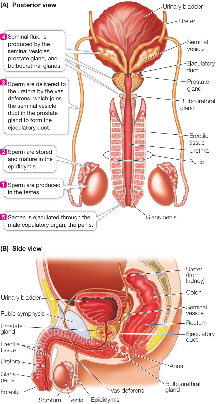
Figure 41.9 Reproductive Tract of the Human Male The male reproductive organs are shown (A) from the rear and (B) from the side. Q: What problem might an enlarged prostate gland produce?
When the second meiotic division is complete, each primary spermatocyte has produced four spermatids (see Figure 41.4A). The spermatids develop into spermatozoa as they migrate toward the lumen of the seminiferous tubule. The nucleus becomes compact, and the surrounding cytoplasm is lost. A flagellum—the sperm tail—develops. The mitochondria that provide the energy for sperm motility become condensed into a midpiece between the head and tail. An acrosome forms over the nucleus in the head of the sperm. Immature sperm are shed into the lumen of the seminiferous tubule.
From the seminiferous tubules, sperm move into the epididymis (see Figure 41.9A), where they mature, become motile, and are stored. The epididymis connects to the urethra via the vas deferens (plural vasa deferentia) and the ejaculatory duct. The urethra originates in the bladder, runs through the penis, and opens to the outside of the body at the tip of the penis. It serves as the common final duct for urine and semen.
The components of the semen other than sperm come from three accessory glands. About 60% of the volume of semen is secreted by the paired seminal vesicles that secrete into the vas deferens just before it joins the urethra. Seminal fluid is thick because it contains mucus and fibrinogen, a protein also found in the blood, where it can polymerize to form blood clots. Seminal fluid also contains the monosaccharide fructose, an energy source for the sperm.
The prostate gland contributes about 30% of the volume of the semen. Prostate fluid is alkaline, so it neutralizes the acidity in the male and female reproductive tracts and makes these environments more hospitable to sperm. The prostate secretes a clotting enzyme that causes fibrinogen from the seminal vesicles to clot and convert the semen into a gelatinous mass that can be ejaculated into the female reproductive tract. Another prostate enzyme, fibrinolysin, dissolves the clotted semen after it is ejaculated, thereby liberating the sperm. Prostaglandins—hormones produced by the prostate—stimulate contractions of the female reproductive tract.
The bulbourethral glands produce a small volume of an alkaline, mucoid secretion that helps neutralize acidity in the urethra and lubricate it to facilitate the passage of semen during the climax of sexual intercourse.
The penis and the scrotum are the male external genitalia
The shaft of the penis is covered with normal skin, but the highly sensitive tip, the glans penis, is covered with thinner, more sensitive skin that is especially responsive to sexual stimulation. A fold of skin called the foreskin covers the glans of the human penis. The procedure known as circumcision removes a portion of the foreskin.
Sexual stimulation triggers responses in the nervous system that result in penile erection. This response is mediated by nerves to the penis from the central nervous system (brain and spinal cord) that release the neurotransmitter acetylcholine (ACh) onto endothelial cells lining the penile blood vessels. These
Male Sex Organs Produce and May Deliver Sperm
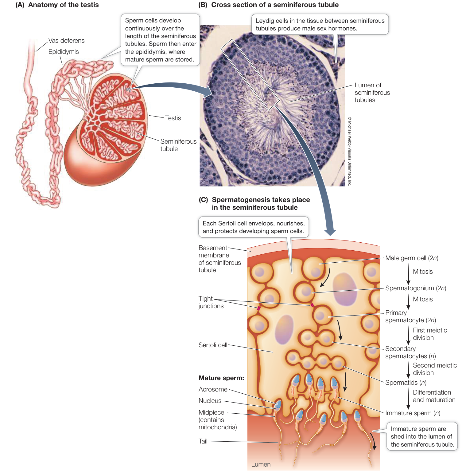
Figure 41.10 Spermatogenesis Takes Place in the Seminiferous Tubules (A) Seminiferous tubules fill the testes, continuously producing millions of sperm. (B) Cross section of seminiferous tubules and the Leydig cells in the spaces between them. (C) This longitudinal diagram shows how, as sperm mature, they move from the outer layer of the tubule toward the center, where they are shed into the lumen of the tubule. Activity 41.2 Spermatogenesis
endothelial cells respond by synthesizing and releasing a gaseous signal—nitric oxide (NO) (Figure 41.11; see also Figure 7.13). The NO diffuses into the smooth muscle cells that control the diameter of the penile arteries and stimulates them to produce the second messenger cyclic GMP (cGMP). Increased cGMP in these muscle cells causes a decrease in cytoplasmic by closing channels and stimulating reuptake by the endoplasmic reticulum. The decrease in causes the smooth muscles to relax; the arteries dilate and carry more blood into the penis. Increased blood flow swells the shafts of spongy erectile tissue located along the length of the penis (see Figure 41.9). The enlargement of these blood-filled cavities compresses the vessels that carry blood out of the penis, and the erectile tissue becomes engorged with blood. The penis becomes hard and erect, facilitating its insertion into the vagina. Many species of mammals (though not humans) have a bone in the penis, but these species still depend on erectile tissue for copulation.
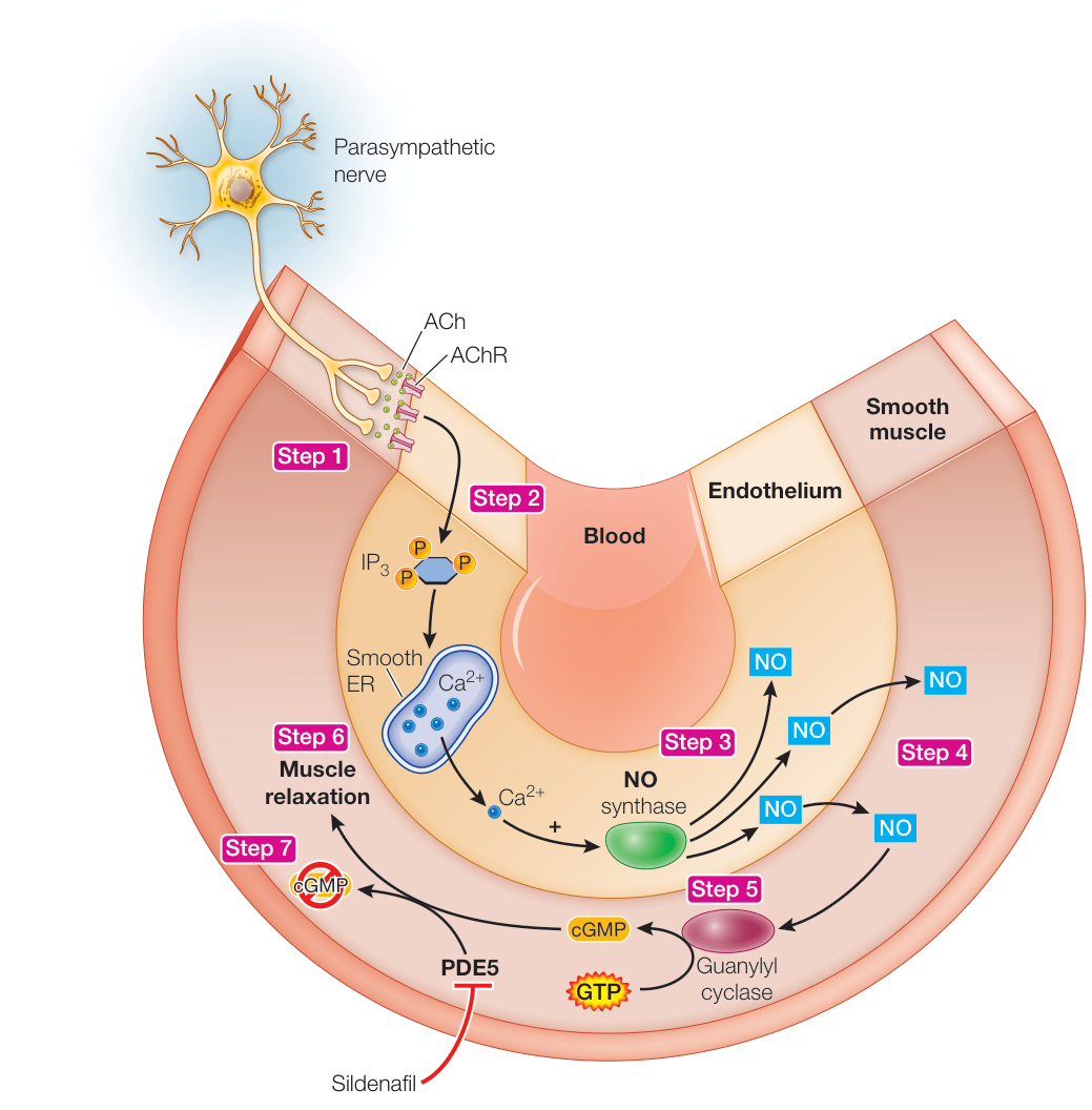
Figure 41.11 Neural Control of Erection Involves a Gaseous Signal Neural release of acetylcholine (ACh) onto endothelial cells of the penile blood vessels activates second messenger (inositol triphosphate) which stimulates the synthesis and release of nitrous oxide (NO), which diffuses to the surrounding smooth muscle cells. NO induces the production of cyclic GMP (cGMP), which lowers cytoplasmic , causing the smooth muscles to relax and thereby allow greater blood flow and an erection. A phosphodiesterase breaks down the cGMP, ending the erection. Drugs such as sildenafil that inhibit phosphodiesterase prolong the erection.
Connect the Concepts The mechanism whereby the neurotransmitter ACh causes production of the gas NO in smooth muscle is described in Key Concept 7.3 (see Figure 7.13), and how intracellular controls smooth muscle contraction is described in Key Concept 46.1 (see Figure 46.9).
At the climax of copulation, 2–6 milliliters of semen are propelled through the vasa deferentia and the urethra in two steps, emission and ejaculation. During emission, rhythmic contractions of smooth muscles in the vasa deferentia and accessory glands move the semen into the urethra at the base of the penis.
Ejaculation is caused by contractions of other muscles at the base of the penis surrounding the urethra. These contractions force the coagulum of semen through the urethra and out of the penis. The muscle contractions of ejaculation are accompanied by feelings of intense pleasure known as orgasm. They are also accompanied by transient increases in heart rate, blood pressure, breathing, pupil dilation, and skeletal muscle contractions throughout the body.
After ejaculation, NO release decreases and a phosphodiesterase enzyme breaks down cGMP (see Figure 41.11). With decreased cGMP, the cytoplasmic concentration increases, causing constriction of the blood vessels carrying blood into the penis. Blood pressure in the erectile tissue decreases, relieving the compression of the blood vessels leaving the penis, and the erection declines.
Erectile dysfunction (ED), or impotence, is the inability to achieve or sustain an erection. ED may have different causes, including cardiovascular disease. Drugs such as sildenafil are used to treat ED because they inhibit the phosphodiesterase that breaks down cGMP, thus enhancing the effect of NO released in the penis and thereby improving the ability to achieve and maintain an erection.
Male sexual function is controlled by hormones
Spermatogenesis and maintenance of male secondary sexual characteristics such as facial hair and a deep voice depend on testosterone produced by the Leydig cells of the testes. As described in Key Concept 39.3, increased production of testosterone at puberty results from an increased release of gonadotropin-releasing hormone (GnRH) by the hypothalamus. GnRH stimulates anterior pituitary cells to secrete luteinizing hormone (LH) and follicle-stimulating hormone (FSH) (Figure 41.12). Higher levels of LH stimulate the Leydig cells to increase their production and release of testosterone. Testosterone exerts negative feedback on the anterior pituitary and the hypothalamus. At the time of puberty, the sensitivity of the hypothalamus to negative feedback from testosterone declines; as a result, the level of circulating testosterone increases.
Increased testosterone in pubertal boys causes the development of pubic and facial hair, a deeper voice, enlarged genitals, and an increased growth rate. Testosterone also promotes increased muscle mass and maturation of the testes. Continued production of testosterone after puberty is essential for the maintenance of secondary sexual characteristics and the production of sperm.
Spermatogenesis is controlled by the influence of FSH and testosterone on the Sertoli cells in the seminiferous tubules. The
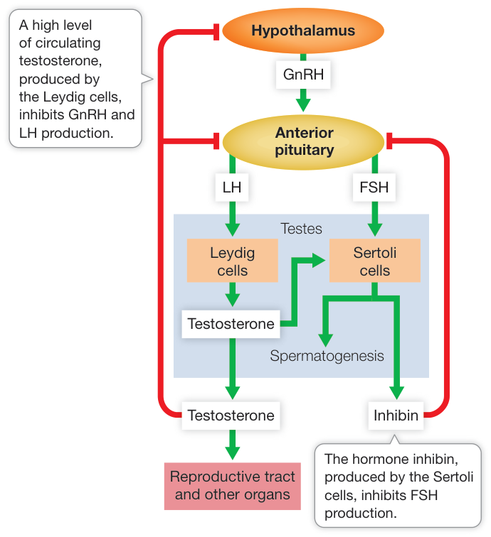
Figure 41.12 Male Reproductive Hormones The male reproductive system is under hormonal control by the hypothalamus and the anterior pituitary. Red lines indicate inhibition; green lines indicate stimulation and secretion.
Sertoli cells also produce a hormone called inhibin that exerts negative feedback on the anterior pituitary cells that produce and secrete FSH.
Recap and Assess
Sperm are produced in the testes and are contained in the seminal fluid produced by accessory glands. The resulting semen is ejaculated into the female reproductive tract during copulation. Prior to copulation, sexual stimulation causes the penis to become erect by increasing the blood flow into the erectile tissues. The male reproductive system is under hormonal control by the hypothalamus and anterior pituitary, with negative feedback coming from hormones produced by the testes.
- What makes ejaculated semen thick and gelatinous, and how do sperm escape the gel to move up the female reproductive tract?
- What are the roles of Sertoli cells in spermatogenesis?
- How does an inhibitor of phosphodiesterase treat erectile dysfunction?
- What is the significance of the fact that the testes of a prepubertal male are responsive to FSH and LH?
Whereas the male reproductive system produces sperm continuously throughout reproductive life, the female system produces eggs that can be fertilized by sperm periodically. We now turn to the cyclical processes by which the female reproductive system produces and releases eggs and prepares the female reproductive tract to receive a developing embryo should fertilization occur.
Female Sex Organs Produce Eggs and May Nurture Embryos
Learning Objectives
- 41.4.1 Link the events in the ovarian and menstrual cycles.
- 41.4.2 Explain how the number of eggs that are ovulated in each cycle is controlled.
- 41.4.3 Explain how the blastocyst prevents menstruation.
- 41.4.4 Describe how mechanical and hormonal stimuli interact during labor.
The human female reproductive system is illustrated in Figure 41.13. Eggs are produced in and released from the ovaries, located on each side of the lower abdominal cavity. When an egg leaves the ovary (ovulation), it enters the abdominal cavity, but it does not go far. The ovaries are close to the openings of the oviducts (also known as the fallopian tubes). The openings are surrounded by undulating, fringed tissues called fimbria that sweep the egg into the oviduct. Cilia lining the oviduct propel the egg toward the uterus, a muscular, thick-walled cavity shaped in humans like an upside-down pear. The uterus is where the embryo will develop if the egg is fertilized. At its bottom, the uterus narrows into a region called the cervix, which opens into the vagina.
Externally, the vagina is enclosed by two sets of skin folds. The inner, more delicate folds are the labia minora; the outer, thicker folds are the labia majora. At the anterior tip of the labia minora is the clitoris, a small bulb of highly sensitive erectile tissue that has the same developmental origins as the male gland penis. Clitoral tissues, homologous with the erectile tissues of the penis, extend deep under the labia minora and the walls of the vagina. The labia minora and the clitoral tissues become engorged with blood in response to sexual stimulation. Just as stimulation of the gland penis during sexual intercourse culminates in orgasm in the male, stimulation of the clitoral tissues results in orgasm in the female.
The external opening of an infant’s vagina is usually, but not always, partly covered by a thin membrane, the hymen. Eventually the hymen can be torn by vigorous physical activity or by first sexual intercourse; it can sometimes make first intercourse difficult or painful for the woman.
Sperm deposited in the vagina swim and are propelled by contractions of the female reproductive tract through the cervical opening, up through the uterus, and most of the way up an oviduct, where fertilization takes place. Before meeting a sperm, the egg is still a secondary oocyte. Fertilization stimulates the egg to complete its second meiotic division. Following that division, the haploid nuclei of the sperm and the egg can fuse to produce a diploid zygote nucleus.
The zygote undergoes its first few cell divisions to become a blastocyst while still in the oviduct. The blastocyst moves down the oviduct to the uterus, where it attaches itself to the epithelial lining of the uterus—the endometrium. Once attached, the blastocyst implants in the endometrium and interacts with it to form the placenta, as you will see in Key Concept 42.2. The mother’s body nurtures the embryo through the placenta, which also produces hormones that sustain pregnancy.
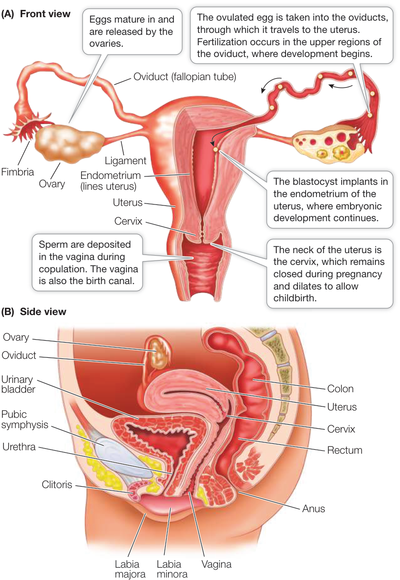
Figure 41.13 Reproductive Tract of the Human Female The female reproductive organs are shown (A) from the front and (B) from the side. Activity 41.3 The Human Female Reproductive Tract
The maturation of eggs and ovulation are cyclical events in the ovaries. Changes in the uterus are also critical processes. These cycles in humans average about 28 days, but they can vary considerably in an individual and among different individuals. In anticipation of receiving a blastocyst, the endometrium thickens and develops lots of blood vessels. The endometrium regresses if a blastocyst does not arrive within a certain window of time. Thus female reproductive functions consist of two linked cycles: an ovarian cycle that produces eggs and hormones; and a uterine, or menstrual, cycle that prepares the endometrium for the arrival of a blastocyst (Figure 41.14).
The two cycles must be synchronized so that a blastocyst arrives in the uterus at the optimal time to implant in the endometrium and continue its development.
Ovarian cycles produce mature eggs
A newborn baby girl has about a million primary oocytes in each ovary. By the time she reaches puberty, she has only about 200,000; the rest have degenerated. During a woman’s fertile years, her ovaries go through about 450 ovarian cycles. During each cycle, 10–20 primary oocytes begin to mature, but usually only one matures completely and is ovulated; the others degenerate (see Figure 41.14A). At around the age of 50, a woman reaches menopause—the end of fertility—and may have few oocytes left in each ovary. For more than 50 years, the gradual age-related decline in oocytes was taken as evidence that oogenesis in humans is limited to prenatal life. However, in recent years oogonial stem cells (OSCs) have been isolated from adult ovaries of mice and of humans. These OSCs have been shown to be capable of division and production of oocytes. Thus menopause may not be the absolute end of fertility.
A primary oocyte surrounded by a layer of ovarian cells is the functional unit of the ovary—the follicle (see the photo in Figure 41.14A). The follicle cells surrounding the oocyte supply it with nutrients and growth factors. The follicle cells are also the main site of production of the female hormones estrogen and progesterone. Usually only one follicle matures completely and reaches the stage of ovulation, releasing its egg at midcycle. Events that lead up to and follow ovulation are illustrated in Figure 41.14B. Follicle maturation occurs in the first half of the cycle, therefore called the follicular phase. The notable hormonal event leading up to ovulation is the gradual and then escalating increase in estrogen, which peaks just prior to midcycle (see Figure 41.14B).
The second half of the ovarian cycle is called the luteal phase because the follicle cells left in the ovary after the egg is ovulated develop into an endocrine gland—the corpus luteum (“yellow body”)—secreting estrogen and progesterone. If the egg is not fertilized and does not embed into the endometrium, the corpus luteum becomes inactive and degenerates in 12–14 days.
Media Clip 41.1 Human Ovulation Life12e.com/mc41.1
The uterine cycle prepares an environment for a fertilized egg
The uterine cycle parallels the ovarian cycle and consists of a build-up and then a breakdown of the endometrium (see Figure 41.14B). The breakdown of the endometrium, or menstruation (from the Latin menses, “months”), marks the beginning of each uterine and ovarian cycle. About 5 days after menstruation begins, the endometrium starts to thicken in preparation for receiving a blastocyst. The uterus attains its maximal state of preparedness about 5 days after ovulation and remains in that state for another 9 days. If a blastocyst has not arrived by that time, the endometrium breaks down and the sloughed-off tissue, including blood, flows from the body through the vagina as a menstrual discharge.
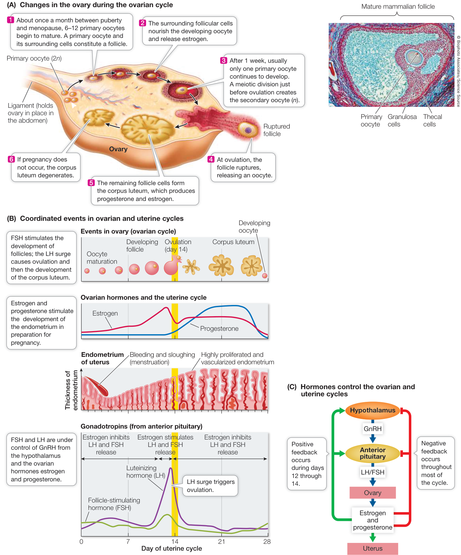
Figure 41.14 The Ovarian and Uterine Cycles (A) The ovarian cycle progresses from the development of a follicle to ovulation and then to growth and finally to degeneration of the corpus luteum. (B) During a woman’s ovarian and uterine cycles, coordinated changes occur in the development of the ovarian follicle, the release of female sex steroids, and the endometrial lining of the uterus. The release of hypothalamic GnRH stimulates the release of pituitary LH and FSH, which in turn control the development of the follicle and eventually ovulation. The cycles begin with the onset of menstruation; ovulation is at mid-cycle (yellow bar). (C) The ovarian and uterine cycles are under a complex series of positive- and negative-feedback controls involving several hormones. Q: Hormonal contraceptive pills contain estrogen and progesterone. Which portion of the ovarian cycle do they simulate, and how do they prevent ovulation? Animation 41.2 The Ovarian and Uterine Cycles
The uterine cycles of most mammals other than humans do not include menstruation; instead, the uterine lining typically is reabsorbed. In these species, the most obvious correlate of the ovarian cycle is a state of sexual receptivity called estrus (“heat”) around the time of ovulation. You may be aware of the bloody discharge that occurs in dogs at the time of estrus. This discharge is not the same as menstruation—in fact it is exactly the opposite. Bleeding in dogs occurs during the proliferation of the uterine lining, which occurs just before ovulation. When the female mammal comes into estrus, she actively solicits male attention and may be aggressive to other females. Humans are unusual among mammals in that females are potentially sexually receptive throughout their ovarian cycles and at all seasons of the year.
Hormones control and coordinate the ovarian and uterine cycles
The ovarian and uterine cycles are coordinated and timed by the same hormones that initiate sexual maturation. Gonadotropins (FSH and LH) secreted by the anterior pituitary are the central elements of this control (see Figure 41.14B). Before puberty (i.e., before about 11 years of age), the secretion of FSH and LH is low and the ovaries are inactive. At puberty the hypothalamus increases its release of GnRH, stimulating the anterior pituitary to secrete FSH and LH. In response to FSH and LH, ovarian tissue grows and produces estrogen. The rise in estrogen causes the maturation of the accessory sex organs and the development of female secondary sexual characteristics. Between puberty and menopause, interactions of GnRH, gonadotropins, and sex steroids control the ovarian and uterine cycles.
A few days before menstruation begins, the anterior pituitary begins to increase its secretion of FSH and LH. In response, several follicles begin to mature in the ovaries, and these follicles steadily increase their production of estrogen. After about a week, all but one of the follicles wither away.
For the first 12 days of a 28-day ovarian cycle, estrogen exerts positive-rather than negative-feedback control on the pituitary. As a result, a surge of LH and a lesser surge of FSH occur (see Figure 41.14B). The LH surge triggers the mature follicle to rupture and release its egg, and it stimulates the cells of the ruptured follicle to develop into a corpus luteum.
Estrogen and progesterone secreted by the corpus luteum following ovulation are crucial to growth and maintenance of the endometrium. These sex steroids also exert negative-feedback control on the pituitary, inhibiting gonadotropin release and thus preventing new follicles from maturing.
If the egg is not fertilized, the corpus luteum degenerates on about day 26 of the cycle. Without production of progesterone by the corpus luteum, the endometrium sloughs off and menstruation occurs. The decrease in circulating steroids also releases the hypothalamus and pituitary from negative-feedback control, so GnRH, FSH, and LH all begin to increase. The increase in these hormones induces the next round of follicle development, and the ovarian cycle begins again.
FSH receptors determine which follicle is ovulated
Early in the ovarian cycle several follicles begin to develop, but only one reaches full maturity. What determines which one will survive? There are two types of follicle cells, arranged in two layers. The inner layer immediately surrounding the ovum is made up of granulosa cells, and the outer layer consists of thecal cells. Thecal cells are stimulated by LH to produce testosterone (similar to Leydig cells in the male). Testosterone diffuses into the granulosa cells, where the enzyme aromatase converts the testosterone to estrogen. Estrogen, along with FSH, stimulates the growth and maturation of the granulosa cells (similar to the Sertoli cells in the male). The estrogen plays two important roles in the selection of the follicle that is ovulated: (1) estrogen stimulates the granulosa cells to express more FSH and LH receptors, and (2) estrogen entering the circulation feeds back on the pituitary to decrease the production of FSH. The granulosa cells also produce inhibin (similar to Sertoli cells in the male), and inhibin also decreases the production of FSH. As FSH and LH levels fall, the follicle with the most FSH and LH receptors survives and matures while the others regress.
In pregnancy, hormones from the extraembryonic membranes take over
If the egg is fertilized and a blastocyst arrives in the uterus and implants in the endometrium, a new hormone comes into play. A layer of cells covering the blastocyst begins to secrete human chorionic gonadotropin, or hCG (Figure 41.15). This gonadotropin, a molecule similar to LH, stimulates the corpus luteum to continue to produce estrogen and progesterone to support the growth and maintenance of the endometrium and thereby prevent menstruation. Because it is present only in the blood of pregnant women, the presence of hCG is the basis for pregnancy testing. Pregnancy tests use an antibody to detect hCG in urine; they take only minutes and can be done at home.
Blastocyst and endometrial tissues form the placenta, which nourishes the embryo. The placenta also replaces the corpus luteum as the major producer of estrogen and progesterone. Continued high levels of estrogen and progesterone prevent the pituitary from secreting gonadotropins; thus the ovarian cycle ceases for the duration of pregnancy. This mechanism underlies the action of birth control pills, which contain synthetic hormones resembling estrogen and progesterone that exert negative-feedback control on the hypothalamus and pituitary.
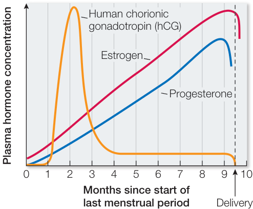
Figure 41.15 Hormones of Pregnancy When a fertilized ovum implants in the uterus, cells surrounding it produce human chorionic gonadotropin, which acts like LH and keeps the corpus luteum functioning as an endocrine gland. The ovarian and uterine cycles are put on hold for the duration of pregnancy. (After B. Pansky. 1982. Review of Medical Embryology. Macmillan: New Jersey.)
Breast feeding may delay the return of the ovarian cycle
Following birth, estrogen and progesterone levels drop because of the loss of the placenta, and therefore negative feedback on the hypothalamus and pituitary imposed by these hormones during pregnancy is gone. Nevertheless, women who breast feed their infants may not have their ovarian and menstrual cycles return for some time—a phenomenon called lactational delay. In Figure 39.4 you learned that the pituitary hormone prolactin stimulates the growth of the mammary glands and the production of milk. Prolactin release is stimulated by the suckling of the infant. Even the sight or sound of the infant can stimulate prolactin release in the mother. Investigations have now shown that in addition to stimulating the mammary glands, prolactin also inhibits GnRH release and LH release, possibly explaining lactational delay. Going back to the case of the tammar wallaby in the opening to this chapter, could prolactin release stimulated by the suckling of the joey in the pouch be responsible for the delayed implantation seen in this species? Investigating Life: The Control of Diapause in the Tammar Wallaby examines this question.
Childbirth is triggered by hormonal and mechanical stimuli
Throughout pregnancy, the muscles of the uterine wall periodically undergo slow, weak, rhythmic contractions called Braxton Hicks contractions. These contractions become stronger during the third trimester of pregnancy and are sometimes called false labor. The contractions of true labor mark the beginning of childbirth. Both hormonal and mechanical stimuli contribute to the onset of labor (Figure 41.16A).
Progesterone inhibits and estrogen stimulates contractions of uterine muscle. Toward the end of the third trimester, the estrogen–progesterone ratio shifts in favor of estrogen. The onset of labor is marked by increased secretion of the hormone oxytocin by the posterior pituitaries of both mother and fetus. Oxytocin is a powerful stimulant of uterine muscle contraction. Manufactured oxytocin is used to induce labor when that is necessary.
Mechanical stimuli come from the stretching of the uterus by the fully grown fetus and the pressure of the fetal head on the cervix. These mechanical stimuli increase the release of oxytocin by the mother’s posterior pituitary, which in turn increases the activity of uterine muscle, causing even more pressure on the cervix. This positive-feedback loop converts the weak, slow, rhythmic Braxton Hicks contractions into stronger labor contractions.
In the early stage of labor, hormonal changes and pressure created by the contractions cause the cervix to dilate (expand) until it is large enough to allow the baby to pass through. Gradually the contractions become more frequent and more intense. This stage of labor lasts an average of 12–15 hours in a first pregnancy but is usually 8 hours or less in subsequent ones.
The second stage of labor begins when the cervix is fully dilated to a diameter of about 10 centimeters (Figure 41.16B). The baby’s head can now move into the vagina. Passage of the fetus through the vagina is assisted by the mother’s bearing down (“pushing”) with her abdominal and other muscles. Once the head and shoulders of the baby clear the cervix, the rest of its body eases out rapidly, but it is still connected to the placenta by the umbilical cord. Once the baby clears the birth canal, it
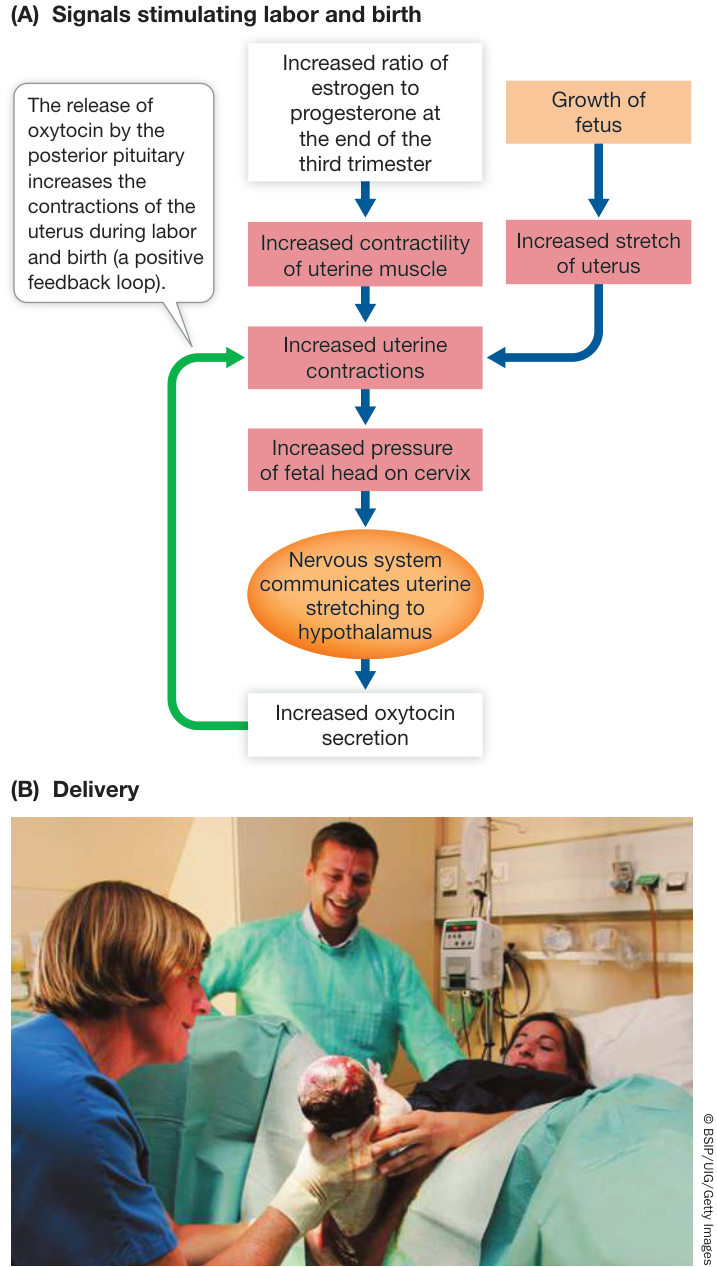
Figure 41.16 Labor and Delivery (A) Both mechanical and hormonal signals are involved in stimulating the uterine contractions of labor and delivery. (B) A new person comes into the world headfirst.
Investigating Life: The Control of Diapause in the Tammar Wallaby
starts breathing and is independent of its mother’s circulation. The umbilical cord may then be clamped and cut. The segment still attached to the baby dries up and sloughs off in a few days, leaving behind its distinctive signature, the belly button—more properly called the umbilicus. The third stage of labor, the detachment and expulsion of the placenta and fetal membranes, takes from a few minutes to an hour, and may be accompanied by uterine contractions. A baby’s suckling at the breast immediately following birth stimulates additional secretion of oxytocin, which augments uterine contractions, reduces the size of the uterus, and helps stop bleeding.
Experiment
Original Paper: L. A. Hinds and C. H. Tyndale-Biscoe. 2013. Daily prolactin pulse inhibits the corpus luteum during lactational quiescence in the marsupial, Macropus eugenii. Reprod Fertil Dev 25: 456–461.
In Chapter 39 (see Figure 39.4) we discussed how the hormone prolactin (PRL) has evolved to serve many physiological processes in different vertebrate groups, but these various processes all have some connection to reproduction. Since prolactin is involved in controlling lactation in mammals, could prolactin be the signal in the tammar wallaby that maintains embryonic diapause as long as suckling continues?
HYPOTHESIS Pulses of prolactin in lactating female tammar wallabies prevent new embryos from developing as long as previous offspring are suckling.
METHOD The hormone progesterone (P) prepares the uterine lining for implantation of an embryo and maintains the uterine lining during pregnancy. Thus measurements of blood levels of P reveal when diapause is broken in tammar wallabies. A stimulus known to break diapause in these animals is the removal of the prior young from the pouch (RYP). The RYP method was used to study the effects of prolactin on maintenance of diapause.
Remove young from the pouches of three groups of five wallabies.
Following RYP, administer the following treatments:
Group A: Daily saline injections for 8 days following RYP (controls)
Group B: PRL injection every 24 hr for 8 days following RYP
Group C: PRL injection every 48 hr for 8 days following RYP
- Take daily blood samples to measure P levels.
RESULTS Data are approximate means of the actual measurements, progesterone concentrations (P) expressed in picograms per milliliter (pg/mL).
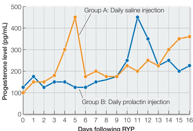
| Days following RYP | P level (picograms/mL) | ||
|---|---|---|---|
| Group A | Group B | Group C | |
| 0 | 100 | 125 | 100 |
| 1 | 150 | 175 | 113 |
| 2 | 150 | 125 | 125 |
| 3 | 180 | 150 | 135 |
| 4 | 300 | 150 | 175 |
| 5 | 450 | 125 | 215 |
| 6 | 175 | 125 | 240 |
| 7 | 200 | 150 | 325 |
| 8 | 175 | 16 | 210 |
| 9 | 175 | 175 | 240 |
| 10 | 225 | 250 | 335 |
| 11 | 200 | 450 | 260 |
| 12 | 250 | 350 | 200 |
| 13 | 225 | 225 | 225 |
| 14 | 300 | 250 | 230 |
| 15 | 350 | 200 | 210 |
| 16 | 360 | 225 | 225 |
CONCLUSION When tammar wallabies were treated with daily saline injections (controls), they broke diapause 4 or 5 days following RYP. However, when they were treated with daily PRL injections, they broke diapause 11 or 12 days following RYP. Hence daily PRL pulses can prolong diapause and delay implantation of a new blastocyst.
Work with the Data
Following the experiment showing that daily pulses of PRL can maintain diapause, the investigators asked whether pulses of PRL every 2 days could maintain diapause. The data are shown in the table as Group C. Plotting the data will show if there is a difference in the response to PRL when pulses were given every other day following RYP.
QUESTION
- Plot the data as was done for Groups A and B. Was the 48-hr PRL pulse effective in preventing the breaking of diapause?
Birth as well as reproduction is timed
Reproduction in many species is seasonal. This seasonality in many species of vertebrates involves mechanisms of photoperiodism. Briefly, changes in day length enable animals to anticipate the best time of year for the rearing of young and therefore time their reproduction and birth accordingly. How do animals measure day length? As we will discuss in more detail in Chapter 51, animals have endogenous daily rhythms called circadian (circa, “day,” + dia, “length”) rhythms. As day length changes, sunrise and sunset occur at different phases of the circadian rhythm. The release of melatonin from the pineal gland is controlled by the circadian rhythm but is inhibited by light. Therefore as days get longer, the nightly pulse of melatonin gets shorter, and conversely as days get shorter, the melatonin pulse gets longer. Day length determines when many species come into reproductive condition and mate. That is not true for the tammar wallaby that was introduced at the beginning of this chapter. The wallaby mates at any time of year soon after it gives birth. However, diapause in the wallaby is sensitive to night length—that is, to the duration of the melatonin pulse. A non-lactating wallaby will break diapause when night length reaches 12 hours. Similarly, when non-lactating wallabies are held at a night length of 9 hours but given melatonin injections 2.5 hours before sunset, they break diapause. These results support the hypothesis that short daily melatonin pulses prolong diapause and that long daily melatonin pulses are a signal to break diapause. The significance for the nocturnal wallaby is that birth tends to be at the time of year when nights are longest.
Humans are not seasonal breeders, but there is a timing component of childbirth. Most women go into labor at night. However, it is quite common for labor to cease when women arrive in the brightly lit hospital. Bright light interrupts the nightly melatonin pulse in humans.
Recap and Assess
The reproductive system of women produces oocytes (eggs) and hormones, and these functions are controlled by hypothalamic and anterior pituitary hormones. The follicle is the functional unit of the ovary. Usually one follicle matures and is ovulated each month as controlled by a cyclical feedback interplay between ovarian hormones and the hypothalamic–pituitary axis. These hormonal cycles also coordinate the uterine, or menstrual, cycle so that the endometrium is prepared to receive a blastocyst should fertilization occur. A blastocyst implanted in the uterus produces hCG, which suspends the ovarian and uterine cycles by sustaining the ovarian production of progesterone and estrogen until the placenta can take over this function. Childbirth and lactation are controlled by mechanical and hormonal mechanisms.
- What events in the ovary are responsible for the onset of menstruation and what prevents it if pregnancy occurs?
- What are the roles of thecal cells and granulosa cells in the ovarian cycle, and how do they limit the number of eggs that are ovulated?
- How does positive feedback between hormonal and mechanical processes play a role in childbirth?
Understanding the physiology of human reproduction has led to numerous methods and technologies for controlling reproduction, either to prevent unwanted pregnancies or to overcome infertility.
Fertility Can Be Controlled
Learning Objectives
- 41.5.1 Discuss the relative advantages and disadvantages of different contraceptive methods.
- 41.5.2 Compare contraception and contragestational techniques.
- 41.5.3 Explain how procedures that use genetic analysis and in vitro fertilization can inform decisions about therapeutic abortions.
Sexual issues and sexual behavior are dominant aspects of our society, and reproductive technologies have had huge impacts on our sexual and reproductive lives.
Humans use a variety of methods to control fertility
According to a 2011 study, 45% of the 6.1 million pregnancies that occurred in the United States that year were unintended. Although a very high number, the rate of unintended pregnancies had dropped considerably from three years earlier when it was 54% and had been around that level for many years. This considerable drop is likely due to an increase in use of highly effective contraceptive methods. Even with this encouraging decrease in unintended pregnancies, however, the number is still much too high for a modern, educated society. It is important to keep in mind, that for women of college age, a single act of unprotected intercourse in the 2 days prior to ovulation carries a chance of conception as high as 50%.
The only failure-proof methods of preventing pregnancy are complete abstinence from sexual activity or surgical removal of the gonads. Those options are not acceptable to most people, and they turn to other methods to prevent pregnancy. Many of these methods prevent fertilization or implantation (conception) and are referred to as contraception. Table 41.1 lists some commonly used contraceptive methods and their relative failure rates. These methods vary enormously in their effectiveness. Most methods are used by the woman; only some are used by the man.
Table 41.1 Methods of Contraception
| Method | Mode of action | Failure rate a | Comments |
|---|---|---|---|
| UNPROTECTED | No form of birth control. | 85 | High risk of pregnancy, especially for women 15–30. |
| NONTECHNOLOGICAL METHODS | |||
| Rhythm method | The couple abstains from intercourse between days 10 and 20 of the ovarian cycle (peak fertility). | 5–24 | High failure rate due to miscalculation and/or variation of individual cycles. |
| Coitus interruptus | The man withdraws his penis prior to ejaculation, with the intention of not depositing sperm into the vagina. | 4–20 | Requires self-control, especially by the man. Very high failure rate. |
| BARRIER METHODSb | |||
| Condom (male or female) | A sheath of impermeable material (often latex) is fitted over the erect penis or into the vagina. Semen is trapped in the condom, so no sperm are deposited in the vagina. | 13–20 | If fitted correctly, an intact condom can prevent pregnancy. Failure can be due to material failure or improper use. Condoms are the only contraceptive method that protects against sexually transmitted diseases (STDs), including HIV (AIDS). |
| Spermicidal jellies | Applied deep inside the vagina, these chemical compounds kill or immobilize sperm. | 18–28 | Used alone, spermicidal compounds have a high failure rate. |
| Diaphragms, cervical caps, cervical sponges containing spermicide | Inserted by the woman prior to intercourse, these devices work by blocking the cervix so that sperm cannot pass into the uterus. | 15–30 | Approximately the same failure rate as condom use by men, but do not protect against STDs. Can be used in conjunction with spermicidal jelly for extra protection. |
| HORMONE-BASED CONTRACEPTIVES | |||
| Oral hormones (“the pill”) | A daily pill for women containing a combination of synthetic estrogens and progesterone. These hormones mimic pregnancy to the extent that the ovarian cycle and ovulation are suspended. The uterine cycle can continue by including a week of non-hormone administration every 21–28 days. | 0.3–7 | Requires medical consultation and prescription. Taken correctly, oral contraceptives are extremely effective. In the United States, more than 12 million women use them each year; they are sometimes prescribed to treat menstrual disorders. |
| Non-orally administered hormones | Making use of the same hormonal actions as the pill, these methods include long-acting injections, patches that release hormones transdermally (through the skin), and a hormone-containing vaginal ring. Implantable progestin releasing tubes have a failure rate of <0.1% and can last up to 3 years. | <7 | Same as oral hormones. A slightly lower failure rate because the woman does not have to remember to take a daily pill. |
| Progestin-only pill (Plan B ® ) | An oral contraceptive meant to be taken within 72 hours after unprotected sex. A high dose of progestin in two pills prevents ovulation in the same manner birth control pills do. | 5–40 | Not an “abortion pill”; this drug will not terminate an existing pregnancy. Currently available to women over 17 without a prescription. Failure rate varies widely depending on when taken. |
| IMPLANTATION BLOCKERS | |||
| Intrauterine device (IUD) | A medical professional inserts a small plastic or metal device into the uterus. The resulting inflammation reaction (see Chapter 39) releases prostaglandins, which prevent implantation of the fertilized egg. | 0.1–0.5 | A highly effective contraceptive, and the most widely used birth control device in China (and hence the world). With medical monitoring, can remain in place for several years. |
| Contragestational pill | Mifepristone, or RU-486, is a drug that blocks the action of progesterone, which is necessary to maintain the endometrium during implantation and pregnancy. | 15–40 | Prevents implantation when taken up to several days after unprotected intercourse. Can terminate a pregnancy up to the time of the first missed menstrual period. In the United States, available from specialized providers. |
| When used with a prostaglandin analogue | 1-5 | ||
| STERILIZATION | |||
| Vasectomy | The vasa deferentia (see Figure 41.9) are cut and tied off so that sperm can no longer pass into the urethra. Sperm continue to be produced but are reabsorbed by the man’s body. Male hormone levels and sexual responses are not affected. | 0.1 | A simple surgical procedure performed under local anesthetic in a doctor’s office. Although theoretically it can be reversed, vasectomy should be considered permanent. |
| Tubal ligation | The oviducts (see Figure 41.13A) are tied off so that eggs cannot reach the uterus and sperm cannot reach the egg. As with vasectomy, hormone levels and sexual responses are not affected. | 0.5 | This surgical procedure is somewhat more complex than vasectomy. It is often performed in conjunction with childbirth when a woman has decided that her family is complete. |
aFailure rate refers to the number of pregnancies per 100 women per year.
bAll of these barrier methods are routinely available without medical prescription.
Sources: Centers for Disease Control. 2017. Effectiveness of family planning methods. (tinyurl.com/ya8t92y4); A. Sundaram, et al. 2017.
Perspectives on Sexual and Reproductive Health 49: 7–16; O. M. Avrech et al. 1991. Fertil Steril 56: 385–393.
Once a fertilized egg is successfully implanted in the uterus, any termination of the pregnancy is called an abortion. A spontaneous abortion is the medical term for what is commonly called a miscarriage. Spontaneous abortions frequently occur early in pregnancy and are usually the result of either a chromosomal abnormality in the fetus or a breakdown in the process of implantation. Many spontaneous abortions occur before the woman even realizes she is pregnant.
Abortions that result from medical intervention may be performed either for therapeutic purposes or for fertility control. A therapeutic abortion may be necessary to protect the health of the mother, or it may be performed because prenatal testing reveals that the fetus has a severe defect. In a medical abortion, the cervix is dilated and some of the endometrium, along with the implanted fetus, is removed. When performed in the first trimester of a pregnancy, a medical abortion carries less risk of death to the mother than a full-term pregnancy. The risk rises after the first 12 weeks of pregnancy but remains less than that of a full-term pregnancy through the second trimester.
Reproductive technologies help solve problems of infertility
About 15% of couples in the United States are infertile—that is, they can’t have children. The reasons for infertility are many, and are about equally distributed between men and women. Several technologies have been developed to overcome barriers to both conceiving and bearing a child. The simplest of these is artificial insemination, in which the physician places sperm in the woman’s reproductive tract. This technique is useful if the male’s sperm count is low, if his sperm lack motility, or if problems in the female’s reproductive tract prevent the normal movement of sperm up the oviducts. Artificial insemination is also widely used in the production of domesticated animals such as cattle.
Assisted reproductive technologies, or ARTs, involve procedures that remove unfertilized eggs from the ovary, combine them with sperm outside the body, and then place fertilized eggs or egg-sperm mixtures in the appropriate location in the woman’s reproductive tract for development to take place. The first successful ART was in vitro fertilization (IVF). In IVF the woman is treated with hormones that stimulate many follicles in her ovaries to mature. Eggs are collected from these follicles, and sperm are collected from the intended father. Eggs and sperm are combined in a culture medium outside the body, where fertilization takes place. The resulting embryos can be injected into the mother’s uterus in the blastocyst stage or kept frozen for implantation later. The first “test tube baby” resulting from IVF was born in England in 1978. Since then, millions of babies have resulted from this ART.
A major cause of IVF failure is inability of sperm to gain access to the egg cell membrane (see Figure 41.5). To solve this problem, methods have been developed to inject a sperm cell directly into the cytoplasm of an egg. In intracytoplasmic sperm injection (ICSI), an egg is held in place by suction applied through a polished glass pipette. A slender, sharp pipette is then used to penetrate the egg and inject a sperm (Figure 41.17). This ART was used successfully for the first time in 1992 by researchers in Belgium;
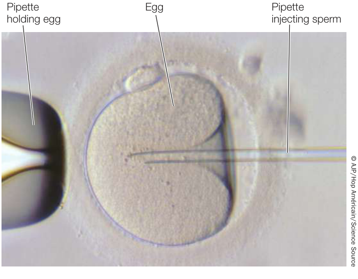
Figure 41.17 Intracytoplasmic Sperm Injection In this procedure, a sperm is injected directly into a mature egg cell. The fertilized egg is then placed in the female reproductive tract, where it can implant and develop into a fetus.
now thousands of these procedures are performed in U.S. clinics each year, with a success rate of about 25%.
IVF, coupled with techniques of genetic analysis, can eliminate the risk that adults who are carriers of genetic diseases will produce affected children. It is now possible to take a cell from a human embryo at the four-or eight-cell stage (see Figure 42.4B) without damaging its developmental potential. The sampled cell can be subjected to molecular analysis to determine whether it carries the harmful gene. This procedure, called preimplantation genetic diagnosis (PGD), makes it possible to determine whether an embryo produced by IVF carries the genetic defect of concern.
Recap and Assess
Controlling fertility is an important aspect of modern human life. Decreasing the probability of pregnancy is achieved through methods that prevent sperm and egg from meeting and that prevent implantation. Pregnancies can be facilitated through technologies that extract eggs from the ovary and fertilize them in vitro before reintroducing them into the female reproductive tract.
- Which method of contraception is the only one to offer protection against sexually transmitted diseases (STDs)? Explain why.
- Both the contraceptive pill (“the pill”) and the contragestational pill (RU-486) involve progesterone signaling. How do their mechanisms of action differ?
- How can IVF be used to prevent transmission of a genetic disease?
The fertilized egg of a sexually reproducing organism is a single cell containing all the genetic information needed to create a new organism. Chapter 21 introduced some of the molecular aspects of the process of development in multicellular animals. Chapter 42 will describe the physiological and anatomical events of animal development.
Investigating Life: QA What signal controls the breaking of diapause in the tammar wallaby?
The experiment presented in Investigating Life: The Control of Diapause in the Tammar Wallaby exploited the fact that removal of the young joey from the pouch of its mother (RYP) stimulated the blastocyst in her uterus to break diapause, implant, and continue development. What diapause-breaking signal might be involved in conjunction with RYP? Researchers hypothesized that RYP resulted in cessation of prolactin release stimulated by suckling of the young, and that the absence of those prolactin pulses initiated the breaking of diapause. The test of that hypothesis was to apply RYP to groups of mother wallabies and test whether applications of prolactin could prevent the breaking of diapause. The results showed that injections of prolactin every 24 hours for 8 days following RYP delayed the progesterone rise that indicates the breaking of diapause. Prolactin injections at intervals of 48 hours or longer were less effective or ineffective in maintaining diapause. The conclusion was thus reached that prolactin pulses stimulated by suckling are a signal for the maintenance of diapause, and that the removal of that signal results in the breaking of diapause. Another signal, melatonin, could be involved in the breaking of diapause. The prolactin experiments were all done at the time of year when night length was appropriate for the breaking of diapause.
Future directions
Reproductive success depends on many factors, including ecological, physiological, and socio-behavioral factors. It is therefore not surprising that evolutionary adaptations have exploited many kinds of information to control the timing of reproduction in various species. While these controlling factors are diverse, in vertebrates they all work through the hypothalamic, pituitary, and gonadal regulatory pathways. Therefore whatever we can learn about the physiology of reproduction in one vertebrate species may also apply to other species, or at least inform us as to what questions we should ask of other species, including humans. The answers to these questions can help us preserve endangered species, improve domesticated species, solve reproductive problems in humans, and even help control human population growth.

© Tui De Roy/Minden Pictures
Visual Summary

You should be able to relate each summary to the adjacent figures. If you go to this visual summary in Achieve, you can follow links to figures, animations, activities, and simulations that will help you consolidate the material.
Asexual Reproduction Is Efficient but Limits Genetic Variability
- ■ Asexual reproduction produces offspring that are genetically identical to their parent.
- ■ Budding, regeneration, and parthenogenesis are types of asexual reproduction.
- ■ Budding and regeneration produce new individuals by mitotic divisions of dedifferentiated somatic cells.
- ■ Parthenogenesis produces new individuals by development of unfertilized eggs.
Questions
- What environmental conditions favor asexual reproduction, and why?
- What is the role of sexual behavior in the all-female populations of whiptail lizards?
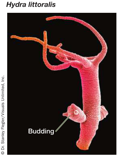
Figure 41.2
Sexual Reproduction Involves the Union of Haploid Egg and Sperm
- ■ Gametogenesis and fertilization are similar in all animals, but spawning and mating include a great variety of anatomical, physiological, and behavioral adaptations.
- ■ In testes and ovaries, the germ cells proliferate mitotically, undergo meiosis, and mature into gametes. Each spermatogonium can produce four sperm, but each oogonium produces only one egg. In humans, spermatogenesis continues throughout life, whereas oogenesis ceases before birth and oocytes undergo developmental arrest after the first meiotic division.
- ■ Fertilization can be external or internal. Fertilization involves sperm activation, binding to the egg, digestion of a path through the coverings of the egg, and fusion of sperm and egg cell membranes. Fusion triggers blocks to polyspermy, and in mammals signals the egg to complete meiosis and begin development.
- ■ Hermaphrodites are monoecious, meaning individuals have both male and female reproductive systems, either sequentially or simultaneously. Dioecious species have separate male and female individuals.
- ■ The young of oviparous species begin development outside the mother’s body. The young of viviparous species begin development inside the mother’s body.
Questions
- What are four differences between gametogenesis in human males and females?
- What is the difference between a viviparous and an ovoviparous species?
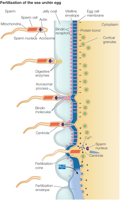
Figure 41.5
Male Sex Organs Produce and May Deliver Sperm
- ■ Semen consists of sperm suspended in seminal fluid. Sperm are generated in the seminiferous tubules of the testes, mature in the epididymis, and are delivered to the urethra through the vasa deferentia. Other components of semen are produced in the seminal vesicles, prostate gland, and bulboure-thral glands. Semen is ejaculated through the erect penis by muscle contractions at the climax of copulation.
- ■ Spermatogenesis depends on testosterone secreted by the Leydig cells of the testes, which are under the control of hormones produced by the hypothalamus and the anterior pituitary. The production of these hormones is controlled by negative feedback from testosterone and from inhibin, a hormone produced by the Sertoli cells of the testes.
- Describe the path taken and the transitions made by a male gamete from being a spermatogonium to ejaculated sperm.
- What are the functions of Leydig and Sertoli cells, and how are those functions controlled?
- What are the sources and functions of the components of semen other than sperm?
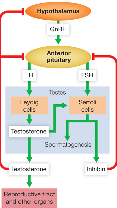
Figure 41.12
Female Sex Organs Produce Eggs and May Nurture Embryos
- ■ An ovarian follicle contains an oocyte surrounded by granulosa cells and thecal cells. Each month a small number of follicles respond to pituitary FSH by growing, producing estrogen, and promoting development of the oocyte, but usually only one of these follicles is ovulated
- ■ An ovulated oocyte is picked up by the fimbria of an oviduct. Sperm deposited in the vagina during copulation move up through the cervix and uterus into the oviducts, where fertilization occurs. In humans, maturation and release of eggs constitute an ovarian cycle. The uterine, or menstrual, cycle prepares the uterus for receipt of a blastocyst. If no blastocyst implants, the lining of the uterus sloughs off in the process of menstruation. The ovarian and uterine cycles are controlled by hypothalamic and pituitary hormones, which are under feedback control of estrogen and progesterone.
- ■ Childbirth is initiated by hormonal and mechanical stimuli that increase the contractility of uterine muscle.
Questions
- What is the relationship between ovarian hormones and the endometrium of the uterus during the ovarian cycle?
- Explain the role of human chorionic gonadotropin in pregnancy.
- How does the birth process involve positive feedback?
Fertility Can Be Controlled
- ■ Contraceptive methods and technologies decrease the probability of fertilization but differ in efficacy.
- ■ Assisted reproductive technologies (ARTs) can increase fertility.
Questions
- Apart from sterilization, what are the two most effective contraceptive methods?
- What are three of the least effective contraceptive methods, and why?
- In cases of infertility, what technology would be appropriate if the woman’s oviducts are blocked? What technology would be appropriate if the man’s sperm are not motile?
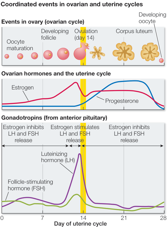
Figure 41.14
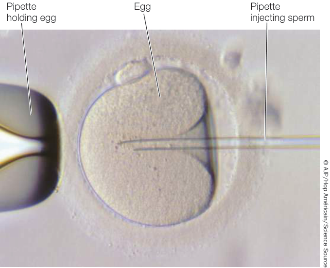
Figure 41.17