Chapter 43Neurons, Glia, and Nervous Systems
Courtesy of Philip Heydon
Key Concepts
43.1 Neurons and Glia Are Unique Cells of Nervous Systems
43.2 Neurons Generate and Transmit Electric Signals
43.3 Neurons Communicate with Other Cells
43.4 Neurons and Glia Form Information-Processing Circuits
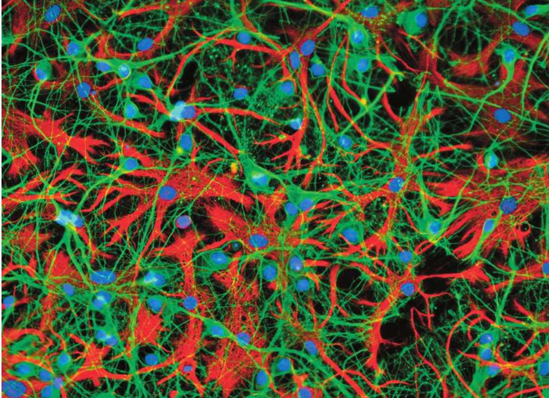
Neurons, colored blue in this photo, code, transmit, integrate, and store information and send commands to non-neuronal cells. The activities of neurons are supported and modulated by glial cells, colored red in this photo.
Investigating Life: Balancing the Brain
Your brain enables you to learn the material in this chapter—to read the words, understand the illustrations, and store information to allow you to answer questions. You must study to master this chapter, but if you do you will learn how the brain receives and processes information.
Imagine what it would be like if you could not learn any of this material—no matter how much you poured over the book. This is the situation faced by individuals with a condition known as Down syndrome, which affects approximately 1 out of every 700 children born in the United States.
Individuals with Down syndrome are born with three copies of most of chromosome 21, one of the smallest of the human chromosomes (containing about 225 confirmed genes). Having an extra copy of these genes causes numerous developmental and functional problems, including learning disability. Can we understand the cause of this disability and perhaps find a way to remedy it?
One productive way of investigating the causes and possible treatments for a human disease or deficit is to develop an animal model. Through genetic engineering, researchers created a “Down syndrome mouse” that has most of the same genes triplicated as those in humans with Down syndrome. These mice cannot perform in mouse memory tests as normal mice do. The mouse Down syndrome model revealed that excess inhibition in the brain causes the learning disability. What is meant by “excess inhibition in the brain”? We tend to think mostly about the excitatory actions of the brain, likening the brain to a puppet master who pulls the strings that activate the muscles and organs of the body. In fact the brain is more like an orchestra conductor, making some sections louder, some softer, speeding up, slowing down. The brain must constantly maintain a delicate balance of excitation and inhibition, acting on some signals and ignoring others. In the brains of Down syndrome model mice there is consistently too much inhibition. When inhibition is experimentally reduced with drugs, the mice are able to learn. Research like this is a first step toward bringing potential therapies for humans into clinical trials in the hope of improving the cognitive functions of individuals with Down syndrome.

© John Birdsail/AGE Fotostock
QA Can learning be restored in mouse models of Down syndrome? (Find the answer on page 981.)
Neurons and Glia Are Unique Cells of Nervous Systems
Learning Objectives
- 43.1.1 Describe the parts of a neuron and explain their functions.
- 43.1.2 Describe in general terms how information is transmitted by axons and across synapses to dendrites.
- 43.1.3 Explain the different functions of glial cells.
Nervous systems are information systems. They encode, process, and store a wide variety of information from the external and internal environments, and they use that information to control and regulate the physiology and behavior of the organism. The nervous systems of all animals can carry out these functions because of the properties of a unique class of cells: nerve cells, or neurons. Vertebrate nervous systems also include another unique class of cells called glial cells, or glia.
There are many types of neurons, which vary in size, shape, and function, but they all generate and conduct electric signals. Glia do not generate and conduct electric signals, and there are two major types—macroglia and microglia. Macroglia support and modulate the functions of neurons. Microglia are phagocytic cells derived from blood-forming stem cells. Microglia are motile, are activated by foreign matter or damage to nervous tissue, and are the first and major immune defense mechanism in the nervous system.
Nervous systems in all but the simplest invertebrates are divided into central and peripheral divisions. The central division includes dense assemblages of neurons that process and store information and generate commands. In invertebrates these assemblages of neurons are generally called ganglia. In more complex invertebrates some large ganglia are designated as brains. In vertebrates the central nervous system (CNS) includes the brain and spinal cord. The peripheral nervous system (PNS) provides communication between the CNS and all of the rest of the body.
Vertebrate neurons and macroglia originate in the embryonic neural tube
Neurons and macroglia have a common origin in the neural tube of the early vertebrate embryo (see Figure 42.13). The neural tube contains rapidly dividing neuronal stem cells. Initially the divisions of these neural stem cells are symmetrical—all of the progeny are stem cells. Eventually, however, the cell divisions become asymmetrical, with one daughter cell remaining a stem cell and the other daughter cell becoming either a neural progenitor cell or a macroglial progenitor cell—a neuroblast or a gliablast. These progenitor cells produce the neurons and glia that together form the various structures of the nervous system.
The structure of neurons reflects their functions
Neurons vary enormously in structure and appearance, but they have a basic structure that includes four regions (Figure 43.1).
- A cell body contains the nucleus and most of the cell’s organelles.
- Shrub-like projections called dendrites (Greek dendron, “tree”) may extend from the cell body. Dendrites receive information from other neurons or sensory cells and bring it to the cell body.
- In most neurons, one projection—the axon—is much longer than the others. Axons carry information from the cell body to target cells.
- At the target cell, the axon divides into a spray of fine nerve endings. The tips of these tiny nerve endings have swellings called axon terminals.
Neurons have a wide variety of forms that reflect different functionalities (Figure 43.2). Neurons with few dendrites receive information from specific and limited sources, whereas neurons with large arrays of dendrites can collect and integrate information from a wide range of sources. Some neurons communicate over very short distances, but others communicate over extremely long distances and therefore have very long axons. For example, axons from neurons in your spinal cord control the muscles that move your toes.
Regardless of form and function, all neurons process and communicate information through changes in the electric potentials across their membranes. As in all cells, the electric charge on the inside of the neuronal cell membrane is slightly negative in comparison with the outside. Specialized neurons called sensory neurons respond to specific stimuli such as light, temperature, and pressure by changes in the flow of electrically charged ions across their cell membranes. These changes in local electric potential travel along the cell membrane to the base of the cell’s axon, where a small change in membrane potential generates a large, rapidly reversed change in membrane potential that is called an action potential (AP). Axons conduct APs over long distances. Non-sensory neurons receive inputs from other neurons through connections in their dendrites. Local changes in the dendrites’ membrane potentials travel to the membrane of the cell body where they are summed and thereby influence the firing of APs in the neuron’s axon.
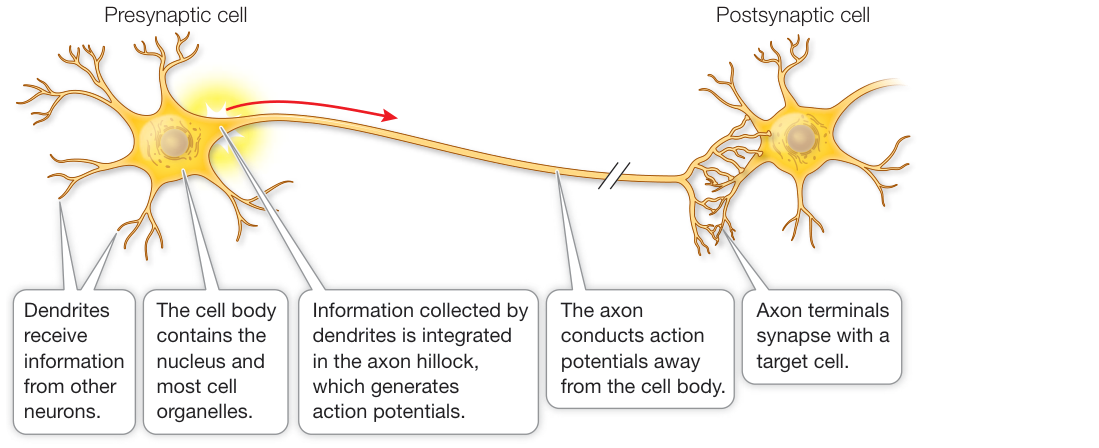
Figure 43.1 A Generalized Neuron The diagram shows the features typical of most neurons. The forms of these features, including the length of the single axon and the density and branching patterns of the dendrites, vary greatly across the many different types of neurons.
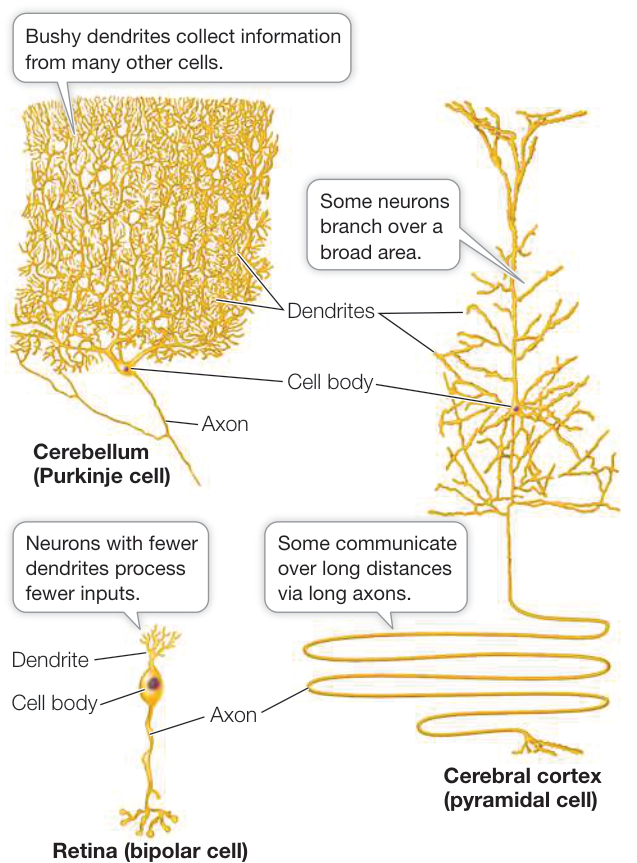
Figure 43.2 Neurons Have Many and Varied Forms The morphological differences in neurons from different parts of the mammalian nervous system are related to their specific functional adaptations. The small sample here shows two neuronal types from the human brain (a Purkinje cell and a pyramidal cell) and a sensory neuron from the retina of the human eye. Q: Which of these neurons is likely to receive a greater number of inputs?
In summary, a neuron receives specific stimuli (e.g., light, sound, pressure, chemicals) that cause changes in the electric potentials across the cell membranes of its dendrites. These electric signals from dendrites spread to and are averaged by the cell body. Changes in the electric potential of the membrane of the cell body at the base of its axon can cause an AP that is conducted along the axon to its terminals. APs can travel at speeds up to 100 meters per second (360 kilometers per hour), making it possible for an individual to sense, process, and act on information very quickly.
What happens when the AP reaches the axon terminals? Axon terminals come extremely close to the membrane of the target cell, which can be another neuron, a muscle cell, or a secretory cell. Structures called synapses form at these locations. The synapse transfers the information conveyed by the AP from the presynaptic cell to the postsynaptic cell (see Figure 43.1).
Synapses can be either electrical or chemical. Electrical synapses allow the AP to pass directly between two neurons. In vertebrates most synapses are chemical. At chemical synapses a space about 25 nanometers wide (about 1/2000th the width of a human hair) separates the presynaptic and postsynaptic membranes. An AP arriving at an axon terminal causes it to release chemical messenger molecules called neurotransmitters. Neurotransmitters diffuse across the synaptic space and bind to receptors on the cell membrane of the postsynaptic (target) cell. The binding of the neurotransmitter to its receptor alters the activity of the postsynaptic neuron. Some neurotransmitter–receptor combinations inhibit activity of the postsynaptic neuron, and other neurotransmitter–receptor combinations excite it. Neurons integrate information by summing excitatory and inhibitory inputs.
Glia are the “silent partners” of neurons
The human brain has at least as many glial cells as neurons, and maybe more. A neurobiologist once said “flashy neurons get all of the attention, but glial cells do most of the brain’s work and are the cause of many of its diseases.” It has been easier to study the functions of neurons because their APs can be observed. The mostly silent glia are more difficult to study, and therefore we know much less about them. Our knowledge of glia will grow enormously in the years to come.
Like neurons, glia come in several forms and have diverse functions. As we mentioned earlier, microglia are macrophages (see Figure 40.4) and provide immune defense responses for the nervous system. Macroglia include Schwann cells, oligodendrocytes, astrocytes, and ependymal cells. In the brain and spinal cord, oligodendrocytes wrap around the axons of neurons, covering them with concentric layers of cell membrane. You can think of this wrapping as being insulation, like the insulation on electrical wires. It prevents electric current from leaking out of the axon. Outside the brain and spinal cord, Schwann cells provide this same function for the peripheral nerves that communicate between the brain and spinal cord and all parts of the body (Figure 43.3). Myelin is the wrapping produced by oligodendrocytes and Schwann cells, and it gives many parts of the nervous system a glistening white appearance. Not all axons are myelinated, but those that are conduct APs more rapidly than can axons that are not myelinated, for reasons described in Key Concept 43.2. Ependymal cells line the central fluid-filled chambers in the brain called ventricles and produce the cerebrospinal fluid that bathes the brain and spinal cord.
Diseases that affect myelin can be devastating because they impair conduction of APs. The most common of these demyelinating diseases is multiple sclerosis (MS)—literally “multiple scars”—which occurs in about 1 in 700 people in the United States. The cause of MS is not known, but it involves inflammatory autoimmune damage to the myelin in the brain and spinal cord. The symptoms of the disease depend on where in the nervous system the myelin is damaged. Motor impairment is common. An example of a demyelinating disease that attacks myelin outside the brain and spinal cord is Guillain–Barre syndrome, which is usually the result of a severe infection. Environmental factors such as pesticide exposure can also damage myelin. There are no known cures for demyelinating diseases.
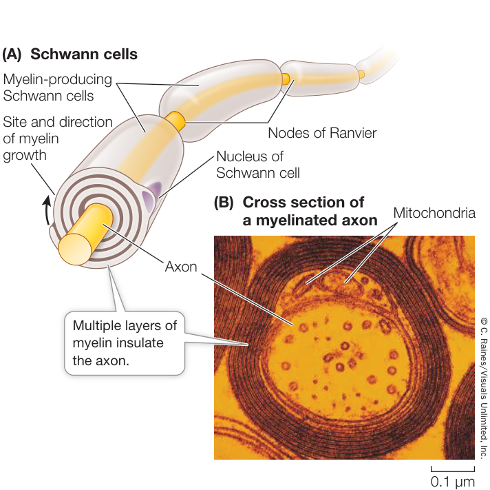
Figure 43.3 Glial Cells Insulate Axons (A) Schwann cells produce layers of myelin, a type of cell membrane that provides electrical insulation to the axon. At the intervals between Schwann cells—the nodes of Ranvier—the axon is exposed. APs travel along the axon by “jumping” from node to node, as described in Key Concept 43.2. (B) A myelinated axon, seen in cross section through an electron microscope.
The fourth type of macroglia, astrocytes (so named because they look like stars), contributes to the blood–brain barrier that protects the brain from toxic chemicals in the blood. Blood vessels throughout the body are very permeable to many chemicals, including toxic ones that could reach the brain if this barrier did not exist. The contribution of the astrocytes to the blood–brain barrier are the end-feet of their many projections that surround the blood vessels in the brain. The barrier is not perfect, however. Because it consists of cell membranes, it is permeable to fat-soluble substances such as anesthetics and alcohol (which explains why these substances have such rapid and marked effects on the nervous system).
Recently a new function of astrocytes is being investigated: ridding the brain of metabolic wastes. In the blood–brain barrier there is a small space—the perivascular space—between the membranes of the blood vessels and the astrocyte end-feet that surround them. Fluid can leak out of incoming blood vessels into the perivascular spaces. The astrocyte membranes have water channels (aquaporins) that can take up fluid from the perivascular spaces and deliver it to wherever the astrocytes project. The fluid can then leave the astrocytes and enter the interstitial fluid. In a reverse process, interstitial fluid can enter the perivascular spaces around the blood vessels leaving the brain, taking metabolic waste products with it. This system of perfusion of extracellular fluid in the brain is called the glymphatic system (Figure 43.4).
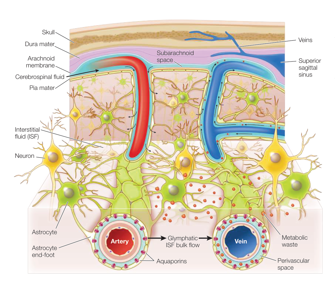
Figure 43.4 The Glymphatic System Interstitial fluid enters the brain through the perivascular spaces between the blood vessels (arteries) and the astrocytes that surround them. The astrocytes have water channels (aquaporins) that enable them to take up fluid from the perivascular spaces and distribute it to the interstitial fluid compartment of the brain. That fluid leaves the brain through the perivascular spaces of the outgoing blood vessels (veins), taking metabolic waste products with it. (After M. Nedergaard. 2013. Science 340: 1529–1530.)
Connect the Concepts The glymphatic system is to the brain what the lymphatic system is to the rest of the body (see Figure 40.3).
Astrocytes have several functions at the synapse:
- They can take up neurotransmitter that has been released into the synapse and thereby control communication between the pre-and postsynaptic cells.
- They can supply neurons with nutrients. Neurons have no energy reserves, but astrocytes store glycogen that they can break down to supply the neurons with fuel.
- They have signaling properties. Even though most astrocytes do not generate APs, they do release neurotransmitters that can alter the activities of neurons.
- They aid in the repair and regeneration of neurons.
- They make contact with both blood vessels and neurons and can therefore signal changes in the composition of the blood.
Astrocytes play crucial yet poorly understood roles in modulating synapse activity. The projections of a single astrocyte may make contact with more than 100,000 synapses. The contact of the astrocyte with the neuronal components of the synapse is so intimate that it has inspired the concept of the tripartite synapse—the idea that a synapse includes not only the pre-and postsynaptic neurons but also connections from astrocytes.
Although astrocytes do not generate APs, they do communicate with each other. They are connected to each other through electrical synapses, and the electric signals traveling across these synapses cause changes in the content of the postsynaptic astrocyte. When -sensitive dyes are applied to nervous tissue, waves can be seen traveling through extensive networks of astrocytes. The functions of these waves are not understood.
Recap and Assess
Nervous systems have two unique types of cells: neurons and glia. There are many types of neurons, but they can all generate and conduct electric signals, and most generate APs. A neuron has four regions: cell body, dendrites, axon, and axon terminals. Neurons communicate with target cells at synapses, which can be chemical or electrical. Although glia do not generate APs, they have a variety of functions, including contributing to the blood-brain barrier, insulating axons, modulating synaptic activity, and providing neurons with metabolic fuel.
- Explain how a neuron integrates information received from several other neurons and communicates it to another neuron.
- Is communication faster over chemical synapses or electrical synapses? Explain your answer.
- What is a tripartite synapse, and how can it modulate information flow in neural circuits?
The one feature common to neurons is that they process information in the form of changes in the electric potential across their cell membranes. In the next key concept we focus on how electric signals are generated and transmitted by neurons.
Neurons Generate and Transmit Electric Signals
Learning Objectives
- 43.2.1 Describe the ionic mechanisms of resting potentials and action potentials.
- 43.2.2 Explain how action potentials are conducted along unmyelinated and myelinated axons.
- 43.2.3 Explain the difference between action potentials and graded potentials, and how each can represent quantitative information.
Ion pumps create ion concentration gradients across animal cell membranes (see Key Concept 6.4). For example, concentrations are higher outside cells and concentrations are higher inside. The positive charges of these ions are balanced by negatively charged ions both inside and outside the cell. But across the cell membrane there is an electric charge difference, with the inside of the cell being negative relative to the outside. This electric charge is due to the cell membrane being differentially permeable to certain ions. For neurons at rest, their membranes are mostly permeable to , and that permeability is primarily responsible for the electric charge across their membranes.
Because the concentration of is higher inside the cell than in the extracellular fluid, tends to diffuse out of the cell down its concentration gradient. But when leaks out of the cell, it leaves behind an unbalanced negative electric charge that tends to pull back into the cell. An equilibrium is reached when the tendency for to diffuse out is countered by the electric charge pulling back in. The resulting charge difference across the membrane is called the membrane potential, with the inside of the cell negative relative to the outside. Membrane potentials exist in all cells.
In neurons the steady-state membrane potential is called the resting potential. Stimuli that cause changes in the permeability of the cell membrane cause local changes in that membrane resting potential. These local changes can generate APs that are conducted along the axon of the neuron and convey information about the stimulus that caused the initial small change in resting potential. The AP is a sudden, large, transient change in membrane potential generated by sudden changes in permeability to ions. Before describing the mechanisms of these permeability changes and the resulting APs in detail, it may be useful to review some simple concepts of electricity.
Simple electrical concepts underlie neuronal function
Voltage (electric potential difference) is a force that causes electrically charged particles to move between two points. Voltage is to the flow of electrically charged particles as pressure is to the flow of water. If the negative and positive poles of a battery are connected by a wire, an electric current will flow through the wire because there is a voltage difference between the two poles. This flow of electric current can be used to do work, just as a current of water can be used to do work.
In wires, electric current is carried by electrons, but in solutions and across cell membranes, electric current is carried by ions. The major ions that carry electric charges across the cell membranes of neurons are sodium (), potassium (), calcium (), and chloride (). Recall that ions with opposite charges attract one another and those with like charges repel one another. How do these basic principles of bioelectricity establish the resting potential of the neuronal cell membrane? And how is the flow of ions through membrane channels turned on and off to generate APs?
Activities of neurons are recorded as changes in membrane potential
We record electrical events in a cell using electrodes. Figure 43.5 shows how this technique is applied across the cell membrane of a neuron to measure the resting potential, which is usually between and millivolts (mV). The minus sign indicates that the inside of the cell is electrically negative compared with the outside.
As just explained, the resting potential is largely due to a balance between the tendency of ions to diffuse down their concentration gradient and the electric potential that holds them back. This resting potential provides a means for a neuron to respond to stimuli. If some determinant of this balance of forces is altered, the membrane potential will change. The concentration gradient of the different ions inside and outside the neuron may not change, but if the permeability of the membrane to ions changes, the membrane potential will change. Therefore any chemical or physical stimulus that changes the permeability of the cell membrane to ions will cause a change in the cell’s membrane potential. For example, what would happen to the membrane potential if there was a sudden increase in the permeability of the membrane to ions? ions are more abundant outside the cell than inside. Also, the inside of the resting cell is negatively charged, which attracts the positively charged ions. Thus a sudden increase in permeability of the membrane to would cause a change of the membrane potential in the positive direction. If that change in permeability were transient, the membrane potential would rapidly return to its resting, negative value. Such a rapid change in membrane potential is an AP.
Ion transporters and channels generate membrane potentials
The cell membranes of neurons, like those of all other cells, are lipid bilayers that are impermeable to ions but contain many protein molecules that serve as ion transporters and ion channels. Ion transporters and channels are responsible for the distribution of charges across the membrane that determine membrane potential.
Connect the Concepts The passive and active mechanisms that transport materials across cell membranes are discussed in Key Concepts 6.3 and 6.4. The passive mechanisms are membrane proteins that form channels (see Figure 6.11), and the active mechanisms (which require ATP) are membrane proteins engaged in active transport (see Figure 6.14).
Ion transporters require energy to move ions against their concentration or electrical gradients and are therefore called ion pumps. A major ion transporter in the cell membranes of neurons (and all other cells) is the sodium–potassium ( – ) pump, so called because it actively expels ions from inside the cell, exchanging them for ions from outside the cell (see Figure 6.14). The – pump is also known as sodium–potassium ATPase, a term emphasizing that it is an enzyme complex requiring ATP to do its work. The – pump keeps the concentration of inside the cell greater than the concentration of the extracellular fluid, and the concentration of inside the cell less than that of the extracellular fluid. The concentration differences established by this active transporter mean that would diffuse out of the cell and would diffuse in if the ions could cross the lipid bilayer. How do these concentration gradients relate to the electrical gradients we discussed earlier?
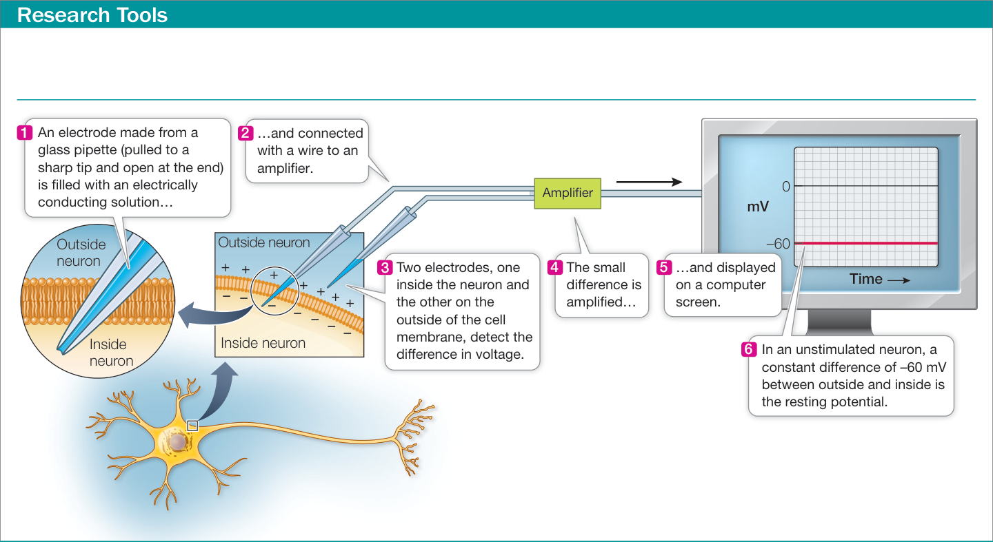
Figure 43.5 Measuring the Membrane Potential An electrode can be made from a glass pipette with a very sharp tip filled with a solution that conducts electric charges. If one electrode is placed inside the neuron and another one on the outside of the cell membrane, the difference in voltage across the cell membrane can be measured.
Ion channels permit the diffusion of ions across membranes. These channels are water-filled pores formed by proteins that span the lipid bilayer and are generally selective, allowing some types of ions to pass through more easily than others. Thus there are potassium channels, sodium channels, chloride channels, and calcium channels, and there are different kinds of channels for each ion. Ions can diffuse through these channels in either direction. The direction and magnitude of the net movement of ions through a channel depend on the concentration gradient of that ion type across the cell membrane, as well as on the voltage difference across that membrane. These two motive forces acting on an ion are termed its electrochemical gradient. The electrochemical gradient drives the movement of ions through channels, but that movement is modified by gates that open and close the channels.
Potassium channels are the most common open, or leak, channels in the cell membranes of resting (non-stimulated) neurons. As a consequence, resting neurons are more permeable to than to any other ion. Thus open potassium channels are largely responsible for the resting membrane potential. Because the potassium channels make the cell membrane permeable to , and because the – pump keeps the concentration of inside the cell much higher than that outside the cell, tends to diffuse down its electrochemical gradient, out of the cell. But if ions leave the cell, they leave behind unbalanced negative charges, generating an electric potential across the membrane that tends to pull back into the cell.
The membrane potential at which the net diffusion of out of the cell ceases (that is, the point at which diffusion out due to the concentration gradient is balanced by its inward movement due to the negative electric potential) is the potassium equilibrium potential, or . The value of can be calculated from the concentrations of on the two sides of the membrane using the Nernst equation (Figure 43.6A). This equation, developed in the late 1800s, shows that the existence of ion channels in neuronal membranes was hypothesized long before their specific structures and properties were discovered.
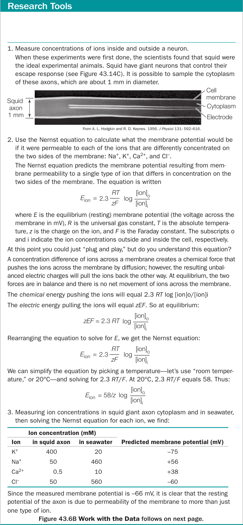
Figure 43.6A Using the Nernst Equation The Nernst equation calculates membrane potential when only one type of ion can cross a membrane that separates solutions with different concentrations of that ion.
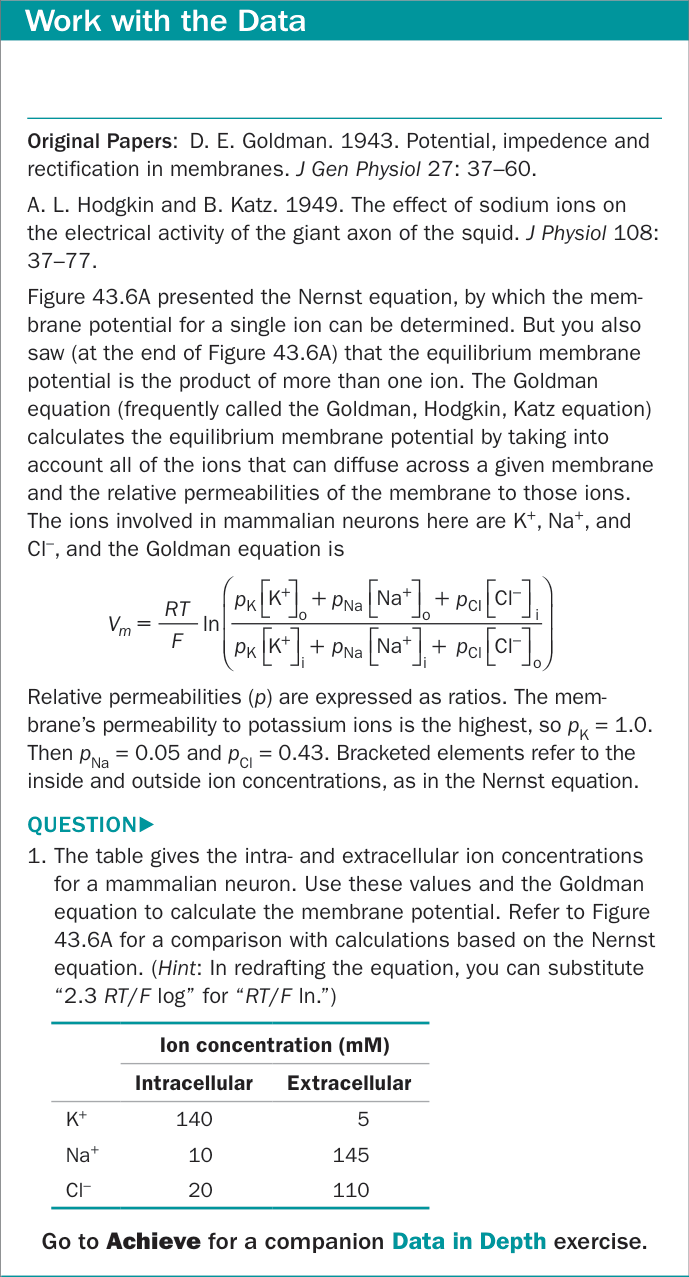
Figure 43.6B Equilibrium Membrane Potential: The Goldman Equation
In the late 1940s, A. L. Hodgkin and A. F. Huxley at the University of Cambridge set out to study the electrical properties of axonal membranes. With the techniques available at the time, the necessary measurements could be made only if you had a very large axon to work with. Such an axon exists in nature—the huge neuron that controls the escape response of squid. Hodgkin and Huxley used electrodes to measure the voltage across the cell membrane of this large axon, as seen in Figure 43.6A, and to pass electric current into it to change its membrane potential. They also changed the concentrations of and both inside and outside the squid axon and measured the resulting changes in membrane potential.
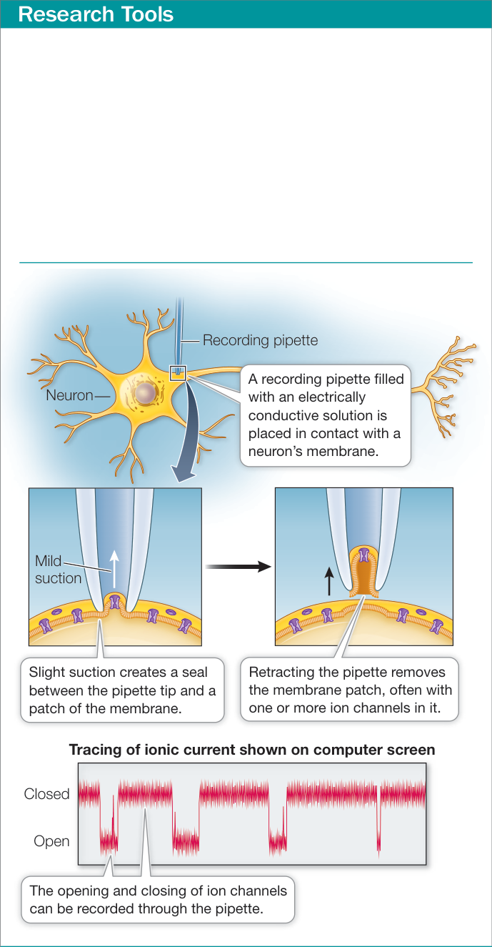
Figure 43.7 Patch Clamping The patch clamp electrode is a glass micropipette with a polished tip and is filled with an electrically conductive solution that has the same composition as extracellular fluids. When this pipette/electrode is positioned against the membrane of a cell and slight suction is applied, a seal forms. If a single ion channel or a few ion channels are within the patch of membrane bounded by the seal, the openings and closings of individual channels can be recorded by the electrode. If the pipette is retracted, it can tear the patched membrane away from the cell, and the activities of the ion channels in the patch can continue to be recorded.
On the basis of their many careful experiments, Hodgkin and Huxley developed virtually all of our basic concepts about the electrical properties of neurons, and they shared a Nobel Prize in 1963.
The resting potential of a neuron is less negative than the calculated from the Nernst equation. This means that the resting potential is not due solely to leak channels. The neuronal membrane is slightly permeable to other ions, especially and , and movements of these ions influence the resting potential. A different equation takes into account (1) all of the ions that can cross the membrane and (2) the relative permeability of the membrane to those ions. This equation, called the Goldman equation, predicts the membrane potential more accurately than does the Nernst equation (Figure 43.6B).
Ion channels and their properties can be studied directly
Because Hodgkin and Huxley were working long before there were laboratory techniques enabling the investigation of ion channels, they could only hypothesize ion channel properties. These hypotheses could not be tested until the late 1970s, when B. Sakmann and E. Neher developed a technique called patch clamping, for which they shared the Nobel Prize in 1991. Patch clamping, described in Figure 43.7, is widely used by neurobiologists, allowing them to record in real time the tiny electric currents caused by the openings and closings of single ion channels.
Gated ion channels alter membrane potential
The ion channels called leak channels are always open, but other ion channels in the cell membranes of neurons behave as if they contain “gates”; they are open under some conditions and closed under other conditions. Voltage-gated channels open or close in response to a change in the voltage across the cell membrane. Chemically gated channels open or close depending on the presence or absence of a specific molecule that binds to the channel protein, or to a separate receptor that in turn alters the channel protein. Mechanically gated channels open or close in response to mechanical force applied to the cell membrane. Gated channels play important roles in neuronal function.
Openings and closings of gated channels alter the membrane potential. Earlier, we considered what would happen if the permeability of the membrane to suddenly increased. Such a change in local membrane permeability is generated by the opening of channels. When those channels open, ions move down their electrochemical gradient until the membrane approaches the equilibrium potential for (). Therefore the inside of the cell becomes less negative. When the inside of a neuron becomes less negative (or more positive) than it is in its resting condition, its cell membrane is depolarized (Figure 43.8). The opposite change in the membrane potential occurs if gated channels open. Opening of these channels increases the permeability of the cell membrane to , and the membrane potential becomes even more negative than when only the leak channels were open, and the cell membrane is said to be hyperpolarized.
The openings and closings of ion channels that result in changes in the voltage across the cell membrane are the basic mechanisms by which neurons respond to stimuli, be they electrical, chemical, or mechanical. How do such local changes in membrane potential get communicated to other parts of the cell?
A local change in membrane potential causes a flow of ions that spreads the change in membrane potential to adjacent regions of the membrane. For example, when channels in an axon open, making that region of the axon positive, a rapid flow of ionic electric current in the axoplasm causes a decay in that electric potential change, and it does not spread very far.
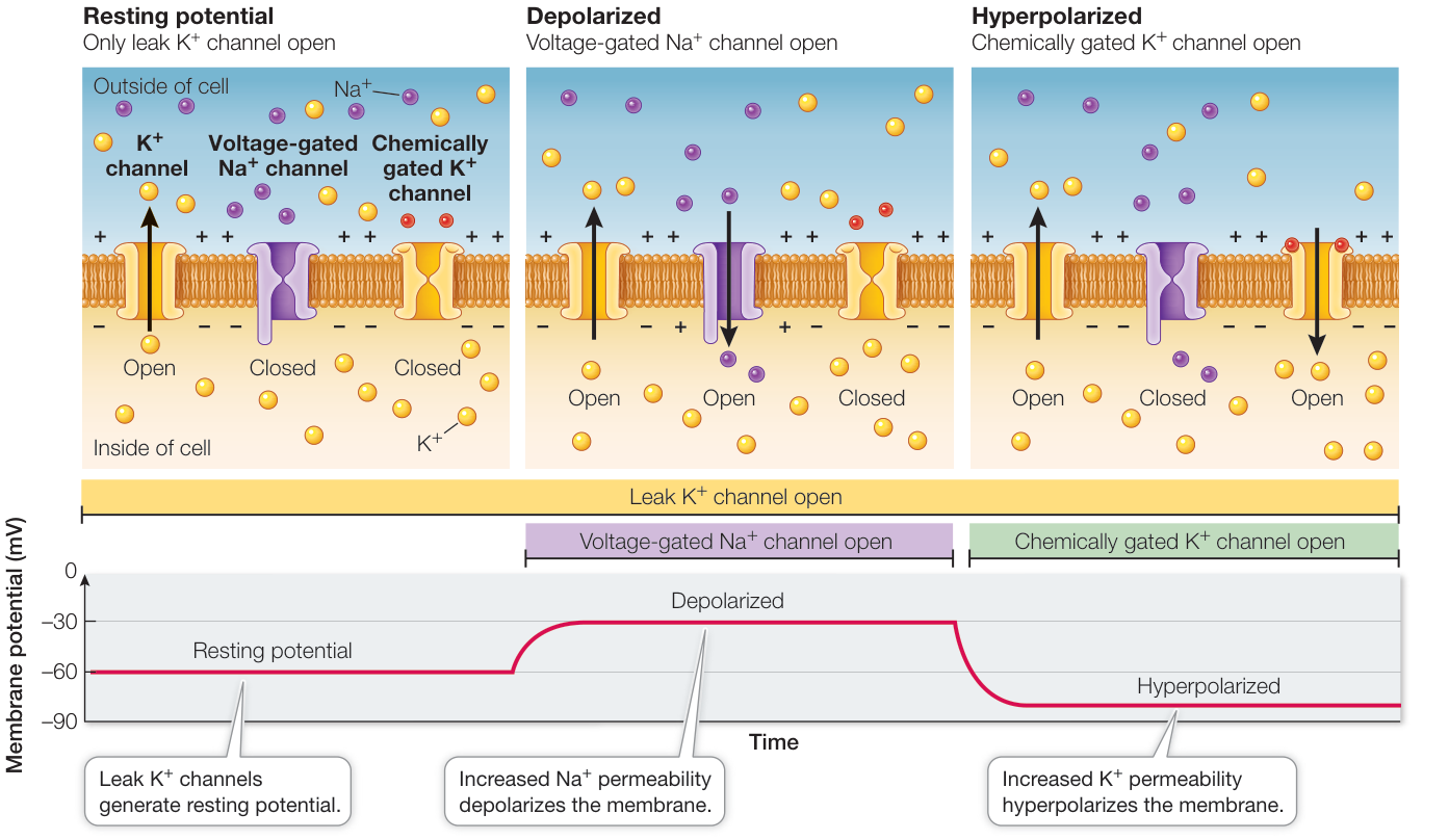
Figure 43.8 Membranes Can Be Depolarized or Hyperpolarized The resting potential is produced mostly by leak channels with a small contribution from leak channels (not shown here). A shift from the resting potential to a less negative membrane potential caused by increased permeability is called depolarization. Hyperpolarization occurs when the membrane potential becomes more negative, as when the permeability of the membrane to increases.
Graded changes in membrane potential can integrate information
The flow of ionic electric current along cell membranes can only extend over a short distance, but it causes a graded change in the local membrane potential. A graded membrane potential is a change from the resting potential that is proportional to the magnitude of a stimulus. That stimulus can be chemical or mechanical. Graded potentials are a means of integrating stimuli because the membrane can respond with proportional amounts of depolarization or hyperpolarization to each stimulus, and those changes in membrane potential are summed. In Chapter 44 you will learn how graded potentials play important roles in sensory systems. However, the spread of graded potentials can only be local and cannot be transmitted down long axons. Therefore axons code information as discrete APs that travel along their membranes. Graded potentials, however, play an important role in the generation of APs.
Sudden changes in and channels generate action potentials
APs are sudden, transient, large changes in membrane potential. In unmyelinated axons (those not wrapped in myelin by oligodendrocytes or Schwann cells), APs can be conducted at speeds of up to 2 meters per second, but in myelinated axons the conduction velocity can be 100 meters per second. Think of running the 100-meter dash—the world record is slightly under 10 seconds.
If we place the tips of a pair of electrodes on either side of the cell membrane of a resting axon and measure the voltage difference, the reading might be about mV, as you saw in Figure 43.5. If these electrodes are in place when an AP travels down the axon, they register a rapid change in membrane potential, from mV to about mV, followed by a rapid return to the resting level of mV (Figure 43.9).
The AP is generated by the openings and closings of voltage-gated and channels in the cell membrane of the axon. At the resting potential, most of these channels are closed except for the leak channels (balloon 1 in Figure 43.9). A slight depolarization of the membrane can cause some voltage-gated channels to open. Specifically, if a neuron is stimulated sufficiently to cause the membrane of its cell body to depolarize slightly, that graded potential spreads by local current flow to the axon hillock, the region of the cell body at the base of the axon (see Figure 43.1). Voltage-gated channels are concentrated in the axon hillock. A slight depolarization of the cell membrane in this area causes some of these voltage-gated channels to open briefly—for less than a millisecond (balloon 2 in Figure 43.9). When these channels open, the increased permeability depolarizes the membrane even more, causing more channels to open—a positive-feedback effect. When the membrane is depolarized about – mV above the resting potential, a threshold is reached; a large number of sodium channels open (balloon 3 in Figure 43.9), and the membrane potential becomes positive—an AP. The rising phase of the AP halts abruptly in 1–2 milliseconds, and the membrane potential rapidly becomes negative once again.
What causes the axon to return to resting potential? There are two contributing factors: the voltage-gated channels close, and voltage-gated channels open (balloon 4 in Figure 43.9). Voltage-gated channels open in response to the same depolarization that triggered the channels, but they do so more slowly and they stay open longer, allowing to carry excess positive charges out of the axon. As a result, the membrane potential returns to a negative value and usually becomes even more negative than the resting potential until the voltage-gated channels close (balloon 5 in Figure 43.9).
Another feature of the voltage-gated channels is that once they open and close, they have a refractory period of 1–2 milliseconds during which they cannot open again. This property can be explained by the channels having two gates, an activation gate and an inactivation gate (see Figure 43.9). Under resting conditions, the activation gate is closed and the inactivation gate is open. Depolarization of the membrane to the threshold level causes both gates to change state, but the activation gate responds faster. As a result, the channel is open for a brief time between the opening of the activation gate and the closing of the inactivation gate. Inactivation gates remain closed for 1–2 milliseconds before they spontaneously open again, thus explaining the refractory period. By the time the inactivation gate reopens, the activation gate is closed, and the membrane is poised to generate another AP. Another contribution to the refractory period is the duration of the opening of the voltage-gated channels, as you saw earlier. The dip in the membrane potential following an AP is called the after-hyperpolarization or undershoot.
The difference in the concentration of across the cell membrane and the negative resting potential constitute the “battery” that drives APs. How rapidly does the battery run down? It might seem that a substantial number of ions would have to cross the membrane for the membrane potential to change from mV to mV and back to mV again. In fact, only a vanishingly small number of ions move through the channels during the passage of an active potential. Thus the effect of a single AP on the concentration gradients of and is very small, and the sodium–potassium pump can keep the “battery” charged, even when the neuron is generating many APs every second.
Action potentials are conducted along axons without loss of signal
An AP is conducted over long distances with no loss of signal. If we place two pairs of electrodes at two different locations along an axon, we can record an AP at those two locations as it travels along the axon (Figure 43.10A). The magnitude of the AP does not change between the two recording sites. This constancy is possible because an AP is an all-or-none, self-regenerating event.
- An AP is all-or-none because of the interaction between the voltage-gated channels and the membrane potential. If the membrane is depolarized slightly, some voltage-gated channels open. Some sodium ions cross the cell membrane and depolarize it even more, opening more voltage-gated channels, and so on, generating an AP. This positive-feedback mechanism ensures that APs always rise to their maximum value.
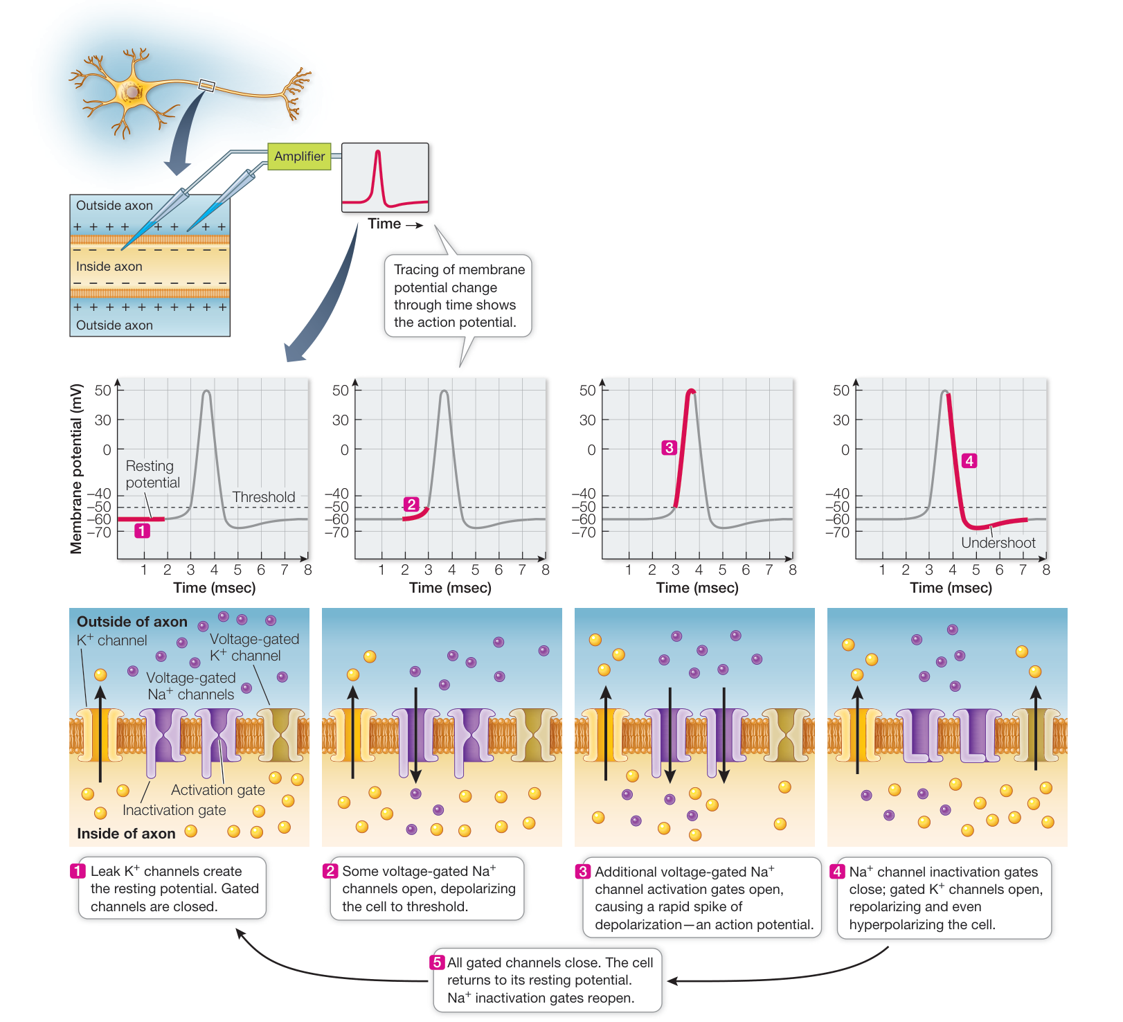
Figure 43.9 The Course of an Action Potential APs result from rapid changes in voltage-gated Na+ and K+ channels. Animation 43.2 The Action Potential
- An AP is self-regenerating because it spreads by local current flow to adjacent regions of the cell membrane. The resulting depolarization brings those neighboring areas of membrane to threshold. Therefore when an AP occurs at one location on an axon, it stimulates the adjacent region of axon to generate an
AP, and so on down the length of the axon. Although we say that the AP travels down the axon, what is really happening is that new APs are being created sequentially.
We can use an electrode to stimulate an axon, causing it to depolarize and to fire an AP that is then conducted along the axon. Figure 43.10B shows the changes in the ion channels in the membrane that are responsible for conducting the AP along the axon without a reduction in amplitude. Normally an AP is propagated in only one direction—away from the cell body. It cannot reverse itself because the voltage-gated channels in the region of the membrane it came from are in their refractory period (Figure 43.10C).
APs are not conducted at the same speed in all axons. They travel faster in large-diameter axons than in small-diameter axons because the resistance to ionic current flow decreases as an axon’s diameter gets bigger. They travel faster in myelinated than in unmyelinated axons because they can move down the axon in short “jumps” as described in the next section (Figure 43.11). Invertebrates depend on increased axon diameter for fast conduction, but vertebrates mostly depend on myelination of axons to increase AP conduction velocity.
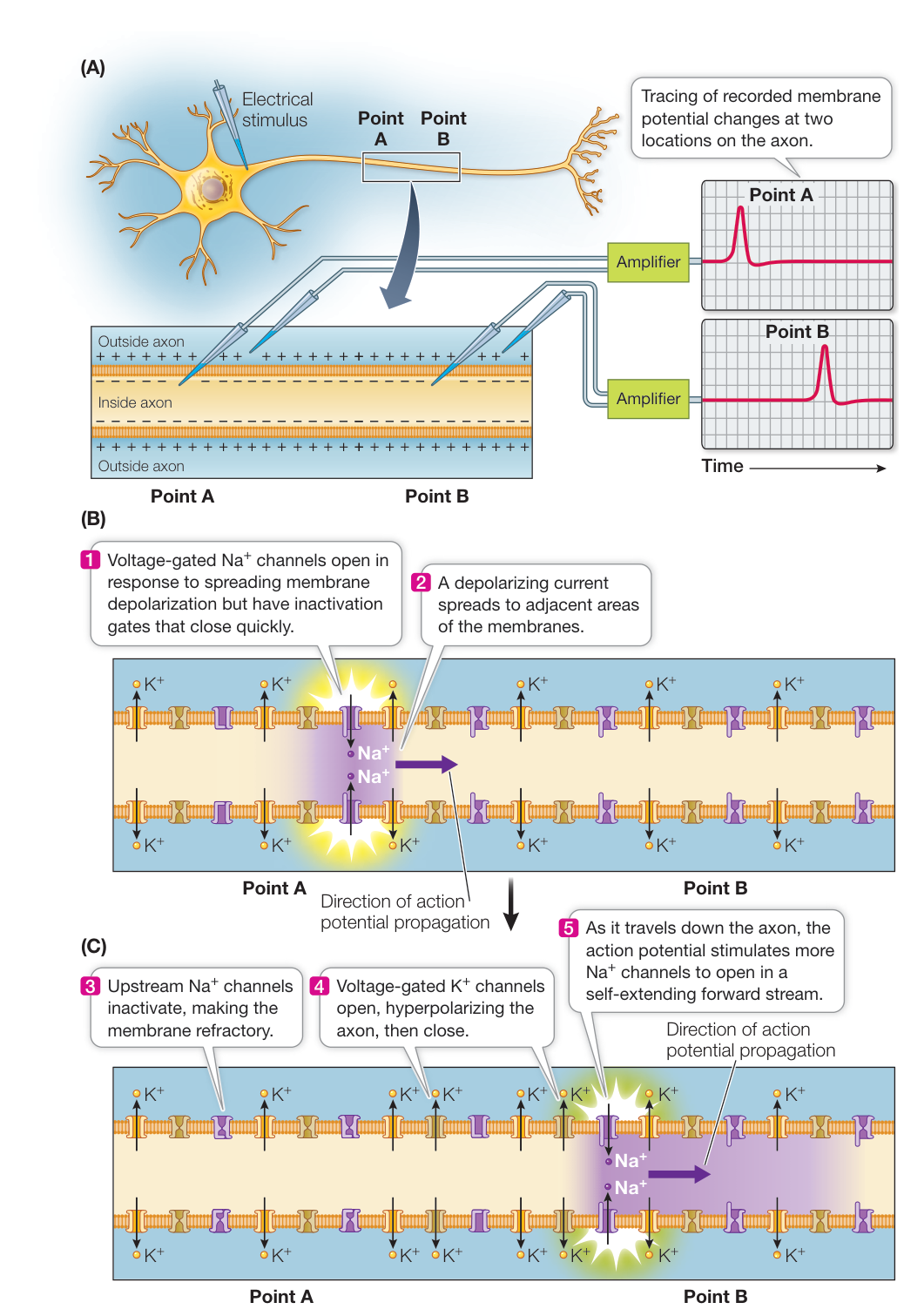
Figure 43.10 Action Potentials Travel along Axons (A) There is no loss of signal as an AP travels along an axon. (B) When an AP is stimulated in one region of membrane, ionic current flows to and depolarizes adjacent areas of membrane. (C) The advancing wave of depolarization causes more channels to open, and the AP is generated anew in the next section of membrane. Meanwhile, in the region where the AP has just fired, the channels are inactivated and the voltage-gated channels are still open, rendering this section of the axon incapable of generating an AP. Hence the AP cannot “back up,” but moves continuously forward, regenerating itself as it goes.
Action potentials jump along myelinated axons
In vertebrate nervous systems, increasing the speed of APs by increasing the diameter of axons is not feasible because of the huge number of axons involved. Each of our eyes, for example, has about a million axons connecting it to the brain. These axons conduct APs at about the same speed as does the squid giant axon—about 20 meters per second—yet the diameter of each is 200 times smaller than the squid axon’s diameter. Imagine having optic tracts 200 times bigger. A different way of increasing conduction velocity of axons has evolved in vertebrates, and that adaptation is myelination.
When glia wrap around axons, they cover the axons with concentric layers of myelin (see Figure 43.3). However, they leave regularly spaced gaps called nodes of Ranvier, where the axon is not covered (see Figure 43.11). Underneath the myelin sheaths, there are no or channels, and therefore APs cannot propagate under the myelin sheath. However, an AP firing at a node of Ranvier creates a local electric field inside the axon that spreads almost instantaneously to the next node of Ranvier. The resulting depolarization of that next node triggers another AP, and so on down the axon. Thus the APs appear to jump from node to node, and their conduction down the axon is very fast.
The speed of conduction is increased in these myelin-wrapped axons because electric current flows much faster through the cytoplasm than ion channels can open and close. This form of rapid impulse propagation is called saltatory conduction (Latin saltare, “to jump”).
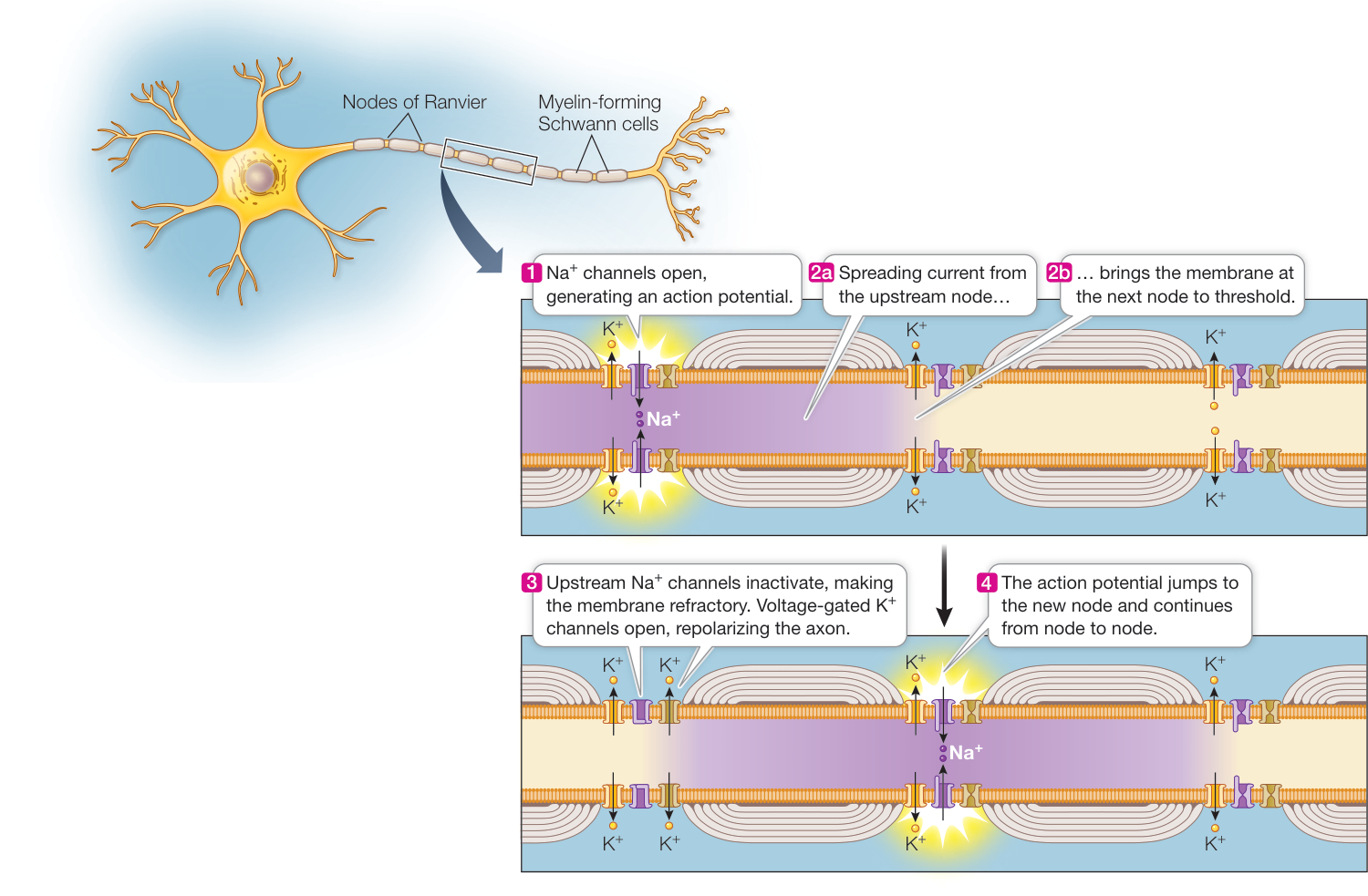
Figure 43.11 Saltatory Action Potentials APs “jump” from node to node in myelinated axons, allowing faster transmission of information.
Recap and Assess
Neurons have membrane potentials due to ion concentration differences across their membranes and the different permeabilities of the membrane to those ions created by ion channels. Leak channels generate the resting potential, and opening and closing of gated channels can create graded changes in membrane potentials depending on how many of which channel types open or close. Voltage-gated channels have threshold potentials for opening. If some voltage-gated channels open, they create a local depolarization that brings other channels to threshold—a positive-feedback mechanism that generates APs. Sudden closure of channels and the opening of channels terminate the AP. APs are rapid, all-or-none changes in membrane potential that are conducted along axons from the cell body to the axon terminals. In myelinated axons, APs jump between nodes of Ranvier and conduction is more rapid than in unmyelinated axons.
- How would the resting potential of neurons be different if the neuronal cell membrane were more permeable to than to , and how would the APs of such a neuron look if they were due to voltage-gated channels with thresholds lower than the resting potential?
- Why does the Goldman equation produce a more accurate calculation of membrane resting potential than the Nernst equation does?
- Why is an AP self-regenerating in both unmyelinated and myelinated neurons?
- How do graded membrane potentials enable a neuron to generate signals that reflect the inputs from many neurons?
Having described how APs are generated and transmitted along axons, we next address the question of what happens when an AP reaches the axon terminals. How is the AP’s signal communicated to the next cell—which could be another neuron, a muscle cell, or a secretory cell?
Neurons Communicate with Other Cells
Learning Objectives
43.3.1 Describe the events at a neuromuscular junction, beginning with the action potential in the presynaptic neuron and ending with the response of the postsynaptic cell.
43.3.2 Compare the characteristics of electrical versus chemical synapses and explain why chemical synapses present a greater variety of postsynaptic responses than do electrical synapses.
43.3.3 Describe the mechanisms that make the actions of neurotransmitters transitory.
Neurons communicate with each other and with other cells at synapses. In electrical synapses, which are common among invertebrates, the AP spreads directly from presynaptic to postsynaptic cell. The most common type of synapse in the vertebrate nervous system is the chemical synapse, in which neurotransmitters released from a presynaptic cell induce changes in a membrane potential of a postsynaptic cell. This key concept begins with a discussion of the synapses between neurons and muscle cells. We then consider the diversity in synapses between neurons and how they integrate information.
The neuromuscular junction is a model chemical synapse
Neuromuscular junctions are synapses between neurons and skeletal muscle cells. They are excellent models for how chemical synaptic transmission works. Neurons that control muscles are called motor neurons. Like other neurons, a motor neuron has only one axon, but that axon can branch into numerous axon terminals that form many synapses with muscle cells. At each axon terminal an enlarged knob or buttonlike structure (called a synaptic bouton) contains membrane-bound vesicles filled with neurotransmitter molecules. The neurotransmitter used by motor neurons to skeletal muscles in all vertebrates is acetylcholine (ACh). ACh is released by exocytosis when the membrane of a vesicle containing this neurotransmitter fuses with the presynaptic membrane of the axon terminal.
The postsynaptic membrane of the neuromuscular junction is a modified part of the muscle cell membrane called a motor end plate. It appears as a depression in the muscle cell membrane, and the terminals of the motor neuron sit in the depression. The space between the presynaptic membrane and the postsynaptic membrane is the synaptic cleft, which in chemical synapses is about 20–40 nanometers wide. ACh released into the cleft by the presynaptic cell diffuses across to the postsynaptic membrane (Figure 43.12).
The arrival of an action potential causes the release of neurotransmitter
Neurotransmitter is released when an AP arrives at the axon terminal and causes the opening of voltage-gated channels in the presynaptic membrane. Because the concentration is greater outside the cell than inside, enters the axon terminal. This increase in inside the axon terminal causes the vesicles containing neurotransmitter to fuse with the presynaptic membrane and empty their contents into the synaptic cleft.
In neuromuscular synapses, vesicle fusion and emptying is allor-none. The vesicle membrane is incorporated into the presynaptic membrane, which actually gets larger as a result—at least until the extra membrane is recycled through endocytosis. The internalized membrane is processed by the cell into new vesicles that are filled with neurotransmitter.
Synaptic functions involve many proteins
Our description of the release of neurotransmitter from the presynaptic membrane may seem simple, but it involves hundreds of proteins that are responsible for various aspects of the process: vesicle formation, transport of neurotransmitter into vesicles, anchoring of vesicles to cytoskeletal elements, docking of the vesicles with the presynaptic membrane, fusion of the vesicular and cell membranes, and endocytosis of the vesicle membrane for recycling.
Some of these proteins are the targets of toxins. For example, botulinum and tetanus toxins from bacteria of the genus Clostridium act on several of the proteins necessary for the docking of vesicles to the presynaptic membrane, resulting in diseases that are frequently fatal. Botulinum toxin impairs muscle contraction, whereas tetanus toxin causes uncontrolled muscle contraction. Poisons can become medicines. Botulinum toxin (in the form of Botox®) is used therapeutically to subdue muscle spasms and cosmetically to reduce wrinkles.
The postsynaptic membrane responds to neurotransmitter
When acetylcholine is released at a synapse, some of it diffuses across the synaptic cleft and binds to ACh receptors (AChRs) on the postsynaptic membrane. The postsynaptic membrane of the motor end plate is highly folded. AChRs are on the crests of the folds, and voltage-gated cation channels are at the bottoms of the folds and in the surrounding muscle cell membrane (see Figure 43.12). The AChRs are gated channels that allow both and to flow through, but since the electrochemical gradients favor a net influx of , the response of the motor end plate to ACh is to depolarize. The amount of depolarization depends on the number of receptors activated, and therefore the response to ACh is a graded potential. If the graded potential is sufficient, the membrane depolarization spreads to the depths of the folds of the motor end plate membrane and to the surrounding muscle cell membrane, where there are voltage-gated channels.
The spreading depolarization of the motor end plate activates the voltage-gated channels and causes the firing of an AP. This AP is then conducted throughout the muscle cell’s system of membranes, causing the cell to contract. We will discuss the neural control of contraction of muscle cells in greater detail in Key Concept 46.1.
How much neurotransmitter is enough? Neither a single ACh molecule nor the contents of an entire vesicle (about 10,000 ACh molecules) will depolarize the membrane of a muscle cell to threshold. However, a single AP in an axon terminal releases the contents of about 100 vesicles—more than enough to fire an AP in the muscle cell and cause it to contract.
Synapses can be excitatory or inhibitory
The synapses between vertebrate motor neurons and muscle cells are always excitatory. The motor end plates always respond to ACh with a graded potential that is less negative than the resting potential (depolarization). However, synapses between neurons are frequently inhibitory; such a synapse causes hyperpolarization of the postsynaptic membrane or simply makes it less likely that its membrane potential will reach threshold for the voltage-gated channels. For example, there are more ions outside than inside the cell. However, in a mammalian neuron the equilibrium potential for is around . Given a resting potential that is also around , opening of channels will not hyperpolarize the membrane, but if these channels are open, it will make it more difficult to raise the membrane potential to the threshold for the voltage-gated channels.
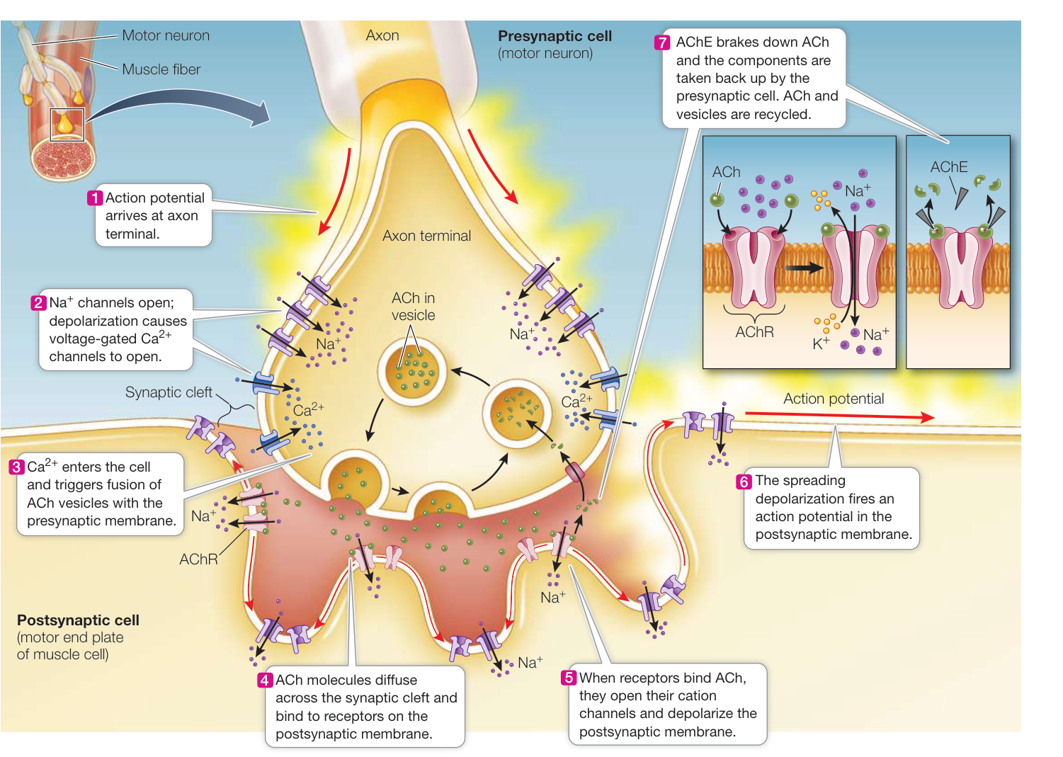
Figure 43.12 Chemical Synaptic Transmission Begins with the Arrival of an Action Potential The neuromuscular junction is a typical chemical synapse. Events shown here are similar for other neurotransmitters at other synapses. Both chemically gated and voltage-gated ACh receptors (AChRs) are found in the motor end plate. These are nonselective cation channels but are more permeable to than to (inset). When one of the chemically gated receptors binds ACh, its channel pore opens and the net increase in permeability of the postsynaptic membrane depolarizes it. The depolarization spreads beyond the motor end plate, activating voltage-gated channels in the muscle cell membrane, depolarizing it and initiating an AP. The enzyme acetylcholinesterase (AChE) breaks down ACh in the synapse, closing the chemically gated channels. The breakdown products (acetate and choline) are taken up by the presynaptic membrane and resynthesized into more ACh. Q: If this synapse were between neurons, and the neurotransmitter were inhibitory rather than excitatory, what chemically gated channels would you expect to be on the postsynaptic membrane? Animation 43.3 Synaptic Transmission Media Clip 43.1 Put Some ACh Into It! Life12e.com/mc43.1
Recall that most neurons have many dendrites. Axon terminals from many other neurons can form synapses with those dendrites and with the cell body. The axon terminals of different presynaptic neurons can store and release different neurotransmitters, and the cell membrane of the dendrites and cell body of a postsynaptic neuron can have receptors for a variety of neurotransmitters. The mix of synaptic activity impinging on a neuron will cause it to have a graded membrane potential that can be either more positive or more negative than its resting potential and thereby increase or decrease its probability of firing APs.
The postsynaptic neuron sums excitatory and inhibitory input
What determines when an individual neuron fires an AP? As you just learned, the sum of excitatory and inhibitory postsynaptic potentials creates a graded membrane potential in the postsynaptic cell body. This summation ability is the major mechanism by which the nervous system integrates information. A neuron may receive 1,000 or more synaptic inputs, but it has only one output: APs in a single axon. At any one time, the information from all of the active inputs is translated into the rate at which that neuron generates APs in its axon.
For most neurons, summation takes place in the axon hillock at the base of the axon. The cell membrane of the axon hillock is not insulated by glia and has many voltage-gated channels. Excitatory and inhibitory postsynaptic potentials from synapses are always on the dendrites or the cell body spread to the axon hillock by local current flow. If the resulting graded potential depolarizes the axon hillock to threshold, it fires an AP. Because postsynaptic potentials decrease in strength as they spread from the site of the synapse, a synapse at the tip of a dendrite has less influence than a synapse on the cell body, near the axon hillock.
Excitatory and inhibitory postsynaptic potentials are summed over space and over time. Spatial summation adds up the simultaneous influences of synapses at different sites on the postsynaptic cell (Figure 43.13A). Temporal summation adds up postsynaptic potentials generated at the same site in a rapid sequence (Figure 43.13B).
Electrical synapses are fast but do not integrate information well
Electrical synapses are different from chemical synapses because they couple neurons electrically. Electrical synapses contain numerous gap junctions. At these synapses, the presynaptic and postsynaptic cell membranes are separated by a space of only 2–3 nanometers, and membrane proteins called connexins link the two neurons by forming pores that connect the cytoplasm of the two cells (see Figure 7.16A). Electric current as well as ions and small molecules can pass directly from cell to cell through these pores. As a result, transmission at electrical synapses is very fast and can proceed in either direction. In comparison, transmission at chemical synapses is slow and unidirectional.
Connect the Concepts The proteins that form gap junctions between cells, including neurons, are discussed in Key Concept 6.2.
The properties of electrical synapses make them especially well adapted for processes that have to be fast—for example, escape from danger. They are also effective means of achieving synchronous activity of populations of cells. Electrical synapses are less common in the nervous systems of vertebrates than are chemical synapses. The major reason is that they offer fewer means for integration of information. Electrical synapses are almost exclusively excitatory, whereas chemical synapses are both excitatory and inhibitory. Electrical synapses are not well suited for temporal summation of inputs. Also, because of their relative simplicity of structure and function, electrical synapses offer fewer possible mechanisms for plasticity, which is the foundation of learning and memory.
The action of a neurotransmitter depends on the receptor to which it binds
More than 100 neurotransmitters are now recognized, and more will surely be discovered. Acetylcholine (ACh) is an important neurotransmitter because it is the substance the nervous system uses to signal muscles to contract. ACh also plays roles in certain synapses between neurons in the brain, but it accounts for only a small percent of the total neurotransmitter content of the brain.
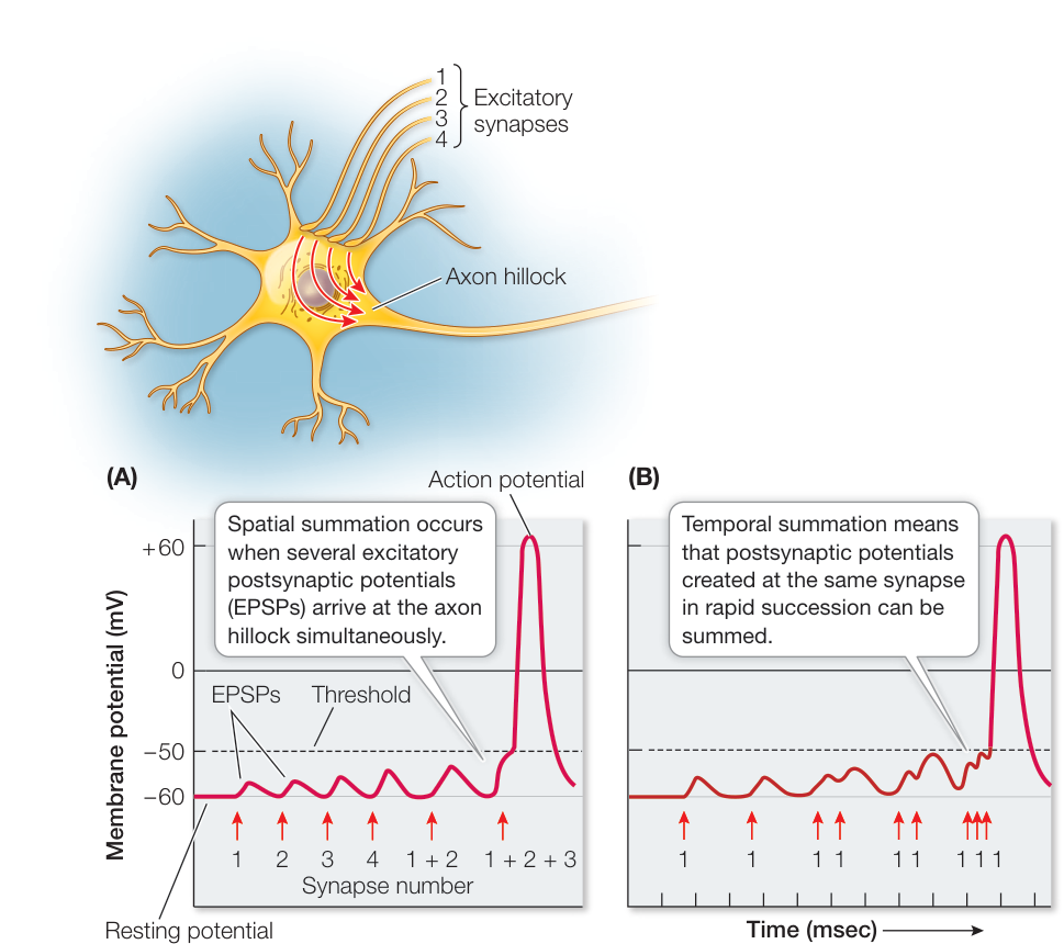
Figure 43.13 The Postsynaptic Neuron Sums Information Individual neurons sum excitatory and inhibitory postsynaptic potentials over space (A) and time (B). When the sum of the potentials depolarizes the axon hillock to threshold, the neuron generates an AP.
The workhorse neurotransmitters of the brain are three simple amino acids: glutamate, which is usually excitatory, and glycine and -aminobutyric acid (GABA), which are usually inhibitory. The integration of information at the cellular level is a balance between excitation and inhibition, so it is understandable that excessive inhibition such as you saw in the Down syndrome mice described at the start of this chapter could impair an animal’s ability to process and integrate new information. Indeed, when GABA’s inhibitory action in these mice was reduced by a drug that blocks the GABA receptor, the learning ability of the mice improved, as described in Investigating Life: Can Learning Be Restored in Mouse Models of Down Syndrome?
Another important group of neurotransmitters in the brain is the monoamines, which are derivatives of amino acids. They include dopamine and norepinephrine (derivatives of tyrosine) and serotonin (a derivative of tryptophan). Peptides also function as neurotransmitters; for example, endorphins and enkephalins are the body’s opiates and modulate the sensation of pain. Another peptide, substance P, transmits pain sensations. Even a gas, nitric oxide, is used by neurons as an intercellular messenger (see Figure 7.13).
Investigating Life: Can Learning Be Restored in Mouse Models of Down Syndrome?
Experiment
Original Paper: D. Colas et al. 2013. Short-term treatment with the GABA antagonist pentylenetetrazole produces a sustained procognitive benefit in a mouse model of Down’s syndrome. Br J Pharmacol 169: 963–973.
If the learning disability in the mouse model of Down syndrome (DS) is due to overinhibition, then reducing the activity of the major inhibitory neurotransmitter, GABA, might improve learning in these mice.
HYPOTHESIS The inability of DS model mice to learn and remember experiences as well as wild-type (WT) mice is due to over-active GABA signaling in their brains. If GABA signaling is reduced, the ability of mice to perform learning tasks should improve.
METHOD
Keep DS and WT mice of the same age on a 12-hour:12-hour light-dark cycle. At the same time each morning, for 2 weeks, inject the mice in the experimental group with the drug pentylenetetrazole (PTZ), which blocks the action of GABA. Use three different doses. Inject the control mice with saline. The doses are measured as mg PTZ/kg body weight of mouse.
Following completion of the 2-week drug-dosing protocol, evaluate the mice using the Novel Object Recognition (NOR) test. For training, place each mouse in a 4-square-foot arena that contains two objects such as chessmen or small bottles. Allow the mouse to explore the objects for 10 minutes and then return it to its home cage. An acceptable baseline is when the mouse spends the same amount of time exploring each object. Test again 1 day later, placing the mouse back in the arena but with one of the objects changed (the novel object). If the mouse spends more time exploring the novel object, that is evidence that it remembered the original object from the day before.
- Quantify the mouse’s behavior as a NOR preference (time spent exploring object A/time spent exploring objects A and B 100%). For the testing trial, A is the novel object. A NOR preference of 50% means the mouse spent equal amounts of time with each object, and therefore did not remember the objects it explored previously. A NOR preference significantly greater than 50% means the mouse spent more time exploring the novel object and is evidence that the mouse remembered the objects it saw before.
RESULTS Data are NOR preference percentages 1 SEM (standard error of the mean). An asterisk (*) indicates for difference between training (blue) and testing (orange) results as determined by a t-test: **, .
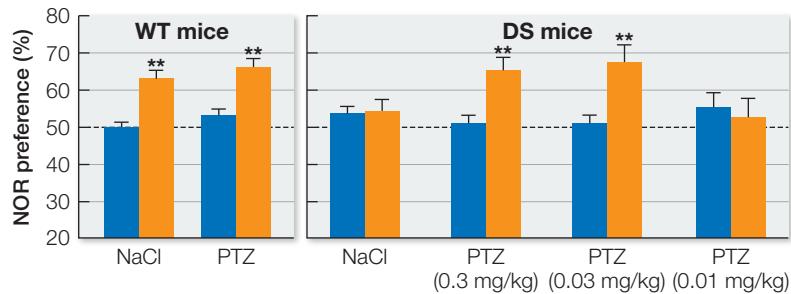
CONCLUSION The saline treatments (controls) show that the WT mice can recognize the novel object 24 hours after training and that the DS mice cannot. PTZ treatment had no effect on the WT mice. PTZ treatment at 0.3 and 0.03 mg/kg improved the ability of the DS mice to remember the familiar objects and therefore recognize the novel object, whereas treatment at 0.01 mg/kg did not.
Neurotransmission is complex in part because each neurotransmitter has multiple receptor types. ACh, for example, has two receptor types: nicotinic receptors and muscarinic receptors. All of the AChRs in skeletal muscles are nicotinic. Both types of AChRs are found in the brain and spinal cord, where nicotinic receptors tend to be excitatory and muscarinic receptors tend to be inhibitory. ACh actions can differ outside the brain and spinal cord as well. ACh acting through nicotinic receptors causes the smooth muscle of the gut to increase its motility, but ACh acting through muscarinic receptors causes cardiac muscle to hyperpolarize and therefore to slow down. There are many more examples of neurotransmitters that have different effects in different tissues, but the important thing to remember is that the action of a neurotransmitter depends on the receptor to which it binds. In addition, turning neurotransmitter action off is as important as turning it on.
Work with the Data
All of the PTZ injections in the experiment described above were given to the mice early in the light phase of their day. This is the time when these nocturnal animals are normally sleeping. The researchers therefore asked whether the same results could be obtained if the treatments were delivered during the dark phase of the day, when the mice are usually active and exploring. They obtained the following data. Data are NOR preference percentages 1 SEM. An asterisk (*) indicates for difference between saline and PTZ treatments.
| Group | Treatment | |
|---|---|---|
| Saline | PTZ 0.3 mg/kg | |
| WT Mice Training | 55.0 3.1 | 55.4 2.4 |
| DS Mice Training | 51.9 2.4 | 50.7 2.0 |
| WT Mice Testing | 65.8 5.3* | 67.4 4.9* |
| DS Mice Testing | 53.7 4.7 | 51.3 6.9 |
QUESTIONS
- How did the learning ability of the WT and DS mice compare during the dark phase of the day, and did this result differ from when the training and testing were done during the light phase?
- Describe any difference between the effect of the PTZ treatment on the performance of the WT and DS mice on the NOR task during the light and dark phases of the day.
Synapses must be cleared of neurotransmitter to turn off responses
If released neurotransmitter molecules simply remained in the synaptic cleft, the postsynaptic membrane would become saturated and receptors would be constantly activated. The postsynaptic cell would remain hyperpolarized or depolarized and would be unresponsive to additional APs arriving at the presynaptic cell. The more rapidly neurons can respond to input, the more information they can process in a given amount of time. Thus neurotransmitter must be cleared from the synaptic cleft shortly after it is released by the axon terminal.
Neurotransmitter action can be terminated in several ways. First, enzymes can destroy the neurotransmitter. Acetylcholine, for example, is rapidly destroyed by the enzyme acetylcholinesterase (AChE), which is present in the synaptic cleft in close association with AChRs on the postsynaptic membrane (see Figure 43.12). When AChE is inhibited, ACh lingers in the synaptic cleft, causing spastic (contracted) muscle paralysis that can lead to death. Some of the most deadly nerve gases developed for chemical warfare worked by inhibiting AChE. Some agricultural insecticides, such as malathion, also inhibit AChE and can poison farm workers if used without safety precautions.
Neurotransmitter can also simply diffuse away from the cleft or be taken back up by the presynaptic membrane to be recycled. Glial cells also take up neurotransmitters. The antidepressant drug fluoxetine (commonly prescribed under the brand name Prozac®) slows the reuptake of the neurotransmitter serotonin, thus enhancing serotonin’s activity at the synapse.
The diversity of receptors makes drug specificity possible
Many drugs used to treat the nervous system act by modulating specific synaptic interactions. Drugs that mimic or potentiate the effect of a neurotransmitter are called agonists; those that block the actions of neurotransmitters are called antagonists. For example, morphine is an agonist at the endorphin receptor and therefore blocks pain. Propranolol, a widely used beta blocker, is an antagonist of receptors in the fight-or-flight response and therefore decreases panic attacks and anxiety. A major emphasis in neurobiology is to identify neurotransmitter receptor subtypes and design drugs that selectively bind to them to have highly specific effects on nervous system activity.
Recap and Assess
Chemical synapses involve the release of neurotransmitter molecules stored in vesicles in the presynaptic axon terminal. APs reaching that terminal cause the fusion of vesicles with the presynaptic membrane, releasing neurotransmitter that can then bind to receptors on the postsynaptic membrane and influence its membrane potential. At excitatory synapses, the neurotransmitter causes depolarization of the postsynaptic membrane, and at inhibitory synapses the neurotransmitter hyperpolarizes or stabilizes the postsynaptic membrane potential. There is a great diversity of neurotransmitters and their receptors. At electrical synapses the AP moves directly between pre-and postsynaptic cells.
What are the two major types of cation channels in the motor end plate, and what are their functions?
- How can the same neurotransmitter be excitatory to some postsynaptic neurons and inhibitory to others?
- Sarin is a colorless, odorless liquid that can quickly evaporate into the environment and is highly lethal. It irreversibly inactivates the enzyme acetylcholinesterase (AChE). Explain why victims of sarin poisoning generally die of asphyxiation.
- Explain why parts of nervous systems that integrate complex information use chemical rather than electrical synapses.
Now that you understand how neurons communicate with each other, let’s explore how they are organized into circuits and systems that process sensory information and generate responses to that information.
Neurons and Glia Form Information-Processing Circuits
Learning Objectives
- 43.4.1 Distinguish the central from the peripheral nervous system and describe how these systems range in complexity among different species.
- 43.4.2 Using the knee-jerk reflex, explain how a neural circuit involves both afferent and efferent neurons.
Nervous systems can process information because their neurons are organized into neural networks. These networks include three functional categories of neurons that are responsible for input, output, and integration:
- Afferent neurons carry sensory information into the nervous system. That information comes from specialized sensory cells that transduce (convert) various kinds of sensory stimuli (e.g., light, heat, pressure) into APs.
- Efferent neurons carry commands to physiological and behavioral effectors such as muscles and glands.
- Interneurons integrate and store information and communicate between afferent and efferent neurons.
Nervous systems range in complexity
Simple animals such as cnidarians (sea anemones, for example) process information with a limited number of simple neural networks that do little more than provide direct lines of communication from sensory cells to effectors; there is little integration or processing of signals (Figure 43.14A). The cnidarian’s nerve net is most developed around the tentacles and the oral opening, where it facilitates detection of food or danger and causes tentacles to extend or retract.
Animals that are more complex and move about in search of food and mates must process and integrate larger amounts of information. Even earthworms fit this description, and their increased need for information processing is met by higher numbers of neurons organized into clusters of neuronal cell bodies called ganglia (singular ganglion). Ganglia serving different functions may be distributed around the body, as in earthworms (Figure 43.14B). In animals that are bilaterally symmetrical, ganglia frequently come in pairs, one on each side of the body (Figure 43.14C). Also, as animals increase in complexity, some ganglia may become enlarged or fused together at the anterior end, forming a brain. Small nervous systems of invertebrates can be remarkably complex. Consider the nervous systems of spiders, which have programmed within them the thousands of precise movements necessary to construct an intricate web without prior experience or opportunities to learn the specific web architecture of their species.
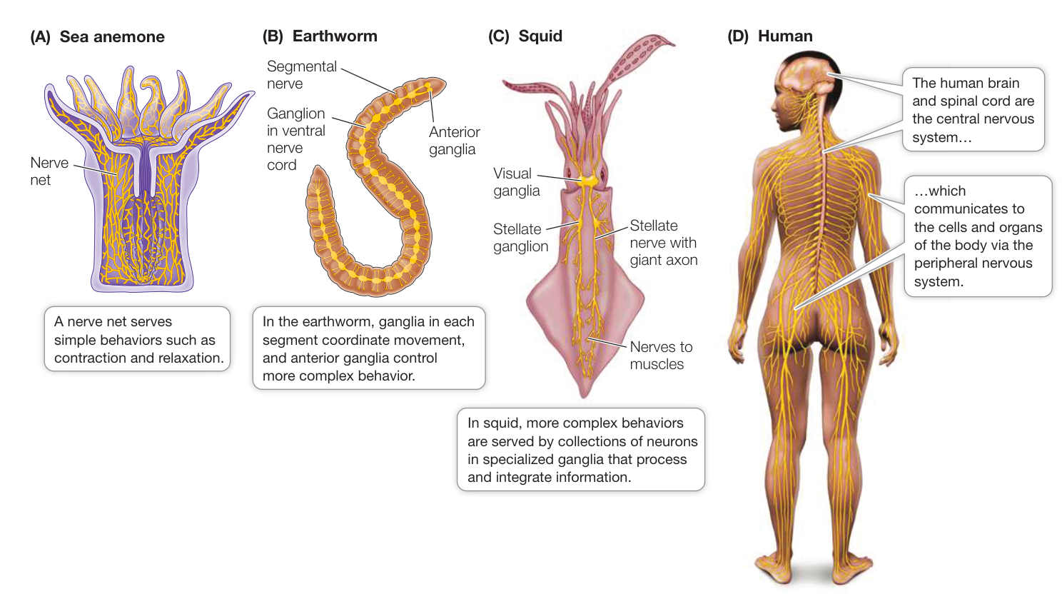
Figure 43.14 Nervous Systems Vary in Size and Complexity In animal species with increasingly complex sensory and behavioral abilities, information processing is increasingly centralized in ganglia (collections of neurons) or in a brain.
In vertebrates, most cells of the nervous system are found in the central nervous system (CNS). The CNS includes the brain and the spinal cord, which are the sites of most information processing, storage, and retrieval (Figure 43.14D). Information is transmitted from sensory cells to the CNS and from the CNS to cells that respond (effectors) via neurons that extend or reside outside the brain and the spinal cord. These sensory and effector neurons and their supporting cells are the peripheral nervous system.
Reflexes are controlled by simple circuits involving sensory neurons, interneurons, and effectors
A reflex is a behavioral or physiological response that does not require conscious information processing—it is autonomic, meaning an involuntary or unconscious reaction. Examples are salivating at the sight or smell of food, and the jerk of your lower leg when a physician taps your knee with a small rubber hammer. The knee-jerk reflex involves a very simple neural circuit involving just a few neurons that connect with each other in the spinal cord. Thus it is called a spinal reflex. A cross section of the human spinal cord reveals a central area of gray matter in the shape of a butterfly, surrounded by areas of white matter (see Figure 43.15). In the nervous system, gray matter is rich in neuronal cell bodies, and white matter contains myelinated axons. The gray matter of the spinal cord contains the cell bodies of the spinal neurons; the white matter contains myelinated axons that conduct information up and down the spinal cord.
Spinal nerves extend from the spinal cord at regular intervals on each side. Each spinal nerve has two roots, one connecting with the dorsal horn of the gray matter, the other with the ventral horn. The afferent (sensory) axons in a spinal nerve enter the spinal cord through the dorsal root, and the efferent (motor) axons leave through the ventral root. The neural circuit of the knee-jerk reflex is diagrammed in Figure 43.15. The tap of the physician’s rubber hammer stretches the tendon going over the knee. That tendon attaches the muscle of the upper leg to bone in the lower leg. Stretching the tendon stretches muscle fibers in the upper leg, and stretch receptors in that muscle transduce the physical stimulus into APs. The APs are then conducted by a sensory neuron into the dorsal horn of the spinal cord. That sensory neuron goes all the way to the ventral horn and synapses onto a motor neuron, which fires APs. The axon of that motor neuron travels out through the ventral horn of the spinal cord and extends all the way to the same muscle that initially was stretched, causing that muscle to contract.
What is the function of the spinal knee-jerk reflex? This simple circuit senses increased load on a muscle and adjusts its level of contraction to match the load. Most spinal reflexes are more complex. For example, limb movement is controlled by antagonistic sets of muscles that work against each other. When one member of an antagonistic set of muscles contracts, it bends (flexes) the limb; it is therefore called a flexor. The antagonist muscle, the extensor, straightens (extends) the limb. For a limb to move, one muscle of the pair must relax while the other contracts. Thus sensory input that activates the motor neuron of one muscle also inhibits its antagonist. This coordination is achieved by an interneuron between the sensory neuron and the motor neuron of the antagonist muscle (see Figure 43.15). Thus the reciprocal inhibition of antagonist muscles involves at least two synapses.
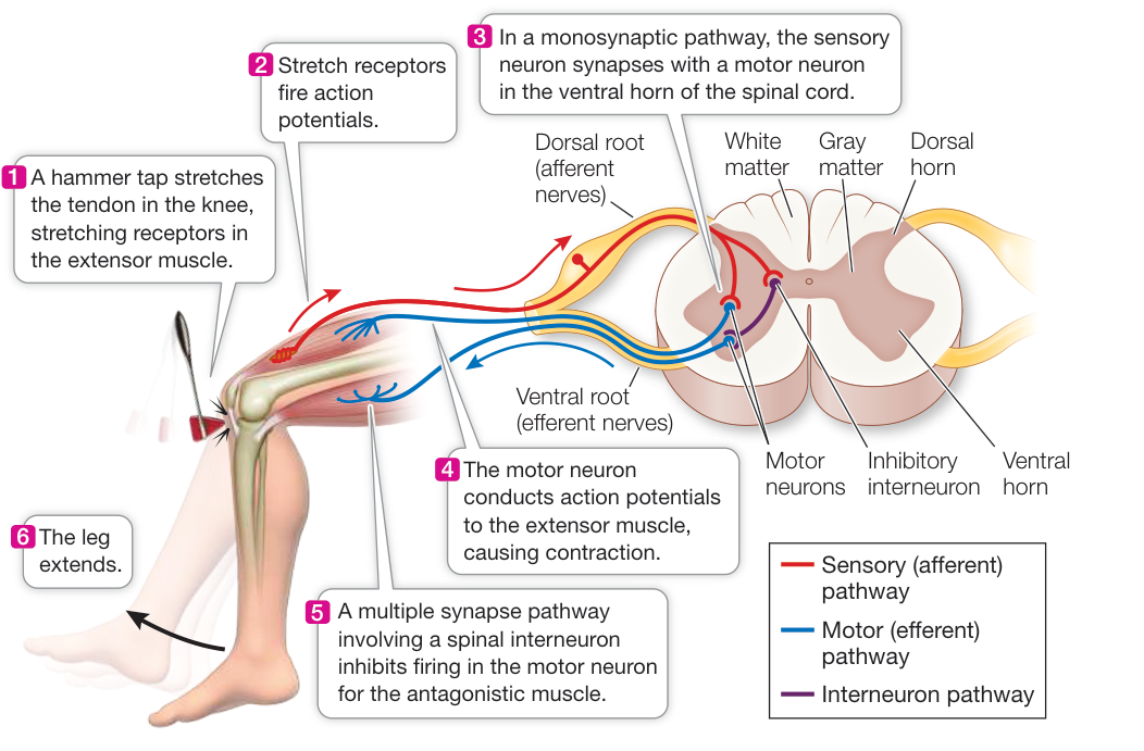
Figure 43.15 A Neural Network in the Spinal Cord Generates the Knee-Jerk Reflex Sensory (afferent) information enters the spinal cord through the dorsal horns, and motor (efferent) output leaves it via the ventral horns. Information travels to the brain in white matter tracts. Interneurons make connections within the spinal cord that result in a complex, coordinated behavior pattern. Animation 43.4 Information Processing in the Spinal Cord
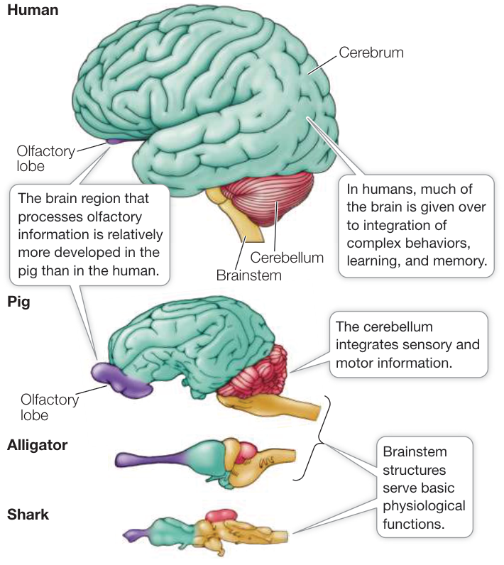
Figure 43.16 Brains Vary in Size and Complexity The brains of four vertebrate species—all of which may have a similar body mass—show immense differences. Note that the brainstem, which is involved in physiological regulation and stereotypic behavior, differs less among these species than does the cerebrum, which is responsible for complex behavior and learning. Q: What sensory capability do you think is well developed in the pig?
The withdrawal reflex is an example of a polysynaptic spinal reflex that involves many interneurons. When you step on a tack, you immediately pull back your foot, but you don’t fall over because many other muscles change your posture. The tack stimulates pain receptors in the foot, and the sensory neurons transmit APs into the dorsal horn of the spinal cord on the same side of the body. In the dorsal horn, these neurons synapse with a variety of interneurons. Some send the pain information through their axons to the brain, resulting in the conscious sensation of pain. But even before the brain is aware of the pain, other interneurons coordinate the withdrawal of the foot with actions of other muscles to shift your weight onto the other leg. Thus a rather complex suite of movements is coordinated by a network of interneurons in the spinal cord. By extension, you can appreciate how much more complex the neural networks are that enable you to execute complex movements in time with music and coordinated with another person—in other words, to dance.
The vertebrate brain is the seat of behavioral complexity
Vertebrates differ greatly in their behavioral complexity and in their physiological specializations, and their neural networks reflect this diversity. Figure 43.16 shows the brains of four vertebrate species of similar body mass drawn to the same scale.
The current estimate for the number of neurone in the human brain is . A given neuron in the brain can have 1,000 or
Investigating Life
QA Can learning be restored in mouse models of Down syndrome?
Some neurotransmitter-receptor combinations excite activity in the postsynaptic neuron, depolarizing it and making it likely to fire an AP. Other neurotransmitter-receptor combinations inhibit the responses of the postsynaptic cell. Neurons integrate information by summing excitatory and inhibitory synaptic inputs. Overinhibition in the nervous system can arise when too much inhibitory neurotransmitter (such as GABA) is released, or if postsynaptic cells have too many receptors for inhibitory neurotransmitters. Drugs that decrease the synthesis and release of the inhibitory neurotransmitter can reduce the inhibition, as can drugs that block the receptors for such neurotransmitters. In the case of the Down syndrome model mice, drugs such as pentylenetetrazole, which blocked the GABA receptors (which function also as Cl- channels), reduced the level of inhibition in the mouse nervous system and increased the ability of the mice to learn and form memories.
Future directions
Going from experiments on the effects of drugs on mice to a therapy for humans is a difficult path. To protect patients, the federal Food and Drug Administration (FDA) has a rigorous procedure for approving drugs for human use. After a candidate drug is shown to be effective and safe in animal models, four stages of approval must be passed before a drug is allowed on the market. In Phase 0, the first in-human trials are conducted on a small group of volunteers to study how the body absorbs, distributes, metabolizes, and excretes the drug, and whether it has any obvious side effects. In Phase 1, trials are conducted in a small group of volunteers to evaluate safety and dosage, and again to look for side effects. Phase 2 involves a larger group of subjects who have the disease that the drug is expected to treat, to see if it is effective and safe. The comparison is with a placebo. In Phase 3, trials are conducted with a large group of patients to assess the drug’s effectiveness and safety, and to compare it with other commonly used treatments. Each of these phases must be reviewed by the FDA and approved before proceeding to the next phase.
more synapses. Thus the human brain can contain synapses ( neurons synapses per neuron). Then there are the glia currently estimated to be about the same number as neurons. A single astrocyte might participate in thousands of synapses while at the same time monitoring signals in the extracellular fluid and the blood. In addition, synapses are not constant but can be highly plastic. They can increase or decrease in number and size, and they can become more or less sensitive.
This astronomical number of neurons and synapses is divided into thousands of distinct but interacting networks that function in parallel. The possible number of information networks in the brain is almost infinite, and therein lies the incredible ability of the human brain to process information, to learn, to do complex tasks, to remember, and to have emotions.
Recap and Assess
Nervous systems are composed of neural networks that include afferent neurons, interneurons, and efferent neurons. Nervous systems range in complexity from simple nerve nets to the human brain. The spinal knee-jerk reflex is an example of a simple neural network, but networks that control more complex behavior are much more complicated.
- What are ganglia, and why are they concentrated in the anterior region of many invertebrates?
- Draw or describe the flow of information from stimulus to response in the knee-jerk reflex circuit.
- In the knee-jerk reflex circuit, how can the same stimulus cause contraction in one muscle and relaxation in another?

Courtesy of Philip Haydon
Visual Summary
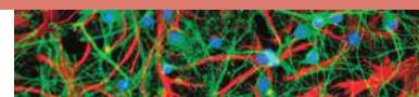
You should be able to relate each summary to the adjacent figures. If you go to this visual summary in Achieve, you can follow links to figures, animations, activities, and simulations that will help you consolidate the material.
Neurons and Glia Are Unique Cells of Nervous Systems
- ■ Neurons generate and conduct electric signals, and most can generate action potentials (APs). Glia support and modulate the activities of neurons but do not generate APs.
- ■ A neuron receives information via its dendrites and transmits information via its axon.
- ■ Neurons communicate with target cells at synapses.
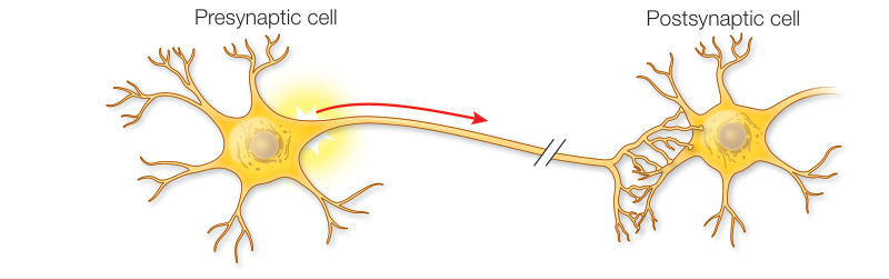
Figure 43.1
- ■ Glia include Schwann cells and oligodendrocytes, both of which generate myelin sheaths on axons.
- ■ Glia also include astrocytes, which support neurons metabolically, modulate synaptic signaling, and contribute to the blood–brain barrier and the glymphatic system.
Questions
- How do neurons integrate information from multiple sources?
- Why are neurons that communicate over chemical synapses better for integrating different inputs than are neurons that communicate over electrical synapses?
- How does the glymphatic system remove metabolic wastes from the brain?
Neurons Generate and Transmit Electric Signals
- ■ The sodium–potassium ( – ) pump concentrates on the inside of a neuron and on the outside. Permeability of the membrane to allows to diffuse out, leaving behind unbalanced negative charges, creating the resting potential.
- ■ The resting potential is altered by the opening or closing of ion channels that depolarize or hyperpolarize the membrane, producing a graded membrane potential.
- ■ An AP is a rapid, all-or-none, transient reversal in charge across a portion of the cell membrane resulting from the sequential opening and closing of voltage-gated and channels.
- ■ APs are conducted down non-myelinated axons because local current brings adjacent regions of membrane to threshold. These events jump between nodes of Ranvier in myelinated axons.
Questions
- Why is there a negative electric potential across neuronal membranes?
- Why does myelination increase AP conduction speed?
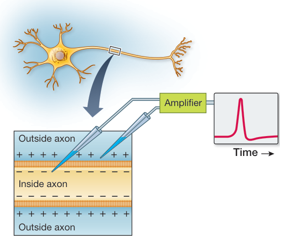
Figure 43.9
Neurons Communicate with Other Cells
- ■ Neurons communicate with other cell types at electrical synapses or chemical synapses.
- ■ The neuromuscular junction is a chemical synapse between a motor neuron and a skeletal muscle cell. Its neurotransmitter is acetylcholine (ACh), which causes depolarization of the postsynaptic motor end plate.
- ■ At chemical synapses, APs cause the release of neurotransmitters, which diffuse across the synaptic cleft and bind to postsynaptic receptors.
- ■ Synapses between neurons can be either excitatory or inhibitory, and postsynaptic neurons can sum excitatory and inhibitory postsynaptic potentials both spatially and temporally.
- ■ There are many different neurotransmitters and types of receptors. The action of a neurotransmitter depends on the type of receptor to which it binds.
- ■ Neurotransmitter molecules must be cleared from the synapse to turn off responses in the postsynaptic cell.
- How does an AP cause release of neurotransmitter at the neuromuscular synapse?
- How do graded membrane potentials relate to integration of information by postsynaptic neurons?
- What are four ways that neurotransmitter action can be terminated?
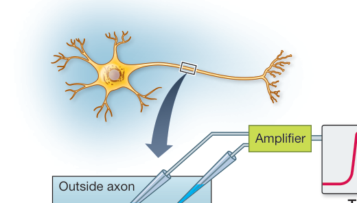
Figure 43.12
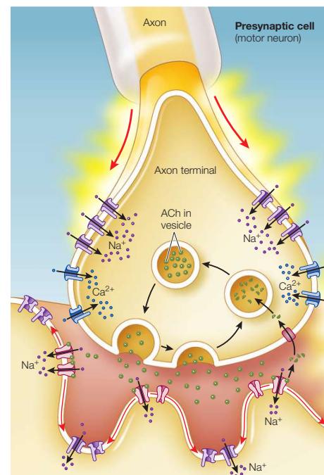
Neurons and Glia Form Information-Processing Circuits
- ■ In vertebrates, the brain and spinal cord form the central nervous system, which communicates with the rest of the body through the peripheral nervous system.
- ■ The CNS increases in complexity from invertebrates to vertebrates and from fishes to mammals.
- ■ Neural networks include afferent neurons and efferent neurons, generally connected through interneurons.
- ■ A spinal reflex is a simple neural network.
Questions
- What are the differences between a nerve net, a ganglion, and a brain?
- When considering fishes, then reptiles, then mammals, which part of the vertebrate brain increases most in size?
- What are the roles of interneurons in the withdrawal reflex?
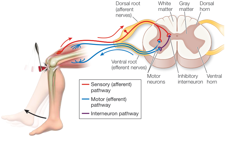
Figure 43.15