Chapter 45The Mammalian Nervous System: Structure and Higher Functions
Key Concepts
45.1 Functions Are Localized in the Nervous System
45.2 Nervous System Functions Rely on Neural Circuits
45.3 Higher Brain Functions Involve Integration of Multiple Systems
The extraordinary ability of taxicab drivers in London to navigate its maze of streets and byways prompted a study that revealed London cabbies to have larger than normal posterior hippocampi—a brain region implicated in the memory of spatial relationships in the environment.

© Jason Hawkes/Getty Images
Investigating Life: The Brains of Taxi Drivers
Compare Google maps of London and New York City at the same scale. In which city do you think it is easier to drive a taxi? Eleanor Maguire at University College London was so impressed with the navigational abilities of London taxi drivers that she investigated whether there was anything “special” about their brains. Using magnetic resonance imaging, Maguire and her colleagues examined the brains of taxi drivers with varying numbers of years of experience and compared them with each other and with the brains of control subjects who were not taxi drivers. The studies revealed significant differences in the sizes of a brain region called the hippocampus.
The hippocampus is involved in learning and memory. The posterior hippocampus in particular is implicated in the memory of spatial relationships among objects in the environment. Maguire found that the posterior hippocampi of taxi drivers were larger than those of control subjects and that, among the cab drivers, there was a positive correlation between the size of the posterior hippocampus and years of driving experience.
In Matt Wilson’s lab at the Massachusetts Institute of Technology, researchers record the activity of hippocampal neurons of rats running in a maze. Specific neurons called “place cells” fire action potentials only when the rat is at a particular location in the maze. Thus when the rat runs the maze from beginning to end, a sequence of cell firing is recorded. Sometimes when the rat is resting, the same sequence is recorded but is speeded up about 20 times, or the reverse sequence is recorded. It seems as if the recordings are reflecting what the resting rat is thinking—where it has been, where it is going to go. In a maze with a choice point (go right or go left), the sequence recorded when the rat is resting at the start position frequently predicts which direction it will turn.
The hippocampus is important for acquiring memories and consolidating them into long-term memory. We also know that sleep is important for learning. What is happening in the hippocampus during sleep to promote memory?
QA Can recordings of hippocampal place cells reveal processes of memory consolidation during sleep? (Find the answer on page 1023.)
Functions Are Localized in the Nervous System
Learning Objectives
- 45.1.1 Classify the types of afferent information to and efferent information from the nervous system.
- 45.1.2 Explain the developmental relationships between the major brain regions, starting with the forebrain, midbrain, and hindbrain and ending with structures such as the amygdala, hypothalamus, primary motor cortex, reticular-activating system, and other components of the adult brain.
- 45.1.3 Sketch and/or list the five lobes of the cerebrum and identify the primary sensory and motor areas.
- 45.1.4 Describe the relationship between body size and brain size in humans and other vertebrates.
The organization of the mammalian nervous system can be described anatomically and functionally. In anatomical terms, all vertebrate nervous systems consist of three parts: a brain, a spinal cord, and a set of peripheral nerves that reach all parts of the body. As discussed in Key Concept 43.4, the brain and spinal cord are the central nervous system, or CNS, and the neurons that connect the CNS to all the tissues and sensors of the body are the peripheral nervous system, or PNS. An additional division of the nervous system exists in the gut; we will discuss this enteric nervous system in Chapter 49.
Recall from Key Concept 43.1 that a neuron is an electrically excitable cell that communicates via an axon. When used in the context of a nervous system, the term nerve refers to a bundle of axons in the PNS that carry information about many things simultaneously. Some axons in a nerve may be carrying information to the CNS while other axons in the same nerve are carrying information from the CNS to the body’s organs. An understanding of the functional organization of the nervous system requires knowledge of these paths of information flow. In this chapter we divide the anatomy of the mammalian brain, spinal cord, and PNS into smaller, discrete functional units.
Functional organization is based on flow and type of information
Figure 45.1 illustrates the major avenues of information flow through the human nervous system. The white boxes identify four divisions of the PNS; two of these bring information from the periphery to the CNS, and two transmit information from the CNS to the periphery.
The afferent portion of the PNS (see Figure 45.1, left) carries information from sensory receptor cells to the CNS. We are conscious of much of this information (e.g., light, sound, skin temperature, limb position), but we are usually unconscious of the information involved in physiological regulation (e.g., blood pressure, deep body temperature, blood oxygen levels).
- The efferent portion of the PNS (see Figure 45.1, right) carries information from the CNS to the muscles and glands of the body. Efferent pathways are divided into a voluntary division that executes our conscious movements; and an involuntary, or autonomic, division that controls physiological functions.
In addition to the neural information it receives from the PNS, the CNS receives chemical information from hormones circulating in the blood. In turn, neurohormones released by neurons enter the circulation and affect neurons and other cells distant from the site of release (see Key Concept 39.1).
The anatomical organization of the CNS emerges during development
Early in the development of a vertebrate embryo, a tube of nervous tissue forms (see Key Concept 42.4). At its anterior end, this neural tube forms three swellings that become the forebrain, midbrain, and hindbrain. The rest of the neural tube becomes the spinal cord. Peripheral nerves sprout from the midbrain and hindbrain (the cranial nerves) and from the spinal cord (the spinal nerves). From these early stages we see the linear axis of information flow in the nervous system. Although the developing brain will fold and become a complex structure, the information flow in the adult nervous system will follow paths that emerge from the simple, linear neural tube.
Each of these three regions of the embryonic brain develops into several structures in the adult brain (Figure 45.2). From the embryonic midbrain come structures that integrate information from the different senses and coordinate motor responses. From the hindbrain come the medulla, the pons, and the cerebellum. The medulla is continuous with the spinal cord, the pons is anterior to the medulla, and the cerebellum is a dorsal outgrowth of the pons. The medulla and pons contain distinct groups of neurons involved in controlling physiological functions such as breathing, circulation, and basic motor patterns such as swallowing and vomiting. All information traveling between the spinal cord and higher brain areas must pass through the pons, the medulla, and the midbrain, which are collectively known as the brainstem.
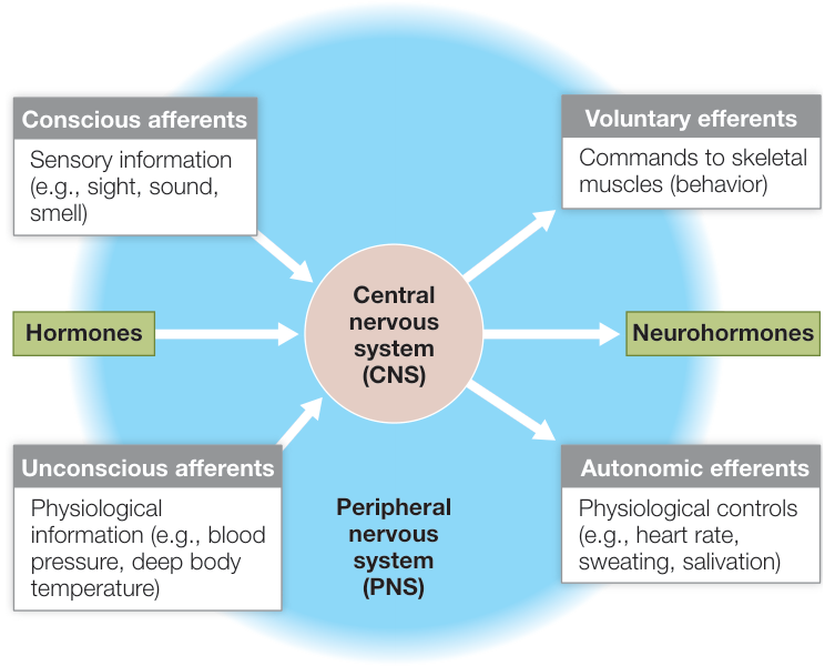
Figure 45.1 Organization of the Nervous System The peripheral nervous system carries information to (afferent) and from (efferent) the central nervous system (center circle in the diagram). The CNS also receives hormonal inputs and produces hormonal outputs (see Key Concept 39.2). Q: How do hormonal inputs reach the CNS and hormonal outputs leave the CNS?
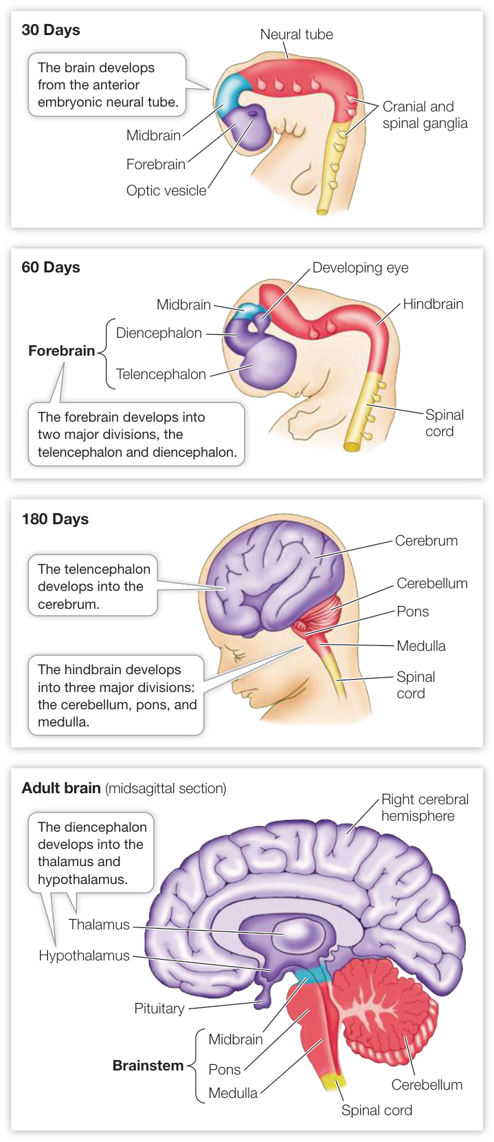
Figure 45.2 Development of the Central Nervous System In vertebrate embryos, the anterior end of the hollow neural tube differentiates into forebrain, midbrain, and hindbrain. Each of these regions develops into several structures of the adult brain. The remainder of the neural tube becomes the spinal cord.
One function of the cerebellum is coordinating muscle activity and maintaining balance. It is like the director of a movie; the cerebellum receives a “script” of the commands going to the muscles from higher brain areas, and it receives information about the actual performance coming up the spinal cord from the “actors”—the joints and muscles. The cerebellum compares the “script” with the performance and refines motor commands accordingly. Damage to the cerebellum results in loss of fine motor control and coordination.
The embryonic forebrain develops a central region called the diencephalon and a surrounding structure called the telencephalon. The diencephalon is the core of the forebrain and consists of an upper structure, the thalamus, and a lower structure, the hypothalamus. The thalamus is the final relay station for sensory information going to the telencephalon. The hypothalamus receives a lot of physiological information of which we are not conscious, and it uses that information to regulate many physiological functions and biological drives. A major function of the hypothalamus is to control the pituitary gland (see Key Concept 39.2).
Connect the Concepts Key Concept 39.2 explains how the endocrine system and nervous system work together.
The embryonic telencephalon gives rise to the cerebrum, consisting of the left and right cerebral hemispheres. The outer layer of the cerebrum is the cerebral cortex, a thin layer rich in cell bodies. If we compare vertebrate groups from fishes through amphibians, reptiles, and mammals, the cerebrum increases in size, complexity, and importance—an evolutionary trend called telencephalization (see Figure 43.16). In humans, the cerebrum is by far the largest part of the brain and plays major roles in sensory perception, learning, memory, and conscious behavior.
The spinal cord transmits and processes information
The spinal cord conveys information to and from the brain. However, the spinal cord is more than an information pipe. As you saw in Key Concept 43.4, the spinal cord carries out integrative functions as well. The knee-jerk reflex (see Figure 43.15) is an example of a circuit between the PNS and the spinal cord that controls a behavioral function. That simple circuit can be built on to control more complex behaviors such as the withdrawal reflex, which involves readjusting tension in many muscles on both sides of the body to coordinate movement and maintain balance.
The brainstem carries out many autonomic functions
Swallowing, salivating, breathing, eye movements, blood pressure regulation, and gut activity are only a few of the many autonomic functions that are localized in the medulla, the pons, and the midbrain. To carry out these functions, the brainstem has its own components of the PNS, the 12 paired cranial nerves. You encountered the olfactory nerve, the optic nerve, and the auditory nerve (cranial nerves I, II, and VIII) in Chapter 44. Another one, cranial nerve X, is called the vagus (“wandering”) nerve because it travels into the body cavity and communicates with many of the organs, including the heart and the gut. You will encounter the vagus nerve in subsequent chapters.
Anatomically distinct groups of neurons in the CNS that work together as a unit are called a nucleus (not to be confused with the nucleus of a single cell). Axons from the neurons of many brainstem nuclei extend to various regions of the brain and modulate brain activity; for example, brainstem nuclei are involved in keeping the higher brain areas awake or allowing them to sleep. All of the sensory information coming up the neural axis from the spinal cord passes through the brainstem on its way to the forebrain, and many of these ascending neuronal tracts give off branches to the awake-promoting nuclei in the brainstem. Because the neuronal circuitry in this part of the brain is so complicated and because activity in these ascending sensory pathways can promote wakefulness, the core of the brainstem has been termed the reticular-activating system (“reticular” means netlike). Damage to the brain or spinal cord below the reticular-activating system can result in paralysis but leave sleep—wake cyclic behavior normal. Damage above the level of the reticular-activating system can interrupt ascending wake-promoting activity, resulting in a coma.
The core of the forebrain controls physiological drives, instincts, and emotions
As mentioned earlier, the diencephalon consists of the thalamus and the hypothalamus. The thalamus communicates sensory information to the cerebral cortex; the hypothalamus receives information about physiological conditions in the body and regulates many homeostatic functions. Key Concept 38.5 described how the hypothalamus is involved in regulating body temperature, and Key Concept 39.2 discussed the intimate association between the hypothalamus and the pituitary gland in the control of many homeostatic functions.
The forebrain of vertebrates includes a phylogenetically old, complex set of structures called the limbic system (Figure 45.3). The limbic system is responsible for some basic instincts and drives such as hunger and thirst, sexual behavior, and emotions. Within the limbic system there are areas that, when stimulated with small electric currents, can cause intense sensations of pleasure, pain, or rage.
Pleasure and pain centers in the limbic system play roles in learning and in physiological drives. One component of the limbic system—the amygdala—is involved in fear and fear memory. If a certain portion of the amygdala is damaged or chemically blocked, an animal becomes tame and cannot learn to be afraid of a situation that would normally induce a strong fear reaction.
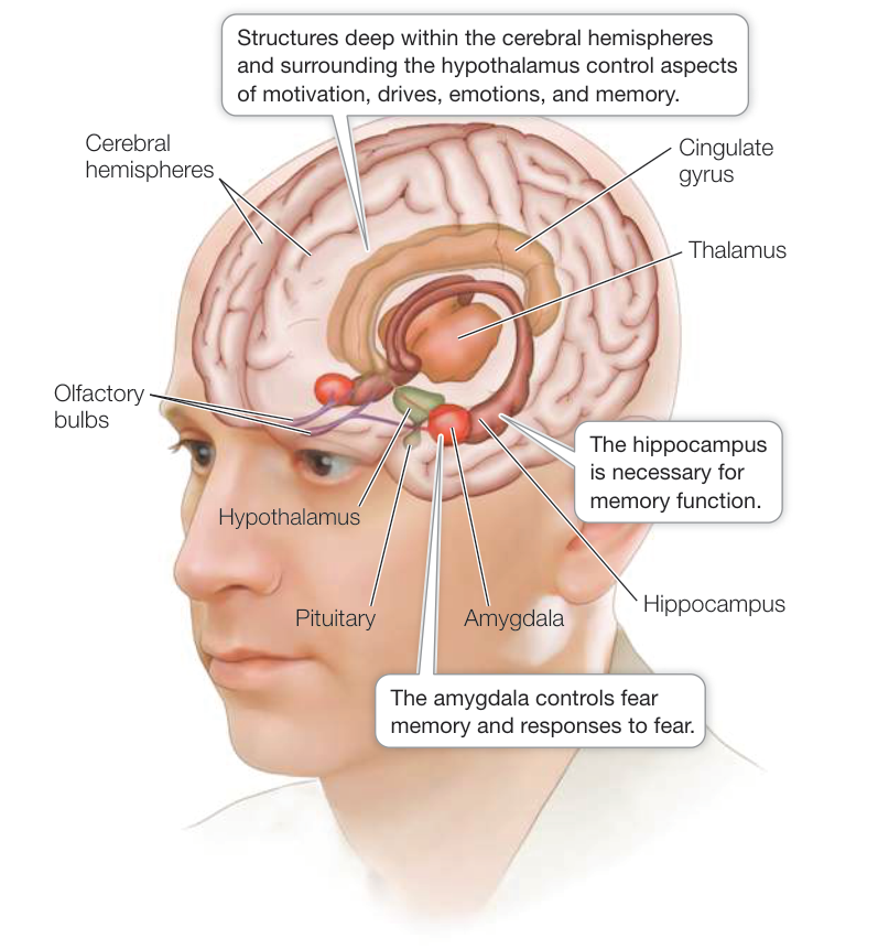
Figure 45.3 The Limbic System The evolutionarily primitive parts of the forebrain are referred to as the limbic system. The hippocampus is involved in forming long-term memory. The amygdala triggers fear emotions and fear memories.
The amygdala is involved in post-traumatic stress disorder (PTSD). Another part of the limbic system, the hippocampus, is involved in the transfer of certain types of short-term memory to long-term memory, as discussed in the chapter opener and in Investigating Life: Place Cells Reveal Processes of Memory Consolidation during Sleep.
The cerebrum is responsible for complex behavior and consciousness
The cerebrum is the dominant structure in the mammalian brain. In humans it is so large that it covers all other parts of the brain except the cerebellum (Figure 45.4A). Layers of cells at the surface of the cerebrum form the cerebral cortex, which is about 4 millimeters thick and is folded into ridges (gyri; singular gyrus) and valleys (sulci; singular sulcus). If the cortex were flattened out, it would be about 1 square meter, but the foldings, or convolutions, enable that extensive surface area of cortex to fit within the skull.
As we explore the infoldings of the cerebral cortex and other parts of the brain, we will occasionally mention an individual whose brain was damaged by an accident or other unfortunate event. Until about 50 years ago the study of such individuals was the main source of functional information about the human brain, but developments of imaging technologies such as positron emission tomography (PET) and magnetic resonance imaging (MRI) greatly expanded opportunities to study the human brain. Advances in imaging technologies are contributing to rapid progress in neurobiology.
Investigating Life: Place Cells Reveal Processes of Memory Consolidation during Sleep
Experiment
Original Paper: A. K. Lee and M. A. Wilson. 2002. Memory of sequential experience in the hippocampus during slow wave sleep. Neuron 36: 1183–1194.
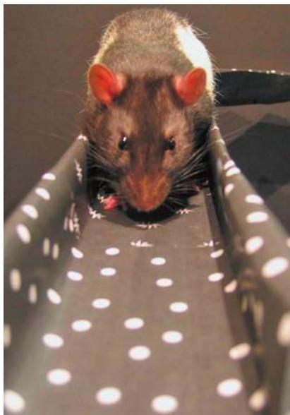
Multiple hippocampal cells were recorded while a rat ran a maze. Individual cells fired only when the rat was at particular locations in the maze; thus the rat’s progress through the maze can be represented by a specific sequence of firing of these “place cells.” Since memory is consolidated during sleep, are the sequences of place cells expressed during the maze-running experience replayed and processed into long-term memory during sleep?
HYPOTHESIS Memory of the maze-running experience is represented by a sequence of firing of hippocampal place cells. This sequence is repeated during sleep as part of the process of memory consolidation.
METHOD
- Record from multiple hippocampal neurons while a rat is running through a maze.
- Identify sequences of cell firing that reoccur whenever the rat runs the maze.
- Continue to record the neuronal activity during sleep and determine if similar sequences are occurring with a probability greater than expected from random activity in that group of cells.
Courtesy of Mark Wattson and Linda Lee
RESULTS
1 After 30 runs through the maze, a group of nine place cells are identified that fire in the same order correlating with position in the maze.
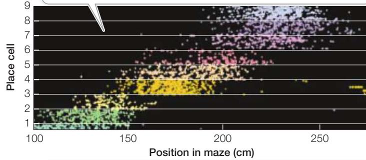
2 The peak firing rate of each neuron can be calculated as a function of position in the maze. When the neurons are arranged in the temporal order of their peak firing rates, you can think of each neuron as being a “letter” in a “word” that codes spatial information about the maze.
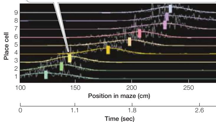
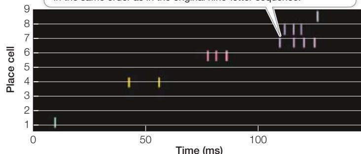
3 Subsequent recordings while the animal is awake, at rest, or asleep are analyzed for the occurrence of the same or similar “words”—complete or partial sequences of place-cell activity in the same order as the base sequence. Thus a similar word to a nine-letter (nine-neuronal) sequence might be a sequence of seven cells firing in the same order as in the original nine-letter sequence.
CONCLUSION
- Sequences of hippocampal-cell firing that correspond to locations in the maze were highly repeatable and probably represent the coding of that place information in the hippocampus.
- During sleep, bursts of cell firing sometimes occurred that correspond wholly or partially to the sequences recorded when the rat was running in the maze.
Work with the Data
To conclude that a particular sequence of cell firing observed during sleep is a significant event—meaning that it could be a replay of the waking experience—we have to compare it with the probability that it would occur by random chance. And we need to consider whether an observed sequence that includes some but not all of the components of the baseline “word” is nevertheless a significant event.
What is the minimum number of elements that would have to match the baseline sequence for it to be considered a significant event with ? To find out, we need to calculate the probabilities of the recorded neurons firing randomly and creating the sequence matches. First, let’s consider the probability of a recurrence by chance of the exact nine-“letter” baseline sequence being recorded in a single burst of that group of nine cells. The total number of possible combinations of those nine letters can be calculated as the factorial :
In other words, the probability of repeating the entire nine-letter sequence would be 1 out of 362,880. That repeat would have an extremely low probability of occurring and would therefore be a highly significant event. But what if the recorded sequence has only six of the nine cells firing in the proper order? How do you determine the probability of that? First, calculate the probability of a combination of six cells out of the nine. So for the first cell you have nine choices, for the second you have eight choices, and so forth. Then you have to calculate the probability of that combination of six cells being in the proper sequence, and that will be 1 out of the total number of possible arrangements of those six cells, or . Then what do you do for probabilities of successive events? You multiply the probabilities. For example, if you flip a coin, the probability of heads is . If you flip it twice, the probability of getting heads both times is , or .
QUESTIONS ►
- What is the probability of the sequence 1, 4, 6, 7, 8, 9 out of a random firing of nine cells?
- What is the probability of a sequence of four letters occurring out of a random firing of nine cells?
- Are both of the recordings from Questions 1 and 2 low-probability events?
- What do you note about the time frame of the sequence during sleep versus during wakefulness?
A curious feature of the human nervous system is that the left side of the body is served (in both sensory and motor aspects) mostly by the right side of the brain, and the right side of the body is served mostly by the left side of the brain. Thus sensory input from the right hand goes to the left cerebral hemisphere, and sensory input from the left hand goes to the right cerebral hemisphere. The exception is the head, where the left side is controlled by the left cerebral hemisphere and the right side by the right cerebral hemisphere. The two hemispheres are not symmetrical with respect to all functions. Language abilities, for example, reside predominantly in the left hemisphere in most people.
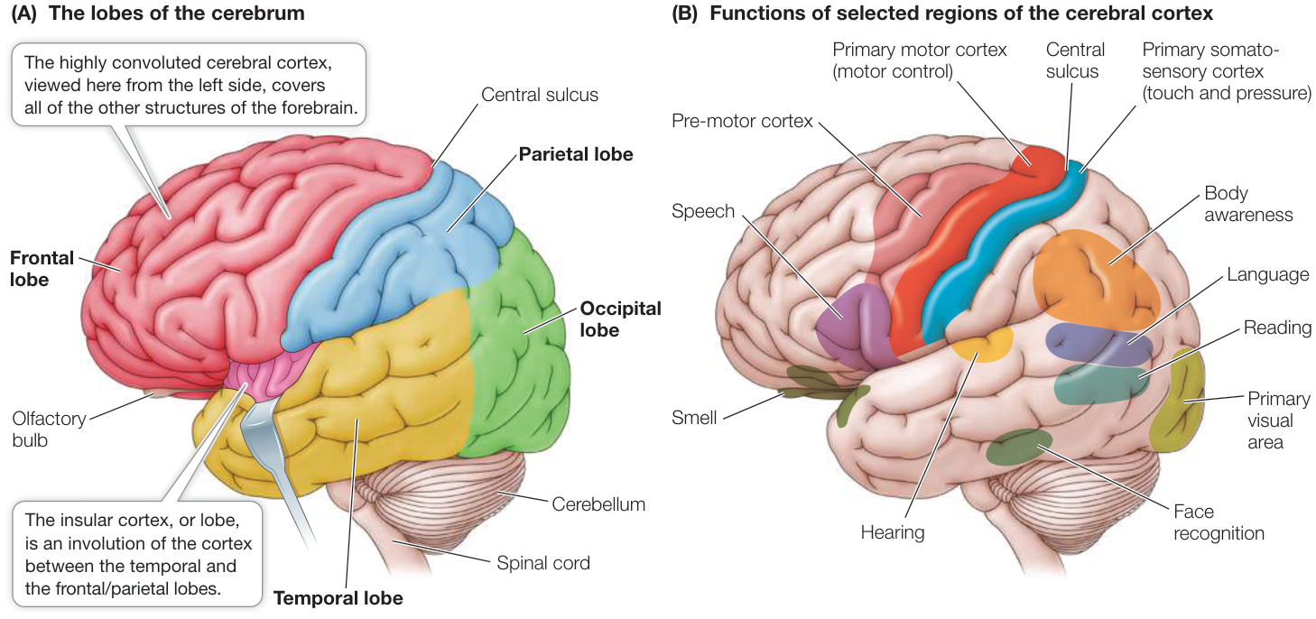
Figure 45.4 The Human Cerebrum (A) Each cerebral hemisphere is divided into frontal, temporal, parietal, occipital, and insular lobes. (B) Different functions are localized in particular areas of the five cerebral lobes.
Different regions of the cerebral cortex have specific functions (Figure 45.4B). Some of those functions are easily defined, such as receiving and processing sensory information or generating motor commands, but in humans most of the cortex is involved in higher-order information processing that is less easy to define. These latter areas are given the general name of association cortex, because they integrate, or associate, information from different sensory modalities and from memory.
To understand the cerebral cortex, it helps to have an anatomical road map. Viewed from the left side, the left cerebral hemisphere looks like a boxing glove for the right hand with the fingers pointing forward, the thumb on the side, and the wrist at the rear. The “thumb” area is the temporal lobe, the fingers the frontal lobe, the back of the hand the parietal lobe, and the wrist the occipital lobe (see Figure 45.4A). What you can’t see from an external view is the insular lobe, which is folded in between the thumb and finger areas of the boxing glove (between the frontal/parietal and the temporal lobes). The right cerebral hemisphere shows a mirror image of this arrangement. We will look at each lobe separately.
THE TEMPORAL LOBE The upper region of the temporal lobe receives and processes auditory information. The association areas of this lobe are involved in recognizing, identifying, and naming objects. Damage to the temporal lobe results in disorders called agnosias, in which the individual is aware of an object but cannot identify it.
Damage to a specific region of the temporal lobe results in the inability to recognize faces. Even old acquaintances cannot be identified by facial features, although they may be identified by other attributes such as voice, body features, and posture. Damage to other association areas of the temporal lobe can cause deficits in understanding spoken language, although speaking, reading, and writing abilities may be intact.
THE FRONTAL LOBE The frontal and parietal lobes are separated by a deep valley called the central sulcus. A strip of the frontal lobe cortex just in front of the central sulcus is called the primary motor cortex (see Figure 45.4B). The neurons in this region control muscles in specific parts of the body; the parts of the body map onto the primary motor cortex, with the head represented in the lower outside region and the legs and feet in the top region near the midline. The graphic drawing of body parts overlying the diagram of the cortex is called a homunculus, and its distortion represents disproportionate innervation (Figure 45.5). Parts of the body with fine motor control, such as the face and hands, have disproportionate representation (see Figure 45.5A). Electrical stimulation of neurons in different regions of the primary motor cortex causes specific muscles of the body to twitch. Just anterior to the primary motor cortex is the pre-motor cortex, which is involved in planning more complex and coordinated movements.
The association functions of the frontal lobe are diverse and best described as having to do with feeling and planning. They are said to have executive function and they contribute significantly to personality. People with frontal lobe damage have drastic alterations of personality and difficulty planning future events. A dramatic case of frontal lobe damage is that of Phineas Gage, who in 1848 was an industrious and responsible young railroad construction foreman. Then a blasting accident shot a meter-long, 3-centimeter-wide iron tamping rod through his brain. The rod entered Gage’s head below his left eye, passed through his frontal lobe, and exited the top of his head (Figure 45.6).
Remarkably, Gage survived, but he had a different personality. In the years following his recovery, he was quarrelsome, impatient, obstinate, and used profane language, which he did not do before. He lost his railroad job and earned money by telling his story and exhibiting his scars (and the tamping iron). Several years later, however, he took a job as a stage coach driver in Chile—a challenging job indicating considerable behavioral recovery. He died of a seizure in 1860, at the age of 38. If you are in Boston, you can pay him a visit—his skull, death mask, and the tamping iron are on display in the Warren Anatomical Museum of Harvard Medical School.
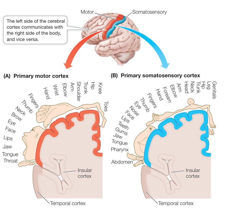
Figure 45.5 The Body Is Represented in Primary Motor and Primary Somatosensory Cortexes Neurons in the primary motor cortex (A) control muscles in specific parts of the body, while neurons in the primary somatosensory cortex (B) receive information from specific parts of the body. The locations of these neurons within each cortex correspond to “maps” on which regions of the body are represented in proportion to the amount of innervation they receive. (After W. Penfield and T. Rasmussen 1950. The Cerebral Cortex of Man: A Clinical Study of Localization of Function. The Macmillan Company: New York.)
THE PARIETAL LOBE The strip of parietal lobe cortex just behind the central sulcus is the primary somatosensory cortex (see Figure 45.4B). This area receives touch and pressure information relayed from the body through the thalamus.
As with the primary motor cortex, the entire body surface can be mapped onto the primary somatosensory cortex (see Figure 45.5B). Areas of the body that have a high density of tactile mechanoreceptors and are capable of making fine discriminations in touch (such as the lips and fingers) have disproportionately large representation. If a very small area of the primary somatosensory cortex is stimulated electrically, the subject reports feeling specific sensations, such as touch, in a localized part of the body.
A major association function of the parietal lobe is attending to complex stimuli. Damage to the right parietal lobe causes a condition called contralateral neglect syndrome, in which the individual tends to ignore stimuli from the left side of the body or the left visual field. Such individuals have difficulty performing complex tasks, such as dressing the left side of the body; an afflicted man may not shave the left side of his face unless he is reminded to do so. When asked to copy simple drawings, a person who exhibits this syndrome can do well with the right side of the drawing but not the left. When reading, the person will tend to start from the middle of the page rather than the left margin of the page.
The parietal cortex is not symmetrical with respect to its role in attention. Damage to the left parietal cortex does not cause the same degree of neglect of the right side of the body. You will see similar asymmetries in cortical function later in the chapter when we discuss language.
THE OCCIPITAL LOBE The occipital lobe receives and processes visual information. The association areas of the occipital cortex are essential for making sense of the visual world and translating visual experience into language. Some deficits resulting from damage to these areas are specific. In one case, a woman with limited damage was unable to see motion. Her vision was intact, but she could see a waterfall only as a still image, and an approaching car only as a series of a stationary object at different distances.
THE INSULAR CORTEX The insular cortex, buried deep in the forebrain, receives a great variety of afferent information. The posterior regions receive somatosensory information (e.g., touch, pain, temperature); the middle region combines that with autonomic regulatory information and drives (e.g., hunger, thirst, sex); and the anterior region, which is well developed in more complex primates, elephants, and marine mammals, receives information about social interactions. Thus in mammals the insular cortex appears to integrate physiological information from all over the body to create a sensation of how the body “feels,” and in more complex mammals it may extend that function into a sense of self.
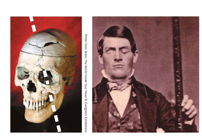
Figure 45.6 A Mind-Altering Experience Phineas Gage miraculously survived a nineteenth-century railroad construction accident that blew an iron rod through his brain. His personality, however, was permanently altered from that of a responsible foreman to a sometimes quarrelsome drifter.
The size of the human brain is off the curve
Humans are sometimes called “big-brain primates,” and that is an accurate characterization. Across vertebrate species there is a correlation between body size and brain size (Figure 45.7). More complex primates such as chimpanzees, baboons, and gorillas all fall above this regression line, but humans stand out because they are so far above the regression line. Gorillas are much larger than humans, but they have smaller brains. Elephants and whales have large brains, but they fall closer to the regression line. Porpoises and humans stand out as having much larger brains than would be predicted by their body sizes.
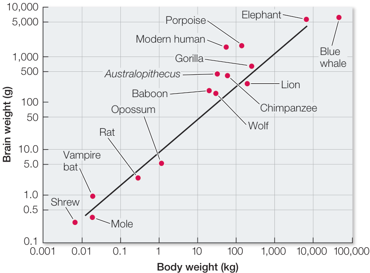
Figure 45.7 Evolution of the Human Brain Brain size scales to body size across a wide range of vertebrates. The more complex primates have larger brains than predicted by the correlation, and humans stand outside this relationship with much bigger brains. The increase in brain size in humans is due mostly to an increase in the cerebral cortex. The human brain is also highly convoluted, and more of it is devoted to associative functions. (After H. J. Jerison. 1969. Am Nat 103: 575–588; data from G. Crile and D. P. Quiring. 1940. Ohio J Sci 40: 219–259.)
Connect the Concepts Key Concept 31.5 described how human brains became larger as jaws became smaller.
The correlation of brain size to body size does not tell the whole story of human brain evolution, however. In Figure 43.16, which compares the brains of four vertebrates, we see that in mammals the forebrain is larger than other brain regions, and another feature is the degree of convolution of the cortex. Since the cortex is a layered, two-dimensional array of neurons, the area of cortex is increased by convolutions, which are greatest in humans. And finally, the percent of the cortex that is association cortex (i.e., devoted to the integration of information) is by far the greatest in humans. It is these evolutionary changes, primarily in the cortex, that provide the resources for the intellectual capacity of humans—a topic to which we will return at the end of the chapter.
Recap and Assess
The central nervous system communicates with the rest of the body through the peripheral nervous system. We are conscious of some sensory information coming into the CNS, but we are not conscious of other afferent information used in physiological regulation. The brain develops from the anterior region of the embryonic neural tube. The forebrain gives rise to the cerebrum, which is divided into five lobes. Specific functions such as somatosensation, vision, hearing, motor control, smell, and speech are localized in specific cortical areas. At the center of the cerebrum is the limbic system, which contributes to the processing of emotions, inborn responses, and some types of memory. Underneath the cerebrum is the hypothalamus, responsible for many autonomic functions, and the thalamus, which directs ascending information to the cortex. The midbrain and hindbrain constitute the brainstem, within which are many nuclei that support autonomic functions, including sleep and wakefulness. Evolution of the human brain has resulted in a greatly increased cerebral cortex devoted to integration of information.
- Given your understanding of afferent and efferent systems operating
in the nervous system, give an example of a brain-body feedback loop
that influences each of the following:
- Voluntary behavior through a conscious sensory stimulus.
- Involuntary response through a conscious sensory stimulus.
- Involuntary response through an unconscious stimulus.
- What is significant about the location of the reading and language areas of the cerebral cortex as shown in Figure 45.4B?
- Key Concept 44.3 described the two-point spatial discrimination test. How do the typical results of that test relate to Figure 45.5B?
- The brain size of humans is significantly above the regression line for mammalian brain size as a function of body size. Why is this observation an understatement of the difference in cerebral cortical capacity in humans as compared with other mammals?
Having briefly described the structure and function of different regions of the nervous system, we now explore some examples of how information is processed by the neural circuitry in some specific brain regions.
Nervous System Functions Rely on Neural Circuits
Learning Objectives
- 45.2.1 Predict the outcome on effector organs when specific peripheral nerves of the sympathetic or parasympathetic divisions of the autonomic nervous system are damaged or stimulated.
- 45.2.2 Explain how receptive fields are generated in the retina through the interactions among photoreceptors, bipolar cells, ganglion cells, horizontal cells, and amacrine cells.
- 45.2.3 Describe how neural connections between the eyes and the visual cortex produce three-dimensional vision.
Specific functions are localized in specific parts of the nervous system and depend on the neural circuits, or networks, in those structures. A major focus of modern neuroscience is to understand how the various functions of the nervous system, ranging from simple reflexes to complex learning and memory, are accomplished by the interactions of neurons in circuits. Two extensively studied examples of how neuronal networks process information are the autonomic nervous system (an output pathway) and the visual system (an input pathway).
Pathways of the autonomic nervous system control involuntary physiological functions
The autonomic nervous system, or ANS, includes CNS and PNS components and controls many involuntary functions, such as heart rate, blood flow, sweating, and digestive activities. Its control of diverse organs and tissues is crucial to homeostasis. The ANS has two divisions, sympathetic and parasympathetic, that work in opposition to each other in their effects on most organs (Figure 45.8). The sympathetic and parasympathetic divisions are distinguished by their anatomy, neurotransmitters, and actions.
The best-known functions of the ANS are those of the sympathetic division that produce the fight-or-flight response: increasing heart rate, blood pressure, and cardiac output—all of which prepare the body for emergencies (see Figure 39.3). In contrast, the parasympathetic division slows the heart and lowers blood pressure; its actions have been characterized as “rest and digest.” It is tempting to think of the sympathetic division as speeding things up and the parasympathetic division as slowing things down, but it is not that simple; for example, the sympathetic division slows down the digestive system whereas the parasympathetic division accelerates it.
Both sympathetic and parasympathetic efferent pathways begin with cholinergic neurons (neurons that use acetylcholine as their neurotransmitter) whose cell bodies are in the brainstem or spinal cord. These cells are called preganglionic neurons because the
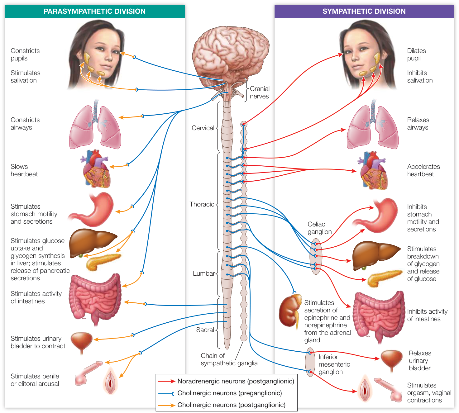
Figure 45.8 The Autonomic Nervous System The autonomic nervous system is divided into the sympathetic and parasympathetic divisions. The two divisions work in opposition to each other in their effects on most organs; one results in an increase and the other a decrease in activity.
Q: Why does a person get an extremely dry mouth when speaking in public?
second neurons in these pathways reside in collections of neurons outside the CNS called ganglia (singular ganglion). The second neurons are called postganglionic neurons because their axons extend out from the ganglia. The axon of a postganglionic neuron synapses with cells in the target organs (see Figure 45.8).
The postganglionic neurons of the sympathetic division mostly use norepinephrine as their neurotransmitter. In contrast, the postganglionic neurons of the parasympathetic division are mostly cholinergic. In organs that receive both sympathetic and parasympathetic input, the target cells respond in an opposite manner to norepinephrine and to acetylcholine. This happens, for example, in a region of the heart called the pacemaker, which initiates the heartbeat. Stimulating the sympathetic nerve to the heart or dripping norepinephrine onto pacemaker cells increases their firing rate and causes the heart to beat faster. In contrast, stimulating the parasympathetic nerve to the heart or dripping acetylcholine onto pacemaker cells decreases their firing rate and causes the heart to beat more slowly (see Figure 48.8).
The sympathetic and parasympathetic divisions of the ANS can also be distinguished by anatomy. The preganglionic neurons of the parasympathetic division come from the cranial nerves of the brainstem and the sacral (lowest) region of the spinal cord; those of the sympathetic division come from the thoracic and lumbar regions of the spinal cord (see Figure 45.8). Most of the ganglia of the sympathetic division are lined up and interconnected in two chains, one on either side of the spinal cord. The parasympathetic ganglia are close to the target organs and are not interconnected.
The ANS is an important link between the CNS and many physiological functions. Its control of diverse organs and tissues is crucial to homeostasis. Despite the complexity of the ANS, work by neurobiologists and physiologists over many decades has made it possible to understand its functions in terms of neuronal properties and circuits.
The visual system is an example of information integration by the cerebral cortex
The visual system is one of the most-studied input pathways to the CNS. Key Concept 44.4 described how light falling on the retina produces signals that are transmitted through the cellular circuits of the retina, resulting in action potentials in the optic nerve. But how does the brain use this information to represent the visual world?
RETINAL RECEPTIVE FIELDS Key Concept 44.4 described how a retinal ganglion cell collects information from several photoreceptors—an example of “convergence of information.” Each ganglion cell is communicating to the brain something more than simply the presence or absence or intensity of light falling on a portion of the retina.
The question of what information the retinal ganglion cell extracts from the photoreceptors was addressed in classic experiments by Stephen Kuffler at Johns Hopkins University. He used electrodes placed in the retinas of anesthetized cats to record the activity of single ganglion cells while stimulating their retinas with spots of light (Figure 45.9). Kuffler’s experiments revealed that each ganglion cell has a well-defined receptive field composed of a group of photoreceptor cells that receive light from a small area of the entire visual field. Stimulating these photoreceptors with light activates the ganglion cell, which sends action potentials to the thalamus and on to the visual cortex (the area of the occipital lobe where visual information is processed; see Figure 45.4). Information from many photoreceptors is therefore communicated to the brain as a single message. Individual photoreceptors may contribute to the receptive fields of multiple ganglion cells, so that receptive fields overlap.
The receptive fields of most ganglion cells are circular, but whether a spot of light falling on a receptive field excites or inhibits its ganglion cell depends both on the nature of the receptive field and on where the spot of light falls on it. Receptive fields have a center and a concentric surround, and can be either “on-center” or “off-center.” Light falling on the center of an on-center receptive field excites the ganglion cell, and light falling on the center of an off-center receptive field inhibits the ganglion cell. Light falling on the surround has the opposite effect: the surround for an on-center receptive field inhibits the ganglion cell, and the surround for an off-center field is excitatory. Thus the activity of the ganglion cell reflects how much of the light stimulus is on the center and how much is on the surround of its receptive field (see Figure 45.9).
Center effects are always stronger than surround effects. Thus a small dot of light directly on the center of a receptive field has the maximum effect, and a larger light stimulus illuminating the center and parts of the surround has a smaller effect. A uniform patch of light falling equally on the center and surround has very little effect on the firing rate of the ganglion cell for that receptive field. The center and surround almost exactly cancel one another, even though the area of the surround is much larger.
As is seen in Figure 45.9, photoreceptors synapse onto bipolar cells and bipolar cells onto ganglion cells. This is the pattern of connectivity between photoreceptors in the center of a receptive field, their bipolar cells, and their ganglion cells. In contrast, the photoreceptors in the surround areas modify communication between the center photoreceptors and their bipolar cells through the lateral connections of horizontal cells and amacrine cells. Thus the receptive field of a ganglion cell results from the integration of synaptic activity between photoreceptors, horizontal cells, amacrine cells, and bipolar cells. A general lesson to learn from this seemingly confusing chain of events is that inhibition can be as important as excitation in neural circuits.
In summary, the neural circuitry of the retina results in the generation of signals in the ganglion cell axons traveling in the optic nerve that communicate simple information about the contrasting patterns of light and dark falling on different parts of the retina. But once the action potentials in the optic nerve reach their destinations, how does the brain integrate them to construct visual images of the outside world?
RECEPTIVE FIELDS OF CELLS IN THE VISUAL CORTEX The axons of the optic nerves terminate in a region of the thalamus that is a relay station receiving information from both the right and left eyes. From the thalamus, the information encoded in the activity of axons in the optic nerves is relayed to the visual cortex in the occipital lobes at the back of the brain. In the 1960s David Hubel and Torsten Wiesel of Harvard University studied the activity of neurons in the visual cortex by shining spots and bars of light on retinas of cats while recording the activities of single cells in the cortex. They found that many neurons in the visual cortex, like retinal ganglion cells, have receptive fields with regions that are mutually antagonistic.
Hubel and Wiesel discovered that neurons in the visual cortex respond selectively to bars of light of different orientations falling on the retina, and in some cases to movement of those bars of light in different directions. The concept that emerges from these experiments is that the brain assembles a mental image of the visual world by analyzing edges in patterns of light falling on the retina. Each retina sends a million axons to the brain, but there are hundreds of millions of neurons in the visual cortex. The action potentials from one retinal ganglion cell are received via the thalamus by hundreds of cortical neurons, each responsive to a different combination of orientation, position, color, and movement of contrasting lines in the patterns of light and dark falling on the retina.
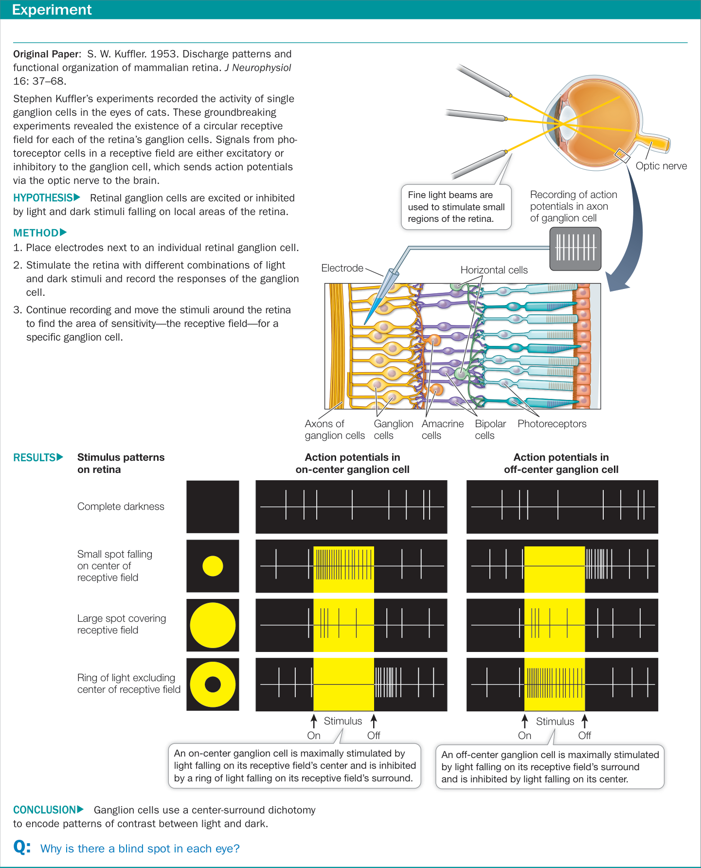
Figure 45.9 What Does the Eye Tell the Brain?
Three-dimensional vision results from cortical cells receiving input from both eyes
How do we perceive objects in three dimensions? The short answer is that our two eyes see overlapping, yet slightly different, visual fields—that is, humans have binocular vision. A person who is blind in one eye has difficulty discriminating distances. Animals whose eyes are on the sides of the head rather than facing front have minimal overlap in their fields of vision and, as a result, poor depth vision except for a narrow field straight ahead; however, they can see predators creeping up from all sides.
The story of how the brain integrates information from two eyes begins with the paths of the optic nerves. The two optic nerves run along the underside of the brain, join just under the hypothalamus, and then separate again (Figure 45.10). The place where they join is called the optic chiasm. Axons from the half of each retina closest to the nose cross in the optic chiasm and go to the opposite side of the brain. The axons from the outer half of each retina do not cross over at the optic chiasm; axons from the outer left retina go to the left side of the brain, and vice versa for axons from the outer right retina.
The functional consequence of the optic chiasm is that all of the visual information from the left side of your field of vision when you are looking straight ahead goes to the right side of your brain, and all of the visual information from the right side of your field of vision goes to the left side of your brain.
(A) Visual field
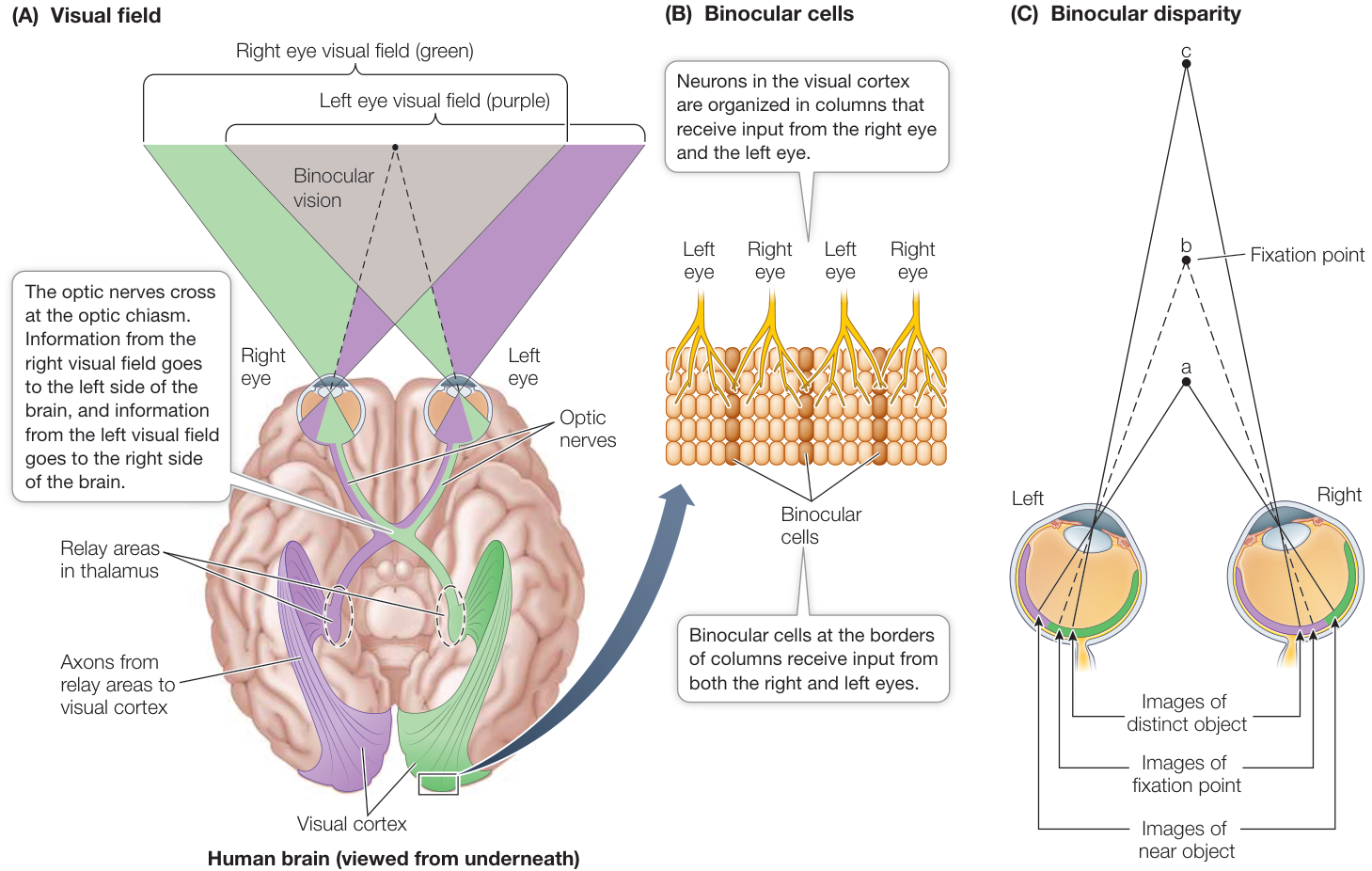
Figure 45.10 How Do We See in Three Dimensions? (A) Our eyes have largely overlapping visual fields, and both eyes transmit information to both sides of the brain. However, because the axons from the inner sides of the retinas cross at the optic chiasm, information from the visual space to the left of straight ahead goes to the right side of the brain, and information from the visual space to the right of straight ahead goes to the left side of the brain. (B) Between these left and right eye columns of neurons are neurons that receive input from both eyes. These are the binocular cells. (C) Light from objects that are different distances away will fall at different locations on the retinas. These disparities between where the image of the fixation point and the images of other objects fall on the retina mean that different binocular cells will receive input from different retinal axons depending on how far away the object is. These disparity-sensitive cells enable depth perception and three-dimensional vision.
However, there is a large area of overlap between the visual fields of the two eyes. These relationships are shown in purple and green in Figure 45.10.
Because of the overlap between the visual fields of the two eyes, there can be neurons in the visual cortex that receive input from both the left and the right eye. These are called binocular cells, and they can detect disparity between where the image of an object falls on the two retinas. What is disparity? Hold your finger out in front of you and look at it closing one eye and then the other. Your finger appears to jump back and forth because its image falls on a different position on each retina. Repeat the exercise with a distant object. It doesn’t jump back and forth as much because there is less disparity in the positions of the image on the two retinas. Certain binocular cells respond optimally to a stimulus falling on both retinas with a particular disparity. Which set of binocular cells is stimulated depends on how far away the stimulus is.
When we look at something, we can detect its shape, color, depth, and movement. Where does all this information come together? Is there a single cell that fires only when a red sports car drives by? The answer to that is probably no. Visual experience comes from simultaneous activity in many different cells. In addition, most visual experiences are enhanced by information from the other senses and from memory, which helps explain why about 75% of the cerebral cortex is association cortex.
Recap and Assess
Information in the nervous system is processed by cellular interactions in neuronal networks. The opposing actions of the sympathetic and parasympathetic divisions of the ANS can be understood in terms of neural pathways consisting of just two neurons. Vision begins with retinal photoreceptors organized into circular retinal receptive fields that influence the activity of retinal ganglion cells. Information from ganglion cells is transmitted in the optic nerves to the thalamus and relayed from there to the visual cortex. The left visual field from both eyes is represented in the right visual cortex, and vice versa for the right visual field. Receptive fields of cortical cells represent input from multiple ganglion cells, and binocular, disparity-sensitive cortical cells enable depth perception.
- Explain how a knife wound to left side of the neck can result in pupillary constriction in the left eye.
- How does the ANS differ in its control of heart rate versus digestive activity?
- Why can a small spot of light on the retina cause a larger response in a retinal ganglion cell than a large spot of light falling on the same area of retina?
By studying the neural circuitry of the visual system and the ANS, you have gained some understanding of how information reaches the CNS and how the CNS controls various functions of the body. But what about the higher functions of the mammalian CNS—the complex functions between input and output, such as language, learning, memory, and dreams?
Higher Brain Functions Involve Integration of Multiple Systems
Learning Objectives
- 45.3.1 Describe the EEG, EOG, and EMG characteristics associated with wake, non-REM and REM sleep and how they change throughout the daily sleep-wake cycle.
- 45.3.2 Describe the flow of information when speaking a written or a heard word, and specify which areas of the brain are involved in speech processing.
- 45.3.3 Explain how the modification of synaptic strength in hippocampal place cells produces a spatial map in the hippocampus.
- 45.3.4 Discuss how evolutionary changes in brain structures led to the development of self-awareness.
The higher brain functions discussed in the remaining pages of this chapter are undeniably complex. Nevertheless, neuroscientists, using a wide range of techniques, are making considerable progress in understanding these functions in terms of their cellular, molecular, and network properties. The following discussion addresses several aspects of brain and behavior that present challenges to neuroscientists: sleep and dreaming, learning and memory, language use, and consciousness.
Sleep and dreaming are reflected in electrical patterns in the cerebral cortex
A dominant feature of behavior is the daily cycle of sleep and waking. All birds and mammals, probably all other vertebrates, and also many invertebrates, sleep. We humans spend one-third of our lives sleeping, yet we do not know why or how. We do know, however, that we need to sleep. Loss of sleep impairs alertness and performance. Many people in our society—certainly most college students—are chronically sleep-deprived. Accidents and serious mistakes that endanger lives can be attributed to impaired alertness caused by lack of sleep. Insomnia (difficulty in falling or staying asleep) is one of the most common medical complaints.
THE ELECTROENCEPHALOGRAM A common tool of sleep researchers is the electroencephalogram, or EEG. Rather than recording the activity of single neurons, the EEG characterizes activity in huge numbers of neurons. EEG electrodes are much larger than the fine electrodes used to detect single cell activity. Placed at different locations on the head and scalp, EEG electrodes record fluctuations in the electric potential differences between electrodes over time (Figure 45.11A). These differences reflect the electrical activity of the neurons in the brain regions under the electrodes, primarily regions of the cerebral cortex. Usually the electrical activity of one or more skeletal muscles is also recorded; this record is called an electromyogram (EMG). Movements of the eyes are recorded as an electroocculogram (EOG).
EEG, EMG, and EOG patterns reveal the transition from being awake to being asleep. They also reveal that there are different states of sleep. In mammals other than humans, two major sleep states are easily distinguished: slow-wave sleep and rapid eye movement (REM) sleep. Slow-wave sleep gets its name from the high-amplitude, slow-frequency waves in the EEG. REM sleep gets its name from jerky movements of the eyeballs that occur during this state. In humans, sleep states are characterized as non-REM sleep and REM sleep. Human non-REM sleep is divided into three stages.
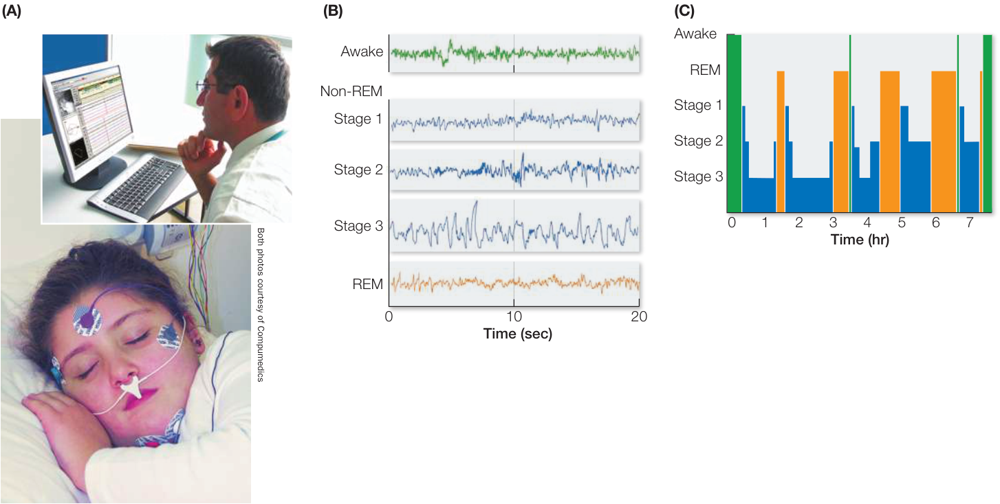
Figure 45.11 Stages of Sleep (A) Electrical activity in the cerebral cortex is detected by electrodes that are placed on the head and record changes in voltage between the electrodes through time. (B) The resulting record is an electroencephalogram, or EEG. (C) Humans cycle through different stages of sleep throughout the night. (B after A. Rechtschaffen and A. Kales. 1968. A manual of standardized terminology, techniques and scoring system for sleep stages of human subjects. NINDB Neurological Information Network (U.S.). Bethesda, MD.)
When you fall asleep, you first enter stage 1 non-REM sleep, which then progresses through stage 2 to stage 3 (Figure 45.11B). Stage 3 is deep, restorative, slow-wave sleep. This first full cycle of non-REM sleep is followed by an episode of REM sleep. Throughout the night you experience four or five cycles of non-REM and REM sleep (Figure 45.11C). About 80% of your sleep is non-REM sleep. The most vivid dreams and nightmares occur during the 20% of sleep that is REM sleep.
CELLULAR CHANGES DURING SLEEP When we are awake, several nuclei (groups of neurons) in the brainstem reticular-activating system (see Key Concept 45.1) are continuously active. Axons from neurons in these nuclei extend to the thalamus and throughout the cerebral cortex, where they release depolarizing neurotransmitters (acetylcholine, norepinephrine, and serotonin). These broadly distributed neurotransmitters keep the resting potential of the neurons of the thalamus and cortex close to threshold and sensitive to synaptic inputs, thereby maintaining the responsiveness of the brain that characterizes being awake.
With the onset of sleep, activity in these brainstem nuclei decreases, and their axon terminals release less neurotransmitter. With the withdrawal of these depolarizing neurotransmitters, the resting potentials of the cells of the thalamus and cortex become more negative (hyperpolarized), and the cells are less sensitive to excitatory synaptic input. Their processing of information is inhibited, and consciousness is lost. You have probably experienced this as “nodding off.” As you enter non-REM sleep, cells of the cortex begin to fire action potentials in bursts. The synchronization of these bursts over broad areas of cerebral cortex results in the EEG slow-wave pattern that characterizes deep non-REM sleep.
At the transition from non-REM to REM sleep, dramatic changes occur. Some of the brainstem nuclei that were inactive during non-REM sleep become active again, causing a general depolarization of cortical neurons. Thus in REM sleep the synchronized bursts of firing cease, and the EEG resembles that of the awake brain. Because the resting potentials of the neurons return to near threshold levels, the cortex can process information, and vivid dreams occur.
So why don’t we act out our dreams? During REM sleep the brain inhibits both afferent (sensory) and efferent (motor) pathways; we are paralyzed during REM sleep. Limb twitches and the jerky eye movements are motor signals breaking through the inhibition. The bizarre nature of dreams may be due to the lack of sensory feedback to the cortex from the body and the outside world. In other words, a functioning cortex is out of touch with reality. The function of muscle paralysis during REM sleep may be to prevent the acting out of dreams.
Knowing the cellular mechanisms of sleep has not yet led to an understanding of its function. Many questions remain. Why do we have two sleep states with very different neurophysiological characteristics? Why does non-REM sleep always occur first? Why do the two states cycle during the rest phase? We know sleep is essential for life, but we don’t know why. One set of hypotheses is that sleep is necessary for the maintenance and repair of neural connections and for the neural changes involved in learning and memory—and forgetting. These hypotheses are supported by many experiments showing that performance of a learned task or recall of declarative information on the day following training is impaired if sleep is prevented, and is best following a good night’s sleep. Another possible function of sleep was introduced in Key Concept 43.1, and that was the clearing of the brain of metabolic waste products by the newly described glymphatic system. Understanding the function, or more likely functions, of sleep is a major challenge for neuroscientists.
Language abilities are localized in the left cerebral hemisphere
No aspect of brain function is as integrally related to human consciousness and intellect as is language. Therefore brain mechanisms that underlie the acquisition and use of language are extremely interesting to neuroscientists. A curious observation about language ability is that it resides principally in one cerebral hemisphere—which in 97% of people is the left hemisphere. This phenomenon is referred to as the lateralization of language functions.
Fascinating research on this subject was conducted by Roger Sperry and his colleagues at the California Institute of Technology. The two cerebral hemispheres are connected by a tract of white matter called the corpus callosum. Recall from Key Concept 43.4 that white matter tracts are bundles of myelin-coated axons transmitting neural information from one area of the CNS to another. In one severe form of epilepsy, bursts of action potentials causing seizures travel between hemispheres via the corpus callosum. Cutting the tract eliminates the problem, and patients function well following surgery. However, these “split-brain” patients display interesting deficits in language ability.
After the surgery, if an object is shown in the right visual field and the left eye is closed (see Figure 45.10), the patient can describe it verbally and in writing. If the object is shown in the left visual field and the right eye is closed, the patient cannot describe it either verbally or in writing, but can use his or her left hand to point to a picture of the object. Without the connecting tissue between the two hemispheres, knowledge or experience of the right hemisphere can no longer be expressed in language.
Individuals who have suffered damage to the left hemisphere frequently suffer from some form of aphasia, a deficit in the ability to use or understand words. Studies of such individuals have identified several language areas in the left hemisphere.
Broca’s area, located in the frontal lobe just in front of the primary motor cortex, is essential for speech. Damage to Broca’s area results in halting, slow, poorly articulated speech or even complete loss of speech, but the patient can still read and understand language.
Wernicke’s area, located in the temporal lobe close to its border with the occipital lobe, is more involved with sensory than with motor aspects of language. Damage to Wernicke’s area can cause a person to lose the ability to speak sensibly while retaining the abilities to form the sounds of normal speech and to imitate its cadence. Such a patient cannot understand spoken or written language.
The angular gyrus, located near Wernicke’s area, is involved in integrating spoken and written language.
Normal language ability depends on the flow of information among various areas of the left cerebral cortex. Input from spoken language travels from the auditory cortex to Wernicke’s area (Figure 45.12A). Input from written language travels from the visual cortex to the angular gyrus to Wernicke’s area (Figure 45.12B). Commands to speak are formulated in Wernicke’s area and travel to Broca’s area and from there to the primary motor cortex. Damage to any one of those areas or the pathways between them can result in aphasia. Using methods of brain imaging such as functional magnetic resonance imaging (fMRI) or positron emission tomography (PET), it is possible to see the metabolic activity in different brain areas when the brain is using language (Figure 45.13).
Some learning and memory can be localized to specific brain areas
Learning is the modification of behavior by experience. Memory is the ability of the nervous system to retain what is learned and experienced. Even very simple animals can learn and remember, but these two abilities are most highly developed in humans. Consider the amount of information associated with learning a language, and then the much greater amount of information that language enables our brains to store and to process. The capacity of memory and the rate at which memories can be retrieved are remarkable features of the human nervous system.
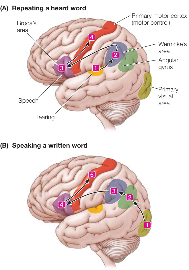
Figure 45.12 Language Areas of the Cortex Different regions of the left cerebral cortex participate in the processes of (A) repeating a word that is heard and (B) speaking a written word.
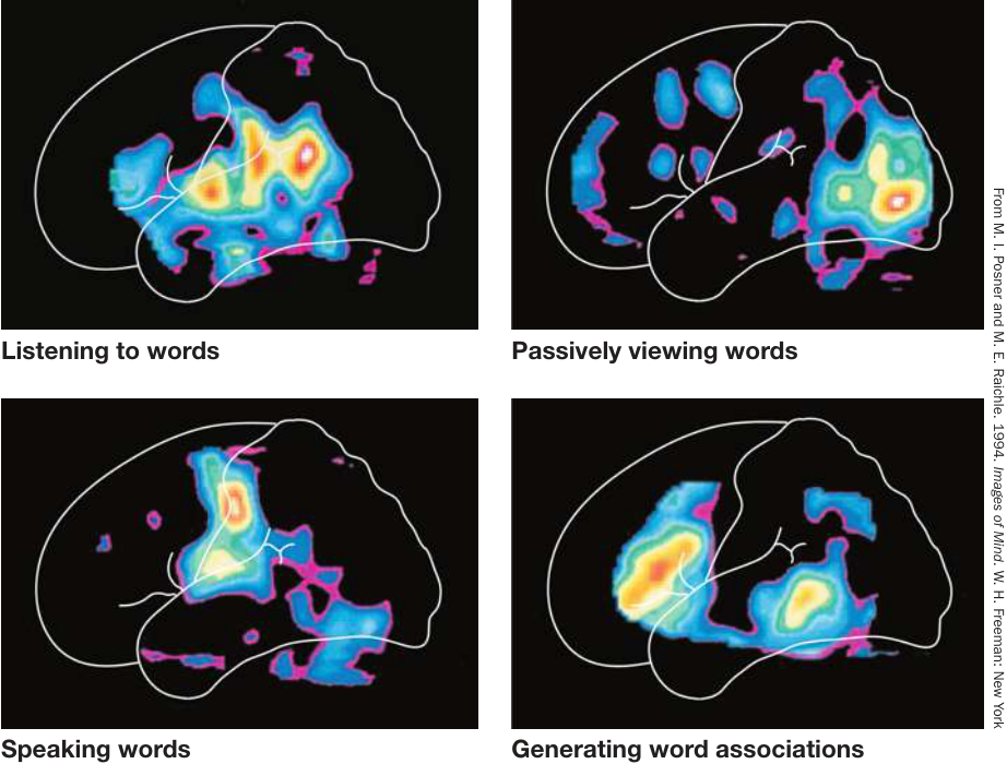
Figure 45.13 Imaging Techniques Reveal Active Parts of the Brain Positron emission tomography (PET) scanning reveals the brain regions activated by different aspects of language use. Radioactively labeled glucose is given to the subject. Brain areas take up radioactivity in proportion to their metabolic use of glucose. The PET scan visualizes levels of radioactivity in specific brain regions when a particular activity is performed. The red, yellow, and white areas are the most active.
Learning that leads to long-term memory and modification of behavior must involve long-lasting synaptic changes. A phenomenon that may explain how long-term synaptic changes might arise is long-term potentiation, or LTP. LTP results from high-frequency electrical stimulation of certain identifiable circuits that makes these circuits more sensitive to subsequent stimulation. In contrast, continuous, repetitive, low-level stimulation of these same circuits reduces their responsiveness, a phenomenon that has been called long-term depression (LTD). LTP and LTD may be fundamental cellular or molecular mechanisms involved in learning and memory.
Above the level of the synapse, memory involves interactions between several brain areas, as we discussed in the opener to this chapter and in Investigating Life: Places Cells Reveal Processes of Memory Consolidation during Sleep. Some of the first insights into memory processes came from surgical treatment of patients with severe seizures. That treatment involves destruction of the excessively active brain area triggering the seizures. To find the right area, the surgery is done under local anesthesia, with the patient remaining conscious. As different regions of the brain are electrically stimulated with electrodes, the patient reports the resulting sensations. Stimulation of some regions of the association cortex elicits recall of vivid memories. Such observations provided the first evidence that specific areas in the brain are associated with specific memories and that memory can be attributed to networks of neurons. Destroying a small area of the brain does not completely erase a memory, however, so it is postulated that memory is a function distributed over many brain regions and can be stimulated via many different routes.
You experience several forms of memory everyday. You have immediate memory for events that are happening now. Immediate memory is almost perfectly photographic but lasts only seconds. Short-term memory contains less information but lasts longer—on the order of 10–15 minutes. When you are introduced to a group of several new people, you probably will have forgotten their names in an hour or so if you have not written them down, used them in a conversation, or made a conscious effort to repeat them. Repetition, use, or reinforcement by something that gets your attention (a title such as “President,” for example) facilitates the transfer of short-term memory to long-term memory, which can last for days, months, years, or a lifetime. There is also intermediate-term memory, which lasts about a day.
Knowledge about neural mechanisms for the transfer of short-term memory to long-term memory—memory consolidation—has come from observations of persons who have lost parts of the limbic system, notably the hippocampus. A famous case is that of the man identified as H.M., whose hippocampus on both sides of the brain was removed to control his severe epilepsy. After the surgery, H.M. was unable to transfer information to long-term memory. If someone was introduced to him, had a conversation with him, and then left the room for several minutes, when that person returned, H.M. could not recognize him—it was as if the conversation had never taken place. Up until his death 55 years later, H.M. remembered events that happened before his surgery but could not remember post-surgery events for more than 10–15 minutes.
The studies of H.M., other such patients, and experiments on animals and even London taxi drivers show that the hippocampus plays an essential role in the acquisition of declarative memories—those that involve people, places, things, and events. The study featured in the opener to this chapter revealed the mapping of spatial information by neuronal networks in the rat, and the replay of that information when the rat is resting before or after running a maze. Subsequent experiments revealed the replay of the same spatial information during sleep, possibly revealing a process that is consolidating the memory and transferring it into long-term storage (see Investigating Life: Place Cells Reveal Processes of Memory Consolidation during Sleep).
We still cannot answer the question “What is consciousness?”
This chapter has only scratched the surface of the organization and functions of the human brain. Even with all of our knowledge of the human brain, and with all of the sophisticated new research tools, we still cannot answer the question “What is consciousness?”
The word “consciousness” is used in everyday language to refer to being awake in contrast to being asleep or in a coma. Here we are referring to being mentally aware of yourself, your environment, and events going on around you in such a way that you can plan for future events and make decisions based on experience, evidence, value systems, and predicted consequences. Speculations about consciousness have been the realm of philosophers, but we are getting closer to a neurobiological understanding.
One requirement for conscious experience is a perception of self that can be integrated with information from the physical and social environment and information from past experience. The basis for a perception of self derives from the huge amount of somatosensory and visceral information that comes from all parts of the body. This information is used for motor control and for homeostatic regulation. It enables animals to find food, seek mates, seek warmth, avoid cold, avoid danger, and so on. Different modes of this afferent information go to appropriate control and regulatory systems in the brainstem and forebrain. But these diverse sources of information also go to the insular cortex. The insular lobe appears to integrate physiological information from all over the body to create a sensation of how the body “feels.” Thus when an animal’s actions restore homeostasis, it “feels” better, and this is motivation to do the right thing for well-being.
In humans and the great apes, the insular lobe is greatly expanded and its circuitry communicates with parts of the brain involved in planning and decision making. In imaging studies, the insular cortex is activated by a great diversity of situations that involve strong feelings such as pleasure, disgust, humor, pain, lust, craving, humiliation, guilt, or empathy. Damage to the insular lobe causes apathy, loss of the ability to enjoy music, loss of sexual responsiveness, and even loss of the ability to distinguish good food from spoiled food. Humans and the few other species (great apes, elephants, and some marine mammals) that have expanded insular lobes are unique in that they can recognize themselves in a mirror. Could it be that this very discrete part of our brains and its circuitry are the neurobiological basis for self-awareness?
Recap and Assess
Complex functions of the nervous system can be understood in terms of the properties of neurons and neuronal networks. Language functions are lateralized to the left cerebral hemisphere. Since information about objects seen in the left visual field is processed in the right visual cortex, patients with a severed connection between their left and right cerebral hemispheres can see and recognize objects in their left visual field, but not describe them in words. Imaging shows the integration of different brain areas in the hearing, reading, and speaking of words. REM and non-REM sleep and awake states involve very different activities in many areas of the brain. Sleep states have a regular organization of expression during the daily rest phase. At the cellular level, learning and memory involve changes in synaptic strength, and memory processes involve interactions of neuronal networks from different brain areas. Declarative memories are acquired through the hippocampus. Consciousness may involve creation of a perception of the self in the insular cortex.
- What is the significance of paralysis of skeletal muscles during REM sleep?
- If a person with a severed corpus callosum is shown an object in his left visual field, how will he be able to communicate to an observer what that object is?
- What does the case of H. M. tell us about the brain mechanisms underlying the acquisition of declarative memories?
- How might the insular lobe of humans contribute to the phenomenon of consciousness?

Activity 45.3 Concept Matching:
The Mammalian Nervous System
Investigating Life
Q&A Can recordings of hippocampal place cells reveal processes of memory consolidation during sleep?
Unfortunately we cannot give a rat an exam to ask what it remembers. However, we can look in other brain areas for electrophysiological patterns of activity that correlate with the hippocampal place-cell patterns. Wilson and colleagues described the fast replay of the hippocampal place-cell patterns as a unique EEG signal called “ripples.” They observed that these ripples were tightly coupled to slower EEG brainwaves that spread to cortical areas of the brain. When they recorded in some of these areas of the cortex, they found similar patterns of firing (i.e., ripples) in those areas that were synchronized to the hippocampal ripples. They hypothesized that the ripples represent memory transcripts that are transferred to and stored in areas of the cortex.
Future directions
The neurophysiological studies of the acquisition and consolidation of declarative memories bring us closer to understanding the physical nature of a memory. The synaptic strengthening known as LTP (long-term potentiation) lasts a long time in contrast to action potentials, but not nearly as long as a memory that can last a lifetime. We can think of a memory as a piece of information that is stored in the brain. We have used the word “information” a lot in this chapter, but we really do not know what “information” is in the brain. It is not simply action potentials; they are brief, transient events. It is not patterns of synapses; they are constantly being altered. It must involve patterns of connectivity in and between neuronal networks in the brain; but what maintains those networks over years? We have much to learn about how our brains work.

Visual Summary

© Jason Hawkes/Getty Images
You should be able to relate each summary to the adjacent figures. If you go to this visual summary in Achieve, you can follow links to figures, animations, activities, and simulations that will help you consolidate the material.
Functions Are Localized in the Nervous System
- ■ The brain and spinal cord make up the central nervous system (CNS); the cranial and spinal nerves make up the peripheral nervous system (PNS).
- ■ The vertebrate brain forms from three swellings at the anterior end of the embryonic neural tube that become the hindbrain, midbrain, and forebrain. The forebrain consists of the telencephalon and diencephalon. The telencephalon develops into the cerebrum (cerebral hemispheres). The diencephalon develops into the thalamus and hypothalamus. The midbrain and hindbrain develop into the brainstem and the cerebellum.
- ■ The spinal cord and the cranial nerves communicate information between the brain and the rest of the body.
- ■ The limbic system is involved in emotions, mood, physiological drives, instincts, and memory.
- ■ The cerebral hemispheres can be divided into the temporal, frontal, parietal, occipital, and insular lobes. Layers of neurons on the surfaces of these lobes are the cerebral cortex, different regions of which have specific functions.
Questions
- What major structures of the adult brain develop from the embryonic forebrain?
- Per square centimeter, which has greater representation in the primary somatosensory cortex, the hand or the foot? What does that mean?
- What are the major functions associated with each of the five cerebral lobes?
Nervous System Functions Rely on Neural Circuits
- ■ The autonomic nervous system (ANS) controls physiological functions of organs and organ systems. Its sympathetic and parasympathetic divisions are characterized by their anatomy, neurotransmitters, and effects on target tissues.
- ■ Information from retinal ganglion cells is communicated via the optic nerve to the thalamus and then to the visual cortex. The visual cortex assembles an image of the visual world by analyzing edges of patterns of light.
- ■ Binocular vision results from information from both eyes being communicated to binocular cells in the visual cortex. These cells interpret distance by measuring the disparity between where the same stimulus falls on the two retinas.
Questions
- What are three major differences between sympathetic and parasympathetic pathways?
- What are the five cell types in the layered organization of the retina, and what role does each play?
- How does binocular vision enable perception of distance?
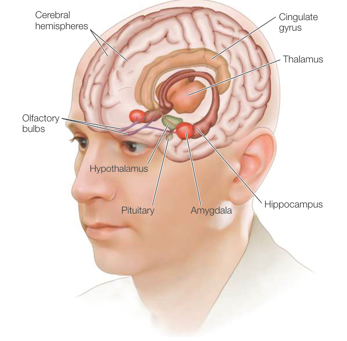
Figure 45.1
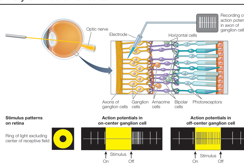
Figure 45.9 1. 2. What are the five cell types in the layered organization of the retina, and what role does each play? 2. 3. How does binocular vision enable perception of distance?
Higher Brain Functions Involve Integration of Multiple Systems
- ■ Humans, like other mammals and birds, have daily cycles of wake and sleep. Sleep can be divided into rapid eye movement (REM) sleep and non-REM sleep.
- ■ Language abilities are lateralized mostly in the left cerebral hemisphere. Different areas of the left hemisphere—including Broca’s area, Wernicke’s area, and the angular gyrus—are responsible for different aspects of language.
- ■ Learning and memory processes are localized to specific brain areas. Long-term potentiation (LTP) and long-term depression (LDP) are important processes in learning and memory.
- ■ Complex memories can be elicited by stimulating small regions of association cortex. Damage to the hippocampus can destroy the ability to form long-term declarative memory.
Questions
- What are the differences between non-REM and REM sleep at the level of cortical neurons, EEG recordings, and behavior?
- Learning to do a behavioral task like riding a bike is forming a procedural memory. Interpret the fact that patient H.M. could get better at a behavioral task over days but could not remember having practiced it.
- Why is it proposed that the insular lobe of the cerebrum is responsible for a generalized perception of feeling good or bad?
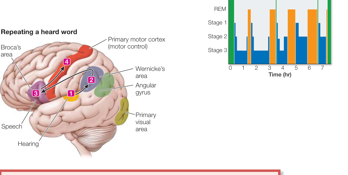
Figure 45.12
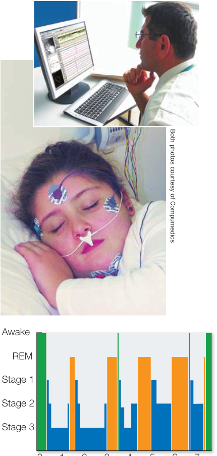
Figure 45.11