Chapter 46Musculoskeletal Systems
Key Concepts
46.1 Interactions of Actin and Myosin Cause Muscles to Contract
46.2 Many Factors Affect Muscle Performance
46.3 Muscles and Skeletal Systems Work Together
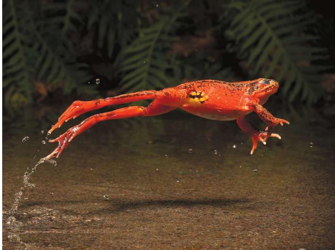
Relative to their size, many animals have more impressive jumping skills than humans. This red-legged frog (Rana aurora) can leap distances up to 20 times its body length.
© Michael Durham/Minden Pictures
Investigating Life: Champion Jumpers
The women’s long jump Olympic record is 7.4 meters, set in 1988 by Jackie Joyner-Kersee. Another world-record long jump was set two years earlier by Rosie the Ribeter, who jumped 6.5 meters. Rosie was a frog competing in the Calaveras County Jumping Frog Contest. Rosie’s jump is more impressive—it was 20 times her body length, whereas Jackie’s was 5 times her body length. And Rosie jumped from a sitting, crouched position, while Jackie’s jump was propelled by a sprint.
Both jumps were powered by skeletal muscle. The cellular mechanisms of muscle contraction are the same in frog and human, so why is the frog’s jump more impressive? The answer involves the concept of leverage—muscles and skeletal elements working together. Muscles pull on bones that are connected at joints to make levers. With a lever you can use the same force to move a large mass a small distance or a small mass a large distance. The ratio of leg length to body mass is greater for the frog than for the human. Thus the frog’s long legs can move its small body mass a long distance.
The power for the frog’s jump comes from a single contraction of its leg muscles (there is no sprint), and those muscles contract only a very short distance compared with the length of the jump. One species of frog, Rana pipiens, has a body length of only 11 centimeters, but longer legs. The muscles that power the jump are only about 3.5 centimeters long and shorten by just 0.75 centimeters (21%) when the frog jumps. Once again leverage comes into play. The thigh muscles are attached to the lower leg bones close to the knee joint and thus can move the opposite ends of those bones over a longer distance and more quickly than if they were attached farther away from the joint. There is a trade-off, however, in terms of force and speed. To use a shovel, you have one hand near the end of the shaft and the other hand farther down the shaft. With hands close together, you move the shovel blade fast, but can’t pick up a heavy load of dirt. With hands farther apart, you can’t move the blade as fast, but you can pick up a heavier load.
QA What adaptations optimize the jumping muscles of the frog? (Find the answer on page 1044.)
Interactions of Actin and Myosin Cause Muscles to Contract
Learning Objectives
- 46.1.1 Describe changes in sarcomere structure and appearance as muscle contraction occurs.
- 46.1.2 Explain the molecular events underlying the sliding filament model of muscle contraction.
- 46.1.3 Describe how ATP is involved in muscle contraction.
- 46.1.4 Beginning with an action potential in a motor neuron, describe the sequence of events at the molecular level that leads to skeletal muscle contraction.
- 46.1.5 Discuss why gap junctions are important for cardiac and smooth, but not skeletal, muscle functions.
Behavior and most physiological functions of animals, such as beating of the heart and moving of food through the digestive tract, depend on muscle contraction. Wherever tissues contract, muscle cells are responsible. As you saw in Figure 38.2, there are three types of vertebrate muscle:
- Skeletal muscle is responsible for all voluntary movements, such as running or playing a piano. It is also involved in many involuntary actions, including breathing, shivering, and maintaining posture.
- Cardiac muscle is responsible for the beating of the heart.
- Smooth muscle creates movement in many hollow internal organs, such as the digestive system, bladder, and blood vessels, and is under the control of the autonomic (involuntary) nervous system.
Skeletal and smooth muscle occur in all animal phyla, but cardiac muscle is unique to vertebrates. All three muscle types share the same underlying molecular mechanisms for generating force. We will use vertebrate skeletal muscle as our primary example. Later in this key concept we discuss the differences in cardiac and smooth muscle that adapt them to their particular functions, and at the end of Key Concept 46.2 we describe interesting specializations of insect muscle.
Sliding filaments of actin and myosin cause skeletal muscle to contract
Skeletal muscles are so named because they attach to skeletal elements and make most movements of the body possible. Skeletal muscles are frequently called striated muscles because of their striped appearance (Figure 46.1; also see Figure 38.2). Skeletal muscle cells, called muscle fibers, are large and long and have many nuclei. These multinucleate cells form in development through the fusion of many individual embryonic muscle cells called myoblasts. A specific muscle such as your biceps (which bends your arm) is composed of hundreds or thousands of muscle fibers bundled together by connective tissue.
Muscle contraction is due to the interaction of the contractile proteins actin and myosin, which exist as long filaments within muscle cells. Actin filaments are thin and myosin filaments are thick, and they lie parallel to each other. When muscle contraction is triggered, the actin and myosin filaments slide past each other in a telescoping fashion.
Connect the Concepts Key Concept 5.3 discussed how microfilaments made of polymerized actin are elements of the cytoskeleton that are involved in maintaining cell shape and cell movements in all eukaryotic cells.
Thin actin and thick myosin filaments are bundled together in an orderly fashion as myofibrils. A muscle cell (fiber) contains many myofibrils (see Figure 46.1). A longitudinal view of a myofibril reveals why skeletal muscle appears striated. The myofibril consists of repeating units called sarcomeres. Each sarcomere is made of overlapping bundles of actin and myosin filaments that create a distinct banding pattern (see Figure 46.1). Before the molecular nature of the muscle banding pattern was known, the bands were given names that are still used today. Each sarcomere is bounded by Z lines, which anchor the thin actin filaments. Centered in the sarcomere is the A band, which contains the myosin filaments. The light-appearing H zone and I band are regions where actin and myosin filaments do not overlap in the relaxed muscle. The dark stripe within the H zone is the M band; it contains proteins that hold the myosin filaments in their regular arrangement.
The bundles of myosin filaments are held in a centered position within the sarcomere by a protein called titin (see Figure 46.2). Titin is the largest protein in the body; it runs the full length of the sarcomere from Z line to Z line. Each titin molecule runs right through a myosin bundle. Between the ends of the myosin bundles and the Z lines, titin molecules are very stretchable, like bungee cords. In a relaxed skeletal muscle, resistance to stretch is mostly due to the elasticity of the titin molecules.
As the muscle contracts, the sarcomeres shorten and the band pattern changes. The H zone and the I band become much narrower, and the Z lines move toward the A band as if the actin filaments were sliding into the H zone, the region occupied by only myosin filaments (Figure 46.2). In the mid-1950s this observation independently led two teams of British biologists to propose the sliding filament model of muscle contraction.
It is not uncommon in science for critical breakthroughs to be made simultaneously in different laboratories, but in this case the coincidences are remarkable. The leaders of the two teams were named Hugh Huxley and Andrew Huxley—but they were not related. Andrew Huxley was a faculty member at Cambridge University in the early 1950s, and Hugh Huxley was a graduate student working in a different laboratory. Both were interested in the structure of muscle and the mechanism of contraction. Hugh Huxley moved to MIT as a postdoctoral fellow where he worked with Jean Hanson using electron microscopy. Andrew Huxley, joined by Rolf Niedergerke, used a new form of light microscopy that the two developed. The two groups proposed the sliding filament model at the same time, and both papers were published in the same issue of the journal Nature.
Actin-myosin interactions cause filaments to slide
To understand the mechanism causing the actin and myosin filaments to slide past each other, we must first examine the structures of actin and myosin (Figure 46.3). A myosin molecule consists of two long polypeptide chains coiled together, each ending in a large globular head. A myosin filament is made up of many myosin molecules arranged in parallel, with their heads projecting sideways at each end of the filament like a bunch of golf clubs.
An actin filament consists of actin monomers polymerized into long chains that look like two strands of beads twisted together.
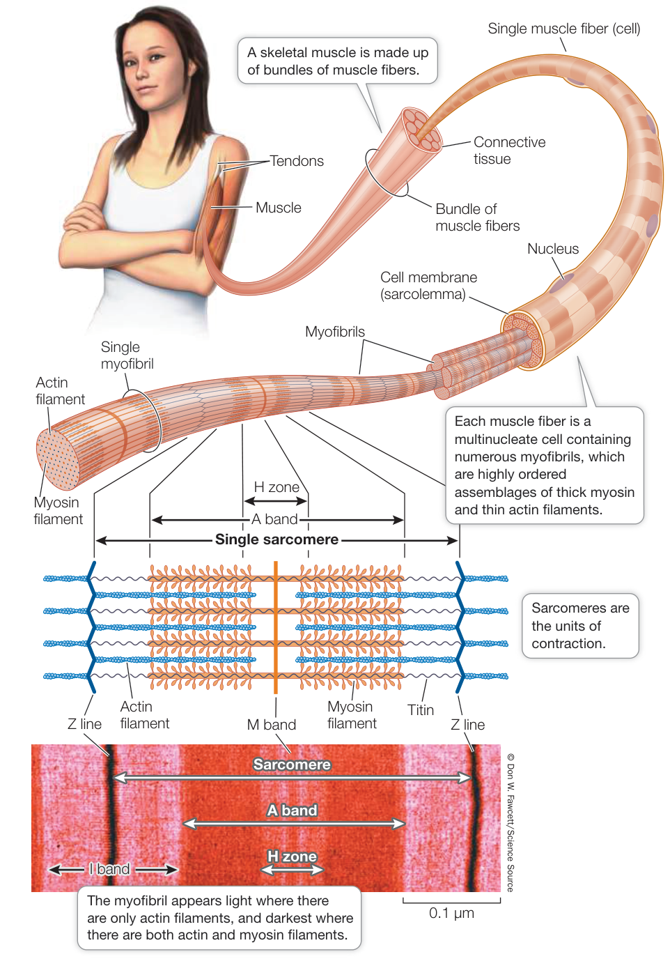
Figure 46.1 The Structure of Skeletal Muscle A skeletal muscle is made up of bundles of muscle fibers. Each muscle fiber is a multinucleate cell containing numerous myofibrils, which are highly ordered assemblages of thick myosin and thin actin filaments. The arrangement of the actin and myosin filaments gives skeletal muscle fibers their characteristic striated appearance.
Twisting around the actin chains is another protein, tropomyosin, and attached to tropomyosin at intervals are molecules of troponin. We discuss these two proteins later in this key concept.
The myosin heads bind reversibly to specific sites on actin and thereby form cross-bridges between the myosin and the actin filaments. When a myosin head binds to an actin filament, the head’s conformation changes. As the head bends, it exerts a tiny force that causes the actin filament to move 5–10 nanometers relative to the myosin filament. When the myosin heads are bound to actin, they can bind and hydrolyze ATP. The energy released when this happens changes the conformation of the myosin head, causing it to release the actin and return to its extended position, from which it can bind to actin again.
Together these details explain the cycle of events that cause the actin and myosin filaments to slide past each other and shorten the sarcomere. They also explain rigor mortis—the stiffening of muscles soon after death. ATP binding causes myosin to release from actin, so when ATP production stops with death, myosin cannot release and the muscles stay contracted. Eventually, however, the proteins lose their integrity and the muscles soften. The timing of these events in different muscles helps a medical examiner estimate the time of death.
We have been discussing the cycle of contraction in terms of a single myosin head. But each myosin filament has many myosin heads at both ends and is surrounded by six actin filaments; thus the contraction of the sarcomere involves a great many cycles of interaction between actin and myosin molecules. That is why when a single myosin head breaks its contact with actin, the actin filaments do not slip backward.
Actin-myosin interactions are controlled by calcium ions
Like neurons, muscle cells are excitable—that is, their cell membranes generate and conduct action potentials. Skeletal muscle action potentials are initiated by motor neurons releasing neurotransmitter at neuromuscular junctions (see Key Concept 43.3). The axon terminals of motor neurons are generally highly branched and form synapses with hundreds of muscle fibers (Figure 46.4). A motor neuron and all of the fibers with which it synapses constitute a motor unit. The fibers contract simultaneously when the unit’s motor neuron fires. One muscle can consist of many motor units. Thus there are two ways to increase a muscle’s strength of contraction—increase the firing rate of an individual motor neuron, or recruit more motor neurons.
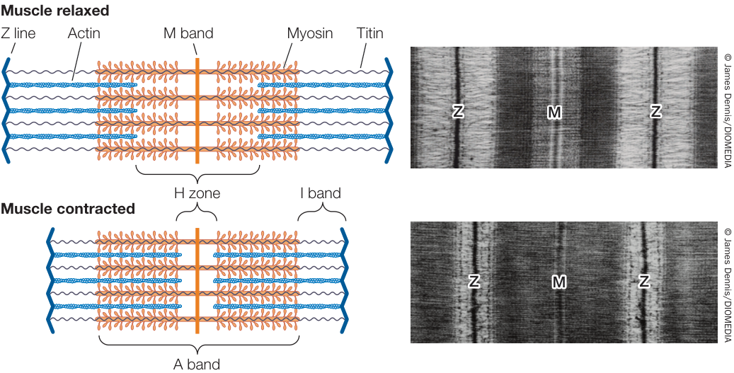
Figure 46.2 Sliding Filaments The banding pattern of the sarcomere changes as it shortens, as seen in electron micrographs. These observations led to the sliding filament model of muscle contraction.
When an action potential arrives at a neuromuscular junction, acetylcholine is released from the motor neuron terminals, diffuses across the synaptic cleft, binds to receptors in the postsynaptic membrane, and causes ion channels in the motor end plate to open (see Figure 43.12). Most of the ions that flow through these channels are , and therefore the motor end plate is depolarized. The depolarization spreads to the surrounding cell membrane of the muscle fiber, which contains voltage-gated sodium channels. When thresholds for those channels are reached, the cell membrane fires an action potential that is conducted rapidly to all points on the surface of the muscle fiber.
An action potential in a muscle fiber also travels deep within the cell. The cell membrane is continuous with a system of T
tubules that descend into the muscle fiber cytoplasm (also called the sarcoplasm). The “T” stands for “transverse.” The contents of the T tubules are outside the cell. The T tubules are like the indentations created in a water-filled balloon by poking your finger into it. The action potential that spreads over the surface membrane of the fiber also spreads through this system of T tubules (Figure 46.5).
The T tubules come very close to the endoplasmic reticulum (ER) of the muscle cell. In muscle cells the ER is called the sarcoplasmic reticulum, and it is a closed compartment surrounding every myofibril. Calcium pumps in the sarcoplasmic reticulum take up from the sarcoplasm. Therefore when the muscle fiber is at rest, there is a high concentration of in the sarcoplasmic reticulum and a low concentration of in the sarcoplasm.
Spanning the space between the T tubule membrane and the sarcoplasmic reticulum are two proteins. One protein, the dihydropyridine (DHP) receptor, is located in the T tubule membrane; it is voltage-sensitive and changes its conformation in response to an action potential spreading through the T tubule system. The other protein, the ryanodine receptor, is located in the sarcoplasmic reticulum membrane and is a channel.
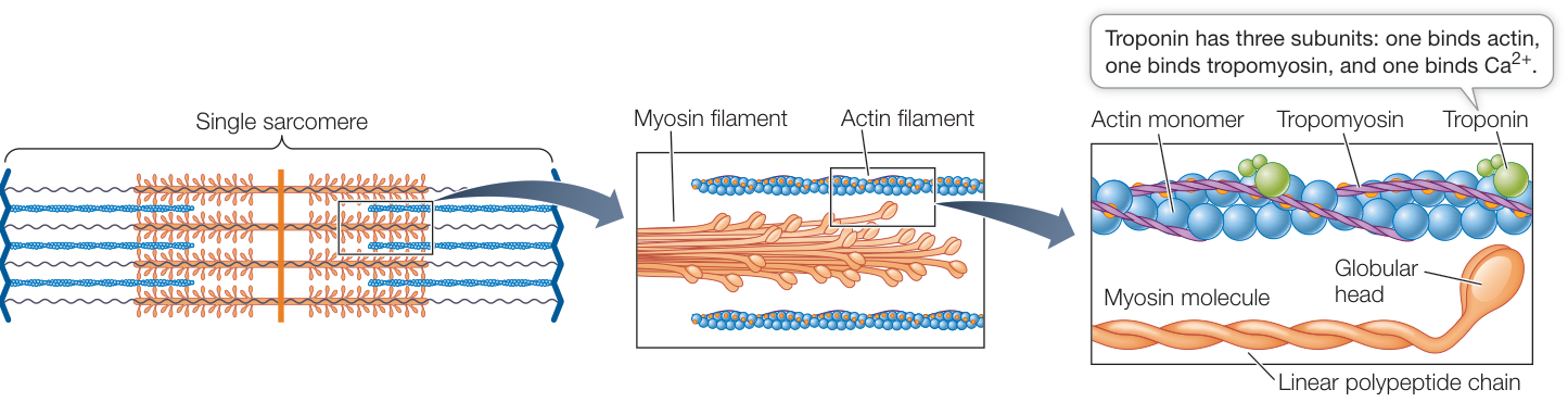
Figure 46.3 Actin and Myosin Filaments Overlap to Form Myofibrils Myosin filaments are bundles of molecules with globular heads and polypeptide tails; the protein titin holds these filaments centered within the sarcomeres. Actin filaments consist of two chains of actin monomers twisted together. They are wrapped by chains of the polypeptide tropomyosin and are studded at intervals with another protein, troponin.
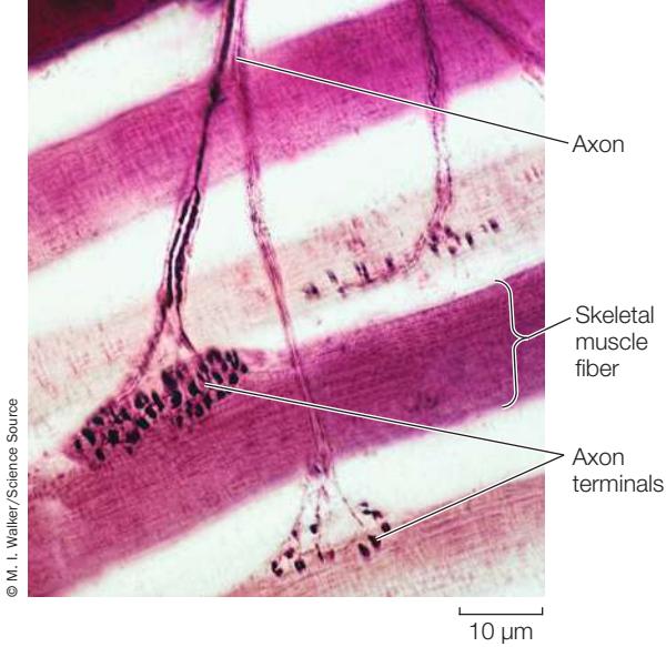
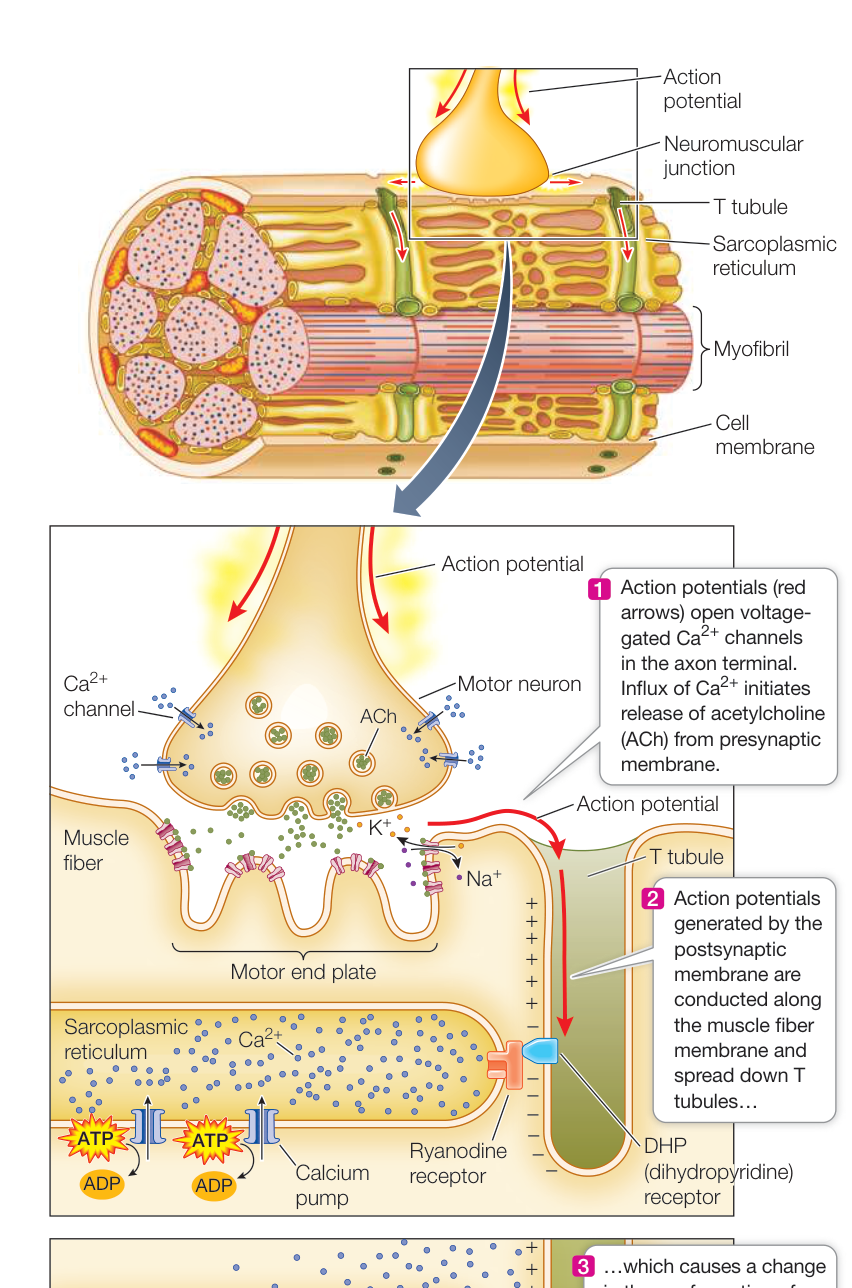
Figure 46.4 The Neuromuscular Junction Axons branching from a single motor neuron end in terminals that innervate multiple skeletal muscle fibers. Q: Is there a relationship between the number of fibers that a motor neuron innervates and fine motor control, between the number of fibers innervated and the exertion of force?
These two proteins are physically connected. When the DHP receptor changes its conformation due to an action potential, it opens the ryanodine channels, and flows from the sarcoplasmic reticulum into the sarcoplasm. ions diffusing through the sarcoplasm surrounding the actin and myosin filaments trigger the interaction of actin and myosin and the sliding of the filaments. How do the ions do this?
An actin filament, as you have seen, is a helical arrangement of actin monomers. Twisted around the actin filament are two strands of the protein tropomyosin (Figure 46.6; see also Figure 46.3). At regular intervals, the filament also includes a globular protein, troponin. The troponin molecule has three subunits: one binds actin, one binds tropomyosin, and one binds .
When the muscle is at rest, the tropomyosin strands are positioned so that they block the myosin binding sites on the actin filaments. When is released into the sarcoplasm, it binds to troponin, changing its conformation. Because the troponin is bound to the tropomyosin, this conformational change twists the tropomyosin
Figure 46.5 T Tubules Spread Action Potentials into the Fiber An action potential at the neuromuscular junction spreads throughout the muscle fiber via a network of T tubules, triggering the release of from the sarcoplasmic reticulum.
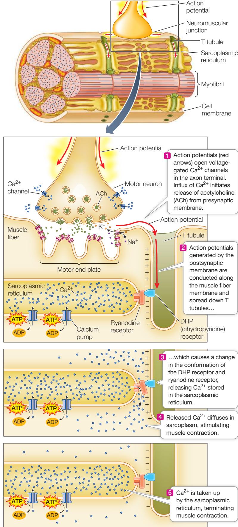
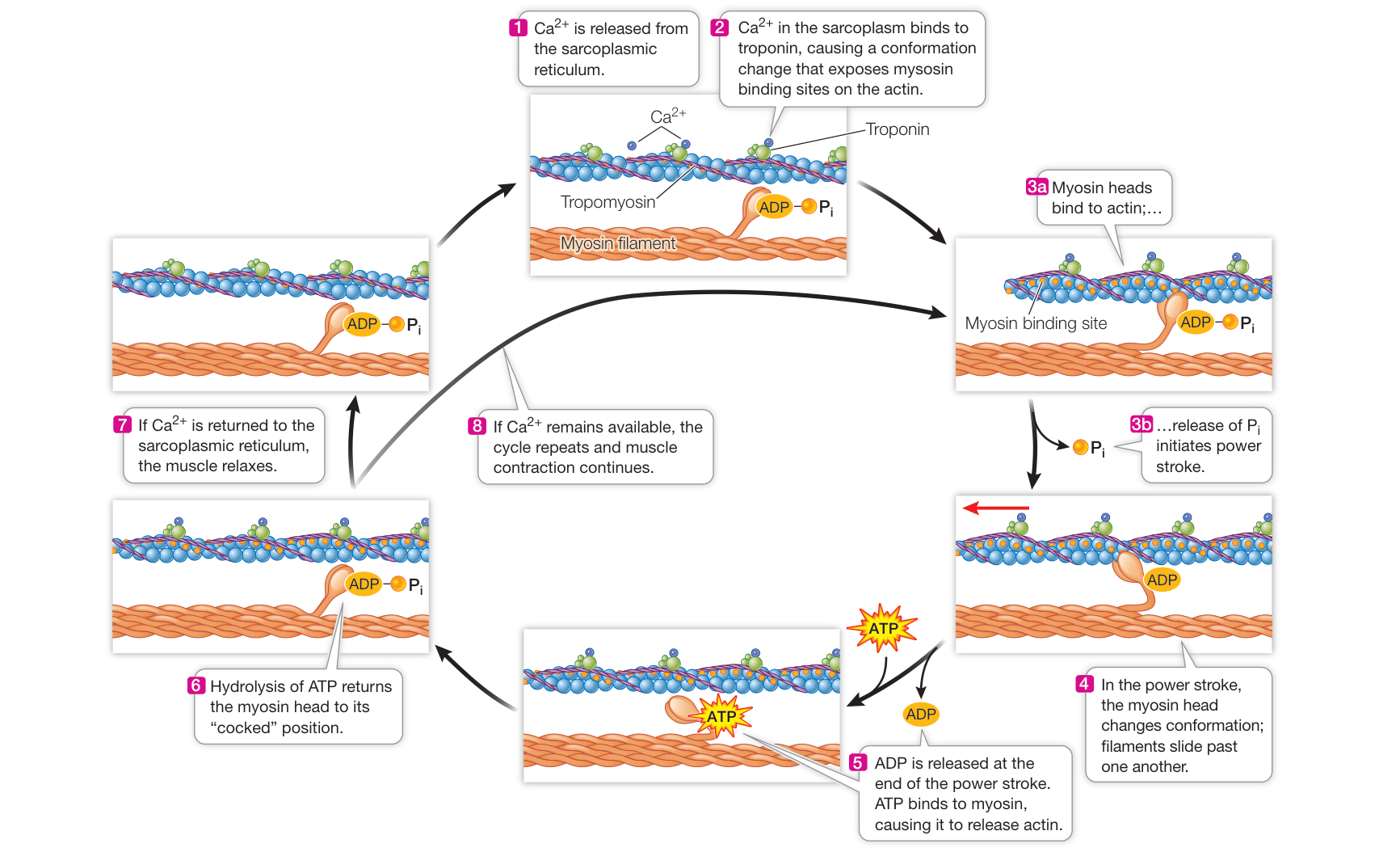
Figure 46.6 Release of from the Sarcoplasmic Reticulum Triggers Muscle Contraction When binds to troponin, it changes the conformation of troponin, exposing myosin binding sites on the actin. As long as binding sites and ATP are available, the cycle of actin and myosin interactions continues and the filaments slide past each other. Note that in the relaxed condition, the myosin heads are energized. Q: What are three roles that ATP plays in skeletal muscle contraction?

Animation 46.1
Molecular Mechanisms of Muscle Contraction
enough to expose the actin–myosin binding sites, and the cycle of making and breaking actin–myosin bonds is initiated. The filaments pull past each other, and the muscle fiber contracts. When the calcium pumps in the sarcoplasmic reticulum membranes remove the ions from the sarcoplasm, the conformation of the tropomyosin reverts and again blocks the binding of myosin heads to actin, and the muscle fiber returns to its resting condition. Figure 46.6 summarizes this cycle.
Cardiac muscle is similar to and different from skeletal muscle
Like skeletal muscle, cardiac muscle appears striated because of the regular arrangement of actin and myosin filaments into sarcomeres (Figure 46.7). Cardiac muscle cells are smaller than skeletal muscle fibers and have only one nucleus each (uninucleate). Cardiac muscle cells branch, and the branches of adjoining cells interdigitate, forming a meshwork resistant to tearing. As a result, the heart walls can withstand high pressures while pumping blood, without the danger of developing leaks. Adding to the strength of cardiac muscle are intercalated discs that provide strong mechanical adhesions between adjacent cells. Gap junctions are an important feature of cardiac muscle. These structures in the intercalated discs allow cytoplasmic continuity between cells (see Figure 7.16A). Because of gap junctions, cardiac muscle cells are electrically coupled. An action potential initiated at one point in a sheet of cardiac muscle spreads rapidly, causing a large number of cardiac muscle cells to contract simultaneously.
Certain cardiac muscle cells are specialized for generating and conducting electric signals. These pacemaker and conducting cells have a low density of actin and myosin filaments, but they initiate and coordinate the rhythmic contractions of the heart. (The molecular basis for this pacemaking function is covered in Key Concept 48.3.) Pacemaker cells make the vertebrate heartbeat autorhythmic, meaning the heartbeat is generated by the heart muscle itself. A heart removed from a vertebrate can continue to beat with no input from the nervous system. Input from the autonomic nervous system modifies the rate at which pacemaker cells fire, but it is not essential for their continued rhythmic function.
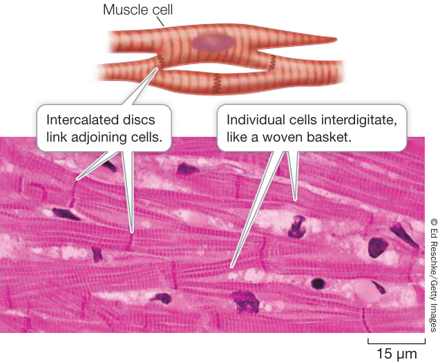
Figure 46.7 Cardiac Muscle Cells Form a Strong Meshwork Cardiac muscle cells branch and interdigitate, forming a tear-resistant mesh that can withstand the blood pressure generated by the contraction of the heart muscle.
The mechanism of excitation–contraction coupling in cardiac muscle cells is different from that in skeletal muscle cells. The T tubules are larger, and the DHP receptor proteins in the T tubules are channels. The DHP receptors in the cardiac T tubules are not physically connected with the ryanodine receptors in the sarcoplasmic reticulum. Instead, when an action potential spreads down the T tubules, it causes the voltage-gated DHP channels to open, allowing extracellular to flow into the sarcoplasm. That increased sarcoplasmic binds to the ryanodine receptors, causing their channels to open. The resulting huge rise in sarcoplasmic concentration stimulates fiber contraction. This mechanism is called -induced release.
Smooth muscle causes slow contractions of many internal organs
Smooth muscle provides the contractile force for most of our internal organs, which are under the control of the autonomic nervous system. Smooth muscle moves food through the digestive tract, controls the flow of blood through blood vessels, and empties the urinary bladder. Smooth muscle also generates “goose bumps” on the skin and causes the hair on the back of a dog or cat to stand erect when the animal is being aggressive or defensive. Structurally, smooth muscle cells are the simplest muscle cells. They are smaller than skeletal muscle cells, usually long and spindle-shaped, and each has a single nucleus. They appear “smooth” because the actin and myosin filaments are not as regularly arranged as they are in skeletal and cardiac muscle, and so do not produce the striated appearance.
Some smooth muscle tissue, such as that from the wall of the digestive tract, is arranged in sheets, and individual cells in a sheet are in electrical contact with one another through gap junctions, as they are in cardiac muscle. As a result, an action potential generated in one smooth muscle cell spreads to all the cells in that sheet of tissue, and the sheet contracts in a coordinated fashion.
The cell membranes of smooth muscle cells are sensitive to stretch, with important consequences. For example, if the wall of the digestive tract is stretched in one location (as by a mouthful of food passing down the esophagus to the stomach), the membranes of the stretched cells depolarize, reach threshold, and fire action potentials, which cause the cells to contract. Thus smooth muscle contracts after being stretched, and the harder it is stretched, the stronger it contracts. This behavior of smooth muscle is important for moving food through the digestive system.
The walls of blood vessels—except capillaries—contain much smooth muscle, especially on the arterial side where the blood is under higher pressure. Changes in vascular smooth muscle tone are responsible for controlling the distribution of blood in the body.
The neural influences on smooth muscle come from the two divisions of the autonomic nervous system (see Figure 45.8). The neurotransmitters of the sympathetic and parasympathetic postganglionic cells alter the membrane potential of smooth muscle cells. For example, in the digestive tract acetylcholine causes smooth muscle cells to depolarize, making them more likely to fire action potentials and contract. Antagonistically, norepinephrine causes these muscle cells to hyperpolarize and thus be less likely to fire action potentials and contract (Figure 46.8). In contrast, norepinephrine acting through G protein-coupled receptors causes the smooth muscle in arteries serving the gut to contract. Remember that the action of the neurotransmitters depends on the receptors in the target tissues. Sympathetic activity is high in a fight-or-flight situation. In an emergency you don’t need to digest your lunch, but shunting blood from the gut to tissues such as major skeletal muscles could be critical for your survival.
Smooth muscle cell contraction is not controlled by the troponin–tropomyosin mechanism, but calcium still plays a critical role. A influx into the sarcoplasm of a smooth muscle cell can be stimulated by action potentials, hormones, or stretching. The that enters the sarcoplasm combines with a protein called calmodulin. The –calmodulin complex activates an enzyme called myosin kinase that phosphorylates myosin heads. When the myosin heads in smooth muscle are phosphorylated, they undergo cycles of binding and releasing actin, causing muscle contraction. As is removed from the sarcoplasm, it dissociates from calmodulin, and the activity of myosin kinase falls. An additional enzyme, myosin phosphatase, dephosphorylates the myosin to help reduce actin–myosin interactions (Figure 46.9).

Animation 46.2 Smooth Muscle Action
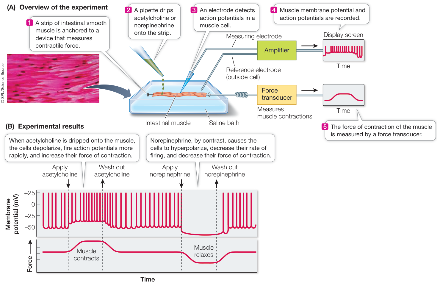
Figure 46.8 How do Autonomic Nervous System Neurotransmitters Control the Firing Rate of Smooth Muscle Cells? (A) When a strip of intestinal smooth muscle is mounted in an incubation bath so that its generation of action potentials and its force of contraction can be recorded, it is possible to test the effects of sympathetic and parasympathetic neurotransmitters on that tissue. (B) Acetylcholine depolarizes (makes less negative) the membranes and increases action potentials; norepinephrine hyperpolarizes (makes more negative) the membranes and decreases action potentials. (B after T. B. Bolton et al. 1979. Physiol Rev 59: 606–718.)
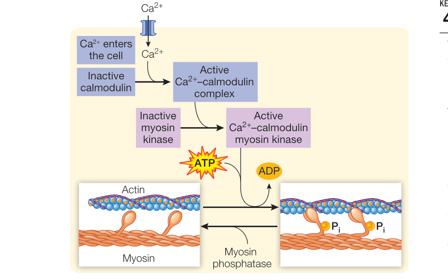
Figure 46.9 The Role of Ca2+ in Smooth Muscle Contraction When a smooth muscle cell is depolarized by an action potential, a neurotransmitter, or stretch, Ca2+ enters the sarcoplasm and binds to calmodulin, which in turn activates an enzyme that phosphorylates the myosin heads, causing them to bind to actin. As long as the myosin remains phosphorylated, actin and myosin go through cycles of binding and release. Thus in smooth muscle the Ca2+-mediated change is on myosin, whereas in skeletal and cardiac muscle it is on the actin–tropomyosin filament. Now that you understand how muscles generate force, let’s look at what determines the characteristics of a muscle, its performance, and how individual muscles can change their characteristics with regular use and conditioning.
Recap and Assess
The three types of muscle are skeletal, cardiac, and smooth. The contractile ability of muscle derives from interactions between actin and myosin filaments. Contraction depends on control by Ca2+ in the sarcoplasm. Tropomyosin and troponin are controlling elements in skeletal and cardiac muscle. Calmodulin is the controlling element in smooth muscle.
- In terms of the arrangement of actin and myosin filaments in the sarcomere, why do the H zone and I band get smaller when the skeletal muscle contracts, and why does the A band stay the same?
- What roles do the three subunits of troponin play in the control of skeletal muscle contraction?
- Why does rigor mortis—the stiffening of muscles after death—occur first in muscles with the least glycogen content?
- The insecticide malathion destroys the enzyme acetylcholinesterase. Accidental exposure can cause extreme rigid muscle paralysis. Why?
- Why are gap junctions important to the function of cardiac and smooth muscle, but not skeletal muscle?
Many Factors Affect Muscle Performance
Learning Objectives
- 46.2.1 Explain what determines how much force or tension is generated by a muscle.
- 46.2.2 Describe how muscle fiber types contribute to performance abilities in aerobic and anaerobic work.
- 46.2.3 Explain why peak running performance declines with distance of the event.
The various functions that muscles perform place different demands on them. Some muscles, such as postural muscles, must sustain a load continuously over long periods of time. Muscles used in walking or running must rapidly vary their strength of contraction over a wide range of positions. In contrast, the jumping muscles of the frog must exert maximum force instantaneously. The muscles that control your fingers generally do not have to sustain long, strong contractions, but they must be able to undergo quick yet graded contractions for fine manual dexterity. What is “quick” for humans, however, doesn’t begin to compare with insect flight muscles that can contract as fast as 1,000 times per second. How are different muscles adapted for specific functions and demands?
The strength of a muscle contraction depends on how many fibers are contracting and at what rate
The arrival of an action potential at a neuromuscular junction of a skeletal muscle fiber causes an action potential in that fiber. That action potential is conducted through the muscle fiber’s T tubule system and causes a minimum unit of contraction, called a twitch. A twitch can be measured in terms of the tension, or force, it generates (Figure 46.10A). A single action potential stimulates a single twitch, but the ultimate force generated by an action potential varies enormously depending on how many muscle fibers it reaches. The level of tension an entire muscle generates depends on two factors: the number of motor units activated, and the frequency at which those motor units fire.
In muscles responsible for fine movements, such as those of the fingers, a motor neuron may innervate only one or a few muscle fibers, but in a muscle that produces large forces, such as the biceps, a motor neuron innervates a large number of muscle fibers.
At the level of a muscle fiber, a single action potential stimulates a single twitch. If action potentials reaching the muscle fiber are adequately separated in time, each twitch is a discrete, all-or-none phenomenon. If action potentials are fired more rapidly, however, new twitches are triggered before the myofibrils have a chance to return to their resting condition. As a result, the twitches sum, and the tension generated by the fiber increases and becomes more sustained. Thus an individual muscle fiber can show a graded response to increased levels of stimulation by its motor neuron.
Twitches sum at high levels of stimulation because the calcium pumps in the sarcoplasmic reticulum (see Figure 46.5) are unable to clear the ions from the sarcoplasm between action potentials. Eventually a stimulation frequency can be reached that results in the continuous presence of in the sarcoplasm at high enough levels to cause continuous activation of the contractile machinery—a condition known as tetanus (Figure 46.10B). (Do not confuse this condition with the disease tetanus, which is caused by a bacterial toxin and is characterized by spastic contractions of skeletal muscles.)
How long a muscle fiber can maintain a tetanic contraction depends on its supply of ATP. Eventually the contracting fiber will become fatigued and be unable to sustain the contraction. It may seem paradoxical that the lack of ATP causes fatigue, since the action of ATP is to break actin–myosin bonds. But remember that the energy released from the hydrolysis of ATP “re-cocks” the myosin heads, allowing them to cycle through another power stroke. When a muscle is contracting against a load, the cycle of making and breaking actin–myosin bonds must continue in order to prevent the load from stretching the muscle. The situation is like rowing a boat upstream. You cannot maintain your position relative to the stream bank by just holding the oars out against the current; you have to keep rowing. Likewise, actin–myosin bonds have to keep cycling to maintain tension in the muscle.
Many muscles of the body maintain a low level of tension even when the body is at rest. For example, the muscles of the neck, trunk, and limbs that maintain our posture against the pull of gravity are always working, even when we are standing or sitting still. Muscle tone comes from the activity of a small but changing number of motor units in a muscle; at any one time, some of the muscle’s fibers are contracting and others are relaxed. The nervous system is constantly readjusting muscle tone.
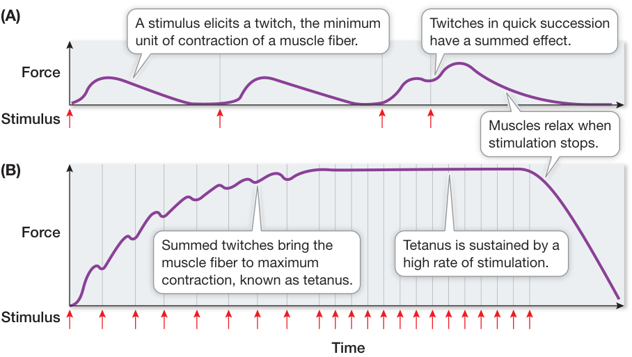
Figure 46.10 Twitches and Tetanus (A) Action potentials from a motor neuron cause a muscle fiber to twitch. Twitches in quick succession can be summed. (B) Summation of many twitches can bring the muscle fiber to the maximum level of contraction, known as tetanus.
Muscle fiber types determine endurance and strength
Not all skeletal muscle fibers are alike, and a single muscle often contains more than one type of fiber. The two major types of skeletal muscle fibers express different genes for their myosin molecules, and these myosin variants have different rates of ATPase activity. Those with high ATPase activity can recycle their actin–myosin cross-bridges rapidly and are therefore called fast-twitch fibers; those with lower ATPase activity are called slow-twitch fibers, and they develop tension more slowly but can maintain it longer (Figure 46.11A).
Slow-twitch fibers are also called oxidative or red muscle because they contain especially large amounts of myoglobin (an oxygen-binding protein similar to the hemoglobin in red blood cells), have many mitochondria, and are well supplied with blood vessels. These characteristics increase the fibers’ capacity for oxidative metabolism and result in their red appearance. The maximum tension a slow-twitch fiber produces is low and develops slowly but is highly resistant to fatigue. Slow-twitch fibers have substantial reserves of fuel, mostly lipids, so they can maintain steady, prolonged production of ATP as long as oxygen is available. Muscles with high proportions of slow-twitch fibers are good for long-term aerobic work (work that requires oxygen). Long-distance runners, swimmers, cyclists, and other athletes whose activities require endurance have leg and arm muscles consisting mostly of slow-twitch fibers (Figure 46.11B).
Fast-twitch fibers, also called glycolytic or white muscle, have few mitochondria, little or no myoglobin, and fewer blood vessels; thus they look pale. Their major energy source is glycolysis, and therefore their major stored fuel is glycogen. Fast-twitch glycolytic fibers can develop maximum tension more rapidly than slow-twitch fibers can, and that maximum tension is greater. However, fast-twitch fibers fatigue rapidly (see Figure 46.11A). The myosin of these fibers puts the energy of ATP to work very rapidly, but the fibers cannot replenish ATP quickly enough to sustain contraction for a long time. Fast-twitch fibers are especially good for short-term work that requires maximum strength, such as sprinting (see Figure 46.11B).
The types of fibers that make up a muscle influence the performance properties of that muscle, and different muscles have different fiber compositions depending on their function. Postural muscles that maintain continuous contractions are mostly composed of slow-twitch fibers. An example is the soleus muscle that runs up the back of the leg from the heel. Its contraction extends the foot and is therefore used in walking, but its continuous contraction is required for standing. If the soleus muscle fatigued, we would fall forward. We can walk or stand for a long period of time because the soleus muscle is resistant to fatigue. In contrast, a muscle that is used mostly for short-term work, such as the biceps, has a higher percentage of fast-twitch fibers than does the soleus. We can use our biceps to pick up a heavy
(B) Fiber type and performance
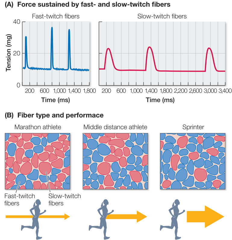
Figure 46.11 Fast- and Slow-Twitch Muscle Fibers (A) Fast-twitch fibers generate a large force quickly but fatigue rapidly. Slow-twitch fibers generate smaller forces, and do so more slowly, but can sustain them for a longer time. (B) The skeletal muscles in these three panels show slow-twitch fibers as red and fast-twitch fibers as blue. Arrows represent the duration (length) and speed (thickness) of running. Q: What would you expect the muscle fiber distribution to be in a champion weight-lifter?
weight, but we cannot hold that weight in a given position for a long period of time.
Can you change the fiber composition of your muscles to optimize your performance in a particular activity? To a limited extent, you can alter the properties of your muscle fibers through training. There are fast-twitch fibers that are somewhat oxidative and therefore intermediate in their properties between slow-twitch and fast-twitch fibers. These intermediate fibers can become more oxidative with endurance training and more glycolytic with strength training. However, the most important determinant of your muscle fiber types is your genetic heritage. There is some truth to the statement that champions are born, not made. A person born with a high proportion of fast-twitch fibers in her legs is unlikely to become a champion marathon runner, and a person born with a high proportion of slow-twitch fibers in her legs is unlikely to become a champion sprinter.
A muscle has an optimal length for generating maximum tension
Two parts of a pull-up exercise are especially difficult. When your arms are fully extended, it is hard to start the pull-up; and when your chin has almost reached the bar, the last little bit is difficult. The structure of the sarcomere is the reason.
When a muscle changes length the sarcomeres also change length, and the spatial relationship between the actin and myosin filaments changes (Figure 46.12). If there is no actin–myosin overlap, cross-bridges cannot form and the sarcomere can’t generate force. This is almost the situation when you are hanging from the pull-up bar. At the other extreme, when the muscle is fully contracted, the actin filaments are overlapping and the myosin filaments are bumping up against the Z lines, so additional force cannot be generated. With these considerations, you can ask how well a specific muscle—such as the frog jumping muscle—will function given a range of sarcomere lengths. In Investigating Life: What Is the Optimal Resting Position for the Jumping Muscle of the Frog?, we examine an experiment done to identify the optimal sarcomere length for a forceful frog jump. As you review this experiment, think about how it applies to deciding on the best starting position for a sprint.
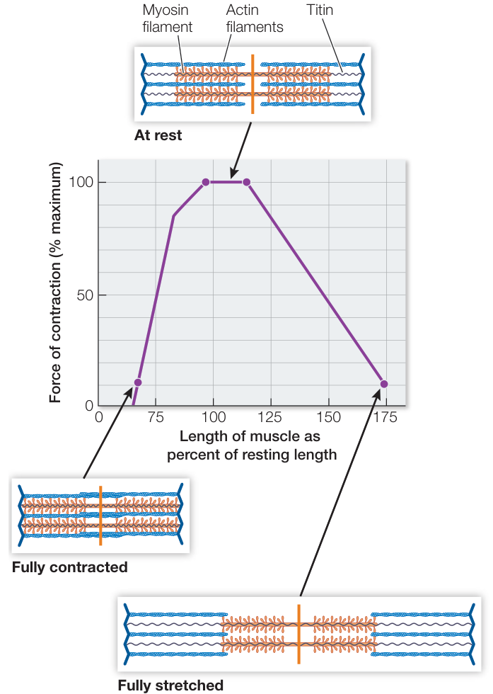
Figure 46.12 Force and Length The amount of force a sarcomere can generate depends on its resting length. When a muscle is stretched and its sarcomeres lengthened, there is less overlap between the actin and myosin filaments, and less force can be produced. When a muscle is fully contracted, the myosin filaments are bumping up against the Z lines of the sarcomeres, so further contraction is not possible. (After A. M. Gordon et al. 1996. J Physiol 184: 170–192.)
Exercise increases muscle strength and endurance
Different types of exercise produce different physical conditioning responses. In general, anaerobic activities such as weight-lifting increase strength, and aerobic activities such as jogging increase endurance. Strength is the maximum force a muscle can exert, and endurance is work capacity or how long a given workload can be sustained. What are the physiological bases for these differences?
Strength is a function of the volume of the muscles: the more actin and myosin filaments in a muscle fiber, and the more muscle fibers in a muscle, the more tension it can produce. When athletes undertake strength training, they use weights or exercises such as pull-ups or bench presses to repeatedly contract specific muscles under heavy loads. Repetitions are usually done until the muscle is completely fatigued. Such stress on a muscle does minor tissue damage—hence the soreness the day or two after a hard work-out—but it also induces the formation of new actin and myosin filaments in existing muscle fibers. The muscle fibers, and hence the muscles, get bigger and stronger. In extreme cases, and after serious muscle damage, new muscle fibers can also be produced from stem cells called satellite cells in the muscle. In general, however, the major effect of strength training is to produce bigger, rather than more, muscle fibers.
Aerobic exercise has a completely different effect on muscles: it enhances their oxidative capacity. This effect comes from increases in the number of mitochondria, in enzymes involved in energy use, and in the density of capillaries that deliver oxygen to the muscle. Myoglobin also increases in skeletal muscle cells. Myoglobin is similar to hemoglobin, but it has a higher affinity for oxygen. Therefore myoglobin accepts oxygen from the blood, facilitates the diffusion of oxygen throughout the muscle, and provides a store of oxygen for use when oxygen delivery by the blood is insufficient. By increasing the capacity of muscle to use oxygen to produce ATP, aerobic training increases the length of time that a given workload can be sustained.
Muscle ATP supply limits performance
Muscles have three systems for supplying the ATP they need for contraction:
- The immediate system uses preformed ATP and creatine phosphate.
- The glycolytic system metabolizes carbohydrates to lactate and pyruvate.
- The oxidative system metabolizes carbohydrates or fats all the way to and .
The capacity of these three systems and the rates at which they can produce ATP determine both work capacity and endurance of a muscle (Figure 46.13).
ATP is present in muscles in very small amounts. However, muscle fibers also contain a storage compound called creatine phosphate (CP). This molecule stores energy in a phosphate bond that it can transfer to ADP. The total energy available in all the muscles of your body in the form of ATP and CP—the immediate energy system—is only about 10 kilocalories. When at rest, you metabolize a kilocalorie of energy in less than a minute. Even though the energy available from ATP and CP is limited, it is available immediately, and it enables fast-twitch fibers to generate a lot of force quickly. During burst activity, the immediate system is exhausted in seconds.
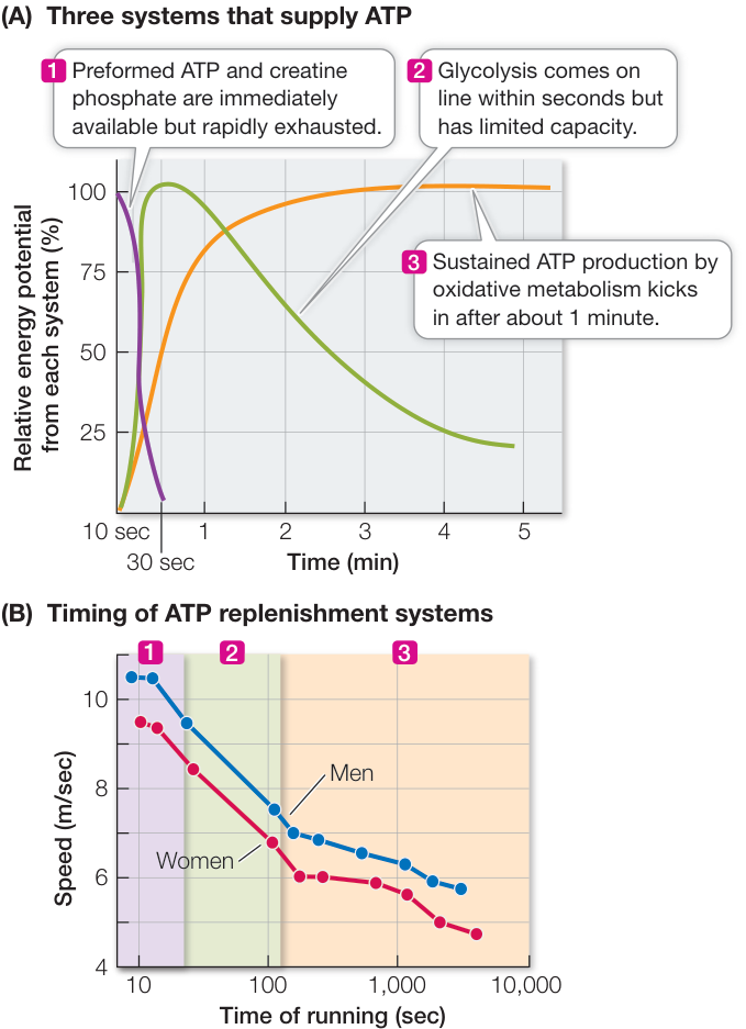
Figure 46.13 Supplying Fuel for High Performance (A) Muscles have three systems for obtaining the ATP they need for contraction during exertion such as running. (B) Looking at a plot of world-record times for running events of different durations, you can see that the performance of the athletes corresponds to the time courses of the three energy systems. (B data from the International Association of Athletics Federations. https://www.iaaf.org/records/by-category/world-records. Accessed 10/2/2019.) Q: Figure 46.13B shows how maximum speed decreased as the duration of the running events got longer. Explain why there are three different slopes for this curve.
▶ Investigating LIFE What Is the Optimal Resting Position for the Jumping Muscle of the Frog?
Experiment
Original Paper: G. J. Lutz and L. C. Rome. 1994. Built for jumping: The design of the frog muscular system. Science 263: 370–372.
The force propelling a frog’s jump should be greatest when there is optimal overlap between the actin and myosin filaments and not when the muscle is stretched. The following experiment attempted to determine the resting sarcomere length that allows a frog to achieve a maximal jump.
QUESTION If you want to get the maximum force out of a slingshot, you stretch it as far as you can. Would the frog’s jump be greater if the jumping muscle were stretched when the frog was crouched before the jump?
METHOD
- Through anatomical studies, measure sarcomere length at different resting positions observed in frogs before they jump and also measure the changes in muscle length and sarcomere length during jumps.
- Excise frog jumping muscle from a frog and mount it in a tissue culture chamber so that the muscle can be held at different lengths and electrically stimulated to contract. Attach one end of the muscle to a force transducer that measures the force generated by the contraction. With this experimental setup you can determine the relationship between sarcomere length and maximum force the muscle can generate.
- Compare the sarcomere length measurements with the forces measured in muscles held at different lengths.
- Measure velocity of contraction of the muscle under different load conditions.
RESULTS
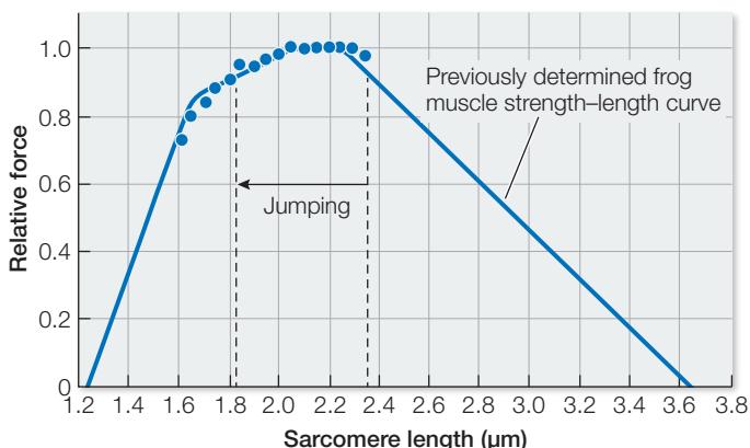
- With methods not described here, it was determined that the jumping muscle shortened from 33.6 mm to 26.1 mm during a jump, and the resting sarcomere lengths varied from 2.34 µm to 1.82 µm.
- The relative forces generated by isometric contraction at different sarcomere lengths corresponded well with a previously determined strength–length curve (data points shown in the graph).
CONCLUSION During the jump, the sarcomeres of the frog shortened over the optimal range for generation of force.
Work with the Data
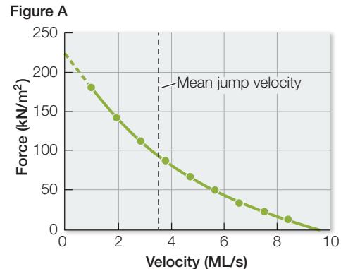
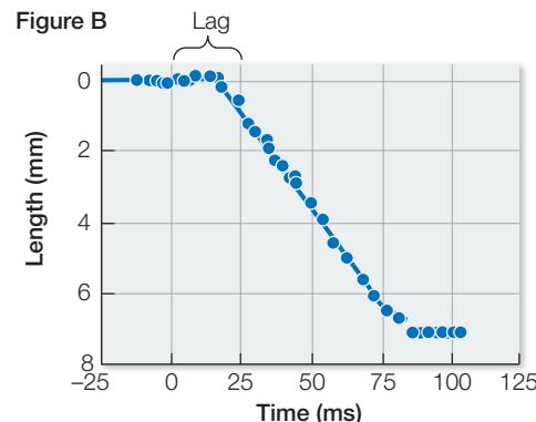
The resting position of the sarcomeres and the extent of their shortening during a jump maximize the force that the frog’s muscle can generate to power the jump. But in physical terms, power is a force applied over a specific time. Therefore the velocity of contraction (measured as muscle length/second [ML/s]) will influence the maximum power the muscle can generate, and the velocity of contraction depends on the load. If the load is too great, the contraction is isometric (velocity = 0). If the load is negligible, the velocity of contraction can be maximal. Figure A shows the relationship between force and velocity of contraction in a frog jumping muscle under different loads. The mean jump velocity is indicated. Figure B shows the change in muscle length (millimeters) during a jump (milliseconds).
QUESTIONS
- Power is work/time and work is force × displacement. Displacement/time = velocity. Therefore power = force × velocity. Using the data in Figure A, plot a curve that shows power as a function of contraction velocity for the frog muscle.
- What is the velocity of contraction of the frog jumping muscle in muscle lengths/second? Use the data in Figure B.
- Are the speed and force of the frog muscle optimal for generating the most powerful jump?
The glycolytic system activates within a few seconds to replace the ATP depleted at the onset of muscle activity. The glycolytic enzymes are located in the cytoplasm of the muscle fiber, and therefore the ATP they generate is rapidly available to the myosin filaments. However, as noted in Chapter 9, glycolysis alone is an inefficient way to produce ATP, and it leads to the accumulation of lactate, which slows the process. Thus the glycolytic system and the immediate system together provide the energy needed by active muscles for less than a minute (see Figure 46.13). Fortunately, oxidative metabolism becomes fully active in about a
Work with the Data
Does Heat Cause Muscle Fatigue?
Original Paper: D. A. Grahn et al. 2012. Work volume and strength training responses to resistive exercise improve with periodic heat extraction from the palm. J Strength Cond Res 26: 2558–2569.
Physical conditioning requires repeated intense physical activity, and the capacity of such workouts is limited by muscle fatigue. Because metabolic heat production raises the temperature of muscles during workouts, it is possible that the rise in temperature contributes to muscle fatigue and limits the capacity of workouts. To test this idea, investigators used rapid-cooling technology to extract heat from subjects during 3-minute rests between ten sets of pull-ups twice a week. In each of the ten sets of pull-ups, the subjects worked until muscle failure—the inability to complete an additional pull-up. The control condition was 3-minute rests without cooling. Each subject was his own control; the subjects were randomly assigned to begin with 6 weeks of training with cooling or 6 weeks of training without cooling, followed by a reversal of the treatments. The results are shown in the figure.
Figure A
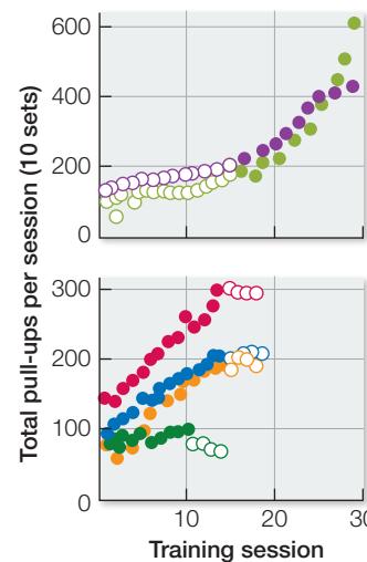
Figure B
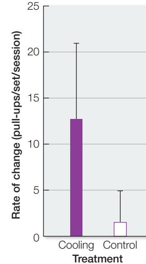
The Effect of Cooling on Workout Capacity (A) The results for individual subjects. Open symbols indicate control treatment, closed symbols indicate cooling treatment; different colors indicate different subjects. (B) The slopes of the lines in Figure A show the rates of increase in workout capacity over the course of the experiment. Mean standard deviation of rate of change in pull-ups per set during the two treatment phases ( , paired t-tests).
QUESTIONS►
- What do these data indicate about the possible role of muscle temperature in muscle fatigue?
- What do these data indicate about the relationship between workout capacity and physical conditioning effects?
minute, producing relatively huge amounts of ATP because it can completely metabolize carbohydrates and fats to and water. However, oxidative metabolism requires many reactions, and it takes place in the mitochondria, so and substrate must get into the mitochondria, and the formed ATP must get from the mitochondria to the myosin filaments in the muscle. These processes are not instantaneous, so the rate at which oxidative metabolism can make ATP available to do work is slower than the rate at which the other two systems can supply ATP.
The fuel resources available to the muscles influence how long someone can sustain a high level of aerobic exercise. From the circulating blood, muscle receives glucose and free fatty acids that it metabolizes to generate ATP. At high levels of aerobic exercise, however, most of the fuel used by muscles to produce ATP comes from the reserve of glycogen stored in the muscle itself. Depletion of muscle glycogen contributes to fatigue.
Connect the Concepts The work performance curves for different activities reflect the complexity of the biochemical processes by which different fuel sources are metabolized to produce ATP. Glycolysis supports anaerobic activities and it only entails ten enzymatic steps, so it can come on fast (see Key Concept 9.2). For aerobic activity, the citric acid cycle and the respiratory chain reactions add more steps and limit the rate of ATP production.
The rate at which muscle glycogen is replenished depends on diet. Glycogen is quickly replenished with a high-carbohydrate diet, but slowly replenished with a high-fat diet. A method used to maximize muscle glycogen is called carbo-loading. About 1 week before an athletic endurance event, athletes typically exercise for 3–5 days at a level that depletes muscle glycogen. Then, 2–3 days before the event, they taper down their level of training and eat a diet rich in complex carbohydrates. The result can be glycogen supercompensation, in which the restoration of muscle glycogen stores “overshoots” and reaches above-normal levels.
Insect muscle has the greatest rate of cycling
Insect flight muscle can produce a wingbeat frequency of up to 1,000 cycles per second. Since neuronal action potentials last 1–3 milliseconds, that number of cycles per second would exceed the capacity of motor neurons, let alone the mechanism of cycling of striated muscle contraction and relaxation. The extremely fast wingbeat of a hummingbird may be only about 50 cycles per second. How do insects do it?
Vertebrate skeletal muscle and much of invertebrate skeletal muscle is called “synchronous” because the cycling of the contractile mechanism is linked to the firing of the motor neurons. Insect flight muscle does not contract in tandem with the firing of motor neurons, however, and is thus called “asynchronous” muscle. The firing of action potentials in the insect flight motor neurons is not particularly fast, but it does cause depolarization of the muscle cell membrane, the spreading of an action potential throughout the membrane, and the release of from the sarcoplasmic reticulum. However, once the asynchronous muscle fiber is stimulated, its cycle of contraction and relaxation proceeds at its own characteristic frequency as long as is available to bind to the troponin. Thus contractile cycling and the resulting wingbeat frequency are not tied to the firing rate of the flight motor neurons.
Recap and Assess
Depending on the function a muscle serves, it may need to generate maximum force rapidly, sustain activity for a long period, or contract and relax at a very rapid rate. Properties of muscles can facilitate these types of activities.
- In terms of the relationships between motor neurons and skeletal muscles, how can the nervous system control the amount of force generated by a muscle?
- Why do postural muscles have a higher percentage of slow-twitch fibers than do muscles used for sprinting?
- How do the three systems used by muscles as sources of ATP relate to intensity and duration of peak performances in different running events?
Regardless of how much force a muscle can generate, how long it can sustain a workload, or how fast it can contract and relax, a muscle needs something to pull on; otherwise it would just be a lump of pulsating, quivering tissue. Let’s look now at how muscle and skeletal elements work together to produce movement.
Muscles and Skeletal Systems Work Together
Learning Objectives
46.3.1 Compare the features of hydrostatic skeletons, exoskeletons, and endoskeletons.
46.3.2 Explain why molting is a universal characteristic of arthropods but not of vertebrates.
46.3.3 Describe how osteoblasts, osteoclasts, and osteocytes work together to maintain bone structure.
46.3.4 Discuss how the load and force arm ratio gives different endoskeletal joints different capabilities.
Muscles can contract and exert force, or they can relax. To create significant movement, they must have something to pull on and something that stretches the muscle back to a longer position. In some cases muscles pull on each other, as in the elephant’s trunk or the arms of an octopus. In most cases, however, skeletal systems are the rigid supports against which muscles pull to create directed movement. In this key concept we examine the three types of skeletal systems: hydrostatic skeletons, exoskeletons, and endoskeletons.
Hydrostatic skeletons depend on pressure differences
Cnidarians, annelids, and other soft-bodied invertebrates have hydrostatic skeletons consisting of a volume of fluid enclosed in a body cavity surrounded by muscle (see Key Concept 29.1). When muscles oriented in one direction contract, the fluid-filled body cavity bulges out in a perpendicular direction.
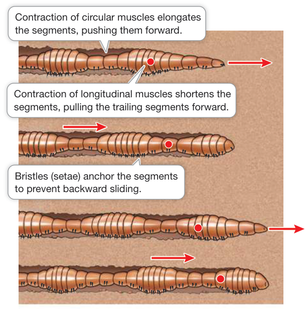
Figure 46.14 A Hydrostatic Skeleton Alternating waves of muscle contraction move the earthworm through the soil. The red dot enables you to follow the changes in one segment as the worm moves forward.
An earthworm uses its hydrostatic skeleton to crawl (Figure 46.14). The earthworm’s body cavity is divided into separate segments filled with extracellular fluid. The body wall surrounding each segment has two muscle layers: a circular layer and a longitudinal layer. If the circular muscles in a segment contract, that segment constricts and elongates. If the longitudinal muscles in a segment contract, it shortens and bulges outward. Alternating contractions of the earthworm’s circular and longitudinal muscles create waves of narrowing and widening, lengthening and shortening, that travel down the body. Bulging, shortened segments serve as anchors as long, narrow segments project forward and longitudinal contractions pull other segments forward. Bristles help the widest parts of the body to hold firm against the substrate, so the body moves forward.
Exoskeletons are rigid outer structures
An exoskeleton is a hardened, rigid outer surface to which muscles can be attached. Contractions of the muscles cause jointed segments of the exoskeleton to move relative to each other. The simplest example of an exoskeleton is the shell of a mollusk. Some marine mollusks, such as clams, have shells composed of protein strengthened by crystals of calcium carbonate (a rock-hard material). These shells can be massive, affording significant protection against predators. The shells of terrestrial snails contain less calcium carbonate, and are therefore lighter.
The most complex exoskeletons are found among the arthropods (see Key Concept 30.1). A type of exoskeleton called a cuticle covers the outer surfaces of the arthropod body, including its appendages. It is made up of chitin (a nitrogen-containing polysaccharide) secreted by a layer of cells just below the exoskeleton. Chitin stiffens and hardens the cuticle everywhere except at the joints, where flexibility must be retained. Muscles attached to the inner surfaces of the arthropod cuticle move its parts around the joints (see Figure 30.4). Growth of animals with exoskeletons is accommodated by molting (see Figure 30.3). Soft-shelled crabs, a gourmet delicacy, are crabs caught while they are molting.
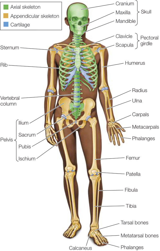
Figure 46.15 The Human Endoskeleton Cartilage and bone make up the internal skeleton of a human.
Vertebrate endoskeletons consist of cartilage and bone
The endoskeleton of vertebrates is an internal scaffolding. Muscles are attached to it and pull against it. Endoskeletons are composed of rodlike, platelike, and tubelike bones connected to one another at a variety of joints that allow a wide range of movements. An advantage of endoskeletons over the exoskeletons of arthropods is that bones in the body can grow without the animal shedding its skeleton.
The adult human skeleton consists of 206 bones, some of which are shown in Figure 46.15. It can be divided into an axial skeleton, which includes the skull, vertebral column, sternum, and ribs; and an appendicular skeleton, which includes the pectoral girdle, pelvic girdle, and bones of the arms, legs, hands, and feet.
The vertebrate endoskeleton consists of two kinds of connective tissue, cartilage and bone, which are produced by two kinds of connective tissue cells that produce extensive extracellular matrices. Cartilage cells produce a matrix that is a tough, rubbery mixture of polysaccharides and proteins—mainly fibrous collagen. Collagen fibers run in all directions like reinforcing cords through the gel-like matrix and give it the well-known strength and resiliency of gristle in meat. Cartilage is found in parts of the endoskeleton where both stiffness and resiliency are required, such as on the surfaces of joints where bones move against one another. Cartilage is also the supportive tissue in stiff but flexible structures such as the larynx (voice box), nose, and ear pinnae. Sharks and rays are called cartilaginous fishes because their skeletons are composed entirely of cartilage. In most other vertebrates, cartilage is the principal component of the embryonic skeleton, but during development most of it is gradually replaced by bone.
Bone also contains collagen fibers, but it gets its rigidity and hardness from an extracellular matrix of insoluble calcium phosphate crystals. Bone serves as a reservoir of calcium for the rest of the body and is in dynamic equilibrium with soluble calcium in the extracellular fluids of the body. This equilibrium is under the control of calcitonin and parathyroid hormone (see Figure 39.15). If too much calcium is taken from the skeleton, the bones are significantly weakened.
The living cells of bone—osteoblasts, osteocytes, and osteoclasts—are responsible for the constant dynamic remodeling of bone (Figure 46.16). Osteoblasts lay down new matrix material on bone surfaces. These cells gradually become surrounded by matrix and eventually become enclosed within the bone, at which point they cease laying down matrix but continue to exist within small lacunae (cavities) in the bone. In this state they are called osteocytes. Despite the vast amounts of matrix between them, osteocytes remain in contact with one another through long cellular extensions that run through tiny channels in the bone. Communication between osteocytes is important in controlling the activities of the cells that are laying down or removing bone.
The cells that break down bone and release calcium into the extracellular fluid are called osteoclasts. They are derived from the same cell lineage that produces white blood cells. Osteoclasts erode bone, forming cavities and tunnels. Osteoblasts follow osteoclasts, depositing new bone. Thus the interplay of osteoblasts and osteoclasts continuously replaces and remodels the bones, allowing a bone to recover from damage and adjust to the forces placed on it.
A dramatic finding from the early days of manned space missions was that the bones of astronauts who spent long periods in zero gravity decalcified. Conversely, in athletes certain bones thicken during training. Both thickening and thinning of bones are experienced by anyone who has had a leg in a cast for a long time: the bones of the uninjured leg carry the person’s weight and thicken while the bones of the inactive leg in the cast thin. We now know that there are multiple mechanisms in bone that transduce physical stress into activation of various cell signaling mechanisms that can control the bone remodeling responses to those stressors.
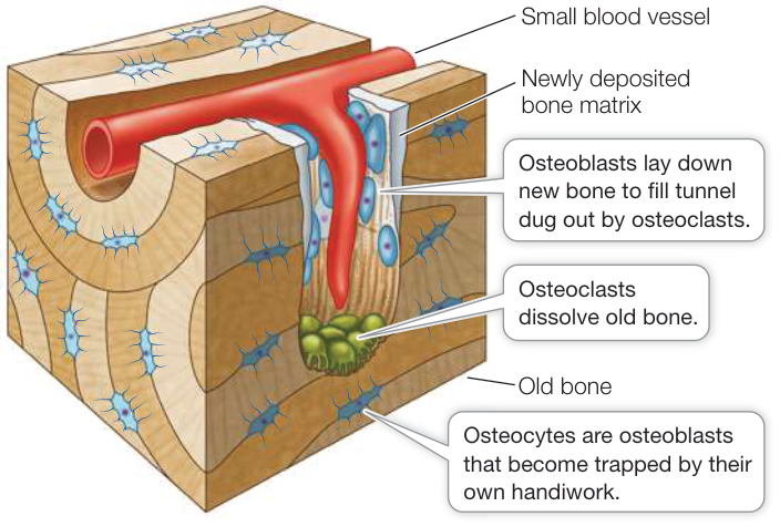
Figure 46.16 Bone Is Living Tissue Bones are constantly being remodeled by osteoblasts, which lay down bone, and osteoclasts, which break down bone.
Because of the positive effects of physical stress on bone deposition, weight-bearing exercise is effective in preventing and treating osteoporosis, which is the loss of bone density (and hence strength). More than 25 million people in the United States suffer from this debilitating condition. Although osteoporosis is most commonly a problem for postmenopausal women, it can occur in younger people as a result of malnutrition. For example, the condition known as female athlete triad includes eating disorders, cessation of menstrual cycling, and osteoporosis. These are interactive conditions in which the eating disorder and excessive training lead to malnutrition that can result in endocrine disruption and osteoporosis. Excessive training and malnutrition can lead to bone loss in males as well.
Bones develop from connective tissues
Bones are divided into two types on the basis of how they develop. Membranous bone forms on a scaffold of connective tissue membrane. Cartilage bone forms first as a cartilaginous structure resembling the future mature bone, then gradually hardens, or ossifies, to become bone. The outer bones of the skull are membranous bones; the bones of the limbs are cartilage bones.
Cartilage bones can grow throughout the ossification process. The long bones of the legs and arms, for example, ossify first at the centers and later at each end (Figure 46.17). The cartilage between these ossification centers forms epiphyseal plates. Growth can continue until these areas of ossification join, eliminating the epiphyseal plates. The membranous bones forming the skull cap grow until their edges meet. The large soft spot on the top of a baby’s head, called a fontanel, is a point where the skull bones have not yet joined. There are multiple fontanels in the neonate where the skull bones eventually join.
The structure of bone may be compact (solid and hard) or cancellous (having numerous internal cavities that make it appear spongy, although it is rigid). The architecture of a specific bone depends on its position and function, but most bones have both compact and cancellous regions. The shafts of the long bones of the limbs, for example, are cylinders of compact bone surrounding central cavities that contain fatty bone marrow called yellow bone marrow. The rigid, tubelike shaft of compact bone can withstand compression and bending forces. Architects and nature alike use hollow tubes as lightweight structural elements.
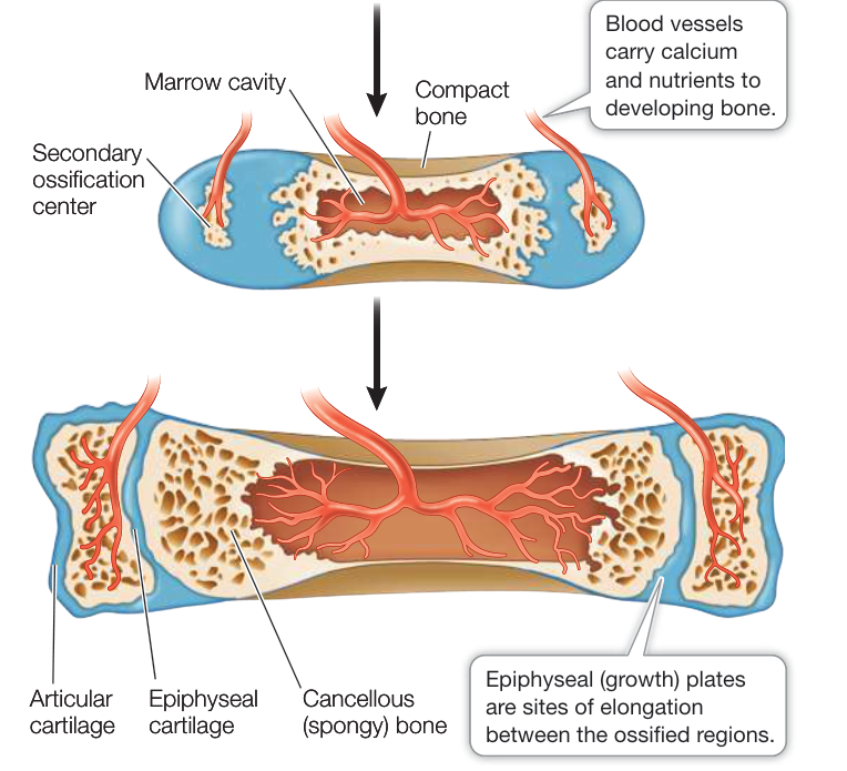
Figure 46.17 The Growth of Long Bones In the long bones of human limbs, ossification occurs first at the centers and later at each end.
The ends of the long bones are cancellous (see Figure 46.17). Cancellous bone is lightweight because of its numerous cavities. These cavities contain the red bone marrow responsible for red blood cell formation as well as other cellular elements of the blood. Cancellous bone is also strong because its internal mesh-work constitutes a support system. It can withstand considerable forces of compression.
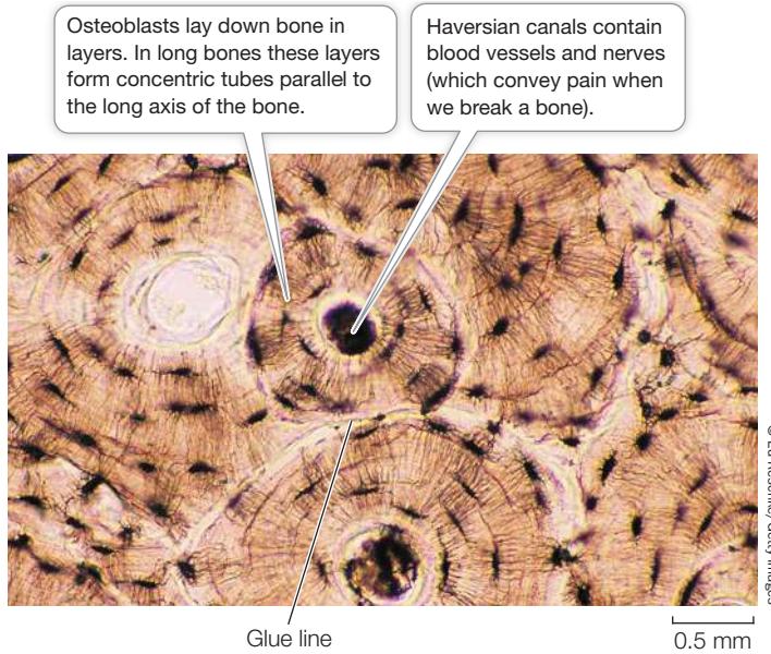
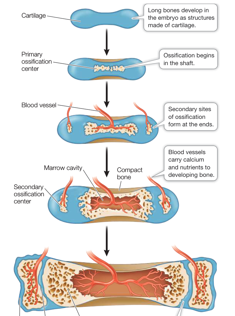
Figure 46.18 Most Compact Bone Is Composed of Haversian Systems A micrograph of a section of a long bone shows Haversian systems with their central canals. Glue lines separate Haversian systems. Osteocytes reside in small spaces and are in communication by means of thin cell processes running through small channels.
Most compact bone in mammals is called Haversian bone because it is composed of structural units called Haversian systems (Figure 46.18). Each Haversian system is a set of thin, concentric bony cylinders that surround a narrow Haversian canal that contains blood vessels and nerves. Osteocytes are arranged in small spaces between the concentric circles of bone surrounding the canals. Adjacent Haversian systems are separated by boundaries called glue lines. Haversian bone is resistant to fracturing because cracks tend to stop at glue lines.
Bones that have a common joint can work as a lever
Muscles and bones work together around joints, where two or more bones come together. Different kinds of joints allow motion in different directions (Figure 46.19), but muscles can exert force in only one direction. Therefore muscles create movement around joints by working in antagonistic pairs: when one muscle contracts, the other relaxes. When both contract, the joint becomes rigid (which is important for maintaining posture, for example).
With respect to a particular joint, such as the knee, we refer to the muscle that bends, or flexes, the joint as the flexor, and the muscle that straightens, or extends, the joint as the extensor. The bones that meet at the joint are held together by ligaments, which are flexible bands of connective tissue. Other straps of connective tissue, called tendons, attach the muscles to the bones. In Figure 46.20, which shows the knee joint, you can see a tendon that connects a flexor muscle in the upper leg (biceps femoris) to a bone in the lower leg. You can also see a tendon connecting the extensor muscle (quadriceps) to the kneecap (patella) and the patellar ligament that connects the patella to the lower leg bone. It is the patellar ligament that is tapped when testing the knee-jerk reflex (see Figure 43.15).
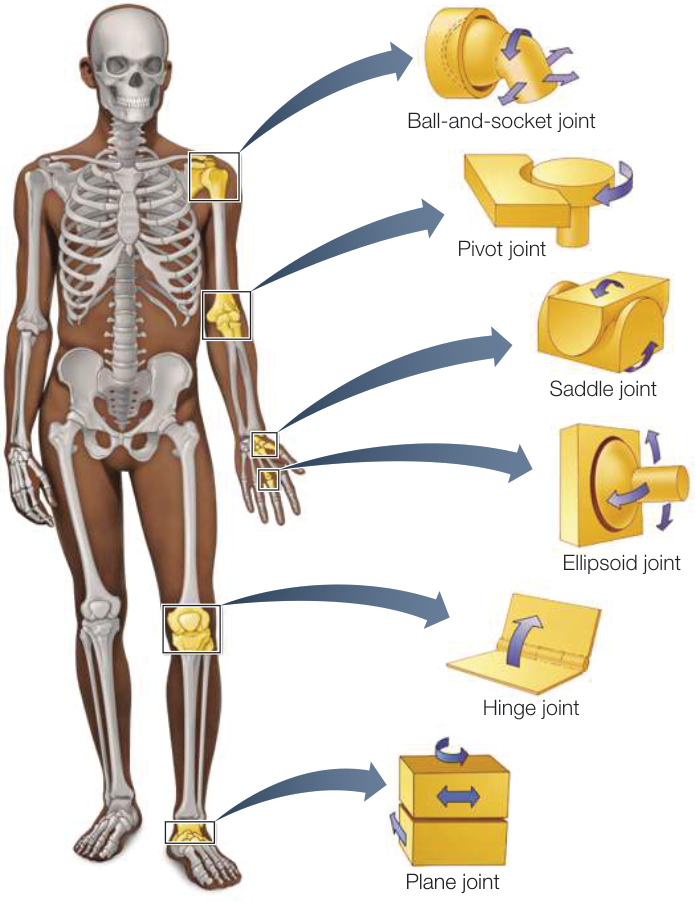
Figure 46.19 Types of Joint Motions The designs of joints are similar to mechanical counterparts and enable a variety of movements. Activity 46.3 Joints
Bones constitute a system of levers that are moved around joints by the muscles. Levers have three parts: load arm, force arm, and fulcrum. The joint is the fulcrum, and the length of the force arm relative to the load arm depends on where the muscle attaches to the bone. Three classes of levers are defined based on the location of the fulcrum (Figure 46.21). In a class 1 lever, the fulcrum is between the load and force arms, as in a seesaw. An example is the joint between the skull and the spinal cord. A class 2 lever is like a wheelbarrow, with the load in the middle. An example is the ankle joint and the muscle in the back of the leg that enables you to walk and stand on tiptoe. In a class 3 lever, the force arm is between the fulcrum and the load. An example is your elbow or your knee. The length ratio of the load and force arms determines whether a particular lever can exert a lot of force over a short distance or is better at translating force into large or fast movements. Think of the powerful jaws of carnivores that can easily crack bones. In contrast, the force arm of the lower legs of Rosie the Ribeter and Jackie Joyner-Kersee, featured in the opener to this chapter, is short relative to the load arm, so they can jump fast and high.
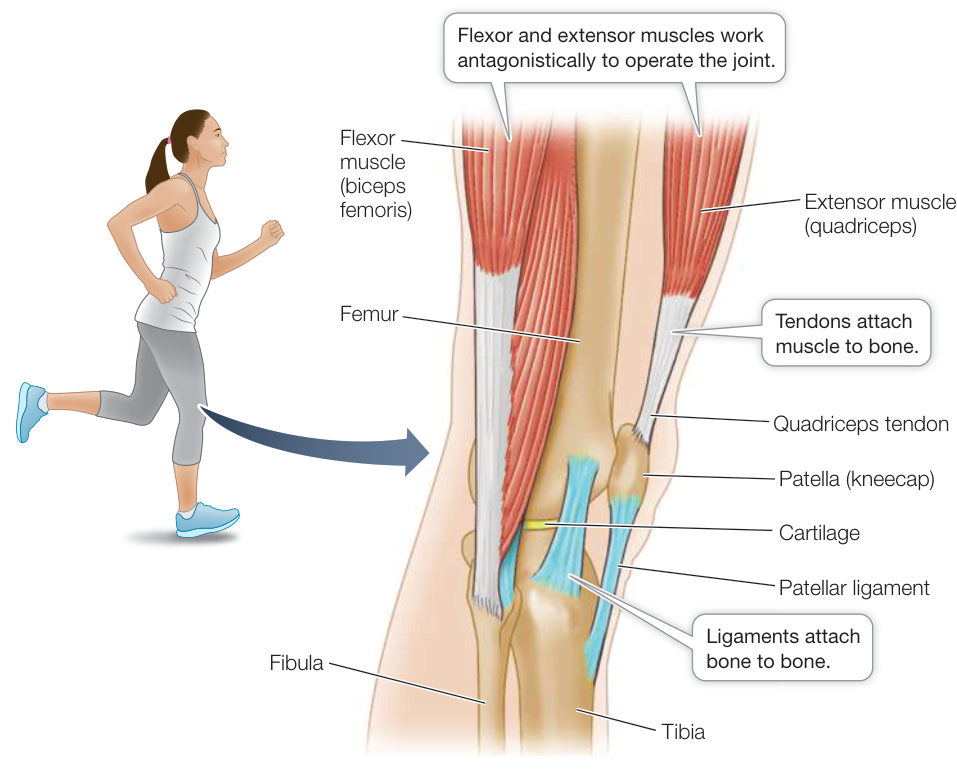
Figure 46.20 Joints, Ligaments, and Tendons A side view of the knee shows the interactions of muscle, bone, cartilage, ligaments, and tendons at this crucial and vulnerable human joint.
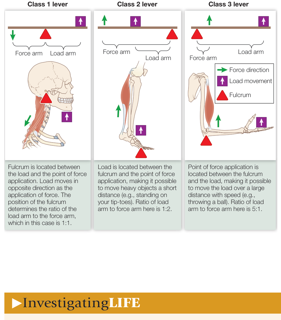
Figure 46.21 Bones and Joints Work as Levers The three classes of levers are defined by the relationship between the fulcrum and the load and force arms. For each class of lever, the load and force arm ratio determines whether force or speed of movement is maximized. Q: What class of lever is the knee joint?
Investigating Life
Q&A What adaptations optimize the jumping muscles of the frog?
Rosie the Ribeter was an exceptional jumper judged by the length of time her record remains unbeaten, but frogs in general are remarkable jumpers. This ability is not due to the molecular structure of their muscles, as the arrangement of sarcomeres is very similar across a wide cross section of animals, and in all cases the sarcomeres and therefore the muscles only contract by about 25%–30% of their length. So how can a muscle only a few centimeters long produce a jump of many meters? One answer, which is considered in the experiment described in Investigating Life: What Is the Optimal Resting Position for the Jumping Muscle of the Frog?, involves maximizing force and power. First, you saw that the resting posture of the frog maintains the sarcomere length where it can generate the maximum force when it contracts. Then by using the measured force/velocity curve, you saw that the velocity of the jump was at the level that maximized the power of the jump. This rather fast velocity of contraction is possible because the jumping muscle contains mostly fast-twitch fibers. As mentioned in the opening story, and further explained in Key Concept 46.3, the legs operate as class 3 levers to move the body mass. Because the legs of the frog are long, the load arms of those levers are long in comparison with the force arms—that is, the length ratio of the load and force arms is high. A frog’s long legs and big feet have another advantage as well. Muscles must have something to exert force against to maximize power, and once the frog leaves the ground, its muscles can no longer generate power. Long legs and feet ensure that as the jumping muscles contract, the feet remain in contact with the ground for a longer time, thus maximizing the power of the jump.
Future directions
Understanding how muscles and skeletal structures work together to generate movements of different types is essential in the growing field of robotics. Robots that repeat the same movement over and over are quite common in manufacturing, but more and more robotic systems are being created to do more variable and complex tasks that mimic and frequently augment the abilities of humans and other animals. For example, robots have been developed to travel over rough terrain, even while carrying heavy loads. Robots have been developed to do household chores such as washing dishes and folding laundry. Robotic systems are improving the functionality of prosthetic limbs for amputees. And melding of robotic systems with normal human functionality is leading to technologies called “exoskeletons” that greatly enhance the physical abilities of humans. The starting point in all of these engineering efforts is a thorough characterization of how the biological musculoskeletal system works.
Recap and Assess
Muscles can only contract and relax; to achieve organized movement, they must pull against rigid structures—other muscles, hydrostatic skeletons, exoskeletons, or endoskeletons. Endoskeletons consist of cartilage and bone. Bone is continuously resorbed by osteoclasts and laid down by osteoblasts. Joints give directionality to movements, and muscle–joint combinations act as levers.
- Why is molting a universal characteristic of arthropods but not of vertebrates?
- What changes in the activities of bone cells might explain the loss of bone mass in astronauts who spend long periods at the International Space Station, and what mechanism in the bone might be responsible?
- What is the significance of the fact that the ratio of the force arm to the load arm is much greater in the jaw joint than in the elbow joint?
Visual Summary
© Michael Durham/Minden Pictures
You should be able to relate each summary to the adjacent figures. If you go to this visual summary in Achieve, you can follow links to figures, animations, activities, and simulations that will help you consolidate the material.
Interactions of Actin and Myosin Cause Muscles to Contract
- ■ Skeletal muscle fibers are multinucleate cells containing myofibrils, which are regularly arranged bundles of actin and myosin filaments forming sarcomeres as the functional units of contraction.
- ■ A motor neuron and the fibers it activates constitute a motor unit. The synapse between a motor neuron and a muscle fiber is a motor end plate.
- ■ Action potentials generated at the motor end plate spread across the muscle cell membrane and through the T tubules, causing to be released from the sarcoplasmic reticulum.
- ■ binds to troponin and changes its conformation, pulling the tropomyosin strands away from myosin binding sites on the actin filament.
- ■ Cardiac muscle cells are striated, uninucleate, branching, and electrically connected by gap junctions.
- ■ Smooth muscle cells are uninucleate and connected by gap junctions.
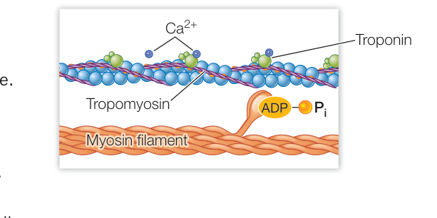
Figure 46.6
Questions
- How does an action potential in the T tubule initiate skeletal muscle contraction?
- Why do skeletal muscles stiffen soon after death?
- What are the structural and functional differences between skeletal, cardiac, and smooth muscle?
Many Factors Affect Muscle Performance
- ■ In skeletal muscle, an action potential causes a minimum unit of contraction called a twitch. Twitches occurring in rapid succession can be summed to achieve graded levels of contraction. Sustained maximal contraction is known as tetanus.
- ■ Slow-twitch fibers are used for extended aerobic work; fast-twitch fibers generate maximum forces for short periods of time. Mixtures of slow-and fast-twitch fibers suit different muscles for different functions.
- ■ Anaerobic exercise stimulates the enlargement of muscle fibers through production of new filaments. Aerobic exercise stimulates greater oxidative capacity of muscle fibers.
- ■ Muscle performance depends on a supply of ATP.
Questions
- If you compared the fiber composition of the large leg muscles of a champion sprinter and a cross-country skier, what differences would you expect to find?
- A high-fat diet results in lower levels of muscle glycogen. If a weight-lifter and a mile-runner went on a high-fat diet, which one would see the greatest decrease in performance, and why?
- Why is it harder to do pull-ups if you go to full extension between pull-ups?
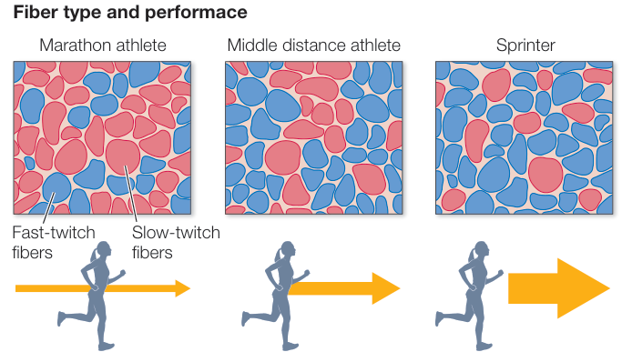
Figure 46.11
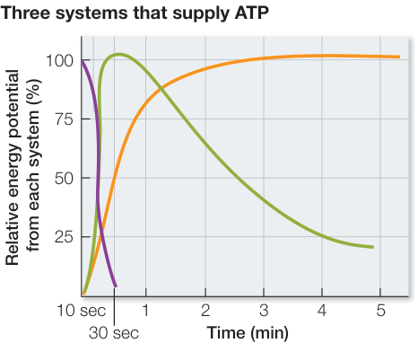
Figure 46.13


Muscles and Skeletal Systems Work Together
- ■ Hydrostatic skeletons are fluid-filled body cavities that are squeezed by muscles.
- ■ Exoskeletons are hardened outer surfaces to which internal muscles are attached.
- ■ Endoskeletons are internal systems of rigid rodlike, platelike, and tubelike supports, consisting of bone and cartilage to which muscles are attached.
- ■ Bone is continually remodeled by osteoblasts, which lay down new bone, and osteoclasts, which resorb bone.
- ■ Bones develop from connective tissue membranes (membranous bone) or from cartilage (cartilage bone) through ossification.
- ■ Bone is compact (solid and hard) or cancellous (containing numerous internal spaces). Most compact bone of mammals is composed of Haversian systems.
- ■ Joints enable muscles to power movements in different directions.
- ■ Tendons connect muscles to bones; ligaments connect bones to one another.
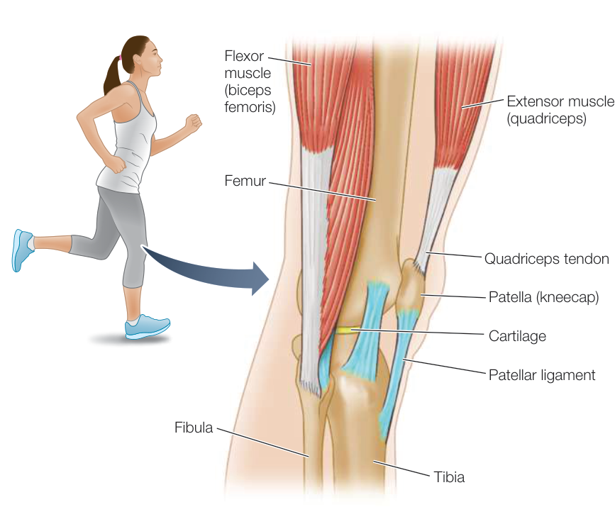
Figure 46.20
Questions
- Earthworm bodies are divided into separate segments. Why is this important for their locomotion?
- Why is the human skull divided into epiphyseal plates?
- Is height an advantage or a disadvantage to a football kicker, and why?