Chapter 47Gas Exchange
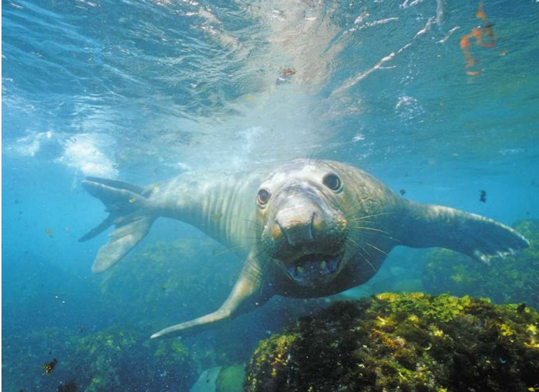
© Bruce Walkins/Animals Animals/AGE Footstock
Key Concepts
47.1 Respiratory Gas Exchange Is Governed by Physical Factors
47.2 Enhancing Diffusion Maximizes Respiratory Gas Exchange
47.3 Humans Have Tidal Respiration
47.4 Respiratory Gases Are Transported by the Blood
47.5 Breathing Is Homeostatically Regulated
Northern elephant seals (Mirounga angustirostris) are champion breath-hold divers. Diving repeatedly, they may spend only 20% of their time at the surface.
Investigating Life: The Breath of Life
Northern elephant seals (Mirounga angustirostris) on a beach are impressive. Typical adult males are 5 meters long and weigh 2,500 kilograms. They fight viciously to defend areas of beach where the females come out of the water to give birth and rear their young. The beach masters are really defending their opportunities to father the next generation.
Physiologically, these animals are even more impressive in the water. Female elephant seals, half the size of the males and therefore easier and safer to manage for experiments, have been fitted with instruments to record the depth and duration of their dives after they leave the beach where they gave birth. They spend up to 7 months at sea, and during that entire time they dive continually—day and night—to feed on eels, skates, rays, squid, and other prey. Most dives are 300–600 meters deep and last 20 minutes, but dives lasting over an hour and exceeding 2,000 meters have been recorded. The seals spend only 4–5 minutes at the surface between dives. Thus for the 80% of their lives that they spend at sea, they are breathing only about 20% of the time—12 minutes per hour!
The best human divers are perhaps the pearl divers of Japan and Korea. They dive repeatedly during their workday to about 20 meters, and their dives last about a minute. Record breath-hold dives for humans resting facedown in a pool are about 10 minutes, but that is a one-time event that cannot be repeated for many hours. The elephant seals are not resting—they are descending and ascending great distances and pursuing prey.
How can these behemoths spend so much time not breathing? How can they survive such crushing depths? How can they not get the bends, which is a serious danger for human divers? How much oxygen do these animals need to carry out their normal behavior? How much oxygen can they store in their bodies during the short times they spend at the surface? And what adaptations do they have to make those oxygen stores last as long as they do? To answer these questions, elephant seals and other diving animals have been studied in unique laboratory facilities and in the field.
QA What adaptations make seals champion breath-hold divers? (Find the answer on page 1066.)
Respiratory Gas Exchange Is Governed by Physical Factors
Learning Objectives
- 47.1.1 Describe the concept of partial pressures of gases and how they relate to concentrations of gases in air and water.
- 47.1.2 Explain adaptations for respiratory gas exchange in terms of Fick’s law of diffusion.
- 47.1.3 Compare water and air as respiratory media.
- 47.1.4 Describe three factors that make it more difficult for fishes to satisfy their respiratory needs at higher temperatures.
- 47.1.5 Explain how elevation influences respiratory gas exchange.
The respiratory gases that animals must exchange are oxygen () and carbon dioxide (). Cells need to obtain from the environment to produce an adequate supply of ATP by cellular respiration (see Chapter 9). is an end-product of cellular respiration, and it must be removed from the body to prevent toxic effects. Gas exchange systems of animals consist of (1) specialized body surface areas where these gases can move between the body and the external environment, (2) ventilation mechanisms that move air or water over the environmental sides of those surfaces, and (3) perfusion mechanisms that circulate extracellular fluids on the internal sides.
Diffusion is the only means by which respiratory gases are exchanged between an animal’s internal body fluids and the outside medium (air or water). There are no active transport mechanisms to move respiratory gases across biological membranes. Because diffusion is a physical process, knowing what physical factors influence rates of diffusion helps us understand the diverse adaptations of gas exchange systems. (You may want to review the coverage of diffusion in Key Concept 6.3.)
Diffusion of gases is driven by partial pressure differences
Diffusion is due to the random motion of molecules, and it results in a net movement of molecules from areas of higher concentration to areas of lower concentration. In other words, diffusion evens out the distribution of molecules. Diffusion of respiratory gases occurs either in water or in air. Concentrations of gases in water or air vary with pressure because gases are compressible, a relationship described in physics by Boyle’s law. For example, there are twice as many gas molecules in a liter of gas at 2 atmospheres of pressure as there are in a liter of gas at 1 atmosphere of pressure. And if that gas is in contact with a liquid such as water, twice as many gas molecules will enter into solution when the gas pressure is 2 atmospheres than when it is 1 atmosphere.
The concentrations of different gases in a mixture are described as the partial pressures of those gases. To calculate the partial pressure of a gas such as oxygen in a mixture of gases such as air, we have to know the total pressure. The total pressure of air inhaled by air-breathing animals is the atmospheric pressure. At sea level, atmospheric pressure is about 760 millimeters of mercury (mm Hg), depending on the weather. Because dry air is 20.9%
, the partial pressure of oxygen () at sea level is 20.9% of 760 mm Hg, or about 159 mm Hg. If two gas mixtures are separated by a membrane permeable to , will diffuse from the mixture where its partial pressure is higher to the mixture where its partial pressure is lower.
To calculate the concentration of in the environment of a water breather, we have to know two things: the partial pressure of in the air in contact with the water, and the solubility of in water. The amount of a gas that dissolves in a liquid depends both on its partial pressure in the gas phase in contact with the liquid and on its solubility in that liquid. The diffusion of a gas between the gas phase and the liquid phase is a function of its partial pressures in those two phases; the gas diffuses from the phase with the higher partial pressure to the phase with the lower partial pressure until equilibrium is reached—the point at which the partial pressures in the two phases are equal. However, the amount of the gas that can be contained in the liquid depends on the solubility of that gas in that liquid. Furthermore, the solubility of a gas in a particular liquid can vary widely depending on conditions. What follows is a practical illustration of these facts.
Solubility of a gas in a liquid, such as oxygen in water, is a function of temperature—solubility is higher at low temperatures. Think of opening a bottle of warm soda in comparison with a bottle of cold soda. When the pressure is relieved by opening the bottle, more gas comes out of solution—there is more fizz—in the warm soda. So if we have similar containers of water in equilibrium with air, but at different temperatures, the concentrations of oxygen in these water containers will be different (less in the warmer one), but the partial pressures of oxygen will be the same. Thus for water-breathing animals, the warmer the water is, the less there is per liter of water. The important point is that for a gas in solution, its concentration is not the same as its partial pressure, but partial pressures are what drive diffusion. Thus in our continuing discussions of respiratory gas exchange, we will always use partial pressure rather than concentration when referring to the diffusion of respiratory gases.
Fick’s law applies to all systems of gas exchange
Whether in air or water, the diffusion rates of respiratory gases between an animal and its respiratory medium—air or water—depend on the partial pressure gradients across the gas exchange surfaces and on other factors that are described quantitatively with a simple equation called Fick’s law of diffusion. All environmental variables that limit respiratory gas exchange and all adaptations that maximize respiratory gas exchange are reflected in one or more components of this equation. Fick’s law is written as
where
- is the rate at which a gas such as diffuses between two locations.
- is the diffusion coefficient, which is a characteristic of the diffusing substance, the medium, and the temperature. For example, perfume has a higher than motor oil vapor, and all substances diffuse faster at higher temperatures and faster in air than in water. Temperature is not expressed explicitly in Fick’s law because diffusion coefficients are usually determined at room temperature (about ).
- is the area across which the gas is diffusing.
- and are the partial pressures of the gas at the two locations.
- is the path length, or distance, between the two locations.
- is a partial pressure gradient.
The strict dependence of animals on diffusion for gas exchange with their environments has selected for various adaptations that maximize , many of which we describe in this chapter. Animals can maximize for respiratory gases by using air rather than water as their gas exchange medium whenever possible. All other adaptations for maximizing respiratory gas exchange must influence the surface area () for gas exchange or the partial pressure gradient across that surface area.
Air is a better respiratory medium than water
The slower diffusion of molecules in water than in air affects both air-and water-breathing animals. Diffusion of in water is so slow that even animal cells with low rates of metabolism cannot function more than a few millimeters away from a good source of environmental . Therefore there are severe size and shape limits on the many species of invertebrates that lack internal systems for distributing . Most of these species are small or, like marine flatworms, have evolved a flat, thin body with a large external surface area (Figure 47.1A). Another strategy is seen in sponges, which have bodies made of cells surrounding water channels through which the external medium flows (Figure 47.1B; also see Figure 29.2B). Critical factors enabling larger, more complex animal bodies are (1) the evolution of specialized respiratory structures with large surface areas that are highly permeable to respiratory gases (Figure 47.1C) and (2) internal systems that facilitate the movement of respiratory gases between the respiratory exchange surfaces and the tissues of the body.
Respiratory systems of air-breathing animals also have had to evolve adaptations to minimize the path length for diffusion of respiratory gases in aqueous media. Eukaryotic cells carry out cellular respiration in their mitochondria, which are located in the cytosol, a gel-like substance. Cells are bathed in extracellular fluid—also an aqueous medium. All respiratory surfaces consist of cells, and to protect them from drying out, they are covered by a thin film of fluid. must diffuse through all of these aqueous respiratory barriers. So even in air-breathing animals, the slow rate of diffusion in water limits the efficiency of distribution from the environment to the sites of cellular respiration.
can be obtained more easily from air than from water for several reasons:
The content of air is much higher than the content of an equal volume of water. The maximum content of a bubbling stream in equilibrium with air is less than 10 milliliters (mL) of per liter of water. The content of the air over the stream is about 200 mL of per liter of air.
diffuses about 8,000 times more rapidly in air than in water. That is why the content of a stagnant pond can be zero only a few millimeters below the surface.
An animal has to work (expend energy) to ventilate its gas exchange surfaces with water or air. More energy is required to move water than air because water is 800 times denser than air and about 50 times more viscous.
You can appreciate how important these facts were for the evolutionary transition of life to the terrestrial environment, because they meant that there were fewer constraints on the evolution of higher metabolic rates.
High temperatures create respiratory problems for aquatic animals
Animals that use water for their respiratory exchange medium are in a double bind when environmental temperatures rise. Most water-breathing animals are ectotherms—their body temperatures are closely tied to the temperature of the water around them. As the water temperature rises, an ectotherm’s body temperature and metabolic rate rise (see Figure 38.8). Thus water breathers need more as their environment gets warmer (Figure 47.2). But as mentioned earlier, warm water holds less dissolved gas than cold water does. In addition, since a water-breathing animal performs work to move water across its gas exchange surfaces, it must expend more energy to breathe as water temperature rises. Therefore as water temperature goes up, a water-breathing animal must extract more and more from an environment that is increasingly deficient, and has a lower percentage of that available to support activities other than breathing.
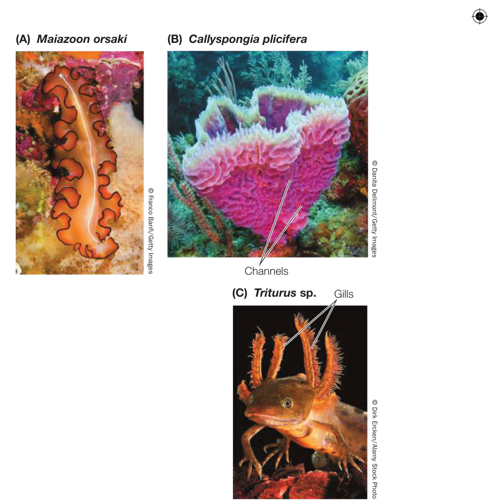
Figure 47.1 Keeping in Touch with the Medium (A) No cell in the leaflike body of this marine flatworm is more than a millimeter away from seawater. (B) Sponges have body walls perforated by many channels, which allow water to flow between the outside world and a central cavity. No cell in the sponge is more than a millimeter away from seawater. (C) A feathery fringe of gills on this larval aquatic salamander provides a large surface area for gas exchange. Blood circulating through the gills comes into close contact with the respiratory medium.
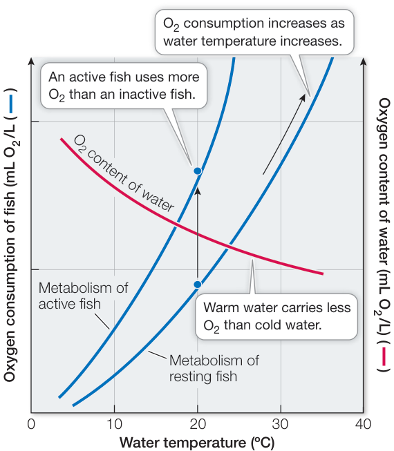
Figure 47.2 The Double Bind of Water Breathers Fishes need more when the water is warmer, but warm water carries less than cold water.
availability decreases with elevation
Just as a rise in water temperature reduces the supply of available to water-breathing animals, an increase in elevation reduces the supply for air breathers. At all elevations, makes up 20.9% of the dry air; however, as you go up in elevation, the total amount of gas per unit of volume decreases, as reflected by the atmospheric pressure. For example, at 5,800 meters, atmospheric pressure is only half what it is at sea level, so the at that elevation is only about 80 mm Hg. At the summit of Mount Everest (8,850 m), is only about 50 mm Hg—roughly one-third what it is at sea level.
Because diffusion of across respiratory gas exchange surfaces and into the body depends on the difference between the air and the body fluids, the reduced in the air at high elevations constrains uptake. Because of this, mountain climbers attempting peaks such as Mount Everest or K2 usually breathe from pressurized bottles.
is lost by diffusion
Respiratory gas exchange is a two-way process: diffuses out of the body as diffuses in. The direction and rate of diffusion of the respiratory gases across the exchange surfaces depend on the partial pressure gradients of the gases. The partial pressure gradients of and across these exchange surfaces are quite different. The amount of in the atmosphere is extremely low (0.03%), so for air-breathing animals there is always a large concentration gradient for diffusion of from the body to the environment. Whereas the partial pressure gradient for decreases with increasing elevation, the gradient driving out of the body hardly changes. The partial pressure of carbon dioxide () in the atmosphere is close to zero both at sea level and atop Mount Everest.
In general, getting rid of is not a problem for water-breathing animals because is more soluble in water than is . Even in stagnant water, where the is higher than in moving water, the lack of becomes a problem for an animal long before exchange difficulties arise.
Recap and Assess
Respiratory gases are exchanged only by diffusion and are therefore a function of partial pressure gradients. All factors influencing respiratory gas exchanges are components of Fick’s law of diffusion. Air is a better respiratory medium than water because a given volume of air has more than the same volume of water. diffuses faster in air than in water, and less work is required to move air over respiratory exchange surfaces. exchange with the external environment occurs readily because of a large concentration gradient from the body to the environment and because is highly soluble in water.
- What is the partial pressure of oxygen in inhaled air at sea level; at 2,000 meters above sea level, where atmospheric pressure is 80% that of sea level; and while breathing air at the same pressure as the environment at 10 meters (= an additional 1 atmosphere of pressure) underwater?
- Flatworms (see Figure 47.1A) are invertebrates that do not have respiratory systems. What two components of Fick’s law enable flatworms to satisfy their need for oxygen?
- The content of water decreases with an increase in water temperature, but the gill ventilation rate of a fish increases much more than is necessary to match that decrease in availability. Why?
- In terms of Fick’s law, describe differences between the ability to exchange and at sea level and at high elevation.
Now that we have discussed the physical factors that influence diffusion rates of respiratory gases between animals and their environments, let’s look at some of the adaptations that have evolved for maximizing respiratory gas exchange.
Enhancing Diffusion Maximizes Respiratory Gas Exchange
Learning Objectives
47.2.1 Explain how insects can sustain high levels of activity without active respiratory systems.
47.2.2 Explain the concept and adaptive significance of countercurrent exchange, using fish gills as the example.
47.2.3 Describe how the avian respiratory system maximizes the partial pressure gradient for uptake.
47.2.4 Determine the total lung capacity (including residual volume) of a human.
Adaptations to maximize respiratory gas exchange are reflected in the values of Fick’s law of diffusion . Such adaptations have to do with one or more of the following:
- Increase the surface area for gas exchange (A)
- Maximize the partial pressure difference driving diffusion ()
- Minimize the diffusion path length (L)
- Minimize the diffusion that takes place in an aqueous medium (maximize )
With these factors in mind, we now look at four different respiratory systems: those of insects, fishes, birds, and mammals.
Respiratory organs have large surface areas
A variety of anatomical adaptations maximize the specialized body surface areas (A) for respiratory gas exchange. Water-breathing animals generally have gills, and air-breathing animals have tracheae or lungs. Gills are highly branched and folded extensions of the body surface that provide a large surface area for gas exchange. Because gills consist of thin, delicate tissues, they minimize the path length () traversed by diffusing molecules of and . External gills are found in larval amphibians and in the aquatic larvae of many insects (Figure 47.3A; see also Figure 47.1C). External gills are vulnerable to damage and are tempting morsels for predators, so in many animals protective body cavities for gills have evolved. Such internal gills are found in many mollusks such as clams, many arthropods such as crabs and lobsters (Figure 47.3B), and in all fishes.
Lungs are highly divided internal cavities with large surface areas for respiratory gas exchange with air (Figure 47.3C). Because lung tissue is elastic, lungs can be inflated with air and deflated as a means of ventilating the respiratory exchange surfaces.
Insects have a respiratory gas exchange system consisting of a network of air-filled tubes called tracheae that branch through all tissues of the insect’s body (Figure 47.3D). The tracheae branch into smaller tracheoles that further branch into extremely fine air capillaries. These air capillaries, which are the respiratory gas exchange surfaces, are so numerous that they have an enormous surface area compared with the external surface area of the insect’s body.
Ventilation and perfusion of gas exchange surfaces maximize partial pressure gradients
Partial pressure gradients, , drive diffusion across gas exchange surfaces; the larger the gradient, the greater the rate of gas exchange. These gradients can be maximized in several ways:
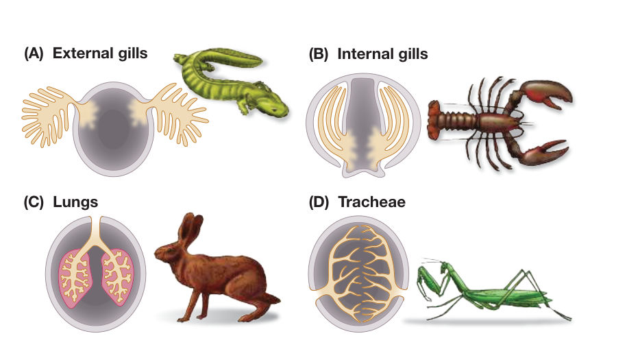
Figure 47.3 Gas Exchange Systems Large surface areas for the diffusion of respiratory gases (tan in these diagrams) are common features of animals. External (A) and internal (B) gills are adaptations for gas exchange with water. Lungs (C) and tracheae (D) are adaptations for gas exchange with air.
- Minimizing path length: Very thin tissues in gills and lungs reduce the diffusion path length ().
- Ventilating: Actively moving the external medium over the gas exchange surfaces (i.e., breathing) regularly exposes those surfaces to fresh respiratory medium containing maximum and minimum concentrations. This maximizes the partial pressure gradients.
- Perfusing: Actively moving the internal medium (e.g., blood) over the internal side of the exchange surfaces transports to those surfaces and away from them, thus maximizing the partial pressure gradients driving diffusion.
Insects have airways throughout their bodies
The tracheal system that enables insects to exchange respiratory gases extends to all tissues in the insect body. Thus respiratory gases diffuse through air most of the way to and from every cell. The insect respiratory system communicates with the outside environment through gated openings called spiracles in the sides of the abdomen and thorax (Figure 47.4A,B). The spiracles open to allow gas exchange and then close to decrease water loss. They open into tubes called tracheae that branch into even finer tubes, the tracheoles, that end in tiny air capillaries—the actual gas exchange surfaces (Figure 47.4C).
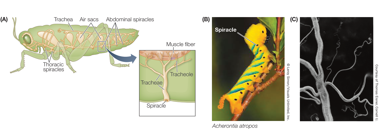
Figure 47.4 The Tracheal Gas Exchange System of Insects (A) In insects, respiratory gases diffuse through a system of air sacs and tubes (tracheae) that open to the external environment through holes called spiracles. (B) The spiracles of a hawkmoth larva run down its sides. (C) A scanning electron micrograph shows an insect tracheae dividing into smaller tracheoles and still finer air capillaries.
Fish gills use countercurrent flow to maximize gas exchange
The internal gills of fishes are supported by gill arches that lie between the mouth cavity and the protective opercular flaps on the sides of the fish just behind the eyes (Figure 47.5A). Water flows unidirectionally into the fish’s mouth, over the gills, and out from under the opercular flaps. Thus there is a constant one-way flow of oxygenated water over the gills, maximizing the on the external gill surfaces. On the internal side of the gill membranes, the circulation of blood minimizes the by sweeping away as rapidly as it diffuses across.
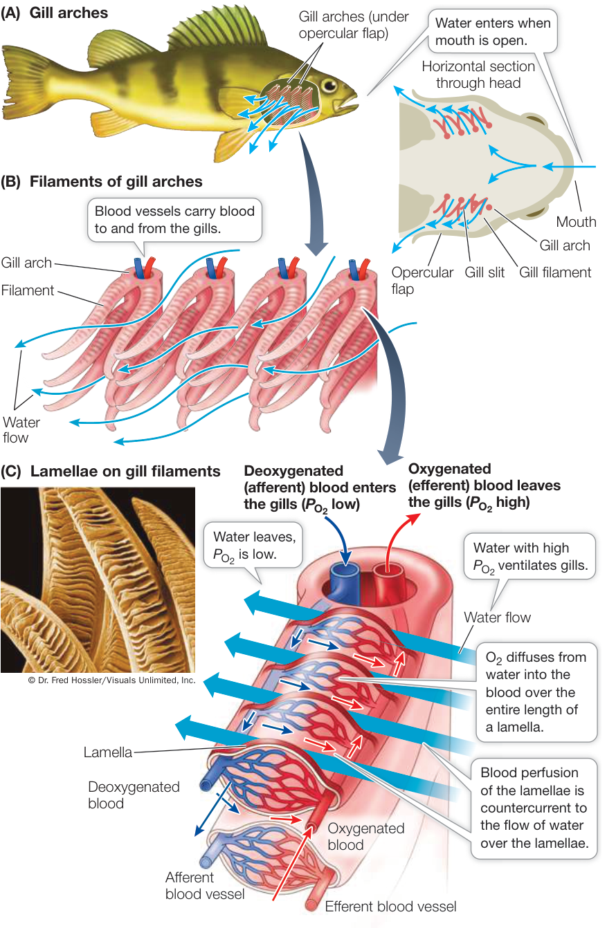
Figure 47.5 Fish Gills (A) Water flows unidirectionally over the gills of a fish, in through the mouth and out through the opercular opening. (B) Gill filaments have a large surface area and thin membranes. (C) Gill filaments are lined with lamellae, which are the gas exchange surfaces. Blood flows through the lamellae (left to right in this drawing; small blue and red arrows) countercurrent to the direction of the flow of water across the gills (right to left in this drawing; large blue arrows).
Gills have an enormous surface area for gas exchange because they are so highly subdivided. Each gill consists of hundreds of ribbonlike gill filaments (Figure 47.5B). The upper and lower flat surfaces of each gill filament are covered with rows of evenly spaced folds, or lamellae (Figure 47.5C). The lamellae are the actual gas exchange surfaces. Because the lamellae are exceedingly thin, the path length () for diffusion of gases between blood and water is minimized. The surfaces of the lamellae consist of highly flattened epithelial cells, so the water and the fish’s red blood cells are separated by little more than 1–3 micrometers.
The flow of blood perfusing the inner surfaces of the lamellae, like the flow of water over the gills, is unidirectional. Afferent blood vessels bring deoxygenated blood to the gills, while efferent blood vessels take oxygenated blood away from the gills (see Figure 47.5C). However, blood flows through the lamellae in the opposite direction to the flow of water over the lamellae. This countercurrent flow maximizes the transfer of from water to blood because at all points along the exchange surface there is a gradient between water and blood. In other words, the in the water and the blood never come into equilibrium as they would if the two parallel streams flowed in the same direction (concurrent flow) (Figure 47.6). Thus even though the concentration gradient at any one point along the gill filament is not large, the effective concentration gradient across the entire length of the gill filament is maximal.
Some fishes, including anchovies, tuna, and certain sharks, ventilate their gills by swimming almost constantly with their mouths open. Most fishes, however, ventilate their gills by means of a two-pump mechanism. The closing and contracting of the mouth cavity pushes water over the gills, and the expansion of the opercular cavity prior to opening of the opercular flaps pulls water over the gills. In both cases the ventilation of the gills is unidirectional and continuous, thus maximizing the on the water side of the lamellar membranes.
These adaptations for maximizing the surface area (A) for diffusion, minimizing the path length (L) for diffusion, and maintaining the a gradient along the entire length of the lamellae allow fishes to efficiently extract from meager environmental sources.
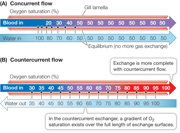
Figure 47.6 Countercurrent Flow Enables More Complete Exchange In these models of concurrent and countercurrent gas exchange, the numbers represent the saturation percentages of blood and water. (A) In a concurrent exchanger, the saturation percentages of blood and water reach equilibrium halfway across the exchange surface. (B) A countercurrent exchanger allows more complete gas exchange because the water always has a higher than the blood; thus gradients of support diffusion of into the blood across the entire length of the exchanger. Q: What could make the concurrent exchanger achieve the same maximum saturation as the countercurrent exchanger?
Birds use unidirectional ventilation to maximize gas exchange
Birds are remarkable for their ability to sustain high levels of activity for a long time—on long-distance flights for example—even at high altitudes where mammals cannot even survive. The first team to climb Mount Everest (8,850 meters) was surprised to see birds flying over the mountain when they themselves could barely move without supplemental . Bar-headed Geese (Anser indicus) regularly migrate over Mount Everest and surrounding peaks, but the highest recorded flight of a bird is that of a Ruppell’s Vulture (Gyps rueppelli) that was sucked into a jet engine at 11,278 meters. Humans cannot survive at such elevations without supplemental . Yet the lungs of a bird are smaller than the lungs of a similar-sized mammal, and bird lungs expand and contract less during a breathing cycle than do mammalian lungs. Most different from mammalian lungs is that the volume of bird lungs decreases during inhalation and increases during exhalation. How do birds accomplish such remarkable feats of respiratory gas exchange?
Figure 47.7 The Respiratory System of a Bird (A) Air sacs in the body cavity (and air spaces in the bones) are unique to birds. (B) Air comes in through the trachea, which divides into a left and a right primary bronchus. The primary bronchi go to the posterior air sacs. Secondary bronchi connect the posterior air sacs to the lungs. Air flows unidirectionally through the lungs in numerous parabronchi and flows in secondary bronchi from the lungs to the anterior air sacs, then out through the trachea. (C) Air capillaries running between the parabronchi are the site of gas exchange.
The structure of bird lungs allows air to flow unidirectionally through the lungs, rather than bidirectionally through all the same airways, as it does in tidally ventilating mammals (discussed in the next section). Because mammalian lungs are never completely emptied of air during exhalation, there is always some lung volume that is not ventilated with fresh air. The air remaining in lungs and airways after exhalation is called dead space. Bird lungs, by contrast, have very little dead space, and the fresh incoming air is not mixed with stale air. In this way, a high gradient is maintained.
Air sacs are an important and unique feature of the avian respiratory system, and they occupy much of the body cavity of the bird (Figure 47.7A). They can be divided into a group of anterior air sacs and a group of posterior air sacs. The air sacs are interconnected with each other, with the lungs, and with air spaces in some of the bones. The air sacs are not gas exchange surfaces; rather, they act as bellows to maintain a unidirectional flow of air through the lungs.
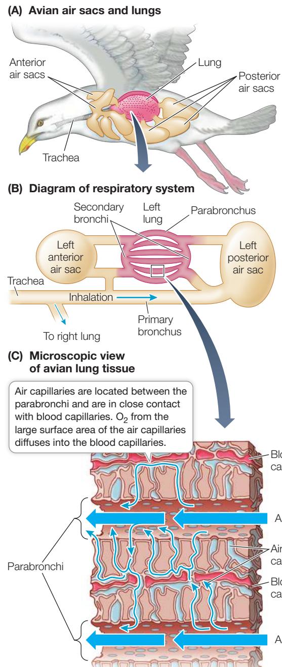
As in other air-breathing vertebrates, air enters and leaves a bird’s gas exchange system through the trachea (commonly known as the windpipe, and not to be confused with the air-conducting tracheae of insects) (Figure 47.7B). The trachea divides into two smaller airways, the primary bronchi (singular bronchus). The primary bronchi extend all the way to the posterior air sacs and also branch into secondary bronchi. The posterior air sacs also have connections to the secondary bronchi. Secondary bronchi divide into tubelike parabronchi that run parallel to one another through the lungs in a posterior to anterior direction.
Branching off the parabronchi and running between them are numerous tiny air capillaries (Figure 47.7C). Air flows through the parabronchi and diffuses into the air capillaries, which are the gas exchange surfaces. The air capillaries are so numerous that they provide an enormous surface area for gas exchange. The blood capillaries are in close contact with the air capillaries, and oxygen from the air diffuses into the blood. The parabronchi coalesce into larger bronchi that take the air out of the lungs and into anterior air sacs and then back to the trachea. Thus the anatomy of a bird’s airways allows air to flow unidirectionally through the lungs: trachea, bronchi, posterior air sacs, parabronchi, bronchi, anterior air sacs, trachea.
The puzzle of how birds breathe was solved by placing small sensors at different locations in birds’ air sacs and airways. The birds were then allowed to inhale pure for just a single breath, which made it possible to track that particular breath by recording when each of the sensors registered a peak value. The experiment showed that a single breath remains in a bird’s respiratory system for two cycles of inhalation and exhalation. During inhalation, the air sacs expand and air flows through the trachea into the posterior air sacs. At the same time, the lungs are contracting as air moves from the lungs into the anterior air sacs. During exhalation, the air sacs contract. The posterior air sacs push air into the lungs, which are expanding, and the anterior air sacs push air into the trachea and out of the respiratory system. This bellows-like action of the air sacs maintains a continuous, unidirectional flow of fresh air through the lungs (Figure 47.8).
The advantages of the avian respiratory system are similar to those of fish gills. The air sacs keep fresh air flowing unidirectionally over the gas exchange surfaces without interruption. Thus those gas exchange surfaces are constantly exposed to a that is very close to that of the ambient air. Even at high elevations where the of the ambient air may be only slightly above that of the blood, can still diffuse continuously from air to blood.
Tidal ventilation produces dead space that limits gas exchange efficiency
Lungs evolved in early lungfishes as outpocketings of the digestive tract. Although lung structure has evolved considerably, lungs remain dead-end sacs in all air-breathing vertebrates except birds (and likely their extinct reptilian ancestors). Ventilation of dead-end sacs with common passageways for inflow and outflow cannot be constant and unidirectional but must be
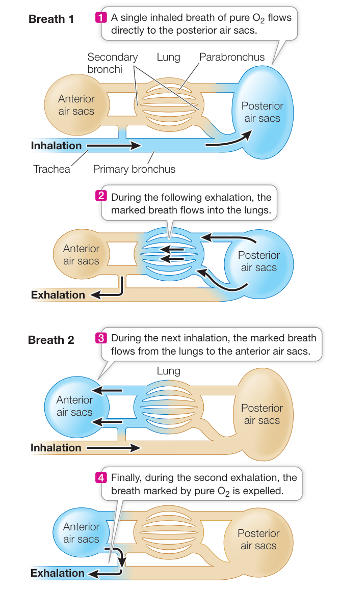
Figure 47.8 An Experiment to Describe Airflow in the Avian Respiratory System Oxygen sensors placed in different locations in the avian respiratory system trace the flow of a single breath of pure oxygen (blue) through the system. A single breath remains in the system for two breathing cycles.
tidal: air flows in and exhaled gases flow out by the same route. Since the lungs and airways can never be completely emptied of air, they always contain dead space. We can easily measure the volumes of air exchanged during breathing, but we have to use an indirect method to measure the dead space contained in the lungs and airways. Measures of dead space are important in assessing lung health and disease.
A flowmeter measures the volume of air breathed in and out (Figure 47.9). Using a human as an example, the amount of air that moves either in or out per breath when at rest is called the tidal volume (TV) (about 500 mL for an average human adult). When we breathe in as much as possible, the additional volume is the inspiratory reserve volume (IRV). Conversely, if we forcefully exhale as much air as possible, the additional amount of air expelled is the expiratory reserve volume (ERV). The maximum capacity for air exchange in one breath, or the vital capacity (VC), is the sum of . The vital capacity of an athlete is generally greater than that of a nonathlete, and vital capacity decreases with age because of stiffening of the lung tissue.
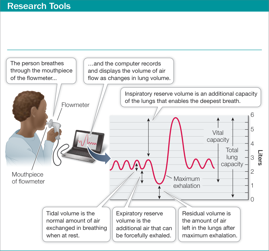
Figure 47.9A Measuring Lung Ventilation A flowmeter measures the volume of air a person breathes through a mouthpiece. The combined tidal volume, inspiratory reserve volume, and expiratory reserve volume are the lungs’ vital capacity. The vital capacity plus the residual volume are the total lung capacity.
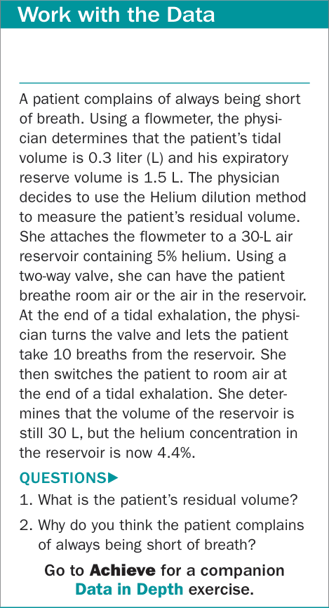
Figure 47.9B Measuring Lung Ventilation
Vital capacity is not the entire lung volume because of the dead space, also called the residual volume (RV). We can’t measure RV directly with the flowmeter, but we can measure it indirectly using the helium dilution method. The flowmeter is attached to a closed reservoir of air of a known volume () so that a person can breathe in and out from that reservoir. If this reservoir has a known amount of helium () in it, and a subject breathes the air in that reservoir through several breathing cycles, the helium will become distributed in the reservoir and in the subject’s respiratory system. Helium is not absorbed into the body from the lungs, so the total amount of helium at the beginning of a test () will equal the total amount at the end of the test ()—it will just be distributed over a larger volume and therefore be less concentrated. The following formula quantifies that relationship:
We start the subject breathing from the reservoir at an end-tidal breath, and we end the breathing from the reservoir at an end-tidal breath. Since the does not change, the decrease in helium concentration [ ] enables us to calculate the subject’s functional residual volume (FRV), which is the .
Since we can measure the ERV with the flowmeter, we can subtract the ERV from the FRV to obtain the RV.
Why is the RV important? Referring to Figure 47.9, you will see that for a normal person the ERV is about 1,000 mL and the RV is 1,000 mL. Thus the FRV is 2,000 mL, but the tidal volume is only 500 mL; this means that the air that reaches the alveoli (the actual gas exchange surfaces) with each breath consists of only 500 mL of fresh air diluted by 2,000 mL of stale air. The maximum in this mixed air is much below the of the outside air, and because of the tidal ventilation pattern, the in the alveoli is steadily dropping during the breathing cycle. The RV is important because it contributes to the FRV and to the dilution of the in the inhaled air. Any disease or condition that increases the RV (such as emphysema or pulmonary fibrosis) compromises a patient’s respiratory ability. Similarly, considering the mixing of fresh air with the FRV, you can understand why reductions in tidal volume can be a problem—and therefore why patients recovering from surgery are encouraged to breathe deeply, even if it hurts.
Small residual volume prevents the bends in seals
In the opening of this chapter we discussed the remarkable abilities of elephant seals to dive to great depths for long times and then spend only minutes breathing at the surface. We asked how elephant seals avoid getting the bends, a very painful condition that develops in scuba divers who surface too rapidly after spending time at depths. The pressure of the water on the body increases by 1 atmosphere for every 10 meters of depth. Body tissues are not compressible, but air in the respiratory system is, so scuba divers breathe air through a regulator that equalizes the air pressure in their lungs to the pressure of the water on the body. As the total air pressure goes up, the partial pressures of the component gases go up, increasing their rate of diffusion into the tissues. Eighty percent of the air is nitrogen, so the amount of nitrogen dissolved in tissues increases. If the diver comes up too rapidly, the dissolved nitrogen comes out of solution in the tissues, creating small bubbles that block blood vessels, damage tissues, cause pain, and can even result in death. To avoid the bends, divers use decompression tables that tell them how to ascend depending on how deep and how long their dives have been. For example, if a diver has spent 1 hour at a depth of 60 meters, he would have to spend 3 hours gradually ascending to shallower and shallower depths to allow the nitrogen dissolved in his tissues to diffuse out through the respiratory system rather than form bubbles in his tissues. Why does a seal that makes repeated dives not face the same consequences? The seal exhales before diving and the residual air in its respiratory system gets compressed into its airways, which are not respiratory exchange surfaces. So when seals dive they do not have compressible air in their lungs that can be forced into tissues at depth and then come out of solution upon resurfacing.
Recap and Assess
The major adaptations that increase efficiency of respiratory gas exchange are large surface areas for exchange and maximized partial pressure gradients across those surfaces. Fishes have countercurrent exchange across their gill filaments. Fish and avian respiratory systems have continuous and unidirectional ventilation of their respiratory exchange surfaces. Mammals have tidal ventilation, resulting in residual volumes of air that dilutes the incoming fresh air.
- Given constant rates of ventilation and perfusion of fish gills, how can the directionality of blood flow inside the gill lamellae and directionality of water flow on the outside the lamellae influence the maximum exchange that can occur?
- How do birds maintain a constant and unidirectional flow of air through their lungs?
- Find the functional residual volume of a person who yields the following data while breathing through a flowmeter from a closed reservoir that has a volume of 30 liters (L) and an initial gas mixture of 20% , 75% nitrogen (N), and 5% helium (He). Beginning with an inhalation, the person inhales and exhales 20 times. The final concentration of He in the flowmeter is 4.6%.
Despite their limitations, mammalian lungs serve the respiratory needs of mammals well. Offsetting the inefficiencies of tidal breathing, mammalian lungs have an enormous surface area and a very short path length for diffusion. Next we look at the human respiratory system as an example.
Humans Have Tidal Respiration
Learning Objectives
- 47.3.1 Explain the roles of surface tension and surfactant in breathing.
- 47.3.2 Explain how changes in volume and pressure in the thoracic cavity are involved in ventilation.
- 47.3.3 Describe the changes (in arbitrary units) in tension between the pleural membranes and the pressure in the alveoli, and airflow during a breathing cycle.
The respiratory exchange surfaces in mammalian lungs are the very thin-walled alveoli that cluster around a huge number of finely branching terminal airways. To understand how these respiratory surfaces are ventilated and perfused, we first examine the functional anatomy of the whole respiratory system (Figure 47.10).
Lungs are ventilated through a branching system of airways
Air enters the lungs through the oral cavity or through the nasal passages, which join together in the pharynx (see Figure 47.10A). Below the pharynx, the esophagus conducts food to the stomach, and the trachea conducts air to the lungs. At the beginning of this airway is the larynx, or voice box, which houses the vocal cords. The larynx is the lump that you can see or feel on the front of your neck. The trachea is about 2 centimeters in diameter. C-shaped bands of cartilage prevent the thin walls of the trachea from collapsing as air pressure changes during the breathing cycle. If you run your fingers down the front of your neck just below your larynx, you can feel a few of these bands of cartilage.
The trachea branches into two primary bronchi, one leading into the right lung and one into the left. The right lung has three lobes and the left lung has two, so there is an asymmetry in the secondary bronchi, with three on the right and two on the left. The bronchi branch repeatedly to generate treelike structures of progressively smaller airways extending to all regions of the lungs. After four branchings, the cartilage supports disappear, marking the transition to bronchioles. After about 16 branchings, the bronchioles are less than a millimeter in diameter, and tiny, thin-walled air sacs called alveoli begin to appear. Alveoli are the sites of gas exchange. After the first alveoli there are about six more branchings of the airways that end in clusters of alveoli (see Figure 47.10B). Because the airways conduct air only to and from the alveoli and do not themselves participate in gas exchange, their volume is dead space.
Human lungs have about 300 million alveoli. Although each alveolus is very small, their combined surface area for diffusion of respiratory gases is about 70 square meters—about one-fourth the size of a basketball court. The walls of each alveolus are made of very thin cells. Wrapped around the alveoli are networks of capillaries that also have very thin walls. Thus where capillary meets alveolus, the diffusion path length between air and blood is less than 2 micrometers (see Figure 47.10C).
Diseases of the bronchioles and alveoli are the fourth leading cause of death in the United States as of 2019 Among these diseases, the most lethal is emphysema, a condition in which inflammation damages and eventually destroys the walls of the alveoli. As a result, the lungs have fewer but larger alveoli, the lungs lose elasticity, and the RV increases. A principal cause of emphysema is smoking and another is air pollution.
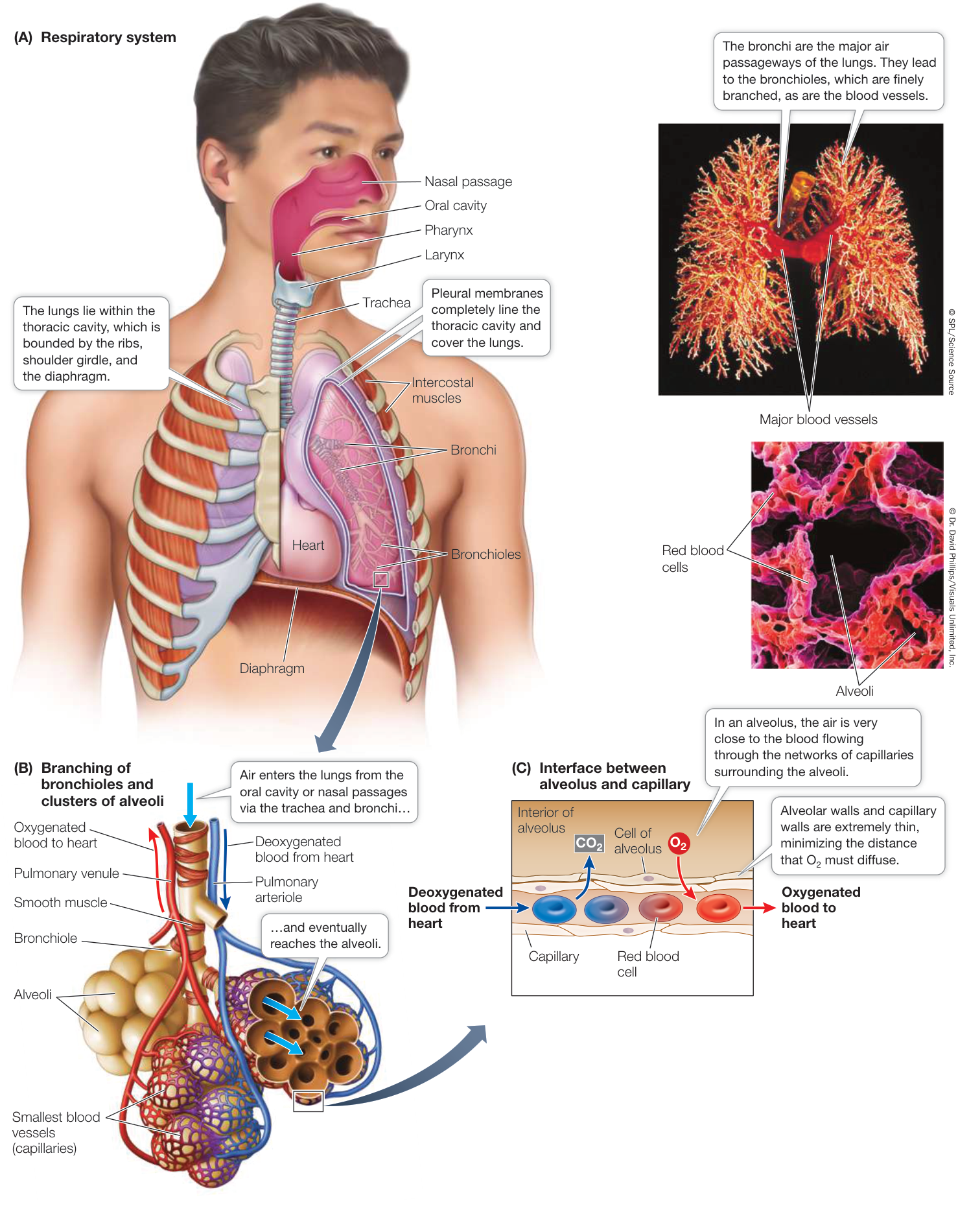
Figure 47.10 The Human Respiratory System The diagrams trace the hierarchy of human respiratory structures, from the airways to the minuscule alveoli. Q: If the trachea branches into two bronchi, each of those bronchi branches into two smaller airways, and so forth for 23 branchings, how many terminal airways are there? Activity 47.1 The Human Respiratory System
Respiratory tract secretions aid ventilation
Mammalian lungs produce two secretions that do not directly influence their gas exchange but do affect the process of ventilation: mucus and surfactant.
Many cells lining the airways produce sticky mucus that captures bits of dirt and microorganisms that are inhaled. Other cells lining the airways have cilia that beat continually and sweep the mucus, with its trapped debris, up toward the pharynx, where it is swallowed or spit out. This phenomenon, called the mucus escalator, can be adversely affected by inhaled pollutants. Smoking one cigarette can immobilize the cilia of the airways for hours. A smoker’s cough results from the need to clear the obstructing mucus from the airways when the mucus escalator is out of order.
A surfactant is a substance that reduces the surface tension of a liquid. Surface tension gives the surface of a liquid the properties of an elastic membrane, and it is why certain insects, such as water striders, can walk on water. As discussed in Key Concept 2.4, surface tension is the result of chemical forces of attraction between water molecules. The attractive forces working on the water molecules at the surface pull from below and from the sides but not from above. This imbalance of forces creates surface tension. The thin film of fluid covering the air-facing surfaces of the alveoli has surface tension that contributes to the lungs’ elasticity. To inflate the lungs, enough force has to be generated to overcome both the elasticity of the lung tissue and the surface tension in the alveoli.
Lung surfactant is a fatty lipoprotein complex substance that reduces the work necessary to inflate the lungs. Certain cells in the alveoli release these surfactant molecules. If a baby is born more than a month prematurely, these cells may not have developed the ability to produce surfactant. A baby with this condition, known as respiratory distress syndrome (RDS), will have great difficulty breathing and may die from exhaustion and lack of . RDS is the leading cause of death for premature babies, but current treatment protocols have greatly reduced mortality due to RDS. Those treatments include application of synthetic or animal-derived surfactants directly into the infant’s lungs. Positive airway pressure applied through nasal tubes also helps ventilate the infant’s lungs.
Laundry detergent is also a surfactant, but lung surfactant differs in a very important way. Detergent dissolves in water; lipoprotein surfactant does not, and remains at the water-air interface. Therefore as the alveoli expand, the surfactant molecules are more thinly distributed over a larger surface area and have less of an effect on surface tension. Why is this property important?
Have you ever wondered how a tiny fan can hold up a collapsible cover over a tennis court or a swimming pool? The answer is LaPlace’s law, which describes the relationships between pressure (), tension (), and radius () of bubbles: . The larger the radius, the less pressure it takes to overcome the tension. Alveoli are not all the same size, so according to LaPlace’s law, inhalation should preferentially inflate the larger alveoli and not the smaller ones. The unique property of lung surfactant solves this problem because as the larger alveoli inflate, their surface tension increases, favoring the inflation of the smaller alveoli. Also, the increased surface tension in the inflated lungs adds to the recoil during exhalation, facilitating the emptying of the lungs.
Lungs are ventilated by pressure changes in the thoracic cavity
Human lungs are suspended in the thoracic cavity, a closed compartment bounded on the bottom by a sheet of muscle called the diaphragm (see Figure 47.10A). Each lung is covered by a continuous sheet of tissue called the pleural membrane that also lines the thoracic cavity adjacent to the lung. A thin film of fluid between the pleural membranes of the lung and the thoracic cavity lubricates the inner surfaces of the pleural membranes so they can slip and slide against each other. As with surface tension, there are forces of attraction between the molecules of fluid covering the pleural membranes. As a result, it is difficult to pull the pleural membranes apart. Think of two wet panes of glass or two wet microscope slides; you can slide them past each other, but it is difficult to separate them. While the inner surfaces of the pleural membranes are “stuck” to each other by surface tension, they can move relative to each other during breathing movements.
Inhalation and exhalation involve changes in the volume of the thoracic cavity (Figure 47.11). Because the pleural membranes covering the cavity wall and the lung surface are stuck to each other by surface tension, any attempt to increase the volume of the thoracic cavity increases the tension between the pleural membranes. Even between breaths, there is tension between the pleural membranes because the rib cage is pulling outward and the elasticity of the lung tissue is pulling inward. This slight negative pressure keeps the alveoli partly inflated even at the end of an exhalation. If the thoracic cavity is punctured—by a knife wound, for example—air can leak into the space between the pleural membranes and cause the lung to deflate. If the wound is not sealed, breathing movements pull air in between the pleural membranes rather than into the lung (a “sucking chest wound”), and there is no ventilation of the alveoli in that lung—a condition called “collapsed lung.”
At rest, inhalation is initiated by contraction of the muscular diaphragm (see Figure 47.11A). As the domed diaphragm contracts, it pulls down, expanding the thoracic cavity and pulling on the pleural membranes. Since the pleural membranes cannot separate, they pull on the lungs, increasing their volume and decreasing the alveolar pressure. As a result, air rushes in through the trachea from the outside. Exhalation begins when contraction of the diaphragm ceases. As the diaphragm relaxes, the elastic recoil of the lung tissues pulls the diaphragm up and pushes air out through the airways (see Figure 47.11B). When a person is at rest, inhalation is an active process and exhalation is a passive process.
The diaphragm is not the only muscle that can change the volume of the thoracic cavity. Between the ribs are two sets of intercostal muscles (see Figure 47.11C). The external intercostal muscles expand the thoracic cavity by lifting the ribs up and outward. The internal intercostal muscles decrease the volume of the thoracic cavity by pulling the ribs down and inward. During strenuous exercise, the external intercostal muscles increase the volume of air inhaled, making use of the inspiratory reserve volume, and the internal intercostal muscles increase the amount of air exhaled, making use of the expiratory reserve volume. The abdominal muscles can also aid in breathing. When they contract, they cause the abdominal contents to push up on the diaphragm and thereby contribute to the expiratory reserve volume.
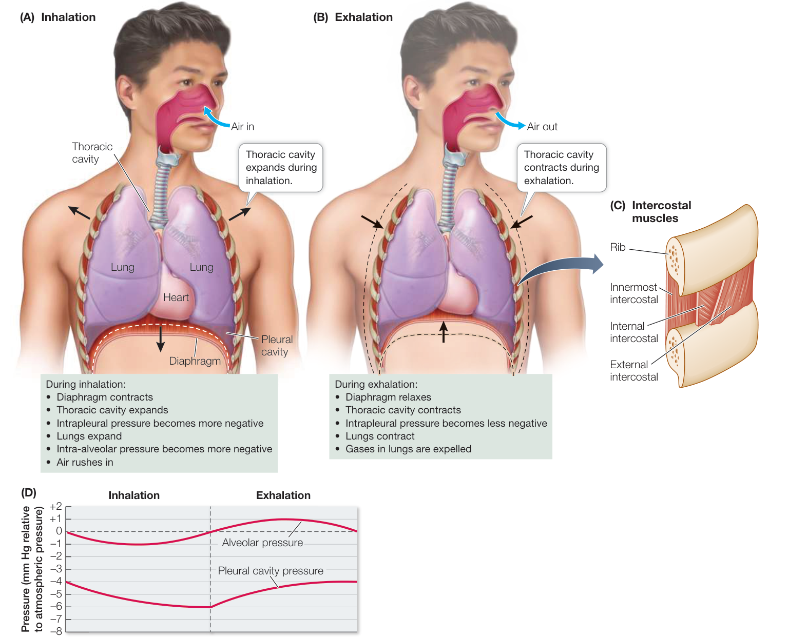
Figure 47.11 Into the Lungs and Out Again (A) Inhalation is an active process spurred by contraction of the diaphragm. (B) Exhalation generally is a passive process as the diaphragm relaxes. (C) Strong inhalations and exhalations also involve the external and internal intercostal muscles. (D) Between breaths, the intra-alveolar pressure is the same as the external barometric pressure, but the interpleural pressure is still negative and keeps the lungs from collapsing. During inhalation the interpleural pressure becomes more negative, expanding the lungs and causing the intra-alveolar pressure to become negative, which pulls air into the lungs. Animation 47.2 Airflow in Mammals
Remember that ventilation and perfusion work together to maximize the partial pressure gradients across the alveolar membranes (see Figure 47.11D). Ventilation delivers to the environmental side of the exchange surface, where it diffuses across and is swept away by the perfusing blood, which carries it to the tissues. The reverse is true for . Perfusion delivers to the exchange surface, where it diffuses across and is swept away by ventilation.
Recap and Assess
The mammalian respiratory system consists of a highly branching system of airways that lead to alveoli—the gas exchange surfaces. Respiratory muscles ventilate the alveoli by creating pressure differences between the lungs and the outside air. Alveolar surfactant decreases the work of inflating the lungs, and surface tension in the alveoli contributes to elastic recoil, facilitating emptying of the lungs. and are exchanged across thin capillary and alveoli walls by diffusion.
- In terms of LaPlace’s law, explain the functional significance of the unique properties of lung surfactant.
- Explain why a puncture wound of the chest causes a collapsed lung.
- In the breathing cycle, what is the alveolar pressure when the tension between the pleural membranes is maximal? Explain your answer.
Having discussed how respiratory gases get to and from the environmental side of the gas exchange membranes through ventilation, we now look at how these gases get to and from the internal side of those membranes through perfusion.
Respiratory Gases Are Transported by the Blood
Learning Objectives
- 47.4.1 Describe the functional properties of the hemoglobin-oxygen dissociation curve.
- 47.4.2 Explain the significance of the effects of 2,3-bisphosphoglycerate and pH on the -binding properties of hemoglobin.
- 47.4.3 Explain how the same enzyme, carbonic anhydrase, catalyzes both the loading of from cells into the blood and the off-loading of from the blood to the alveoli.
Perfusion of the lungs is one of the functions of the circulatory system. The circulatory system uses a pump (the heart) and a network of vessels to transport blood around the body. Circulatory systems are the subject of Chapter 48, so here we discuss only one aspect of perfusion: how blood transports respiratory gases.
The liquid part of blood, the plasma, carries some in solution, but its ability to transport this nonpolar molecule is limited. The blood plasma of a human can contain in solution about 3 mL of per liter of plasma, which is inadequate to support even basal metabolism. However, the blood of vertebrates and of many invertebrates contains molecules that bind and release and thus augment its transport capacity. These molecules pick up where is high and release it where is lower. There are many transport molecules in the animal kingdom, but in vertebrates this role is played by hemoglobin, a protein contained in red blood cells (RBCs). The percent of the blood volume consisting of RBCs is called the hematocrit. The hematocrit of human males averages 45% and that of females 40%. Hemoglobin enables the blood to carry 60 times more oxygen than it could carry just in solution, making high rates of metabolism possible.
Red blood cells are produced in the bone marrow
RBCs are the most abundant human cells in the body. They are produced in the bone marrow at a rate of about 2 million per second, and they circulate around the body for 100–120 days before they are destroyed. Each circuit around the body takes about a minute when at rest. RBCs are among the smallest cells of the body. They have to pass through the tiniest blood vessels, the capillaries. To be so small yet carry a lot of hemoglobin (at least 25% of their volume), RBCs lose their nuclei and most cell organelles prior to being released into the circulation. Because they have no mitochondria, they depend on glycolysis for energy.
Connect the Concepts In Key Concept 9.2, glycolysis is described as a sequence of reactions that by the sixth step has converted one molecule of glucose to two molecules of 1,3-bisphosphoglycerate (see Figure 9.7) and by the tenth step to two molecules of pyruvate in addition to a net gain of two molecules of ATP. However, some of the 1,3-bisphosphoglycerate can be enzymatically converted to 2,3-bisphosphoglycerate, which plays an important role in regulating the oxygen-binding properties of hemoglobin, as we discuss later in this key concept.
Hemoglobin combines reversibly with
RBCs contain enormous numbers of hemoglobin (Hgb) molecules. Hgb is a protein consisting of four polypeptide subunits, each of which surrounds a heme group—an iron-containing ring structure that can reversibly bind a molecule of . Thus each Hgb molecule can bind and release up to four molecules, enabling the blood to carry a large amount of to the body’s tissues.
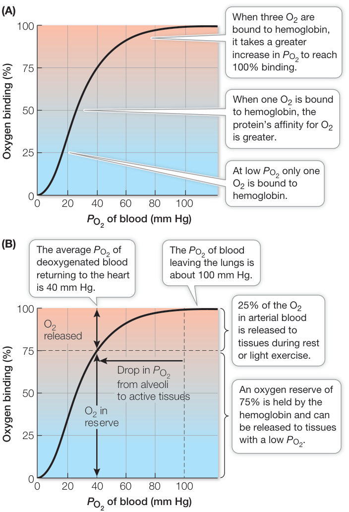
Figure 47.12 Binding of Oxygen to Hemoglobin Depends on (A) The sigmoidal shape of the hemoglobin-oxygen binding/dissociation curve reflects positive cooperativity among hemoglobin’s subunits. (B) Hemoglobin in blood leaving the lungs is 100% saturated (four molecules are bound to each hemoglobin molecule). Most hemoglobin molecules drop only one of their four molecules as they circulate through the body and are still 75% saturated when the blood returns to the lungs. The steep portion of this -binding/dissociation curve comes into play when tissue falls below 40 mm Hg, which is a normal mixed venous blood .
Hgb’s ability to pick up or release depends on the in its environment. When the of the blood plasma is high, as it usually is in the lung capillaries, each Hgb molecule can carry its maximum load of four molecules. As the blood circulates through the rest of the body, it releases some of the it carries when it encounters lower values in tissues.
The relationship between and the amount of bound to Hgb is not linear but S-shaped (sigmoidal). The hemoglobin–oxygen binding/dissociation curve reflects interactions between the four subunits of the Hgb molecule. At low values, only one subunit will bind an molecule (Figure 47.12A). When it does so, the shape of that subunit changes, altering the quaternary structure of the entire Hgb molecule. That structural change makes it easier for the other subunits to bind an molecule; that is, their affinity is increased. Therefore a smaller increase in is necessary to get the Hgb molecule to bind a second molecule (to become 50% saturated) than was necessary to get it to bind one molecule (to become 25% saturated). This change in affinity is reflected in the increased steepness of the -binding curve. The influence of binding to one Hgb subunit on the affinity of the other subunits is called positive cooperativity.
Once the third molecule is bound, the relationship seems to change, as a larger increase in is required for the Hgb to reach 100% saturation. This upper bend of the sigmoid curve is due to a probability phenomenon. The closer we get to having all subunits occupied, the less likely it is that any particular molecule will find a place to bind. Therefore it takes a relatively greater to achieve 100% saturation.
The -binding/dissociation properties of Hgb help get to the tissues that need it most (Figure 47.12B). In the lungs, where the is about 100 mm Hg, Hgb is 100% saturated. The in blood returning to the heart from the body (at rest) is usually about 40 mm Hg. You can see that at this the Hgb is still about 75% saturated. This means that as the blood circulates around the body, it releases only about one in four of the molecules it carries. This system seems inefficient, but because the Hgb keeps 75% of its in reserve, it can meet the demands of highly active tissues.
When a tissue becomes starved of and its local falls below 40 mm Hg, the Hgb flowing through that tissue is on the steep portion of its binding/dissociation curve. That means relatively small decreases in below 40 mm Hg will result in the release of lots of to the tissue. Thus Hgb is very effective in making available to tissues precisely when and where it is needed most.
The transport function of Hgb can rapidly and tragically be disrupted by a common by-product of incomplete combustion: carbon monoxide (CO). If CO from a faulty heating system, engine exhaust, or burning charcoal accumulates in a closed space, the results can be deadly. Because CO binds to Hgb with a 240-fold higher affinity than , it can prevent Hgb from transporting . In the United States, up to 500 people die each year from CO poisoning.
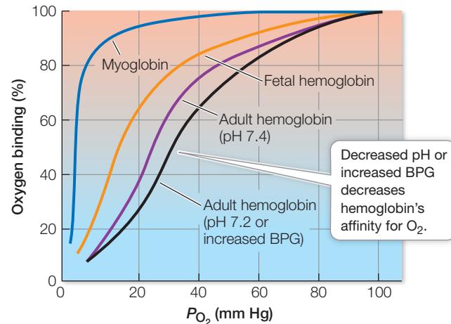
Figure 47.13 Oxygen-Binding Adaptations Myoglobin and the different hemoglobins have different -binding properties adapted for different circumstances. Fetal hemoglobin, for example, has a higher affinity for than does adult hemoglobin, facilitating transfer in the placenta. When high metabolism lowers the pH of the blood, or low increases 2,3-bisphosphoglycerate (2,3-BPG), hemoglobin releases more of its . Q: How would breathing some carbon monoxide affect the hemoglobin–oxygen binding curve?
Myoglobin holds an reserve
Muscle cells have their own -binding molecule, myoglobin. Myoglobin consists of just one polypeptide chain associated with an iron-containing ring structure that can bind one molecule. Myoglobin has a higher affinity for than Hgb does, so it picks up and holds at values at which Hgb is releasing its bound (Figure 47.13).
Connect the Concepts The evolutionary relationships between myoglobin and the various hemoglobin subunits are shown in Figure 21.10.
Myoglobin facilitates the diffusion of in muscle cells and provides an reserve for times when metabolic demands are high and blood flow is interrupted. Interruption of blood flow in muscles is common because contracting muscles squeeze blood vessels. When tissue values are low and Hgb can no longer supply more , myoglobin releases its bound . Diving mammals such as seals have high concentrations of myoglobin in their muscles, which is one reason they can stay underwater for so long (Investigating Life: Seals Are Champion Breath-Hold Divers). Even in nondiving animals, muscles called on for extended periods of work frequently have more myoglobin than do muscles that are used for short, intermittent periods, as noted in Key Concept 46.2.
Hemoglobin’s affinity for is variable
Various factors influence the -binding/dissociation properties of Hgb, thereby influencing delivery to tissues. Three of these factors are the chemical composition of the Hgb, the blood pH, and the presence of 2,3-bisphosphoglycerate (2,3-BPG) in RBCs.
Investigating Life: Seals Are Champion Breath-Hold Divers
Experiment
Original Paper: M. A. Castellini et al. 1992. Metabolic rates of freely diving Weddell seals: Correlations with oxygen stores, swim velocity, and diving duration. J Exper Biol 165: 181–194.
Weddell seals (Leptonychotes weddellii) live near the edges of the ice in Antarctica where they feed by diving in the surrounding ocean. The ice-covered ocean presents a unique opportunity to study the diving and breathing behaviors of these seals if they have to return to the same breathing hole in the ice.
QUESTION How do metabolic requirements of a freely diving seal correlate with its oxygen stores?
METHOD
- Transport Weddell seals several kilometers away from the ice edge, but still on the extensive Antarctic ice sheet.
- Fit the seals with distance and depth gauges to calculate the metabolic costs of individual dives.
- Make a hole in the ice and release the seals so they can return to the sea through the hole.
- Allow the seals to dive and feed naturally.
- When seals return to the hole to breathe, measure consumption and release and determine each seal’s metabolic rate (MR).
RESULTS Most dives were only a few minutes long, but many were over 30 minutes and the longest was 82 minutes. MR was also measured when the seals rested and slept. Seals frequently do not breathe during sleep. The longest sleep episode without breathing was 13.5 minutes. Mean MR is listed with standard deviation (See Appendix B for discussion of statistical concepts.)
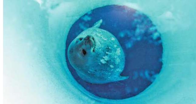
© Aurora Photos/Alamy Stock Photo
| Condition | Mean MR (mL /min kg) ( SD) |
|---|---|
| Rest | 4.1 (0.3) |
| Sleep | 3.5 (0.6) |
| All dives | 4.5 (0.9) |
| Short dives | 5.0 (0.9) |
| Long dives | 3.4 (0.6) |
CONCLUSION
- There were no significant differences in MR during rest, sleep, and the mean for all dives taken together. The relatively consistent measurements of MR indicate a very low metabolic cost of diving and activities during the dives. Going from resting to diving resulted in only a 10% increase in MR. (In humans, by contrast, going from resting to standing involves a 16% increase in MR.)
- MRs during long dives were lower than at rest, but MRs during short dives (<15 minutes) were significantly higher than during long dives, indicating the capacity of the seals to conserve supplies to extend dive duration.
Work with the Data
How do the seal’s oxygen stores compare with its oxygen demands? To calculate the oxygen reserves of a Weddell seal, we have to assess the amount of oxygen in its blood and in the myoglobin in its muscles at the beginning of the dive. The amount of oxygen in its lungs is irrelevant since a seal exhales before diving, so there is little oxygen available in its alveoli during the dive.
The problem requires calculating the amount of in the arterial and venous blood and myoglobin at the beginning of the dive that is available to support the seal’s MR.
Assume the following:
- Average body mass: 355 kg
- Blood volume: 14.8% of mass (7% in humans)
- Hematocrit: 58% (human averages 42%)
- Hemoglobin content of red blood cells: 23.7%
- holding capacity of hemoglobin: 1.34 mL/g
- Blood: 34% is arterial, 95% saturated at beginning of dive, can desaturate to 20%
Available in arterial blood is calculated as follows:
QUESTIONS
- Calculate the available in the venous blood. Assume venous blood is 66% of total blood volume, and that it is 90% saturated at the beginning of the dive and can desaturate to 0%.
- Calculate the available in myoglobin. Assume 33% of body mass is muscle, myoglobin content of seal muscle is 45 g/kg (20–25 g/kg for humans), and holding capacity of myoglobin is 1.34 mL /g.
- What are the total reserves at the beginning of the dive?
- Given the MR values found in the experiment, what do you predict would be the maximum duration of no breathing during sleep?
- Using the table of MR values for all dives in the results portion of the experiment, predict the maximum duration of a Weddell seal dive.
HEMOGLOBIN COMPOSITION There is more than one type of Hgb because the chemical composition of the polypeptide chains that form the Hgb molecule varies. The normal Hgb of adult humans has two each of two kinds of polypeptide chains—two -globin chains and two -globin chains. This normal adult Hgb has the -binding characteristics shown in Figure 47.13.
Before birth, the human fetus has a different form of Hgb, consisting of two -globin and two -globin chains. The functional difference between fetal and adult Hgb is that fetal Hgb has a higher affinity for . Therefore the fetal hemoglobin–oxygen binding/dissociation curve is shifted to the left compared with the adult curve (see Figure 47.13). You can see from these curves that if both types of Hgb are at the same (as they are in the placenta), fetal Hgb will pick up that the adult Hgb releases. This difference in affinities enables the efficient transfer of from the mother’s blood to the fetus’s blood.
HEMOGLOBIN AND pH The -binding properties of Hgb are also influenced by physiological conditions. The influence of pH on the function of Hgb is known as the Bohr effect. As blood passes through metabolically active tissue such as exercising muscle, it picks up acidic metabolites. As a result, blood pH falls. The excess ions bind preferentially to deoxygenated Hgb and decrease its affinity for , and the -binding/dissociation curve of Hgb shifts to the right (see Figure 47.13). This shift means the Hgb will release more in tissues where pH is low—another way that is supplied where and when it is most needed.
2,3-BISPHOSPHOGLYCERATE 1,3-BPG is an intermediate in glycolysis (see Figure 9.7), and therefore an important source of energy for cells. However, 1,3-BPG can be converted to 2,3-BPG by an enzyme in the RBC. 2,3-BPG is an important regulator of Hgb function. Like excess , 2,3-BPG reversibly combines with deoxygenated Hgb and lowers its affinity for . The result is that at any , Hgb releases more of its bound than it otherwise would. In other words, 2,3-BPG shifts the -binding/dissociation curve of mammalian Hgb to the right.
When humans go to high elevations, or when they cease being sedentary and begin to exercise, the level of 2,3-BPG in their RBCs goes up, making it easier for Hgb to deliver more to tissues. During pregnancy, a woman has about a 30% increase in 2,3-BPG, which makes more available in the placenta to be picked up by the fetal Hgb. In addition, fetal Hgb has a left-shifted -binding/dissociation curve because its -globin chains have a lower affinity for 2,3-BPG than do the -globin chains of adult Hgb.
Most is transported as bicarbonate ions in the blood
Delivering to tissues is only half the respiratory function of blood. Blood also carries , a metabolic waste product, away from tissues (Figure 47.14). is highly soluble and readily diffuses through cell membranes, moving from its site of production in the tissues into the blood, where is lower. However, only about 5% of the carried by the blood is dissolved . About 20% of the carried to the lungs in the blood is in chemical combination with Hgb. And most produced by the tissues is transported to the lungs in the form of bicarbonate ions (). diffusing from cells is converted to , transported to the lungs, and then converted back to to be exhaled.
When dissolves in water, some of it slowly reacts with the water molecules to form carbonic acid (), some of which then dissociates into a proton () and a bicarbonate ion (). This reversible reaction is expressed as follows:
In the extracellular fluid, the reaction between and proceeds slowly. But it is a different story in the endothelial cells of the capillaries and in the RBCs, where the enzyme carbonic
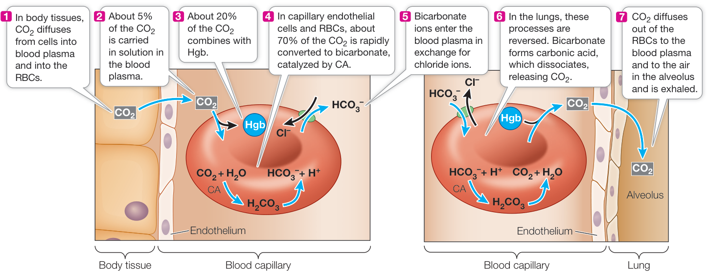
Figure 47.14 Carbon Dioxide Is Transported as Bicarbonate Ions The left panel represents the circulation through the tissues of the body where carbonic anhydrase (CA) in capillary endothelial cells and in RBCs facilitates conversion of produced in the tissues to bicarbonate ions ( ) in the blood. The right panel shows the process reversing in the lungs as diffuses from blood to alveolar air and is exhaled.
anhydrase (CA) speeds up the conversion of to . The newly formed dissociates, and the resulting bicarbonate ions enter the blood plasma in exchange for (see Figure 47.14). By converting to , carbonic anhydrase reduces the in these cells and in the blood plasma, facilitating the diffusion of from tissue cells to endothelial cells, blood plasma, and RBCs.
In the lungs, the reactions involving and bicarbonate ions are reversed. Remember that an enzyme such as carbonic anhydrase only speeds up a reversible reaction; it does not determine its direction. The direction is determined by concentrations of reactants and products. Ventilation keeps the in the alveoli low, so diffuses from the blood plasma into the alveoli, lowering the in the blood, which favors the conversion of into . Also, the oxygenation of Hgb in the alveoli decreases its affinity for , a phenomenon known as the Haldane effect.
Recap and Assess
is transported from the lungs to the body’s tissues in reversible combination with Hgb. Each Hgb molecule can reversibly combine with four molecules; the percent saturation of the binding sites is a function of the in the Hgb’s environment. The hemoglobin-oxygen binding/dissociation curve is shifted by the concentration of and the level of 2,3-BPG in the blood. is transported by the blood mostly in the form of .
- The of atmospheric air at sea level is about 159 mm Hg. What is the functional significance of the fact that human Hgb fully saturates with at a of 100 mm Hg and returns to the heart in venous blood with a of about 40 mm Hg, reflecting 75% saturation?
- When RBCs are stored in the blood bank, they will eventually use their 1,3-BPG for energy. What will be the consequence for their ability to function after transfusion into a patient?
- Why do ions leave the RBCs in systemic venous blood but enter RBCs in the alveolar circulation?
We must breathe every minute of our lives, but most of us usually don’t worry about it, or even think about it very often. In the next key concept we examine how the regular breathing cycle is generated and controlled by the central nervous system.
Breathing Is Homeostatically Regulated
Learning Objectives
47.5.1 Explain the roles of the dorsal and ventral groups of respiratory neurons in the medulla.
47.5.2 Apply knowledge about the physiological processes underlying breathing to explain the body’s response to intense exercise and to decreases in .
47.5.3 Describe where chemosensitive cells are located and how they participate in negative feedback control of breathing.
Breathing is an involuntary function of the central nervous system (CNS). The breathing pattern is adjusted around other activities
(such as speech and eating), and breathing rates change to match the metabolic demands of the body. How is this accomplished?
Breathing is controlled in the brainstem
The basic breathing rhythm is an involuntary function that is modified by feedback information that conveys the body’s need for an adequate supply and elimination. Breathing is driven by a rhythm-generating neural circuit in the brainstem, but where? Breathing ceases if the spinal cord is severed in the neck region, showing that breathing is generated in the brain. If the brainstem is cut just above the pons, breathing is normal. If the brainstem is cut between the pons and the medulla (the segment of the brainstem just above the spinal cord), the breathing pattern becomes irregular (Figure 47.15). Electrophysiological recordings reveal a group of respiratory motor neurons in the dorsal medulla that increase their firing rates just before and as an inhalation occurs. The axons of these neurons leave the CNS in the neck region to form the phrenic nerve that innervates the diaphragm. As more and more of these neurons fire—and fire faster and faster—the diaphragm contracts. All of a sudden these neurons stop firing, the diaphragm relaxes, and exhalation begins. Exhalation is usually a passive process that depends on the elastic recoil of the lung tissues. Another group of respiratory motor neurons is found in the ventral medulla, and they become active when breathing demand is high. These motor neurons communicate through thoracic spinal nerves to the intercostal muscles between the ribs. By expanding and contracting the rib cage, the intercostal muscles increase both the inhalation and the exhalation volumes.
Neurons in the lower region of the pons help regularize the basic respiratory rhythm. Still higher brain areas modify breathing to accommodate speech, ingestion of food, coughing, and emotional states.
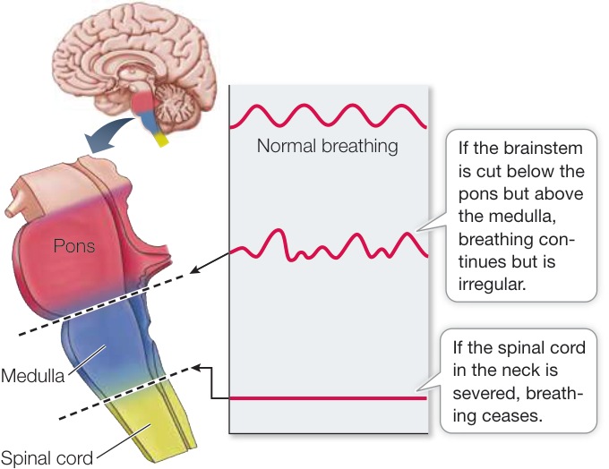
Figure 47.15 Breathing Is Controlled in the Brainstem The basic breathing rhythm is generated in the medulla and is modified by neurons in or above the pons.
Regulating breathing requires feedback
When breathing or metabolism changes, the and in the blood change as well. It is reasonable to surmise that the blood levels of one or both of these gases provide feedback information to the breathing rhythm generator in the medulla. Humans and other mammals are remarkably insensitive to falling levels of in arterial blood but are extremely sensitive to increases in . Arterial can deviate considerably from normal without causing much of an increase in ventilation rate, but even a small rise in arterial causes a large increase in ventilation (Figure 47.16). This relationship is reversed for water-breathing animals, in which is the primary feedback stimulus for gill ventilation.
The lack of sensitivity in humans was tragically demonstrated by an accident at the Kennedy Space Center in 1981 prior to the first flight of the Space Shuttle Program. The space shuttle Columbia was being prepared for launch, and to reduce the danger of fire, the engine compartment was purged with nitrogen and sealed. Unaware of the nitrogen, workers opened a hatch to the compartment for inspection. The first worker to enter became unconscious, and others followed him in a rescue attempt. Three of these workers died, and two others who were stricken recovered. When exposed to lack of oxygen, there is no alerting, emergency response—there is simply loss of consciousness.
Where are partial pressures of gases in the blood sensed? The major site of sensitivity is an area on the ventral surface of the medulla that is in contact with the cerebrospinal fluid that surrounds the brain. The primary sensitivity of these chemosensitive cells is not, however, to . It is ions that stimulate these cells. The ion concentration in the environment of these cells is a direct reflection of the of the blood, but not of the pH of the blood. The cerebrospinal fluid is extracellular fluid, and extracellular fluid has less protein content than the blood plasma. The high protein content of the blood plasma buffers its pH (see Figure 2.18). But, where the of the blood is higher than that of the extracellular fluid, diffuses out of the blood. This interacts with in the extracellular fluid to form carbonic acid (), which dissociates into ions and ions (see Figure 47.14, which describes this same reaction in the RBCs). It is the ions produced by the dissociation of carbonic acid that stimulate the chemosensitive cells on the ventral surface of the medulla that increase respiratory gas exchange. Thus even though we measure blood as the stimulus for breathing, the major stimulus is an increase in ion concentration (lowered pH).
We do have physiological sensitivity to blood in nodes of neural tissue on the large blood vessels leaving the heart: the aorta and the carotid arteries (Figure 47.17). These carotid bodies and aortic bodies are chemosensors. If the blood supply to these structures decreases, or if the blood falls dramatically, the chemosensors are activated and send nerve impulses to the breathing control center. Although we are not very sensitive to changes in blood , the carotid and aortic bodies can stimulate increases in breathing during exposure to high elevations or when blood volume or blood pressure is very low. Also, there is a synergism between and sensing. When blood increases, there is an increased sensitivity to low , and vice versa.
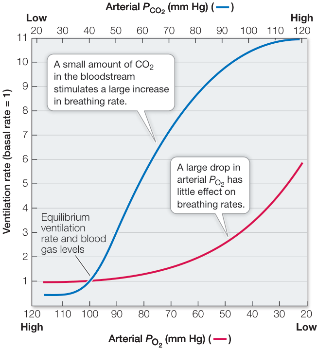
Figure 47.16 Arterial Affects Breathing Rate More Than Low Altering arterial or by breathing air with different concentrations of those gases shows that the regulation of human breathing is more sensitive to increased levels of in arterial blood than to decreased amounts of .
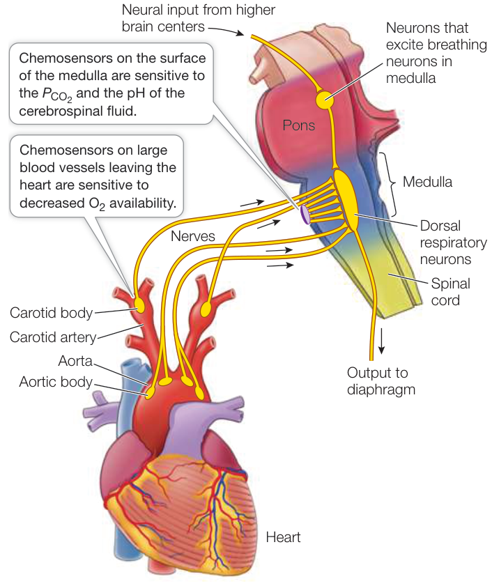
Figure 47.17 Feedback Information Controls Breathing The body uses feedback information from chemosensors in the great arteries leaving the heart and on the ventral surface of the medullary brainstem to match breathing rate to metabolic demand.
Recap and Assess
The rhythmic contractions of the respiratory muscles that drive breathing are generated by neurons in the brainstem. Breathing is regulated predominantly in response to the of the blood. Respiratory chemosensors are cells on the ventral surface of the medulla that are sensitive to the blood , and the carotid and aortic bodies on the large arteries leaving the heart that are sensitive to the blood or to a decrease in the blood they are receiving.
- In terms of respiratory motor neurons and respiratory muscle groups, what changes occur in transitioning from breathing at rest to breathing during exercise that uses more of the lungs’ vital capacity?
- When you go rapidly to high elevation, you may experience a breathing pattern in which periods of no breathing alternate with periods of fast breathing. Explain that response in terms of diffusion gradients for and and the feedback signals regulating breathing.
- Cells in the carotid and aortic bodies have high metabolic rates, and they are sensitive either to a decrease in blood flow or to a decrease in the of the blood. How are these two facts related?
Investigating Life: QA What adaptations make seals champion and breath-hold divers?
Seals have remarkable abilities to dive deep and long and to spend only minutes at the surface to breathe between dives. In the chapter opener about elephant seals, we posed several questions that apply to all seals: Why aren’t seals crushed by the water pressure at extreme depths? Why don’t they get the bends? What are their demands? And what are their reserves? The answers to the first two questions are related. Only air-filled structures of the body are compressible, and the seals exhale at the beginning of a dive. Their residual volume is restricted to their airways, where gas exchanges do not occur. Thus the high pressure of the small amount of gas in their respiratory system does not drive dissolved nitrogen into their tissues. Investigating Life: Seals Are Champion Breath-Hold Divers presented metabolic rate (MR) measurements for Weddell seals resting, sleeping, and diving. No significant differences in MR for various activities were observed, leading to the conclusion that the metabolic costs of diving are very low. In fact, MRs during dives were only about 10% higher than at rest—not much different than for humans going from sitting to standing.
In spite of the low metabolic expenditures of seals, how do they maintain reserves sufficient to support them during dives? The calculations made in the Work with the Data exercise in Investigating Life: Seals Are Champion Breath-Hold Divers showed that the reserve of a sleeping seal would permit it to not breathe for a maximum of 12.8 minutes, which corresponds well with the maximum episodes of sleep observed—13.5 minutes. Using the MR value for all dives, calculations show that the seals’ reserves would only support dives that were under 10 minutes in duration. Therefore there must be additional adaptations enabling the seals to remain active underwater for longer periods even at an average MR similar to what they experience during sleep.
Future directions
Recording devices attached to seals diving in nature reveal adaptations that minimize their demands during a dive. Because they exhale at the beginning of a dive, they are negatively buoyant, meaning they can drift down to depths like a falling leaf. These recordings also answered another question. Since the seals at sea dive continually day and night, when do they sleep? They spend much of their passive descent time sleeping, thus lowering their MR even more. Yet more metabolic savings are needed to enable long dives. One clue comes from measures of the seals’ heart rates during dives. Their heart rates are very much reduced, a phenomenon called diving bradycardia that is accompanied by reduced cardiac output (total systemic blood flow). This reduction in cardiac output is achieved by shutting off blood flow to many tissues that can switch to anaerobic metabolism and in response to low levels of become hypometabolic. The result is that available in the blood is reserved for heart and brain, which are highly sensitive to lack of .
Visual Summary
© Bruce Watkins/Animals Animals/AGE Fotostock
You should be able to relate each summary to the adjacent figures. If you go to this visual summary in Achieve, you can follow links to figures, animations, activities, and simulations that will help you consolidate the material.
Respiratory Gas Exchange Is Governed by Physical Factors
- ■ Exchange of respiratory gases occurs only by diffusion, and any adaptation to maximize gas exchange must influence a factor in Fick’s law of diffusion.
- ■ Diffusion coefficients depend on the diffusing substance, the medium, and the temperature.
- ■ For aquatic organisms, increases in water temperature raise requirements and decrease its availability.
Questions
- In terms of Fick’s law, how does elevation affect respiratory gas exchange?
- Why is water a worse respiratory gas exchange medium than air?
- Why does a rise in water temperature put fishes in a double bind?
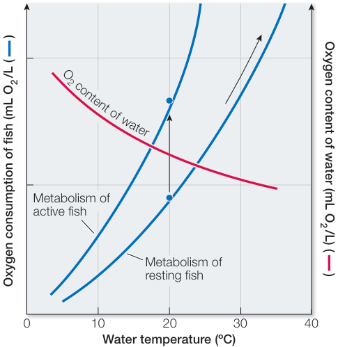
Figure 47.2
Enhancing Diffusion Maximizes Respiratory Gas Exchange
- ■ delivery to cells of the insect body is by diffusion in air. The gills of fishes have large gas exchange surfaces that are ventilated continuously and unidirectionally with water flowing countercurrent to the flow of blood inside the gills. Air inhaled by birds flows continuously and unidirectionally through airways, air sacs, parabronchii, and air capillaries and is in the system for two breathing cycles.
- ■ Except for birds, air-breathing vertebrates have tidal ventilation, creating dead space and resulting in mixing of incoming fresh air with stale air.
Questions
- How does countercurrent flow maximize gas exchange in fish gills?
- What is the main functional difference between parabronchi of birds and bronchi of mammals?
- How would you measure the difference between vital capacity and total lung volume in a human?
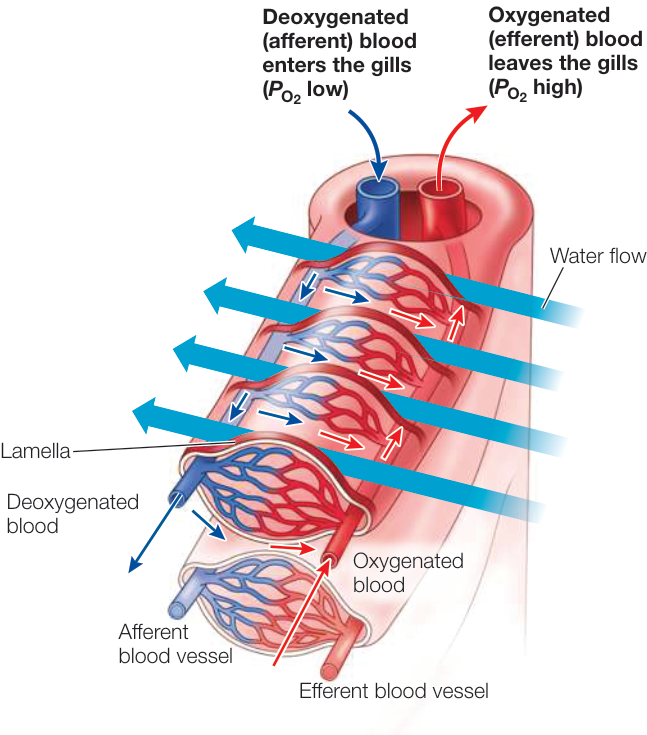
Figure 47.5
Humans Have Tidal Respiration
- ■ Alveoli are the respiratory exchange surfaces of mammalian lungs and are ventilated through a branching system of bronchi and bronchioles and are surrounded by capillaries.
- ■ Lung surfactant reduces surface tension in alveoli, thereby decreasing the pressure necessary to inflate them.
- ■ Contractions of the diaphragm and intercostal muscles drive inhalation and exhalation by changing the pressure in the thoracic cavity.
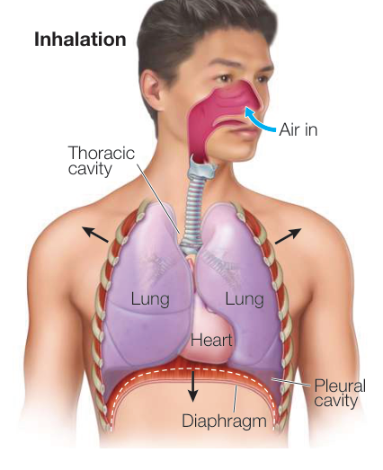
Figure 47.11 Questions
- Describe the path taken by air during breathing.
- How are the properties of lung surfactant important for both inhalation and exhalation?
- How do alveolar pressure and pleural cavity pressure change relative to the outside air pressure during the breathing cycle?
Respiratory Gases Are Transported by the Blood
- ■ Oxygen reversibly binds to hemoglobin (Hgb) in red blood cells, and percent saturation depends on the to which the Hgb is exposed. Myoglobin serves as an reserve in muscle.
- ■ The affinity of Hgb for is altered by the composition of the Hgb, by pH, and by the presence of 2,3-bisphosphoglycerate (2,3-BPG). is transported in the blood principally as bicarbonate ions ().
Questions
- What is the significance of the fact that mixed venous blood can have a of 40 mm Hg and its Hgb is still 75% saturated?
- Why is it important that fetal Hgb has a higher affinity for 2,3-BPG than does adult Hgb?
- What is the role of carbonic anhydrase (CA) in eliminating from the body?
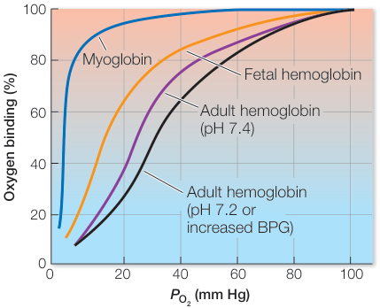
Figure 47.13
Breathing Is Homeostatically Regulated
- ■ Breathing movements are generated by neurons in the medulla and modulated by higher brain centers.
- ■ The breathing rhythm is altered by feedback from chemosensors on the ventral surface of the medulla and in the carotid and aortic bodies on the large vessels leaving the heart.
- ■ The most important feedback stimulus for breathing is the in the blood.
Questions
- What are the locations and roles of respiratory group neurons?
- What is the relationship between and pH in the regulation of breathing?
- What roles do carotid and aortic bodies play in the regulation of breathing?
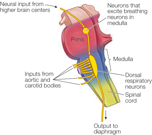
Figure 47.17