Chapter 49Nutrition, Digestion, and Absorption

© Haacov Dagan/Alamy Stock Photo
Key Concepts
- 49.1 Food Provides Energy as well as Materials for Biosynthesis
- 49.2 Diverse Adaptations Support Ingestion and Digestion of Food
- 49.3 The Vertebrate Gastrointestinal System Is a Disassembly Line
- 49.4 Nutrient Availability Is Controlled and Regulated
In Pima populations, historic occurrences of food deprivation may have imposed selection for genes that improve the efficiency of managing the energy obtained from food. With modern diets and lifestyles, these “thrifty genes” can contribute to obesity and diabetes.
Investigating Life: Thrifty Phenotypes
For millennia the Pima of southwestern North America were hunter-gatherers, supplementing their diet with subsistence agriculture. Their environment was arid, so they developed sophisticated irrigation systems; even so, they frequently encountered drought and subsequent starvation. Today most ethnic Pima in North America are clinically obese. As a population they are one of the heaviest in the world.
Obesity contributes to health problems such as diabetes, high blood pressure, and heart disease. Diabetes incidence in the Pima is seven times the national average. Two-thirds of Pima adults over age 40 are diabetic, and diabetes is occurring in younger individuals than previously seen. What has caused such a radical health change in the population? Two interacting factors are involved: genetics and lifestyle.
Geneticists hypothesize that recurring episodes of starvation produce strong selective pressure for “thrifty genes”—particular alleles of the genes involved in digestion, absorption, and energy storage that result in above-average efficiency in converting food into fat. Thrifty genes would carry a strong selective advantage when food is scarce. The Pima display a “thrifty” phenotype. They have low resting metabolic rates and convert food into fat readily. Insulin facilitates conversion of dietary sugar into fat. For many Pima, consuming a standard amount of glucose causes their insulin levels to rise three times higher than it does in people of European ancestry.
Lifestyle also plays a role in the Pima obesity epidemic. Instead of eating their traditional diet, the Pima now eat a high-calorie, high-fat Western diet, and they engage in less physical activity than their ancestors did. A population of Pima living in northern Mexico are genetically the same as the Arizona population. However, they eat traditional foods and live a traditional lifestyle that involves much physical activity. Obesity and diabetes are not prevalent among the Mexico Pima.
A high-calorie diet and sedentary lifestyle affect not just the Pima but contribute to the overall obesity epidemic in the U.S. Recently an additional factor that might contribute to obesity and diabetes has been discovered—the type of food that is eaten. The composition of the diet influences the populations of gut microbes, and those microbes influence outcomes of digestion. Apparently calories are not all equal; their impact depends on their origin.
Q&A If the nutritional values were the same, could different foods have different impacts on health? (Find the answer on page 1117.)
Food Provides Energy as well as Materials for Biosynthesis
Learning Objectives
49.1.1 Describe energy storage adaptations in animal species.
49.1.2 Explain challenges related to obtaining all essential amino acids from the diet.
49.1.3 Describe mineral macro-and micronutrients and their functions.
49.1.4 Give examples of water-soluble and fat-soluble vitamins and their effects on an animal’s body.
Animals are heterotrophs—they derive their nutrition from eating other organisms. In contrast, autotrophs (most plants, some bacteria, some archaea, and some protists) can use solar energy or inorganic chemical energy to synthesize all of their components. Of course, some plants are also heterotrophs.
Connect the Concepts Carnivorous and parasitic plants are discussed in Key Concept 34.4.
Directly and indirectly, heterotrophs take advantage of—indeed, depend on—the organic synthesis carried out by autotrophs and have evolved an enormous diversity of adaptations to exploit this resource (Figure 49.1). In this key concept we describe how animals use food, be it plants or other animals, to obtain energy and building blocks of complex molecules. We also consider the need for special mineral nutrients and organic molecules and the diseases that result when they are lacking in the diet.
Energy needs and expenditures can be measured
Energy—the capacity to do work—comes in different forms, including electric, heat, chemical, and nuclear energy. A calorie (note the small c) is a unit of heat energy; specifically, it is the amount of heat necessary to raise the temperature of 1 gram of water 1 degree Celsius. Because this is such a tiny amount of energy, physiologists commonly use the kilocalorie (kcal) as a unit of measure (1 kcal = 1,000 calories). Nutritionists also use the kilocalorie as a standard unit of energy, but they traditionally refer to it as the Calorie (Cal), which is capitalized to distinguish it from the single calorie.
Just about any food container you pick up in the United States carries the label “Nutrition Facts,” which includes the item “Calories.” How do Calories (or kcal) relate to the discussion in Chapter 9 about how energy in the chemical bonds of food molecules is transferred to the high-energy phosphate bonds of adenosine triphosphate (ATP) and is then used to do cellular work? And why do we use heat energy as a measure of nutrition?
The reason is found in the laws of thermodynamics, which state that energy cannot be created or destroyed, but can be converted from one form to another. However, every energy conversion is inefficient; a large portion of the original energy always ends up as heat. Whether we are using the energy in glucose to make ATP or are using that ATP to power muscle contraction or ion transport, most of the available chemical energy is lost as heat. The bottom line is that if an animal is not growing, not doing any external work, and not changing its body temperature, the heat it loses to the environment is a measure of its total energy expenditure, or metabolism.
Connect the Concepts The laws of thermodynamics and their application to biological energetics are described in Key Concept 8.1.
An animal’s energy needs must be met by the ingestion, digestion, and assimilation of food. The basal energy expenditure (basal metabolic rate, BMR) of a human is 1,300–1,490 Cal/day for an adult female and 1,600–1,800 Cal/day for an adult male. Any physical activity adds to the BMR. For a person doing sedentary work, about 30% of the total Calories expended are used for skeletal muscle activity; for a person doing heavy physical labor, more than 95% of caloric expenditure is for skeletal muscle activity.
Macronutrients are the components of food that provide energy. They are fats, carbohydrates, and proteins. Fats yield 9.5 Cal/gram, carbohydrates 4.2 Cal/gram, and proteins about 4.1 Cal/gram. Figure 49.2 shows some equivalences of food, energy, and energy consumption.
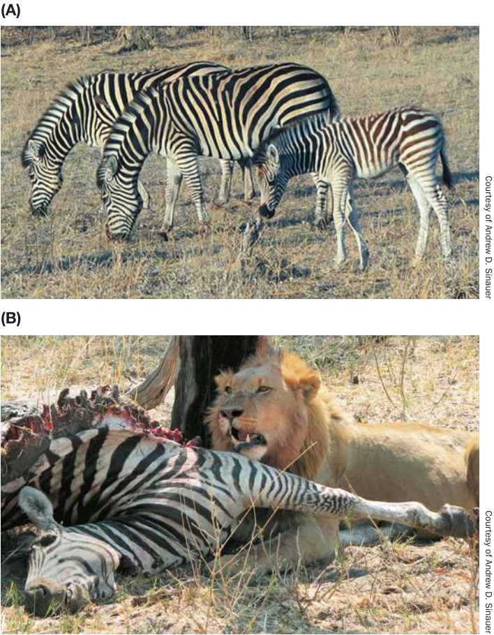
Figure 49.1 Heterotrophs Get Energy from Autotrophs (A) Herbivores get their energy directly from autotrophs. The large herbivores of the African grasslands must consume huge amounts of plant matter to fulfill their nutritional needs. (B) A carnivore’s energy is indirectly obtained from autotrophs, since the energy stored in a prey animal was originally obtained from autotrophs.
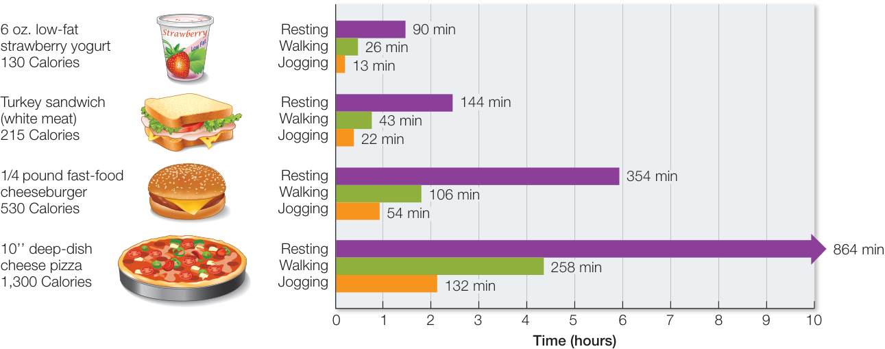
Figure 49.2 Food Energy and How We Use It Estimates of the energy contained in four common food items is shown at the left. Note: Caloric contents of these food examples can vary considerably. The graphs indicate about how long it would take a person with a basal energy requirement of about 1,800 Cal/day to use the equivalent amount of energy while resting, walking, or jogging.
Even though the units calorie, kilocalorie, and Calorie remain in popular use, most scientists now use the International System of Units (ISU). In this system the basic unit of energy is the joule: 1 joule (J) = 0.239 calories, and the measure of energy use is 1 J/second = 1 watt. You are familiar with lightbulb ratings, so think about that when you convert kcal/day into watts. The 1,700 Cal/day energy expenditure of the average man converts to 82 watts (note that a watt includes the time dimension, so it is a rate of energy use).
Thus it is possible to quantify the caloric value of any food an animal eats. It is also possible to quantify the caloric expenditure of any activity or behavior an animal performs. By comparing calories consumed with calories expended, we can construct energy budgets that allow ecologists and evolutionary biologists to apply a cost–benefit analysis to feeding behavior.
Connect the Concepts Any behavior involves energetic costs, opportunity costs, and risk costs and can be measured in terms of its effect on reproductive success. Key Concept 51.4 explores costs and benefits of various behaviors, illustrating how measures of metabolism permit quantitative analysis of evolutionary adaptations.
Sources of energy are stored in the body
Although cells of the body use energy continuously, most animals do not eat continuously and so must store fuel molecules that can be released as needed between meals. Carbohydrates are stored in the liver and in muscle cells as glycogen, but the total glycogen stored represents only about a day’s basal energy requirement. Fat is the most important form of stored energy in the bodies of animals. Not only does fat have more energy per gram than glycogen, but it can be stored with little associated water, making it more compact and less expensive to carry. Migrating birds store energy as fat to fuel their long flights; if they had to store the same amount of energy as glycogen, they would be too heavy to fly. Proteins are not used as energy storage compounds, but the body’s proteins can be metabolized as an energy source of last resort.
If an animal takes in too little food to meet its energy requirements, it starts to metabolize some of the molecules of its own body. This “self-consumption” begins with the energy storage compounds glycogen and fat. Once fat reserves are seriously depleted, the body increases its metabolism of proteins for energy (Figure 49.3A). The first proteins to be sacrificed are those of the blood plasma. The loss of plasma proteins decreases the osmotic concentration of the plasma, resulting in increased loss of fluid from the blood to the interstitial spaces (edema). Accumulation of fluid in the extremities and abdomen is the classic sign of kwashiorkor, a disease caused by chronic protein deficiency (Figure 49.3B). Continued protein loss damages the body’s organs, eventually leading to death.
When an animal consistently takes in more food than it needs to meet its energy requirements, the excess nutrients are stored as increased body mass. First glycogen reserves build up; then additional dietary carbohydrates, fats, and proteins are converted to body fat. In some species, such as those that hibernate, seasonal overnutrition is an important adaptation for surviving periods when food is not available. In humans, however, overnutrition can be a serious health hazard, increasing the risk of high blood pressure, heart attack, diabetes, and other disorders, as seen in the Pima that we discussed in the chapter opener.
Food provides carbon skeletons for biosynthesis
Every animal must take in certain organic molecules that it cannot synthesize for itself but needs to serve as building blocks for its own complex organic molecules. The acetyl group () is one such required building block, supplying the carbon skeleton of larger organic molecules (Figure 49.4). Animals cannot synthesize acetyl groups from carbon, oxygen, and hydrogen molecules but must obtain them from food. Acetyl groups can be derived from the metabolism of almost any food, but they originate in plants.
Acetyl groups are never in short supply for an adequately nourished animal. However, some groups supplying carbon skeletons can be deficient in an animal’s diet even if caloric intake is adequate. One such group includes certain amino acids, the building blocks of proteins. Animals can synthesize some of their own amino acids by using carbon skeletons from acetyl or other groups and transferring to them amine groups () derived from other amino acids. However, most animals cannot synthesize all the amino acids they need and thus must obtain certain essential amino acids from food. If an animal does not take in enough of even one of its essential amino acids, its protein synthesis is impaired and therefore its capacity to maintain many functions such as tissue repair and synthesis of many molecules such as enzymes, ion channels, transporters, hormones, etc. is compromised.
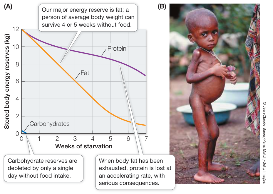
Figure 49.3 The Course of Starvation (A) When a person is subjected to undernutrition, the body’s energy reserves are depleted. (B) The swollen abdomen, hands, and feet of this boy are due to edema. Along with his spindly limbs, these are symptoms of kwashi-orkor, a disease resulting from the body breaking down blood proteins and muscle tissue to fuel metabolism. (A after A. C. Guyton. 1971. Basic Human Physiology: Normal Function and Mechanisms of Disease. W. B. Saunders: Philadelphia.)
Essential amino acids vary by species. Most researchers agree that adult humans must obtain eight essential amino acids from their food: isoleucine, leucine, lysine, methionine, phenylalanine, threonine, tryptophan, and valine. All eight are available in milk, eggs, meat, and soybean products, but most plant foods do not contain adequate quantities of all eight, so a strict vegetarian diet carries a risk of protein malnutrition. A complementary diet of plant foods, however, supplies all eight essential amino acids (Figure 49.5). In general, grains (such as rice, wheat, and corn) are complemented by legumes (such as beans and peas). Long before the chemical basis for complementarity was understood, societies with little access to meat developed complementary diets. Many Central and South American peoples traditionally eat beans with corn, and the native peoples of North America complemented their beans with squash.
Human infants are thought to require four additional amino acids in their diets: histidine, tyrosine, cysteine, and arginine. Also, some amino acids are required by individuals with certain metabolic disorders who cannot synthesize them adequately. For example, individuals with the genetic disease phenylketonuria lack the enzyme for converting phenylalanine to tyrosine (see Key Concept 15.2) and must obtain tyrosine from their diets. They must keep their dietary intake of phenylalanine low to prevent its accumulation to toxic levels.
Why are dietary proteins completely digested to their constituent amino acids before being used by the body? Wouldn’t it be more energy-efficient to reuse some dietary proteins directly? There are several reasons why ingested proteins are not used “as is”:
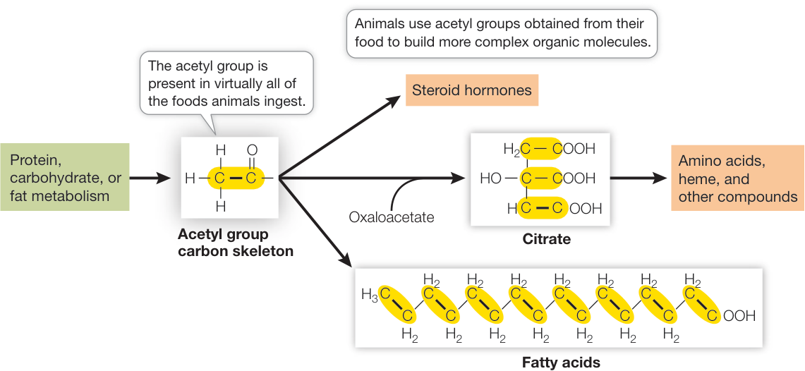
Figure 49.4 The Acetyl Group Is an Acquired Carbon Skeleton Animals cannot synthesize the acetyl group for themselves, but they ingest it in their food and use it to synthesize a wide variety of molecules.
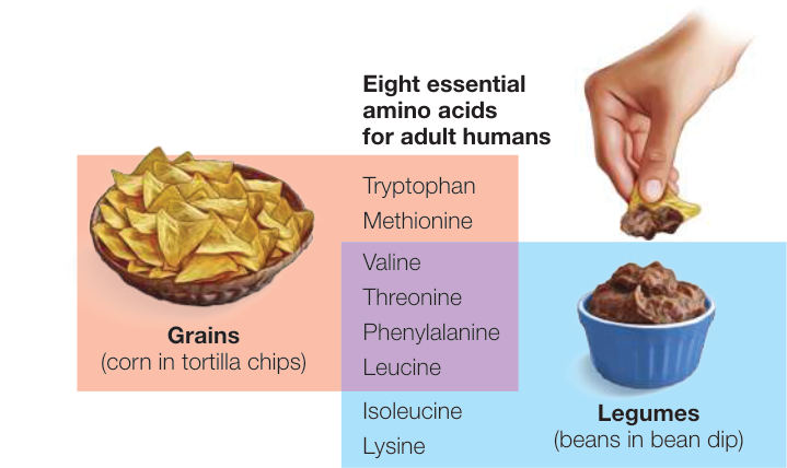
Figure 49.5 A Strategy for Vegetarians By combining cereal grains with legumes, an adult vegetarian can obtain all eight essential amino acids.
Macromolecules such as proteins are not readily absorbed by the cells of the gut, but their constituent monomers (such as amino acids) are readily absorbed.
Protein structure and function are highly species-specific. A protein that functions optimally in one species might not function well in another.
Foreign proteins entering the body directly from the gut would be recognized as invaders and would be attacked by the immune system.
Humans can synthesize almost all the lipids required by the body using acetyl groups obtained from food (see Figure 49.4), but we must have a dietary source of two essential fatty acids—linoleic acid and alpha-linoleic acid—that we cannot synthesize. Linoleic acid is needed by mammals to synthesize other unsaturated fatty acids, such as arachidonic acid, which is a component of several signal molecules, including prostaglandins. Essential fatty acids are also necessary components of membrane phospholipids. A deficiency of linoleic acid can lead to problems such as infertility and impaired lactation, but because it is commonly present in vegetable oils, a deficiency is unlikely in an adequately nourished individual.
Animals need mineral elements for a variety of functions
Principal mineral elements that animals require are listed in Table 49.1. Elements required in large amounts are called mineral macronutrients; those required in only tiny amounts (generally less
Table 49.1 Mineral Elements Required by Animals
| Element | Source in human diet | Major functions |
|---|---|---|
| MINERAL MACRONUTRIENTS | ||
| Calcium (Ca) | Dairy foods, eggs, green leafy vegetables, whole grains, legumes, nuts, meat | Found in bones and teeth; blood clotting; nerve and muscle action; enzyme activation |
| Chlorine (Cl) | Table salt (NaCl), meat, eggs, vegetables, dairy foods | Water balance; digestion (as HCl); principal negative ion in extracellular fluid |
| Magnesium (Mg) | Green vegetables, meat, whole grains, nuts, milk, legumes | Required by many enzymes; found in bones and teeth |
| Phosphorus (P) | Dairy foods, eggs, meat, whole grains, legumes, nuts | Component of nucleic acids, ATP and phospholipids; bone formation; buffers; metabolism of sugars |
| Potassium (K) | Meat, whole grains, fruits, vegetables | Nerve and muscle action; protein synthesis; principal positive ion in cells |
| Sodium (Na) | Table salt, dairy foods, meat, eggs | Nerve and muscle action; water balance; principal positive ion in extracellular fluid |
| Sulfur (S) | Meat, eggs, dairy foods, nuts, legumes | Found in proteins and coenzymes; detoxification of harmful substances |
| MINERAL MICRONUTRIENTS | ||
| Chromium (Cr) | Meat, dairy foods, whole grains, legumes, yeast | Glucose metabolism |
| Cobalt (Co) | Meat, tap water | Found in vitamin B 12 ; formation of red blood cells |
| Copper (Cu) | Liver, meat, fish, shellfish, legumes, whole grains, nuts | Found in active site of many redox enzymes and electron carriers; production of hemoglobin; bone formation |
| Fluorine (F) | Most municipal water supplies | Found in teeth; helps prevent tooth decay |
| Iodine (I) | Fish, shellfish, iodized salt | Found in thyroid hormones |
| Iron (Fe) | Liver, meat, green vegetables, eggs, whole grains, legumes, nuts | Found in active sites of many redox enzymes and electron carriers, hemoglobin, and myoglobin |
| Manganese (Mn) | Organ meats, whole grains, legumes, nuts, tea, coffee | Activates many enzymes |
| Molybdenum (Mo) | Organ meats, dairy foods, whole grains, green vegetables, legumes | Found in some enzymes |
| Selenium (Se) | Meat, seafood, whole grains, eggs, milk, garlic | Fat metabolism |
| Zinc (Zn) | Liver, fish, shellfish, and many other foods | Found in some enzymes and some transcription factors; insulin physiology |
than 100 mg/day) are called mineral micronutrients. Some mineral micronutrients are required in such minute amounts that deficiencies are never observed, but the elements are nevertheless essential.
Calcium is an example of a mineral micronutrient. It is the fifth most abundant element in the body; a 70-kg person contains about 1.2 kg of calcium. Calcium phosphate is the principal structural material in bones and teeth. Muscle contraction, neural function, and many other intracellular functions in animals require calcium ions (). The turnover of calcium in the extracellular fluid is high, as bones are constantly being remodeled and calcium is constantly entering and leaving cells. Calcium is lost from the body in urine, sweat, and feces, so it must be replaced regularly. Adult humans require 800–1,000 mg of calcium per day in their diet.
Connect the Concepts Many physiological processes are affected by ; therefore it is essential that the blood levels stay between 9 and 11 mg/100 mL. This is only 0.1% of the in the body. About 1% is intracellular and 99% is in the bones; thus it is a challenge to maintain tiny, but critical, blood levels in the face of such huge reservoirs. Hormonal regulation of blood is discussed in Key Concept 39.4, and the roles of in muscle function are discussed in Key Concept 46.1.
Iron is an example of a mineral micronutrient. It is found throughout the body because it is the oxygen-binding atom in hemoglobin and myoglobin and is a component of enzymes in the respiratory chain. Nevertheless, the total amount of iron in a 70-kg person is only about 4 g, and since iron is recycled efficiently in the body and is not lost in the urine, we require only about 15 mg per day in our food. Despite the small amount required, insufficient iron is the most common mineral nutrient deficiency in the world today. Iron deficiency leads to anemia, a condition that renders individuals weak and tired all the time.
Animals must obtain vitamins from food
Like essential amino acids and fatty acids, vitamins are carbon compounds that an animal requires for growth and metabolism but cannot synthesize for itself. Most vitamins function as coenzymes or parts of coenzymes (see Key Concept 8.4).
Each species has its own vitamin requirements. Primates, for example, require vitamin C (ascorbic acid). While most mammals can make their own ascorbic acid, primates (including humans) cannot, so for primates, ascorbic acid is a vitamin. If we do not get vitamin C in our food, we develop scurvy, a disease characterized by bleeding gums, loss of teeth, subcutaneous hemorrhages, and slow wound healing. Scurvy was a frequently fatal problem for sailors on long voyages until late in the eighteenth century when a Scottish physician, James Lind, discovered that the disease could be prevented if the sailors ate fresh greens and citrus fruit. The British Admiralty made limes standard provisions for its ships (and British sailors have been called “limeys” ever since). When the active ingredient in limes was isolated, it was named ascorbic (“without scurvy”) acid.
Humans require 13 vitamins; these are divided into two groups, water-soluble and fat-soluble (Table 49.2). When water-soluble vitamins are ingested in excess of the body’s needs, they are simply eliminated in the urine. (This is the fate of much of the large doses of vitamin C that people take.) Fat-soluble vitamins, however, can accumulate in body fat and may build up to toxic levels in the liver if taken in excess.
The fat-soluble vitamin D (calciferol), which is essential for absorbing and metabolizing calcium, is a special case because the body can synthesize it. (As noted in Key Concept 39.4, vitamin D is by definition a hormone.) Certain lipids present in the human
Table 49.2 Vitamins in the Human Diet
| Vitamin | Source | Function | Deficiency symptoms |
|---|---|---|---|
| WATER-SOLUBLE | |||
| (thiamin) | Liver, legumes, whole grains | Coenzyme in cellular respiration | Beriberi, loss of appetite, fatigue |
| (riboflavin) | Dairy foods, meat, eggs, green leafy vegetables | Coenzyme in FAD | Lesions in corners of mouth, eye irritation, skin disorders |
| (niacin) | Meat, fowl, liver, yeast | Coenzyme in NAD and NADP | Pellagra, skin disorders, diarrhea, mental disorders |
| (pantothenic acid) | Liver, eggs, yeast | Found in acetyl CoA | Adrenal problems, reproductive problems |
| (pyridoxine) | Liver, whole grains, dairy foods | Coenzyme in amino acid metabolism | Anemia, slow growth, skin problems, convulsions |
| (biotin) | Liver, yeast, bacteria in gut | Found in coenzymes | Skin problems, loss of hair |
| (cobalamin) | Liver, meat, dairy foods, eggs | Formation of nucleic acids, proteins, red blood cells | Pernicious anemia |
| Folic acid | Vegetables, eggs, liver, whole grains | Coenzyme in formation of heme and nucleotides | Anemia |
| C (ascorbic acid) | Citrus fruits, tomatoes, potatoes | Formation of connective tissues; antioxidant | Scurvy, slow healing, poor bone growth |
| FAT-SOLUBLE | |||
| A (retinol) | Fruits, vegetables, liver, dairy foods | Found in visual pigments | Night blindness |
| D (calciferol) | Fortified milk, fish oils, sunshine | Absorption of calcium and phosphate | Rickets |
| E (tocopherol) | Meat, dairy foods, whole grains | Muscle maintenance, antioxidant | Anemia |
| K (menadione) | Intestinal bacteria, liver | Blood clotting | Blood-clotting problems |
body can be converted into vitamin D by the action of ultraviolet light on the skin. Thus vitamin D must be obtained in the diet by individuals with inadequate exposure to the Sun.
The need for vitamin D may have been an important factor in the evolution of skin color. For humans living in equatorial and low latitudes, dark skin pigmentation is adaptive, as it protects against the damaging effects of ultraviolet radiation. These peoples generally expose extensive areas of skin to the Sun on a regular basis, so their skin synthesizes adequate amounts of vitamin D. Most races that adapted to life in the higher latitudes lost this dark skin pigmentation, probably because lighter skin facilitates vitamin D production in the relatively small areas of skin exposed to sunlight during the short days of winter. The dark-skinned Inuit peoples of the Arctic are an exception to the correlation between latitude and skin pigmentation, but the Inuit obtain ample vitamin D from the large amounts of animal fat (especially whale blubber) and fish oils in their diet.

Activity 49.2 Vitamins in the Human Diet
Nutrient deficiencies result in diseases
The lack of an essential nutrient in the diet produces a state of malnutrition, and chronic malnutrition leads to a characteristic deficiency disease (see Table 49.2). We discussed kwashiorkor (protein deficiency) and scurvy (vitamin C deficiency). Another deficiency disease, beriberi, was directly involved in the discovery of vitamins.
Beriberi, which means “extreme weakness,” became prevalent in Asia in the nineteenth century when it became standard practice to mill rice to a white polish and discard the hulls present in brown rice. A critical observation was that chickens and pigeons developed beriberi-like symptoms when they were fed only polished rice. In 1912 Casimir Funk, a Polish scientist working in England, cured pigeons of beriberi by feeding them discarded rice hulls.
At the time of Funk’s discovery, all diseases were thought either to be caused by microorganisms or to be inherited. Funk suggested that beriberi and some other diseases are dietary in origin and result from deficiencies in specific substances. Funk coined the term “vitamines” from “vital amines” because he mistakenly thought that all these substances vital for life were compounds with amine groups. In 1926 thiamin (vitamin )—the substance lost in the rice milling process—was the first vitamin to be isolated in pure form.
Deficiency diseases can also result from an inability to absorb or process an essential nutrient even if it is present in the diet. Vitamin (cobalamin), for example, is present in all foods of animal origin. Since plants neither use nor produce vitamin , a strictly vegetarian diet (not supplemented with dairy products or vitamin pills) can lead to a deficiency disease called pernicious anemia, characterized by a failure of red blood cells to mature. The most common cause of pernicious anemia, however, is not a lack of vitamin in the diet but an inability to absorb it. Normally cells in the stomach lining secrete a peptide called intrinsic factor that binds to vitamin and makes it absorbable by the small intestine. Conditions that damage the stomach lining, such as alcoholism or gastritis, can thus lead to pernicious anemia.
Inadequate mineral nutrition can also lead to deficiency diseases. Examples are hypothyroidism and goiter resulting from iodine deficiency (see Key Concept 39.4), and anemia resulting from iron deficiency. Iodine deficiency is almost unheard of in the developed world because we add iodide to salt. However, it is still a major health problem in large segments of the human population.
Recap and Assess
As heterotrophs, animals must obtain the energy and molecular building blocks for biosynthesis from their food. Energy can come from the metabolism of carbohydrates, fats, and proteins. Molecular building blocks include carbon skeletons, vitamins, and minerals. Lack of essential nutrients results in deficiency diseases.
- Gray whales (Eschrichtius robustus) migrate each year from their summer range in the Bering and Chukchi Seas to their winter range off Mexico (see Figure 51.13). During their migrations and their stay in Mexican waters, they eat very little, if anything. What is their energy source over this long time, and what are the advantages of this energy source?
- Inuit peoples do not eat the liver of polar bears (Ursus maritimus) because it contains so much vitamin A that consuming it produces a serious disease called hypervitaminosis A. Why isn’t there a disease called hypervitaminosis C?
- Anemia is lack of the mineral micronutrient iron. The incidence of anemia is greater in premenopausal women than in men. Why?
- A comatose patient is fed a nutrient mix through a food tube into the stomach, but a patient with a nonfunctional gastrointestinal system will receive a nutrient mix via blood vessels. Mixes delivered into the stomach include proteins, but mixes delivered into blood vessels do not. Explain why, and what must take the place of proteins in the mix delivered into blood vessels.
We have surveyed the essential elements of nutrition in animals. Next we look at various methods and adaptations by which animals obtain the food they need, and the mechanisms they use to extract nutrients from their food.
Diverse Adaptations Support Ingestion and Digestion of Food
Diverse Adaptations Support Ingestion and Digestion of Food
Learning Objectives
- 49.2.1 Compare the feeding and digestive adaptations of different species.
- 49.2.2 Relate structural features of animal guts to their functions and adaptive significance in digestion.
- 49.2.3 Describe digestive enzymes that are present in animal guts.
- 49.2.4 Explain the importance of the microbiome in the digestion of food.
Heterotrophic organisms can be classified by how they acquire their nutrition. Saprobes (also called saprotrophs) are organisms that absorb nutrients from dead organic matter. Most saprobes are either protists or fungi. Detritivores, or decomposers, such as earthworms and crabs, actively feed on dead organic material. Animals that feed on living organisms are predators: herbivores prey on plants, carnivores prey on animals, and omnivores prey on both. Filter feeders, such as clams and blue whales (Balaenoptera musculus), prey on small organisms by filtering them from the aquatic environment. Fluid feeders include mosquitoes, aphids, leeches, and humming-birds. The anatomical adaptations that enable a species to exploit a particular source of nutrition are usually obvious, but physiological and biochemical adaptations are also important.
The food of herbivores is often low in energy and hard to digest
Most vegetation is coarse, difficult to break down, and has low energy content. Therefore herbivores spend a great deal of time feeding and processing their food. Many have striking adaptations for feeding, such as the trunk (a flexible, gripping nose) of the elephant or the huge bill of the fruit-eating toucan, which can be half as long as its body. Many types of grinding, rasping, cutting, and shredding mouthparts have evolved in invertebrates for ingesting plant material, and the teeth of herbivorous vertebrates have been shaped by selection to tear, crush, and grind coarse plant matter.
The elephant provides an example of the importance of teeth. Elephants have six chewing teeth per quadrant, which appear one at a time. The first is present at birth, and subsequent ones appear at the back of the jaw and move forward as the elephant ages. The first falls out at 2–3 years of age, the second at 4–6 years of age, and so forth until the last, and biggest, one (20 cm long and 4 kg) is worn out sometime past 60 years of age. With the loss of that last tooth, the elephant starves and dies.
The digestive processes of herbivores can be quite specialized. An example is the koala (Phascolarctos cinereus), which almost exclusively eats leaves of eucalyptus trees. These leaves are very fibrous, low in usable energy and protein, and high in toxic chemicals. The koala has strong jaws for grinding the leaves, a very long gut for fermenting them, enzymes in its liver for detoxifying chemicals in the leaves, and a low metabolic rate (i.e., it expends little energy) to compensate for low energy intake.
Carnivores must find, capture, and kill prey
The predatory behaviors of many carnivores are legendary—the hunting skills of hawks, wolves, and tigers, for example. Carnivores have evolved stealth, speed, power, large jaws, sharp teeth, and strong gripping appendages. They also have evolved remarkable means of detecting prey. Bats use echolocation, pit vipers sense infrared radiation from the warm bodies of their prey (see the Chapter 44 opener), and certain fishes detect electric fields created in the water by their prey. There are many fascinating examples of adaptations for capturing prey, such as the immobilizing venom of many snakes, the long sticky tongues of chameleons, and the webs of spiders.
Some predators digest their prey externally. For example, a spider injects insect prey with digestive enzymes and then sucks out the liquefied contents, leaving behind the empty exoskeletons frequently seen in old spider webs. The majority of animals, however, digest their food internally. For many, the process of digestion begins with the physical breaking down of the food items by the teeth.
Vertebrate species have distinctive teeth
Teeth are adapted for the acquisition and initial processing of specific types of foods. Because they are among the hardest structures of the body, an animal’s teeth remain in the environment long after it dies. Paleontologists use teeth to identify animals that lived in the distant past and to deduce their feeding behavior.
In general, teeth of all vertebrates have the same, three-layered structure (Figure 49.6A). An extremely hard material called enamel, composed principally of calcium phosphate, covers the crown of the tooth. Both the crown and root contain a layer of bony material called dentine, inside of which is a pulp cavity containing blood vessels, nerves, and the cells that produce the dentine.
There is a great deal of homology in the dentition of mammals, but the shapes and organization of mammalian teeth are adaptations to different diets (Figure 49.6B). In general, incisors are used for cutting, chopping, and gnawing; canines are used for stabbing, gripping, and ripping; and molars and premolars (the cheek teeth) are used for shearing, crushing, and grinding. The highly varied diet of humans is reflected in our multipurpose set of teeth, as is common among omnivores.
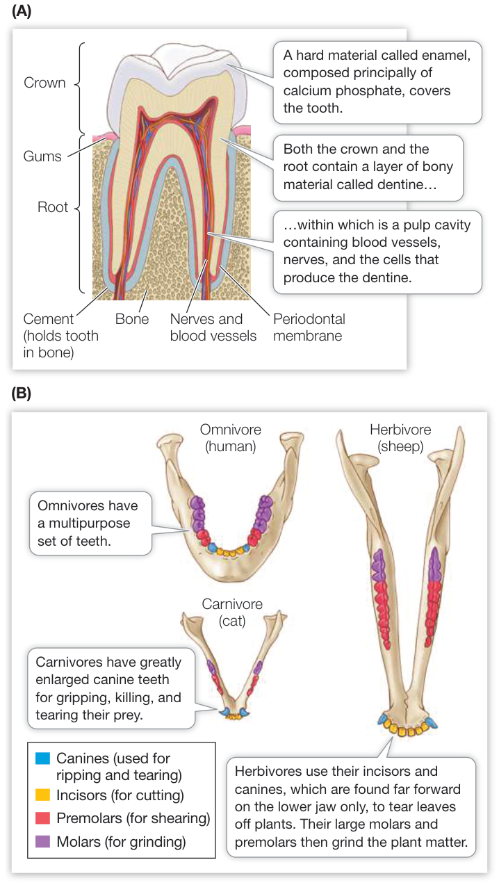
Figure 49.6 Mammalian Teeth (A) A mammalian tooth has three layers: enamel, dentine, and a pulp cavity. (B) The teeth of different mammalian species are specialized for different diets. This illustration depicts the teeth of the lower jaw, viewed from above.
Digestion usually begins in a body cavity
Animals take food into a body cavity that is continuous with the outside environment. They secrete digestive enzymes into that cavity, and the enzymes break down the food into nutrient molecules that can be absorbed by the cells lining the cavity.
The simplest digestive system is found in the simplest animals such as sponges. Water flows from the environment through the body of the sponge in water channels, and individual cells capture food particles from the water (see Figure 29.2B). A slightly more specialized digestive system is a gastrovascular cavity, which connects to the outside world through a single opening. Cnidarians, such as jellyfishes, capture prey using stinging nematocysts and use their tentacles to cram the prey into their gastrovascular cavities. Enzymes in the gastrovascular cavity partially digest the prey. Cells lining the cavity take in small food particles by endocytosis. The vesicles created by endocytosis then fuse with lysosomes containing digestive enzymes, and intracellular digestion completes the breakdown of the food. Nutrients are released to the cytoplasm as the vesicles break down.
Tubular guts have an opening at each end
Animals classified as bilaterians (see Key Concept 29.1) have a tubular digestive system called a gastrointestinal system or simply gut. The gut begins with a mouth that takes in food. The food is moved through the length of the gut as it is sequentially disassembled or digested, and the smallest units of the digested food are absorbed across the gut wall. Solid digestive wastes are eliminated through an anus. Different regions in the tubular gut are specialized for particular functions (Figure 49.7). These functions must be coordinated so they occur in the proper sequence, and they must be regulated so they occur slowly enough to allow complete digestion but quickly enough to supply the energy needs of the animal.
In the mouth cavity, food may be physically fragmented by teeth (in many vertebrates), by a radula (in snails), or by mandibles (in many arthropods). In most birds, food is ground by small stones in an early, muscular portion of the gut called the gizzard. Some animals, such as snakes, simply ingest whole prey with little or no fragmentation. Teeth in these species are angled backward to keep the prey moving in only one direction while being slowly swallowed. Stomachs and crops are storage chambers that enable animals to ingest relatively large amounts of food when it is available, and then digest it gradually. In these storage chambers, food may be further fragmented and mixed, and in most vertebrates it is an important site of digestion. Food delivered into the next section of the gut, the intestine, is in small particles, well mixed, and usually partially digested.
Most digestion occurs in the intestine, and nutrients, water, and ions are absorbed across its walls. Glands secrete digestive enzymes into the intestine, and other enzymes are produced and secreted by cells lining the intestine. The final segment of the intestine recovers water and ions and stores undigested wastes, or feces, so they can be released to the environment at an appropriate time or place. A muscular rectum near the anus assists in expelling feces.
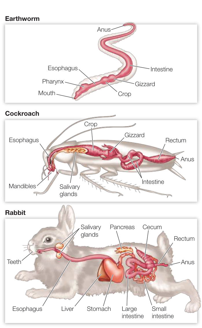
Figure 49.7 Compartments for Digestion and Absorption Most invertebrates and all vertebrates have a tubular gut that begins with a mouth where food is taken in, and ends in an anus where waste is eliminated. Between these two structures are specialized regions for digestion and nutrient absorption; the structures in these regions are adapted to different diets and vary from species to species.
In many animals, the parts of the gut that absorb nutrients have greater surface areas than would be expected of a simple tube. The simplest way to increase the surface area of a tube without changing its diameter is to produce an infolding. Such an infolding of the gut, called a typhlosole, is seen in earthworms (Figure 49.8A). Sharks, which have large stomachs but short intestines, have a unique adaptation called a spiral valve (Figure 49.8B). The lower region of the intestine is enlarged and has an internal structure that forces the food to pass through it in a spiral fashion (like going down a spiral staircase). The walls of the spiral present a large surface area for absorption of nutrients. This spiral passage, however, will not accommodate large chunks of food or undigestible matter, so food remains in the stomach for a long time to be broken down, and some larger, undigestible items leave the stomach only by regurgitation.
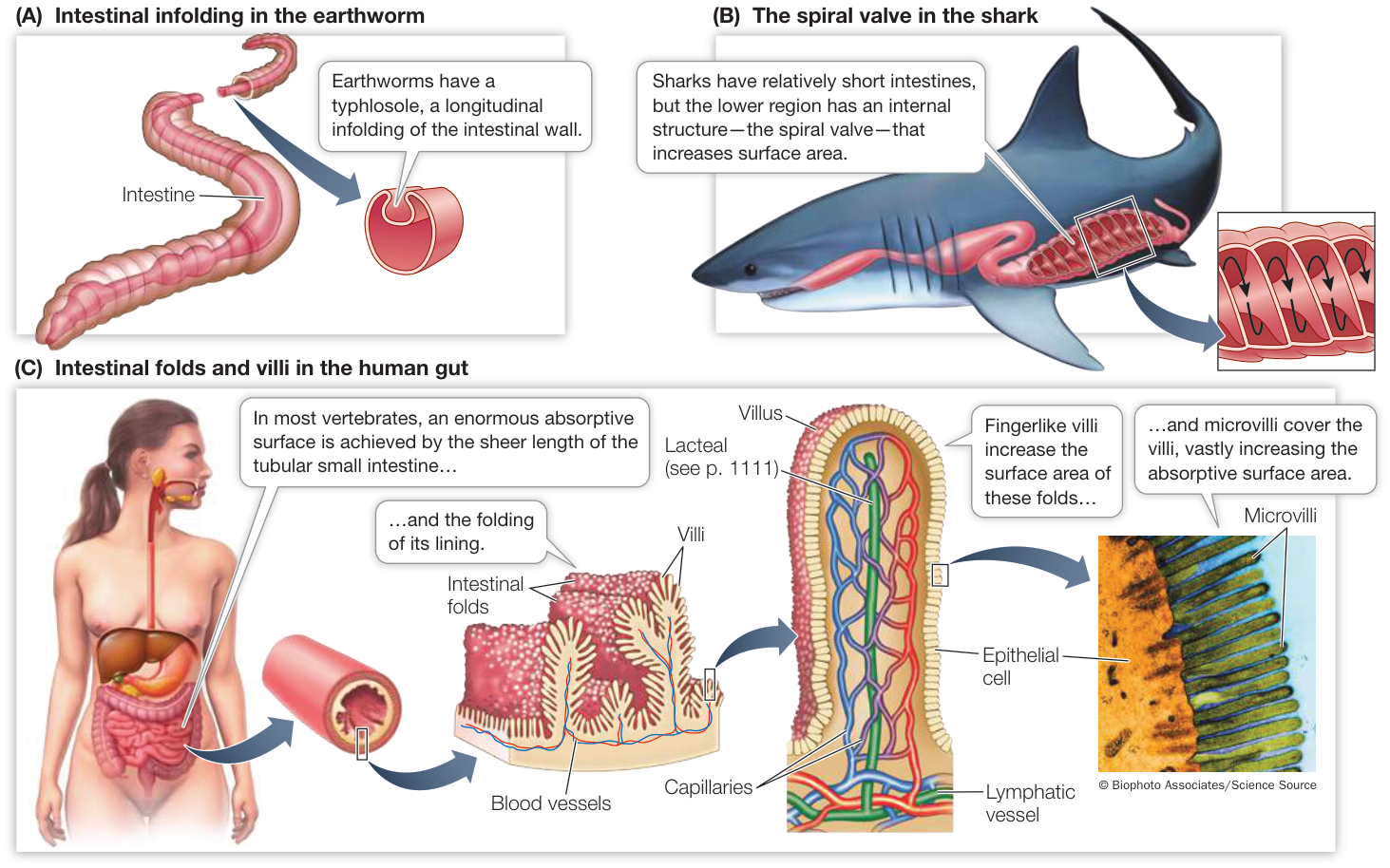
Figure 49.8 Intestinal Surface Area and Nutrient Absorption Maximizing the surface area of the gut increases an animal’s ability to absorb nutrients.
The wall of the intestine in humans and most other vertebrates is highly folded, with the individual folds bearing legions of tiny fingerlike projections called villi (Figure 49.8C). The cells that line the surfaces of the villi, in turn, have microscopic projections called microvilli. The microvilli give the intestine an enormous internal surface area for absorbing nutrients.
Digestive enzymes break down complex food molecules
Protein, carbohydrate, and fat macromolecules are broken down into their simplest monomeric units by hydrolytic enzymes produced at different locations in the digestive tract. Many are secreted into the lumen of the gut, and others remain associated with the membranes of the microvilli. All of these enzymes cleave the chemical bonds of macromolecules through hydrolysis, a reaction that adds a water molecule (see Figure 3.4B). Digestive enzymes are classified according to the substances they hydrolyze: proteases break the bonds between adjacent amino acids in proteins; carbohydrases hydrolyze carbohydrates; peptidases break down peptides; lipases, fats; and nucleases, nucleic acids. We discuss the digestive enzymes of humans in detail in Key Concept 49.3.
The gut microbiome contributes to digestion
The gut is colonized by a huge population of microorganisms, mostly bacteria. Collectively the foreign species associated with an organism are called its microbiota, and their cumulative genomes are called the organism’s microbiome. The microbiomes of the skin are different from the microbiomes of the mouth, gut, respiratory tract, and urogenital tracts. The microbiota of the gut obtain their nutrition from the food passing through the gut while contributing to the host’s digestive processes. For example, members of the leech genus Hirudo produce no enzymes that can digest the proteins in the blood they suck from vertebrates; instead they depend on bacteria to perform this service. The resulting amino acids are subsequently used by both the leech and the bacteria. The microbiota of the human gut also provide important services in digestion, prevent the establishment of harmful microorganisms, and even produce some vitamins (vitamin K and biotin). It is estimated that the human body consists of cells, but our guts contain probably three times that number of unicellular organisms representing at least 490 different species (see Figure 24.19). Recent studies have revealed that gut microbiomes are not constant, but are influenced by diet and other factors, including medications we ingest. The effects of different microbiomes may go far beyond simply facilitating digestion (Investigating Life: How Does the Gut Microbiome Contribute to Obesity and Metabolic Disease?).
Investigating Life: How Does the Gut Microbiome Contribute to Obesity and Metabolic Disease?
Experiment
Original Paper: R. Caesar et al. 2015. Crosstalk between gut microbiota and dietary lipids aggravates WAT inflammation through TLR signaling. Cell 22: 658–668.
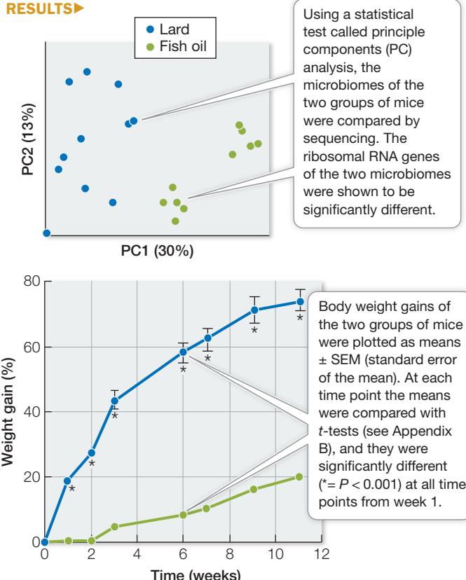
Fats differ in fatty acid composition, and fatty acids differ in the number of single or double bonds between their carbon atoms (see Key Concept 3.4). Saturated fatty acids have no double bonds, and unsaturated fatty acids do. Diets rich in saturated fatty acids are implicated in several health concerns, but what is it about saturated fatty acids that is unhealthy? Recent research examines the effect of diet on the gut microbiome, and in turn, the effect of the microbiome on health. Does the amount of saturated fat in the diet influence the composition of the microbiome, and does the microbiome influence the health consequences of a diet rich in saturated fats? We examine the first question in the experiment below, and the second question in an online companion Data in Depth exercise.
HYPOTHESIS The type of fat in the diet can influence the composition of the gut microbiome.
METHOD
- Raise two groups of 15 mice on isocaloric diets rich in saturated fats (lard) or unsaturated fats (fish oil).
- Each week, record the mice’s body weight.
- At the end of 11 weeks, compare the mice’s microbiomes (by sequencing the ribosomal RNA genes) and body weight gain.
RESULTS
CONCLUSION
- The fat composition of the diet influenced the composition of the microbiome.
- Mice on the diet containing saturated fats gained more weight and gained weight faster than mice on the isocaloric diet containing unsaturated fats.
Work with the Data
The researchers hypothesized that mice consuming more saturated fats would have a higher probability of becoming obese and developing symptoms of diabetes. To test for dietary effects on the development of metabolic disease, the researchers collected additional data from the two cohorts of mice: fasting blood glucose levels (measured in millimolars, mM) and fasting blood insulin levels (measured in nanograms per milliliter, ng/mL). They also did an insulin tolerance test (effect of a dose of insulin on blood glucose level). Data are reported as means ( SEM).
| Fasting blood glucose (mM) | Fasting blood insulin (ng/mL) |
|---|---|
| Lard (N = 14) |
Fish oil (N = 14) |
| 11 (0.22) | 10.32 (0.23) |
| Lard (N = 10) |
Fish oil (N = 14) |
| 3.96 (0.36) | 1.09 (0.17) |
| Blood glucose (% of the time 0 level) | ||
|---|---|---|
| Time after insulin injection (min) | Lard | Fish oil |
| 0 | 100 | 100 |
| 30 | 59.8 (3.05) | 49.8 (4.09) |
| 60 | 54.5 (3.51) | 37.2 (4.07) |
| 90 | 63.1 (3.91) | 39.1 (4.09) |
| 120 | 75.6 (3.06) | 56.9 (3.31) |
QUESTIONS
- Using a t-test, determine whether the difference in the fasting blood glucose levels for the lard-fed and fish-oil-fed mice is significant.
- Using a t-test, determine whether the difference in the fasting blood insulin levels for the lard-fed and fish-oil-fed mice is significant.
- Plot the glucose over time data. Did the two groups of mice differ in their response to the same dose of insulin? At which time points?
- What conclusions can you make about the effect of the type of dietary fat on carbohydrate metabolism?
Recap and Assess
Heterotrophs have diverse adaptations for acquiring food. Once captured or ingested, food is digested extracellularly by secreted enzymes to release nutrients that are absorbed into the animal’s body, usually via a tubular gut. The gut microbiome plays a role in digestion.
- Why do herbivores typically spend more time feeding than do carnivores?
- What do the actions of all digestive enzymes have in common?
- Why is it common for individuals to have digestive problems after antibiotic therapy?
- When people with celiac disease eat food containing gluten, their immune system damages their intestinal villi. If undiagnosed, such people are likely to experience bloating, abdominal pain, diarrhea, fatty stool, loss of body weight, and iron deficiency. Why do these symptoms occur?
Once ingested by an animal, food may be fragmented and moved into the gut for digestion by hydrolytic enzymes. The processes of digestion release nutrients that are absorbed into the animal’s body. Next we focus on how those processes occur in vertebrates.
The Vertebrate Gastrointestinal System Is a Disassembly Line
Learning Objectives
49.3.1 Explain the mechanisms moving food through the vertebrate digestive system.
49.3.2 Discuss the role of stomach acid and how it is produced.
49.3.3 Describe the role of autocatalysis in digestion.
49.3.4 Explain the role of bile in digestion.
49.3.5 Discuss the roles of microbiota in the digestive systems of different vertebrates.
Digestion in vertebrates occurs in the gastrointestinal system, which includes a tubular gut running from mouth to anus and several accessory structures that produce secretions that play important roles in digestion (Figure 49.9). In this key concept we consider three important processes of this system: the movement of food through it, the sequential steps of digestion, and the absorption of nutrients. We use as our primary example a typical vertebrate, the human.
The vertebrate gut consists of concentric cellular layers
Tissues of the vertebrate gut are arranged in concentric layers that have a similar organization throughout its length (Figure 49.10). The mucosa lines the internal cavity, or lumen, of the gut. The mucosa consists of delicate epithelial cells with underlying connective tissue. Some cells of this mucosal epithelium secrete mucus to lubricate and protect the walls of the gut; some secrete digestive enzymes; and some secrete hormones. Mucosal epithelial cells in the stomach secrete hydrochloric acid, and as we noted in Key Concept 49.1, some secrete intrinsic factor to aid the absorption of vitamin B12. In some regions of the gut, nutrients are absorbed by mucosal epithelial cells. The apical cell membranes of these absorptive cells have microvilli that increase the surface area over which absorption can take place (see Figure 49.8C).
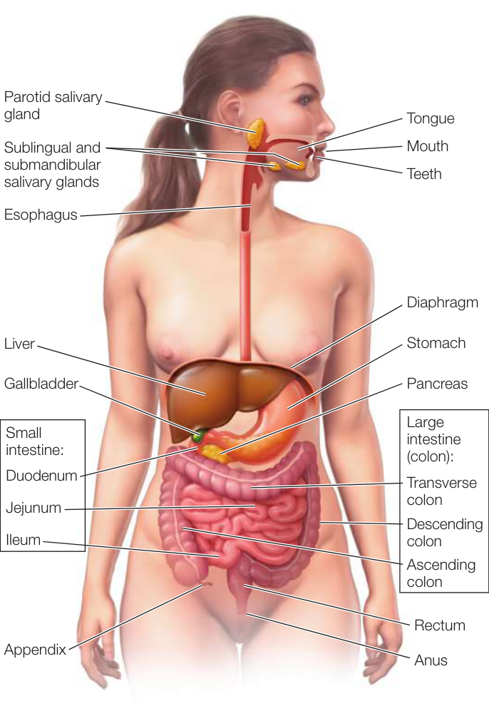
Figure 49.9 The Human Digestive System Different compartments in the long tubular gut specialize in digesting food, absorbing nutrients, and storing and expelling wastes. Accessory organs contribute secretions containing enzymes and other molecules. Activity 49.4 The Human Digestive System
At the base of the mucosa there is a thin layer of smooth muscle cells with random orientations. The function of this cell layer is to move the mucosal epithelium to maximize its contact with the gut contents. Just under the mucosa is the submucosal tissue layer where blood and lymph vessels pick up nutrients to transport to the rest of the body. The submucosa also contains a network of nerves; the neurons in this network have sensory functions (responsible for stomach aches) and also control various secretory functions of the gut.
External to the submucosa are two layers of smooth muscle responsible for large movements of the gut. Innermost is the circular muscle layer, with its cells oriented around the gut; outermost is the longitudinal muscle layer, with its cells oriented along the length of the gut (see Figure 49.10). The circular muscles constrict the gut, and the longitudinal muscles shorten it. Between the two layers of smooth muscle is another nerve network that controls and coordinates the movements of the gut. The coordinated activity of the two smooth muscle layers mixes contents of the gut and moves them continuously toward the anus.
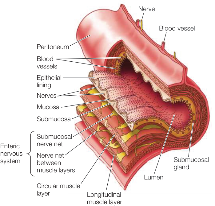
Figure 49.10 Tissue Layers of the Vertebrate Gut The organization of tissue layers is the same in all compartments of the gut, but specialized adaptations of specific tissues characterize different regions.
Media Clip 49.1 Following Food from Mouth to Gut Life12e.com/mc49.1
Nerve nets in the submucosa and between the smooth muscle layers are called the enteric nervous system, and they are unusual. The cell bodies of most peripheral nerves are in the central nervous system (CNS)—in the brain or spinal cord. The only exceptions are the postganglionic cells of the autonomic nervous system and the cells of the enteric nervous system. In addition, most neurons of the peripheral nervous system either receive synapses from neurons in the CNS or contribute synapses to neurons in the CNS. In contrast, most of the neurons in the enteric nervous system form synapses only with other neurons in their network. Thus they are responsible for communication within the gut. The CNS can influence activity in the enteric nervous system and receive information from it, but the gut truly has “a mind of its own.”
A tissue membrane called the peritoneum surrounds the gut and most of the organs of the abdominal cavity as well as lining the wall of the cavity. The peritoneum includes connective and epithelial tissues that secrete a lubricating fluid that enables the organs to easily slide against each other in the body cavity.
Gut motility moves food through the gut and aids digestion
In humans and most other mammals, food is chewed in the mouth and mixed with saliva. Periodically the tongue pushes a bolus (mass) of the chewed food toward the throat. By making contact with the soft palate at the back of the mouth cavity, the food bolus initiates swallowing, which is a complex series of reflexes. Swallowing propels the food through the pharynx (where the mouth cavity and nasal passages join) and into the esophagus (food tube). To prevent food from entering the trachea (windpipe), the larynx (voice box) closes, and a flap of tissue called the epiglottis covers the entrance to the larynx (Figure 49.11A).
Once a bolus of food enters the esophagus, it is moved toward the stomach both by gravity and by waves of muscle contraction called peristalsis (Figure 49.11B). The muscle of the upper region enters the esophagus, peristalsis propels it from mouth to anus by coordinated actions of the circular and longitudinal muscle layers of the gut.
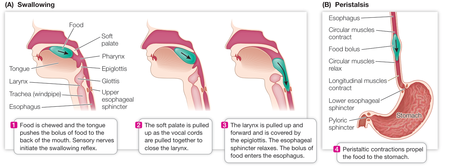
Figure 49.11 Swallowing and Peristalsis (A) Food pushed to the back of the mouth triggers the swallowing reflex. (B) Once a food bolus enters the esophagus, peristalsis propels it from mouth to anus by coordinated actions of the circular and longitudinal muscle layers of the gut.
of the esophagus is striated (i.e., skeletal muscle) and is controlled by the CNS reflexes of swallowing. The muscles of the rest of the esophagus are smooth muscles controlled by the autonomic and enteric nervous systems.
Smooth muscles of the gut contract in response to being stretched. When a bolus of food reaches the smooth muscle region of the esophagus and stretches it, the muscle responds by contracting, thus pushing food toward the stomach. Why doesn’t the contraction of the esophageal smooth muscle push food back toward the mouth? The nerve net between the two smooth muscle layers coordinates the muscles so that when a region of the gut smooth muscle contracts, the circular smooth muscle just beyond it relaxes. This action, controlled by the enteric nervous system, is called an anticipatory wave of relaxation. The contractions of smooth muscles behind the relaxing area push food into it—in the direction of the anus. The resulting stretch of that region stimulates its circular smooth muscle to contract as the anticipatory wave of relaxation causes the smooth muscle of the next region to relax. The peristaltic waves of contraction and relaxation of the gut smooth muscle move food along the gut from the mouth to the anus.
At the junction of the esophagus and stomach is an esophageal sphincter, a ring of circular smooth muscle that is usually constricted. Waves of peristalsis cause the sphincter to relax enough to let food pass from the esophagus into the stomach. Sphincter muscles are found elsewhere in the digestive tract as well: the pyloric sphincter governs the passage of stomach contents into the small intestine; the ileocaecal sphincter controls the flow of food between the small and large intestines; and the anal sphincter relaxes to allow defecation.
Some movements of the stomach are not as coordinated as the peristaltic movements of the esophagus and serve to move the contents of the stomach around and mix them with the digestive juices. However, peristaltic waves of contraction reorganize in the lower region of the stomach and push the semi-digested material up against the pyloric sphincter which controls the opening to the small intestine. In the small intestine, peristaltic activity continues to move the contents of the gut toward the large intestine.
Chemical digestion begins in the mouth and the stomach
Salivary glands secrete the enzyme amylase into the mouth where it is mixed with the food being chewed. Amylase hydrolyzes bonds between glucose monomers that make up carbohydrate molecules. The action of amylase is what makes a chewed piece of bread or cracker taste slightly sweet if you hold it in your mouth and chew it long enough.
The main role of the stomach is to store food, continue its enzymatic digestion, and deliver it to the small intestine at an appropriate rate so that digestion can occur more slowly than ingestion. The stomach produces secretions that facilitate digestion and also kill potential pathogens that are ingested with food. Gastric pits in the stomach walls (Figure 49.12A,B) have three types of secretory cells: (1) mucus-secreting cells, (2) parietal cells that secrete hydrochloric acid (HCl), and (3) chief cells that secrete the inactive proteolytic enzyme pepsinogen (Figure 49.12C). The low pH of the stomach juices kills most microorganisms that are ingested with food; denatures proteins, making them more accessible to the hydrolytic actions of pepsin; and converts pepsinogen to pepsin by cleaving away a sequence of amino acids that masks the active site of the enzyme. Newly activated pepsin activates other pepsinogen molecules, creating a positive-feedback process called autocatalysis. Acid and the proteolytic enzyme pepsin could damage the stomach walls, but the mucus-secreting cells provide a protective coating for the walls of the gastric pits and stomach.
The production of HCl by gastric pit parietal cells is impressive. It amounts to about 2 liters of a highly concentrated solution per day—enough to bring the pH of the stomach contents below 1, which is the same as battery acid and ten times more acidic than pure lemon juice. This means that across their cell membranes, parietal cells create a ion concentration difference of 3 million-fold. Such a feat of transport is not seen anywhere else in the body. How do the gastric pits do it? Enzymes and transporters are involved.
The enzyme carbonic anhydrase in parietal cells catalyzes the hydration of to , which dissociates into and a bicarbonate ion (). An antiporter transport protein (see Figure 6.13) exchanges for on the blood side of the gastric pits, and an antiporter on the gastric pit side exchanges for (Figure 49.12D). However, leaks out of the parietal cell down its concentration gradient. Thus the inward transport of acts like an endless conveyer belt moving out into the stomach lumen. also passively leaks out of the gastric lumen side of the parietal cells to maintain electrical neutrality.
The stomach gradually releases its contents to the small intestine
The acidic, fluid mixture of gastric juice and partly digested food in the stomach is called chyme. A few substances can be absorbed across the stomach wall, including alcohol (hence its rapid effects), aspirin, and caffeine, but even these substances are absorbed in rather small quantities from the stomach; most absorption takes place in the small intestine. At the bottom of the stomach, peristaltic contractions push the chyme against the pyloric sphincter. Associated waves of anticipatory relaxation cause the pyloric sphincter to relax briefly so that little squirts of chyme enter the small intestine. The human stomach empties gradually over a period of approximately 4 hours. This slow introduction of food into the small intestine enables it to work on a little material at a time.
Most chemical digestion occurs in the small intestine
In the small intestine, digestion of carbohydrates and proteins continues, and digestion of fats and absorption of nutrients begin. The small intestine takes its name from its diameter; it is in fact a very large organ, about 6 meters long in an adult human. Given its length and the folds, villi, and microvilli of its epithelial cells, its inner surface area is roughly the size of a tennis court. Across this surface the small intestine absorbs the nutrient molecules derived from food.
The small intestine of humans has three sections. The initial section (about 25 cm long) is called the duodenum and is the site of most digestion; the jejunum and the ileum (together about 600 cm) carry out 90% of the absorption of nutrients (see Figure 49.9).
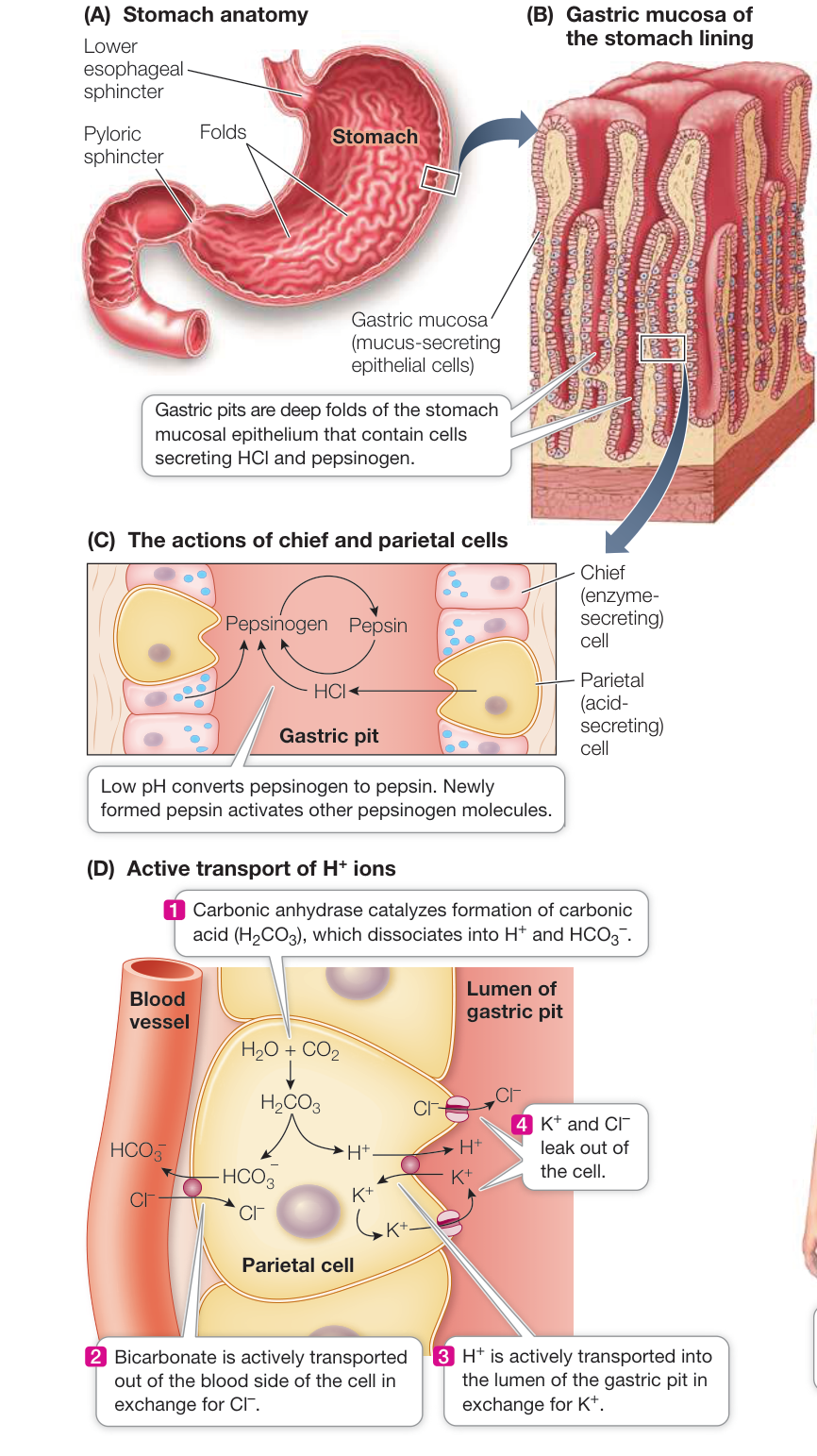
Figure 49.12 Action in the Stomach (A) The human stomach stores ingested food and mixes it with digestive secretions. (B) Gastric pits are folds in the stomach lining. (C) Parietal cells in the gastric pits secrete hydrochloric acid (HCl), and chief cells secrete pepsinogen. HCl and autocatalysis convert inactive pepsinogen into active pepsin. (D) Parietal cells create a tremendous concentration difference between the gut lumen and the extracellular fluid by actively transporting into the lumen of the gastric pit ions produced through the catalytic action of carbonic anhydrase.
Digestion in the small intestine involves many specialized enzymes, as well as several other secretions. Two accessory organs that are not part of the digestive tract—the liver and the pancreas—produce most of these secretions and deliver them to the lumen of the intestine through ducts.
LIVER The liver synthesizes bile from cholesterol. Bile also includes other substances, such as phospholipids and bilirubin (the breakdown product of hemoglobin). Bile flows from the liver through the hepatic duct. A side branch off the hepatic duct called the cystic duct goes to the gallbladder, where bile is stored. Below this junction, the hepatic duct is called the common bile duct. Before it reaches the duodenum, the common bile duct is joined by the pancreatic duct (Figure 49.13).
Fat entering the duodenum stimulates cells of the duodenal epithelium to release the hormone cholecystokinin (CCK), which stimulates the walls of the gallbladder to contract rhythmically. As a result, bile is squeezed out of the gallbladder and through the cystic duct to the common bile duct. Peristalsis moves the bile down the bile duct, and a small sphincter at the junction of the common bile duct with the duodenum relaxes in response to waves of peristalsis and allows squirts of bile to enter the duodenal lumen.
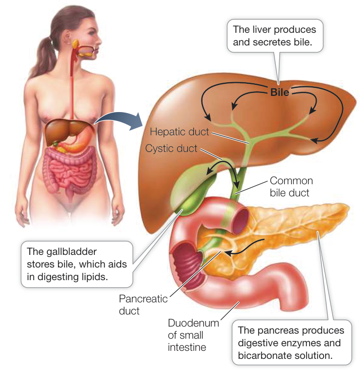
Figure 49.13 Ducts of the Gallbladder and Pancreas Bile produced in the liver flows from the liver via the hepatic duct. Branching off this duct is the gallbladder, which stores bile. Below the gallbladder, the hepatic duct is called the common bile duct and is joined by the pancreatic duct before entering the duodenum.
To understand the role of bile in fat digestion, think of an oil-and-vinegar salad dressing. Oil is hydrophobic and tends to aggregate in large globules. For that reason, many salad dressings include an emulsifier—something that prevents oil droplets from aggregating. Mayonnaise, for example, is oil and vinegar with egg yolk added as an emulsifier. Bile emulsifies fats in the chyme. One end of each bile molecule is lipophilic (soluble in fat), and the other end is hydrophilic (soluble in water). The lipophilic ends of bile molecules merge with the fat droplets, leaving their hydrophilic ends sticking out. As a result, bile prevents small fat droplets from congealing together into large fat droplets, and thereby the surface area of the fats exposed to the fat-digesting enzymes (lipases) is greatly enlarged. The very small fat particles that result from emulsification by bile are called micelles (Figure 49.14A).
PANCREAS The pancreas is a large gland that lies just behind and below the stomach (see Figures 49.9 and 49.13). It is both an endocrine gland (secreting hormones into the circulation; see Key Concept 39.1) and an exocrine gland (secreting digestive juices through the pancreatic duct to the gut lumen). The exocrine tissues of the pancreas produce a host of digestive enzymes, including lipases, amylases, proteases, and nucleases (Table 49.3). As in the stomach, the protease enzymes are released in inactive forms called zymogens; if proteases were not in this inactive state, they would digest the pancreas and its ducts before ever reaching the duodenum. Once in the duodenum, the zymogen trypsinogen is activated by the enzyme enterokinase (secreted by cells lining the duodenum) to produce the active protease trypsin. Trypsin cleaves other zymogens, releasing other proteases as well as more active trypsin.
The mixture of zymogens produced by the pancreas can be dangerous if the pancreatic duct is blocked or if the pancreas is injured by infection or physical trauma such as a blow to the abdomen. A few activated trypsin molecules can initiate a chain reaction of enzyme activity that digests the tissues of the pancreas (a condition called pancreatitis), destroying both its endocrine and exocrine functions.
The ducts of the pancreas produce a secretion rich in bicarbonate ions (). Bicarbonate ions are alkaline (basic) and neutralize the acidic pH of the chyme that enters the duodenum from the stomach. Intestinal enzymes function best at a neutral or slightly alkaline pH. The production of bicarbonate and its secretion into the pancreatic ducts is similar to the production of ions and their secretion into the gastric pits—but with opposite spatial orientations. In both types of secretory cells, carbonic anhydrase catalyzes the hydration of to produce that dissociates into and ions. Whereas in the gastric pits the is secreted into the lumen of the pit, in the pancreatic duct it is the that is secreted into the lumen, and the is transported into the circulation.
Nutrients are absorbed in the small intestine
The final step in digesting proteins and carbohydrates and absorbing their components occurs among the intestinal microvilli. Mucosal epithelial cells produce peptidases that cleave small peptides into absorbable amino acids. These epithelial cells also produce the enzymes maltase, lactase, and sucrase that cleave the common disaccharides into absorbable monosaccharides—glucose, galactose, and fructose. There is also some lipase activity that facilitates fat digestion.
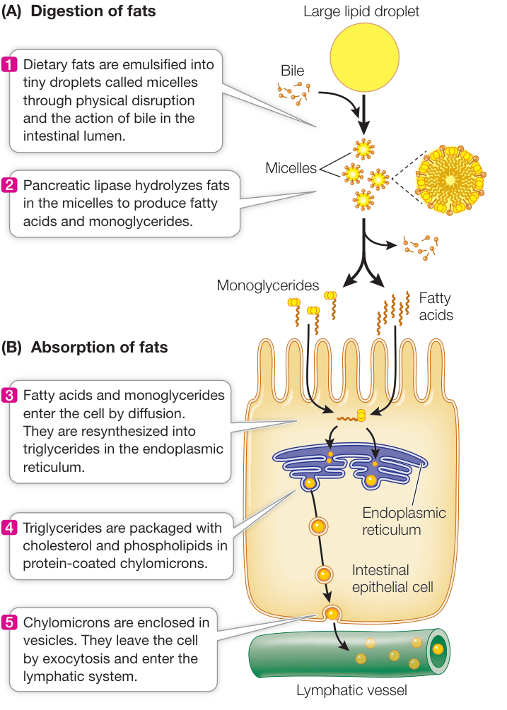
Figure 49.14 Digesting Fats (A) Dietary fats are broken up by bile into small micelles that present a large surface area to lipases. (B) The products of fat digestion are absorbed by intestinal mucosal cells, where they are resynthesized into triglycerides and exported to lymphatic vessels. CHAPTER 49 Nutrition, Digestion, and Absorption Q: A small percentage of people who have had their gallbladders removed must thereafter eat a low-fat diet, or they will experience diarrhea. Why?
Many humans stop producing the enzyme lactase in childhood and thereafter have difficulty digesting lactose (the sugar in milk). Lactose is a disaccharide and cannot be absorbed without being cleaved into its constituents, glucose and galactose. Unabsorbed lactose is metabolized by bacteria in the large intestine, causing gas, diarrhea, and abdominal cramps.
The mechanisms by which cells of the intestinal epithelium absorb nutrients and inorganic ions are diverse and include diffusion, facilitated diffusion, active transport, and secondary active
Table 49.3 Major Digestive Enzymes of Humans
| Source/enzyme | Action |
|---|---|
| SALIVARY GLANDS | |
| Salivary amylase | Starch → Maltose |
| STOMACH | |
| Pepsin | Proteins → Peptides; autocatalysis |
| PANCREAS | |
| Pancreatic amylase | Starch → Maltose |
| Lipase | Fats → Fatty acids and glycerol |
| Nuclease | Nucleic acids → Nucleotides |
| Trypsin | Proteins → Peptides; zymogen activation |
| Chymotrypsin | Proteins → Peptides |
| Carboxypeptidase | Peptides → Shorter peptides and amino acids |
| SMALL INTESTINE | |
| Aminopeptidase | Peptides → Shorter peptides and amino acids |
| Dipeptidase | Dipeptides → Amino acids |
| Enterokinase | Trypsinogen → Trypsin |
| Nuclease | Nucleic acids → Nucleotides |
| Maltase | Maltose → Glucose |
| Lactase | Lactose → Galactose and glucose |
| Sucrase | Sucrose → Fructose and glucose |
transport. Many inorganic ions such as sodium, calcium, and iron are actively transported by these cells. For example, active transporters exist on the basal and lateral sides of the epithelial cells. They maintain a low concentration of in those cells so that can diffuse in from the chyme in the intestinal lumen. About 30 grams of are transported this way every day, and follows.
The transport of and other ions is important for water absorption because it creates an osmotic concentration gradient. At least 7–8 liters of water per day move through the spaces between the epithelial cells in response to this osmotic gradient. Because the water moves through spaces between the cells and not through the cells themselves, it can carry with it nutrients that are in solution—a transport mechanism called solvent drag or bulk transport.
Many different kinds of transport proteins function in epithelial cell membranes. Some, such as the transport protein for fructose, only facilitate diffusion, and that requires a concentration gradient. Once fructose enters the cell it is converted to glucose, so the concentration of fructose in the cell is always low and the concentration gradient is maintained. Transport proteins known as symporters (see Figure 6.13) exploit the concentration gradient of between the inside and outside of the cell that is maintained by the – pump common to all cells (see Figure 6.14). Symporters combine the transport of and another molecule, such as glucose, galactose, or an amino acid. As moves down its concentration gradient into the cell, the “hitchhiking” molecules are carried along with it.
The absorption of the products of fat digestion is relatively simple. Triglycerides are hydrolyzed to diglycerides, monoglycerides, and fatty acids, all of which are lipid-soluble and thus able to pass through the cell membranes of the microvilli. Once these molecules are in the intestinal epithelial cells, they are resynthesized into triglycerides, combined with cholesterol and phospholipids, and coated with protein to form water-soluble chylomicrons (Figure 49.14B). Rather than enter the blood directly, chylomicrons pass into blind-ended lymph vessels called lacteals that are inside each villus (see Figure 49.8C). The chylomicrons then flow through the lymphatic system and enter the bloodstream through the thoracic ducts at the base of the neck. After a meal rich in fats, chylomicrons can be so abundant in the blood that they give the blood plasma a milky appearance. Chylomicrons deliver their triglyceride and cholesterol cargo as they circulate through tissues.
The bile molecules that emulsify fats are not absorbed along with the monoglycerides, diglycerides, and fatty acids, but are shuttled back and forth between the gut contents and the microvilli. In the ileum, bile is actively reabsorbed and returned to the liver via the bloodstream.
Absorbed nutrients go to the liver
Blood leaving the digestive tract flows to the liver in the hepatic portal vein. This large vein delivers the blood to small spaces called sinusoids between groups of liver cells. These cells absorb the nutrients coming from the digestive tract and either store them or convert them to molecules the body needs. Glucose, galactose, and fructose are used to synthesize glycogen. Amino acids are used to build proteins. Lipids from the chylomicrons are either stored as triglycerides or used to make lipoproteins that are released by the liver and carry the triglycerides and cholesterol to other tissues (as we discuss in Key Concept 49.4).
Water and ions are absorbed in the large intestine
The motility of the small intestine gradually pushes its contents through another sphincter—the ileocaecal sphincter—and into the large intestine, or colon. Most of the available nutrients have been removed from the chyme that enters the colon, but it contains a lot of water and inorganic ions. A major type of motility in the colon is called segmentation movements. These movements are mostly due to contractions of circular muscles that repeatedly divide the colon into separate segments. Functionally, segmentation movements cause the chyme to slosh around in the colon and promote absorption of ions and water by increasing contact between the chyme and the epithelium of the colon. As ions and water are absorbed, the chyme becomes a semisolid mass that forms feces. Absorption of too much water from the colon can cause hard feces and constipation. The opposite condition, diarrhea, results if too little water is absorbed. Excessive diarrhea caused by diseases such as cholera can produce such rapid loss of water and electrolytes that death can occur in hours. Simple rehydration therapy with electrolytes can save lives.
Fecal matter is stored in the descending colon and in the rectum until defecation occurs. This is usually once a day and is preceded by strong peristaltic activity. The distension of the walls of the rectum by fecal matter initiates a parasympathetic reflex, causing the rectal muscle to contract and the internal anal sphincter to relax. In addition, there is an external anal sphincter that is under conscious control so that defecation is not entirely an involuntary act.
Herbivores rely on their microbiota to digest cellulose
Cellulose is the primary component of plant cell walls, and is therefore the principal component of the food of herbivores. Most herbivores, however, cannot produce cellulases, the enzymes that break down cellulose. (Exceptions include earthworms, shipworms, and the silverfishes that eat books and stored papers.) From termites to cattle, herbivores rely on the microbiota of their digestive tracts to digest cellulose.
Ruminants (cud chewers) such as cattle ferment their plant-based food in special stomachs before digestion. Their stomachs are large, four-chambered organs (Figure 49.15). The first two chambers, the rumen and the reticulum, are packed with endosymbiotic microorganisms that break down cellulose by fermentation. The ruminant periodically regurgitates the contents of the rumen (the cud) into the mouth for more chewing. When swallowed again, the vegetal fibers present more surface area to the microorganisms. The microorganisms metabolize cellulose and other nutrients to simple fatty acids that are nutrients for their host.
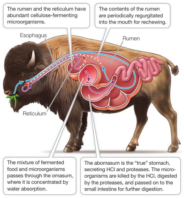
Figure 49.15 A Ruminant’s Stomach Bison (Bison bison), like their relatives domestic cattle, have a specialized stomach with four compartments that enables them to obtain energy from coarse plant material through bacterial fermentation of the otherwise indigestible plant material. The bacteria themselves become an important source of nutrition. Q: Functionally, why do the rumen and reticulum come before the true stomach, the abomasum?
Enormous numbers of microorganisms leave the rumen along with the partially digested food. This mass is concentrated by water absorption in the omasum before it enters the true stomach, the abomasum, where the microorganisms are killed by secreted hydrochloric acid, partially digested by proteases, and passed on to the small intestine for further digestion and absorption. A cow derives more than 100 grams of protein per day from digestion of its microbiota. The rate of multiplication of microorganisms in the rumen offsets their loss, so a well-balanced, mutually beneficial relationship is maintained.
Some mammalian herbivores have a microbial fermentation chamber called a cecum extending from the large intestine. An example is the rabbit (see Figure 49.7). Since the cecum empties into the large intestine, absorption of nutrients produced by the microorganisms in the cecum is inefficient because of the limited surface area of the large intestine. Such species frequently produce two kinds of feces—ones that are pure waste and ones that contain cecal material. In a behavior known as coprophagy, these species reingest the cecal feces directly from the anus so they can digest and absorb the nutrients that would otherwise be lost. In humans the cecum is small and ends in the vestigial appendix which serves no digestive function.
Recap and Assess
The vertebrate gastrointestinal system is a tubular gut that is adapted to ingest food, fragment it, digest it, and absorb nutrients. Peristalsis moves food through the gut. Segmentation movements mix the gut contents. Enzymatic digestion and then absorption of nutrients occur mostly in the small intestine; water and ions are absorbed in the large intestine.
- Explain why in the weightlessness of space, food still travels from the mouth to the stomach.
- When some biochemists ground up a piece of stomach wall and stimulated it with the hormone that causes the stomach to secrete HCl, no change in the pH of the solution occurred. Why not?
- How does bile assist in the digestion of fats, and why aren’t equivalent molecules necessary to facilitate transport of lipids in the lymph and blood?
- Explain why the gut microbiota is a significant source of nutrition for ruminants but not for humans.
- Explain why in the ruminant digestive system HCl is secreted in the abomasum but not in the rumen, the reticulum, or the omasum.
- Why would the autocatalytic property of pepsin foster a positive-feedback mechanism of protein hydrolysis in the stomach?
The steps included in the ingestion and digestion of food—from fragmentation in the mouth to the digestive processes in the stomach and intestines—make the nutrients in food available for absorption and ultimately for metabolism. We look next at how the processes of digestion are controlled and at how nutrients are handled by the body once food has been digested.
Nutrient Availability Is Controlled and Regulated
Learning Objectives
- 49.4.1 Describe how hormones control the rate of delivery of chyme to the small intestine and how they control secretions of digestive juices.
- 49.4.2 Explain how the metabolic interconversions that take place in the liver and muscles meet the body’s demands for glucose.
- 49.4.3 Describe the classes of lipoproteins.
- 49.4.4 Explain the effects of insulin on skeletal muscle cells, adipose cells, and liver cells.
- 49.4.5 Cite evidence that supports the role of leptin as a satiety signal and ghrelin as a hunger signal.
- 49.4.6 Interpret results from experiments designed to elucidate the mechanisms underlying regulation of body mass and food-intake behaviors.
As stated earlier, the vertebrate gut is a disassembly line, and as with a standard assembly line, control of the sequential processes is critical. Neuronal and hormonal controls coordinate digestive processes. And once the products of digestion are absorbed, their availability to the cells of the body must also be controlled and regulated.
Salivation at the sight or smell of food is an autonomic reflex, as is swallowing. Other autonomic reflexes include the release of digestive juices and increased stomach motility stimulated by good food smells. Reflexes also initiate gagging at the smell of rotting meat. These reflexes are called the “cephalic phase” of control of digestive processes because they involve the CNS. Most of the control of digestive processes, however, involves neural and hormonal mechanisms that are intrinsic to the gut.
Neuronal messages travel from one region of the digestive tract to another in the enteric nervous system. One function of the gut’s nervous system is coordinating the movement of food through the gut. Of course, this intrinsic nervous system communicates information to the CNS and receives input from the CNS, but its most important role is to coordinate actions throughout the digestive tract. In spite of this marvelous intrinsic nervous system, however, much of the control and regulation of the digestive system and nutrient management involves hormonal mechanisms.
Hormones control many digestive functions
Several hormones control the activities of the digestive tract and its accessory organs (Figure 49.16). The first hormone ever discovered came from the duodenum; it was called secretin because it causes the pancreas to secrete digestive juices. We now know that secretin is only one of several hormones that control pancreatic secretion; specifically, secretin stimulates the cells of the pancreatic ducts to secrete a solution rich in bicarbonate ions.
The stimulus that causes the duodenum to release secretin is the low pH of the acidic chyme from the stomach. Similarly, the presence of fats and proteins in the chyme stimulates the release of cholecystokinin (CCK), the hormone that stimulates the gall-bladder to release bile. CCK also stimulates the pancreas to release digestive enzymes. Both CCK and secretin have negative-feedback effects on the stomach. They slow the movements of the stomach, thus slowing the delivery of chyme into the small intestine to allow more complete digestion in the duodenum.
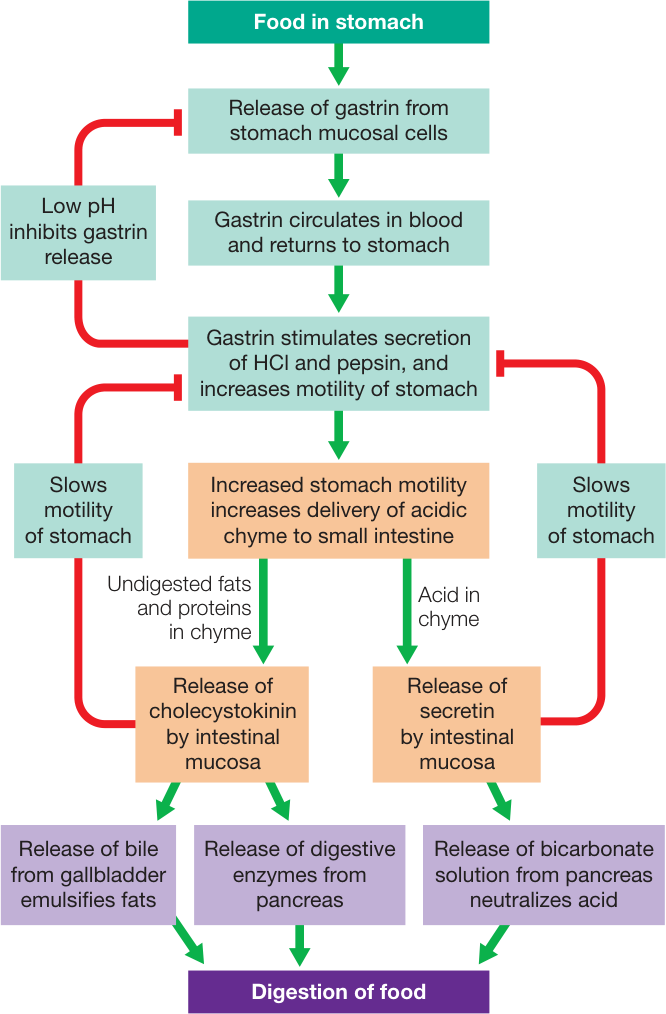
Figure 49.16 Hormones Control Digestion The hormones gastrin, cholecystokinin (CCK), and secretin are involved in feedback loops that control the sequential processing of food in the digestive tract. Red lines indicate inhibitory actions; green lines indicate stimulatory actions. Q: Why do secretin and CCK stimulate the pancreas but inhibit the stomach?
The presence of food in the stomach stimulates cells in the lower region of the stomach to secrete a hormone called gastrin. Gastrin returns to the stomach in the blood and stimulates the secretion of digestive juices and also increases the motility of the stomach. Gastrin release begins to be inhibited when the pH of the stomach contents falls below 3—another example of negative feedback.
The liver stores and releases the molecules that fuel metabolism
Most animals do not eat continuously, so they can be in either an absorptive state (food in the gut) or a postabsorptive state (no food in the gut). Nutrient requirements for energy metabolism and biosynthesis are continuous, however. Thus nutrient traffic must be controlled so that during the absorptive state energy reserves accumulate in the liver, muscle, and adipose (fat) tissue, and during the postabsorptive state these energy reserves are used efficiently. When fuel molecules are abundant in the blood, the liver stores them in the form of glycogen and fats. The liver also synthesizes blood plasma proteins from circulating amino acids. When levels of fuel molecules in the blood decline, the liver taps its reserves and delivers nutrients into the blood.
The liver has an enormous capacity to interconvert fuel molecules. Liver cells convert monosaccharides into either glycogen or fats during the absorptive state and then can reverse the process, returning glucose to the blood during the postabsorptive state. The liver can also convert certain amino acids and some other molecules, such as pyruvate and lactate, into glucose—the process of gluconeogenesis (see Key Concept 9.5). Gluconeogenesis provides an indirect pathway for exercising muscle to contribute to blood glucose levels. At high levels of aerobic activity, muscle cells break down their stores of glycogen to provide glucose for metabolic fuel. The glucose released from muscle glycogen, however, has to be used in the cells that produce it. Glucose cannot leave muscle cells as it can leave liver cells. However, when the activity of the muscle becomes anaerobic, pyruvate and lactate build up, leave muscle cells, and enter the circulation. Circulating pyruvate and lactate are taken up by the liver and converted to glucose that can then move out of the liver cells, circulate in the blood, and be taken up by muscle cells.
The liver produces lipoproteins
The liver is the major controller of fat metabolism through its production of lipoproteins. A lipoprotein is a particle made up of a core of hydrophobic fat and cholesterol with a covering of hydrophilic protein that allows it to be suspended in the extracellular fluids. Lipoproteins are the most abundant fuel reserve in the body, and they move fats from sites of absorption or synthesis to sites of storage, and from sites of storage to sites of use. You saw in Key Concept 49.3 how bile in the intestine solves the problem of processing hydrophobic fats in an aqueous medium. Transport of fats in the circulatory system presents the same problem, and lipoproteins provide the solution.
The chylomicrons produced by the mucosal cells of the intestine are the largest lipoprotein particles in the blood. As the circulation carries chylomicrons through muscle, adipose tissue, and liver, lipoprotein lipases attached to the endothelial cells of capillaries break down the chylomicrons’ triglycerides so that the component fatty acids, monoglycerides, and glycerol can be taken up by the cells. The chylomicron remnants have lipid cores with a lot of cholesterol, and they are taken up by liver cells.
Lipoproteins different from chylomicrons are synthesized in the liver. These lipoproteins can be classified according to their density. Fat has a low density (it floats on water) and protein has a high density, so the greater the fat-to-protein ratio in the lipoprotein, the lower its density.
High-density lipoproteins (HDLs) transport lipids and cholesterol from tissues to the liver where the cholesterol can be used to synthesize bile. HDL consists of about 49% protein, 35% lipids, and 15% cholesterol. These are called good lipoproteins, and their levels are higher in people who exercise and are fit.
Low-density lipoproteins (LDLs) transport cholesterol around the body for use in biosynthesis and for storage. LDL consists of about 25% protein, 25% lipids, and 49% cholesterol. These lipoproteins are associated with a high risk for cardiovascular disease.
Very low-density lipoproteins (VLDLs) transports triglycerides and cholesterol from the liver to peripheral cells that use the triglycerides for fuel and the cholesterol for membrane synthesis. These lipoproteins contain mostly triglycerides, and are about 2% protein, 94% lipids, and 3% cholesterol. As they give up their triglyceride, they become LDLs. VLDLs are associated with obesity and diabetes as well as a high risk for cardiovascular disease.
Insulin and glucagon control fuel metabolism
During the absorptive state, blood glucose levels rise as carbohydrates are digested and absorbed (Figure 49.17). During this time, the pancreas releases the hormone insulin, which plays a major role in directing glucose to where it will be used or stored. In Key Concept 49.3 we mentioned that the pancreas has both an exocrine function (secretions involved in digestion) and endocrine functions. The endocrine functions are located in clusters of cells in the pancreas called the islets of Langerhans. One class of islet cells, the beta cells, produces and releases insulin. Another class, the alpha cells, produces and releases the hormone glucagon (discussed shortly). The actions of insulin vary in different tissues, but they are all aimed at promoting the use of glucose for metabolic fuel and directing the excess glucose into storage as either glycogen or fat.
Glucose enters cells by diffusion. This diffusion is facilitated by transporters, but they are not active transporters—they require a concentration gradient, which is why it is important to regulate blood glucose levels so there is always an adequate glucose concentration gradient across the cell membranes. There are several kinds of glucose transporters, and those in skeletal muscle and adipose tissues are normally sequestered in cytoplasmic vesicles until insulin binds its receptors on the cell surface and triggers the insertion of transporters into the cell membrane.
Insulin plays many roles in controlling how cells use the glucose they take up from the circulation. In adipose cells, insulin inhibits lipase and promotes fat synthesis from glucose. In the liver, insulin activates an enzyme, glucokinase, that phosphorylates glucose as it enters the liver cell so it cannot diffuse back out again, thereby enhancing the overall diffusion of glucose into the cells. At the same time, insulin inhibits the enzyme glucose phosphatase that enables glucose to leave the cell. Insulin also activates glycogen synthase, an enzyme in liver cells that catalyzes the incorporation of glucose into glycogen, and activates enzymes that increase the flow of glucose into glycolysis (Figure 49.18).
To maintain blood glucose levels during the postabsorptive state, liver cells break down their stored glycogen, releasing glucose into the blood. The multiple processes that make it possible for the liver to release glucose depend on a fall in blood insulin levels (see Figure 49.18). Falling insulin inhibits the enzyme responsible for glycogen synthesis and activates the enzyme that breaks down glycogen. Also, with less insulin, the enzyme that phosphorylates glucose is inhibited and activity of glucose phosphatase is increased. The result is a breakdown of glycogen and the return of glucose to the blood. Another consequence of the fall in insulin levels is the increased activity of lipases in the liver and adipose tissue, releasing fatty acids to the blood. Most cells preferentially use fatty acids as their metabolic fuel during the postabsorptive state. Overall, the most important control of fuel metabolism in the postabsorptive state is the lack of insulin.
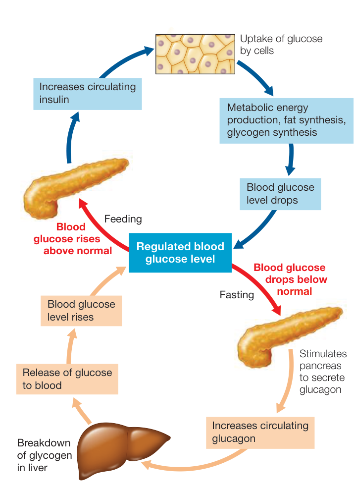
Figure 49.17 Regulating Glucose Levels in the Blood The hormone insulin (blue) acts to promote glucose use and storage and thereby lower blood glucose. The hormone glucagon (brown) acts on the liver to break down glycogen and release glucose into the blood. Q: The nervous system depends on a constant supply of glucose, yet none of the actions of insulin or glucagon in this figure refer to supplying glucose to the nervous system. Why?
One tissue that does not switch fuel sources when an animal is postabsorptive is the nervous system. The cells of the nervous system require a constant supply of glucose and can use other fuels only to a limited extent. Most neurons do not require insulin to absorb glucose from the blood, but they do need an adequate glucose concentration gradient to drive the facilitated diffusion of glucose across their cell membranes. Therefore it is critical that blood glucose levels are maintained when an animal is postabsorptive. The overall dependence of neural tissues on glucose, and their requirement for constant blood glucose levels, are the reasons it is so important for other cells of the body to shift to fat metabolism during the postabsorptive state.
The metabolism of fuel molecules during the postabsorptive state is mostly controlled by the lack of insulin, but if blood glucose falls below a certain level, glucagon is released from the pancreas. Glucagon’s effect is opposite that of insulin: it stimulates liver cells to break down glycogen and to carry out gluconeogenesis. Thus, under the influence of glucagon, the liver produces glucose and releases it into the blood. Note that under conditions that stimulate glucagon release, the effects of low insulin are already in play—low glycogen synthase activity, low glucokinase activity, and high glucose phosphatase activity.
The brain plays a major role in regulating food intake
Multiple brain areas and signals are involved in the regulation of food intake. Long ago it was discovered that damaging a region in the rat hypothalamus close to the midline (the ventromedial hypothalamus) resulted in the rats increasing their food intake and becoming obese. Damage to the lateral hypothalamus, however, led to decreased food intake and the rats became thin. In both cases the rats eventually reached a new equilibrium body weight; thus it appeared that a capacity for regulation remained, but the set point was altered.
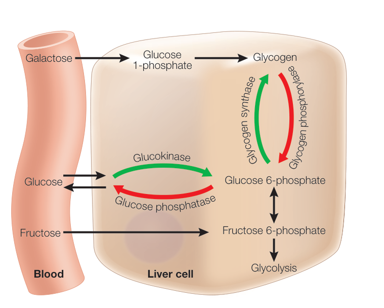
Figure 49.18 Insulin Controls Glucose Traffic in the Liver Glucose freely enters and leaves liver cells by diffusion. But when insulin is present (green arrows), glucose is phosphorylated and cannot leave the cell. The glucose 6-phosphate either enters glycolysis or is synthesized into glycogen. Insulin activates the enzymes of glycogen synthesis. Without insulin (red arrows), glucokinase is inhibited and glycogen phosphorylase and glucose phosphatase are activated. Thus glycogen is broken down to glucose phosphate, which is dephosphorylated so that it can leave the cell and enter the blood. Other monosaccharides—galactose and fructose—can also diffuse into the cell and be converted to glucose through these pathways.
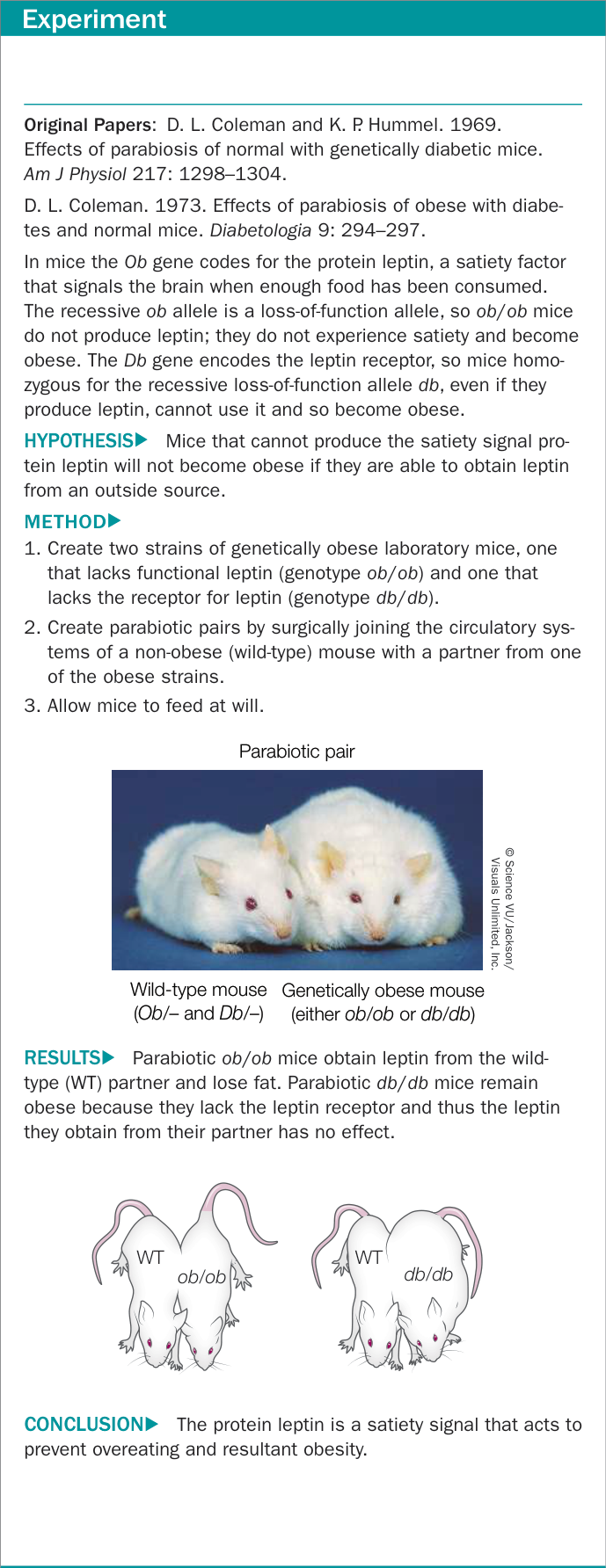
Figure 49.19A A Single-Gene Mutation Leads to Obesity in Mice Figure 49.19 A Single-Gene Mutation Leads to Obesity in Mice
We now know that another region of the hypothalamus, the arcuate nucleus, plays an important role in integrating a variety of feedback signals that influence food intake and body mass. Cells within the arcuate nucleus send axons to the ventromedial and dorsal hypothalamus, as well as to other brain areas that influence food intake and metabolism. One group of arcuate neurons projects to brain areas that inhibit food intake, while the other projects to brain areas that stimulate food intake. But what stimulates or inhibits the activity of the arcuate neurons?
Several factors have been identified that reflect the body’s energy balance. Three of these are the proteins insulin, leptin, and ghrelin. Insulin, as detailed earlier, is released when blood glucose levels are high, and it induces a decline in food intake. Leptin (Greek leptos, “thin”) is released by fat cells in proportion to how much lipid they contain. Evidence that leptin is a satiety signal is described in Figure 49.19. Ghrelin is released by the stomach when it is empty; its levels rise before meals and fall after meals. In the arcuate nucleus, insulin and leptin activate the neurons that inhibit feeding and inhibit the neurons that stimulate feeding. Ghrelin has the opposite effect on these two groups of neurons.
An integrative signal that could be playing a central role in regulation of feeding is the enzyme AMP-activated protein kinase (AMPK). When most cells are nutrient deprived, they produce AMPK, which stimulates the oxidation of substrates to replenish ATP. Fasting increases AMPK levels in the hypothalamus, and feeding reduces them. Insulin and leptin decrease AMPK activity in the hypothalamus, and ghrelin increases it. Thus AMPK could be a common pathway for various signals controlling food intake.
Recap and Assess
The major factors controlling gut function are autonomic reflexes, the enteric nervous system, and the hormones gastrin, secretin, and cholecystokinin. Insulin is the major hormonal controller of fuel metabolism through its effects on liver, muscle, and adipose tissues. The hypothalamus controls food intake by generating sensations of hunger and satiety influenced by feedback from blood glucose and hormones, including insulin, leptin, and ghrelin.
- For various reasons, patients may be fed through a tube going into their stomach or a tube going all the way to their jejunum, the middle region of their small intestine. How would the feeding formula used in the jejunal tube have to differ from the one used in the stomach tube?
- How do the three classes of lipoproteins differ in structure and function?
- How are the effects of high and low insulin levels the same and different in liver and muscle?
- In the experiments in which the lateral or the ventromedial hypothalamus was lesioned, what result enabled the conclusion that the regulation of body mass was altered rather than just the ability to produce satiety or hunger by the nutritional state of the body?
- When exercising muscles do not have an adequate supply of oxygen, they continue to produce small amounts of ATP through glycolysis, but they also produce pyruvate and lactate. Explain how this pyruvate and lactate can help support continuing glycolysis.
- What evidence supports the hypothesis that leptin influences satiety?
Investigating Life
Q&A If the nutritional values were the same, could different foods have different impacts on health? Future directions
The Pima of Arizona were among the first communities to be examined with a genome-wide survey. Many candidate genes were shown to be associated with obesity, but weakly so. The conclusion was that there are genetic contributions to the propensity to develop obesity, but except for a few rare cases, obesity is a multigenic trait that is highly influenced by environment. One aspect of the environment is physical activity, but another one is diet. The caloric content of the diet is obviously an important factor, but what about the composition of the diet? The experiments described in Investigating Life: How Does the Gut Microbiome Contribute to Obesity and Metabolic Disease? show that the composition of the diet can influence the composition of the gut microbiome, and the gut microbiome can influence the nutritional and other health effects of the diet. Thus, apart from differences in caloric intake, the consequences of the
Pima switching from a traditional diet to a high-fat Western diet could have resulted in changes in their microbiomes that have contributed to obesity and diabetes.
Future directions
It would be of interest to compare the microbiomes of the Arizona and the Mexican Pima peoples, and to compare those with the microbiomes of other populations on similar and different diets. If a particular microbiome profile is associated with the obesity-diabetes phenotype, it might be possible to change that microbiome through dietary change to promote better health. Just as we now do genomic screening to identify health risks, we might consider microbiome screening. Whereas we cannot change our genetic makeup, we could change our microbiome either through diet, or even therapeutically by uses of antibiotics and microbiome transplants. A new branch of medicine could develop around the testing, diagnosis, and therapy of the gut microbiome.

Visual Summary

© Yaacov Dagan/Alamy Stock Photo
You should be able to relate each summary to the adjacent figures. If you go to this visual summary in Achieve, you can follow links to figures, animations, activities, and simulations that will help you consolidate the material.
Food Provides Energy as well as Materials for Biosynthesis
- ■ Animals are heterotrophs that directly or indirectly derive energy and molecular building blocks from autotrophs.
- ■ Carbohydrates, fats, and proteins in food are macro-nutrients that provide energy. Energy content of food is measured in kilocalories (kcal), and excess caloric intake is stored as glycogen and fat.
- ■ Food provides essential carbon skeletons that animals cannot synthesize themselves.
- ■ Animals must obtain vitamins and essential amino acids and fatty acids from their diet. They also need mineral macronutrients (in large amounts) and mineral micronutrients (in small amounts).
- ■ Malnutrition results when any essential nutrient is lacking from the diet. Chronic malnutrition causes deficiency diseases.
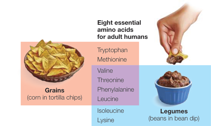
Figure 49.5
Questions
- Why is the energy content of food measured in quantities of heat (kcal)?
- Why does severe starvation result in a swollen abdomen, hands, and feet?
- What are the differences between macronutrients and mineral macro-and micronutrients?
Diverse Adaptations Support Ingestion and Digestion of Food
- ■ Animals are characterized by how they acquire nutrients and include saprobes, detritivores, filter feeders, herbivores, and carnivores.
- ■ Digestion is the breakdown of complex food molecules into monomers that can be absorbed and used by cells.
- ■ Vertebrate guts are characterized by a large surface area for nutrient absorption.
- ■ The microbiome of the gut participates in digestive processes, makes certain nutrients available for absorption, and can be altered by diet and other factors.
Questions
- Why are fossil teeth of great interest to paleontologists?
- What is meant when someone claims that most of the DNA in our bodies is not human?
- What are four features of the human gut that maximize its surface area for nutrient absorption?
The Vertebrate Gastrointestinal System Is a Disassembly Line
- ■ Swallowing is a neural reflex. Peristalsis and segmentation movements move gut contents through the entire length of the gut.
- ■ Sphincters block the gut at certain locations but are opened by peristaltic waves of anticipatory relaxation.
- ■ Salivary amylase begins carbohydrate digestion in the mouth; pepsin and hydrochloric acid begin digestion of protein in the stomach. Mucus secretion facilitates food processing and protects the lining of the gut.
- ■ Pancreatic enzymes carry out most digestion in the duodenum, where bicarbonate ions from the pancreatic ducts neutralize the pH of the gut contents.
- ■ Bile from the liver and gallbladder emulsifies fats into micelles.
Questions
- How do parietal cells in the stomach mucosa acidify the stomach contents?
- Carbonic anhydrase also catalyzes the hydration of in pancreatic ducts. How does this process differ from that in the gastric pits?
- Explain two ways in which active transport of ions by gut mucosal cells results in the movement of nutrients such as glucose into the body.
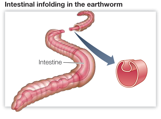
Figure 49.8
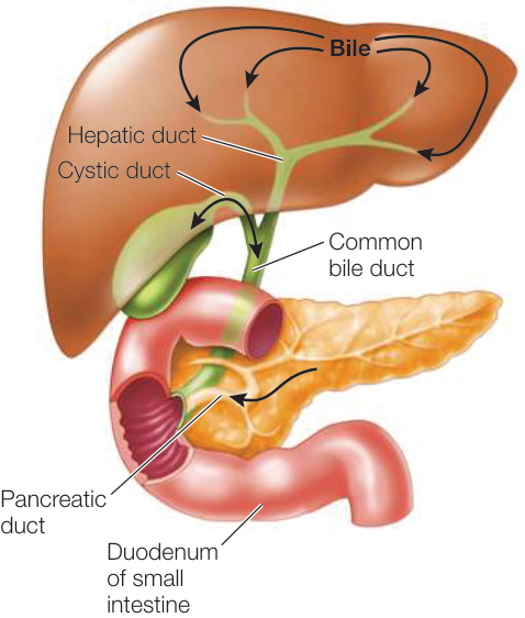
Figure 49.13
Nutrient Availability Is Controlled and Regulated
- ■ Actions of the stomach and small intestine are largely controlled by the hormones gastrin, secretin, and cholecystokinin (CCK).
- ■ Fat and cholesterol are shipped out of the liver as very low-density lipoproteins (VLDLs). Low-density lipoproteins (LDLs) transport cholesterol around the body. High-density lipoproteins (HDLs) bring cholesterol back to the liver.
- ■ Insulin promotes glucose uptake and storage as glycogen and fat during the absorptive state. In the postabsorptive state, lack of insulin blocks the uptake and use of glucose by most cells of the body except neurons. If blood glucose levels fall, glucagon secretion increases, stimulating the liver to break down glycogen and release glucose to the blood.
- ■ Sensations of hunger and satiety are controlled by feed-back signals that include insulin, leptin, and ghrelin.
Questions
- What does gastrin control, and what controls gastrin?
- How do secretin and CCK speed up and slow down digestive processes?
- What are the roles of lipoproteins in lipid management?
- How does insulin control the management of glucose in the liver?
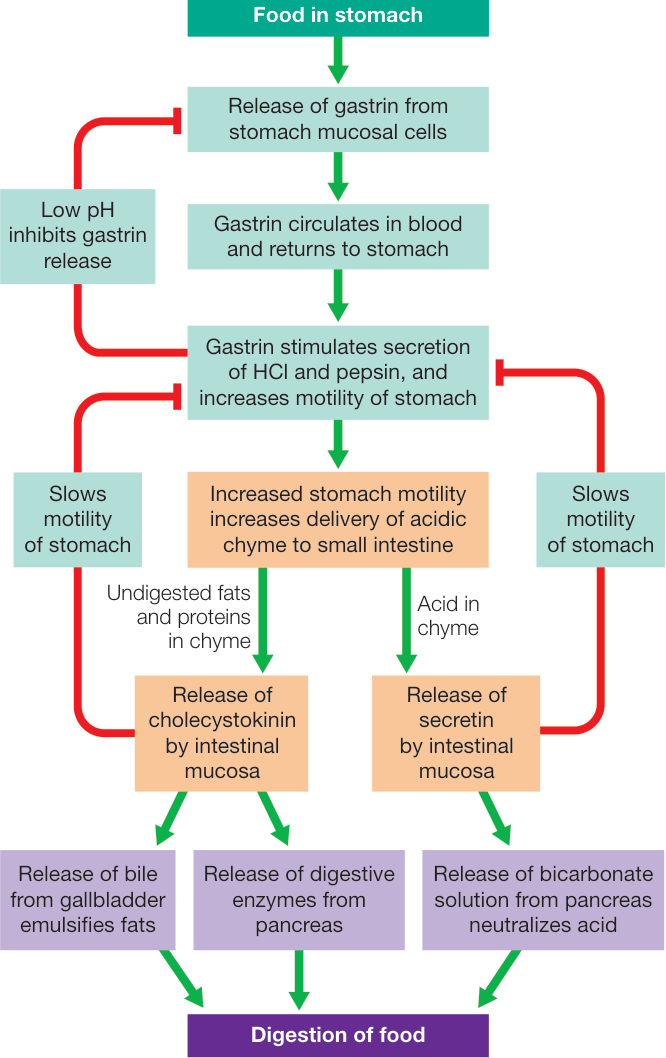
Figure 49.16