Chapter 50Salt and Water Balance and Nitrogen Excretion
Key Concepts
50.1 Excretory Systems Regulate Osmotic and Ionic Concentrations
50.2 Animals Excrete Nitrogen as Ammonia, Urea, or Uric Acid
50.3 Invertebrate Excretory Systems Use Filtration, Secretion, and Reabsorption
50.4 The Nephron Is the Basic Functional Unit of Vertebrate Excretory Systems
50.5 The Mammalian Kidney Can Produce Concentrated Urine
50.6 Kidney Function Is Regulated
The common vampire bat (Desmodus rotundus) is able to adjust its excretory physiology rapidly from water-excreting to water-conserving, depending on whether it is ingesting or digesting its blood meal.
© Michael & Patricia Foglieny/Minden Pictures
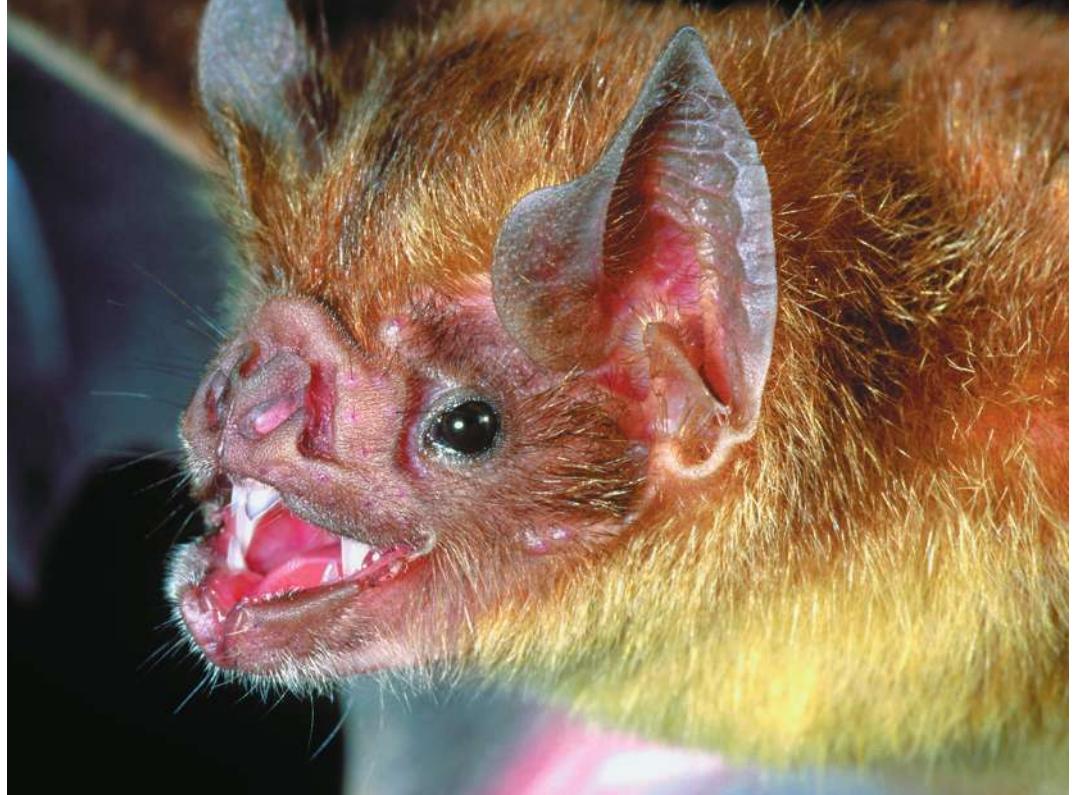
Investigating Life: How Vampire Bats Use Blood as Fast Food
Blood, sweat, and tears have the composition of extracellular fluid and taste salty. The composition and volume of the extracellular fluid are regulated by the excretory system. Challenges to an animal’s excretory system depend on its environment and lifestyle. Some desert animals rarely encounter free water, so they must conserve water by excreting their excess ions and nitrogenous wastes with minimal water loss. Animals living in fresh water have the opposite challenge: water continuously enters their bodies by osmosis and with their food, so they must excrete water and conserve ions. Most vertebrate animals living in the ocean face a challenge similar to that of the desert dwellers; they lose body water by osmosis and therefore need to conserve water and excrete ions.
Adaptations animals have to maintain salt and water balance are similar, but they are used in different ways to solve the problems unique to each species. Consider vampire bats feeding on the blood of animals such as goats and cattle. The bats use their sharp incisor teeth to make a small incision (usually on the ankle or leg of a sleeping victim) and then lap up the blood. A vampire bat feeds only once a day, and has to do so quickly, before the victim awakens and shakes it off.
Blood contains nutritious protein but is mostly water. To meet its daily energy needs, a vampire bat must take in a huge volume of blood relative to its body size, and it has to be able to fly back to its roost. To maximize protein intake and still be able to fly, the bat gets rid of water fast as a copious flow of dilute urine.
Back in the roost, the bat’s challenge is reversed. To digest a high-protein meal, the bat must excrete a lot of nitrogenous waste with minimal loss of water, as it may not get more water until its next meal. Within a few hours, the bat’s excretory system has to switch from rapid excretion of water to conservation of water, in spite of the need to excrete lots of nitrogenous wastes. The vampire bat’s excretory system rapidly switches from producing lots of dilute urine to producing small amounts of concentrated urine.
QA How does the excretory system of vampire bats process their high-protein liquid diet? (Find the answer on page 1139.)
Excretory Systems Regulate Osmotic and Ionic Concentrations
Learning Objectives
- 50.1.1 Describe the roles of filtration, secretion, and reabsorption in the excretory systems of animals with closed circulatory systems.
- 50.1.2 Determine the osmoregulatory characteristics of a species from data about the composition of its extracellular fluid and its environment.
Homeostasis of the extracellular fluid (the blood plasma and interstitial fluid; see Key Concept 38.2) is critical because:
- The solute concentration of the extracellular fluid determines the water balance of the cells of the body.
- The specific ionic composition of the extracellular fluid influences many cell functions.
- The health of cells requires the elimination of nitrogenous wastes.
The problems that have to be solved to maintain homeostasis of the extracellular fluid depend on the environment in which a species lives (salt water, fresh water, or terrestrial) and its life-style, as you saw in the case of the vampire bat in the opening story. Animals depend on excretory systems to maintain the volume, concentration, and composition of their extracellular fluids, and to excrete wastes.
Osmosis causes water to enter or to leave cells
The volume of a cell depends on whether it takes up water from or loses water to the extracellular fluid. The movement of water across cell membranes via osmosis depends on differences in solute concentration on the two sides of the membrane and on the permeability of the membrane (see Key Concept 6.3). If the solute concentration of the extracellular fluid is less than that of the cytoplasm, water moves into the cells, causing them to swell and possibly burst (see Figure 6.10). If the solute concentration of the extracellular fluid is greater than that of the cytoplasm, the cells lose water and shrink. Thus the solute concentration of the extracellular fluid affects both the volume and the solute concentrations of the cells.
Animal physiologists use the term osmolarity in discussing osmosis. The osmolarity of a solution is the number of moles of osmotically active solutes per liter of solvent. Thus a 1-molar solution of glucose is also a 1-osmolar (1 osmole per liter) solution, but a 1-molar solution of sodium chloride (NaCl) is a 2-osmolar solution, because each NaCl molecule dissociates into two osmotically active ions.
Connect the Concepts As described in Key Concept 2.4, a mole is the amount of a substance in grams that is numerically equal to its molecular weight.
Excretory systems control extracellular fluid osmolarity and composition
Excretory systems control the osmolarity and composition of the extracellular fluids by excreting solutes that are present in excess (such as NaCl when we eat lots of salty food) and conserving solutes that are valuable or in short supply (such as glucose and amino acids). Excretory systems also eliminate the toxic waste products of protein metabolism. The output of the excretory system is urine.
Three basic processes are common to a wide variety of animal excretory systems: filtration, secretion, and reabsorption. Filtered extracellular fluid contains no cells or large molecules, such as proteins. In animals with a closed circulatory system, the blood plasma is filtered from capillaries into associated tubules. The walls of the capillaries and of the tubules are the filter, and the filtration is driven by blood pressure. As the filtrate flows through the tubules, its composition and concentration are modified through processes of secretion and reabsorption to form the urine that leaves the body.
In all of the discussions that follow about the movement of water across membranes, it is important to remember that there are no mechanisms for the active transport of water. The movement of water is due either to a pressure difference (filtration) or to a difference in solute concentration (osmosis). Water molecules move down a pressure gradient or up a solute concentration gradient.
Aquatic invertebrates are either ionic conformers or regulators
Most invertebrates that live in seawater conform to the osmotic concentration of their environment over a fairly wide range of salinities and are therefore called osmoconformers (Figure 50.1). The osmolarity of seawater in the open ocean is about 1,000 milliosmoles/liter (mOsm/L), but it can vary quite a bit in estuaries where it is diluted by an influx of fresh water or in evaporating tide pools as the salt gets concentrated. Osmoconformity can result in considerable energetic savings, as it costs metabolic energy to move ions across membranes to achieve osmotic regulation.
Osmoconformity is not an option for freshwater animals. No cell can do without ions or nutrients, so freshwater animals always have to expend energy to conserve salts and excrete water.
Some marine invertebrates maintain a rather constant osmolarity of their extracellular fluids as the osmolarity of the environment changes, and these animals are therefore called osmoregulators (see Figure 50.1). Osmoregulators in the marine environment mostly maintain their osmotic concentration considerably below that of the environment and are therefore engaged in hypotonic regulation. Occasionally, however, seawater is diluted by an influx of fresh water, as in estuaries, and animals must maintain their osmotic concentrations above that of the environment—a process called hypertonic regulation.
The brine shrimp Artemia illustrated in Figure 50.1 is an osmoregulator with an enormous range of tolerances. Artemia are found in huge numbers in extremely salty environments, such as Utah’s Great Salt Lake and in coastal evaporation ponds where salt is concentrated for commercial purposes (see Figure 24.17)
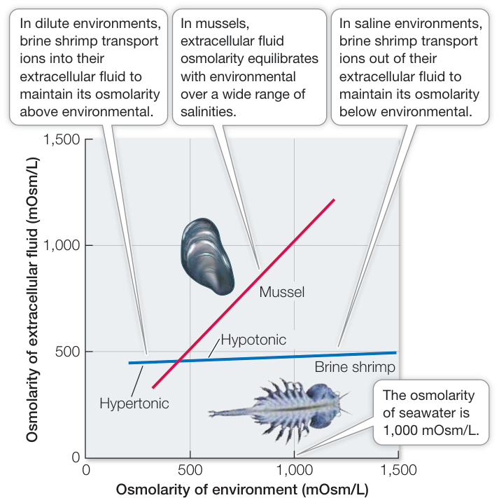
Figure 50.1 Some Marine Invertebrates Osmoregulate Some aquatic animals, such as mussels, experience an extreme range of salt concentrations in their environment and are osmoconformers over much of that range. Other aquatic animals, such as brine shrimp, are osmoregulators in that they maintain a relatively constant osmolarity of their extracellular fluids as environmental salinity varies. (Mussel: After W. T. W. Potts and G. Parry, 1964. Osmotic and Ionic Regulation in Animals. Pergamon, Oxford; Brine Shrimp: P. C. Croghan. 1958. J Exp Biol 35: 219–233.)
and can reach an osmolarity of 2,500 mOsm/L. No animal could survive with internal osmolarities that high; such a solute concentration would cause proteins to denature. Artemia are able to exploit these environments because of their ability to regulate hypotonically by actively transporting NaCl from their extracellular fluid out across their gill membranes to the environment. Artemia cannot survive in fresh water, but they can live in dilute seawater by reversing the direction of transport of NaCl across their gill membranes to maintain the osmolarity of their extracellular fluids above that of the environment, thus becoming hypertonic regulators.
Invertebrates that are osmoconformers still have to be ionic regulators with respect to certain ions. Although the concentrations of and in their extracellular fluids may be the same as in seawater, many other ions are regulated at different levels. For example, the concentration of is usually higher, and the concentrations of and of are usually lower, in the extracellular fluids than in the seawater. Active transporters maintain the regulated ions in the extracellular fluid at optimal concentrations.
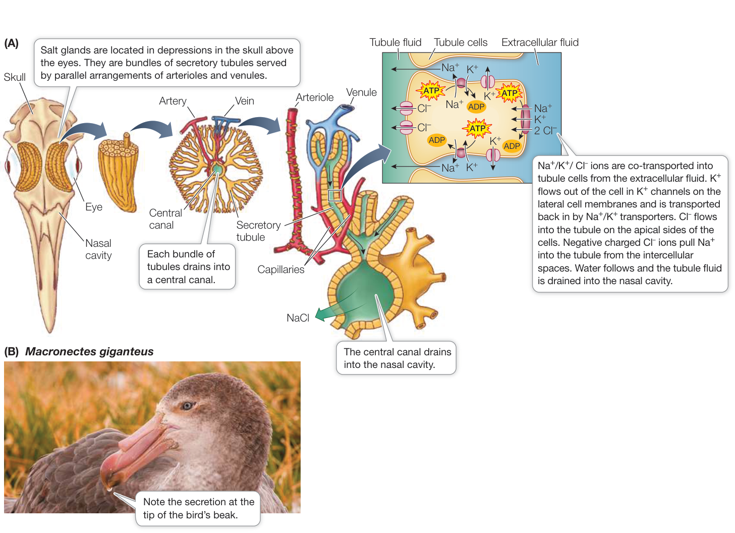
Figure 50.2 Nasal Salt Glands Excrete Excess Salt (A) Marine birds have nasal salt glands that excrete the excess salt they ingest with their food. (B) This Southern Giant Petrel (Macronectes giganteus) has returned from a feeding trip at sea and is secreting salt through its nasal salt gland.
Vertebrates are osmoregulators and ionic regulators
All aquatic vertebrates, with the exception of two classes, regulate the osmolarity of their extracellular fluids at around 300 mOsm/L. In doing so, they are selective in which ions they conserve and which ions they excrete; thus they are ionic regulators and osmoregulators. The exceptions are the hagfishes and the cartilaginous fishes (sharks and rays). Hagfishes are an ancient vertebrate group of jawless fishes (see Figure 31.11). Hagfishes are osmoconformers as well as ionic conformers for most of the ions found in seawater. The cartilaginous fishes are osmoconformers, but in a unique way. They retain in their extracellular fluid two organic solutes, urea and trimethylamine oxide (TMAO), that are products of protein metabolism. As a result, their extracellular fluid is slightly hypertonic to the seawater and they gain water by osmosis rather than lose water to their salty environment.
Terrestrial vertebrates obtain their salts mostly from food, and they regulate the ionic composition of their extracellular fluids by conserving some ions and excreting others. For example, herbivores have to conserve because most plants have low concentrations of . In contrast, birds that feed on marine animals must excrete the excess sodium they ingest with their food. Such birds, which include penguins and gulls, excrete excess salt through nasal salt glands. These glands are activated by parasympathetic innervation in response to increased osmolarity of the blood. The glands consist of bundles of secretory tubules in close relationship to blood vessels and emptying into central canals (Figure 50.2). Activation causes opening of potassium channels on the lateral membranes of the tubule cells and opening of channels on the apical side of the cells (facing into the tubule). Activation also stimulates cotransporters on the basal sides of the cells (in contact with interstitial fluid). Increased in the cells is transported out of the lateral cell membrane in exchange . The leaving the apical membranes creates an electrochemical gradient that pulls the excess in the lateral spaces between the tubule cells to flow into the tubule lumen through intercellular junctions. The resulting concentrated solution of flows into a series of canals that empty into the nasal cavity. These birds can be seen frequently sneezing or shaking their heads to get rid of the salty droplets excreted from their nasal salt glands.
Recap and Assess
Excretory systems control water and salt balance and the excretion of nitrogenous waste products through three mechanisms: filtration of body fluids to form urine, active secretion of substances into the urine, and active reabsorption of substances from the urine. Animals can be osmoconformers or osmoregulators.
What is the significance of a closed circulatory system for osmoregulation in a freshwater vertebrate?
- Why is it important for an animal like Artemia that lives in the intertidal zone to be able to osmoregulate, and how does Artemia do that at the extremes of salinities in its environment?
- How does water get into the nasal salt gland ducts of marine birds?
In addition to maintaining salt and water balance, animals must eliminate the waste products of metabolism from their extracellular fluids. The major problem is nitrogen. When nitrogen-containing molecules are broken down by metabolism, the end-product can be toxic.
Animals Excrete Nitrogen as Ammonia, Urea, or Uric Acid
Learning Objectives
- 50.2.1 Discuss the properties of ammonia, urea, and uric acid as nitrogenous waste products.
- 50.2.2 Describe the relative advantages and disadvantages of excreting nitrogenous waste as urea or as uric acid.
The end-products of the metabolism of carbohydrates and fats are water and carbon dioxide, which are not difficult to eliminate. Proteins and nucleic acids, however, contain nitrogen, so their metabolism produces nitrogenous wastes in addition to water and carbon dioxide.
Ammonia is toxic
The most common nitrogenous waste is ammonia (). Ammonia is highly toxic, so it must be excreted continuously to prevent its accumulation or it must be detoxified by conversion into urea or uric acid (Figure 50.3). Ammonia is highly soluble in water and diffuses rapidly, so its continuous excretion is relatively simple for many aquatic animals that continuously lose ammonia from their blood to the environment by diffusion across their gill membranes. Animals that excrete ammonia, such as aquatic invertebrates and bony fishes, are called ammonotelic.
Ammonia can also be lost by diffusion across the alveolar membranes of air-breathing animals, but with tidal ventilation, the buildup of ammonia in the extracellular fluids would be greater and therefore toxic. Therefore these animals detoxify ammonia by converting it into urea or uric acid.
Urea is highly soluble in water
Mammals, amphibians, cartilaginous fishes (sharks and rays), and hagfishes excrete urea as their principal nitrogenous waste product and are therefore called ureotelic. Urea is quite soluble in water, but its excretion can result in a large loss of water that many animals can ill afford. Mammals have somewhat solved this problem through the evolution of excretory systems that can produce urine with a high concentration of urea, thereby decreasing the water loss associated with excretion of urea. Also, recall from Key Concept 50.1 that sharks and rays solve the water loss problem of living in seawater by retaining high concentrations of urea and TMAO in their extracellular fluid, so that it is hyperosmotic to seawater.
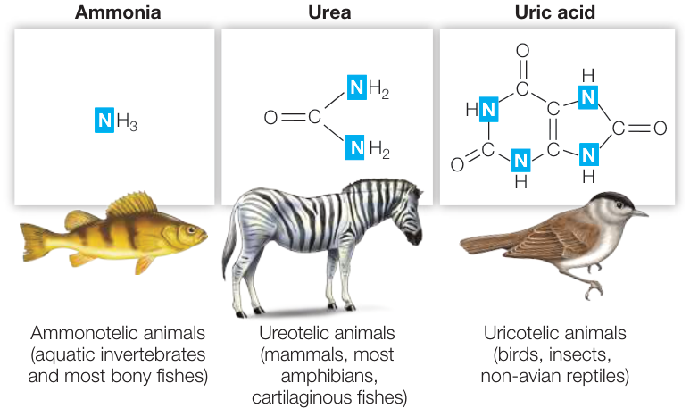
Figure 50.3 Nitrogenous Waste Products of Metabolism The metabolism of proteins and nucleic acids produces nitrogenous wastes. Many aquatic animals, including most fishes, excrete nitrogenous wastes as ammonia, which is highly diffusible and soluble in an aqueous environment. Most terrestrial animals and some aquatic animals excrete either urea or uric acid. Urea is more soluble in water and is the major nitrogenous excretory product for mammals, amphibians, and some fishes. Uric acid is not very soluble in water and is the major nitrogenous excretory product for reptiles (including birds), insects, and some amphibians. 1. 1. When amino acids are broken down, a first step is removal of the amine group, forming ammonia. Why is it important to get rid of that ammonia, and how do fishes and mammals achieve this excretion? 2. 2. Why do sharks and rays produce urea rather than uric acid to raise the osmotic concentration of their extracellular fluids? 3. 3. For terrestrial animals, what is the advantage of excreting uric acid as the nitrogenous waste product?
Uric acid is not very soluble in water
A single molecule of uric acid eliminates twice the amount of nitrogen as does a molecule of urea (see Figure 50.3), but uric acid is not very soluble in water. Therefore it tends to precipitate out of solution and form a colloidal suspension in the urine, requiring it to be excreted as a semisolid (for example, the whitish material in bird droppings). Because of the precipitation of the uric acid out of solution in the excretory system, water can be reabsorbed from the urine and thereby reduce the water loss necessary to excrete the nitrogenous waste. Animals that excrete uric acid are uricotelic, and they include insects, reptiles (including birds), and some amphibians. Many uricotelic species live in very arid environments.
Most species produce more than one nitrogenous waste
Humans are ureotelic, but we also excrete uric acid. The uric acid in human urine comes largely from the metabolism of nucleic acids. If uric acid levels in the extracellular fluid rise too high, uric acid can precipitate in joints, causing the age-old malady gout. Because solubility goes down with temperature, uric acid usually precipitates first in the extremities, especially the big toe. Pain in the big toe is a telltale symptom of gout. The risk for developing gout goes up with excessive consumption of protein, beer, and spirits (less so with wine) and in general with obesity and insulin resistance. Therefore in the past it was called “the disease of rich men.” Gout has been around for a very long time; the first documented case was in Egypt around 2500 BCE.
Humans can also excrete ammonia, which is an important mechanism for regulating the pH of the extracellular fluids. As you will see later in this chapter, excreted ammonia buffers the urine and enables the excretion of excess hydrogen ions.
Species that live in different habitats at different developmental stages may use more than one mechanism of nitrogen excretion. The tadpoles of frogs and toads, for example, excrete ammonia across their gill membranes, but adult frogs and toads generally excrete urea. Some adult amphibians that live in arid habitats excrete uric acid.
Recap and Assess
Ammonia is a common metabolic waste product of nitrogen-containing molecules. Most aquatic animals excrete ammonia by diffusion into the water. Terrestrial animals and some aquatic animals detoxify ammonia by converting it to urea or uric acid.
Animals exhibit a variety of adaptations for dealing with the challenges of salt and water balance in different environments. All of these adaptations, however, are based on two basic mechanisms—namely, filtration and tubular processing of the filtrate to conserve some solutes and excrete others.
Invertebrate Excretory Systems Use Filtration, Secretion, and Reabsorption
Learning Objectives
50.3.1 Describe the structure and function of a protonephridium.
50.3.2 Diagram the process of urine formation by a metanephridium.
50.3.3 Explain how Malpighian tubules enable insects to live in very arid habitats.
Freshwater and terrestrial invertebrates have a wide variety of adaptations for maintaining salt and water balance and excreting nitrogen. Here we explore three examples of invertebrate excretory systems: protonephridia, metanephridia, and Malpighian tubules. Each of these systems produces an extract of interstitial fluid lacking large molecules. They then change the solute composition (ions and small molecules) of that fluid to form an excretory product.
Protonephridia of flatworms excrete water and conserve salts
Many free-living flatworms, such as Planaria, live in fresh water. These animals excrete water through an elaborate network of tubules running throughout their bodies. The tubules end in flame cells, so called because each cell has a tuft of cilia projecting into its tubule (Figure 50.4). The beating of the cilia gives the appearance of a flickering flame. A flame cell and a tubule together form a protonephridium (plural protonephridia; Greek proto, “before,” + nephros, “kidney”). Flame cells are also called cap cells because they cap off the tubule.
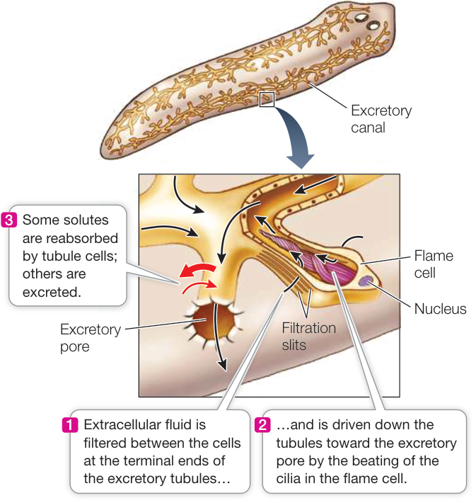
Figure 50.4 Protonephridia in Flatworms The protonephridia of the freshwater flatworm Planaria consist of tubules ending in flame cells. Extracellular fluid is filtered through the filtration slits in the flame cells. The composition of the filtrate is modified as it flows down the tubule. Black arrows indicate direction of flow of tubular fluid. Q: Do you expect the fluid coming out of the excretory pore to be hypertonic or hypotonic to the flatworm’s interstitial fluid?
Extracellular fluid enters the tubules by filtration through slits in the flame cells. The beating of the cilia causes a slight negative pressure in the tubule, and movements of the animal create positive pressure in the extracellular fluid. This pressure difference causes extracellular fluid to be filtered through the filtration slits in the flame cells. The filtrate flows toward the animal’s excretory pore, and along the way the cells of the tubules modify the composition of the fluid by reabsorption and secretion of specific ions and molecules. Because more ions are reabsorbed than are secreted, the urine that leaves the flatworm’s body is less concentrated than the extracellular fluid. Thus the protonephridium conserves ions and excretes water and wastes.
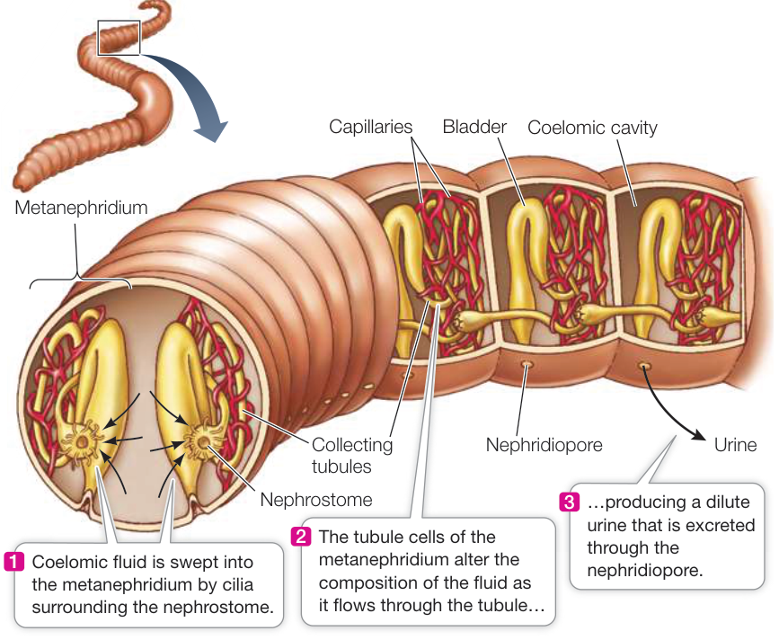
Figure 50.5 Metanephridia in Earthworms The metanephridia of annelids are arranged segmentally. The cross section at the left end shows a pair of metanephridia. Three longitudinal sections (right) show only one metanephridium of the two in each segment. Coelomic fluid enters the nephrostome and flows through tubules leading to the nephridiopore. A close association of the tubules and blood capillaries facilitates the active exchange of substances between the blood and the tubular fluid. Activity 50.1 Annelid Metanephridia
Metanephridia of annelids process coelomic fluid
Filtration of body fluids and modification of urine by tubules are highly developed processes in annelids such as the earthworm. Annelids are segmented, and in each segment they have a fluid-filled body cavity called a coelom (see Figure 30.11). Annelids have a closed circulatory system through which blood is pumped under pressure. The pressure causes the blood to be filtered across the thin, permeable capillary walls into the coelom. Some waste products, such as ammonia, diffuse directly from the tissues into the coelom. Where does this coelomic fluid go?
Each segment of the earthworm contains a pair of metanephridia (singular metanephridium; Greek meta, “akin to”). Each metanephridium begins as a ciliated, funnel-like opening called a nephrostome. The nephrostome resides in one segment and continues as a tubule in the next segment. The tubule ends in a pore, called a nephridiopore, that opens to the outside of the animal (Figure 50.5). Coelomic fluid is swept into the metanephridia through the ciliated nephrostomes. As the fluid passes through the tubules, their cells actively reabsorb certain molecules from it and actively secrete other molecules into it. What leaves the animal through the nephridiopores is a dilute urine containing nitrogenous wastes and other solutes.
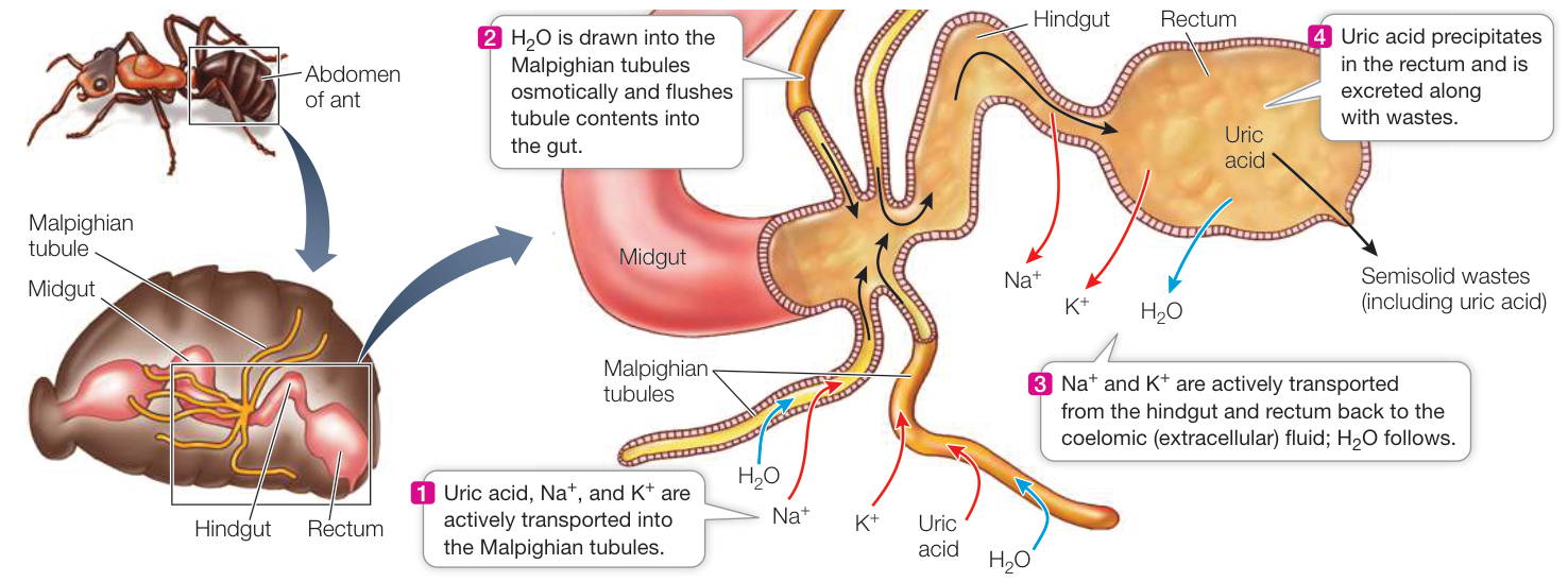
Figure 50.6 Malpighian Tubules in Insects The blind, thin-walled Malpighian tubules are attached to the junction of the insect’s midgut and hindgut and project into the spaces containing extracellular fluid. This system makes it possible to excrete wastes with very little loss of water.
Q: Why are and ions transported into the Malpighian tubule but then transported out of the hindgut and rectum?
Malpighian tubules of insects use active transport to excrete wastes
Insects can excrete nitrogenous wastes with very little loss of water and can therefore live in the driest habitats on Earth. The insect excretory system consists of Malpighian tubules. An individual insect has from 2 to more than 100 of these blind-ended tubules that open into the gut between the midgut and hindgut (Figure 50.6).
Insects have an open circulatory system and therefore cannot use a pressure difference to filter extracellular fluids into the Malpighian tubules. Instead, the cells of the tubules actively transport uric acid, potassium ions, and sodium ions from the extracellular fluid into the tubules. The high concentration of solutes in the tubules causes water to follow osmotically, which flushes the tubule contents toward the gut.
The epithelial cells of the hindgut and rectum actively transport sodium and potassium ions from the gut contents back into the extracellular fluid. This local transport of salts creates an osmotic gradient that pulls water out of the rectal contents. As its concentration increases, the uric acid forms a colloidal suspension, freeing even more water to be reabsorbed. Remaining in the rectum is the uric acid mixed with other wastes; this semisolid matter is what the insect excretes. If you ever park your car under a tree being visited by bees, you will find the little excretory droplets from the bees all over the car. The Malpighian tubule system is a highly effective mechanism for excreting nitrogenous wastes and some salts without giving up much water.
Recap and Assess
Protonephridia and metanephridia work by creating a filtrate of the body fluids that is modified by the secretion and reabsorption of specific substances before being excreted. Insect Malpighian tubules actively secrete uric acid and other solutes into closed tubules.
- How do protonephridia enable flatworms to live in fresh water?
- What are the relationships among blood plasma, coelomic fluid, and urine in an annelid worm?
- How does the insect excretory system conserve water?
Having described how several invertebrate groups handle nitrogen excretion, we next consider the nephron—the basic unit of the vertebrate excretory system—and how it evolved to be able to respond to a variety of salt-and water-balance challenges and maintain a relatively constant internal environment.
The Nephron Is the Basic Functional Unit of Vertebrate Excretory Systems
The Nephron Is the Basic Functional Unit of Vertebrate Excretory Systems
Learning Objectives
- 50.4.1 Discuss the structural and behavioral characteristics of amphibians and reptiles that enable them to minimize water loss.
- 50.4.2 Diagram and/or describe the organization of blood vessels in the nephron.
- 50.4.3 Explain the mechanism of filtration of blood plasma from the glomerular capillaries into Bowman’s capsule.
- 50.4.4 Explain why the renal filtrate changes in composition as it passes through the nephron.
The main excretory organ of vertebrates is the kidney, and the functional unit of the kidney is the nephron, which has a blood vessel component and a tubule component. The blood vessel (vascular) component begins with a knot of capillaries that are highly permeable and filter the blood into the tubule component. Processes of secretion and reabsorption in the tubules produce urine, which differs in composition from the extracellular fluids of the body.
The evolution of vertebrates is thought to have begun with a marine ancestor that moved into fresh water. The excretory system of this vertebrate ancestor would have evolved to excrete large quantities of water while conserving salts. But if the early vertebrate excretory system evolved in response to the need to excrete water, how did subsequent vertebrate lineages adapt to environments where water must be conserved and salts excreted? The answer to this question differs among vertebrate groups. Even among marine fishes, the excretory adaptations of the bony fishes differ from those of the cartilaginous fishes. Reptiles, including birds, and mammals have excretory systems that conserve water. The reptiles achieve this mainly by being uricotelic and producing a semisolid excretory product that contains little water. Mammals, in contrast, are ureotelic; they excrete a liquid waste product but have evolved the ability to produce highly concentrated urine.
Marine fishes must conserve water
Marine bony fishes osmoregulate their extracellular fluids to maintain them at one-third to one-half the osmolarity of seawater. Thus marine bony fishes must conserve water, which they are constantly losing through osmosis, and must excrete excess solutes. Marine bony fishes cannot produce urine that is more concentrated than their extracellular fluids, so they minimize water loss by producing very little urine. In contrast, freshwater fishes produce lots of dilute urine.
How do marine bony fishes deal with the large salt loads they ingest with food? Marine bony fishes do not absorb from their guts some of the ions they take in, especially divalent ions such as and . NaCl, the major salt ingested, is actively excreted across the gill membranes. As mentioned earlier, bony fishes can lose their nitrogenous waste, ammonia, by diffusion across their gill membranes.
Sharks and rays are osmoconformers but not ionic conformers. As mentioned earlier, they raise the osmolarity of their body fluids by retaining urea and TMAO, making their extracellular fluids hyperosmotic to seawater. These species have adapted to a concentration of urea in the body fluids that would be toxic to other vertebrates. Sharks and rays still have the problem of excreting the large amount of salts they take in with their food. They solve this problem by having a gland in the rectum that actively secretes NaCl by a mechanism similar to that of the nasal salt glands of seabirds.
Terrestrial amphibians and reptiles must avoid desiccation
Most amphibians live in or near fresh water, and they usually stay in humid habitats when they do venture from the water. Like freshwater fishes, most amphibians produce large amounts of dilute urine and conserve salts. Some amphibians, however, have adapted to habitats that require water conservation.
Amphibians living in dry terrestrial environments have skin with a reduced permeability to water. Some secrete a waxy substance over the skin for waterproofing. Several species of frogs that live in arid regions of Australia burrow into the ground and remain there during long dry periods. They enter estivation, a state of very low metabolic activity and therefore low water turnover. When it rains, the frogs come out of estivation, feed, and reproduce. Their most interesting adaptation is an enormous urinary bladder. Before entering estivation, they fill the bladder with dilute urine, which can amount to one-third of their body weight. This dilute urine serves as a water reservoir that is gradually reabsorbed into the blood during the long period of estivation. Aboriginal peoples have learned to locate these buried frogs and use them as an emergency source of water.
Reptiles occupy habitats ranging from aquatic to extremely hot and dry. In fact, snakes, lizards, and birds are among the most prominent members of many desert faunas. Three major adaptations have freed reptiles from needing to have a close association with water (see Key Concept 31.4):
- Reptiles are amniotes that do not need fresh water to reproduce because they employ internal fertilization and lay eggs with shells that retard evaporative water loss.
- Reptiles have a dry epidermis (skin) that retards evaporative water loss.
- Reptiles excrete nitrogenous wastes as uric acid semisolids, losing little water in the process.
Mammals can produce highly concentrated urine
Mammals occupy diverse habitats, many of which present special excretory system challenges. The most challenging environments are those in which water is severely limited. Mammals have a variety of adaptations to conserve water, but chief among them is the ability to produce urine that is more concentrated than their extracellular fluids. They are able to concentrate their urine because of adaptations of their kidneys that we explore in detail in Key Concept 50.5. To understand how these adaptations work, you must first know the structure and function of the vertebrate nephron.
The nephron is the functional unit of the vertebrate kidney
Urine formation in vertebrate nephrons involves three main processes (Figure 50.7):
- Filtration. Each nephron has a dense knot of capillaries called a glomerulus (plural glomeruli). The glomerulus is highly permeable to water, ions, and small molecules but impermeable to large molecules. Blood pressure drives the movement of water and small-molecular-weight solutes out of the glomerular capillaries.
- Tubular reabsorption. The filtrate from the glomerulus flows into the renal tubule. Cells in the renal tubule modify the filtrate by reabsorbing specific ions, nutrients, and water, returning these to the blood, and leaving behind and concentrating excess ions and waste products such as urea.
- Tubular secretion. The filtrate in the renal tubule is further modified by tubule cells transporting substances into the tubule. These are substances that the body needs to excrete.
Blood enters the glomerular capillaries via an afferent arteriole and leaves the glomerulus in an efferent arteriole. This short vessel is called an arteriole because it feeds another capillary bed, called the peritubular capillaries. These capillaries intimately surround the renal tubules and deliver substances to the renal tubules that the cells of the tubules secrete into the urine. The peritubular capillaries also carry away substances that the tubule cells reabsorb from the urine.
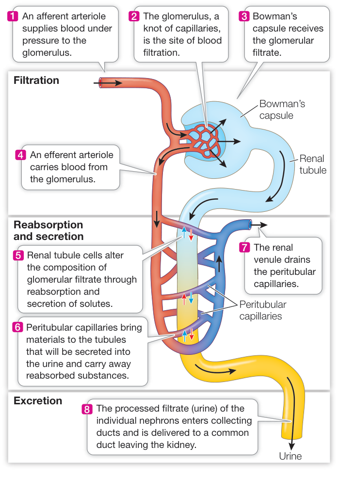
Figure 50.7 The Vertebrate Nephron The vertebrate nephron consists of a renal tubule closely associated with two capillary beds, the glomerulus and the peritubular capillaries. Activity 50.2 The Vertebrate Nephron
Blood is filtered into Bowman’s capsule
The renal tubule begins with Bowman’s capsule (see Figure 50.7), which encloses the glomerulus (Figure 50.8A,B). The glomerulus appears to be pushed into Bowman’s capsule much like a fist pushed into an inflated balloon. The cells of the capsule that are in direct contact with the glomerular capillaries are called podocytes (Figure 50.8C). These highly specialized cells have numerous extensions, each with hundreds of fine processes. The podocytes wrap around the capillaries so that their processes interdigitate and intimately cover the capillaries.
The glomerulus filters the blood, producing a fluid (the renal filtrate) that lacks cells and large molecules. The walls of the capillaries, the basal lamina of the capillary endothelium, and the podocytes of Bowman’s capsule all participate in filtration. Fenestrations in the walls of the capillaries (see Key Concept 48.4) allow water and many solute molecules, but not red blood cells, to pass through. The meshwork of the basal lamina and the spaces between the processes of the podocytes are even finer and prevent large molecules from leaving the capillaries (Figure 50.8D). The arterial pressure of the blood entering the permeable capillaries causes the filtration of water and small molecules out of the glomerular capillaries and into Bowman’s capsule. The glomerular filtration rate is high because blood pressure in the glomerular capillaries is high, and because the capillaries of the glomerulus, along with their covering of podocytes, are more permeable to water than are other capillary beds in the body.
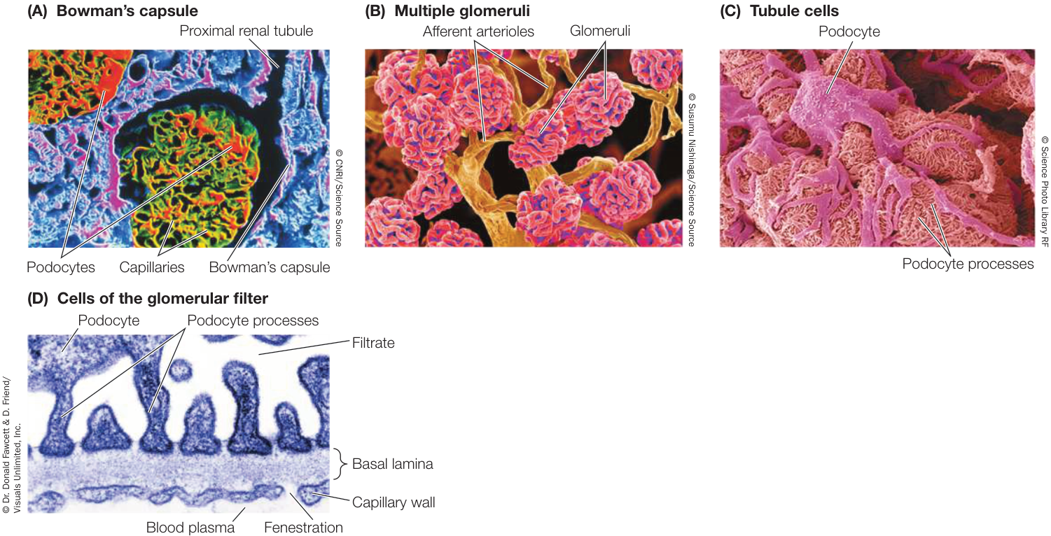
Figure 50.8 A Tour of the Vertebrate Nephron Scanning electron micrographs illustrate the anatomical basis for blood filtration in the kidneys. (A) This cross section of an intact glomerulus shows the tubule cells that form Bowman’s capsule surrounding the glomerular capillaries. (B) In a preparation showing only the blood vessels (tubular tissue has been digested away), the glomeruli appear as balls of capillaries served by arterioles. (C) Higher magnification of a glomerulus with the tubule cells intact shows the podocytes that wrap around the glomerular capillaries. (D) The glomerular filter has three layers: the fenestrated endothelial cells of the capillaries, the meshwork of collagen fibers making up the basal lamina, and the filtration slits between the podocyte processes. All three layers have negative charges, which contribute to their ability to prevent the passage of protein molecules. (D) Cells of the glomerular filter
Recall that marine bony fishes produce very little urine. The explanation for this is that their kidneys have evolved to have few if any glomeruli, whereas the kidneys of freshwater fishes have many glomeruli.
The renal tubules convert glomerular filtrate to urine
The composition of the filtrate that enters the renal tubule is similar to that of the blood plasma, except the filtrate lacks high-molecular-weight solutes such as proteins. Reabsorption and secretion cause the composition of this fluid to change as it passes down the renal tubule. Cells of the tubule actively reabsorb certain molecules from the tubule fluid (which are returned to the blood flowing through the peritubular capillaries). For example, glucose and amino acids are reabsorbed. Most NaCl is reabsorbed. Other substances in the blood of the peritubular capillaries are actively secreted into the tubule fluid. An example is para-aminohippuric acid (PAH), which is produced in the liver from benzoic acid, a common food preservative. Because of the actions of the renal tubules, the excreted urine is very different from the original glomerular filtrate. As you learn all the things that renal tubules do, keep in mind that renal tubules all consist of a single layer of epithelial cells
Recap and Assess
The kidney is the major excretory organ of vertebrates. Its functional unit is the nephron, which includes a glomerulus that filters blood and a renal tubule that secretes and reabsorbs solutes, modifying the filtrate to produce urine. As seen in freshwater fishes, the nephron evolved as a mechanism for excreting excess water while conserving valuable solutes. Vertebrates have various adaptations for conserving water. Marine bony fishes produce very little urine but lose nitrogenous waste as ammonia across their gills and also excrete salts from their gill membranes. Marine cartilaginous fishes produce urea and retain it, so their extracellular fluids are hyperosmotic to the environment and therefore take up water by osmosis. Many terrestrial vertebrates conserve water by excreting nitrogenous waste as uric acid, by minimizing evaporative water loss, and by active secretion of salts. Mammals have the ability to conserve water by producing urine that is more concentrated than their extracellular fluid.
- What is the connection between the need to conserve water and the evolution of internal fertilization?
- What are the roles of the afferent and efferent arterioles in the nephron?
- What are the anatomical components of the filtration mechanism in the glomerulus?
- How does the composition of the urine become different from the composition of the blood?
The adaptations that enable the mammalian kidney to produce urine more concentrated than extracellular fluids were important steps in vertebrate evolution, and they were largely achieved through changes in the structure and regional functions of the renal tubules. These changes converted a kidney that evolved to excrete water into an organ that conserves water.
The Mammalian Kidney Can Produce Concentrated Urine
Learning Objectives
- 50.5.1 Summarize the internal organization of the mammalian kidney.
- 50.5.2 Describe the mechanism whereby most of the glomerular filtrate is reabsorbed in the proximal convoluted tubule.
- 50.5.3 Explain how the loop of Henle maintains a concentration gradient in the extracellular fluid of the medulla of the kidney.
- 50.5.4 Describe how the mammalian excretory system is involved in regulating pH of the extracellular fluid.
Mammals have high body temperatures and high metabolic rates, and therefore have the potential for a high rate of water loss and a high level of nitrogenous waste production. Having an excretory system that minimizes water loss made it possible for these highly active species to occupy arid habitats.
Kidneys produce urine and the bladder stores it
Mammalian excretory systems are similar, so we will use that of humans as our example. Humans have two kidneys at the back of the upper region of the abdominal cavity (Figure 50.9A). Each kidney filters blood, processes the filtrate into urine, and releases that urine into a duct called the ureter. The ureter of each kidney leads to the urinary bladder, where the urine is stored until it is excreted through the urethra, a short tube that opens to the outside of the body.
Two sphincter muscles surrounding the base of the urethra control urination. One of these sphincters is a smooth muscle and is controlled by the autonomic nervous system. As the bladder fills, stretch receptors in the walls of the bladder trigger a spinal reflex that relaxes this sphincter. This reflex is the only control of urination in infants, hence their frequent “accidents.” The other sphincter is a skeletal muscle and is controlled by the voluntary nervous system. When the bladder is very full, only deliberate conscious effort prevents urination. Toilet training of children teaches them to control this sphincter.
Nephrons have a regular arrangement in the kidney
The kidney is shaped like a kidney bean. When sliced along its long axis on the midline, its key anatomical features are revealed (Figure 50.9B). The ureter and the renal artery and renal vein enter
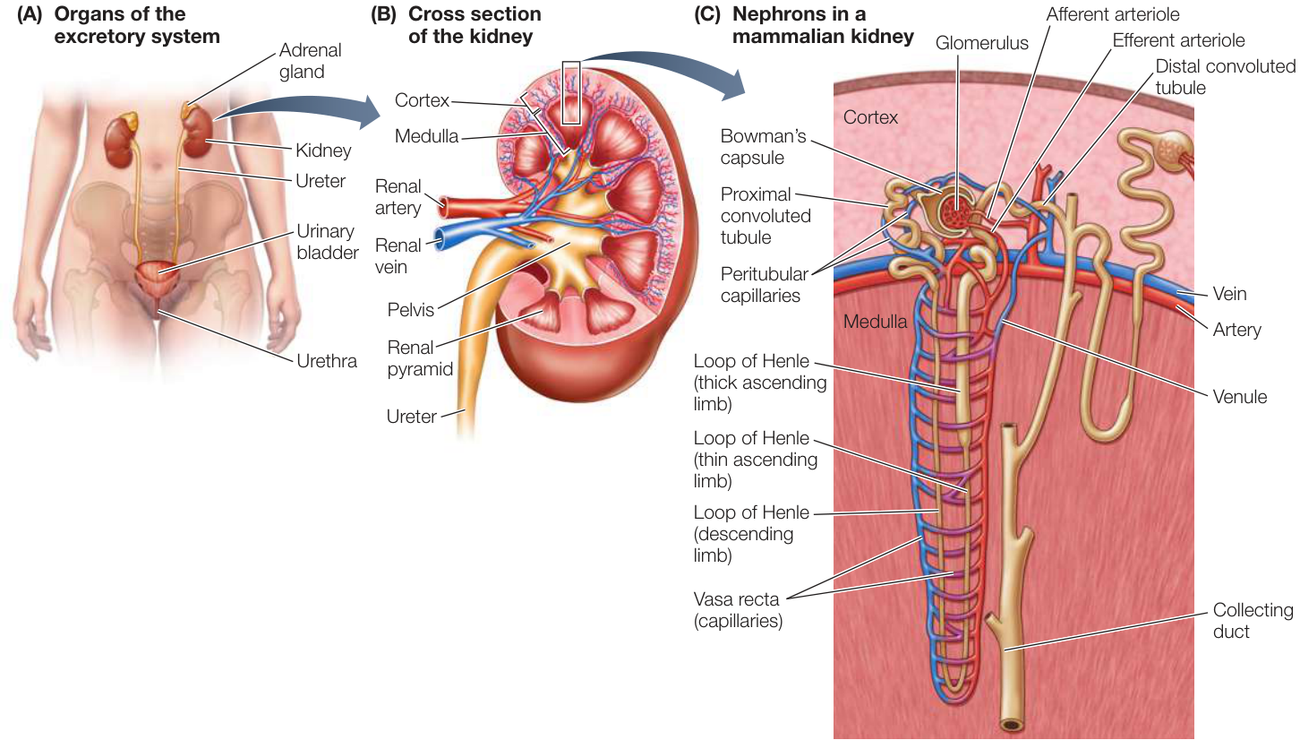
Figure 50.9 The Human Excretory System (A) The kidneys lie against the back wall of the abdominal cavity, in the region of the middle back. The adrenal glands (see Figure 39.18) are positioned on top of the kidneys; they are the source of the hormone aldosterone, which plays a role in the regulation of kidney function. (B) A highly organized internal structure is the basis for kidney function. Certain parts of the nephrons are in the organ’s outer region, the cortex; other parts are in the internal region, the medulla. (C) The glomeruli and the proximal and distal convoluted tubules are located in the cortex of the kidney. The loops of Henle run in parallel as straight sections down into the renal medulla and back up to the cortex. Collecting ducts run from the cortex to the inner surface of the medulla, where they open into the ureter. The vasa recta are peritubular capillaries that parallel the loops of Henle and are permeable to salts and water. Q: How do you think the osmolarity of the blood in the vasa recta compares with the osmolarity of the interstitial fluid at different levels of the medulla?
Activity 50.3 The Human Excretory System the kidney on its concave (punched-in) side. Inside the kidney, the ureter is continuous with structures that envelop kidney tissues called renal pyramids. The renal pyramids make up the internal core, or medulla, of the kidney. The medulla is covered by an outer layer, or cortex, that has a granular appearance because it contains all of the glomeruli. Between the cortex and the medulla, the renal artery divides into the many arterioles that serve the nephrons. In this same region, the renal vein collects blood from the many venules that drain the peritubular capillaries.
The organization of nephrons within the kidney is very regular. All of the glomeruli with their Bowman’s capsules are located in the cortex. The initial segments of the renal tubules that come from Bowman’s capsules are called the proximal convoluted tubules—“proximal” because they are closest to the glomerulus, and “convoluted” because they are twisted (Figure 50.9C). All of the proximal convoluted tubules are located in the cortex.
At the point at which the proximal convoluted tubule enters the medulla, it becomes thin, straight, and descends directly down toward the tip of a renal pyramid. In the medulla the tubule makes a hairpin turn and ascends back to the cortex, forming what is called a loop of Henle. Some nephrons have longer loops of Henle than others. Some 20%–30% of human nephrons that have glomeruli deep in the cortex (i.e., near the border with the medulla) have long loops of Henle that go deep into the medulla.
Nephrons that have glomeruli farther up in the cortex generally have short loops of Henle that descend only a short distance into the medulla. As you will see, the long loops are the critical adaptation of the mammalian nephron that enables the kidney to concentrate the urine.
The ascending limb of the loop of Henle becomes the distal convoluted tubule when it reaches the cortex—“distal” because it is farther from the glomerulus. The distal convoluted tubules of many nephrons join a common collecting duct in the cortex. The collecting ducts descend back down through the renal pyramid, parallel to and past the tips of the loops of Henle, and empty into a funnel-shaped structure called the pelvis. Divisions of the pelvis that surround each renal pyramid join together to leave the kidney as the ureter (see Figure 50.9B).
The organization of the blood vessels of the kidney closely parallels the organization of the nephrons (see Figure 50.9C). Smaller arteries branch from the renal artery and radiate into the cortex, forming the afferent arterioles that carry blood to each glomerulus. Each glomerulus is drained by an afferent arteriole that gives rise to the peritubular capillaries, most of which surround the proximal and distal convoluted tubules. The intimate associations of the glomerular and peritubular capillaries with the renal tubules enable exchanges between the blood and the specialized regions of the tubules.
Some of the peritubular capillaries run into the medulla in parallel with the loops of Henle and the collecting ducts, forming a vascular network called the vasa recta. All of the peritubular capillaries from a nephron join back together into a venule that joins with venules from other nephrons and eventually leads to the renal vein. The concentrating ability of the mammalian kidney depends on water reabsorption in the renal medulla, and the vasa recta are the avenue by which that water gets out of the renal medulla and back into the circulation.
Most of the glomerular filtrate is reabsorbed by the proximal convoluted tubule
Most of the water and solutes filtered by the glomerulus are reabsorbed and do not appear in the urine. We can reach this conclusion by comparing the rate of filtration by the glomeruli with the rate of urine production. The kidneys receive about 1 liter of blood per minute, or about 1,500 liters of blood per day. How much of this huge volume is filtered out of the glomeruli? The answer is about 12%. This is still a large volume—180 liters per day! We normally urinate less than 2 liters per day, so about 99% of the fluid volume that is filtered out of the glomerulus is returned to the blood. Where and how is this enormous amount of fluid reabsorbed?
The proximal convoluted tubule (PCT) is responsible for most of the reabsorption of water and solutes from the glomerular filtrate. The cells of this section of the renal tubule have many microvilli that increase their apical (facing into the tubule) surface area for reabsorption, and they have many mitochondria—an indication that they are metabolically active. PCT cells actively transport (with following) and other solutes, such as glucose and amino acids, out of the tubule fluid.
Almost all glucose and amino acid molecules that are filtered from the blood are actively reabsorbed by PCT cells and transported into the extracellular fluid. The active transport of solutes from the proximal tubule into the interstitial fluid causes water to follow osmotically. The water and solutes moved into the interstitial fluid are taken up by the peritubular capillaries and returned to the venous blood. These processes accomplish the reabsorption of more than 75% of the fluid that initially enters the nephron.
Despite the bulk reabsorption of water and solutes by the PCT, the overall osmolarity of the fluid flowing through the PCT does not change. Thus the process that is occurring in the PCT is called isosmotic reabsorption. The fluid that enters the loop of Henle has the same osmolarity as the blood plasma, although its composition is different. How then does the kidney produce urine that is more concentrated than the blood plasma?
The loop of Henle creates a concentration gradient in the renal medulla
Humans can produce urine that is four times more concentrated than their blood plasma. The vampire bat you encountered at the beginning of this chapter can produce urine that is 15 times more concentrated than its blood plasma (Investigating Life: How Can Vampire Bats Use Blood as Fast Food?). The concentrating ability of the mammalian kidney arises from a countercurrent multiplier mechanism made possible by the anatomical arrangement of the loops of Henle. The term “countercurrent” refers to the opposing directions of fluid flow in the descending and ascending limbs. The term “multiplier” refers to the ability of this system to create a solute concentration gradient in the renal medulla.
The loops of Henle do not themselves produce concentrated urine; rather, they increase the osmolarity of the extracellular fluid in the medulla in a graduated way. In humans, for example, the extracellular fluid at the top of the medulla bordering the cortex will be about 300 mOsm/L (the concentration of blood plasma). But at the bottom of the medulla, where the loops of Henle make their hairpin turns, the extracellular fluid can be 1,200 mOsm/L (Figure 50.10). How do the loops produce this effect?
The cells that make up the different segments of the loop of Henle differ anatomically and functionally. Cells of the descending limb and the initial cells of the ascending limb are thin, with no microvilli and few mitochondria. They are not specialized for transport. Partway up the ascending limb, the cells become specialized for active transport. These cells are thick and have many mitochondria. Accordingly, the segments of the loop of Henle are named the thin descending limb, the thin ascending limb, and the thick ascending limb (see Figure 50.9C).
The countercurrent multiplier mechanism is best understood by first considering events occurring in the thick ascending limb (see Figure 50.10, note 1). The apical (luminal side) membranes of the cells of the thick ascending limb cotransport from the tubule fluid into the tubule cells, and cotransport out of the basal membranes into the interstitial fluid. The basal membranes also actively transport out and into the cells. The end result is a reabsorption of and from the thick ascending tubule into the interstitial fluid. (In the following discussion, we distinguish between the two components of extracellular fluid—the blood plasma and the interstitial fluid.) The thick ascending limb is not permeable to water, so the reabsorption of and from the tubular fluid raises the concentration of those solutes in the surrounding interstitial fluid and decreases the concentration of the tubular fluid entering the distal convoluted tubule.
The thin descending limb, in contrast, is highly permeable to water but not very permeable to and . Since the local interstitial fluid has been made more concentrated by the and reabsorbed from the neighboring thick ascending limb, water is withdrawn osmotically from the fluid in the descending limb. Therefore the fluid in the descending limb becomes more concentrated as it flows toward the hairpin turn at the bottom of the renal medulla (see Figure 50.10, note 2).
The thin ascending limb, like the thick ascending limb, is not permeable to water. It is, however, permeable to and . As the concentrated tubule fluid flows up the thin ascending limb, it is more concentrated than the surrounding interstitial fluid, so and diffuse out. When the tubule fluid reaches the thick ascending limb, active transport continues to move and from the tubule fluid to the interstitial fluid.
Because of this countercurrent multiplier mechanism, the tubule fluid reaching the distal convoluted tubule is less concentrated than the blood plasma (see Figure 50.10, note 3), and the solutes that have been left behind in the renal medulla have created a concentration gradient in the interstitial fluid of the medulla (indicated by the background color gradient in Figure 50.10).
Investigating Life: How Can Vampire Bats Use Blood as Fast Food?
Experiment
Original Paper: C. Busch. 1988. Consumption of blood, renal function and utilization of free water by the vampire bat, Desmodus rotundus. Comp Biochem Physiol A Mol Integr Physiol 90: 141–146.
You learned at the beginning of the chapter that vampire bats feed once a day. After sunset they fly out, search for a victim, and then return to their roost. This nightly excursion generally takes less than 2 hours. Therefore in a very short time these small animals must take in a large enough volume of their liquid blood diet to supply their energy and water needs for a 24-hour period. How do they do it?
HYPOTHESIS Vampire bats maximize their food intake by rapidly excreting water, but they then conserve water by producing a highly concentrated urine while digesting their protein meal.
METHOD
- Offer a colony of captive vampire bats a beef blood meal once a day for 2 hours, to mimic the natural once-a-day feeding behavior of the bats.
- Do not allow bats access to free water.
- Measure the daily amount of blood consumed and the time course of subsequent urine volume and concentration.
RESULTS The table shows the average body mass and average amount of blood consumed by nine vampire bats over a period of 5–8 days.
| Average body mass (g) | Average amount of blood consumed (g) |
|---|---|
| 25.0 | 19.7 |
| 26.3 | 11.3 |
| 26.4 | 13.6 |
| 28.6 | 16.8 |
| 29.6 | 15.7 |
| 30.3 | 17.8 |
| 32.4 | 24.7 |
| 36.0 | 24.4 |
| 36.2 | 18.6 |
CONCLUSION In a short daily feeding period, vampire bats consume on average 60% of their body mass.
Work with the Data
To consume 60% of body mass and still be able to fly, the bats must rapidly excrete a large percentage of the water content of their food. The time course of urine flow and its concentration were measured during and after daily feeding for three vampire bats for 1 week and averaged. The data are shown in the table (—, no data reported). Create a double plot of the flow and concentration data as a function of time.
| Time after beginning to feed (min) | Average urine flow rate (g/g body mass/hr) | Average urine osmolality (Osm) |
|---|---|---|
| 10 | 0.13 | 0.5 |
| 20 | 0.24 | 0.25 |
| 45 | 0.17 | 0.40 |
| 90 | 0.05 | 0.70 |
| 150 | 0.01 | — |
| 210 | 0.01 | — |
| 270 | 0.005 | — |
| 330 | 0.003 | 3.3 |
| 390 | 0.02 | 3.4 |
QUESTIONS
- If you averaged the urine flow rates over the first 45 min following the beginning of the meal, and you assumed that the total blood intake was 60% of body mass and body mass was 30g, what percentage of the ingested volume would be excreted in that first 45 min?
- Considering that the normal osmolality of interstitial fluid in a mammal is 300 mOsm, what is the maximum concentrating factor in the vampire bats in this experiment?
- Three data points are missing in the urine osmolality curve. Would it be reasonable to plot these data by connecting the 90-minute sample and the 330-minute sample with a straight line? Why or why not?
You may wonder why the blood flow through the medulla does not wash out the concentration gradient established by the loops of Henle. The parallel arrangement of the descending and ascending peritubular capillaries in the medulla—the vasa recta—helps preserve the concentration gradient in the medulla. These capillaries are permeable to both salts and water. Therefore as blood flows down the descending limb of the vasa recta into the increasingly concentrated interstitial fluid of the medulla, it loses water and gains solutes. As blood flows up from the bottom of the medulla in the ascending limb of the vasa recta, the opposite happens (water is gained and solutes are lost) because now the blood is more concentrated than the surrounding interstitial fluid (see Figure 50.10, notes 4–6). The dynamics of this countercurrent exchange of salts and water between the blood in the vasa recta and the interstitial fluids result in little net change in the composition of the interstitial fluid in the medulla.
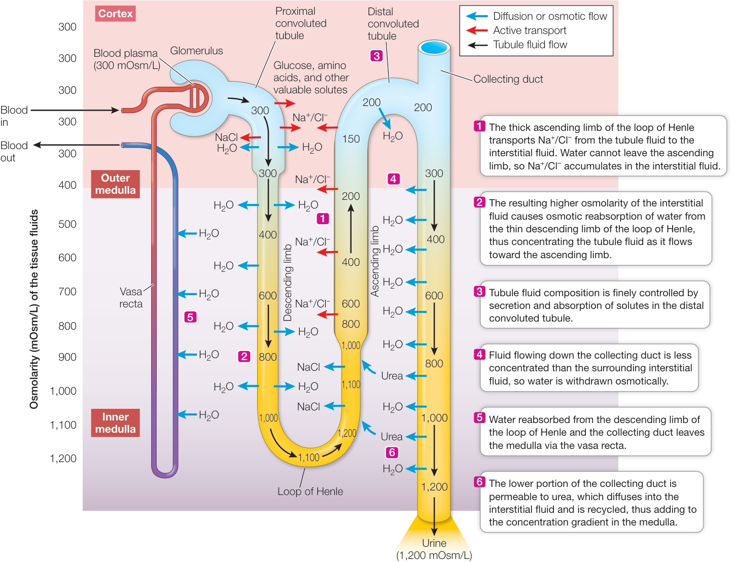
Figure 50.10 Concentrating the Urine A countercurrent multiplier mechanism enables the mammalian kidney to produce urine that is far more concentrated than the blood plasma. The composition—but not the concentration—of the filtrate is changed by the proximal convoluted tubule, which reabsorbs valuable ions and molecules (including ). Bulk reabsorption of water follows osmotically. The urine concentration process begins in the thick ascending limb of the loop of Henle, which reabsorbs but is impermeable to . This reabsorbed creates an osmotic differential that pulls water out of the thin descending limb; also can diffuse into the thin descending limb, making the fluid in that limb more concentrated. Through this continuous process, a concentration gradient in the interstitial fluid of the renal medulla is created as indicated by the gradient of color in the medulla. As urine in the collecting duct passes through this concentration gradient, it can lose water osmotically, becomes more concentrated as indicated by the shading of color, and can become almost as concentrated as the interstitial fluid at the bottom of the renal medulla. Q: What would be the effect of a drug called a “loop diuretic” that blocks the transport in the thick ascending limb of the loop of Henle?
Water permeability of kidney tubules depends on water channels
We have noted that some tubule regions, such as the PCT, are highly permeable to water whereas others, such as the thick ascending limb of the loop of Henle, are impermeable to water. What causes these differences in water permeability in different regions of the nephron? Aquaporins are a class of membrane proteins that form water channels. Regions of the nephron that are highly permeable to water have greater numbers of aquaporins. Thus aquaporins are abundant in kidney PCT cells and in descending limbs of the loops of Henle, but not in the ascending limbs of the loop of Henle.
Connect the Concepts As explained in Key Concept 6.3, water does not cross cell membranes easily, but aquaporins are protein channels that make cells permeable to water.
As an interesting evolutionary note, aquaporins are also important in maintaining water balance in amphibians. Many amphibians can gain water from a moist substrate because they have aquaporins in the epithelial cells of their belly skin. Thus water can cross their skin into the interstitial fluid by osmosis.
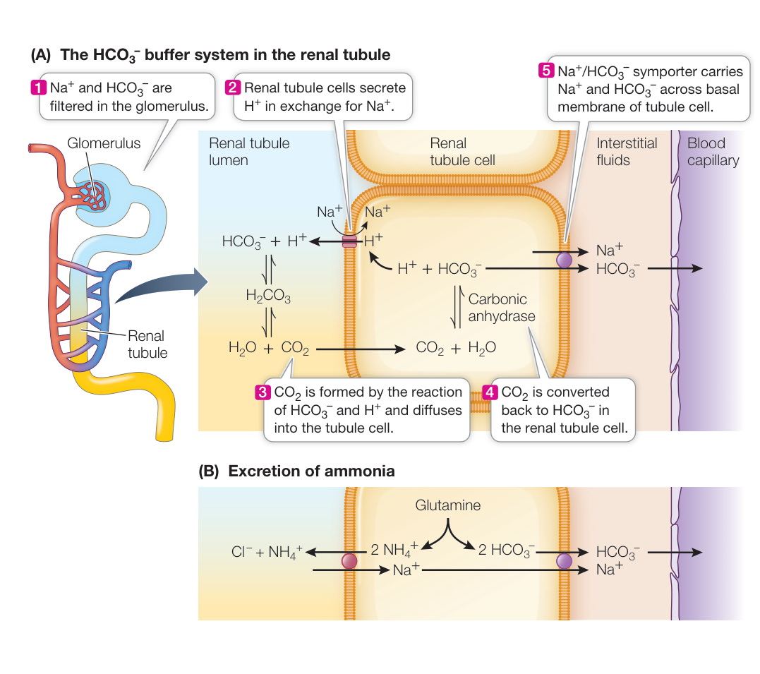
Figure 50.11 The Kidney Excretes Acids and Conserves Bases (A) Bicarbonate ions are filtered out of the blood at the glomerulus, and renal tubule cells secrete hydrogen ions into the tubule fluid. In the renal tubule, the filtered bicarbonate buffers the secreted hydrogen ions and keeps the urine from becoming too acidic. The formed by the reaction of bicarbonate and hydrogen ions is converted back to bicarbonate by the renal tubule cells and transported back into the interstitial fluid. (B) Excretion of ammonium ions ( ) by renal tubule cells is also important for acid–base balance.
The distal convoluted tubule fine-tunes the composition of the urine
The first portion of the distal convoluted tubule is similar to the thick ascending limb of the loop of Henle. and are transported out of the tubule fluid, and water cannot follow. As a result, the tubule fluid becomes even more dilute. The later sections of the distal convoluted tubule, however, can be permeable to water, and therefore water can be osmotically drawn from the hypotonic tubule fluid into the interstitial fluid. As the tubule fluid flows through the distal tubule to the collecting duct, it equilibrates with the osmolarity of the blood plasma.
An important function of the distal tubule is the fine-tuning of the ionic composition of the urine. Even though bulk reabsorption of substances such as calcium, phosphate, bicarbonate, and potassium occurs in the proximal convoluted tubule, changes in the concentrations of these substances occur in the distal convoluted tubule. In the case of potassium, for example, if a person is potassium depleted, this ion is reabsorbed in the distal convoluted tubule, but if a person has an abundance of potassium, this ion is secreted in the distal convoluted tubule. As you will see in Key Concept 50.6, this exchange of is controlled by the hormone aldosterone. Another example is reabsorption of in the distal convoluted tubule, which is controlled by the actions of vitamin D. The fine-tuning of urine composition continues in the collecting duct. As you can imagine, the list of ion transporters in the distal convoluted tubule is large.
Urine is concentrated in the collecting duct
The tubule fluid entering the collecting duct is at about the same solute concentration as the blood plasma, but its solute composition is considerably different from that of the plasma. The major solute in the tubule fluid is now urea, since salts were reabsorbed earlier in the nephron. As the tubule fluid flows down the collecting duct, it loses water osmotically to the interstitial fluid, and that water returns to the circulatory system via the vasa recta (see Figure 50.10, note 4).
The concentration gradient established in the renal medulla by the countercurrent multiplier actions of the loops of Henle creates the osmotic potential that withdraws water from the collecting ducts. The collecting ducts begin in the renal cortex and run through the renal medulla before emptying into the ureter at the tips of the renal pyramids. During this journey, the solute concentration of the surrounding interstitial fluid increases, and more and more water can be absorbed from the urine in the collecting duct. By the time the urine reaches the ureter, it can become greatly concentrated, with urea as the major solute.
As water is withdrawn from the collecting duct, some urea also leaks out into the medullary interstitial fluid, adding to its osmotic potential. This urea diffuses back into the loop of Henle and is returned to the collecting duct. The recycling of urea in the renal medulla contributes significantly to the concentration gradient and therefore the ability of the kidney to concentrate the urine in the collecting duct. The ability of a mammal to concentrate its urine is determined by the maximum concentration gradient it can establish in its renal medulla.
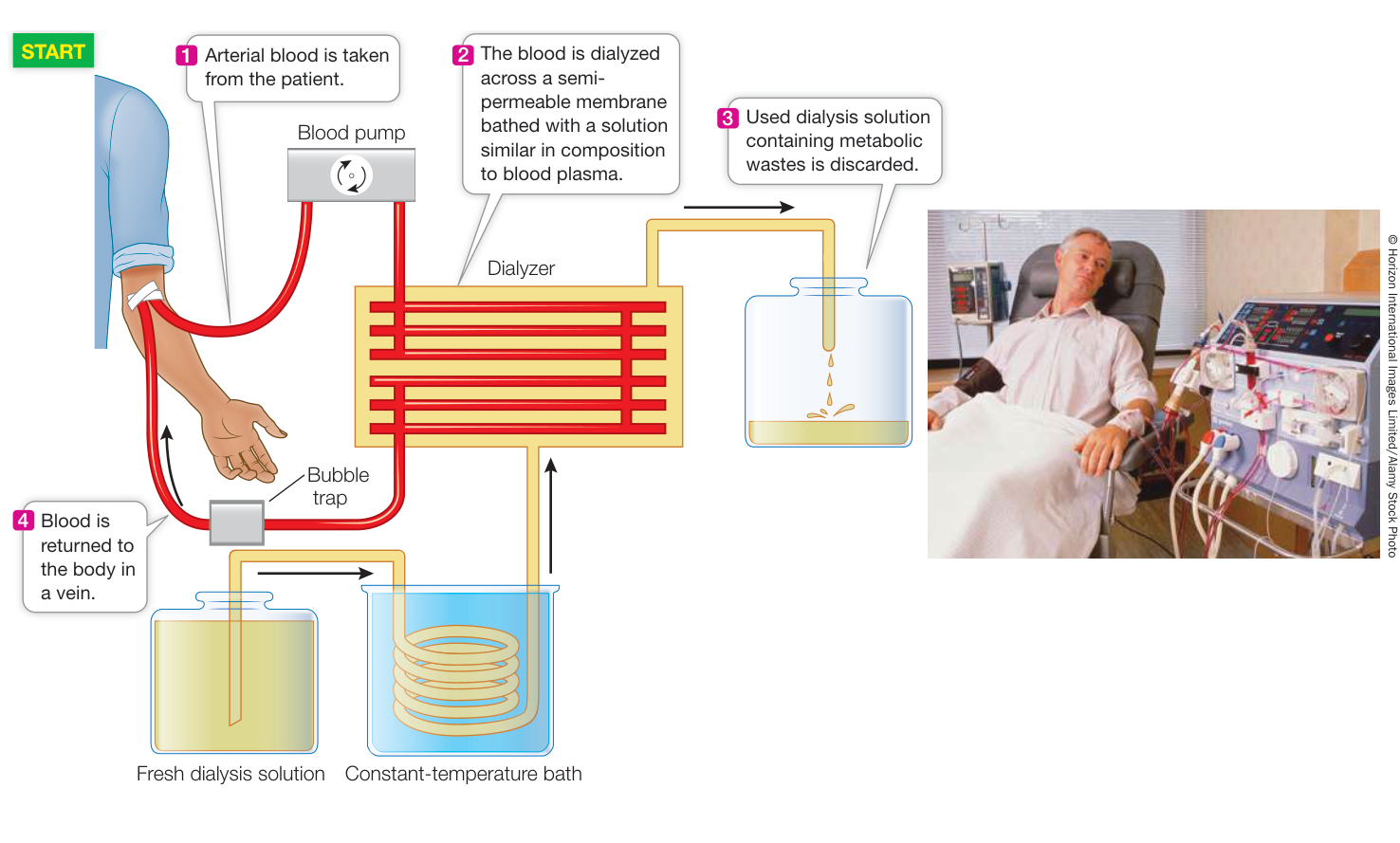
Figure 50.12 Renal Dialysis Patients with kidney failure can have their blood cleansed of wastes by renal dialysis machines. Blood flows through channels of semipermeable membranes that allow diffusion of waste molecules from the blood to a dialysis fluid.
The kidneys help regulate acid–base balance
Besides regulating salt and water balance and excreting nitrogenous wastes, the kidneys have another important role: they regulate the hydrogen ion concentration (the pH) of the extracellular fluids. pH is a critical variable because it influences the structure and function of proteins.
One way to minimize pH changes in a chemical solution is to add a buffer—a substance that can either absorb or release hydrogen ions (see Key Concept 2.4). The major buffer in the blood is bicarbonate ions ( ; see Figure 47.14) that are formed from the dissociation of carbonic acid, which in turn is formed by the hydration of according to the following equilibrium reaction:
From this equation, you can see that if excess hydrogen ions are added to this reaction mixture, the reaction will move to the left and absorb the excess . If hydrogen ions are removed from the reaction mixture, however, the reaction will move to the right and supply more .
The buffer system is important for controlling the pH of the blood, and therefore of the interstitial fluids as well, because the reaction can be pushed to the right and pulled to the left physiologically. The lungs control the levels of in the blood, thus altering the acid portion of the reaction. is considered the acid portion of the reaction because if you add additional , the reaction shifts to the right, producing more ions. The kidneys control the base portion of the reaction by removing from the blood and returning to the blood. How does this occur?
is filtered in the glomerulus and is therefore present in the tubule fluid. As illustrated in Figure 50.11A, tubule cells transport into the tubule fluid in exchange for . In the tubule, the excreted combines with the filtered to produce that then disassociates into and . The diffuses into the tubule cells, where in the presence of the enzyme carbonic anhydrase it produces that is transported out of the basal end of the cell into the interstitial fluid and thence to the blood. Thus for each secreted into the tubule fluid, a ion is released into the blood.
Another mechanism for secretion and reabsorption involves ammonium ions (). The metabolism of glutamine in tubule cells produces and (Figure 50.11B). The is reabsorbed into the interstitial fluid. The is transported into the tubule fluid and combines with , which is excreted in the urine. This process results in the addition of a new ion to the blood. The is transported into the tubules by means of an transporter.
Kidney failure is treated with dialysis
Loss of kidney function (renal failure) results in the retention of salts and water (hence high blood pressure), retention of urea (uremic poisoning), and a decreasing pH (acidosis). A person who suffers complete renal failure will die within 2 weeks if not treated. A drastic but highly successful treatment is kidney transplant, but it is usually necessary to sustain a patient for considerable time while waiting for a kidney to become available. Therefore artificial kidneys, or renal dialysis machines, are essential modes of treatment.
In a dialysis machine, the patient’s blood flows through many small channels made of semipermeable membranes (Figure 50.12). A dialysis solution flows on the other side of these membranes, through which small molecules can diffuse. Molecules and ions diffuse from an area of higher concentration to an area of lower concentration, so the composition of the dialysis fluid is crucial. The concentrations of the molecules or ions that need to be conserved must be at the same concentration in the dialysis fluid as they are in the blood. The concentrations of molecules and ions that need to be removed from the blood are zero in the dialysis fluid. The total osmotic potential of the dialysis fluid must equal that of the blood plasma.
About 500 milliliters of the patient’s blood is in the dialysis machine at any one time, and the unit processes several hundred milliliters of blood per minute. A patient with no kidney function must be on the dialysis machine for 4–6 hours three times a week.
Recap and Assess
The anatomical organization of nephrons makes it possible for the mammalian kidney to produce a urine more concentrated than the blood, thereby conserving water to maintain extracellular fluid volume. Bulk reabsorption of salts, other valuable solutes, and water takes place in the proximal convoluted tubule. The loops of Henle act as a countercurrent multiplier, creating a concentration gradient of the interstitial fluids in the renal medulla. Collecting ducts run through the renal medulla and lose water osmotically to the surrounding interstitial fluids, concentrating the urine. The renal tubules contribute to acid–base balance by excreting ions.
- If you could measure the concentration of mitochondria in mammalian kidney tissues, where would you find the highest concentration, the lowest concentration, and an intermediate concentration? Explain your answer.
- How do the loops of Henle act as a countercurrent multiplier?
- If you compared a water rat and a desert rat of the same size, how would you expect their kidneys to differ in anatomical structure? Explain your answer.
- How does the active secretion of ions by the renal tubule cells result in an increase in the bicarbonate concentration in the blood?
The kidneys contribute to homeostasis in several ways, including regulating extracellular fluid volume, maintaining the osmotic concentration and ionic composition of the extracellular fluid, and regulating pH. As you will see next, the kidneys also play a major role in regulating blood pressure.
Kidney Function Is Regulated
Learning Objectives
- 50.6.1 Discuss how renin, angiotensin, and aldosterone affect glomerular filtration rate (GFR) and blood pressure.
- 50.6.2 Given an event that lowers blood pressure, predict the response of the renin–angiotensin–aldosterone system.
- 50.6.3 Describe ADH’s role in regulating kidney function in response to changes in blood osmolarity or blood pressure.
- 50.6.4 Explain the mechanisms whereby the afferent and efferent arterioles regulate the GFR, and how those responses are controlled.
Several regulatory mechanisms act on the kidneys to maintain blood pressure, blood osmolarity, and blood composition. We discuss these mechanisms separately, but keep in mind that they are always working together.
Glomerular filtration rate is regulated
If the kidneys stop filtering blood, they cannot accomplish any of their functions. The maintenance of a constant glomerular filtration rate (GFR) depends on an adequate blood supply to the kidneys at an adequate blood pressure. Renal arteries usually deliver blood to the kidneys at high pressure because they are early branches off the aorta. In addition, autoregulatory mechanisms ensure adequate blood supply and blood pressure for kidney function regardless of what is happening elsewhere in the body. The kidney’s autoregulatory adjustments compensate for decreases in cardiac output or decreases in blood pressure so that the GFR remains constant.
One autoregulatory mechanism is the dilation (expansion) of the afferent renal arterioles when blood pressure falls. This dilation decreases the resistance in the arterioles and helps maintain blood pressure in the glomerulus. If arteriole dilation does not keep the GFR from falling, the kidney releases an enzyme, renin, into the blood. Renin converts a circulating protein, angiotensinogen, into angiotensin I, which is then acted on by angiotensin-converting enzyme (ACE) in the lungs and other tissues to form the active hormone angiotensin II, or simply angiotensin (Figure 50.13). Angiotensin has several effects that help restore the GFR to normal:
- It constricts the efferent renal arterioles, raising the resistance for blood leaving the glomerulus. Like putting a finger over the end of a garden hose, this restriction of drainage elevates blood pressure in the glomerular capillaries.
- It constricts peripheral blood vessels all over the body, an action that elevates blood pressure.
- It stimulates the adrenal cortex to release the hormone aldosterone. Aldosterone stimulates sodium reabsorption by the kidney, making its reabsorption of water more effective. Enhanced water reabsorption helps maintain blood volume and therefore blood pressure.
- It acts on the brain to stimulate thirst. Increased water intake in response to thirst increases blood volume and blood pressure. Thus the renin–angiotensin–aldosterone system, or RAAS, coordinates many responses to maintain blood pressure and kidney function.
Regulation of GFR uses feedback information from the distal tubule
A remarkable anatomical feature of the nephron is that where its renal tubule returns to the cortex and becomes the distal convoluted tubule, it makes contact with the afferent and efferent arterioles of its glomerulus. At this location the cells of the tubule are modified to form a structure called the macula densa (see Figure 50.13), and the arteriole cells are also modified and are called juxtaglomerular cells. The macula densa cells sense the concentration of NaCl in the fluid entering the distal convoluted tubule. If GFR is high, flow through the loop of Henle is high and the cells of the thick ascending limb cannot reabsorb all of the NaCl passing through it. If GFR
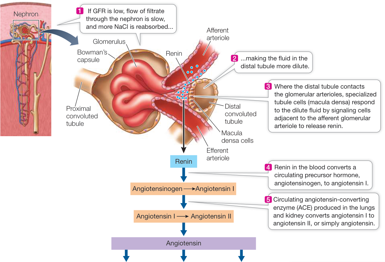
Figure 50.13 The Renin-Angiotensin-Aldosterone System Helps Regulate GFR When glomerular filtration rate (GFR) falls, tubular fluid flows more slowly through the loops of Henle, more NaCl is reabsorbed from the thick ascending limb, and the fluid reaching the distal convoluted tubule is more dilute. Dilute fluid in the distal convoluted tubule (where it comes into contact with the afferent and efferent glomerular arterioles) stimulates release of the enzyme renin into the circulation. Renin starts a cascade of events that work together to raise blood volume, blood pressure, and GFR.
Simulation 50.1 Kidney Regulation
is low, flow through the loop of Henle is slower and the cells of the thick ascending limb can reabsorb more of the NaCl. If the NaCl level in the distal convoluted tubule drops too low, the macula densa cells signal the juxtaglomerular cells to release renin and trigger the RAAS response. Thus the concentration of NaCl in the fluid passing over the macula densa is a function of GFR and is information that enables fine control of the RAAS.
Blood osmolarity and blood pressure are regulated by ADH
Cells in the hypothalamus can stimulate the release of a hormone called antidiuretic hormone (ADH), also called vasopressin) from the posterior pituitary. ADH stimulates cells of the collecting duct to insert aquaporins (water channels) into their cell membranes. The aquaporins increase the permeability of these membranes to water, and therefore more water is reabsorbed from the collecting duct fluid into the interstitial fluid of the renal medulla. The higher the circulating levels of ADH, the greater the number of aquaporins. Various factors can stimulate or inhibit the release of ADH. Of key importance to kidney function are (1) osmoreceptors that monitor blood osmolarity and (2) baroreceptors that monitor blood pressure (Figure 50.14).
Osmoreceptor neurons in the hypothalamus are activated by a rise in blood osmolarity, and they increase the release of ADH. ADH helps regulate blood osmolarity by controlling water reabsorption. The osmoreceptors also stimulate thirst. The resulting water retention and water intake dilute the blood as they expand blood volume.
Baroreceptors in the walls of the aorta and the carotid arteries (see Figure 48.21) that detect an increase in blood pressure will inhibit the release of ADH. With less circulating ADH, less water is reabsorbed, which increases urine volume and decreases blood volume, hence lowering blood pressure.
If blood pressure falls, as when you lose blood volume through hemorrhage or excessive evaporative water loss, activity of the baroreceptors in the aorta and carotid arteries decreases. Input via cranial nerves to the hypothalamus from these receptors inhibits the release of ADH, so when the firing rates of these baroreceptors fall, ADH release increases. More ADH results in more efficient water reabsorption and therefore a protection of blood volume and blood pressure.
Alcohol inhibits ADH release, explaining why excessive beer drinking leads to excessive urination and dehydration, which contribute to the symptoms of a hangover.
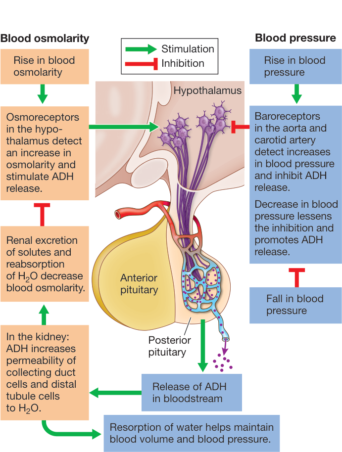
Figure 50.14 Antidiuretic Hormone Increases Blood Pressure and Promotes Water Reabsorption ADH is produced by neurons in the hypothalamus and released from nerve endings in the posterior pituitary. The release of ADH is stimulated by hypothalamic osmoreceptors and inhibited by baroreceptors in the large arteries. Red lines indicate inhibitory actions; green lines show stimulatory actions.
As already mentioned, the presence of aquaporins in cell membranes determines their water permeability. Aquaporins play an important and unique role in the collecting duct. Several members of the aquaporin family of water channels are found in the cell membranes of the collecting duct cells. At least two aquaporins (AQP-3 and AQP-4) are localized in the basolateral membranes (facing the blood vessels). A different aquaporin, AQP-2, is found in the apical cell membranes (facing into the tubule). The presence of AQP-2 in these membranes is controlled by ADH (Figure 50.15).
When ADH levels are low, such as when a person is well hydrated, most of the AQP-2 protein is sequestered in the membranes of intracellular vesicles, and the collecting duct permeability is low. A rise in ADH levels stimulates the insertion of these vesicles along with their AQP-2 channels into the apical cell membranes. As a result, the membranes become more permeable to water. Water that enters the collecting duct cells passes into the interstitial fluid through the aquaporins in the basolateral membranes. ADH also stimulates the synthesis of new AQP-2 proteins. Thus circulating ADH controls the number of AQP-2 water channels in the cell membranes of the collecting duct cells, and therefore the permeability of the collecting duct to water.
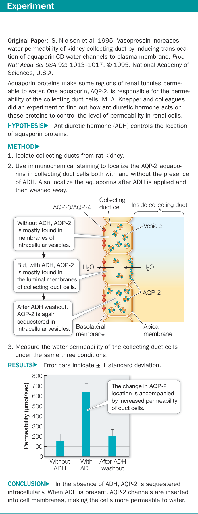
Figure 50.15 ADH Induces Insertion of Aquaporins into Cell Membranes
The heart produces a hormone that helps lower blood pressure
You may not think of the heart as an endocrine organ, but it is. When blood volume is high, blood pressure is high, putting strain on the heart. Under these conditions, the increased venous return stretches the atria of the heart. When the atrial muscle fibers are overstretched, they release a peptide hormone called atrial natriuretic peptide (ANP). This peptide hormone enters the circulation, and in the kidney it decreases the reabsorption of sodium. If less sodium is reabsorbed, less water is reabsorbed, and more passes into the urine. Thus ANP has the effect of lowering blood volume and therefore blood pressure.
Recap and Assess
Glomerular filtration is essential for kidney function and is sustained by autoregulatory mechanisms. These autoregulatory mechanisms include changes in dilation and constriction of the glomerular afferent arterioles and hormonal control over constriction of the efferent glomerular arteriole mediated by the renin–angiotensin–aldosterone system (RAAS). Angiotensin controls the release of aldosterone, which controls sodium reabsorption. Sensors that monitor blood pressure and blood osmolality stimulate or inhibit the release of ADH, which controls the water permeability of the collecting duct.
- What are the mechanisms whereby the afferent and efferent arterioles regulate the GFR, and how are those responses controlled?
- Aldosterone stimulates activity of the exchangers in the distal convoluted tubule and the initial segments of the collecting ducts. Aldosterone release is triggered by angiotensin. Given this information, what is the significance of the fact that the exchanger in the tubule cells secretes two ions for every three ions it reabsorbs?
- If you drank a lot of salty chicken broth that had an osmotic concentration of 300 mOsm/L, what two mechanisms would play roles in the response of your kidneys to excrete the excess fluid volume and excess ? Explain your answer.

Activity 50.4 The Major Organ Systems
Investigating Life
Q&A How does the excretory system of vampire bats process their high-protein liquid diet? Future directions
As we described in the opening story, vampire bats conserve water by producing urine that can be as much as 15 times more concentrated than their blood. For comparison, the maximum urine concentration of humans is about four times that of the blood.
Recall from Key Concept 50.5 that the mammalian body’s ability to produce concentrated urine depends on the concentration gradient set up in the renal medulla by the loops of Henle. An important adaptation for increasing the concentration gradient is to increase the lengths of the loops of Henle relative to overall kidney size. Some tiny desert gerbils, for example, have such long loops of Henle that the renal pyramid (each of the rodent kidneys has only one, in contrast to humans) extends far out of the concave surface of the kidney and into the ureter (Figure 50.16). The large concentration gradient that results draws most of the water out of the urine as it passes down the collecting duct. Desert gerbils are so effective in conserving water that they can survive on the water released by the metabolism of their food.
The vampire bat kidney is also particularly good at concentrating urea. Within minutes after feeding, the urea concentration in its urine can increase by more than 500-fold.
Future directions
Understanding the diverse mechanisms whereby physiological systems are controlled and regulated can contribute to the discovery and development of new pharmaceuticals. The renal system is an example. Many diseases involve problems with salt and water balance. A common problem is water retention. A large number of drugs have been developed that increase the excretion of water in urine, such as occurs naturally in vampire bats. These drugs are called diuretics, and since they act through different mechanisms, they have different effects on , , and levels in the blood, which can cause serious side effects. Excessive loss of , for example, can lead to heart arrhythmia, muscle weakness, and paralysis. Excessive retention of can also lead to heart arrhythmia, along with muscle cramps and paralysis. Loss of too much can result in central nervous system deficits. Too much has many negative side effects, ranging from depression and fatigue to vomiting and gout. Thus it is important to understand how drugs work on physiological systems, to avoid negative side effects.
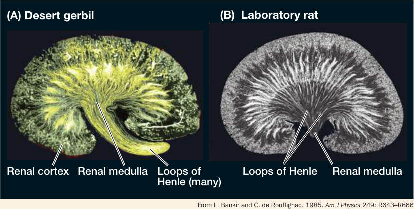
Figure 50.16 The Ability to Concentrate The ability of the mammalian kidney to concentrate urine depends on the lengths of its loops of Henle relative to the overall size of the kidney. (A) The kidney of a desert gerbil has a single renal pyramid with loops of Henle so long that the pyramid extends far into the ureter (ureter not shown). (B) An ordinary laboratory rat has much shorter loops of Henle. Q: Do you think the vampire bat has loops of Henle resembling those of the laboratory rat or the desert gerbil?
© Michael & Patricia Fogden/Minden Pictures
You should be able to relate each summary to the adjacent figures. If you go to this visual summary in Achieve, you can follow links to figures, animations, activities, and simulations that will help you consolidate the material.
Visual Summary
Excretory Systems Regulate Osmotic and Ionic Concentrations
■ Excretory systems maintain the osmolarity and volume of the extracellular fluids and eliminate the waste products of nitrogen metabolism through the processes of filtration, secretion, and reabsorption. Urine is the output of excretory systems.
■ Marine animals can be osmoconformers or osmoregulators. Freshwater animals must be osmoregulators. Terrestrial animals are osmoregulators, but the nature of their regulation depends on their environment and lifestyle.
■ Apart from regulating osmolarity of cells and extracellular fluids, animals must also regulate their ionic composition by conserving some ions and secreting others. Salt glands are adaptations for secretion of and .
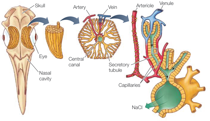
Figure 50.2 Questions
- What are the two factors that cause water to move across biological membranes?
- How can a marine shark be an osmoconformer and still have an extracellular concentration less than that of seawater?
- Why are there limits to osmoconformity in marine invertebrates?
Animals Excrete Nitrogen as Ammonia, Urea, or Uric Acid
■ Aquatic animals that breathe water can eliminate nitrogenous wastes such as ammonia by diffusion across their gill membranes. Terrestrial animals and some aquatic animals detoxify ammonia by converting it to urea or uric acid before excretion.
■ Depending on the form in which they excrete their nitrogenous wastes, animals are classified as ammonotelic, ureotelic, or uricotelic.
- Why don’t water-breathing animals have to detoxify the waste product ammonia?
- Why are many terrestrial animals that live in arid habitats uricotelic?
- Historically, why was gout was called the disease of kings?
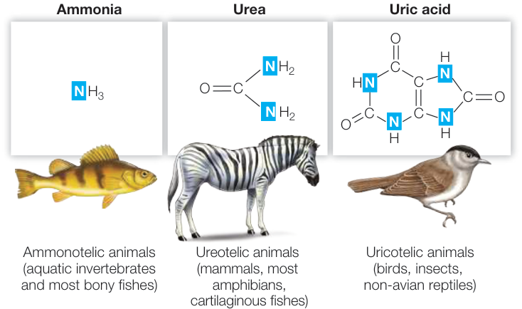
Figure 50.3
- ■ The protonephridia of flatworms consist of flame cells and excretory tubules. Extracellular fluid is filtered into the tubules, which process the filtrate to produce dilute urine.
- ■ In annelid worms, blood pressure causes filtration of the blood across capillary walls. The filtrate enters the coelomic cavity, where it is taken up by metanephridia, which alter the composition of the filtrate by active transport mechanisms.
- ■ The Malpighian tubules of insects receive ions and nitrogenous wastes by active transport across the tubule cells.
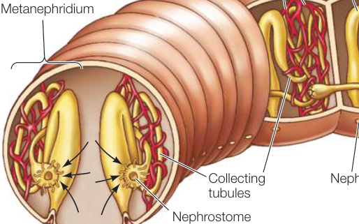
Figure 50.5 Questions
- What is the function of metanephridia?
- Why is filtration not a function of the insect excretory system?
The Nephron Is the Basic Functional Unit of Vertebrate Excretory Systems
- ■ Marine bony fishes produce little urine.
- ■ Reptiles, including birds, have skin with low water permeability and excrete nitrogenous wastes as uric acid in a semisolid form.
- ■ Mammals produce urine that is more concentrated than their extracellular fluids.
- ■ The nephron, the functional unit of the vertebrate kidney, consists of a glomerulus, which filters the blood, a renal tubule, which use active secretion and reabsorption to convert the glomerular filtrate into urine, and a system of peritubular capillaries, which surround the tubule and support its functions of secretion and reabsorption.
- Why do marine bony fishes have few glomeruli?
- What is a podocyte?
- Why does the glomerulus have an efferent arteriole rather than a venule?
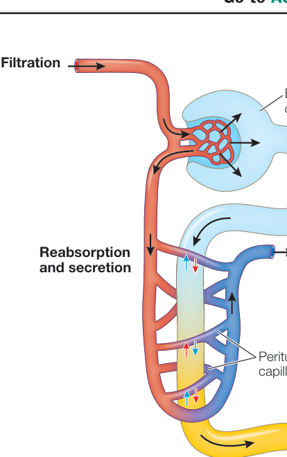
Figure 50.7
The Mammalian Kidney Can Produce Concentrated Urine
and Animation 50.1
- The glomeruli and the proximal and distal convoluted tubules are located in the cortex of the kidney. Certain molecules are actively reabsorbed from the glomerular filtrate by the tubule cells, and other molecules are actively secreted. Straight sections of renal tubules called loops of Henle and collecting ducts are arranged in parallel in the medulla of the kidney.
- Salts, water, and valuable molecules such as glucose and amino acids are reabsorbed in the proximal convoluted tubule without the renal filtrate becoming more concentrated, although its composition changes.
- The loops of Henle create a concentration gradient in the interstitial fluid of the renal medulla by a countercurrent multiplier mechanism. Urine flowing down the collecting ducts to the ureter is concentrated by the osmotic reabsorption of water caused by the concentration gradient in the surrounding interstitial fluid.
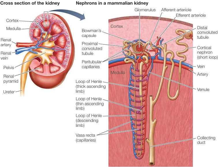
Figure 50.9
Questions
- Where in the nephron does the greatest reabsorption of water and solutes occur?
- Which part of the nephron is not permeable to water?
- Acetazolamide is a carbonic anhydrase inhibitor used to treat altitude sickness. Explain why one of its side effects is increased urine production.
Kidney Function Is Regulated
- Kidney function in mammals is controlled by autoregulatory mechanisms that maintain a constant high glomerular filtration rate (GFR) even if blood pressure varies.
- The renin–angiotensin–aldosterone system (RAAS) is an important autoregulatory mechanism. Renin is released by the kidney when blood pressure falls. Renin activates angiotensin, which causes the constriction of efferent glomerular arterioles and peripheral blood vessels, causes the release of aldosterone (which enhances water reabsorption), and stimulates thirst.
- Changes in blood pressure and osmolarity influence the release of antidiuretic hormone (ADH), which controls the permeability of the collecting duct to water and therefore the amount of water that is reabsorbed from the urine.
Questions
- How does a fall in blood pressure result in a signal that increases activation of angiotensin?
- How does angiotensin raise GFR?
- How does ADH change the permeability of the collecting duct to water?
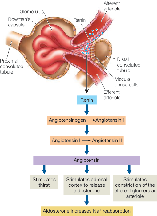
Figure 50.13