Chapter 10Protein Interface and Active Site Redesign
This chapter describes two quite different but complementary approaches to enzyme redesign: minimal redesign of residues in the active site responsible for catalysis or active site conformation [1, 2], and the use of directed evolution to rescue function in chimeric, domain-swapped enzymes [3].
Minimalist Active Site Redesign
Enzymes use only a handful of their hundreds of residues to create a local chemical environment customized to lower the activation energy of their reactions. They have traditionally been viewed as fine-tuned to catalyze a single reaction not only with tremendous efficiency, but also with catalytic and substrate specificity. This is true, but in reality many enzymes also display reactive promiscuity; they can perform the same reaction on a variety of similar substrates (though still with one overwhelmingly dominating the minor species), or even catalyze a handful of minor reactions in addition to the major one. Redesign by necessity involves undoing some of the specificity evolved for a major reaction and redirecting as much of it as possible in a new direction. Theoretically, this should be easier if the new reaction is already a minor function of the wild-type protein. Promiscuity can be divided into three types: substrate (performing the same reaction on different substrates), product (yielding different products from the same substrate), and catalytic (a combination of both) [1]. In these pioneer days of protein (re)design, substrate and reaction promiscuity offer obvious targets for investigation into the mechanisms by which enzymes can be persuaded to perform new functions.
Often proteins from the same homology family, with remarkably similar folds, use similar catalytic machinery to perform many different tasks—even though their primary sequences may share less than 50% identity [4]. This catalytic variation can be traced to key differences at a handful of residue positions, most often at the active site, that alter either part of the catalytic machinery or the nature (size, local charge, solvent exposure, etc.) of the substrate binding pocket. Enzyme redesign can be seen as an attempt to extend a given homology family by creating new members. Protein engineers have exploited both enzyme homology and promiscuity to devise an array of novel activities from simple, often single-residue active site mutations that leave the wild-type scaffold unaltered. While these novel enzymes rarely approach the efficiency of their evolution-streamlined progenitors, they point the way for future work in design.
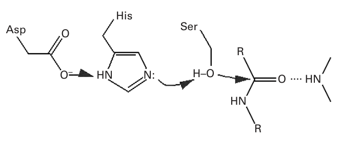
Figure 10.1 The Ser-His-Asp catalytic triad.
Subtilisin
The Ser-His-Asp catalytic triad (figure 10.1) is responsible for the name and activity of serine proteases, which come in two very distinct forms (the result of convergent evolution) that nonetheless share their method of catalysis. The aspartate forms a very strong hydrogen bond to histidine, raising its , which deprotonates serine, which in turn performs a nucleophilic attack on the carbonyl group of an aromatic residue, converting into a tetrahedral intermediate that is stabilized by the active site residues (the oxyanion hole). This moiety’s return to a carbonyl severs the peptide bond.
Subtilisin is a particularly well-studied serine protease. One early experiment used chemical modifications to replace the catalytic serine oxygen of subtilisin with a sulfur, effectively mutating the serine to cysteine [5]. Cysteine proteases are a natural enzyme family possessing an active site geometry similar to that of the serine proteases, so the cysteine variant of subtilisin (dubbed thiolsubtilisin) was expected to have an even higher rate of amide bond hydrolysis than the original, given the greater nucleophilicity of sulfur compared to oxygen. In fact, thiolsubtilisin showed a -fold drop in protease activity, but was able to catalyze the addition of amines to activated esters to form new peptide bonds, a reaction eschewed by subtilisin in favor of hydrolysis. On the premise that the active site of subtilisin was fine-tuned for the alcohol of serine, a proline near the catalytic triad was mutated to alanine to reduce the steric crowding caused by the addition of the bulkier sulfur atom [6]. This double mutant, called subtiligase, lost more protease activity but displayed ten-fold more ligase activity than thiolsubtilisin. Subtiligase operates on the same substrates as subtilisin, showing that the substrate specificity mechanisms of (at least some) enzymes are disjoint from their catalytic machinery. Another subtilisin variant, selenosubtilisin, replaces the sulfur of the cysteine with selenium and acts as both a peroxidase as well as ligase [7]. See figure 10.2.
Figure 10.2Reactions catalyzed by subtilisin and its derivatives. Subtilisin favors hydrolysis, while its synthetic analogs perform aminolysis [1]. Copyright Wiley-VCH Verlag GmbH & Co. KGaA. Reproduced with permission.
Interconverting Homologous Enzymes
Homologous enzymes share the same overall scaffold, and often use the same chemical step to catalyze their various reactions, so comparing their structures can lead to fruitful active-site mutations. L-Ala-D/L-Glu epimerase (AEE), muconate lactonizing enzyme II (MLE II), and ortho-succinylbenzoate synthase (OSBS) are all members of the enolase superfamily that remove a proton from a carboxylate substrate to form an enediolate intermediate stabilized by a ligand [8]. AEE and MLE II were mutated to obtain OSBS activity, the former via rational design and the latter using random mutagenesis and screening for the desired activity. In both cases, a single mutation (D297G in AEE and E323G in MLE II) that allowed the bulkier OSBS substrate to fit into the redesigned active sites was enough to confer OSBS activity, albeit at lower rates than OSBS and alongside the original reaction. Directed evolution of the variant enzymes increased their novel catalytic efficiencies by tuning the active site environment toward the new reaction. Still, the novel enzymes did not approach the catalytic efficiency of the original OSBS, a common issue in minimalist active site redesign.
3-alpha-HSD and 5-beta reductase are members of the NADPH-dependent aldo-keto reductase family that use a catalytic tetrad (Tyr-Lys-Asp-Xaa) to catalyze consecutive reactions in steroid metabolism; 3-alpha-HSD’s substrate is the product of 5-beta reductase [9]. The identity of the Xaa residue is histidine in 3-alpha-HSD and glutamate in 5-beta reductase. Simply mutating the His of 3-alpha-HSD to Glu is enough to give it 5-beta reductase activity. Similarly, sequence alignment of the monofunctional base-excision repair enzyme MutY with bifunctional repair enzymes suggested that the addition of a single lysine conserved in the bifunctional enzymes could confer strand scission activity on MutY, which it accomplished with only slight effects on the original base excision activity. The addition of a single aspartate was also sufficient to convert the activities of HisA and HisF, involved in histidine biosynthesis, into that of the analogous tryptophan synthesis enzyme TrpF [10]. For HisA, this involved only a change in substrate specificity, since it already catalyzed the analogous reaction in histidine synthesis, but for HisF this single mutation altered both substrate and reaction specificity by enhancing a promiscuous activity that was already present in the wild-type species.
Introduction of Catalytic Machinery
Whereas activity swapping of homologous enzymes uses the catalytic machinery inherent in the original wild types, researchers can also add new activity by replacing the original machinery, while retaining the native substrate specificity. The creation of selenosubtilisin is one such example. This can even work in proteins with no inherent catalytic activity, such as the phospho(serine/threonine/tyrosine)-binding protein STYX. Comparison to a class of phosphatases that binds the same substrates in a similar way suggested that a cysteine necessary for activity in the phosphatases was reduced to a glycine in STYX. Mutation of this gly to cys indeed added phosphatase activity to STYX [11]. In an even more dramatic instance, the Ser-His-Asp catalytic triad was added into the active site of cyclophilin, which normally catalyzes the cis-trans isomerization of proline peptide bonds, to form the novel serine protease cyproase, which maintained the substrate specificity of cyclophilin [2]. Subsequent mutations in the second shell of residues surrounding those of the active site further improved cyproase activity. See figure 10.3 (plate 6).
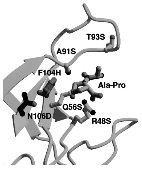
Figure 10.3 (plate 6) Cyclophilin active site residues mutated in cyproase. Those residues highlighted in yellow were separately mutated to serine in the first round of design. Upon selection of A91S as the best protease, the F104H and N106D mutations were added to reproduce the catalytic triad [2]. Reprinted by permission from Macmillan Publishers Ltd.
Removal of Catalytic Nucleophiles
Removal of one reactive group from an active site usually destroys catalytic activity, at least for the main reaction of an enzyme. However, if it does not obliterate substrate binding it can also reveal heretofore blocked secondary reactions, allowing a mechanism previously blocked by nucleophilic attack to proceed. The E54D mutation of glutaconate-CoA-transferase is one such example [12]. The original enzyme transfers CoA from glutaryl-CoA to acetate. The mutant lacks transferase activity because the shorter aspartate carboxyl cannot reach CoA, but does activate a water molecule for hydrolysis. Similarly, removal of the catalytic cysteine in GAPDH by mutation to alanine obliterates phosphorylation, but induces dehydrogenase activity by the addition of water [13]. Finally, removal of the nucleophilic glutamate from the active site of glycosidases prevents hydrolysis but not substrate binding, allowing for the addition of nucleophilic thiosugars (glycoside ligation) [14].
Partitioning of Reaction Intermediates
Enzyme active sites usher reaction intermediates along particular reaction pathways, promoting some and blocking others. Active site redesign can work to alter this partitioning, as in the case of hydrolases and ligases. Both form acyl-enzyme intermediates, with the former transferring the acyl to a water and the latter to a designated acceptor [1]. Mutations favoring one reaction over the other can convert hydrolases to ligases and vice versa. Partitioning redesign can also be as simple as limiting the size of the pocket available to polymerization reactions, such as that catalyzed by farnesyl diphosphate polymerase, in which a single phe to ala mutation alters the length of the major product [15]. See figure 10.4.
Another type of partitioning redesign involves the rescue of enzymes from suicide inhibitors. Organophosphorus compounds such as the nerve agent sarin form tight phosphoester bonds with the catalytic serines of cholinesterases, causing irreversible inhibition. The mutation of an active-site glycine to histidine in human butyrylcholinesterase allows the enzyme to escape this fate by hydrolyzing the bond, either by activating a water molecule for nucleophilic attack on the phosphorus or positioning the inhibitor for such an attack [16].
Controlling Stereo-and Regiochemistry
Rational design can switch the stereo-and regiospecificity of wild-type enzymes by minimal active-site mutations. This can be done by altering the relative orientation of reactive groups and substrates, either by affecting substrate positioning or transferring reactive groups from one side of the active site to another. For example, the Asp170Ser/Thr457Glu double mutant of vanillyl-alcohol oxidase switched its major product from the R to the S form [17], whereas a single mutation in adenylate kinase was sufficient to alter the substrate-binding orientation and subsequently the stereospecificity of product production [18]. Similar alterations to active site architecture alter the regioselectivities of sesquiterpene synthases, in a less well-understood manner, though the effects of the various mutations on product regioselectivity have been characterized [19]. See figure 10.5 (plate 7).
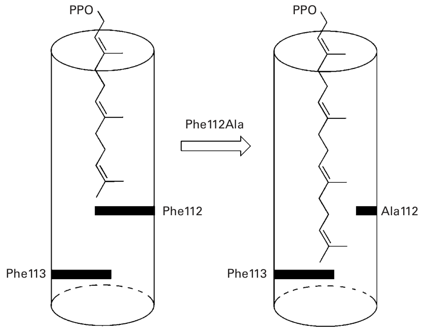
Figure 10.4 Binding pocket size effects the length of polymerization reaction products [1]. Copyright Wiley-VCH Verlag GmbH & Co. KGaA. Reproduced with permission.
Improving Promiscuity
Redesign also proceeds by enhancing the rates of promiscuous activities, often furthered by comparison to homologous enzymes that perform the target’s promiscuous reaction as their major activity (such as in HisF). Original activities in enzymes redesigned to enhance a promiscuous activity are usually more robust than those in enzymes redesigned to perform a wholly novel reaction, losing only a few orders of magnitude of catalytic efficiency for the original reaction while gaining many more for the promiscuous target. Promiscuity enhancement can proceed by directly altering residues responsible for catalysis or substrate binding (depending on the type of promiscuity being designed for), or can be more indirect, altering cofactor specificity or orientation. Altering the bound orientation of flavin in bacterial luciferase changed it from a monooxygenase to an oxidase [20]; a similar mutation converts 1-lactate monooxygenase into an oxidase [21]. Metals are an important class of cofactor, used in many different enzymes, and altering either the identity of the metal binding site or its orientation to the substrate and/or other catalytic groups can have profound effects on catalysis. For example, altering the primary sequence location of a key histidine in the heme group of myoglobin enhances its promiscuous peroxidase activity 200-fold [22].
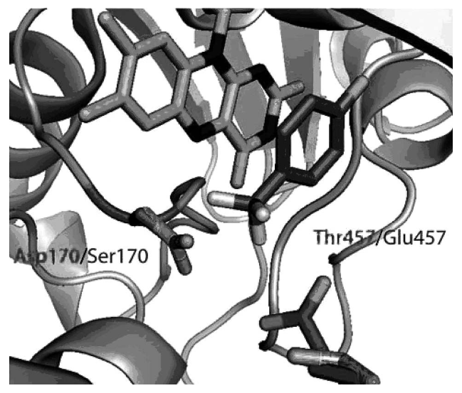
Figure 10.5 (plate 7) Altering the stereospecificity of vanillyl-alcohol oxidase. Here the flavin cofactor is in orange and a substrate analog in green. Note how the mutations shift the catalytic carboxylate from one side of the substrate to the other [1]. Copyright Wiley-VCH Verlag GmbH & Co. KGaA. Reproduced with permission.
Pyridoxal phosphate (PLP) is used as a cofactor in a great variety of reactions, which forge an imine bond between the substrate and PLP that stabilizes carbanion formation on breaking of one of the substrate bonds. Dunathan’s hypothesis proposes the method that allows this variety: The bond to be broken must be oriented perpendicular to the PLP pyridinium ring. Redesign efforts have exploited this relationship, converting alanine racemase into a D-amino acid aldolase and ornithine decarboxylase into a transaminase, both via single mutations [23, 24]. See figure 10.6.
Protein Domain Interface Redesign via Directed Evolution
Nonribosomal peptide synthetases (NRPS) are a class of multidomain enzymes that catalyze the construction of small peptides in a manner reminiscent of a factory assemblyline. The adenylation (A) domain of each module activates its substrate amino acid for addition to the growing polypeptide, and is responsible for the majority of specificity in amino acid incorporation. Genetic evidence suggests that A domain rearrangement has occurred in the evolution of several NRPS, and their modular nature makes domain-swapping between homologous NRPS an attractive potential method for the generation of novel antibiotics. Unfortunately, the resultant chimeric proteins display very low or nonfunctional activities, presumably because of the incompatibility of their constituents’ domain interfaces. Using directed evolution to rescue protein function in chimeric multidomain proteins offers a solution to this problem.
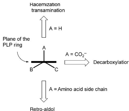
Figure 10.6 Dunathan’s hypothesis on PLP-dependent catalysis [1]. Copyright Wiley-VCH Verlag GmbH & Co. KGaA. Reproduced with permission.
Walsh and colleagues first attempted domain swapping with the serine-specific A domain of E. coli enterobacteria, swapping in a serine-specific domain from P. syringae syringomycin [3]. The initial chimeric protein showed 30-fold less activity than the original. Following mutagenic polymerase chain reaction (PCR) and growth under screening conditions, the fastest growing clone was selected for another round of mutagenesis and screening. Of the three evolved clones, the best showed 8-fold improvement over the original chimera, within 4-fold of wild-type enterobacteria. Later tests focused on the NRPS responsible for the production of the antibiotic andrimid, incorporating an A domain with a new substrate specificity chosen for its promiscuity in the hopes of generating novel antibiotics. Three rounds of mutagenesis and screening later, a mutant was isolated that produced 10.7 more andrimid per unit of culture than the original chimera, only 3-fold less than the wild-type enzyme. These results showed that directed evolution could restore function in chimeric multidomain enzymes. The mutations they incorporated were spread throughout the A domain structure in an unpredictable manner, suggesting the appropriateness of directed evolution for this task as opposed to a more focused rational design approach. It would be interesting to see whether such interfaces could be successfully and reliably redesigned using modern structure-based protein design algorithms, such as those in the dead-end elimination (DEE) family. Such algorithms are introduced in chapter 11. See figure 10.7 (plate 8).
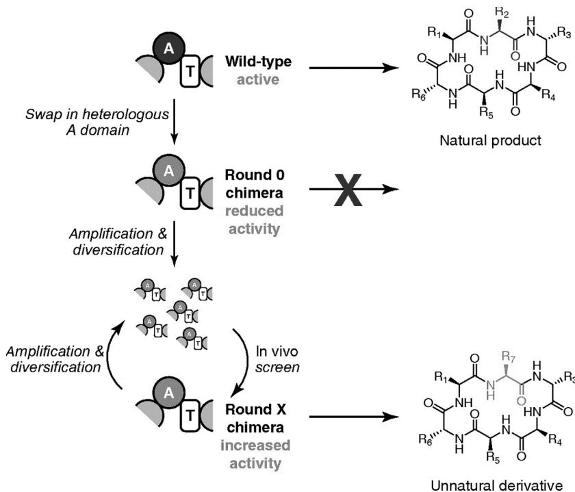
Figure 10.7 (plate 8) Directed evolution in the rescue of chimeric NRPS activity [3]. Copyright 2007 National Academy of Sciences.
References
- [1] M. D. Toscano, K. J. Woycechowsky, and D. Hilvert. Minimalist active-site redesign: Teaching old enzymes new tricks. Angewandte Chemie International Edition 46;18 (2007): 3212–3236.
- [2] E. Quemeneur, M. Moutiez, J.-B. Charbonnier, and A. Menez. Engineering cyclophilin into a proline-specific endopeptidase. Nature 391(1998): 301–304.
- [3] M. A. Fischbach, J. R. Lai, E. D. Roche, C. T. Walsh, and D. R. Liu. Directed evolution can rapidly improve the activity of chimeric assembly-line enzymes. Proceedings of the National Academy of Sciences of the USA 104;29 (2007): 11951–11956.
- [4] M. E. Glasner, J. A. Gerlt, and P. C. Babbitt. Evolution of enzyme superfamilies. Current Opinion in Chemical Biology 10(2006): 492–497.
- [5] L. Polgar, and M. L. Bender A new enzyme containing a synthetically formed active site: Thiol-subtilisin. Journal of the American Chemical Society 1966; 88(13): 3153–3154.
- [6] L. Abrahmsen, J. Tom, J. Burnier, K. A. Butcher, A. Kossiakoff, and J. A. Wells. Engineering subtilisin and its substrates for efficient ligation of peptide bonds in aqueous solution. Biochemistry 30;17 (1991): 4151–4159.
- [7] Z.-P Wu and D. Hilvert. Conversion of a protease into an acyl transferase: Selenolsubtilisin. Journal of the American Chemical Society 111;12 (1989): 4513–4514.
- [8] D. M. Z. Schmidt, E. C. Mundorff, M. Dojka, E. Bermudez, J. E. Ness, S. Govindarajan, et al. Evolutionary potential of alpha/beta-barrels: Functional promiscuity produced by single substitutions in the enolase. Biochemistry 42;28 (2003): 8387–8393.
[9] J. M. Jez, and T. M. Penning. Engineering steroid 5-reductase activity into rat liver 3-hydroxysteroid dehydrogenase. Biochemistry 37;27 (1998): 9695–9703.
[10] C. Juergens, A. Strom, D. Wegener, S. Hettwer, M. Wilmanns, and R. Sterner. Directed evolution of a (beta alpha) 8-barrel enzyme to catalyze related reactions in two different metabolic pathways. Proceedings of the National Academy of Sciences of the USA 97;18 (2000): 9925.
[11] M. J. Wishart, J. M. Denu, J. A. Williams, and J. E. Dixon. A single mutation converts a novel phosphotyrosine binding, domain into a dual-specificity phosphatase. Journal of Biological Chemistry 270;45 (1995): 26782–26785.
[12] M. Mack, and W. Buckel. Conversion of glutaconate CoA-transferase from Acidaminococcus fermentans into an acyl-CoA hydrolase by site-directed mutagenesis. FEBS Letters 405;2 (1997): 209–212.
[13] C. Corbier, F. D. Setta, and G. Branlant. A new chemical mechanism catalyzed by a mutated aldehyde dehydrogenase. Biochemistry 31;49 (1992): 12532–12535.
[14] M. Jahn, H. Chen, J. Müllegger, J. Marles, R. A. J. Warren, and S. G. Withers. Thioglycosynthases: Double mutant glycosidases that serve as scaffolds for thioglycoside synthesis. Chemical Communications (2004): 274–275.
[15] L. C. Tarshis, P. J. Proteau, B. A. Kellogg, J. C. Sacchettini, and C. D. Poulter. Regulation of product chain length by isoprenyl diphosphate synthases. Proceedings of the National Academy of Sciences of the USA 93;26 (1996): 15018–15023.
[16] O. Lockridge, R. M. Blong, P. Masson, M.-T. Froment, C. B. Millard, and C. A. Broomfield. A single amino acid substitution, Gly117His, confers phosphotriesterase (organophosphorus acid anhydride hydrolase) activity on human butyrylcholinesterase. Biochemistry 36;4 (1997): 786–795.
[17] R. H. H. van den Heuvel, M. W. Fraaije, M. Ferrer, A. Mattevi, and W. J. H. van Berkel. Inversion of stereospecificity of vanillyl-alcohol oxidase. Proceedings of the National Academy of Sciences of the USA 97;17 (2000): 9455–9460.
[18] R.-T. Jiang, T. Dahnke, and M.-D. Tsai. Mechanism of adenylate kinase. 10. Reversing phosphorus stereospecificity by site-directed mutagenesis. Journal of the American Chemical Society 113;14 (1991): 5485–5486.
[19] D. B. Little, and R. B. Croteau. Alteration of product formation by directed mutagenesis and truncation of the multiple-product sesquiterpene synthases delta-selinene synthase and gamma-humulene synthase. Archives of Biochemistry and Biophysics 402;1 (2002): 120–125.
[20] L. Xi, K.-W. Cho, M. E. Herndon, and S.-C. Tu. Elicitation of an oxidase activity in bacterial luciferase by site-directed mutation of a noncatalytic residue. Journal of Biological Chemistry 265;8 (1990): 4200–4203.
[21] W. Sun, C. H. Williams, Jr., and V. Massey. Site-directed mutagenesis of glycine 99 to alanine in L-lactate monooxygenase from Mycobacterium smegmatis. Journal of Biological Chemistry 271;29 (1996): 17226–17233.
[22] T. Matsui, S.-I. Ozaki, E. Liong, G. N. Phillips, Jr., and Y. Watanabe. Effects of the location of distal histidine in the reaction of myoglobin with hydrogen peroxide. Journal of Biological Chemistry 274;5 (1999): 2838–2844.
[23] F. P. Seebeck, and D. Hilvert. Conversion of a PLP-dependent racemase into an aldolase by a single active site mutation. Journal of the American Chemical Society 125;34 (2003): 10158–10159.
[24] L. K. Jackson, H. B. Brooks, A. L. Osterman, E. J. Goldsmith, and M. A. Phillips. Altering the Reaction Specificity of Eukaryotic Ornithine Decarboxylase. Biochemistry 39;37 (2000): 11247–11257.