Chapter 3Proteins, Carbohydrates, and Lipids

© Jeff Spoon Stockhouse
Key Concepts
3.1 Macromolecules Characterize Living Things
3.2 Proteins Are Polymers with Highly Variable Structures
3.3 Carbohydrates Are Made from Simple Sugars
3.4 Lipids Are Defined by Their Insolubility in Water
Molecular Superpower Spider silk protein has a structure that makes it one of the strongest fibers in nature.
Investigating Life: Weaving a Web
A spider web is an amazing structure. It is not only beautiful to look at, but an architectural wonder serving as the spider’s home, its mating place, and its way to capture food. Consider a fly that chances to interact with a spider web. The fibers of the web must slow down the fly, but they cannot break, so they need to stretch to dissipate the energy of the fly’s movement. The fibers holding the fly cannot stretch too much; they must be strong enough to hold the web in place. Web fibers are far thinner than a human hair, yet they are very strong. The fibers can also be long; for example, the Darwin’s bark spider makes strands up to 25 meters long.
Chemical investigations reveal that spider silk is made of variations on a single type of large molecule called a protein. Proteins are polymers: long chains of individual smaller units called amino acids. The proteins in spider silks have characteristic structures and amino acid compositions that determine particular functions. Proteins in the stretchy web fibers have amino acids that allow them to curl into spirals, and these spirals can slip along one another to change the fiber’s length. Another kind of spider silk is the dragline silk, which is less stretchy and used to construct the outline of the web, its spokes, and the lifeline of the spider. The proteins in these strong fibers are made up of amino acids that cause the proteins to fold into flat sheets with ratchets, so that parallel sheets can fit together like Lego blocks. This arrangement makes these fibers hard to pull apart.
Just how strong is spider silk? A popular series of films features the fictional character Spider-Man. He often uses his web to ensnare evildoers and protect the innocent. In one scene, he stops a train by using ten ropes made of spider silk, each about 1 centimeter thick. Scientists who study spider silk have estimated the number of silk fibers in each rope and calculated that this scene would indeed be possible. All of this from covalent bonds holding long chains of amino acids together and weaker forces between the chains! Given the amazing strength shown by these proteins, it is not surprising that there is a lot of interest in using spider silk for human activities.
QA What are practical uses for spider silk? (Find the answer on page 65.)
Macromolecules Characterize Living Things
Learning Objectives
3.1.1 Give an example of two molecules that have the same functional groups but different properties, and explain the reason for the differences.
3.1.2 Sketch the chemical structures of two biological monomers, and show how a condensation reaction produces a covalent bond between the two.
3.1.3 Distinguish between structural, cis-trans, and optical isomers.
Four kinds of molecules are characteristic of living things: proteins, carbohydrates, lipids, and nucleic acids. With the exception of the lipids, these biological molecules are polymers (poly, “many,” + mer, “unit”) constructed by the covalent bonding of smaller molecules called monomers. Each kind of biological molecule is made up of monomers with similar chemical structures:
- Proteins are formed from different combinations of 20 amino acids, all of which share chemical similarities.
- Carbohydrates can form giant molecules by linking together chemically similar sugar monomers (monosaccharides) to form polysaccharides.
- Nucleic acids are formed from four kinds of nucleotide monomers linked together in long chains.
- Lipids also form large structures from a limited set of smaller molecules, but in this case noncovalent forces maintain the interactions between the lipid monomers that are held together by covalent bonds.
Polymers containing thousands or more atoms are called macromolecules. The proteins, carbohydrates, and nucleic acids of living systems fall into this category. Although large lipid structures are not polymers in the strictest sense, it is convenient to treat them as macromolecules (see Key Concept 3.4). Green plants have the living world’s most abundant protein (rubisco; see Chapter 10), most abundant carbohydrate (cellulose in plant cell walls), and most abundant lipid (monogalactosyl diglyceride in leaves).
Functional chemical groups determine the structures of macromolecules
Certain small groups of atoms, called functional groups, occur frequently in biological molecules (Figure 3.1). Each functional group has specific chemical properties, and when it is attached to a larger molecule, it confers those properties on the larger molecule. One of these properties is polarity. Looking at the structures in Figure 3.1, can you determine which functional groups are the most polar? (Hint: look for C—O, N—H, and P—O bonds.) The chemical properties of functional groups help us understand the properties of the molecules that contain them.
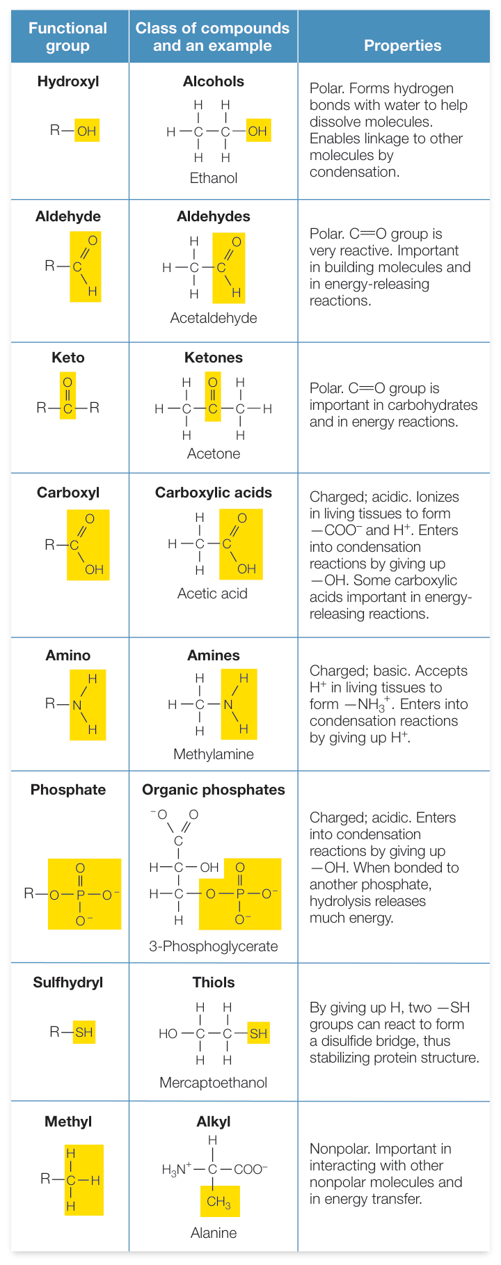
Figure 3.1 Some Functional Groups Important to Living Systems Highlighted here are the eight functional groups most commonly found in biologically important molecules. “R” is a variable chemical grouping. Q: Which functional group(s) are hydrophobic? Why? Activity 3.1 Functional Groups Animation 3.1 Proteins, Carbohydrates, and Lipids


Macromolecules have many different functional groups. A single large protein may contain nonpolar, polar, and charged functional groups, each of which gives different specific properties to local sites on the macromolecule. As we will see, sometimes these different groups interact within the same macromolecule. They help determine the shape of the macromolecule as well as how it interacts with other macromolecules and with smaller molecules.
Using the same atoms, molecules can differ from one another because their functional groups can be arranged differently. Isomers are molecules that have the same (“iso”) chemical formula—the same kinds and numbers of atoms—but with the atoms arranged differently. Of the different kinds of isomers, we will consider three: structural isomers, cis-trans isomers, and optical isomers.
- Structural isomers differ in how their atoms are joined together. Consider two simple molecules, each composed of four carbon and ten hydrogen atoms bonded covalently, both with the formula . These atoms can be linked in two different ways, resulting in different molecules (Figure 3.2A).
- cis-trans isomers typically involve a double bond between two carbon atoms, where the carbons share two pairs of electrons. When the remaining two bonds of each of these carbons are to two different atoms or groups of atoms (e.g., a hydrogen and a methyl group; Figure 3.2B), these can be oriented on the same side or different sides of the double-bonded molecule. If the different atoms or groups of atoms are on the same side, the double bond is called cis; if they are on opposite sides, the bond is trans. These molecules can have very different properties.
- Optical isomers occur when a carbon atom has four different atoms or groups of atoms attached to it. This pattern allows for two different ways of making the attachments, each the mirror image of the other (Figure 3.2C). Such a carbon atom is called an asymmetric carbon, and the two resulting molecules are optical isomers of one another. You can envision your right and left hands as optical isomers. Just as a glove is specific for a particular hand, some biochemical molecules that can interact with one optical isomer of a carbon compound are unable to “fit” the other.
The structures of macromolecules reflect their functions
The four kinds of biological macromolecules are present in roughly the same proportions in all living organisms (Figure 3.3). Also, a particular protein that has a certain three-dimensional structure and function in an apple tree probably has a similar structure and function in a human being, because the protein’s chemistry is the same wherever it is found. This impressive biochemical unity reflects the evolution of all life from a common ancestor, by descent with modification. An important advantage of biochemical unity is that some organisms can acquire needed raw materials by eating other organisms. When you eat bread, the molecules you take in include carbohydrates, lipids, and proteins that can be broken down into monomers which are then polymerized into the varieties of those molecules needed by humans. For example, the storage proteins in wheat grains have a particular composition and sequence of the 20 amino acids. When you eat them in bread, these wheat proteins are broken down into amino acids. In your muscle, these amino acids from wheat are used to make the human proteins actin and myosin, which compose muscle fibers.
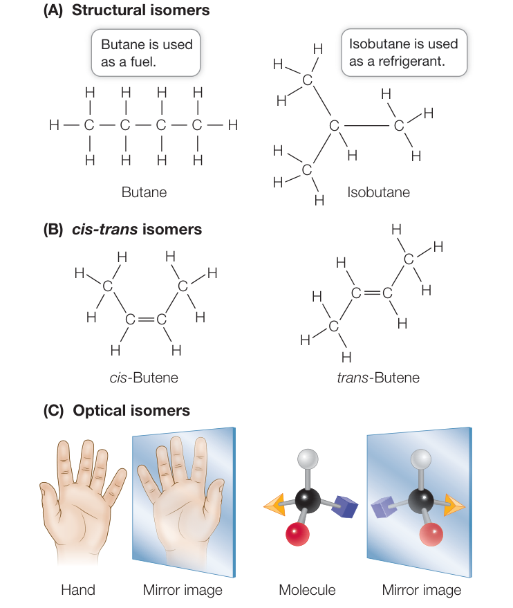
Figure 3.2 Isomers Isomers have the same chemical formula, but the atoms are arranged differently. Pairs of isomers often have different chemical properties and functions. Q: Draw the cis and trans isomers of dichloroethene: .
The sequence and chemical properties of the chain of monomers in a macromolecule determine its three-dimensional shape and function. Some macromolecules fold into compact forms with surface features that make them water-soluble and capable of binding and interacting with other molecules. Some proteins and carbohydrates form long, fibrous structures (such as those found in spider silk or hair) that provide strength and rigidity. The specific structure of a macromolecule determines its function in a given environment, regardless of its origin. For example, composite spider silk fiber made by another organism (e.g., the silkworm, a moth larva) has the same properties as spider silk made by a spider (Investigating Life: Making Spider Silk).
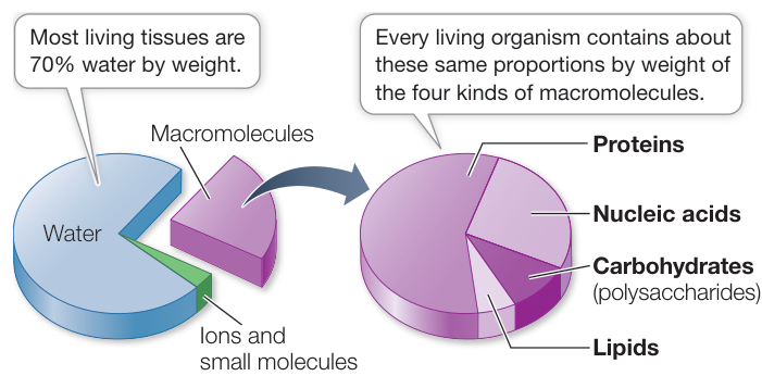
Figure 3.3 Substances Found in Living Tissues The substances shown here make up the non-mineral components of living tissues (bone is an example of a mineral component).
Most macromolecules are formed by condensation and broken down by hydrolysis
Polymers are formed from monomers by a series of condensation reactions (in this case called dehydration reactions; both terms refer to the loss of water). Condensation reactions result in the formation of covalent bonds between monomers. A molecule of water is released with each covalent bond formed (Figure 3.4A). The condensation reactions that produce the different kinds of polymers differ in detail, but in all cases polymers form only if water molecules are removed and energy is added to the system. In living systems, specific energy-rich molecules supply the necessary energy.
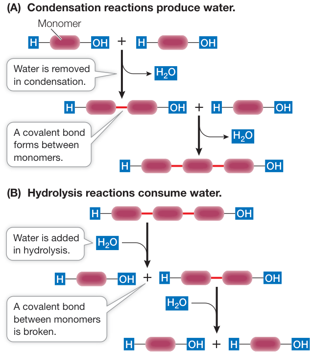
Figure 3.4 Condensation and Hydrolysis of Polymers (A) Condensation reactions link monomers into polymers and produce water. (B) Hydrolysis reactions break polymers into individual monomers and consume water.
The reverse of a condensation reaction is a hydrolysis reaction (hydro, “water,” + lysis, “break”). Hydrolysis reactions result in the breakdown of polymers into their component monomers. Water reacts with the covalent bonds that link the polymer together. For each covalent bond that is broken, a water molecule splits into two ions ( and ), each of which becomes part of one of the products (Figure 3.4B).
Recap and Assess
The four kinds of large molecules that distinguish living tissues are proteins, lipids, carbohydrates, and nucleic acids. Most are polymers: chains of monomers linked by dehydration reactions. Very large polymers are called macromolecules. Biological molecules carry out a variety of functions.
What are the differences between structural, cis-trans, and optical isomers?
Examine the isomers mannose and galactose below. What makes them isomers of each other? Which functional groups do these carbohydrates contain, and what properties do these functional groups give to the molecules?
- Below are the general structures of three monomers in a biological polymer where A, B, and C are numerous atoms. Draw the two reactions that link them together to form a polymer ABC. H—A—OH H—B—OH H—C—OH
The four types of macromolecules can be seen as the building blocks of life. We will cover the unique properties of the nucleic acids in Chapter 4. The remainder of this chapter will describe the structures and functions of the proteins, carbohydrates, and lipids.
Proteins Are Polymers with Highly Variable Structures
Learning Objectives
- 3.2.1 Describe how amino acids differ in their side chains.
- 3.2.2 Describe how amino acids are joined to form a peptide.
- 3.2.3 Distinguish primary, secondary, tertiary, and quaternary structure and predict how a protein’s structure will be affected by a change in an amino acid in its primary structure.
- 3.2.4 Predict the effects of various environmental factors on a protein’s structure.
Proteins have many roles. You’ll learn about the many functions of proteins (Table 3.1) in virtually every chapter of this book. Proteins
Investigating Life: Making Spider Silk
Experiment
Original Paper: F. Teule et al. 2012. Silkworms transformed with chimeric silkworm/spider silk genes spin composite silk fibers with improved mechanical properties. Proc Natl Acad Sci USA 109: 923–928.
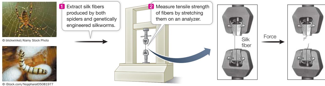
Spider silk is one of the strongest materials known, but it is very hard to harvest in quantity. Industrial biologists are therefore using genetic engineering to produce the protein fibers. The scientists have coaxed silkworms, insect larvae that make silk fibers used in clothing, to make a much stronger composite of silkworm and spider silk.
HYPOTHESIS Genetically engineered silkworms can produce silk with physical properties like those of silk made by spiders.
METHOD
Work with the Data
Spider silk is a protein-based material that is very strong because of its secondary structure. Because of its potential uses by humans, it is desirable to obtain large amounts of these fibers. Spiders spin out the silk fibers for their web, but the amounts are insufficient for human use. Silkworms make abundant silk fibers that are used in textiles but are much weaker than spider silk. An international team of scientists led by Randy Lewis at the University of Wyoming genetically engineered silkworms to make composite silkworm-spider silk fibers in large quantities. They then tested the properties of these fibers and compared them with those of native spider silk.
QUESTIONS
To evaluate the properties of the protein fibers, researchers stretched them and measured the forces involved as well as the resulting lengthening of the fibers. You can liken this to stretching a rubber band until it breaks. Stress is the force required to break the fiber; it is measured in millipascals (mPa; 1 Pa is the force in newtons per unit of area in m2). Strain is a measure of how much the fiber lengthens (as a percentage of its original length). Table A compares the results from silk from spiders and from genetically engineered silkworms. Did the genetically engineered silkworms make fibers that were similar to native spider silk?
Why was fiber thickness (diameter) reported in the data?
What statistical test would you perform to determine if the measured properties were significantly different (see Appendix B)?
The scientists measured the properties of composite silkworm-spider silk compared with those of Kevlar (a synthetic fiber with many uses, including in bicycle tires, racing sails, and body armor) and steel. The results are shown in Table B. What can you conclude about the composite silkworm-spider silk?
Table A
| Silk source | Diameter (µm) | Max. strain (%) |
|---|---|---|
| Silkworms | 21.8 ± 1.6 | 22.0 ± 5.8 |
| Silkworms, genetically engineered | 21.1 ± 1.4 | 31.8 ± 5.2 |
| Spiders | 8.1 ± 0.4 | 19.7 ± 4.8 |
Table B
| Material | Energy required to break filament (joules/kg) | Weight (g/mm 3 ) |
|---|---|---|
| Composite silkworm-spider silk | 120,000 | 1.3 |
| Kevlar | 40,000 | 1.4 |
| Steel | 3,500 | 7.84 |
Source: https://www.kraiglabs.com/spider-silk/
Table 3.1 Proteins and Their Functions
| Category | Function |
|---|---|
| Enzymes | Catalyze (speed up) biochemical reactions |
| Structural proteins | Provide physical stability and movement |
| Defensive proteins | Recognize and respond to nonself substances (e.g., antibodies) |
| Signaling proteins | Control physiological processes (e.g., hormones) |
| Receptor proteins | Receive and respond to chemical signals |
| Membrane transporters | Regulate passage of substances across cellular membranes |
| Storage proteins | Store amino acids for later use |
| Transport proteins | Bind and carry substances within the organism |
| Gene regulatory proteins | Determine the rate of expression of a gene |
| Motor proteins | Cause movement of structures in the cell |
are polymers made up of 20 amino acids in different proportions and sequences. Proteins range in size, from small ones such as the human hormone insulin, which has 51 amino acids and a molecular weight of 5,733, to huge molecules such as the muscle protein titin, with 24,000-36,000 amino acids and a molecular weight of 2,993,451. Proteins consist of one or more polypeptide chains—unbranched (linear) polymers of covalently bonded amino acids. Variation in the sequences of amino acids in the polypeptide chains allows for the vast diversity in protein structure and function. Each chain folds into a particular three-dimensional shape that is specified by the sequence of amino acids present in the chain.
Monomers of proteins link together to make the macromolecule
Each amino acid has both a carboxyl functional group and an amino functional group (see Figure 3.1) attached to the same carbon atom, called the (alpha) carbon. Also attached to the carbon atom are a hydrogen atom and a side chain, or R group, designated by the letter R (Figure 3.5).
The carbon in an amino acid has four available electrons for covalent bonding. It is asymmetrical because it is bonded to four atoms or groups of atoms. Therefore amino acids can exist as optical isomers (see Figure 3.2) called D-amino acids and L-amino acids. D and L are abbreviations of the Latin terms for right (dextro) and left (levo). Only L-amino acids (with the configuration shown in Figure 3.5) are found in the proteins of most organisms, and their presence is an important chemical “signature” of life.
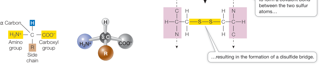
Figure 3.5 An Amino Acid The structure of glycine, an amino acid that has an R group (side chain) of . Note that at the pH in living tissues, both the amino group and carboxyl group are ionized. Q: At a low pH, what will be the ionization status of the amino and carboxyl groups? Figure 3.6 A Disulfide Bridge Two cysteine molecules in a polypeptide chain can form a disulfide bridge ( ) by oxidation (removal of H atoms). Q: Why is it that all cysteines in a protein are not involved in forming disulfide bridges?
At the pH levels typically found in cells (usually pH 7.0–7.4), both the carboxyl and amino groups of amino acids are ionized: the carboxyl group has lost a hydrogen ion:
and the amino group has gained a hydrogen ion:
Thus amino acids show both acid and basic properties.

Activity 3.2 Features of Amino Acids
Connect the Concepts You can review the chemistry and properties of acids and bases in Key Concept 2.4.
The side chains (or R groups) of amino acids contain functional groups that are important in determining the three-dimensional structure and thus the function of the protein. As Table 3.2 shows, the 20 amino acids found in living organisms are grouped and distinguished by their side chains:
- Five amino acids have electrically charged (ionized, +1 or -1) side chains at pH levels typical of living cells. These side chains attract water (are hydrophilic) and attract oppositely charged ions of all sorts.
Table 3.2 The Twenty Amino Acids
(A) Amino acids with electrically charged hydrophilic side chains
(B) Amino acids with polar but uncharged side chains (hydrophilic)
(C) Special cases
(D) Amino acids with nonpolar hydrophobic side chains
• Five amino acids have polar side chains ( and ). They tend to form hydrogen bonds with water and other polar substances. They are also hydrophilic.
• Seven amino acids have side chains that are nonpolar hydrocarbons and are thus hydrophobic. In the watery environment of the cell, these hydrophobic groups may cluster together in the interior of the protein.
Three amino acids—cysteine, glycine, and proline—are special cases, although the side chains of the latter two are generally hydrophobic.
• The cysteine side chain, which has a terminal —SH group, can react with another cysteine side chain in an oxidation reaction to form a covalent bond (Figure 3.6). Such a bond, called a
disulfide bridge or disulfide bond (—S—S—), helps determine how a polypeptide chain folds. Note that most cysteines in a protein are not involved in disulfide bridges.
• The glycine side chain consists of a single hydrogen atom. It is small enough to fit into tight corners in the interiors of protein molecules where larger side chains could not fit. The small side chain allows the protein to be more flexible at the locations where glycine occurs.
• Proline possesses a modified amino group that lacks a hydrogen atom and instead forms a covalent bond with the hydrocarbon side chain, resulting in a ring structure. This limits both its hydrogen-bonding ability and its ability to rotate around the carbon. So proline is often found where a protein bends or loops.
Peptide bonds form the backbone of a protein
Amino acids can form short polymers of 20 or fewer amino acids called oligopeptides or simply peptides. These include some hormones and other molecules involves in signaling between parts of an organism. Even with their short chains, peptides have distinctive three-dimensional structures. More common are the longer polymers called polypeptides, each with a unique sequence of amino acids.
Linking amino acids involves a reaction between carboxyl and amino groups attached to the carbon. The amino group of an adding amino acid interacts in a condensation reaction with the carboxyl group of the last amino acid in the growing chain, forming a peptide bond. Figure 3.7 depicts this reaction.
Just as a sentence begins with a capital letter and ends with a period, polypeptide chains have a beginning and an end. The “capital letter” marking the beginning of a polypeptide is the amino group of the first amino acid added to the chain and is known as the N terminus. The “period” is the carboxyl group of the last amino acid added; this is the C terminus.
Two characteristics of the peptide bond are especially important in the three-dimensional structures of proteins:
- In the C—N bond, the adjacent carbons () are not free to rotate fully, which limits the folding of the polypeptide chain.
- The oxygen bound to the carbon () in the carboxyl group carries a slight negative charge (), whereas the hydrogen bound to the nitrogen () in the amino group is slightly positive (). This asymmetry of charge favors hydrogen bonding within the protein molecule itself and between molecules. These bonds contribute to the structures and functions of many proteins.
In addition to these characteristics of the peptide bond, the particular sequence of amino acids—with their various R groups—in the polypeptide chain also plays a vital role in determining a protein’s structure (see Figure 3.8A) and function.
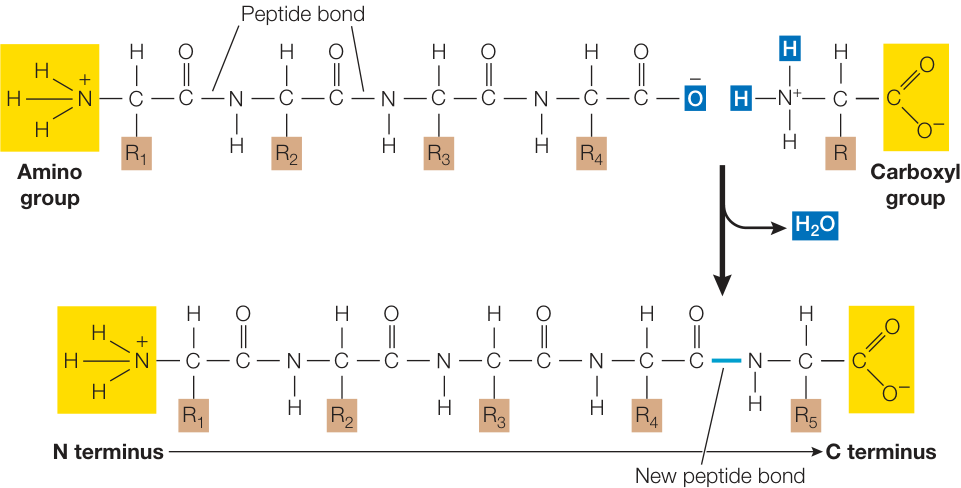
Figure 3.7 Peptide Bond Formation Peptide bonds are formed by condensation reactions in which the amino group of one amino acid reacts with the carboxyl group of another amino acid, releasing a molecule of water.
The primary structure of a protein is its amino acid sequence
The precise sequence of amino acids in a polypeptide chain held together by peptide bonds constitutes the primary structure of a protein (Figure 3.8A). The single-letter abbreviations for amino acids (see Table 3.2) are used to record the amino acid sequence of a protein. Here, for example, are the first 20 amino acids (out of a total of 124) in the protein ribonuclease from a cow:
KETAAAKFERQHMDSSTSAA
The theoretical number of different proteins is enormous. Since there are 20 different amino acids, there could be distinct dipeptides (two linked amino acids) and different tripeptides (three linked amino acids). Imagine this process of multiplying by 20 extended to a protein made up of 100 amino acids (which would be considered a small protein). There could be (that’s approximately ) such small proteins, each with its own distinctive primary structure. How large is the number ? Physicists tell us there aren’t that many electrons in the entire universe!
The sequence of amino acids in the polypeptide chain determines its final shape. The properties associated with each functional group in the side chains of the amino acids (see Table 3.2) determine how the protein can twist and fold, thus adopting a specific stable structure that distinguishes it from every other protein. In contrast to primary structure, which is determined by covalent bonds, most of the forces involved with folding are noncovalent, such as hydrogen bonds and ionic bonds.
The secondary structure of a protein requires hydrogen bonding
A protein’s secondary structure consists of regular, repeated spatial patterns in different regions of a polypeptide chain. There are two basic types of secondary structure, both determined by hydrogen bonding between the amino acids that make up the primary structure: the helix and the pleated sheet (Figure 3.8B).
THE ALPHA HELIX The (alpha) helix is a right-handed coil that turns in the same clockwise direction as a standard wood screw (see Figure 3.8B and Figure 3.9). The R groups extend outward from the peptide backbone of the helix. The coiling results from hydrogen bonds that form between the hydrogen of the N—H of one amino acid and the oxygen of the C=O of another. When this pattern of hydrogen bonding is established repeatedly over a segment of the protein, it stabilizes the coil.
THE BETA PLEATED SHEET A (beta) pleated sheet is formed from two or more polypeptide chains that are almost completely extended and aligned side by side. The sheet is stabilized by hydrogen bonds
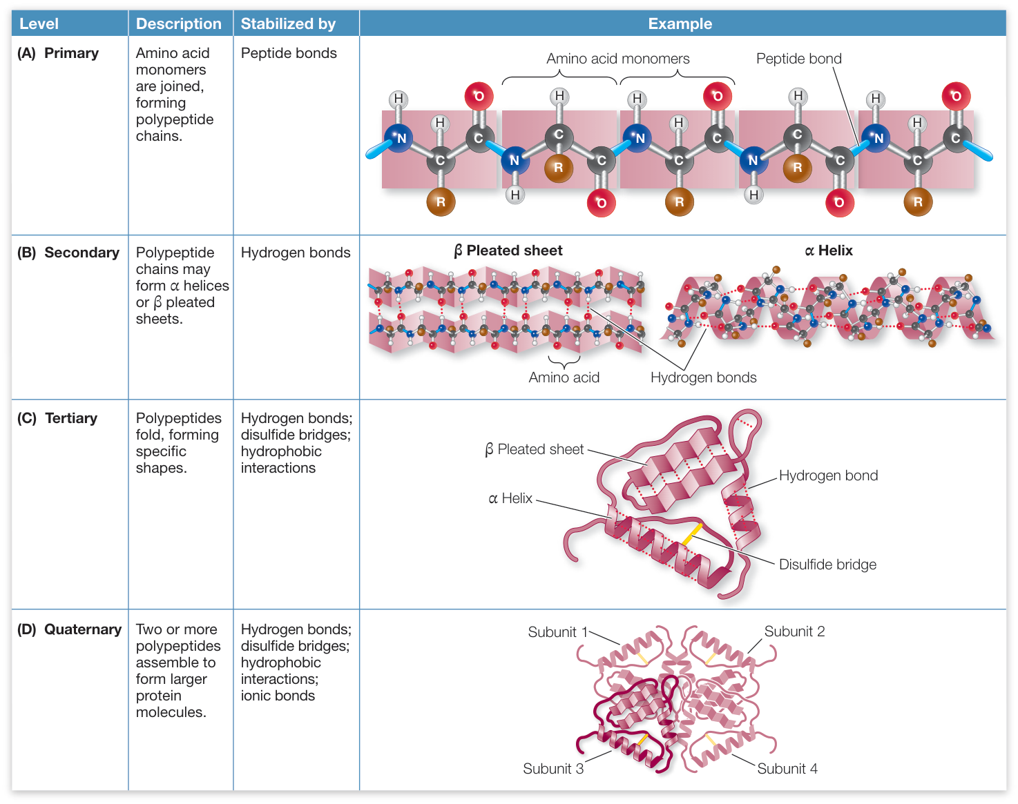
Figure 3.8 The Four Levels of Protein Structure Secondary, tertiary, and quaternary structure all arise from the primary structure of the protein.
Q: If a protein is gently heated to break hydrogen bonds, what level(s) of structure will be unaffected, and why?
between the N—H groups on one chain and the C=O groups on the other (see Figure 3.8B). A pleated sheet may form between separate polypeptide chains or between different regions of a single polypeptide chain that is bent back on itself. The ratcheted, stacked sheets in dragline spider silks (described at the beginning of the chapter and in Investigating Life: Making Spider Silk) are made up of pleated sheets. Many proteins contain regions of both helix and pleated sheet in the same polypeptide chain.
The tertiary structure of a protein is formed by bending and folding
In many proteins, the polypeptide chain is bent at specific sites and then folded back and forth, resulting in the tertiary structure of the protein (Figure 3.8C). Although helices and pleated sheets contribute to the tertiary structure, usually only portions of the macromolecule have these secondary structures, and large regions consist of tertiary structure unique to a particular protein. For example, the proteins found in stretchy spider silks (see the opening story) have repeated amino acid sequences that cause the proteins to fold into spirals. Tertiary structure is a macromolecule’s definitive three-dimensional shape, often including a buried interior as well as a surface that is exposed to the environment.
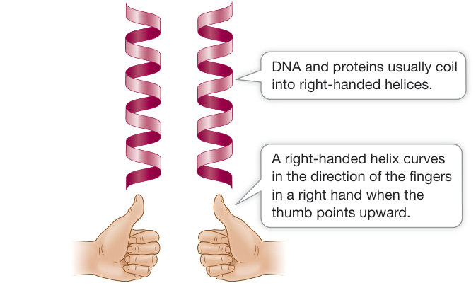
Figure 3.9 Left- and Right-Handed Helices A protein will often have one or more right-handed helices as part of its secondary structure.
The protein’s exposed outer surfaces present functional groups capable of interacting with other molecules in the cell. These molecules might be other macromolecules, including proteins, nucleic acids, carbohydrates, and lipid structures, or smaller chemical substances.
You saw that hydrogen bonding between the N—H and C=O groups within and between chains is responsible for secondary structure. For tertiary structure, the interactions between R groups—the amino acid side chains—and between R groups and the environment are key. Recall the various strong and weak interactions between atoms described in Key Concept 2.2. Here is how these interactions are involved in determining and maintaining tertiary structure:
- Covalent disulfide bridges can form between specific cysteine side chains (see Figure 3.6), holding a folded polypeptide in place.
- Hydrogen bonds between side chains also stabilize folds in proteins.
- Hydrophobic side chains can aggregate together in the interior of the protein, away from water, folding the polypeptide in the process. Close interactions between the hydrophobic side chains are stabilized by van der Waals forces.
- Ionic bonds can form between positively and negatively charged side chains, forming salt bridges between amino acids. Salt bridges can be near the surfaces of polypeptides or buried deep within a protein, away from water. These interactions occur between positively and negatively charged amino acids, for example glutamic acid (which has a negatively charged R group) and arginine (which is positively charged) (see Table 3.2):
A protein folds into its final shape in a way that maximizes all the interactions noted and minimizes inappropriate interactions, such as two positively charged residues (a term identifying monomers in a polymer) being near one another, or a hydrophobic residue being near water. A complete description of a protein’s tertiary structure would specify the location of every atom in the molecule in three-dimensional space relative to all the other atoms. Figure 3.10 shows three ways of modeling the structure of the protein lysozyme. Each way has its uses. The space-filling model might be used to study how other molecules interact with specific sites and R groups on the protein’s surface. The stick model emphasizes the sites where bends occur, resulting in folds in the polypeptide chain. The ribbon model, perhaps the most widely used, shows the different types of secondary structure and how they fold into the tertiary structure.
If a protein is heated slowly and moderately, the heat energy will disrupt only the weak interactions, causing the secondary and tertiary structure to break down. A protein whose structure is disrupted is said to be denatured. A comparison of native (untreated) and denatured proteins shows major differences:
- Native proteins are compact; denatured proteins have a larger volume.
- Native proteins exist in one, preferred shape; denatured proteins can take many shapes.
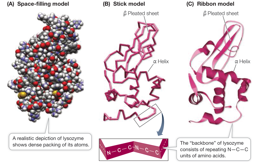
Figure 3.10 Three Representations of Lysozyme Different molecular representations of a protein emphasize different aspects of its tertiary structure: surface features, sites of bends and folds, or sites where alpha or beta structures predominate. These three representations of lysozyme are similarly oriented. (Data from PDB 1IVM. T. Obita et al. 2003. Cell Mol Life Sci 60: 176–184.)
(A) Space-filling model
- Native proteins have hydrogen bonds that stabilize the structure internally; denatured proteins have hydrogen bonds on the exterior, to water.
You can’t “unboil” an egg after it has been hard-boiled; the egg proteins are irreversibly denatured. Amazingly, in some cases a protein can return to its normal tertiary structure when it cools, demonstrating that all the information needed to specify the unique shape of a protein is contained in its primary structure. This was first shown (using chemicals instead of heat to denature the protein) by biochemist Christian Anfinsen for the protein ribonuclease (Figure 3.11).
If a protein has changes in amino acid sequence such that regions that ordinarily are on the inside of the protein (hydrophobic) are now on the outside, the protein may adopt a flat, beta configuration instead of a three-dimensional tertiary structure. These flat sheets act like fibrous proteins and aggregate, forming amyloids. Amyloids are characteristic of a rare brain disease called spongiform encephalopathy (or “mad cow disease,” since amyloids made from fragments of a protein called a prion can enter humans from cows). More commonly, amyloids of a different protein are a hallmark of the brains of people with Alzheimer’s disease.
The quaternary structure of a protein consists of subunits
Many functional proteins contain two or more polypeptide chains, called subunits, each of them folded into its own unique tertiary structure. The protein’s quaternary structure results from the ways in which these subunits bind together and interact (Figure 3.8D).
Hemoglobin has four polypeptide chains interacting to form quaternary structure (Figure 3.12). Hydrophobic interactions, van der Waals forces, hydrogen bonds, and ionic bonds all help hold the four subunits together to form a hemoglobin molecule. However, the weak nature of these forces permits small changes in the quaternary structure to aid the protein’s function—which is to carry oxygen in red blood cells. As hemoglobin binds one molecule, the four subunits shift their relative positions slightly, changing the quaternary structure. Ionic bonds are broken, exposing buried side chains that enhance the binding of additional molecules. The quaternary structure changes back when hemoglobin releases its molecules to the cells of the body.
Shape and surface chemistry contribute to protein function
The shapes of proteins allow specific sites on their exposed surfaces to bind noncovalently to other molecules, which may be large or small. The binding is usually very specific because only certain compatible chemical groups will bind to one another. The specificity of protein binding depends on two general properties of the protein: its shape, and the chemistry of its exposed surface groups.
- Shape. When a small molecule collides with and binds to a much larger protein, it is like a baseball being caught by a catcher’s mitt: the mitt has a shape that binds to the ball and fits around it. Just as a hockey puck or a ping-pong ball does not fit a baseball catcher’s mitt, a given molecule will not bind to a protein unless there is a general “fit” between their three-dimensional shapes.
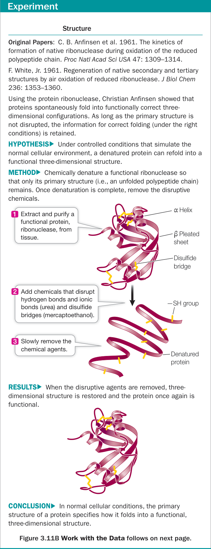
Figure 3.11A Primary Structure Specifies Tertiary Structure
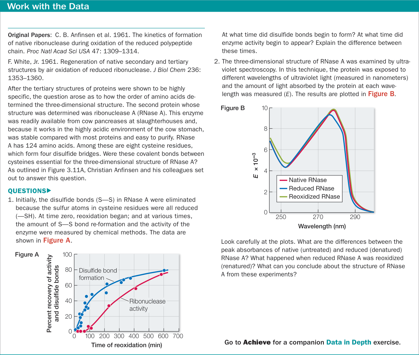
Figure 3.11B Primary Structure Specifies Tertiary Structure
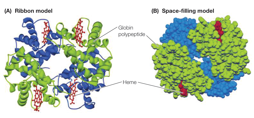
Figure 3.12 Quaternary Structure of a Protein Hemoglobin consists of four folded polypeptide subunits that assemble themselves into the quaternary structure represented by the ribbon model (A) and space-filling model (B). In both representations, each type of subunit is a different color ( subunits are blue and subunits are green). The heme groups (red) contain iron and are the oxygen-carrying sites. (Data from PDB 2HHB. G. Fermi et al. 1984. J Mol Biol 175: 159–174.) Q: What forces hold the subunits of hemoglobin together? (B) Space-filling model
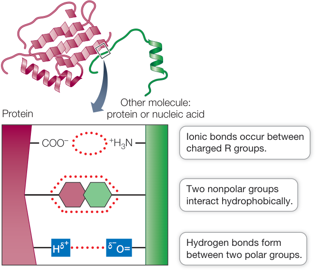
Figure 3.13 Noncovalent Interactions between Proteins and Other Molecules Noncovalent interactions allow a protein (red) to bind tightly to another molecule (green) with specific properties. Noncovalent interactions also allow regions within the same protein to interact with one another.
- Chemistry. The exposed R groups on the surface of a protein permit chemical interactions with other substances (Figure 3.13). Three types of interactions may be involved: ionic, hydrophobic, or hydrogen bonding. Many important functions of proteins involve interactions between surface R groups and other molecules. In later chapters you will learn about proteins called enzymes that bind small molecules and enhance their chemical reactivity, as well as proteins that bind to nucleic acids and participate in the expression and regulation of their activities. In these and many other cases, the interactions between the molecules are noncovalent.
Environmental conditions affect protein structure
Because they are largely determined by weak forces, the three-dimensional structures of proteins are influenced by environmental conditions. Conditions that would not break covalent bonds can disrupt the weaker, noncovalent interactions that determine secondary, tertiary, and quaternary structure. Such alterations may affect a protein’s shape and thus its function. Various conditions can alter the weak, noncovalent interactions:
Increases in temperature cause more rapid molecular movements and thus can break hydrogen bonds and hydrophobic interactions.
Changes in pH can change the ionization pattern of exposed carboxyl and amino groups in the R groups of amino acids, thus disrupting the pattern of ionic bonds and repulsions. For example, as the pH rises, ions are removed from the charged groups of lysine and arginine, making them less charged and less polar; and COOH groups on aspartic and glutamic acids ionize, making them negative and therefore more polar. These differences in polarity change the tertiary structure of the protein, with more polar groups on the outside (interacting with water) and fewer polar groups on the inside (away from water).
High concentrations of polar substances such as urea can disrupt the hydrogen bonding that is crucial to protein structure. Urea was used in the experiment on reversible protein denaturation shown in Figure 3.11.
Nonpolar substances may also disrupt normal protein structure in cases where hydrophobic interactions are essential to maintain the structure.
Protein shapes can change
As we saw in the case of hemoglobin, which undergoes subtle shape changes when it binds oxygen, the shapes of proteins can change as a result of their interactions with other molecules. Proteins can also change shape if they undergo covalent modifications.
- Proteins interact with other molecules. Proteins do not exist in isolation. In fact, if a biochemist “goes fishing” with a particular protein, by attaching the protein to a chemical “hook” and inserting it into cells, the protein will often be attached to something else when it is “reeled in.” These molecular interactions are reminiscent of the interactions that make up quaternary structure (see above). If a polypeptide comes into contact with another molecule, R groups on its surface may form weak interactions (e.g., hydrophobic, van der Waals) with groups on the surface of the other molecule. This may disrupt some of the interactions between R groups within the polypeptide, causing it to undergo a change in shape (Figure 3.14A). You will see many instances of this in the coming chapters.
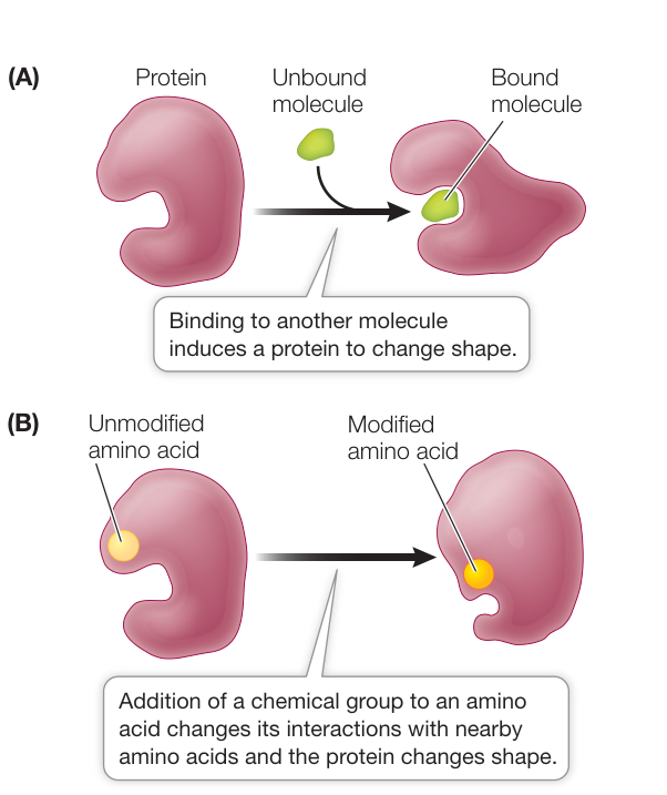
Figure 3.14 Protein Structure Can Change Proteins can change their tertiary structure when they bind to other molecules (A) or are modified chemically (B).
- Proteins undergo covalent modifications. After it is made, the structure of a protein can be modified by the covalent bonding of a chemical group to the side chain of one or more of its amino acids. The chemical modification of just one amino acid can alter the shape and function of a protein. An example is the addition of a charged phosphate group to a relatively nonpolar R group. This can cause the amino acid to become more hydrophilic and to move to the outer surface of the protein, altering the shape of the protein in the region near the amino acid (Figure 3.14B).
Connect the Concepts Covalent modifications of proteins with subsequent changes in shape and function underlie many biological processes, ranging from signaling within the cell (see Key Concept 7.3) to the action of plant hormones on growth (see Key Concept 36.2).
Molecular chaperones help shape proteins
Within a living cell, a polypeptide chain is sometimes in danger of binding the wrong substance. There are two major situations when this can occur:
- Just after a protein is made. When a protein has not yet folded completely, it can present a surface that binds the wrong molecule.
- Following denaturation. Certain conditions, such as moderate heat, can cause some proteins in a living cell to denature without killing the organism. Before the protein can refold, it may present a surface that binds the wrong molecule. In these cases, the inappropriate binding may be irreversible.
Many cells have a special class of proteins, called chaperones, that protect the three-dimensional structures of other proteins. They bind to their partner proteins just as they are being made and also when they become denatured. Like the chaperones at a high school dance, chaperone proteins prevent inappropriate interactions and enhance appropriate ones. Typically, a chaperone protein has a cagelike structure that pulls in a polypeptide, causes it to fold into the correct shape, and then releases it (Figure 3.15).
Tumors make chaperone proteins, possibly to stabilize proteins important in the cancer process, and so chaperone-inhibiting drugs are being designed for use in chemotherapy. In some clinical situations, treatment with these inhibitors results in the inappropriate folding of proteins in tumor cells, causing the tumors to stop growing. As noted above, misfolding of proteins to form amyloid aggregates underlies certain brain diseases, and there is an effort to use the principles of chaperones to induce the proteins to fold correctly and prevent aggregation.
Recap and Assess
Proteins are polymers of amino acids. The sequence of amino acids in a protein is its primary structure. Secondary, tertiary, and quaternary structures arise through noncovalent and covalent interactions between the amino acids. A protein’s three-dimensional shape and exposed chemical groups establish its binding specificity for other substances.
- Suppose that, in a given protein, one lysine is replaced by aspartic acid (see Table 3.2). Does this change occur in the primary structure or in the secondary structure? How might it result in a change in tertiary structure? In quaternary structure?
- A single amino acid change in a protein can change its shape. For
example, the amino acid glycine may normally reside at a certain
position in a protein. If glycine is replaced with either glutamic acid
or arginine, the protein shape near that amino acid changes
significantly. There are two possible explanations for this:
- A small amino acid at that position in the polypeptide is necessary for normal shape.
- An uncharged amino acid is necessary for normal shape.
Further amino acid substitutions are done to distinguish between these possibilities. Replacing glycine with serine or alanine results in normal shape, but replacing glycine with valine changes the shape. Which of the two possible explanations is supported by the observations? Explain your answer.
- Human hair is composed of the protein keratin. At the hair salon, two techniques are used to modify the three-dimensional shape of hair. Styling involves heat, and a perm involves cleaving and re-forming disulfide bonds. How would you investigate these phenomena in terms of protein structure?
The seemingly infinite number of protein configurations made possible by the biochemical properties of the 20 amino acids has driven the evolution of life’s diversity. The linkage configurations of sugar monomers (monosaccharides) determine the structures of the next group of macromolecules, the carbohydrates, which provide energy for life.
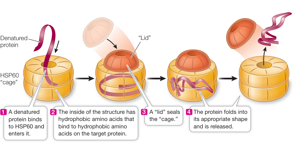
Figure 3.15 Molecular Chaperones Protect Proteins from Inappropriate Binding Chaperone proteins surround new or denatured proteins and prevent them from binding to the wrong substances. Heat shock proteins such as HSP60, shown here, make up one class of chaperone proteins. Q: Why are heat shock proteins important for a cell?
Carbohydrates Are Made from Simple Sugars
Learning Objectives
- 3.3.1 Identify the primary functions of carbohydrates in living organisms.
- 3.3.2 Explain why polysaccharides are useful structures for energy storage.
- 3.3.3 Use the chemical structures of polysaccharides to explain their properties.
Carbohydrates make up a large group of molecules that all have similar atomic compositions but differ greatly in size, chemical properties, and biological functions. Carbohydrates usually have the general formula , ( stands for a number), which makes them appear as “hydrates of carbon” (a hydrate refers to water), hence their name. However, carbohydrates are not really “hydrates” because the water molecules are not intact. Rather, the linked carbon atoms are bonded with hydrogen atoms () and hydroxyl groups (), the components of water.
Carbohydrates have four major biochemical roles:
They are a source of stored energy that can be released in a form usable by organisms.
They are used to transport stored energy within complex organisms.
They serve as carbon skeletons that can be rearranged to form new molecules.
- They form extracellular assemblies such as cell walls that provide structure to organisms.
Some carbohydrates are relatively small, with molecular weights of less than 100. Others are true macromolecules, with molecular weights in the hundreds of thousands.
There are four categories of biologically important carbohydrates, defined by the number of monomers:
- Monosaccharides (mono, “one,” + saccharide, “sugar”), such as glucose, are simple sugars. They are the monomers from which the larger carbohydrates are constructed.
- Disaccharides (di, “two”) consist of two monosaccharides linked together by covalent bonds. The most familiar is sucrose, which is made up of covalently bonded glucose and fructose molecules.
- Oligosaccharides (oligo, “several”) are made up of several (3–20) monosaccharides.
- Polysaccharides (poly, “many”), such as starch, glycogen, and cellulose, are polymers made up of hundreds or thousands of monosaccharides.
Monosaccharides are simple sugars
All living cells contain the monosaccharide glucose; it is the “blood sugar” used to store and transport energy in humans. Cells use glucose as an energy source, breaking it down through a series of reactions that convert stored energy to more usable chemical-bond energy and produce carbon dioxide; this is a cellular form of the combustion reaction you saw in Key Concept 2.3.
Glucose exists in straight chains and in ring forms. The ring forms predominate in virtually all biological circumstances because they are more stable in water. There are two forms of the glucose ring, called - and -glucose, which differ only in the orientation of the and groups attached to carbon 1 (Figure 3.16). The and forms interconvert and exist in equilibrium when dissolved in water.
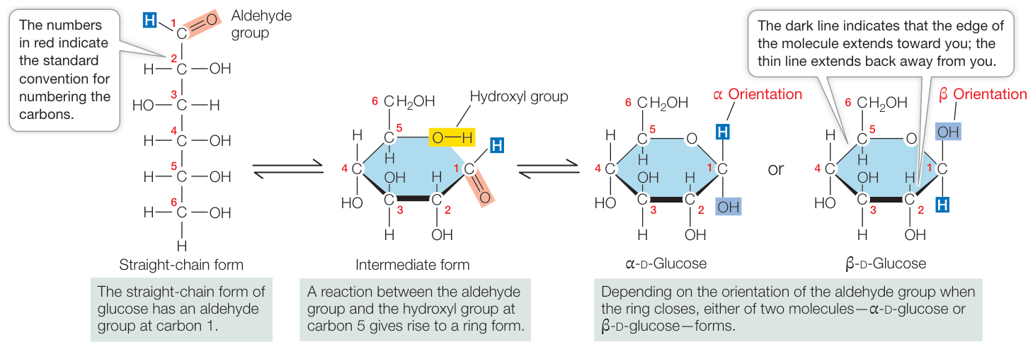
Figure 3.16 From One Form of Glucose to the Other All glucose molecules have the formula , but their structures vary. When dissolved in water, the and “ring” forms of glucose interconvert. The convention used here for numbering the carbon atoms is standard in biochemistry.
Activity 3.3 Forms of Glucose
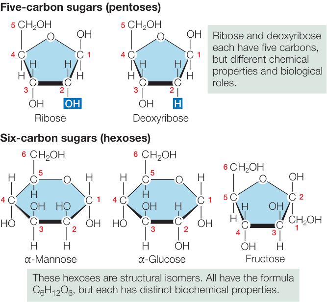
Figure 3.17 Monosaccharides Are Simple Sugars Monosaccharides are made up of varying numbers of carbons. Some hexoses are structural isomers that have the same kind and number of atoms, but the atoms are arranged differently. Fructose, for example, is a hexose but forms a five-membered ring like the pentoses.
Different monosaccharides contain different numbers of carbons. Some monosaccharides are structural isomers, with the same kinds and numbers of atoms but in different arrangements (Figure 3.17). Such seemingly small structural changes can significantly alter their properties. Most of the monosaccharides in living systems belong to the D (right-handed) series of optical isomers.
- Pentoses (pente, “five”) are five-carbon sugars. Two pentoses are of particular biological importance: the backbones of the nucleic acids RNA and DNA contain ribose and deoxyribose, respectively (see Key Concept 4.1). These two pentoses are not isomers of each other; rather, one oxygen atom is missing from carbon 2 in deoxyribose (de-, “absent”). The absence of this oxygen atom is an important distinction between RNA and DNA.
- The hexoses (hex, “six”) shown in Figures 3.16 and 3.17 are a group of structural isomers with the formula . Common hexoses are glucose, fructose (so named because it was first found in fruits), mannose, and galactose.
Glycosidic bonds link monosaccharides
The disaccharides, oligosaccharides, and polysaccharides are all constructed from monosaccharides that are covalently bonded together by condensation reactions that form glycosidic bonds (Figure 3.18). A single glycosidic bond between two monosaccharides forms a disaccharide. For example, sucrose—common table sugar (the real stuff, not artificial sweetener) in the human diet and the major disaccharide in plants—is formed from a glucose and a fructose molecule.
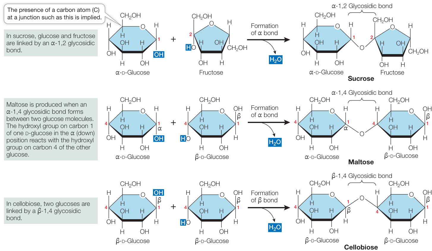
Figure 3.18 Disaccharides Form by Glycosidic Bonds Glycosidic bonds between two monosaccharides can create many different disaccharides. The particular disaccharide formed depends on which monosaccharides are bonded, on the site of bonding (i.e., which carbon atoms are involved), and on the form ( or ) of the bond.
The disaccharides maltose and cellobiose are made from two glucose molecules (see Figure 3.18). Maltose and cellobiose are structural isomers, both having the formula . However, they have different chemical properties and are recognized by different enzymes in biological tissues. For example, maltose can be hydrolyzed into its monosaccharides in the human body, whereas cellobiose cannot.
Connect the Concepts Enzymes are an important class of proteins that may change shape when they come into contact with a reactant in a biochemical reaction. Each enzyme is specific for the reactant it binds. See Key Concept 8.4.
Oligosaccharides contain several monosaccharides bound by glycosidic bonds at various sites. Many oligosaccharides have additional functional groups, which give them special properties.
Oligosaccharides are often covalently bonded to proteins and lipids on the outer cell surface, where they serve as recognition signals. The different human blood groups (e.g., the ABO blood types) get their specificities from oligosaccharide chains.
Polysaccharides store energy and provide structural materials
Polysaccharides are large (sometimes gigantic) polymers of monosaccharides connected by glycosidic bonds (Figure 3.19). In contrast to polypeptides, polysaccharides are not necessarily linear chains of monomers. Each monomer unit has several sites that are capable of forming glycosidic bonds, and thus branched molecules are possible.
STARCH Starches make up a family of large molecules with similar structures. While all starches are polysaccharides of glucose
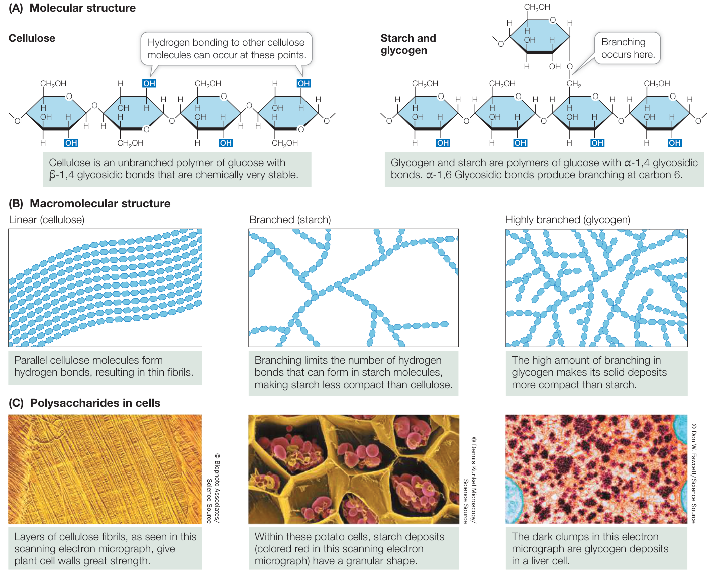
Figure 3.19 Representative Polysaccharides Cellulose, starch, and glycogen have different levels of branching and compaction of the polysaccharides.
Q: When beer is made, initially mashed-up barley seeds are soaked in water and maltose is produced. What kind of reaction is involved in maltose production in this case?
with -glycosidic bonds ( -1,4 and -1,6 glycosidic bonds; see Figure 3.19A), the different starches can be distinguished by the amount of branching that occurs at carbons 1 and 6 (see Figure 3.19B). Starch is the principal energy storage compound of plants. Some plant starches, such as amylose, are unbranched; others are moderately branched (for example, amylopectin). Starch readily binds water; if you’re a cook you know this. However, when water is removed, hydrogen bonds form between the unbranched polysaccharide chains, which then aggregate. Large starch aggregates called starch grains can be observed in the storage tissues of plant seeds (see Figure 3.19C). These aggregates are broken up when starch is heated, breaking the hydrogen bonds. The starch becomes less solid and crystalline and water is absorbed, making the starch even more amorphous. This is what happens in baking with wheat flour, and is what gives bread its texture. Next time you eat some bread, think of hydrogen bonds!
GLYCOGEN Glycogen is a water-insoluble, highly branched polymer of glucose. It is used to store glucose in the liver and muscles and is thus an energy storage compound for animals, as starch is for plants. Both glycogen and starch are readily hydrolyzed into glucose monomers, which in turn can be broken down to liberate their stored energy.
But if it is glucose that is needed for fuel, why store it in the form of glycogen? The reason is that 1,000 glucose molecules would exert 1,000 times the osmotic pressure of a single glycogen molecule, causing water to enter cells where glucose is stored (see Key Concept 6.3). If it were not for polysaccharides, many organisms would expend a lot of energy expelling excess water from their cells.
CELLULOSE As the predominant component of plant cell walls, cellulose is by far the most abundant organic compound on Earth. Like starch and glycogen, cellulose is a polysaccharide of glucose, but its individual monosaccharides are connected by - rather than by -glycosidic bonds. Starch is easily degraded by the actions of chemicals or enzymes. Cellulose, however, is chemically more stable because of its -glycosidic bonds. Thus whereas starch is easily broken down to supply glucose for energy-producing reactions, cellulose is an excellent structural material that can withstand harsh environmental conditions without substantial change.
Chemically modified carbohydrates contain additional functional groups
Some carbohydrates are chemically modified by the addition of functional groups such as phosphate, amino, or -acetyl groups (Figure 3.20). For example, a phosphate group may be added to one or more of the —OH sites (see Figure 3.20A). Some of the resulting sugar phosphates, such as fructose 1,6-bisphosphate, are important intermediates in cellular energy reactions, which you’ll see in Chapter 9.
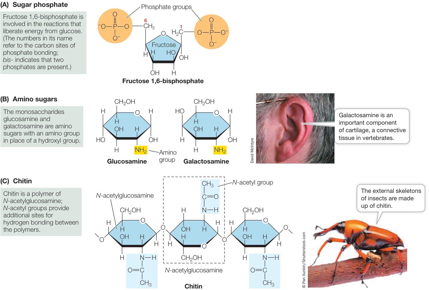
Figure 3.20 Chemically Modified Carbohydrates Added functional groups can modify the form and properties of a carbohydrate.
When an amino group () is substituted for an —OH group, amino sugars, such as glucosamine and galactosamine, are produced (see Figure 3.20B). These compounds are important in the extracellular matrix (see Key Concept 5.4), where they form parts of glycoproteins, which are molecules involved in keeping tissues together. Galactosamine is a major component of cartilage, the material that forms caps on the ends of bones and stiffens the ears and nose. A derivative of glucosamine is present in the polymer chitin (see Figure 3.20C), the principal structural polysaccharide in the external skeletons of insects and many crustaceans (such as crabs and lobsters), and a component of the cell walls of fungi.
Recap and Assess
Carbohydrates are composed of carbon, hydrogen, and oxygen and have the general formula . They provide energy and structure to cells and are precursors of numerous important biological molecules. Monosaccharide monomers can be connected by glycosidic bonds to form disaccharides, oligosaccharides, and polysaccharides.
- What qualities of the polysaccharides starch and glycogen make them useful for energy storage?
- After looking at the cellulose molecule in Figure 3.19A, can you see why a large number of hydrogen bonds are present in the linear structure of cellulose shown in Figure 3.19B? Why is this structure so strong?
You have seen how amino acid monomers form protein polymers and how sugar monomers form the polymers of carbohydrates. Now we will look at the lipids, which are unique among the four classes of large biological molecules in that they are not, strictly speaking, polymers.
Lipids Are Defined by Their Insolubility in Water
Learning Objectives
- 3.4.1 Compare and contrast triglycerides in fats and oils.
- 3.4.2 Describe how the polar and nonpolar natures of phospholipids result in the formation of a membrane bilayer.
- 3.4.3 Explain why carotenoids, steroids, and waxes are classified as lipids.
Lipids—colloquially called fats—are hydrocarbons that are insoluble in water because of their many nonpolar covalent bonds. As you saw in Key Concept 2.2, nonpolar hydrocarbon molecules are hydrophobic and preferentially aggregate together, away from water, which is polar (see Figure 2.13). The huge macromolecular aggregations that can form are not polymers in a strict chemical sense, because the individual lipid molecules are not covalently bonded.
There are several different types of lipids, and they play numerous roles in living organisms:
- Fats and oils store energy.
- Phospholipids play important structural roles in cell membranes.
- Carotenoids and chlorophylls help plants capture light energy.
- Steroids and modified fatty acids play regulatory roles as hormones and vitamins.
- Fat in animal bodies serves as thermal insulation.
- A lipid coating around nerves provides electrical insulation.
- Oil or wax on the surfaces of skin, fur, feathers, and leaves repels water and prevents excessive evaporation of water from terrestrial animals and plants.
Fats and oils are triglycerides
Chemically, fats and oils are triglycerides, also known as simple lipids. Triglycerides that are solid at room temperature (around ) are called fats; those that are liquid at room temperature are called oils. Triglycerides are composed of two types of building blocks: fatty acids and glycerol. Glycerol is a small molecule with three hydroxyl (—OH) groups (thus it is an alcohol). A fatty acid is made up of a long nonpolar hydrocarbon chain and an acidic polar carboxyl group (—COOH). These chains are very hydrophobic because of their abundant C—H and C—C bonds, which have similar electronegativity values and are therefore nonpolar.
Connect the Concepts As discussed in Key Concept 2.2, electronegativity is a measure of the attraction an atomic nucleus exerts on electrons in a covalent bond. When one interacting atom is much more electronegative than the other, a complete transfer of one or more electrons may take place.
A triglyceride contains three fatty acid molecules and one molecule of glycerol. Making a triglyceride involves three condensation (dehydration) reactions. In each reaction, the carboxyl group of a fatty acid bonds with a hydroxyl group of glycerol, resulting in a covalent bond called an ester linkage and the release of a water molecule (Figure 3.21). The three fatty acids in a triglyceride molecule need not all have the same hydrocarbon chain length or structure; some may be saturated fatty acids, whereas others may be unsaturated:
- In saturated fatty acids, all the bonds between the carbon atoms in the hydrocarbon chain are single bonds—there are no double bonds. That is, all the bonds are saturated with hydrogen atoms (Figure 3.22A). These fatty acid molecules are relatively straight, and they pack together tightly, like pencils in a box. They are called saturated because additional hydrogen atoms cannot be added to the carbon atoms in the chain.
- In unsaturated fatty acids, the hydrocarbon chain contains one or more double bonds. Linoleic acid is an example of a polyunsaturated fatty acid that has two double bonds near the middle of the hydrocarbon chain, causing kinks in the molecule (Figure 3.22B). Such kinks prevent the unsaturated fat molecules from packing together tightly. Plants store unsaturated fats, as in corn oil and soybean oil.
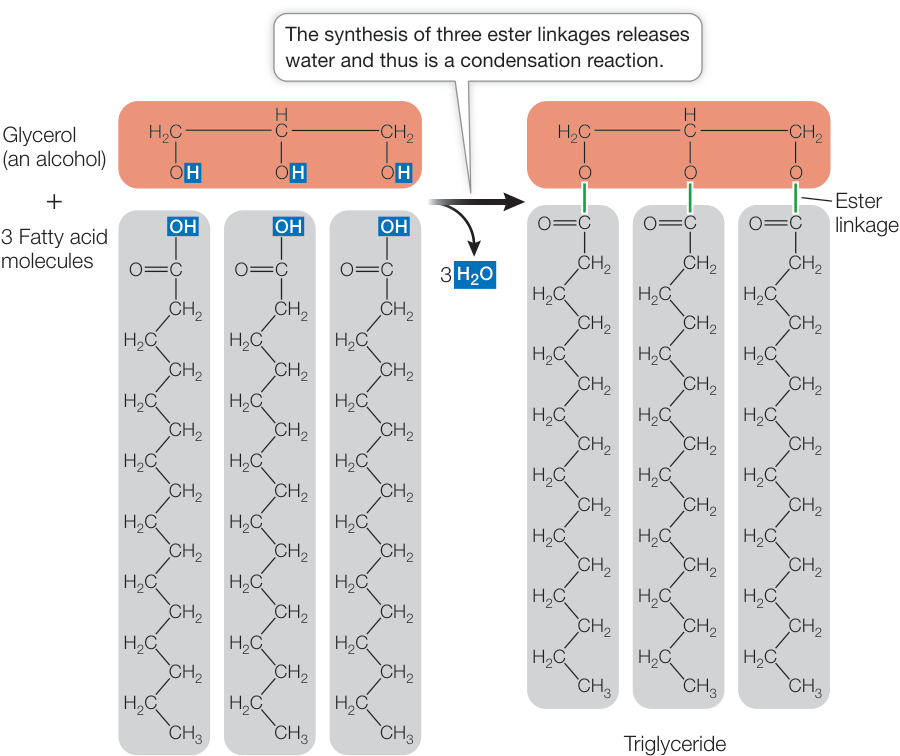
Figure 3.21 Synthesis of a Triglyceride In living things, the reaction that forms a triglyceride is more complex, but the end result is the same as shown here.
The kinks in fatty acid molecules are important in determining the fluidity and melting points of lipids. The triglycerides of animal fats (as in meat) tend to have many long-chain saturated fatty acids packed tightly together; these fats are usually solids at room temperature and have high melting points. The triglycerides of plants, such as corn oil, tend to have short or unsaturated fatty acids. Because of their kinks, these fatty acids pack together poorly and have low melting points, and these triglycerides are usually liquids at room temperature.
The kinks in fatty acids are possible because the H atoms around the double bond are in the same plane of the molecule (cis; see Figures 3.22 and 2.8). However, in some fatty acids the H atoms are on opposite sides of the plane of the molecule (trans). In these fatty acids, there are no kinks and the fat containing them—called trans fat—packs tightly and is more fluid at room temperature. As plant oils became more available, consistent, and cheaper than animal fat, the food industry devised methods to make the oils semisolid by adding hydrogen to saturate the
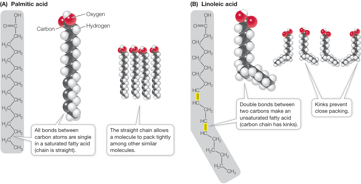
Figure 3.22 Saturated and Unsaturated Fatty Acids (A) The straight hydrocarbon chain of a saturated fatty acid allows the molecule to pack tightly with other, similar molecules. (B) In unsaturated fatty acids, kinks in the chain prevent close packing. The color convention in the models shown here (gray, H; red, O; black, C) is commonly used.
Q: If hydrogen gas () is bubbled over a suspension of unsaturated fatty acids, which will happen to their structure?
double bonds (by bubbling gas). Thus margarine and shortening were developed. A side effect of this industrial process was that some of the cis fatty acid double bonds were converted to the trans configuration. So the foods ended up with some trans fats. As more people consumed trans fats, they had higher levels of low-density lipoproteins (LDL; see Figure 3.23C), which may result in accumulation of lipids on the inner walls of arteries, leading to heart disease and stroke. Food labels now show the content of trans fats, and restaurants have banned them.
Unsaturated fatty acids are identified by the position of the carbon atom of the first double bond, with the first carbon atom—called the omega carbon—being at the opposite end of the chain from the carboxylic acid group. For example, in Figure 3.22B the first bond in linoleic acid is at carbon 6, so it is called an omega-6 fatty acid. Certain other fatty acids have their first bond at position 3 and so are called omega-3 fatty acids. There is good evidence that omega-3 fatty acids protect against heart attacks and stroke, so government agencies in many countries recommend consuming foods containing them.
Fatty acids are excellent storehouses for chemical-bond energy. As you will see in Chapter 9, when the bond is broken, it releases significant energy that an organism can use for its own purposes, such as movement or building up other complex molecules.
Phospholipids form biological membranes
We have mentioned the hydrophobic nature of the many and bonds in fatty acids. But what about the carboxyl functional group at the end of the molecule? When it ionizes and forms , it is strongly hydrophilic. So a fatty acid is a molecule with a hydrophilic end and a long hydrophobic “tail.” It has two opposing chemical properties—part hydrophobic and part hydrophilic; the technical term for this is amphipathic.
Like triglycerides, phospholipids contain fatty acids bound to glycerol by ester linkages. In phospholipids, however, any one of several phosphate-containing compounds replaces the first or third fatty acid, giving phospholipids amphipathic properties (Fig. ure 3.23A). The phosphate functional group has a negative electric charge, so this portion of the molecule is hydrophilic, attracting polar water molecules. But the two fatty acids are hydrophobic, so they tend to avoid water and aggregate together or with other hydrophobic substances.
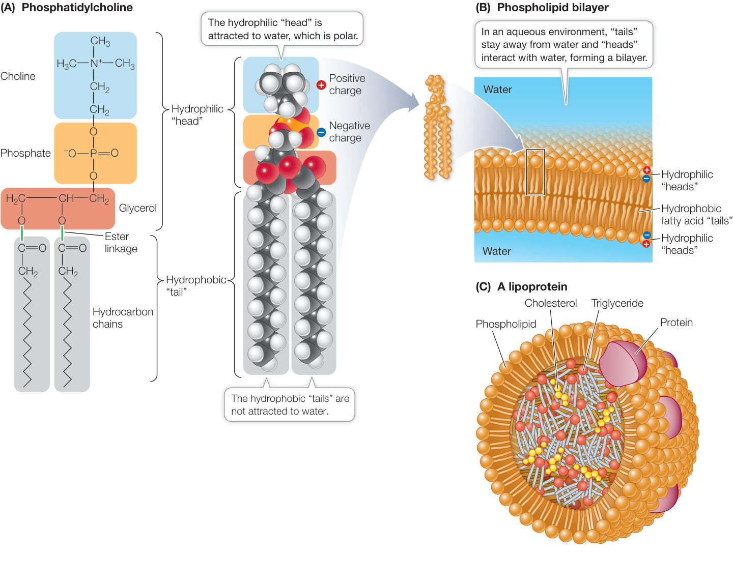
Figure 3.23 Phospholipids (A) Phosphatidylcholine (lecithin) demonstrates the structure of a phospholipid molecule. In other phospholipids, the amino acid serine, the sugar alcohol inositol, or other compounds replace choline. (B) In an aqueous environment, hydrophobic interactions bring the “tails” of phospholipids together in the interior of a bilayer. The hydrophilic “heads” face outward on both sides of the bilayer, where they interact with the surrounding water molecules. (C) Phospholipids, in conjunction with specialized proteins, can form lipoproteins, which transport lipids such as cholesterol primarily in the blood.
In an aqueous environment, phospholipids line up in such a way that the nonpolar, hydrophobic “tails” pack tightly together and the phosphate-containing “heads” face outward, where they interact with water. The phospholipids can form a bilayer: a sheet two molecules thick, with water excluded from the core (Figure 3.23B). Biological membranes have this kind of phospholipid bilayer structure, and we will devote Chapter 6 to their biological functions. In animals, phospholipids form single-layered spherical structures called lipoproteins that have hydrophilic exteriors and hydrophobic interiors (Figure 3.23C). The latter can bind to hydrophobic molecules such as cholesterol (see the next section) and transport it in the blood to various tissues in the body.
Some lipids have roles in energy conversion, regulation, and protection
In the paragraphs above we focused on triglycerides and phospholipids—lipids that are involved in energy storage and cell structure. However, there are other nonpolar and amphipathic lipids that have different structures and roles (Figure 3.24).
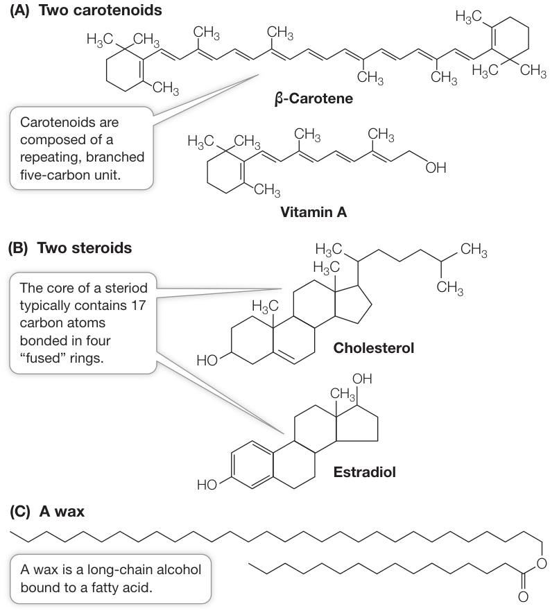
Figure 3.24 More Lipids Because of their insolubility in water, carotenoids (A), steroids (B), and waxes (C) are classified as lipids. Q: Why might it be dangerous for a person to eat meat from animals treated with steroid hormones?
CAROTENOIDS The carotenoids are a family of light-absorbing pigments found in plants and animals. Beta-carotene ( -carotene) is one of the pigments that traps light energy in leaves during photosynthesis. In humans, a molecule of -carotene can be broken down into two vitamin A molecules. Vitamin A is used to make the pigment cis-retinal, which is required for vision. Carotenoids are responsible for the colors of carrots, tomatoes, pumpkins, egg yolks, and butter. The brilliant yellows and oranges of autumn leaves are also from carotenoids.
STEROIDS The steroids are a family of organic compounds whose multiple rings are linked through shared carbons. The steroid cholesterol is an important constituent of membranes, helping maintain membrane integrity (see Key Concept 6.1). Other steroids function as hormones: chemical signals that carry messages from one part of the body to another (see Chapter 40). Cholesterol is synthesized in the liver and is the starting material for making steroid hormones such as testosterone and estrogen.
WAXES Birds and mammals have glands in their skins that secrete waxy coatings onto their feathers or hair. These coatings repel water and help keep the hair and feathers pliable. The shiny leaves of plants such as holly, familiar during winter holidays, also have waxy coatings. Waxy coatings on plants can help them retain water and exclude pathogens. Bees make their honeycombs out of wax. Waxes are substances that are hydrophobic and plastic, or malleable, at room temperature. Each wax molecule consists of a saturated, long-chain fatty acid and a saturated, long-chain alcohol joined by an ester linkage. The result is a very long molecule with 40–60 CH2 groups.
Recap and Assess
Lipids include both hydrophobic and amphipathic molecules that are largely composed of carbon and hydrogen. They are important in energy storage, light absorption, intercellular communication, and biological structures. A phospholipid is composed of two hydrophobic fatty acids linked to glycerol and a hydrophilic phosphate group. Cell membranes contain phospholipid bilayers.
- If phospholipids were placed in a nonpolar solvent, would a membrane-type bilayer structure form? Explain your answer, using a diagram.
- What is the difference between fats and oils?
- Why are steroids classified as lipids?
In this chapter we discussed three of the classes of macromolecules that are characteristic of living organisms. All organisms are made up of these three types of macromolecules, a fact that shows the wonderful biochemical unity of life. This unity in turns implies that all life has a common origin (see Key Concept 1.1). Essential to this origin was a fourth class of macromolecule, the nucleic acids. In the next chapter we will turn to the related topics of nucleic acids and the origin of life.

Simulation 3.1 Macromolecule Building
Investigating Life: QA What are practical uses for spider silk?
Spider silk protein has many glycine and alanine residues, which are hydrophobic and cause the protein to fold into a fibrous shape. More than 1,000 individual polypeptide chains hydrogen-bond and hydrophobically interact with one another to form long silk fibers. The strength—the ability of silk to withstand forces tending to make it longer—and toughness—the ability of silk to resist breakage—are comparable to steel, and this has prompted engineers to propose human uses for spider silk. Using genetic engineering, biologists have been able to harvest composite silk fibers produced by silkworms in industrial quantities. Now that this composite silkworm—spider silk is available, numerous applications (functions related to structure) can be contemplated. For example, a recent study compared surgical sutures made from spider silk with widely used sutures made from synthetic fibers to hold together injured tendons.
The spider silk sutures were far superior, holding their strength over many cycles of tendon movement during healing. Another application on the horizon is bulletproof vests: the energy required to break a spider silk filament is at least 100 times greater than the energy of a typical bullet in impact.
Future directions
Perhaps the most widespread use for spider silk may be in textiles. By definition, textiles are flexible materials made of natural or artificial fibers (threads) that are woven together. The clothes you are wearing are textiles, perhaps made of cotton fibers (made up of cellulose) or polyester fibers (an artificial polymer) or a combination. Of course, silk fibers from silkworms have a long history in textiles, particularly in Asia. The addition of spider silk fibers, or combined spider-silkworm silk to existing textiles, holds great promise to increase the strength and in some cases the flexibility of textiles.
© AF archive/Alamy Stock Photo
You should be able to relate each summary to the adjacent figures. If you go to this visual summary in Achieve, you can follow links to figures, animations, activities, and simulations that will help you consolidate the material.
Visual Summary
Macromolecules Characterize Living Things
- ■ Functional groups have particular chemical properties that they confer on any larger molecule of which they are a part.
- ■ Macromolecules are polymers constructed by the formation of covalent bonds between smaller molecules called monomers.
Questions
- One of the most common chemical reactions in cells is phosphorylation, whereby a phosphate functional group is covalently added to an existing molecule. If the molecule is nonpolar, how will the addition of phosphate alter the molecule’s properties?
- Could hydrolysis reactions to break down a polymer occur in a nonpolar environment?
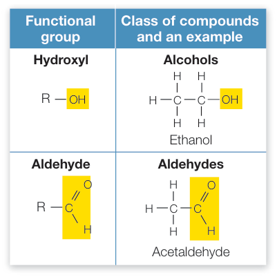
Figure 3.1
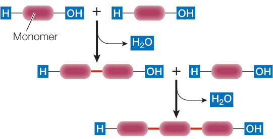
Figure 3.4
Proteins Are Polymers with Highly Variable Structures
- ■ Peptide bonds covalently link amino acids into poly-peptide chains. These bonds form by condensation reactions between the carboxyl and amino groups.
- ■ Proteins have four levels of structure that result in a three-dimensional macromolecule.
- ■ The specific shape and structure of a protein allow it to bind noncovalently to other molecules.

Figure 3.7
Questions
- Is the tertiary structure of a protein determined by covalent bonding, noncovalent interactions, or both? Give examples, using specific amino acids.
- Polypeptide chains are said to “grow in the N to C direction”. Explain.
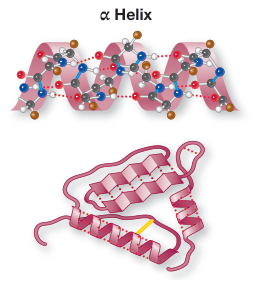
Figure 3.8
Carbohydrates Are Made from Simple Sugars
- ■ Carbohydrates contain carbon bonded to hydrogen and oxygen atoms and have the general formula .
- ■ Monosaccharides are built into larger molecules by glycosidic bonds.
- ■ Starch, glycogen, and cellulose are polymers used to store energy-rich molecules and to provide structure.
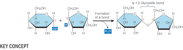
Figure 3.18
Questions
- What is the difference between the isomers maltose and cellobiose?
- Whole-grain wheat flour contains abundant starch and cellulose, while processed flour contains much less cellulose. Why is bread baked from whole-grain flour more fibrous?
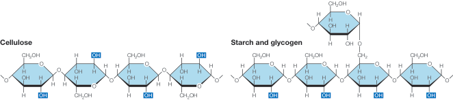
Figure 3.19
Lipids Are Defined by Their Insolubility in Water
- ■ Lipids are hydrocarbons that are insoluble in water because of their many nonpolar covalent bonds. They play roles in energy storage, membrane structure, light harvesting, intercellular communication, and protection.
- ■ A triglyceride is composed of three fatty acids covalently bonded to a molecule of glycerol by ester linkages.
- ■ A saturated fatty acid has a hydrocarbon chain with no double bonds. These molecules can pack together tightly. The hydrocarbon chain of an unsaturated fatty acid has one or more double bonds that bend the chain, preventing close packing.
- ■ A phospholipid has a hydrophobic hydrocarbon “tail” and a hydrophilic phosphate “head”; that is, it is amphipathic. In water, the interactions of the tails and heads of phospholipids generate a phospholipid bilayer. The heads are directed outward, where they interact with the surrounding water. The tails are packed together in the interior of the bilayer, away from water.
Questions
- How are a triglyceride and a phospholipid similar and different?
- Olive oil and vinegar (acetic acid) are both liquids. Why don’t they mix in salad dressing?
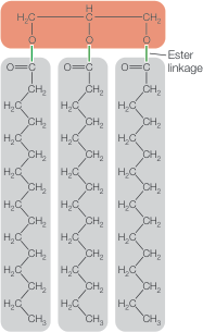
Figure 3.21
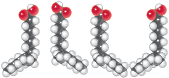
Figure 3.22
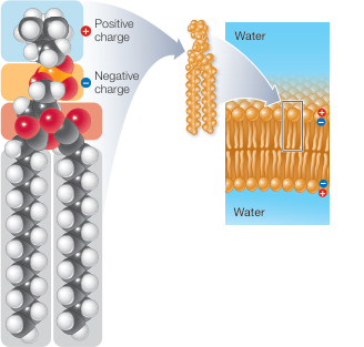
Figure 3.23