Chapter 11The Cell Cycle and Cell Division
PART FOUR Genes, Genomes, and Heredity
The Cell Cycle and 11 Cell Division
© Steve Garrison/Sandra Sheridan
Key Concepts
- 11.1 All Cells Derive from Other Cells
- 11.2 The Eukaryotic Cell Division Cycle Is Regulated
- 11.3 Eukaryotic Cells Divide by Mitosis
- 11.4 Cell Division Plays Important Roles in the Sexual Life Cycle
- 11.5 Meiosis Leads to the Formation of Gametes
- 11.6 Cell Death Is Important in Living Organisms
- 11.7 Unregulated Cell Division Can Lead to Cancer
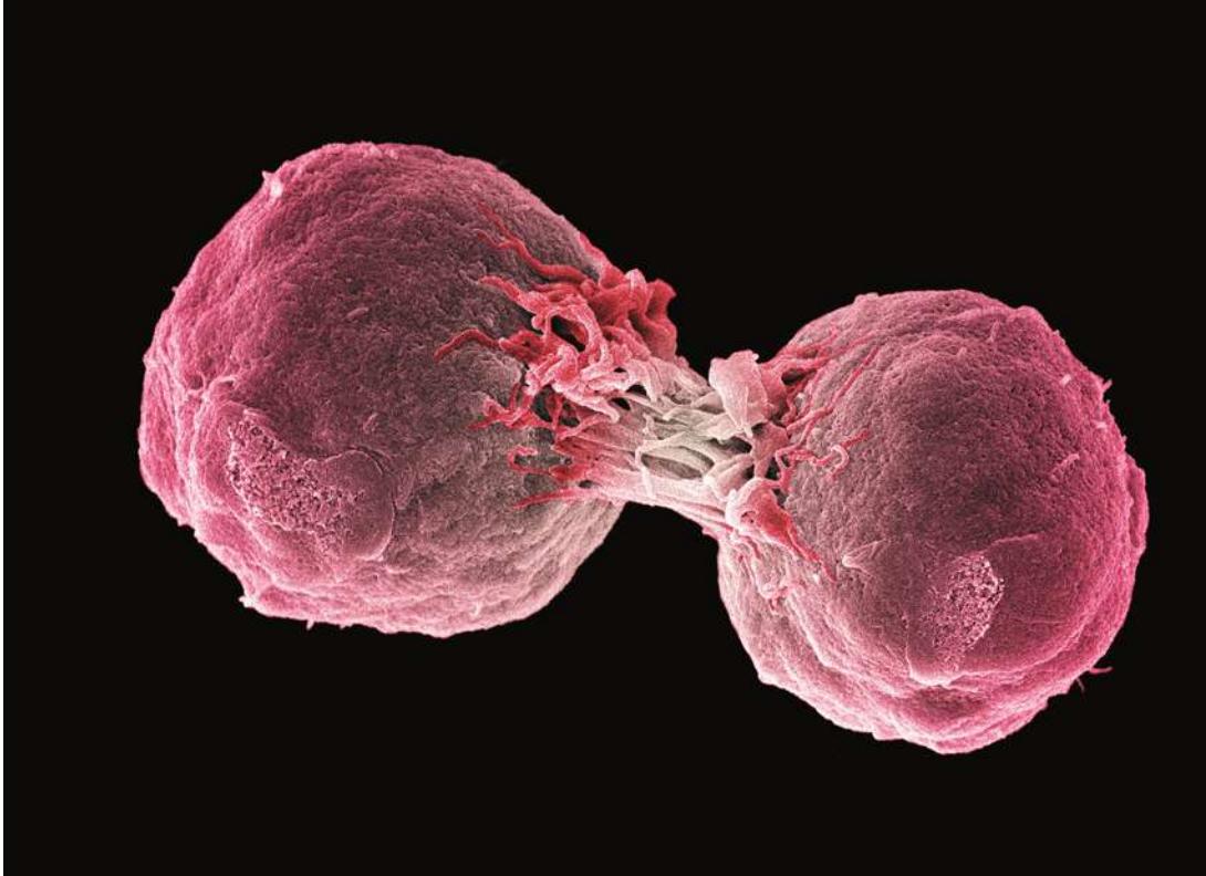
Henrietta Lacks’s tumor cells have far outlived her sad demise from cancer. They reproduce rapidly on the surface of a solid medium and have been grown in labs since her death in 1951.
Investigating Life: Immortal Cells
On January 29, 1951, 30-year-old Henrietta Lacks visited the nearby Johns Hopkins Hospital in Baltimore, Maryland, because she had been bleeding abnormally after the birth of her last child. The physician found the reason for the bleeding: a tumor the size of a quarter on her cervix. A piece of the tumor was sent to a pathologist in a clinical laboratory, who reported that the tumor was malignant.
A week later Ms. Lacks returned to the hospital, where physicians treated her tumor with radiation to try to kill it. But before the treatment began, they took a small sample of cells and sent them without her permission—a practice common then but not ethical today—to the research lab of George and Margaret Gey, two scientists at the hospital who had been trying for 20 years to coax human cells to live and multiply outside the body. If they could do so, they thought, they might find a cure for cancer. The Geys hit pay dirt with Lacks’s cells; they grew and reproduced more vigorously than any cells they had ever seen and were given the name “HeLa cells.” Unfortunately, they also grew fast in Lacks’s body, and in a few months they had spread to almost all of her organs. Lacks died on October 4, 1951.
Because of their robust ability to reproduce, HeLa cells quickly became a staple of cell biology research. In controlled settings they could be infected with viruses, and they were instrumental in developing the supply of polioviruses that led to the first vaccine against that dread disease. HeLa cells have been used for important basic and applied research ever since, especially on the ways that human cells reproduce by cell division. Although Lacks had never been outside Virginia and Maryland, her cells have traveled all over the world and even into space on the space shuttle. Over the past 65 years, tens of thousands of research articles have been published using information obtained from Lacks’s cells. You’ll study one of them in this chapter.
Understanding the cell division cycle and its control is clearly an important subject for understanding cancer. But cell division is not just important in medicine. It underlies the growth, development, and reproduction of all organisms.
QA What controls the reproduction of cancer cells? (Find the answer on page 242.)
All Cells Derive from Other Cells
Learning Objectives
- 11.1.1 Describe the four events that characterize cell division.
- 11.1.2 Explain how replication and segregation co-occur during cell division in prokaryotes.
- 11.1.3 Explain how binary fission results in identical daughter cells.
- 11.1.4 State the two ways in which DNA replication differs in prokaryotes and eukaryotes.
The life cycle of an organism, from birth to death, is intimately linked to cell division. Cell division plays important roles in the development of an organism from a single cell, in the growth and repair of tissues in multicellular organisms, and in the reproduction of all organisms (Figure 11.1). Cell division in both prokaryotes and eukaryotes entails the following four events:
- Cell division signals: One or more signals are required to initiate cell division. The signals may originate from either inside or outside the cell.
- DNA replication: The dividing cell’s genetic material (DNA) must be duplicated so that each of the two new cells will have a full complement of genetic information.
- DNA segregation: The replicated DNA must be distributed appropriately to the two daughter cells, so that each receives a copy of every chromosome.
- Cytokinesis: The cytoplasm must divide to form the two new cells, each surrounded by a cell membrane, and a cell wall in organisms that have one.
As you will see, these four events happen differently in prokaryotes and eukaryotes.
Prokaryotes divide by binary fission
In prokaryotes, cell division results in the reproduction of the entire single-celled organism. The cell grows in size, replicates its DNA, and then separates the cytoplasm and DNA into two new daughter cells in a process called binary fission.
CELL DIVISION SIGNALS External factors such as environmental conditions and nutrient concentrations are common signals for the initiation of cell division in prokaryotes. For example, the bacterium Bacillus subtilis can divide in under an hour if abundant sources of carbohydrate and mineral nutrients are available and if the pH and temperature are optimal. When nutrient levels are low, or the temperature is too hot or cold, or the pH too acidic or basic, the bacterium slows down its growth and eventually stops dividing. When conditions improve, growth and division resume. The bacterium Escherichia coli, which is widely used in genetic studies, can divide every 20 minutes under optimal conditions.
DNA REPLICATION As you saw in Key Concept 5.3, a chromosome consists of a long, thin DNA molecule that contains the genetic information for the organism, as well as various associated proteins. Most prokaryotes have just one main chromosome; when a cell divides, its chromosome must be replicated, and each of the two new daughter cells must receive a copy.
If the E. coli chromosome were spread out into a full circle, the circle would be about 500 micrometers () in diameter, more than 200 times larger than the cell! To fit into the cell, bacterial DNA must be compacted. The DNA folds in on itself, and positively charged (basic) proteins bound to the negatively charged (acidic) DNA contribute to this folding.
Two regions of the prokaryotic chromosome play functional roles in cell reproduction:
- ori: the site where replication of the circular chromosome starts (the origin of replication)
- ter: the site where replication ends (the terminus of replication)
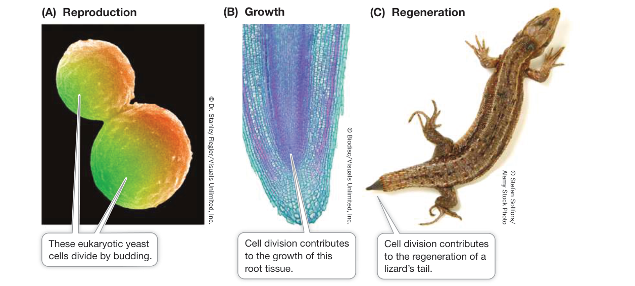
Figure 11.1 Important Consequences of Cell Division Cell division is the basis for (A) reproduction, (B) growth, and (C) repair and regeneration of tissues.
Chromosome replication takes place as the DNA is threaded through a replication complex of proteins near the center of the cell. Replication begins at the ori site and moves bidirectionally toward the ter site. While the DNA replicates, anabolic metabolism is active, resulting in cell growth. When replication is complete, the two daughter DNA molecules separate and segregate from one another at opposite ends of the cell. In rapidly dividing prokaryotes, DNA replication occupies the entire time between cell divisions.
DNA SEGREGATION Replication begins near the center of the cell. Once replicated, the ori regions move toward opposite ends of the cell as replication of the rest of the parent molecule proceeds (Figure 11.2A). DNA sequences adjacent to each of the ori regions bind proteins that are essential for this segregation. This is an energy-requiring process, since the binding proteins hydrolyze ATP.
CYTOKINESIS Immediately after chromosome replication is finished, cytokinesis begins. At first, the cell membrane pinches in to form a ring of fibers, termed the Z ring, similar to a purse string. The major component of these fibers is a protein called FtsZ that is related to eukaryotic tubulin (which makes up microtubules; see Figure 5.17). As the membrane pinches in, new cell wall materials are deposited, which finally separate the two cells (Figure 11.2B).
Eukaryotic cells divide by mitosis followed by cytokinesis
As in prokaryotes, cell reproduction in eukaryotes entails four events. The details, however, are quite different:
- Cell division signals: Like prokaryotes, single-celled eukaryotes often undergo cell division when environmental conditions are appropriate. For multicellular eukaryotes, the internal environmental conditions experienced by cells are often suitable for cell division, but this does not mean that cell division necessarily occurs. The reason is that cell division signals in multicellular eukaryotes are related to the function of the entire organism. For example, there is no need to produce additional liver cells when the liver has reached its optimal size. Once a cell is stimulated to begin dividing, internal signals come into play to ensure that cell division occurs correctly. We will discuss these signals in the next key concept.
- DNA replication: Eukaryotes have more DNA than prokaryotes, and it is contained in more than one chromosome (human cells, for example, have 46 chromosomes). While the mechanics of DNA replication are essentially the same in eukaryotes and prokaryotes, there are two major differences. First, eukaryotic DNA replication starts at numerous origins of replication, not at just one as in prokaryotes. Second, DNA replication in eukaryotes is usually limited to part of the period between cell divisions and does not overlap with the segregation of DNA into daughter cells.
- DNA segregation: Because eukaryotes have multiple chromosomes, segregation of the two sets of DNA molecules produced by replication is somewhat more complicated than it is in prokaryotes, and a mechanism called mitosis segregates them into two new nuclei. During mitosis, one copy of each chromosome in the parent cell ends up in each of the two new daughter cells. Chromosomes become highly condensed before they segregate, which makes them easier to move into the two new nuclei. The spindle, a special cytoskeletal structure composed of microtubules, is involved in moving chromosomes during segregation.
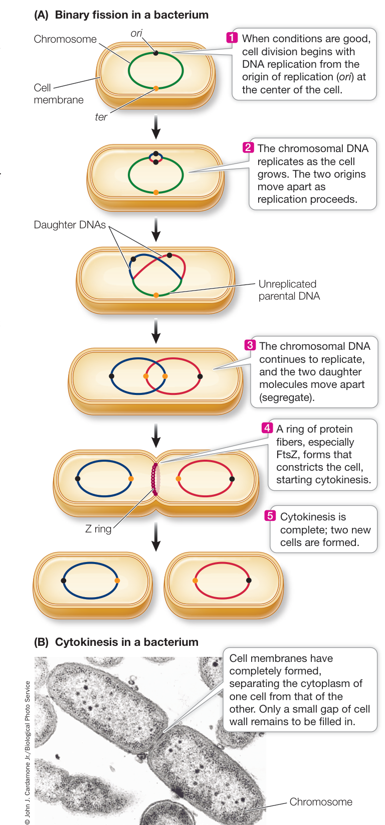
Figure 11.2 Prokaryotic Cell Division (A) DNA replication begins at the ori site and continues to the ter site, replicated DNA segregates to either end of the cell, and cytokinesis produces two cells. (B) Two identical daughter cells produced by binary fission. (A after K. Jonas. 2014. Curr Opin Microbiol 18: 54–60.)
- Cytokinesis: Cytokinesis proceeds differently in plant cells (which have a cell wall) than in animal cells (which do not).
The cells resulting from mitosis are identical to the parent cell in the amount and kind of DNA they contain.
There is another mechanism for nuclear division called meiosis, which is involved in gamete formation. We will return to meiosis and describe differences with mitosis in Key Concept 11.5.
Recap and Assess
Four events are required for cell division: cell division signals, replication of the genetic material (DNA), segregation of replicated DNA, and separation of the two daughter cells (cytokinesis). Prokaryotes often have just one chromosome, and cell division can be rapid. Eukaryotes usually have multiple chromosomes, and the process of cell division is more intricate, involving either mitosis or meiosis.
- What commonly controls whether a prokaryotic cell will begin binary fission?
- Why must DNA be replicated and segregated before the cell can divide?
- Why are DNA replication and cell division more complex in eukaryotes than in prokaryotes?
What determines whether a eukaryotic cell will divide? How does mitosis lead to identical cells? In the key concepts that follow, we will describe the details of mitosis.
The Eukaryotic Cell Division Cycle Is Regulated
Learning Objectives
11.2.1 Sketch and describe the four phases of the eukaryotic cell cycle.
11.2.2 Explain how the progress of a eukaryotic cell through the cell cycle is controlled by cyclin-dependent kinases.
11.2.3 Predict conditions under which external factors might stimulate the eukaryotic cell cycle.
As you will see throughout this book, different cells have different rates of cell division. Some cells, such as those in an early embryo, divide rapidly and continuously. Others, such as neurons in the brain, don’t divide at all. This suggests that the signaling pathways for cells to divide are highly controlled.
In eukaryotes, the phases through which a single cell passes to produce daughter cells by cell division is known as the cell cycle. The cell cycle can be divided into four phases: G1, S, G2, and M. M phase includes mitosis and cytokinesis. The other three phases together make up interphase. During interphase, the cell nucleus is visible and typical cell functions occur, including DNA replication (during S phase) (Figure 11.3).
The duration of the cell cycle varies considerably in different cell types. In the early embryo the cell cycle may be as short as 30 minutes, whereas rapidly dividing cells in an adult human typically complete the cycle in about 24 hours. In general, cells spend most of their time in interphase. So if we take a snapshot through the microscope of a cell population, only a few cells will be in mitosis or cytokinesis at any given moment. In a cell cycle of 24 hours, the four phases would typically last for 11 hours (G1), 8 hours (S), 4 hours (G2), and 1 hour (M).
Here we describe the events of interphase, especially those that trigger mitosis.
- G1 phase. During G1 phase, each chromosome is a single, unreplicated DNA molecule with associated proteins. Variations in the duration of G1 account for most of the variability in the length of the cell cycle in different cell types. Some rapidly dividing embryonic cells dispense with it entirely, whereas other cells may remain in G1 for weeks or even years. G1 ends at the G1-to-S transition, when the commitment is made to DNA replication and subsequent cell division.
- S phase. DNA replication occurs during S phase (see Key Concept 13.3 for a detailed description of DNA replication). Each chromosome is duplicated and thereafter consists of two sister chromatids (the products of DNA replication). The sister chromatids remain joined together until mitosis, when they segregate into two daughter cells.
- G2 phase. During G2 phase, the cell makes preparations for mitosis—for example, by synthesizing and assembling the structures that move the chromatids to opposite ends of the dividing cell.
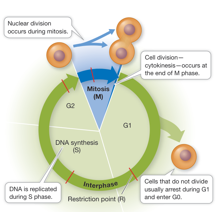
Figure 11.3 The Eukaryotic Cell Cycle The cell cycle consists of four phases: M, during which mitosis and cytokinesis take place, and G1, S, and G2, together termed interphase, during which cells grow and DNA replication occurs. Red lines indicate checkpoints for progression through the cell cycle. Q: If the DNA content of a human skin cell at the beginning of mitosis is 12 picograms ( ), what is the DNA content of a cell in G1?
The initiation, termination, and operations of these phases are regulated by specific signals. Not all cells follow a continuous progression through the phases. Some cells in G1 enter an inactive resting phase of the cell cycle called G0. These cells sometimes return to G1 and the rest of the cell cycle under certain environmental conditions, such as an extracellular signal. Otherwise, cells may stay in G0; examples in humans are cells of the heart (cardiac muscle) and brain (neurons).
Specific internal signals trigger events in the cell cycle
Cell fusion experiments were used to reveal the existence of internal signals that control the transitions between stages of the cell cycle. For example, an experiment involving the fusion of HeLa cells (the cells described in the opening story) at different phases of the cell cycle showed that a cell in S phase produces a substance that activates DNA replication (Investigating Life: What Controls the Reproduction of Cancer Cells?). Similar experiments pointed to the existence of signals controlling entry into M phase.
Investigating Life: What Controls the Reproduction of Cancer Cells?
Experiment
Original Paper: P. N. Rao and R. T. Johnson. 1970. Mammalian cell fusion: Studies on the regulation of DNA synthesis and mitosis. Nature 225: 159–164.
Nuclei of cells in G1 do not undergo DNA replication, but nuclei in S phase do. Rao and Johnson wondered whether substances present in cells in S phase could be used to induce DNA replication in cells in G1.
HYPOTHESIS A cell in S phase contains an activator of DNA replication.
METHOD
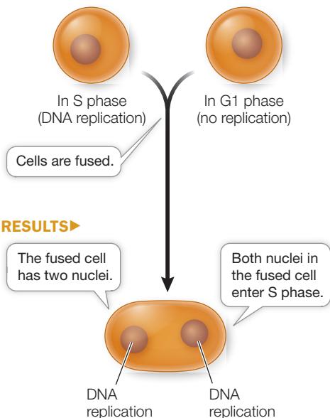
CONCLUSION The S phase cell contains a substance that diffuses to the G1 nucleus and activates DNA replication.
Work with the Data
The fusion of cellular membranes is a natural process; it occurs during endocytosis and exocytosis, and in fertilization (the fusion of gametes). Membrane fusion also occurs when membrane-enclosed viruses infect their host cells. Occasionally these viruses also induce the fusion of adjacent host cells, creating a multinucleate cell. This observation led to the use of Sendai virus, a membrane-enclosed mouse respiratory virus, as a tool in the laboratory to fuse cells experimentally. Rao and Johnson used this strategy to study the regulation of the cell cycle.
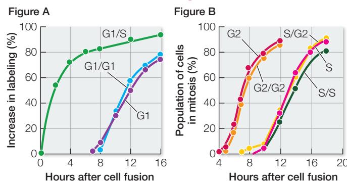
In their experiment, Rao and Johnson used HeLa cells, which divide continuously (see the opening story of this chapter). First, they isolated cells in either G1 or S phase. Before fusion, the cells in S phase were exposed to a radioactively labeled component of DNA (thymidine). The radioactivity was incorporated into these cells’ newly replicated DNA, labeling their nuclei. The S and G1 cells were then fused using Sendai virus (resulting in G1/S fusions) and again exposed to labeled thymidine. At various times after fusion, the scientists calculated the percent of previously unlabeled (G1) nuclei that had incorporated new label (i.e., had replicated their DNA) (Figure A). In a second series of experiments, S and G2 cells were fused in various combinations and then the numbers of cells in mitosis were counted and expressed as a percent of all cells in the population (Figure B).
QUESTIONS
- According to Figure A, how long did it take for all the G1 nuclei in the G1/S cells to become labeled?
- Examine the data for fused G1/G1 cells and unfused G1 cells in Figure A. Explain why these were appropriate controls for the experiment. When did these nuclei become labeled? Compare these times with each other and with that for the G1/S nuclei and discuss.
- Examine the data in Figure B. Why did it take longer for the S phase cells to begin mitosis than the G2 cells?
- According to Figure B, did fusion with G2 cells alter the timing of mitosis in the S cell nuclei? Explain what this means in terms of regulation of the cell cycle.
Progress through the cell cycle depends on the activities of cyclin-dependent kinases, or CDKs. Recall from Key Concept 7.2 that a protein kinase is an enzyme that catalyzes the transfer of a phosphate group from ATP to a target protein; this phosphate transfer is called phosphorylation. A particular CDK controls the G1-to-S transition, which is a control point in the cell cycle called the restriction point (R) (see Figure 11.3). Other CDKs control other parts of the cell cycle.
CDKs are not enzymatically active as protein kinases unless they are bound to another class of protein, the activators called cyclins. The binding of its cyclin—an example of allosteric regulation—activates the CDK by altering its shape and exposing its active site to substrates (Figure 11.4). The cyclin–CDK that controls passage from G1 to S phase is not the only such complex involved in regulating the eukaryotic cell cycle. There are different cyclin–CDK complexes, composed of particular cyclins and their associated CDKs, that act at different stages of the cycle. The details of how these complexes form and function vary among eukaryotic organisms, but we will focus here on the complexes found in mammalian cells. As an example, let’s take a closer look at the cyclin–CDK complex that controls the G1-to-S transition.
Connect the Concepts As discussed in Key Concept 8.5, allosteric regulation occurs when another molecule induces a change in the three-dimensional structure of an enzyme. This changes the ability of the enzyme to bind its substrate, and so changes the rate of the reaction catalyzed by the enzyme.
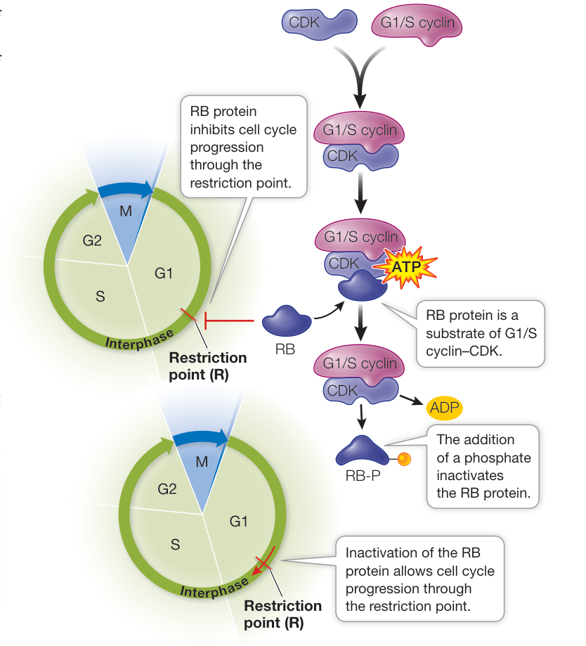
Figure 11.5 Phosphorylation of RB Protein Controls Cell Cycle Progression The G1/S cyclin binds to and activates its CDK, which then phosphorylates the retinoblastoma protein (RB), inactivating it and allowing the cell cycle to progress from G1 to S
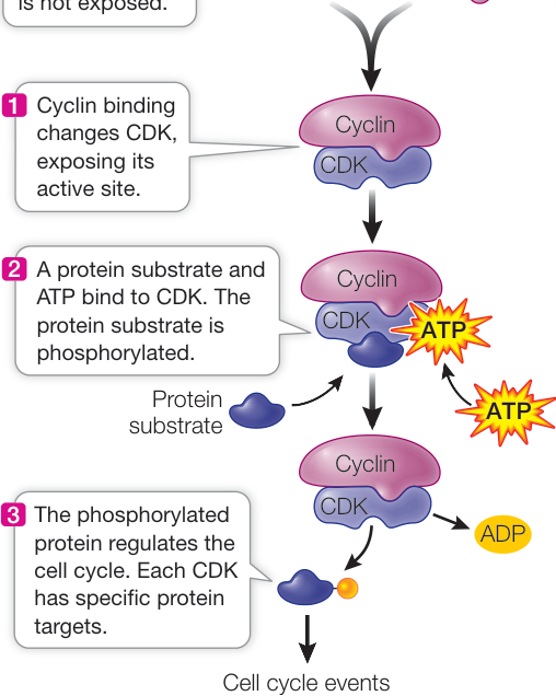
Figure 11.4 Cyclin Binding Activates CDK Binding of a cyclin changes the three-dimensional structure of an inactive CDK, making it an active protein kinase. Each cyclin–CDK complex phosphorylates a specific target protein in the cell cycle.
The G1/S cyclin–CDK catalyzes the phosphorylation of a protein called retinoblastoma protein (RB, named because of its role in cancer; see Key Concept 11.7). In many cells, RB acts as an inhibitor of the cell cycle at the restriction point (R). To begin S phase, a cell must get by the RB block. Here is where the G1/S cyclin–CDK comes in: it catalyzes the phosphorylation of RB molecules. This causes a change in the three-dimensional structure of RB, thereby inactivating it (Figure 11.5). With RB out of the way, the cell cycle can proceed.
Progress through the cell cycle is regulated by the activities of several CDKs, and so regulating them is a key to regulating cell division. An effective way to regulate CDKs is to regulate the presence or absence of cyclins (Figure 11.6). Simply put, if a cyclin is not present, its partner CDK is not active. As their name suggests, cyclins are present cyclically: they are made only at certain times in the cell cycle.
The different cyclin–CDKs act at cell cycle checkpoints, signaling pathways that regulate the cell cycle’s progress. For example, if a cell’s DNA is substantially damaged by radiation or toxic chemicals, the cell may be prevented from successfully completing a cell cycle. During interphase, there are three checkpoints, with a fourth during mitosis (see Figure 11.3). Table 11.1
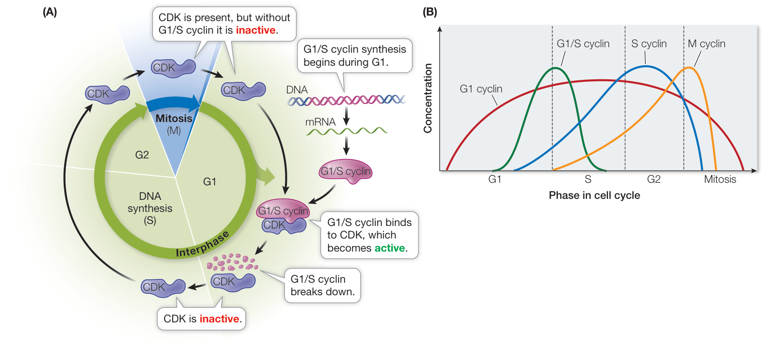
Figure 11.6 Cyclins Are Transient in the Cell Cycle Cyclins are made at a particular time and then break down. (A) The G1/S cyclin is present during G1 and activates a CDK at that time. (B) Other cyclins control progression through other phases of the cell cycle. (B after WikiMiMa|Michael Matthes.)
lists some of the triggers that will cause the cell cycle to pause at each checkpoint.
As an example, let’s consider the G1-to-S checkpoint (R). If DNA is damaged by radiation during G1, a signaling pathway results in the production of a protein called p21. (The p stands for “protein” and the 21 stands for its molecular weight—about 21,000 daltons.) The p21 protein binds to the G1/S CDK, preventing cyclin binding. This keeps the CDK in its inactive state, and the cell cycle pauses while repairs are made to the DNA (you will learn more about DNA repair in Key Concept 13.4). When the DNA damage pathway is no longer operating, p21 breaks down, allowing the cyclin–CDKs to function, and the cell cycle proceeds. If DNA damage is severe and cannot be repaired, the cell will undergo programmed cell death (apoptosis, which we will discuss later in this chapter). Such controls prevent defective cells from proliferating and potentially harming an organism.
Growth factors can stimulate cells to divide
Cyclin–CDKs provide cells with internal controls of their progress through the cell cycle. But the cell cycle is also influenced by external signals. Not all cells in an organism go through the cell cycle on a regular basis. Some cells either no longer go through the cell cycle and enter G0, or go through it slowly and divide infrequently. If such cells are to divide, they must be stimulated by external chemical signals called growth factors. Here are a few examples:
- If you cut yourself and bleed, specialized cell fragments called platelets gather at the wound to initiate blood clotting. The platelets produce and release a protein called platelet-derived growth factor which diffuses to the adjacent cells in the skin and stimulates them to divide and heal the wound.
- Red and white blood cells have limited lifetimes and must be replaced through the division of immature, unspecialized blood cell precursors in the bone marrow. Two types of growth factors, interleukins and erythropoietin, stimulate the division and specialization, respectively, of precursor cells of white blood cells and red blood cells.
Growth factors bind to specific receptors on target cells and activate signal transduction pathways (see Chapter 7) that end with cyclin synthesis, thereby activating CDKs and the cell cycle.
Recap and Assess
The eukaryotic cell cycle is under both external and internal control. CDKs control the eukaryotic cell cycle, and their activities are regulated by binding of cyclins. External signals such as growth factors can initiate the cell cycle.
- Draw a cell cycle diagram with each of its phases.
- How do cyclin–CDKs control the progress of the cell cycle?
- What are growth factors, and how do they act to control the cell cycle?
- The protein p16 acts on the cell cycle in a fashion similar to that of p21. Recently it was found that there is more p16 protein in the cells of older people than in younger people. What are the implications of this for aging?
Table 11.1 Cell Cycle Checkpoints
| Cell cycle phase | Checkpoint trigger |
|---|---|
| G1 | DNA damage |
| S | Incomplete replication or DNA damage |
| G2 | DNA damage |
| M | Chromosome unattached to spindle |
Once the cell cycle is initiated and interphase is completed, the cell enters mitosis, where the replicated DNA is segregated into two daughter cells.
Eukaryotic Cells Divide by Mitosis
Learning Objectives
- 11.3.1 Describe DNA packaging and how it varies during the cell cycle.
- 11.3.2 List the five stages of mitosis and state what events occur in each.
- 11.3.3 Explain how the spindle is involved in segregation of sister chromatids.
- 11.3.4 Describe the differences between cytokinesis in animals and plants.
DNA is a very long polymer, up to several centimeters in length. At every stage of the cell cycle, these threads must be packaged into compact structures. A eukaryotic chromosome consists of one or two linear, double-stranded DNA molecules bound with many proteins (the complex of DNA and proteins is referred to as chromatin). Before S phase, each unreplicated chromosome contains only one double-stranded DNA molecule. After it replicates during S phase, however, there are two double-stranded DNA molecules: the sister chromatids (Figure 11.7). Throughout G2 the sister chromatids are held together along most of their length by a protein complex called cohesin. At mitosis most of the cohesin is removed, except in a region called the centromere, where the chromatids remain held together. At the end of G2 and the beginning of mitosis, a second group of proteins called condensins coats the DNA molecules and makes them more compact.
If all of the DNA in a typical human cell were put end to end, it would be nearly 2 meters long. Yet the nucleus is only 5 µm (0.000005 meters) in diameter and therefore DNA must be extensively packaged in a highly organized way (Figure 11.8). This packing is achieved largely by proteins called histones (histos, “web” or “loom”), which are positively charged at cellular pH because of their high content of the basic amino acids lysine and arginine. The charged R groups on these amino acids bind to the negatively charged phosphate groups on DNA by ionic bonds. These DNA–histone interactions, as well as histone–histone interactions, result in the formation of beadlike units called nucleosomes (see Figure 11.8).
During interphase, the chromatin that makes up each chromosome consists of single DNA molecules wrapped around vast numbers of nucleosomes that resemble beads on a string. During this phase of the cell cycle, the DNA is accessible to proteins involved in replication and transcription. Once a mitotic chromosome is formed, its compact nature makes it inaccessible to replication and transcription factors, and so these processes cannot occur.
Mitosis during M phase of the cell cycle leads to the production of two nuclei that are genetically identical to each other and to the nucleus of the cell that entered the cell cycle in G1 (i.e., the parent cell). Mitosis ensures the accurate segregation of each copy of a eukaryotic cell’s multiple chromosomes into the daughter nuclei. While mitosis is a continuous process in which each event flows smoothly into the next, it is convenient to subdivide it into a series of stages: prophase, prometaphase, metaphase, anaphase, and telophase (Table 11.2, Figure 11.9).
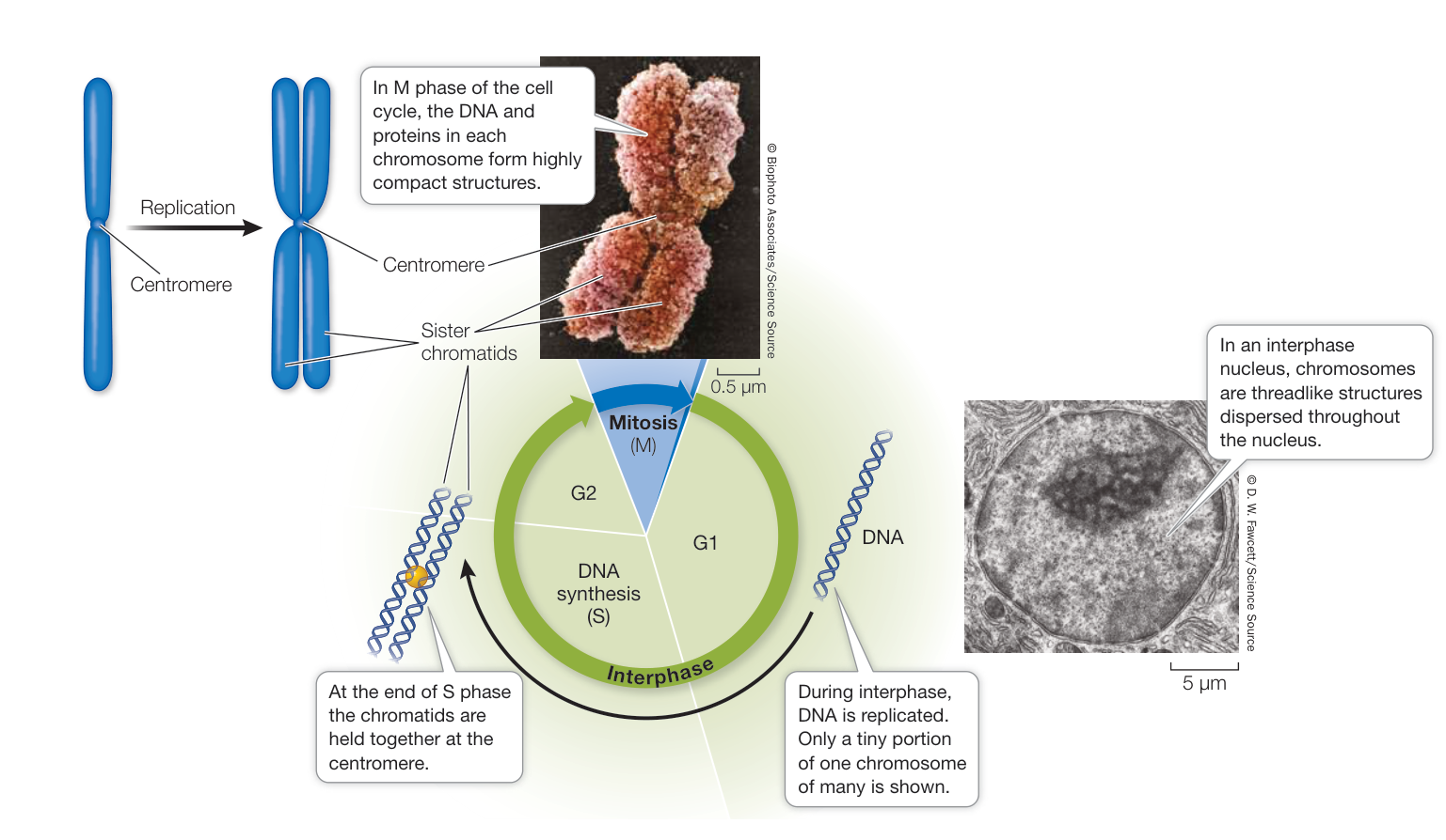
Figure 11.7 Chromosomes and Sister Chromatids DNA in the interphase nucleus is diffuse and becomes compacted as mitosis begins.
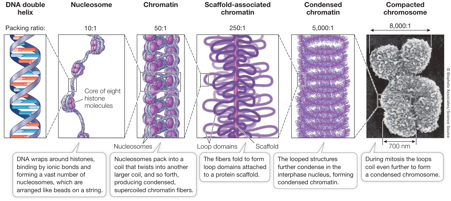
Figure 11.8 DNA Is Packed into a Mitotic Chromosome The nucleosome, formed by DNA and histones, is the essential building block in this highly compacted structure.
Let’s take a closer look at two cellular structures that contribute to the orderly segregation of the chromosomes during mitosis—the centrosomes and the spindle.
The centrosomes determine the plane of cell division
The spindle (also called the mitotic spindle or spindle apparatus) is a dynamic microtubule structure that moves sister chromatids apart during mitosis. Before the spindle can form, its orientation is determined by the centrosome (“central body”), an organelle in the cytoplasm near the nucleus. In many organisms the centrosome consists of a pair of centrioles, each one a hollow tube formed by nine microtubule triplets. During S phase the centrosome duplicates, and at the beginning of prophase the two centrosomes separate from one another, moving to opposite ends of the nuclear envelope. These identify the “poles” toward which chromosomes move during anaphase. The cells of plants and fungi lack centrosomes, but distinct microtubule organizing centers at each end of the cell play the same role.
The positions of the centrosomes determine the plane where the animal cell will divide; therefore they determine the spatial relationship between the two new cells. This relationship may be of little consequence to single free-living cells such as yeasts, but it is important for cells in a multicellular organism. For example, during development from a fertilized egg to an embryo, the daughter cells from some divisions must be positioned correctly to receive signals to form new tissues.
The spindle begins to form during prophase
During interphase, only the nuclear envelope, the nucleoli (see Key Concept 5.3), and a barely discernible tangle of chromatin are visible under the light microscope. The appearance of the nucleus changes as the cell enters prophase. At this stage, most of the cohesin that has held the two products of DNA replication together since S phase is removed, so the individual chromatids become visible. They are still held together by a small amount of cohesin at the centromere. Late in prophase, specialized
Table 11.2 Summary of Cell Cycle Events
| Phase | Events |
|---|---|
| G1 | Growth |
| S | DNA replication |
| G2 | Spindle synthesis begins; preparation for mitosis |
| M | |
| Mitosis | |
| Prophase | Condensation of chromosomes; spindle assembly |
| Prometaphase | Nuclear envelope breakdown; chromosome attachment to spindle |
| Metaphase | Alignment of chromosomes at metaphase plate |
| Anaphase | Separation of chromatids; migration to poles |
| Telophase | Chromosomes decondense; nuclear envelope re-forms |
| Cytokinesis | Cell separation; cell membrane and/or wall formation |
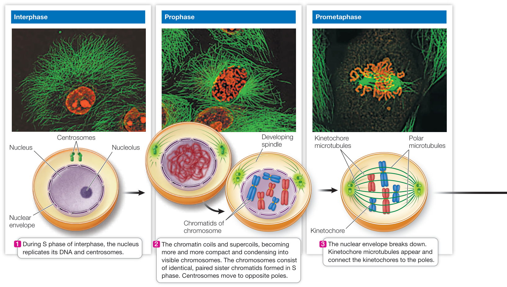
Figure 11.9 The Phases of Mitosis in an Animal Cell Mitosis results in two new genetically identical nuclei, each one destined for one of the two daughter cells produced when a cell divides. In the micrographs, the green dye stains microtubules (and thus the spindle); the red dye stains the chromosomes. The chromosomes in the diagrams are stylized to emphasize the fates of the individual chromatids. For instructional purposes, the chromosomes from one parent are colored blue and those from the other parent are red.
structures called kinetochores develop in the centromere region, one on each chromatid. These structures will be important for chromosome movement.
Each of the two centrosomes, now on opposite sides of the nucleus, serves as a mitotic center, or pole, toward which the chromosomes will move (Figure 11.10A). During prophase and prometaphase, microtubules form between the poles and the chromosomes to make up the spindle. The spindle serves as a structure to which the chromosomes attach and as a framework keeping the two poles apart. Each half of the spindle develops as tubulin dimers aggregate from around the centrioles and form long fibers that extend into the middle region of the cell. The microtubules are initially unstable, constantly forming and falling apart, until they contact kinetochores or microtubules from the other half-spindle and become more stable.
There are two groups of microtubules in the spindle:
Polar microtubules form the framework of the spindle and run from one pole to the other.
- Kinetochore microtubules, which form later, attach to the kinetochores on the chromosomes. The two sister chromatids in each replicated chromosome become attached to kinetochore microtubules from opposite poles (Figure 11.10B). This ensures that the two chromatids will eventually move to opposite poles.
Movement of the chromatids achieves the central goal of mitosis. It accomplishes the segregation of the genetic material that must occur before the cell can divide and complete the cell cycle. Prophase prepares for this movement, and the actual segregation takes place in the next three phases of mitosis.
Chromosome separation and movement are highly organized
During the next three phases of mitosis—prometaphase, metaphase, and anaphase—dramatic changes take place in the cell and the chromosomes (see Figure 11.9). During prometaphase, the nuclear envelope breaks down and spindle formation is completed. During metaphase, the chromosomes line up at the equatorial position (metaphase plate) of the cell. Now let’s consider two key processes of anaphase: separation of the chromatids, and the mechanism of their actual movement toward the poles.
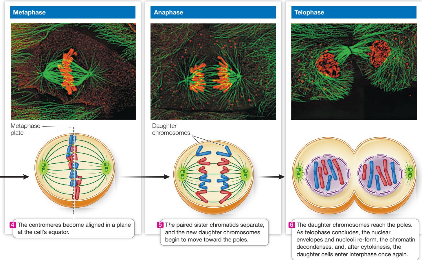
CHROMATID SEPARATION The separation of chromatids occurs at the beginning of anaphase. It is controlled by an M phase cyclin–CDK, which activates another protein complex
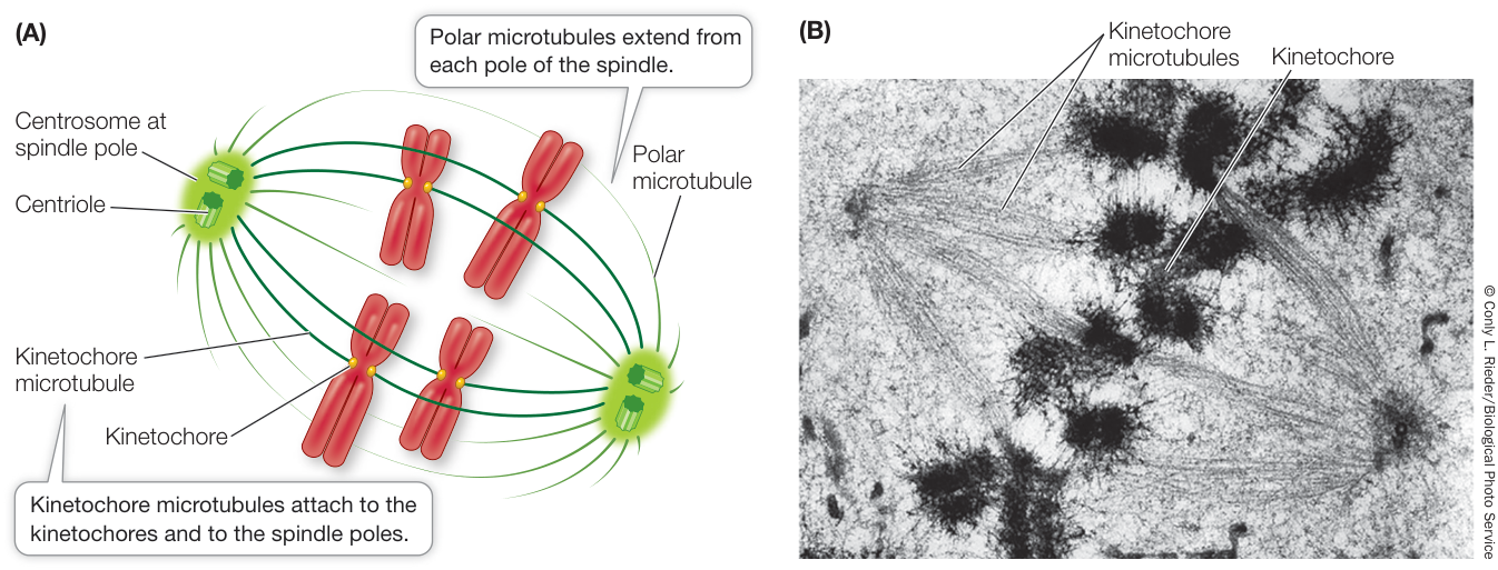
Figure 11.10 The Mitotic Spindle Consists of Microtubules (A) The spindle in an animal cell at metaphase. In plant cells, centrioles are not present. (B) An electron micrograph of metaphase, emphasizing the kinetochore microtubules.
Activity 11.2 The Mitotic Spindle
called the anaphase-promoting complex (APC). Separation occurs because one subunit of the cohesin protein holding the sister chromatids together is hydrolyzed by a specific protease, appropriately called separase (Figure 11.11). A cell cycle checkpoint, often called the spindle assembly checkpoint, occurs at the end of metaphase to inhibit the APC if one of the chromosomes is not attached properly to the spindle. When all chromosomes are attached, the APC is activated and the chromatids separate. After separation the chromatids are called daughter chromosomes. Note the difference between sister chromatids and daughter chromosomes:
- Sister chromatids share a centromere.
- Daughter chromosomes have their own centromere.
CHROMOSOME MOVEMENT The migration of the two sets of daughter chromosomes to the poles of the cell is a highly organized, active process. Three mechanisms operate to move the chromosomes along. First, the kinetochores contain molecular motor proteins, including kinesins and cytosolic dynein (see Figures 5.20 and 5.21), which use energy from ATP hydrolysis to do the work of moving the chromosomes along the microtubules. Second, the kinetochore microtubules shorten, drawing the chromosomes toward them. Third, the centrosomes move apart, aiding in separation. The last stage of mitosis is telophase, when the spindle disappears and a nuclear envelope forms around each set of daughter chromosomes (see Figure 11.9). Finally, the cytoplasm divides to form the two daughter cells during cytokinesis: the last event in cell division.
Cytokinesis divides the cytoplasm
Cytokinesis divides the cell’s cytoplasm after nuclear division in mitosis. There are substantial differences between the process in animal and plant cells. In animal cells, cytokinesis begins with a furrowing of the cell membrane, as if an invisible thread were cinching the cytoplasm between the two nuclei (Figure 11.12A). This contractile ring is composed of microfilaments of actin and associated myosin, which form a ring on the cytoplasmic surface of the cell membrane. These two proteins interact to produce a contraction, pinching the cell in two. The microfilaments assemble rapidly from actin monomers that are present in the interphase cytoskeleton.
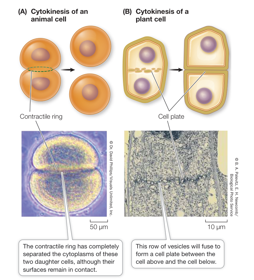
Figure 11.12 Cytokinesis Differs in Animal and Plant Cells (A) A sea urchin zygote (fertilized egg) that has just completed cytokinesis at the end of the first cell division of its development into an embryo. (B) A dividing plant cell in late telophase. Plant cells divide differently from animal cells because plant cells have cell walls.
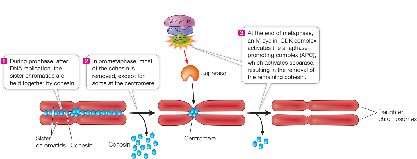
Figure 11.11 Chromatid Attachment and Separation The cohesin protein complex holds sister chromatids together at the centromere. The enzyme separase hydrolyzes cohesin at the end of metaphase and beginning of anaphase, allowing the chromatids to separate into daughter chromosomes.
The plant cell cytoplasm divides differently because plants have rigid cell walls. In plant cells, as the spindle breaks down after mitosis, membranous vesicles derived from the Golgi apparatus appear along the plane of cell division, roughly midway between the two daughter nuclei. The vesicles are propelled along microtubules by the motor protein kinesin, and fuse to form a new cell membrane. At the same time they contribute their contents to a cell plate, which is the beginning of a new cell wall (Figure 11.12B).
Following cytokinesis, each daughter cell contains all the components of a complete cell. A precise distribution of chromosomes is ensured by mitosis. In contrast, organelles such as ribosomes, mitochondria, and chloroplasts do not need to be distributed equally between daughter cells, as long as some of each are present in each cell. Accordingly, there is no mechanism with a precision comparable to that of mitosis to provide for their equal allocation to daughter cells.
Recap and Assess
Mitosis is the ordered division of a eukaryotic cell nucleus into two nuclei with identical sets of chromosomes. The process of mitosis, while continuous, can be viewed as a series of events (prophase, prometaphase, metaphase, anaphase, and telophase). Once two nuclei have formed, the cell divides into two cells by cytokinesis.
- Describe the differences between a replicated and an unreplicated chromosome, and identify during what phases of the cell cycle each one is found.
- What are the various levels of “packing” by which the genetic information contained in linear DNA is condensed during prophase?
- Paclitaxel is an anticancer drug that binds to microtubules, preventing them from adding or removing subunits and causing them to form bundles. How do you think this drug works to stop cancer? Is Paclitaxel specific to cancer cells?
- How does cytokinesis differ in plant and animal cells?
- How would the cell cycle be affected if the cohesion protein was nonfunctional?
Mitosis results in two cells that are genetically identical. But there is another eukaryotic cell division process, meiosis, that results in genetic diversity. In the next key concept we will discuss the roles of mitosis and meiosis in sexual reproduction, and then turn to the details of meiosis in Key Concept 11.5.
Cell Division Plays Important Roles in the Sexual Life Cycle
Learning Objectives
- 11.4.1 Compare and contrast asexual and sexual reproduction.
- 11.4.2 Describe the roles of meiosis and fertilization in the sexual life cycle.
- 11.4.3 Differentiate between haploid and diploid cells and organisms.
- 11.4.4 Compare and contrast different sexual life cycles.
Repetition of the mitotic cell cycle allows a single cell to give rise to many cells that are genetically identical. Meiosis, by contrast, produces four daughter cells that are not genetically identical, with each carrying half the amount of DNA as the original G1 parent cell. Mitosis and meiosis are both involved in reproduction but in different ways: asexual reproduction involves only mitosis, whereas the sexual reproduction cycle involves both mitosis and meiosis.
Asexual reproduction is based on the mitotic division of the nucleus. An organism that reproduces asexually may be single-celled like yeast, reproducing itself with each cell cycle, or it may be multicellular like an aspen in a forest in the Wasatch Mountains of Utah (Figure 11.13). Aspen can reproduce sexually, with male and female plants, but in many aspen stands all the trees are the same sex, and DNA analyses have shown that they are clones of a single parent organism: the offspring are genetically identical to the parent. Any genetic variation among the trees is most likely due to a relatively small number of changes in the DNA that occur during DNA replication. As you will see, this small amount of variation contrasts with the extensive variation possible in sexually reproducing organisms.
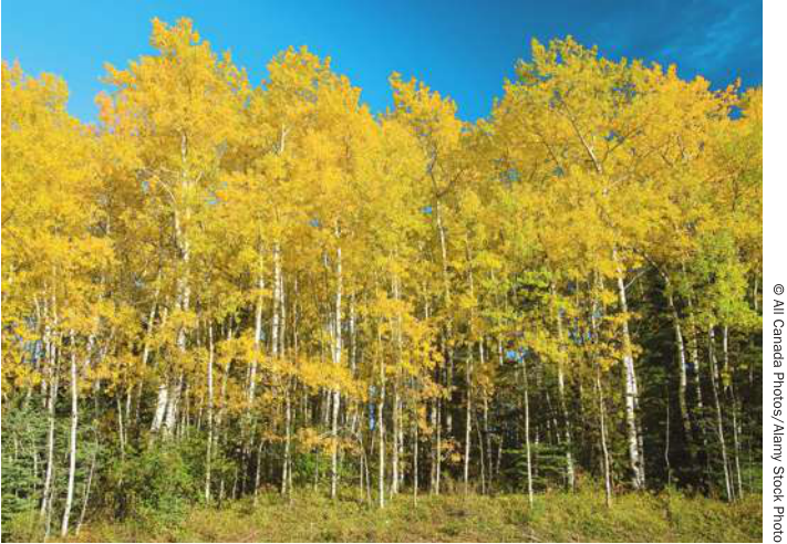
Figure 11.13 Asexual Reproduction on a Large Scale In this forest, the aspen trees arose from a single tree by asexual reproduction. They are virtually identical genetically.
Connect the Concepts Asexual reproduction is common in land plants, and you can learn about this in Key Concept 36.3.
Unlike asexual reproduction, sexual reproduction results in an organism that is not identical to its parents. Sexual reproduction requires gametes created by meiosis; two parents each contribute one gamete to each of their offspring. Meiosis produces gametes—and thus offspring—that differ genetically from each other and from the parents. Because of this genetic variation, some offspring may be better adapted than others to survive and reproduce in a particular environment. While asexually reproducing organisms can have a small amount of genetic diversity, meiosis generates much more diversity, which is available for natural selection and evolution.
Sexual life cycles produce haploid and diploid cells
In most multicellular organisms, the body cells that are not specialized for reproduction, called somatic cells, each contain two sets of chromosomes, which are found in pairs. One chromosome of each pair comes from each of the organism’s two parents; in humans, for example, 23 of the 46 chromosomes come from the biological mother and 23 from the biological father. The members of such a homologous pair are similar in size and appearance.
The two chromosomes in a homologous pair (called homologs) bear corresponding, though not identical, genetic information. For example, a homologous pair of chromosomes in a plant may carry different versions of a gene that controls seed shape. One homolog may carry the version for wrinkled seeds while the other may carry the version for smooth seeds. You’ll see the seed-shape gene in Chapter 12.
There is no simple relationship between the size of an organism and the number of chromosomes in its genome. A housefly has 5 chromosome pairs and a horse has 32, but the smaller carp (a fish) has 52 pairs. Probably the highest number of chromosomes in any organism is in the fern Ophioglossum reticulatum, which has 1,260 (630 pairs)!
In contrast to somatic cells, gametes contain only a single set of chromosomes—that is, one homolog from each pair. The number of chromosomes in a gamete is denoted by n, and the cell is said to be haploid. During reproduction, two haploid gametes fuse to form a zygote in a process called fertilization. The zygote thus has two sets of chromosomes; its chromosome number is denoted by 2n, and it is said to be diploid. Depending on the organism, the zygote may divide by either meiosis or mitosis. Either way, a new mature organism develops that is capable of sexual reproduction.
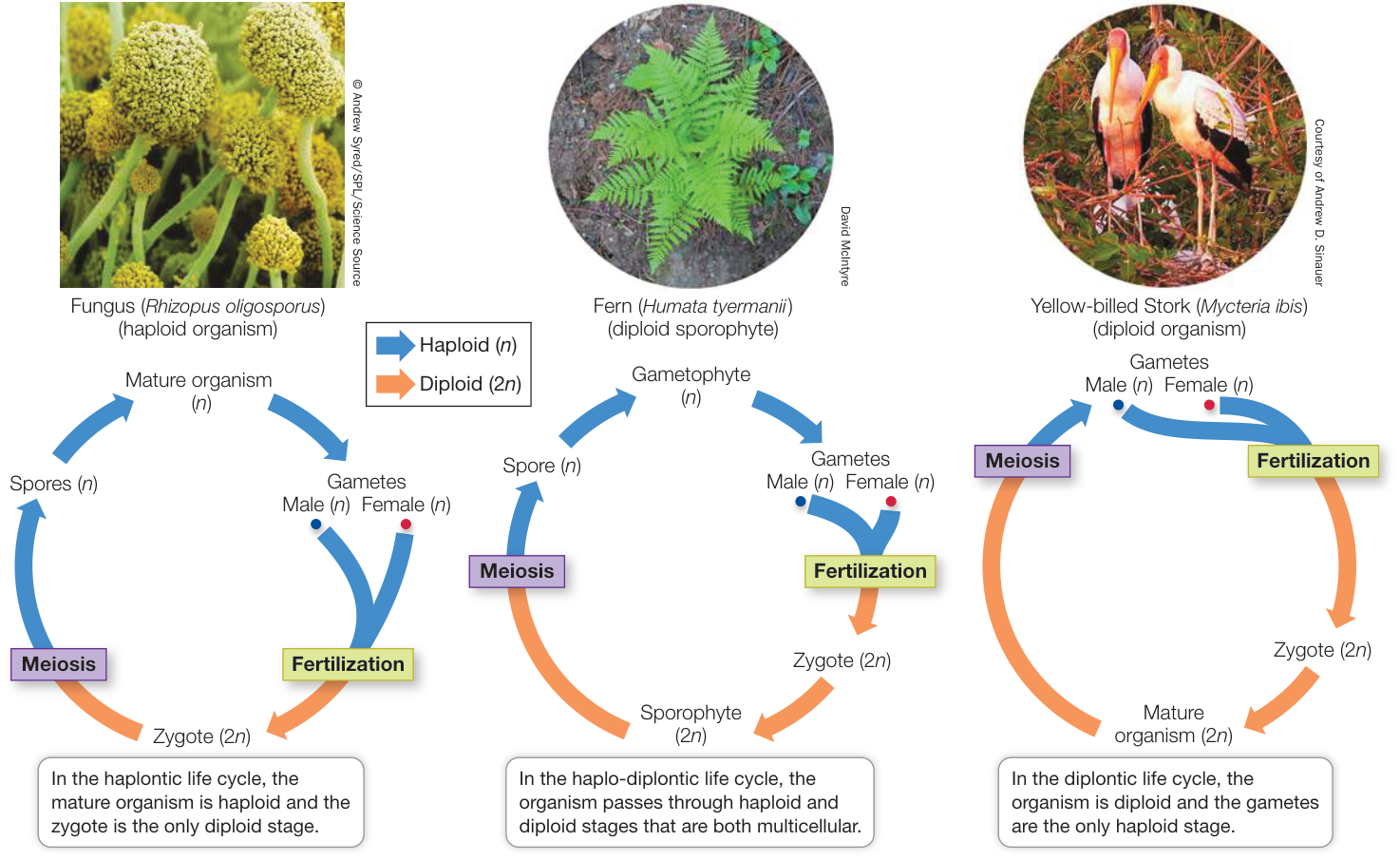
Figure 11.14 Fertilization and Meiosis Alternate in Sexual Reproduction In sexual reproduction, haploid (n) cells or organisms alternate with diploid (2n) cells or organisms.
All sexual life cycles involve meiosis to produce haploid cells and fertilization to produce a zygote. Figure 11.14 presents three types of sexual life cycles. In some life cycles the products of meiosis undergo cell division, resulting in a mature organism with haploid cells. Specialized cells in these haploid organisms become gametes by mitosis. In other life cycles the gametes form directly from the products of meiosis. In all cases, gametes fuse to produce a zygote, beginning the diploid stage of the life cycle. Since the origin of sexual reproduction, evolution has generated many different versions of the sexual life cycle.
The essence of sexual reproduction is the random selection of half of the diploid chromosome set—one member of each genetically similar chromosome pair—to make a haploid gamete, followed by fusion of two haploid gametes to produce a diploid cell. Both of these steps contribute to a shuffling of genetic information in the population, so that no two individuals have exactly the same genetic makeup (unless they are identical twins). The diversity provided by sexual reproduction opens up enormous opportunities for evolution.
Recap and Assess
Sexual reproduction involves meiosis, which generates four haploid cells from a diploid parent cell, and fertilization, which produces a diploid cell from the fusion of two haploid cells. Sexual reproduction increases genetic diversity, the raw material of evolution by natural selection.
- What general features do all sexual life cycles have in common?
- You have a sample of a small green plant. How would you tell if it is haploid or diploid?
- Diagram haplontic and diplontic life cycles, clearly showing the haploid and diploid phases of the life cycle.
Meiosis, unlike mitosis, results in daughter cells that differ genetically from the parent cell. We will now look at the details of meiosis to see how this genetic shuffling occurs.
Meiosis Leads to the Formation of Gametes
Learning Objectives
- 11.5.1 List the stages of meiosis and state what events occur in each.
- 11.5.2 Calculate the number of different gametes produced by independent assortment during meiosis.
- 11.5.3 Explain how crossing over during meiosis generates genetic diversity.
- 11.5.4 Explain how errors in mitosis or meiosis can lead to aneuploidy in cells.
- 11.5.5 Distinguish polyploidy from aneuploidy and explain how it occurs.
In the last key concept you learned about the role and importance of meiosis in sexual reproduction. Now you’ll see how meiosis produces haploid cells. Unlike the products of mitosis, the products of meiosis are genetically different from one another and from the parent cell.
Meiosis has three main effects:
- It reduces the chromosome number from diploid () to haploid ().
- It ensures that each of the haploid products has a complete set of chromosomes.
- It generates genetic diversity among the products.
Meiosis reduces the chromosome number
Meiosis consists of two nuclear divisions that together reduce the number of chromosomes to the haploid number, in preparation for sexual reproduction. Although the nucleus divides twice during meiosis, the DNA is replicated only once during the S phase that precedes it. The events of meiosis are illustrated in Figure 11.15. You should refer to it often as you read about meiosis in detail. Two unique features characterize the first nuclear division, called meiosis I:
- Homologous chromosomes come together to pair along their entire lengths. No such pairing occurs in mitosis.
- The homologous chromosome pairs separate, but the individual chromosomes, each consisting of two sister chromatids, remain intact. (The chromatids will separate during meiosis II.)
Like mitosis, meiosis I is preceded by an interphase with an S phase, during which each chromosome is replicated. As a result, each chromosome consists of two sister chromatids, held together by cohesin proteins. Unlike mitosis, meiosis can occur only in diploid cells; haploid cells do not undergo meiosis. At the end of meiosis I, two haploid nuclei form, each with half of the original chromosomes (one member of each homologous pair). Since the centromeres did not separate, these replicated chromosomes are still composed of two sister chromatids. The sister chromatids are separated during meiosis II (similar to mitosis), which follows meiosis I. As a result, the products of the two divisions that make up meiosis are four cells, each containing the haploid number of unreplicated chromosomes. But these four cells are not genetically identical.
Chromatid exchanges during meiosis I generate genetic diversity
Meiosis I begins with a long prophase I (the first three panels of Figure 11.15), during which the chromosomes change markedly. The homologous replicated chromosomes pair by adhering along their lengths in a process called synapsis. (This does not happen in mitosis.) This pairing process lasts from prophase I to the end of metaphase I. Each synapsed pair of homologous chromosomes is called a bivalent. For example, in a human cell at the end of prophase I there are 23 bivalents, each consisting of two replicated homologs.
Throughout prophase I and metaphase I, the chromatin continues to coil and compact, so that the chromosomes appear ever thicker. At a certain point, the homologous chromosomes begin to
MEIOSIS I
Early prophase I
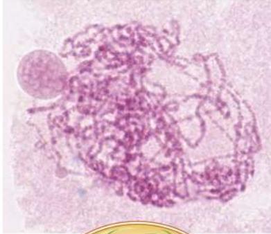
1 The chromatin begins to condense following interphase.
Mid-prophase I
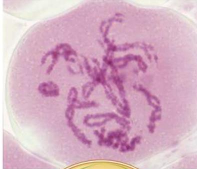
2 Synapsis aligns homologs, and replicated chromosomes condense further.
Late prophase I-Prometaphase
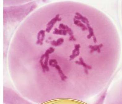
3 The chromosomes continue to coil and shorten. The chiasmata reflect crossing over, the exchange of genetic material between nonsister chromatids in a homologous pair. In prometaphase the nuclear envelope breaks down.
MEIOSIS II
Prophase II
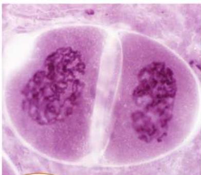
7 The chromosomes condense again, following a brief interphase in which DNA does not replicate.
Metaphase II
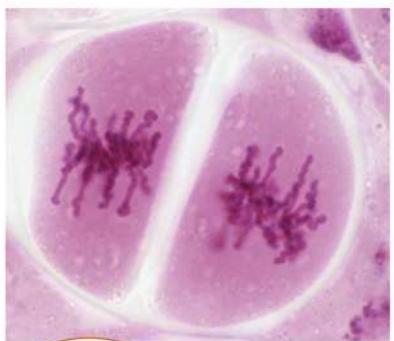
8 The centromeres of the paired chromatids line up across the metaphase II plate of each cell.
Anaphase II
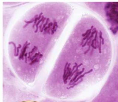
9 The sister chromatids separate and move to opposite poles. Independent alignment of homologous pairs at metaphase I and crossing over guarantee that each gamete will be different.
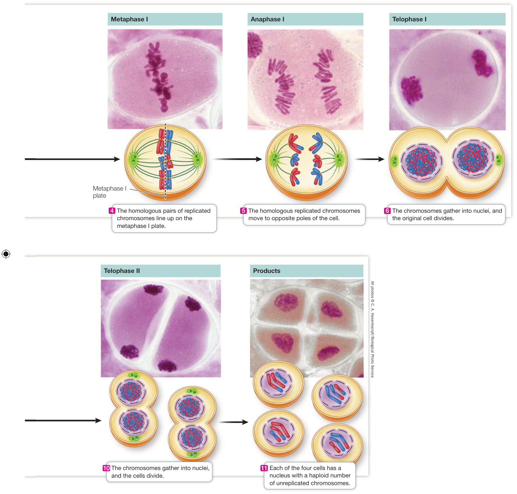
Figure 11.15 Meiosis: Generating Haploid Cells In meiosis, four daughter nuclei are produced, each of which has half as many chromosomes as the original cell. Four haploid cells are the result of two successive nuclear divisions. The micrographs show meiosis in the male reproductive organ of a lily; the diagrams show the corresponding phases in an animal cell.
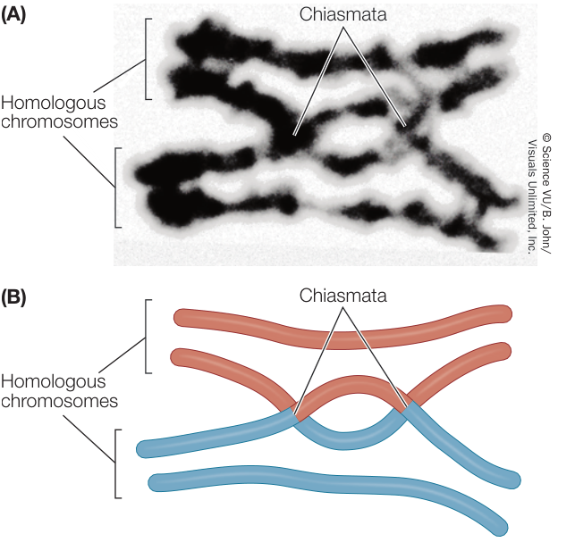
Figure 11.16 Chiasmata: Evidence of Genetic Exchange between Chromatids (A) This micrograph shows a pair of homologous chromosomes, each with two chromatids, during prophase I of meiosis. Two chiasmata are visible. (B) Using colors to represent the two homologs emphasizes where they have undergone genetic exchange.
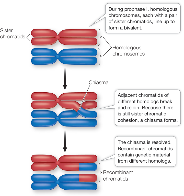
Figure 11.17 Crossing Over Forms Genetically Diverse Chromosomes The exchange of genetic material by crossing over results in new combinations of genetic information on the recombinant chromosomes. The two different colors distinguish the chromosomes contributed by the male and female parents.
be pulled apart by spindle microtubules, especially near the centromeres, but they remain held together by physical attachments mediated by cohesins. Later in prophase I, regions having these attachments take on an X-shaped appearance (Figure 11.16) and are called chiasmata (singular chiasma, “cross”).
A chiasma reflects an exchange of genetic material between nonsister chromatids on homologous replicated chromosomes—what geneticists call crossing over (Figure 11.17). The chromosomes usually begin exchanging material shortly after synapsis begins, but chiasmata do not become visible until later, when the homologs are beginning to separate. Crossing over results in recombinant chromatids, and it increases genetic variation among the products of meiosis by shuffling genetic information among the homologous pairs. In Chapter 12 we will explore further the genetic consequences of crossing over.
At this point, pause and think about what is going on at the molecular level: Each chromatid is a double-stranded DNA molecule, and the homologous chromatids therefore are homologous in DNA nucleotide sequence. Breakage and reunion of DNA molecules to form recombinant chromatids involve breakage of phosphodiester bonds that hold adjacent nucleotides on DNA together (see Figure 4.2) and formation of new bonds linking DNA molecules of two homologous, nonsister chromatids.
Mitosis seldom takes more than an hour or two, but meiosis can take much longer. In human males, the cells in the testis that undergo meiosis take about a week for prophase I and about a month for the entire meiotic cycle. In females, prophase I begins long before a woman’s birth, during her early fetal development, resumes and proceeds to meiosis II as much as decades later, during the monthly ovarian cycle, and is completed only when fertilization occurs (see Key Concept 41.2).
During meiosis homologous chromosomes separate by independent assortment
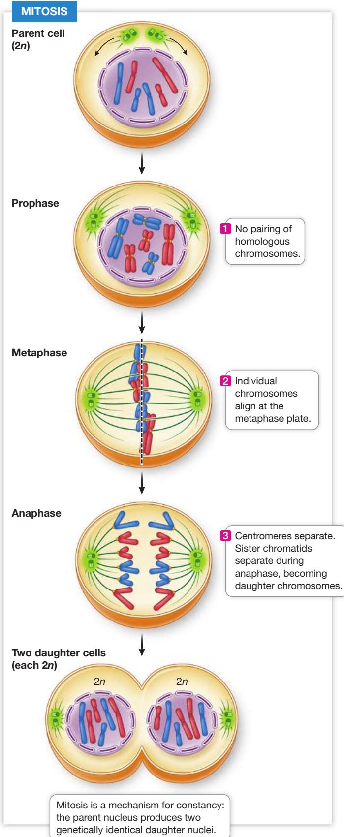
A diploid organism has two sets of chromosomes (): one set derived from its male parent (the paternal set), and the other from its female parent (the maternal set). As the organism grows and develops, its cells undergo mitotic divisions. In mitosis, each chromosome behaves independently of its homolog, and its two sister chromatids are sent to opposite poles during anaphase. Each daughter nucleus ends up with chromosomes, which are identical to those of the parent cell. In meiosis, things are very different. Figure 11.18 compares the two processes.
Figure 11.18 Mitosis and Meiosis: A Comparison ▶
Meiosis I differs from mitosis chiefly by the pairing of homologs and by the continued association of sister chromatids during anaphase I, which results in a reduction in chromosome number (from replicated chromosomes to replicated chromosomes). Meiosis II and mitosis are essentially equivalent, with the sister chromatids of each chromosome ending up at opposite pole.
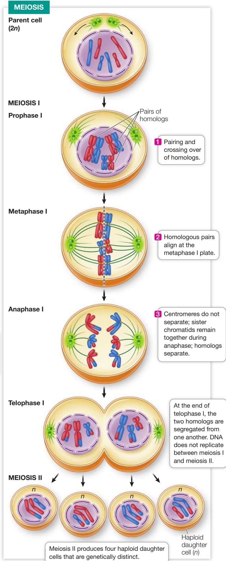
In meiosis I, chromosomes of maternal origin pair with their paternal homologs during synapsis. To clarify, you have two copies of chromosome 1 of the human genome in your cells. Your maternal copy came from your biological mother and your paternal copy from your biological father. In meiosis I, your maternal and paternal chromosome 1’s pair up. And so on with the other 22 pairs of chromosomes. Pairing of maternal and paternal homologs does not occur in mitosis. Segregation of the homologs during meiotic anaphase I ensures that each newly formed cell receives one member of each homologous pair (see steps 4–6 of Figure 11.15). Returning to the example of chromosome 1, at the end of meiosis I in your cells, each daughter nucleus contains one copy of chromosome 1 (either the maternal or paternal copy), whereas the diploid cell that began meiosis had two copies. Taking the other 22 pairs of chromosomes into account, at the end of meiosis I in humans, each of the two daughter cells has 23 of the original 46 chromosomes.
Recall that before meiosis begins each chromosome is duplicated in S phase of interphase. So the beginning cell of meiosis had two replicated copies of each chromosomal DNA molecule. After meiosis I each daughter cell therefore has one copy of each replicated chromosome. In meiosis II, each product of meiosis I divides further to form two haploid gametes, with one copy of each unreplicated chromosome. So the end product of meiosis II is four haploid gametes.
Crossing over is one reason for the genetic diversity among the products of meiosis; another is independent assortment, whereby each haploid cell receives an entire set of genes, but only one of each pair from each parent, from the diploid cell in a random fashion. It is a matter of chance which member of a homologous pair goes to which daughter cell at anaphase I. For example, imagine there are two homologous pairs of chromosomes in the diploid parent nucleus. A particular daughter nucleus could receive the paternal chromosome 1 and the maternal chromosome 2. Or it could get paternal 2 and maternal 1, or both maternal, or both paternal. It all depends on how the homologous pairs line up at metaphase I.
Connect the Concepts Mendel proposed the concept of independent assortment after he observed the results of genetic crosses with pea plants. See Key Concept 12.1.
Note that of the four possible chromosome combinations just described, only two produce daughter nuclei with full complements of either maternal or paternal chromosome sets (apart from the material exchanged by crossing over). The greater the number of chromosomes, the less probable it is that the original parental combinations will be reestablished, and the greater the potential for genetic diversity. Most species of diploid organisms have more than two pairs of chromosomes. In humans, with 23 chromosome pairs, (8,388,608) different combinations can be produced just by the mechanism of independent assortment. Taking the extra genetic shuffling afforded by crossing over into account, the number of possible combinations is very large indeed! Crossing over and independent assortment, along with the processes that result in mutations, provide the genetic diversity needed for the differential survival and reproduction of diverse individuals—the basis of evolution by natural selection.
You have seen how meiosis I is fundamentally different from mitosis. In contrast, meiosis II is similar to mitosis in that it involves the separation of sister chromatids into daughter nuclei (see steps 7–11 in Figure 11.15). However, because of crossing over during meiosis I, the sister chromatids are not necessarily identical to one another as they would be in mitosis. Chance assortment of the sister chromatids during meiosis II contributes further to the genetic diversity of the meiotic products. The final products of meiosis I and meiosis II are four haploid daughter cells, each with one set (n) of chromosomes, each of which is genetically unique.
Meiotic errors lead to abnormal chromosome structures and numbers
In the complex processes of mitosis and meiosis, things occasionally go wrong. In meiosis I, a pair of homologous chromosomes may fail to separate, and in mitosis or meiosis II, sister chromatids may fail to separate. Conversely, homologous chromosomes may fail to remain together during metaphase I of meiosis, and then both may migrate to the same pole in anaphase I. These are all examples of nondisjunction, which results in the production of aneuploid cells. Aneuploidy is a condition in which one or a few chromosomes are either lacking or present in excess. If nondisjunction occurs during meiosis, it can lead to offspring with either one too many, or one too few, chromosomes in all of their cells (Figure 11.19).
There are many different causes of aneuploidy, but one cause may be a breakdown in the cohesins that keep sister chromatids and bivalents joined together during prophase. These and other proteins ensure that when the chromosomes line up at the metaphase I plate, for example, one homolog will face one pole and the other homolog will face the other pole. If the cohesins break down at the wrong time, both homologs may go to the same pole.
Aneuploidy resulting from nondisjunction during meiosis is often lethal for the affected offspring. In a few cases, the affected offspring survive but may have certain abnormalities. An example in humans is Down syndrome, which occurs when a gamete has two copies of chromosome 21. If, for example, an egg with two of these chromosomes is fertilized by a normal sperm, the resulting zygote will have three copies of the chromosome: it will be trisomic for chromosome 21. A child with Down syndrome has mild to moderately impaired intellectual ability; characteristic abnormalities of the hands, tongue, and eyelids; and an increased susceptibility to cardiac abnormalities. About 1 child in 800 is born with Down syndrome. If an egg that did not receive chromosome 21 is fertilized by a normal sperm, the zygote will have only one copy: it will be monosomic for chromosome 21, and this is lethal during fetal development.
Trisomies and the corresponding monosomies are surprisingly common in human zygotes, with 10%–30% of all conceptions showing aneuploidy. But most of the embryos that develop from such zygotes do not survive to birth, and those that do often die before the age of 1 year (trisomies for chromosome 21 are the viable exception). At least one-fifth of all recognized pregnancies are spontaneously terminated (miscarried) during the first 2 months, largely because of trisomies and monosomies. The actual proportion of spontaneously terminated pregnancies is certainly higher, because the earliest ones often go unrecognized.
Other abnormal chromosomal events can also occur. In a process called translocation, a piece of a chromosome may break away and become attached to another chromosome. For example, a large part of one chromosome 21 may be translocated to another chromosome. Individuals who inherit this translocated piece along with two normal copies of chromosome 21 will also have Down syndrome.
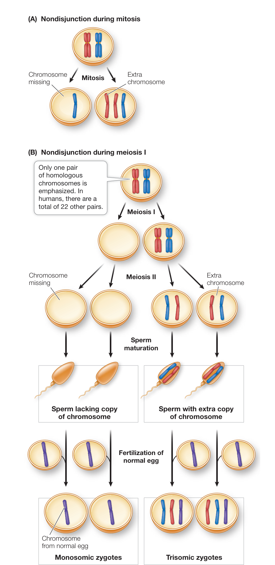
Figure 11.19 Nondisjunction Leads to Aneuploidy (A) In mitosis, nondisjunction results in both daughter cells being aneuploid: one cell has an extra copy of a chromosome and one cell lacks a copy. (B) Nondisjunction in meiosis I during sperm production results in four aneuploid gametes: two sperm have an extra copy of the chromosome and two sperm lack a copy. Fertilization of normal eggs produces aneuploid zygotes. Q: What is the effect of meiosis II nondisjunction on gametes?
The number, shapes, and sizes of the metaphase chromosomes constitute the karyotype
When cells are in metaphase of mitosis, it is often possible to count and characterize their individual chromosomes. If a photomicrograph of the entire set of chromosomes is made, the images of the individual chromosomes can be manipulated to pair them and place them in an orderly arrangement. Such a rearranged photomicrograph reveals the number, shapes, and sizes of the chromosomes in a cell, which together constitute its karyotype (Figure 11.20). In humans, karyotypes can aid in the diagnosis of chromosomal abnormalities such as trisomies or translocations, and this has led to an entire branch of medicine called cytogenetics. However, as you will see in Chapter 15, chromosome analysis with the microscope is replaced in some cases by direct analysis of DNA.
Polyploids have more than two complete sets of chromosomes
As mentioned in Key Concept 11.4, mature organisms are often either diploid (for example, most animals) or haploid (for example, most fungi). Under some circumstances, triploid (3n), tetraploid (4n), or higher-order polyploid nuclei may form. Each of these ploidy levels represents an increase in the number of complete chromosome sets present. If there is nondisjunction of all the chromosomes during meiosis I or II (see the earlier discussion), diploid gametes will form. Such an event might involve breakdown of the meiotic spindle so that anaphase I or II does not occur. A diploid gamete can lead to autopolyploidy after fertilization. Multiple fertilization (polyspermy) can also lead to autopolyploidy. Autotriploids and autotetraploids have been important in some cases in species formation. A diploid nucleus can undergo normal meiosis because there are two sets of chromosomes to make up homologous pairs, which separate during anaphase I. Similarly, a tetraploid nucleus has an even number of each kind of chromosome, so each chromosome can pair with its homolog. However, a triploid nucleus cannot undergo normal meiosis because one-third of the chromosomes would lack partners. As a result, triploid individuals are usually sterile.
Because polyploid nuclei have more chromosome sets, their cells tend to be larger. This has led to the use of polyploid plants in agriculture. Diploid bananas () are smaller and produce inedible seeds; triploid bananas () are larger and seedless. A similar phenomenon is seen in triploid seedless watermelon. Perhaps the best known, and certainly the most important, polyploid crop plant is wheat. In this case, hybridization occurred between species, forming new allopolyploid conditions:
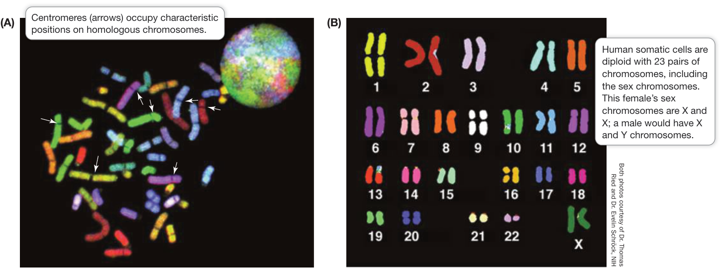
Figure 11.20 The Human Karyotype (A) Chromosomes from a human cell in metaphase. The DNA of each chromosome pair has a specific nucleotide sequence that is stained by a particular colored dye, so that the chromosomes in a homologous pair share a distinctive color. Each chromosome at this stage is composed of two chromatids, but they cannot be distinguished. At the upper right is an interphase nucleus. (B) This karyogram, produced by computerized analysis of the image on the left, shows homologous pairs lined up together and numbered, clearly revealing the individual’s karyotype.
- Haploid gametes from two species (A and B) mated and formed a diploid zygote (chromosomes AB).
- Nondisjunction of all chromosomes occurred during mitosis, resulting in tetraploid (AABB) cells, which then gave rise to fertile flowers (producing AB gametes).
Modern bread wheat is the result of two such events, which occurred about 8,000–10,000 years ago, resulting in an allohexaploid (AABBCC). Wheat’s properties of grain formation and environmental adaptation thus come from three different ancestral species. Other allopolyploid crops include cotton, oats, and sugarcane.
Recap and Assess
Meiosis produces four daughter cells in which the chromosome number is reduced from diploid to haploid. Because of the independent assortment of chromosomes and the crossing over of homologous chromatids, the four products of meiosis are genetically distinct. Meiotic and mitotic errors, such as nondisjunction, can lead to abnormal numbers of chromosomes. Several important crop plants, such as wheat, are polyploid.
- How do crossing over and independent assortment result in unique daughter nuclei?
- Compare prophase I of meiosis with prophase of mitosis. Compare anaphase I of meiosis with anaphase of mitosis.
- Rarely, a person is born with an extra X chromosome (XXY). How might this aneuploidy arise?
- What is polyploidy, and how might it arise?
An essential role of cell division in complex eukaryotes is to replace dead cells. What causes cells to die?
Cell Death Is Important in Living Organisms
Learning Objectives
- 11.6.1 Compare and contrast necrosis and apoptosis.
- 11.6.2 Describe how apoptosis can aid an organism’s survival.
- 11.6.3 Describe how apoptosis is regulated.
Cells within a living organism can die in one of two ways. Necrosis occurs when cells and tissues are damaged by mechanical means or toxins, or are starved of oxygen or nutrients. Necrotic cells often swell up and burst, releasing their contents into the extracellular environment and possibly causing inflammation (see Key Concept 40.2). Apoptosis, or programmed cell death, is a genetically determined series of events that occur during normal developmental processes and in adult tissues as well.
Programmed cell death removes cells that do not benefit the organism
In animals, there are two possible reasons for apoptosis:
- The cell is no longer needed by the organism. For example, before birth, a human fetus has weblike hands, with connective tissue between the fingers. As development proceeds, this unneeded tissue disappears as the cells undergo apoptosis in response to specific signals.
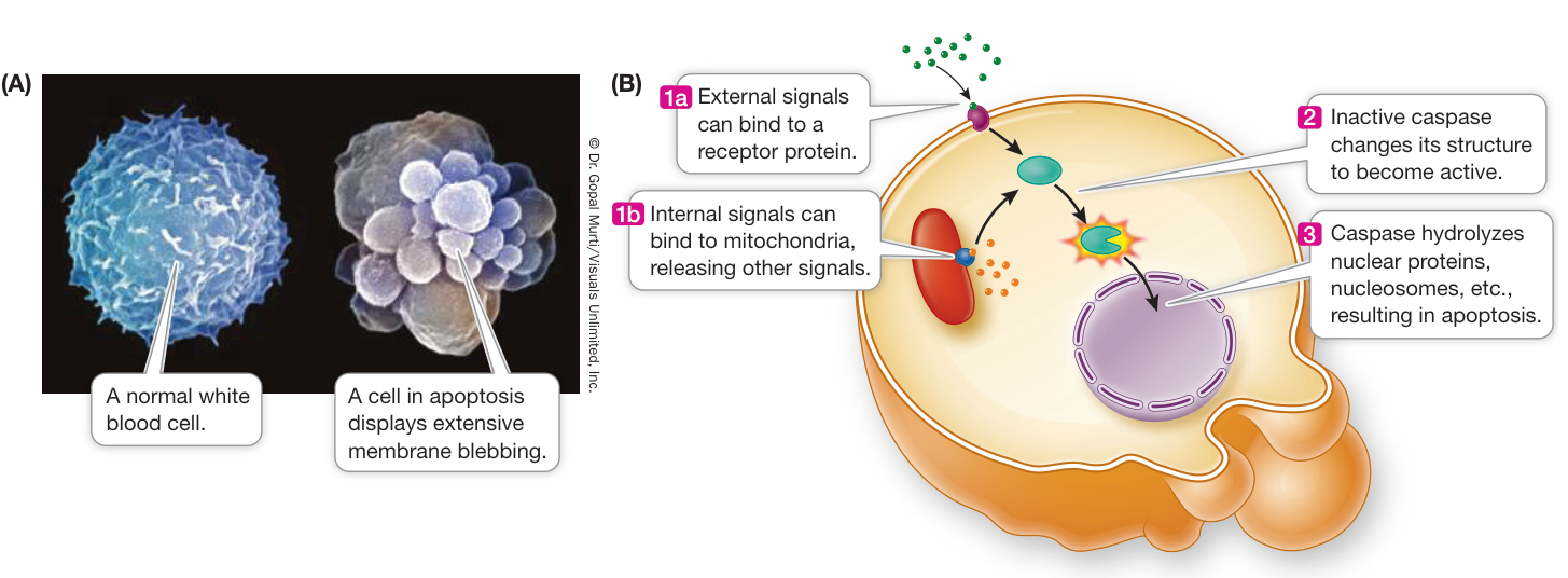
Figure 11.21 Apoptosis: Programmed Cell Death (A) Many cells are programmed to “self-destruct” when they are no longer needed, or when they have lived long enough to accumulate a burden of DNA damage that might harm the organism. (B) Both external and internal signals stimulate caspases, the enzymes that break down specific cell constituents, resulting in apoptosis.
Q: Apoptosis occurs in most organisms, with a common molecular pathway. This indicates that apoptosis is important in terms of evolution. What might be a selective advantage for apoptosis?
- The longer cells live, the more prone they are to genetic damage that could lead to cancer. This is especially true of epithelial cells on the surface of an organism, which may be exposed to radiation or toxic substances. Such cells normally die after only days or weeks and are replaced by new cells.
The events of apoptosis are similar in many organisms. The cell becomes detached from its neighbors and its chromatin is digested by enzymes that cut the DNA (between the nucleosomes) into fragments of about 180 base pairs. The cell forms membranous lobes, or “blebs,” that break up into cell fragments (Figure 11.21A). In a remarkable example of the economy of nature, the surrounding, living cells usually ingest the remains of the dead cell by phagocytosis. Neighboring cells digest the apoptotic cell contents in their lysosomes, and the digested components are recycled.
Apoptosis is also used by plant cells in an important defense mechanism called the hypersensitive response. Plants can protect themselves from disease by undergoing apoptosis at the site of infection by a fungus or bacterium. With no living tissue to grow in, the invading organism is not able to spread to other parts of the plant. Because of their rigid cell walls, plant cells do not form blebs the way animal cells do. Instead, they digest their own cell contents in the vacuole and then release the digested components into the vascular system. You will learn more about this in Chapter 37.
A variety of signals, either external or from inside the cell, can lead to programmed cell death (Figure 11.21B). Such signals include hormones, growth factors, viral infection, certain toxins, or extensive DNA damage. These signals activate specific receptors, which in turn activate signal transduction pathways leading to apoptosis. Some apoptotic pathways target the mitochondria, for example, by increasing the permeability of mitochondrial membranes. The cell quickly dies if its mitochondria can’t carry out cellular respiration. An important class of enzymes called caspases
are activated during apoptosis. These enzymes are proteases that hydrolyze target molecules in a cascade of events. The cell dies as the caspases hydrolyze proteins of the nuclear envelope, nucleosomes, cytoskeleton, and cell membrane.
Recap and Assess
Cell death can occur either by necrosis or by apoptosis. Apoptosis is governed by precise molecular controls.
- What types of events initiate necrosis, and how do these events differ from those that initiate apoptosis?
- In what situations is apoptosis necessary?
- How is apoptosis regulated?
Mitosis adds cells to organisms, and apoptosis removes them. Under normal circumstances these processes are balanced so as to benefit the organism as a whole. In the next key concept we will examine what happens when this balance is disturbed and cell production runs out of control.
Unregulated Cell Division Can Lead to Cancer
Learning Objectives
11.7.1 Compare and contrast cancer cells and normal cells.
11.7.2 Explain how regulation of the cell cycle can become disrupted in cancer cells.
11.7.3 Justify the rationale for targeting events in the cell cycle as a means of treating cancer.
Perhaps no malady affecting people in the industrialized world instills more fear than cancer, and most people realize that it involves an inappropriate increase in cell numbers. One in three Americans will have some form of cancer in his or her lifetime, and at present, one in five will die of it. With 1.7 million new cases and 600,000 deaths in the United States annually, cancer ranks second only to heart disease as a killer.
Cancer cells differ from normal cells in important ways
Cancer cells differ from the normal cells from which they originate in two ways:
- Cancer cells lose control over cell growth.
- Cancer cells can migrate to other locations in the body.
Most cells in the body divide only if they are exposed to extracellular signals such as growth factors. Cancer cells do not respond to these controls, and instead divide more or less continuously, ultimately forming tumors (large masses of cells). By the time a physician can feel a tumor or see one on an X-ray film or CT scan, it already contains millions of cells. Tumors can be benign or malignant:
- Benign tumors resemble the tissue they came from, grow slowly, and remain localized where they develop. For example, a lipoma is a benign tumor of fat cells that may arise in the armpit and remain there. Benign tumors are not cancers, but they must be removed if they impinge on an organ, obstructing its function.
- Malignant tumors do not look like their parent tissue at all. A flat, specialized epithelial cell in the lung wall may turn into a relatively featureless, round, malignant lung cancer cell (Figure 11.22). Malignant cells often have irregular structures, such as variable nucleus sizes and shapes. This characteristic was used to identify the cells in Henrietta Lacks’s tumor as malignant (see the opening investigation of this chapter).
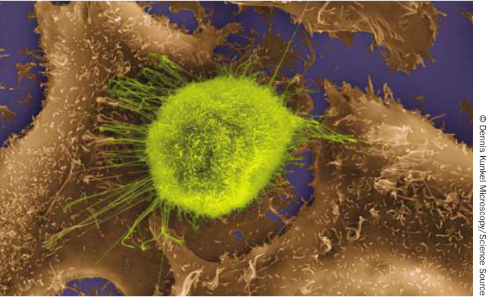
Figure 11.22 A Cancer Cell with Its Normal Neighbors This lung cancer cell (yellow-green) is quite different from the normal lung cells surrounding it. The cancer cell can divide more rapidly than its normal counterparts, and it can spread to other organs. This form of small-cell cancer is lethal, with a 5-year survival rate of only 10%. Most cases are caused by tobacco smoking.
The second and most fearsome characteristic of cancer cells is their ability to invade surrounding tissues and spread to other parts of the body by traveling through the bloodstream or lymphatic ducts. When malignant cells become lodged in some distant part of the body, they go on dividing and growing, establishing a tumor at that new site. This spreading of cancer, called metastasis, results in organ failures and makes the cancer very hard to treat.
Cancer cells lose control over the cell cycle and apoptosis
Earlier in this chapter you learned about proteins that regulate the progress of a eukaryotic cell through the cell cycle:
- Positive regulators such as growth factors stimulate the cell cycle: they are like “gas pedals.”
- Negative regulators such as retinoblastoma protein (RB) inhibit the cell cycle: they are like “brakes.”
Just as driving a car requires stepping on the gas pedal and releasing the brakes, a cell will go through a division cycle only if the positive regulators are active and the negative regulators are inactive.
In most cells, the two regulatory systems ensure that the cells divide only when needed. In cancer cells, these two processes are abnormal.
- Oncogene proteins are positive regulators in cancer cells. They are derived from normal positive regulators that have become mutated to be overly active or that are present in excess, and they stimulate the cancer cells to divide more often. Oncogene products could be growth factors, their receptors, or other components in the signal transduction pathways (see Chapter 7) that stimulate cell division. An example of an oncogene protein is a growth factor receptor in a breast cancer cell (Figure 11.23A). Normal breast cells have relatively low numbers of the human epidermal growth factor receptor HER2. So breast cells are not usually stimulated to multiply in the presence of this growth factor. In about 25% of breast cancers, a DNA change results in the increased production of the HER2 receptor. This results in positive stimulation of the cell cycle, and a rapid proliferation of cells with the altered DNA.
- Tumor suppressors are negative regulators in both cancer and normal cells, but in cancer cells they are inactive. An example is the RB protein, which when active acts at R (the restriction point) in G1 to block the cell cycle (see Figure 11.3). In some cancer cells RB is inactive, allowing the cell cycle to proceed. Some viral proteins can inactivate tumor suppressors. For example, the human papillomavirus infects cells of the cervix and produces a protein called E7. E7 binds to the RB protein and prevents it from inhibiting the cell cycle (Figure 11.23B). Another important tumor suppressor is p53, a transcription factor that is involved in cell cycle checkpoint pathways and apoptosis. The importance of p53 as a tumor suppressor is illustrated by the fact that more than 50% of human tumors have mutations in the gene that encodes p53.
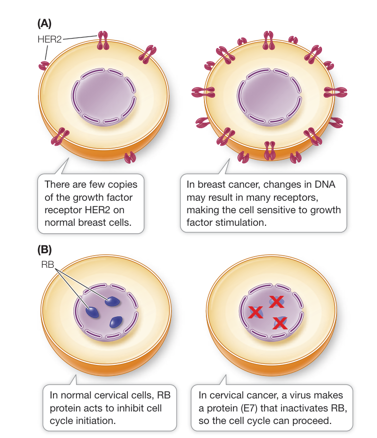
Figure 11.23 Molecular Changes in Cancer Cells In cancer, oncogene proteins become active (A) and tumor suppressor proteins become inactive (B).
With oncogenes (the gas pedal) and tumor suppressor genes (the brakes), it takes more than one protein to allow the cancer cell cycle to proceed. There may be several oncogenes and tumor suppressor genes involved in a single tumor. For example, two important oncogenes in mouse cells are Myc, whose expression stimulates the cell cycle and prevents apoptosis, and Ras, which encodes a signaling molecule you saw in Chapter 7 (see Figure 7.9). Experiments show that it takes the expression of both of these oncogenes to set the cell cycle in motion in mouse cells and convert them into tumor cells (Figure 11.24).
The discovery of apoptosis (see Key Concept 11.6) changed the way biologists think about cancer. In a population of cells, the net increase in cell numbers over time (the growth rate) is the difference between the rate at which cells are added (the rate of cell division) and the rate at which cells are lost (the rate of apoptosis):
Tumor growth rate = rate of cell division – rate of apoptosis
In normal nongrowing tissues, the rate of cell division equals the rate of apoptosis, so the cell population as a whole does not grow. Cancer cells are defective in their regulation of the cell cycle, resulting in increased rates of cell division. In addition, cancer cells can lose the ability to respond to positive regulators of apoptosis (see Figure 11.23B), and this results in lowered rates of cell death. Both of these defects lead to an increased growth rate of the cancer cell population.
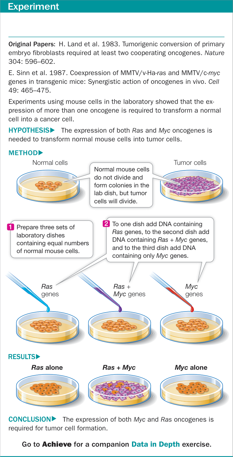
Figure 11.24 Does It Take Multiple Events to Trigger the Cancer Cell Cycle?
Cancer treatments target the cell cycle
The most successful and widely used treatment for cancer is surgery. While physically removing a tumor is optimal, it is often difficult for a surgeon to get all of the tumor cells. (A tumor about 1 centimeter in diameter already has a billion cells!) Tumors are generally embedded in normal tissues. Added to this is the probability that cells of the tumor may have broken off and spread to other organs. This makes it unlikely that localized surgery will be curative. So other approaches are taken to treat or cure cancer, and these generally target the cell cycle (Figure 11.25). The goal is to decrease the rate of cell division and/or increase the rate of apoptosis so that the cancer cell population decreases.
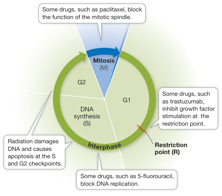
Figure 11.25 Cancer Treatment and the Cell Cycle To prevent cancer cells from dividing, physicians use combinations of therapies that attack the cell cycle at different points. Q: How do these treatments differ from treatments based on specific targets (see Figure 11.23)? Would the side effects of the two types of treatments be different?
An example of a cancer drug that targets the cell cycle is 5-fluorouracil, which blocks the synthesis of thymine, one of the four bases in DNA. The drug paclitaxel prevents the functioning of microtubules in the mitotic spindle. Both drugs inhibit the cell cycle, and apoptosis causes tumor shrinkage. More dramatic is radiation treatment, in which a beam of high-energy radiation is focused on the tumor. DNA damage is extensive, and the cell cycle checkpoint for DNA repair is overwhelmed. As a result, the cell undergoes apoptosis. A major problem with these treatments is that they affect normal cells as well as the tumor cells. These treatments are toxic to tissues with large populations of normal dividing cells such as those in the intestine, skin, and bone marrow (where blood cells are produced).
A major effort in cancer research is to find treatments that target only cancer cells. A promising recent example is trastuzumab, which targets the HER2 growth factor receptor that occurs at high levels on the surfaces of some breast cancer cells (see Figure 11.23A). Trastuzumab binds specifically to the HER2 receptor but does not stimulate it. This prevents the natural growth factor from binding and stimulating cell division. Because cell division is blocked and the apoptosis rate remains the same, the tumor shrinks.
Recap and Assess
Cancer cells differ from normal cells in terms of their rapid cell division and their ability to spread (metastasis). Many proteins regulate the cell cycle, either positively (oncogenes) or negatively (tumor suppressors). In cancer, one or more of these proteins is altered in some way, making its activity abnormal. Radiation and many cancer drugs target genes and proteins involved in the cell cycle.
- How do cells in normal tissue differ from those in benign and malignant tumors?
- How are oncogene proteins and tumor suppressor proteins involved in cell cycle control in normal and cancer cells?
- Cancer-treating drugs are usually given in combination to target several stages of the cell cycle. Why might this be a better approach than using a single drug?
We have now looked at the cell cycle and at cell division by binary fission, mitosis, and meiosis. We have described the normal cell cycle and how its regulation is disrupted in cancer. We have seen how meiosis produces haploid cells in sexual life cycles. In the coming chapters we will examine heredity, genes, and DNA.
Investigating Life: QA What controls the reproduction of cancer cells?
In normal tissues, the rate of cell division is offset by the rate of cell death. Unlike most normal cells, HeLa cells keep growing because they have a genetic imbalance that heavily favors cell reproduction over cell death. Henrietta Lacks was infected with the human papillomavirus, which stimulates cell division in cervical cells. In addition, an enzyme called telomerase, which keeps DNA intact and prevents cell death, is overexpressed in HeLa cells. This combination of traits—increased cell division plus decreased apoptosis—leads to the extraordinary growth rate of HeLa cells.
Because HeLa cells came from a tumor, you might expect that they are still cancer cells, and that they can form a tumor. But such is not the case. They are just cell division machines. What do you think they are missing that tumor cells have?
Future directions
Knowledge of which genes are abnormal in tumors has led to the development of drugs targeted to the specific proteins that are made from these genes. In some instances, such as breast cancers expressing HER2, the results in the clinic have been extraordinary. Patients are living longer, and even being cured by these targeted drugs. Less attention has been given to the other part of the cell population equation—cell death. Many of the widely used drugs that affect the cell cycle in general (see Figure 11.25) work not just by stopping the cell cycle but by damaging cells, triggering apoptosis. But as you may know, these drugs have widespread side effects, stopping cell division and triggering apoptosis in normal cells as well as tumor cells. We know about the molecular events in apoptosis. Perhaps targeted drugs that stimulate this process only in tumor cells could be effective.
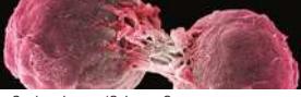
© Steve Gschmeissner/Science Source
You should be able to relate each summary to the adjacent figures. If you go to this visual summary in Achieve, you can follow links to figures, animations, activities, and simulations that will help you consolidate the material.
Visual Summary
All Cells Derive from Other Cells
- Cell division involves cell division signals, DNA replication, DNA segregation, and cytokinesis.
- Prokaryotes reproduce by binary fission.
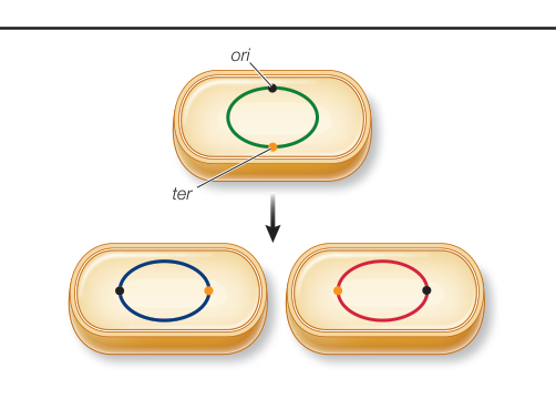
Figure 11.2 Questions
- Briefly describe what happens in each of the four cell division events.
- What are the functions of the ori and ter sequences on the bacterial chromosome?
The Eukaryotic Cell Division Cycle Is Regulated
- The eukaryotic cell cycle has four phases: G1, S (DNA replication), G2, and M (mitosis and cytokinesis).
- Short-lived cyclins and their more permanent CDKs regulate the passage of cells through cell cycle checkpoints.
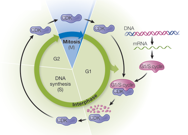
Figure 11.6 Questions
- What events are occurring in the cell during each of the four phases of the eukaryotic cell cycle?
- How are cyclins able to regulate movement through the cell cycle?
Eukaryotic Cells Divide by Mitosis
- DNA is packaged with proteins, especially histones, to form chromatin.
- Mitosis can be divided into prophase, prometaphase, metaphase, anaphase, and telophase and is followed by cytokinesis.
- During mitosis, sister chromatids separate, resulting in identical daughter chromosomes.
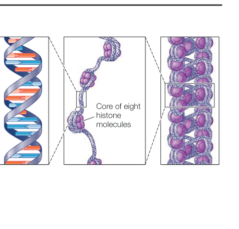
Figure 11.8 1. During mitosis, when do chromosomes align in the middle of the cell? When do sister chromatids move toward opposite poles? When do chromosomes become visible? 2. What is a nucleosome? 3. To what structures are the microtubules that move chromosomes attached?
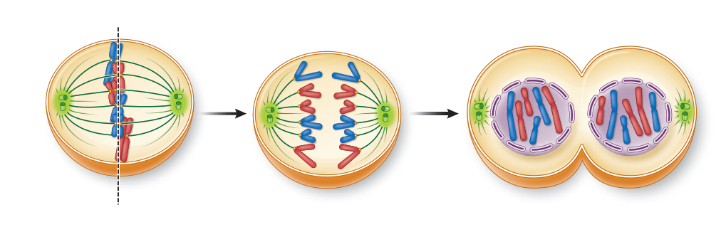
Figure 11.9
Cell Division Plays Important Roles in the Sexual Life Cycle
- ■ Asexual reproduction produces clones, new organisms that are genetically identical to the parent.
- ■ The sexual life cycle involves meiosis and fertilization.
- ■ Sexual life cycles can be haplontic, diplontic, or haplo-diplontic.
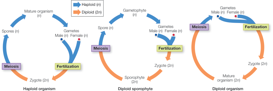
Figure 11.14
Questions
- What two events characterize a sexual life cycle?
- What is the difference between haplontic, diplontic, and haplo-diplontic life cycles?
Meiosis Leads to the Formation of Gametes
- ■ Meiosis produces four genetically distinct haploid gametes from a diploid parent cell.
- ■ Homologs pair during synapsis and exhibit crossing over during prophase I.
- ■ Meiosis I differs from mitosis because homologs separate but sister chromatids remain together.
- ■ Nondisjunction during mitosis or meiosis results in aneuploidy, or polyploidy if all chromosomes are involved.
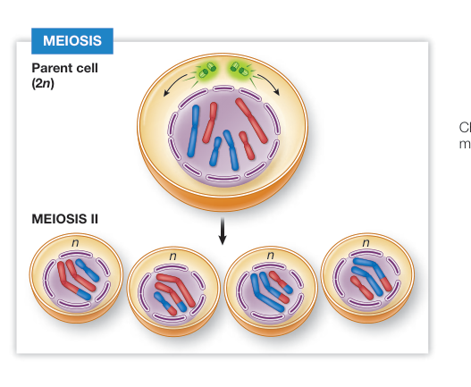
Figure 11.18
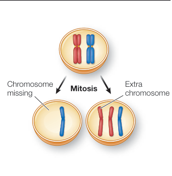
Figure 11.19
Questions
- Draw metaphase I in a diploid cell that has four chromosomes (two pairs of homologs).
- Explain why mitosis, but not meiosis, can occur in a haploid organism.
Cell Death Is Important in Living Organisms
- ■ A cell may die by necrosis, or it may self-destruct by genetically programmed apoptosis.
- ■ Apoptosis is regulated by external and internal signals.
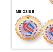
Figure 11.21
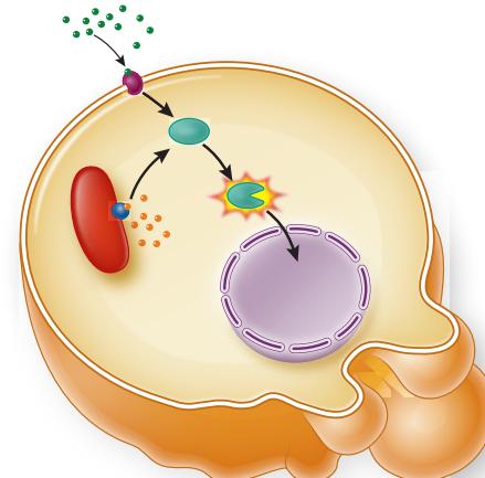
- Some cells instigate apoptosis within a few minutes of being infected by a virus. Why might they do this?
- During development, young healthy cells will sometimes undergo apoptosis. Why might they do this?
Unregulated Cell Division Can Lead to Cancer
- ■ Cancer cells show uncontrolled cell division and metastasis.
- ■ Oncogenes are activated and tumor suppressors are inhibited in cancer cells.
- ■ Cancer treatment often targets the cell cycle in tumor cells.
Questions
- What are two characteristic of cancer cells?
- A misregulated oncogene is usually lethal when inherited in a gamete. In contrast, a misregulated tumor suppressor gene is often not lethal, though cancer rates tend to increase. Explain this difference.
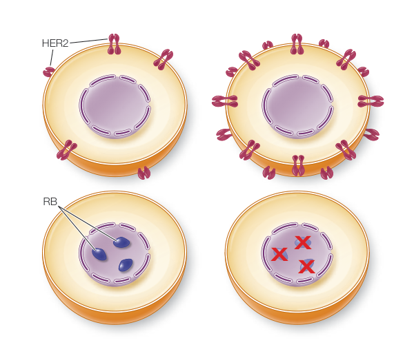
Figure 11.23