Chapter 14From DNA to Protein: Gene Expression
From DNA to Protein: 14 Gene Expression
Courtesy of the National Institute of Allergy and Infectious Diseases (NIAID)
Key Concepts
14.1 Genes Code for Proteins
14.2 Information Flows from Genes to Proteins
14.3 DNA Is Transcribed to Produce RNA
14.4 Eukaryotic Pre-mRNA Transcripts Are Processed prior to Translation
14.5 The Genetic Code Determines the Protein Sequence Encoded by an mRNA
14.6 The Coding Sequence in mRNA Is Translated into Proteins by Ribosomes
14.7 Polypeptides Can Be Modified and Transported during or after Translation
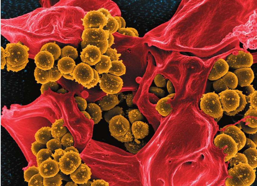
Methicillin-resistant Staphylococcus aureus, a major cause of serious illness and death in the United States and Europe, is treated with antibiotics that target its gene expression.
Investigating Life: Employing the Genetic Code to Combat Superbugs
Staphylococcus aureus is among billions of bacteria that inhabit the human skin and nose, mostly with no ill effect. But sometimes, especially when the immune system has been weakened by age or disease, S. aureus can cause major skin infections and may enter the body through the nose or a wound site, causing serious and sometimes fatal infections of organs such as the heart and lungs.
Until recently, most S. aureus infections were successfully treated with penicillin and related drugs, including methicillin. These antibiotics bind and inactivate several related enzymes involved in the assembly of bacterial cell walls. New bacterial cells produced by cell division of antibiotic-treated bacteria do not survive.
Unfortunately, some S. aureus strains have acquired mutant versions of a penicillin-binding protein that are resistant to antibiotics—the mutant enzyme can catalyze the assembly of cell walls even in the presence of antibiotics. The mutant mecA allele that encodes this mutant protein can be passed from one bacterium to another by bacterial conjugation (see Key Concept 12.6).
What makes the mutant version of mecA different, and how does this difference lead to antibiotic resistance? Compared with the wild-type allele, the mutant mecA allele has a small change in its nucleotide sequence. This results in changes in the amino acid sequence of the expressed protein. This alteration in the protein’s primary structure affects its secondary and tertiary structures: the protein folds into a shape that doesn’t bind the antibiotics.
The fundamental lesson conveyed by this example is that a gene is expressed as a protein, and more specifically, that the sequence of nucleotides in DNA is expressed as a sequence of amino acids in a protein. Understanding how the nucleotide sequence of a gene produces a specific protein came with the discovery of the genetic code, a landmark in biological research.
Mutant bacterial strains that are stubbornly resistant to antibiotics are known as “superbugs.” One greatly feared superbug is methicillin-resistant S. aureus, or MRSA. About 1 person in 50 carries MRSA, so it represents a real threat to public health. MRSA incidence is declining, thanks to careful monitoring and treatment in hospitals and nursing homes, where people are especially vulnerable. The infection is treated with a new class of antibiotics that target bacterial protein synthesis.
QA How does knowledge of the genetic code help us understand the actions of some antibiotics? (Find the answer on page 320.)
Genes Code for Proteins
Learning Objectives
- 14.1.1 Describe the attributes of model organisms used for genetic studies.
- 14.1.2 Determine the order of intermediates in a biosynthetic pathway, given growth data for various mutant strains on these intermediates.
- 14.1.3 Determine the locations of mutant enzymes in a biosynthetic pathway, given growth data for the mutant strains.
In Chapter 4 we introduced DNA and its role in gene expression. Then in Chapter 13 we presented evidence that DNA is the carrier of genetic information and described how DNA is replicated prior to cell division. Here we focus on the evidence that proteins are the major products of gene expression, and we describe how a gene is expressed as protein.
Scientists had a molecular understanding of phenotypes before it was known that DNA was the genetic material; they had studied the chemical differences between individuals carrying wild-type and mutant alleles in organisms as diverse as humans and bread molds and knew that the major phenotypic differences resulted from differences in specific proteins. But just how different proteins arise in different individuals was not understood.
Observations in humans led to the proposal that genes determine enzymes
The identification of a gene product as a protein began with a mutation. In the early twentieth century, English physician Archibald Garrod saw several children with a rare disease. One symptom of the disease was that the urine turned dark brown or black when exposed to air, which was especially noticeable on the infants’ diapers. The disease was given the descriptive name alkaptonuria (“black urine”) (Figure 14.1A).
Garrod noticed that the disease was most common in children whose parents were first cousins. Mendelian genetics had just been “rediscovered,” and Garrod realized that because first cousins share on average one-eighth of their alleles, the children of first cousins might inherit a rare mutant allele from both parents. He proposed that alkaptonuria was a recessive phenotype caused by a mutant allele that causes the disease phenotype.
Garrod took the analysis further by identifying the biochemical abnormality in the affected children. He isolated from them an unusual substance, homogentisic acid, which accumulated in blood, joints (where it crystallized and caused severe pain), and urine (where it turned black). The chemical structure of homogentisic acid is similar to that of the amino acid tyrosine (Figure 14.1B).
The function of enzymes as biological catalysts had just been discovered. Garrod proposed that homogentisic acid was a breakdown product of tyrosine. He suggested that while homogentisic acid ordinarily would be converted to a harmless product, it was not being converted in children with alkaptonuria. Garrod further hypothesized that the enzyme required for the breakdown of homogentisic acid was not being produced in these children. He suggested that a normal human allele (for the wild-type phenotype) was required for the synthesis of an enzyme that catalyzed this conversion. If the allele were mutated, the enzyme would be inactive and homogentistic acid would accumulate instead.
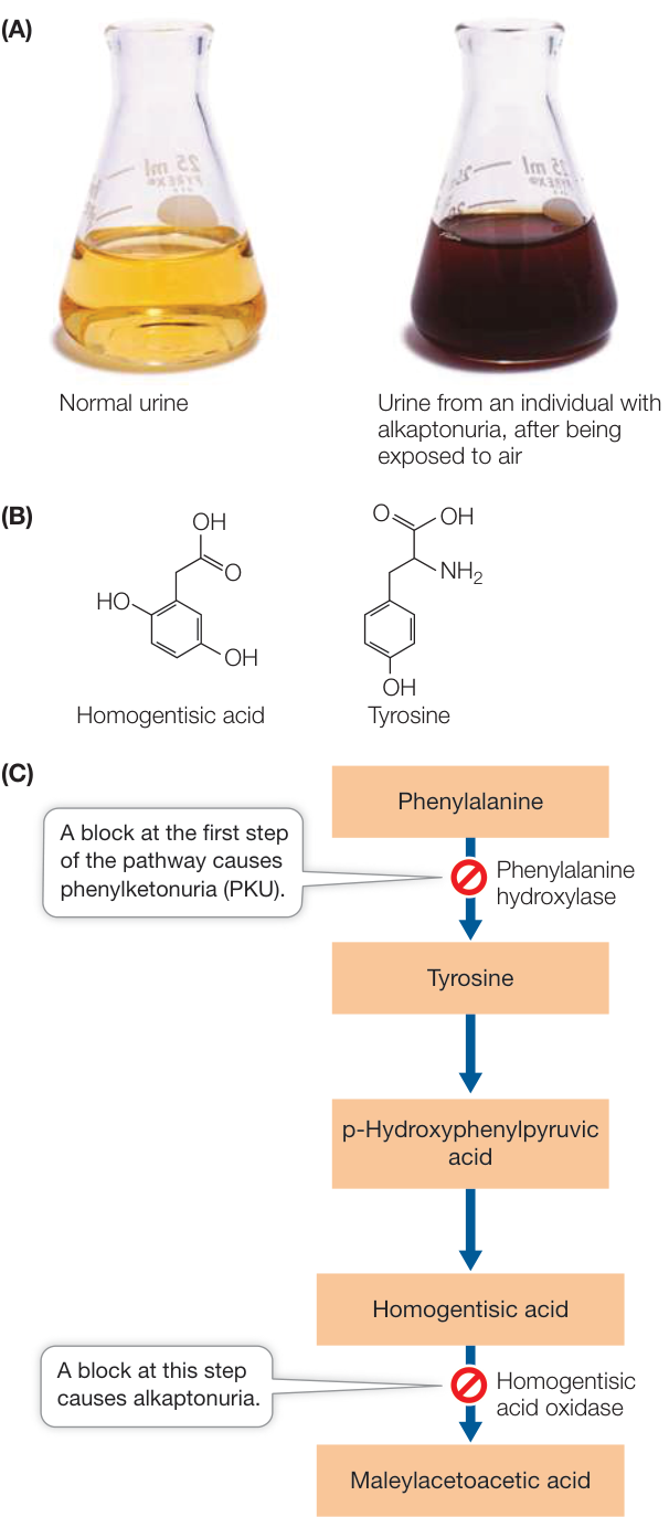
Figure 14.1 Alkaptonuria and Phenylketonuria (A) The physician Archibald Garrod showed that the accumulation of homogentisic acid in the urine of patients with alkaptonuria causes their urine to turn black when exposed to air. (B) Because the chemical structure of homogentisic acid is similar to that of the amino acid tyrosine, Garrod proposed that it was a breakdown product of the amino acid. (C) Both alkaptonuria and phenylketonuria are caused by mutations in specific enzymes in the pathway that breaks down phenylalanine.
Garrod concluded that there must be one gene to one enzyme and coined the term “inborn error of metabolism” to describe this genetically determined biochemical disease. But his hypothesis was not confirmed until the enzyme and pathway were identified (Figure 14.1C). The enzyme, homogentistic acid oxidase, was identified as active in healthy people and inactive in alkaptonuria patients in 1958. The specific DNA mutation that caused the enzyme to be nonfunctional was not described until 1996. An enzyme that catalyzes another step in this pathway is nonfunctional in individuals who have phenylketonuria (PKU). From the pathway, you can see that PKU causes elevated levels of phenylalanine, and this leads to significant intellectual disability if untreated. Fortunately it is now routine to test for high phenylalanine levels in the blood of newborn infants, and if an affected child consumes a diet low in phenylalanine, intellectual disability is avoided.
To relate genes and enzymes more generally, biologists turned to simpler organisms that could be manipulated in the laboratory.
Experiments on bread mold established that genes determine enzymes
As they work to explain the principles that govern life, biologists often turn to organisms that are easy to manipulate experimentally. Such model organisms have certain characteristics that make them attractive experimental subjects. For example, model organisms (1) are easy to grow in the laboratory or greenhouse, (2) have short generation times, (3) often produce large numbers of progeny, and (4) are easy to manipulate genetically, by crossing or by other methods. Biologists use model organisms to discover principles that can then be applied to other organisms. You have encountered some model organisms in previous chapters, including the pea plants (Pisa sativum) used by Mendel (see Key Concept 12.1), the fruit flies (Drosophila) used by Morgan (see Key Concept 12.4), and the E. coli bacteria used by Meselson and Stahl (see Key Concept 13.3).
To this list we now add the bread mold Neurospora, a type of sac fungus (see Chapter 28). In addition to having the traits shared by other model organisms, this mold is haploid for most of its life cycle, so there are no issues with dominance of alleles: all alleles are expressed phenotypically and not masked by a heterozygous condition. Biologists at Stanford University led by George Beadle and Edward Tatum undertook studies to biochemically define the phenotypes in Neurospora.
Like Garrod, Beadle and Tatum hypothesized that the expression of a specific gene results in the activity of a specific enzyme. They set out to test this hypothesis directly. They grew Neurospora on a nutritional medium containing sucrose, minerals, and biotin, which is the only vitamin that wild-type Neurospora cannot synthesize itself. Using this minimal medium, the enzymes of wild-type Neurospora could catalyze all the metabolic reactions needed for growth.
The scientists then treated the wild-type Neurospora with X-rays, which function as a mutagen. A mutagen is something that damages DNA, causing mutations: heritable alterations in the DNA sequence. After the X-ray treatment, some Neurospora strains could no longer grow on the minimal medium. These mutant strains grew only if they were supplied with specific additional nutrients, such as particular vitamins. Beadle and Tatum hypothesized that these genetic strains had mutations in the genes that code for production of enzymes needed to synthesize the additional nutrients. For each mutant strain, the scientists were able to find a single compound that, when added to the minimal medium, supported the growth of that strain. These results suggested that mutations have simple effects, and that each mutation causes a defect in only one enzyme in a metabolic pathway. These conclusions confirmed Garrod’s one-gene, one-enzyme hypothesis.
Mutations provide a powerful way to determine cause and effect in biology. Nowhere has this been more evident than in the elucidation of biochemical pathways. Such pathways consist of sequential steps (chemical reactions) in which each event is dependent on the occurrence of the preceding event. If each reaction is catalyzed by an enzyme, and each enzyme is encoded by a gene, then it should be possible to block a pathway at any step by knocking out (rendering nonfunctional) the gene encoding that enzyme by mutation. Strains with mutated genes will thus not be able to synthesize the final product of the pathway and will exhibit a mutant phenotype. Addition of a compound in the pathway that occurs before the step with the mutated enzyme will not allow the mutant strain to produce the final product of the pathway. In contrast, addition of a compound occurring after the step with the mutated enzyme will allow the mutant strain to produce the final product of the pathway.
Two colleagues of Beadle and Tatum, Adrian Srb and Norman Horowitz, used this reasoning to determine characteristics of the biosynthetic pathway for arginine. They isolated Neurospora mutants that could not survive without the amino acid arginine in their growth medium. By adding particular compounds hypothesized to be in the arginine biosynthetic pathway to the medium, Srb and Horowitz were able to identify a series of steps in the biochemical pathway leading to the synthesis of arginine (Figure 14.2).
One gene determines one polypeptide
The one-gene, one-enzyme relationship has undergone several modifications in light of our current knowledge of molecular biology. Many proteins, including many enzymes, are composed of more than one polypeptide chain, or subunit (recall quaternary protein structure from Key Concept 3.2). So it is more correct to speak of a one-gene, one-polypeptide relationship.
Connect the Concepts Hemoglobin, illustrated in Figure 3.12 is an example of a protein composed of more than one polypeptide chain. Hemoglobin has four polypeptides—two and two subunits, and each kind of subunit is encoded by a separate gene.
So far we have seen that in terms of protein synthesis, the function of a gene is to prescribe the production of a single, specific polypeptide. But not all genes code for polypeptides. As we will see later in this chapter and in Chapter 16, many DNA sequences are transcribed to RNA molecules that are not translated into polypeptides, but instead have other functions.
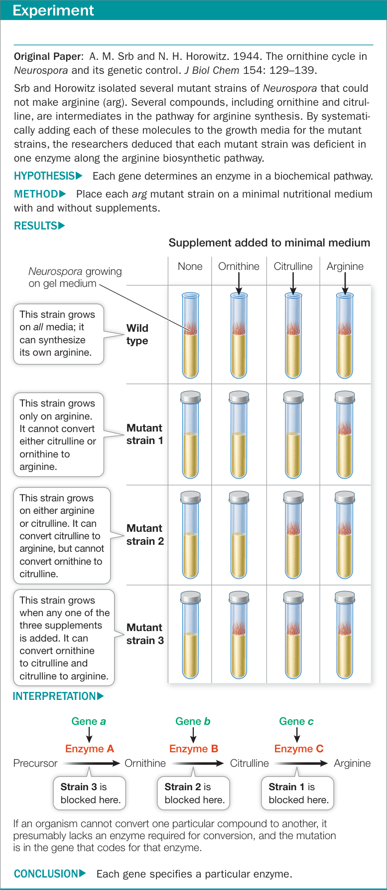
Figure 14.2A One Gene, One Enzyme
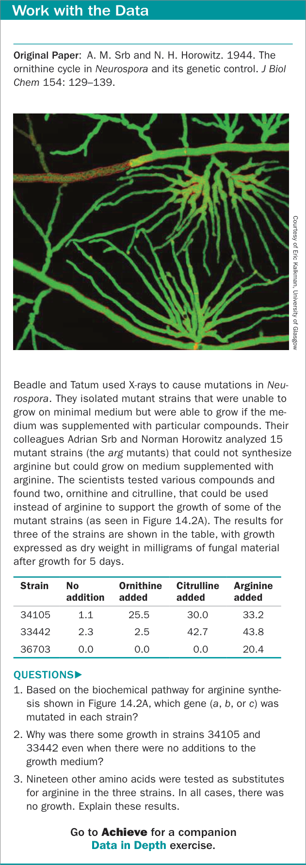
Figure 14.2B One Gene, One Enzyme
Recap and Assess
Studies of mutations in humans and bread molds led to our understanding of the one-gene, one-polypeptide relationship. In most cases, the function of a gene is to code for a specific polypeptide.
- What is a model organism, and why is Neurospora a good model for studying biochemical genetics?
- How were the experiments on mutant strains of Neurospora set up to determine the order of steps in a biochemical pathway?
- In bacteria, the biosynthesis of the amino acid tryptophan (T) from the precursor chorismate (C) involves four intermediate chemical compounds, which we will call D, E, F, and G. Here are the phenotypes of various mutant strains. Each strain has a mutation in a gene for a different enzyme; + means growth with the indicated compound added to the medium, and 0 means no growth. Based on these hypothetical data, order the compounds (C, D, E, F, G, and T) and enzymes (1, 2, 3, 4, and 5) in the tryptophan biosynthetic pathway.
| Mutant strain | Addition to medium | |||||
|---|---|---|---|---|---|---|
| C | D | E | F | G | T | |
| 1 | 0 | 0 | 0 | 0 | + | + |
| 2 | 0 | + | + | 0 | + | + |
| 3 | 0 | + | 0 | 0 | + | + |
| 4 | 0 | + | + | + | + | + |
| 5 | 0 | 0 | 0 | 0 | 0 | + |
Now that you have seen the evidence for the one-gene, one-polypeptide relationship, how does it work? That is, how is the information encoded in DNA used to produce a particular polypeptide?
Information Flows from Genes to Proteins
Learning Objective
14.2.1 Describe the central dogma of molecular biology and give examples of exceptions.
As we discussed in Chapter 13 and in Key Concept 14.1, DNA is the hereditary material and codes for RNAs and proteins. In the remainder of this chapter we focus on the processes that occur when a protein-coding gene is expressed. We briefly outlined gene expression in Key Concept 4.1. To review, this process occurs in two major steps:
- During transcription, the information in a DNA sequence (a gene) is copied into a complementary RNA sequence.
- During translation, this RNA sequence is used to create the amino acid sequence of a polypeptide.
The “central dogma of molecular biology” describes the primary ways in which information flows in a cell: from DNA to DNA, from DNA to RNA, and from RNA to protein (Figure 14.3). We have already discussed replication (see Key Concept 13.3) and now turn to transcription and translation.
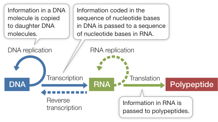
Figure 14.3 The Central Dogma of Molecular Biology The central dogma describes the primary ways in which information flows in the cell: from DNA to DNA, from DNA to RNA and from RNA to protein (solid lines). Information is stored in DNA and copied during replication. Certain DNA sequences are transcribed into RNA, and some RNA sequences are translated into amino acid sequences. Substantially less common, RNA replication (in some RNA viruses) and synthesis of DNA from RNA (reverse transcription) can occur (dashed lines). In no case is amino acid information used to synthesize RNA. Q: Have you seen any examples in previous chapters where information flow does not follow the central dogma (dashed lines)?
Three types of RNA have roles in the information flow from DNA to protein
There are numerous types of RNA. Three of them have vital roles in gene expression and represent the most common RNAs in a typical cell.
MESSENGER RNA When a protein-coding gene is expressed, one of the two DNA strands in the gene is transcribed to produce a complementary RNA strand, which in eukaryotic cells is then processed to produce messenger RNA (mRNA). In eukaryotic cells, the mRNA travels from the nucleus to the cytoplasm, where it is translated into a polypeptide (Figure 14.4). The nucleotide sequence of the mRNA determines the ordered sequence of amino acids in the polypeptide chain, which is synthesized by a ribosome. mRNAs represent about 5% of the RNA in a cell.
RIBOSOMAL RNA The ribosome is essentially a protein synthesis factory composed of multiple proteins and several ribosomal RNAs (rRNAs). One of the rRNAs catalyzes peptide bond formation between amino acids, to form a polypeptide. rRNAs represent about 80% of the RNA in a cell.
TRANSFER RNA Another type of RNA called transfer RNA (tRNA) can both bind a specific amino acid and recognize specific sequences of nucleotides in mRNA. It is the tRNA that recognizes which amino acid should be added next to a growing polypeptide chain. tRNAs represent about 15% of the RNA in a cell.
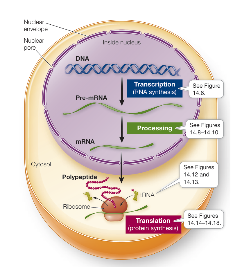
Figure 14.4 From Gene to Protein This diagram summarizes the processes of gene expression in eukaryotes. Note that the nucleus is typically about one-fourth the size shown here. Q: In general, how would this diagram be different for a prokaryotic cell?
In some cases, RNA determines the sequence of RNA or DNA
While we have said that DNA is the genetic material, some viruses present exceptions to this aspect of the central dogma. As we saw in Key Concept 13.1, a virus is a non-cellular infectious particle that reproduces inside cells. Many viruses, such as the ones that cause influenza and polio, have RNA rather than DNA as their genetic material. That is, the nucleotide sequence of viral RNA acts as an information carrier and can be expressed as a protein. Because RNA is usually single-stranded, the question arises: how do these viruses replicate? More specifically, how do they duplicate their genetic material? Most viruses replicate by transcribing from RNA to RNA, making an RNA strand that is complementary to their genomes. This “opposite” strand is then used to make multiple copies of the viral genome.
Not all viruses whose genomes consist of RNA replicate by transcribing from RNA to RNA. Some, such as human immunodeficiency viruses (HIVs) and certain rare tumor viruses, make a DNA copy of their genome after infecting a host cell. This DNA copy is then incorporated into the host’s genome. Synthesis of
DNA from RNA is called reverse transcription, and viruses that employ this kind of transcription are called retroviruses. Retroviruses rely on the host cell’s transcription machinery to make more RNA. This RNA can either be translated to produce viral proteins, or incorporated as the viral genome into new viral particles. You might also recall that telomerase extends telomeres by synthesizing DNA from an RNA template and is thus another example of reverse transcription.
Recap and Assess
The expression of protein-coding genes can be broken down into two fundamental steps: transcription and translation. In transcription one strand of DNA is used to produce a complementary mRNA, whose sequence determines the mature mRNA transcript. The mRNA sequence determines the order of amino acids in a polypeptide. Translation is the process by which this information in mRNA is converted into a polypeptide chain.
- What is the central dogma of molecular biology?
- Do retroviruses violate the central dogma? Explain.
We will revisit viral genetics in later chapters (in Key Concepts 16.3 and 24.4). In the rest of this chapter we focus on gene expression in prokaryotes and eukaryotes. Understanding this process is essential for understanding how organisms function at the molecular level and is key to the application of biology to human welfare, in areas such as agriculture and medicine. We’ll begin by describing how the information in DNA is transcribed to produce RNA.
DNA Is Transcribed to Produce RNA
Learning Objectives
14.3.1 Describe characteristics of RNA polymerases.
14.3.2 Describe the function of the promoter and terminator sequences in transcription.
The synthesis of RNA is directed by DNA. The base sequence of one strand of DNA is used as a template for RNA synthesis, so that the RNA made is complementary in sequence to that DNA strand, with the exception that in RNA there is uracil (U) instead of thymine (T) and in RNA the sugar is ribose instead of deoxyribose. It is important to realize that although the RNA made is a mirror image of its DNA template, the RNA has the same sequence as the other, non-template strand of DNA. So the information content of DNA is indeed preserved in RNA. Since the sequence of nucleotides in the non-template strand is the same as in the protein-coding RNA (except with Ts instead of Us) it is usually referred to as the coding strand.
Transcription—the formation of a specific RNA sequence from a specific DNA sequence—requires several components:
- A DNA template for complementary base pairing—one of the two strands of DNA
Table 14.1 Some RNAs in Eukaryotic Cells
| RNA type | Location of activity | Role |
|---|---|---|
| Ribosomal RNA (rRNA) | Cytoplasm (ribosome) | Component of ribosome, catalysis of peptide bond formation |
| Messenger RNA (mRNA) | Cytoplasm | Carrier of protein-coding sequence |
| Transfer RNA (tRNA) | Cytoplasm | Intermediary between mRNA and protein sequences |
| MicroRNA (miRNA) | Nucleus and cytoplasm | Regulates mRNA stability and translation |
| Small interfering RNA (siRNA) | Nucleus and cytoplasm | Regulates stability of other RNAs |
| Small nuclear RNA (snRNA) | Nucleus | Mediates mRNA processing |
- The four ribonucleoside triphosphates ATP, GTP, CTP, and UTP, to act as substrates
- An RNA polymerase enzyme
- Salts and a pH buffer to create an appropriate chemical environment for RNA polymerase (if transcription is performed in a test tube)
Several kinds of RNA are made from DNA templates. The most important from a genetic point of view is mRNA. But transcription also produces tRNA and rRNA, whose roles in protein synthesis are described in Key Concept 14.6. Like polypeptides, these last two RNAs are encoded by specific genes. Eukaryotes also make many kinds of small RNAs, including small nuclear RNA (snRNA), microRNA (miRNA), and small interfering RNA (siRNA), which are also transcribed. Table 14.1 summarizes some of the RNAs found in eukaryotic cells. We will discuss the roles of miRNA and siRNA in Chapter 16.
RNA polymerases share common features
RNA polymerases from both prokaryotes and eukaryotes catalyze the synthesis of RNA from the DNA template. There is only one kind of RNA polymerase in bacteria, whereas there are several kinds in eukaryotes; however, they all share a common structure (Figure 14.5). Like DNA polymerases, RNA polymerases catalyze the addition of nucleotides in a 5’-to-3’ direction, but unlike DNA polymerases (see Figure 13.12), RNA polymerases do not require a primer.
Transcription occurs in three steps
Transcription can be divided into three distinct processes: (1) initiation, (2) elongation, and (3) termination. Follow these processes in Figure 14.6.
INITIATION Transcription begins when RNA polymerase binds to a special sequence of DNA called a promoter (see Figure 14.6A). Eukaryotic genes generally have one promoter each, whereas in prokaryotes and viruses, several genes often share one promoter. Promoters are important control sequences that “tell” the RNA polymerase two things:
- Where to start transcription
- Which strand of DNA to transcribe
A promoter has directionality, so the RNA polymerase is oriented to the appropriate strand to use as a template. The promoter also determines the initiation site, where transcription begins. Groups of nucleotides lying “upstream” from the initiation site (5’ on the coding strand and 3’ on the template strand) help the RNA polymerase bind. Other proteins, which can bind to specific DNA sequences and to RNA polymerase, help direct the polymerase onto the promoter. These proteins, which include sigma factors in prokaryotes and transcription factors in eukaryotes, help determine the specific genes that are expressed at a particular time in the cell.
Although every gene has a promoter, not all promoters are identical. Some are more effective at transcription initiation than others. Furthermore, there are differences between transcription initiation in prokaryotes and in eukaryotes. We will discuss promoters and their roles in regulating gene expression in Chapter 16.
ELONGATION After RNA polymerase has bound to the promoter, it begins the process of elongation (see Figure 14.6B). DNA unwinds about ten base pairs at a time, and RNA polymerase reads the template strand in the 3’-to-5’ direction. The first nucleotide in the new RNA forms its 5’ end, and subsequent nucleotides complementary to the DNA template are added to its 3’ end. Thus the RNA transcript is antiparallel to the DNA template strand.
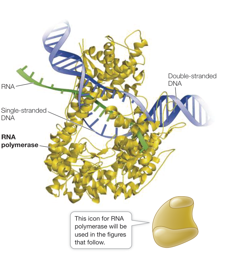
Figure 14.5 RNA Polymerase Interacting with DNA The RNA polymerase enzyme depicted (in yellow-brown) is from bacteriophage T7, but it is representative of most other RNA polymerases. The inset indicates how this enzyme will be represented in the figures that follow. (RNA polymerase by David McIntyre based on data from PDB ID: 1MSW. Y. W. Yin and T. A. Steitz. 2002. Science 298: 1387–1395.)
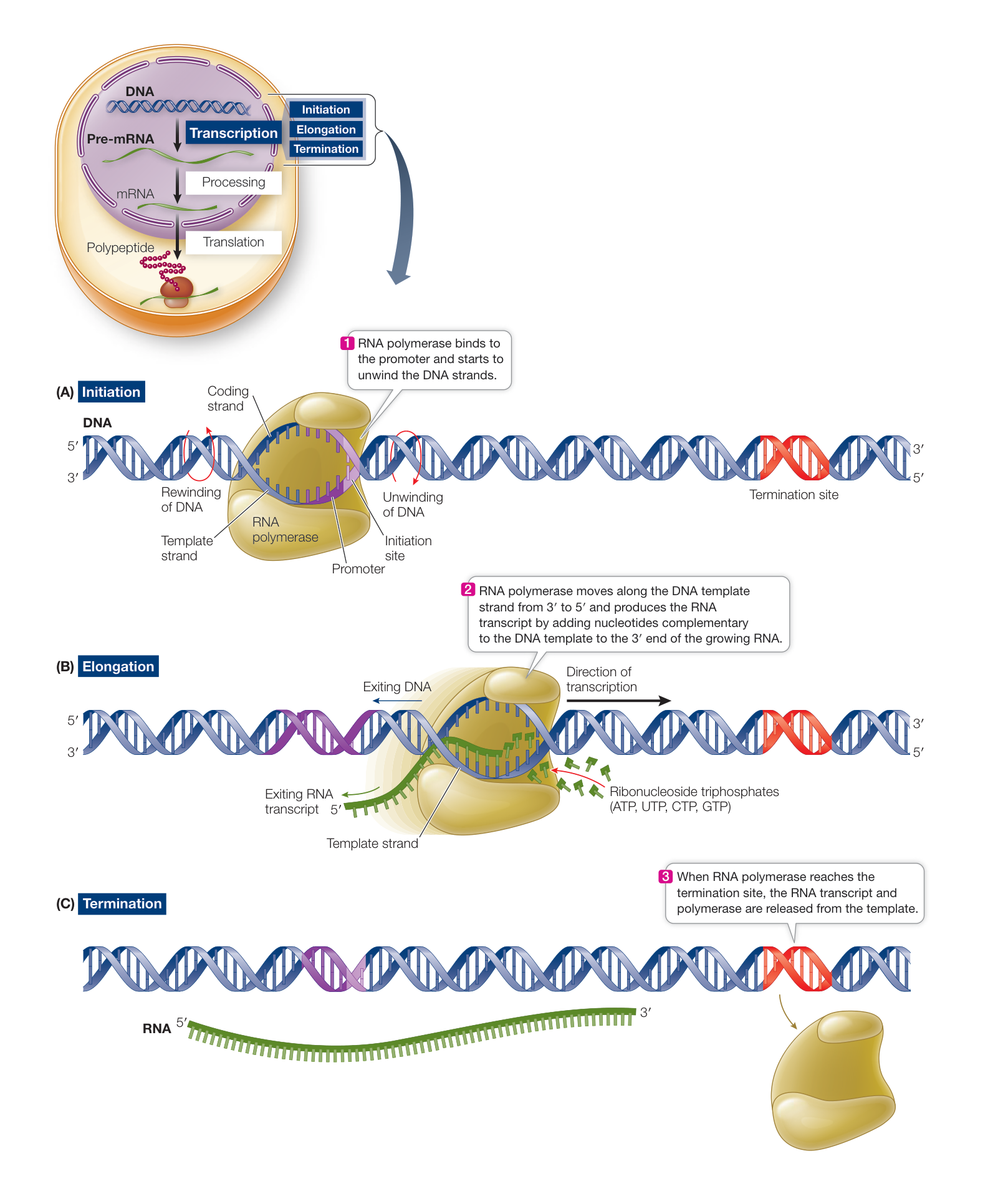
Figure 14.6 DNA Is Transcribed to Form RNA DNA is locally unwound by RNA polymerase, and one strand then serves as a template for RNA synthesis. The RNA transcript is formed and then peels away, allowing the DNA that has already been transcribed to rewind into a double helix. Three distinct processes—initiation (A), elongation (B), and termination (C)—constitute transcription. RNA polymerase is larger in reality than indicated here (see Figure 14.5). Q: Comparing RNA synthesis with DNA replication, what are the common features? Animation 14.1 Transcription
You may recall from Key Concept 13.3 that DNA polymerase uses dNTPs (deoxyribonucleoside triphosphates) as substrates, and forms covalent bonds between each incoming dNTP and the 3’ end of the growing polynucleotide chain (see Figure 13.10). Energy released by the removal of two phosphate groups from the dNTP is used to drive the reaction. Similarly, RNA polymerase uses (ribo)nucleoside triphosphates (NTPs) as substrates, removing two phosphate groups from each substrate molecule and using the released energy to drive the polymerization reaction.
Because RNA polymerases do not proofread, transcription errors occur at a rate of one for every to bases. Because many copies of RNA are made, however, and because they often have only a relatively short life span, these errors are not as potentially harmful as mutations in DNA.
TERMINATION Just as initiation sites in the DNA template strand specify the starting point for transcription, particular base sequences specify its termination (see Figure 14.6C). In prokaryotes there are two mechanisms for ending transcription. For some genes, the newly formed transcript folds back on itself and forms internal hydrogen bonds between bases. A loop forms, and this structure causes the transcript to fall away from the DNA template and the RNA polymerase. In other cases a protein binds to specific sequences on the transcript and causes the RNA to detach from the DNA template. Eukaryotes terminate transcription using methods that are similar to, but more complicated than, those found in prokaryotes.
Recap and Assess
Transcription, which is catalyzed by an RNA polymerase, proceeds in three steps: initiation, elongation, and termination.
- Describe the actions of RNA polymerase during transcription.
- Knocking out (rendering nonfunctional) a particular protein in a bacterium causes many, but not all, genes to produce longer mRNAs. Explain this result.
- Errors in RNA transcription occur about 100,000 times more often than errors in DNA replication. Why can this higher rate be tolerated in RNA but not in DNA synthesis?
The general features of transcription that we have described were first elucidated in model prokaryotes, such as E. coli. Biologists then used the same methods to analyze this process in eukaryotes and found that there are some notable (and important) differences. We turn now to a more detailed description of eukaryotic gene expression.
Eukaryotic Pre-mRNA Transcripts Are Processed prior to Translation
Learning Objectives
- 14.4.1 Describe the mRNA processing steps in eukaryotes.
- 14.4.2 Predict the outcome of a defect in mRNA processing in a eukaryotic cell.
- 14.4.3 Compare the features of a eukaryotic gene with its mature mRNA transcript.
The process of gene expression from DNA to protein is essentially the same in eukaryotes as it is in prokaryotes. In the last key concept we described the basic features of transcription that are common to both. Here we focus on the differences, listed in Table 14.2, between prokaryotic and eukaryotic gene transcription.
In prokaryotes and eukaryotes, the sequence of an mRNA that reaches the ribosome is complementary to the sequence of a gene in the organism’s DNA. One way to show this is by the technique of nucleic acid hybridization, shown in Figure 14.7A. This technique involves two steps:
- A sample of chromosomal DNA containing the gene is denatured to break the hydrogen bonds between the base pairs and separate the two strands.
- The single-stranded mRNA (called a probe) is incubated with the denatured DNA. If the probe has a base sequence complementary to the target DNA, a probe-target double helix forms by hydrogen bonding between the bases. Because the two strands are from different sources, the resulting double-stranded region is called a hybrid.
Hybridization experiments can be performed with various combinations of DNA and RNA (RNA as target and DNA as probe; DNA as both target and probe, etc.). In many hybridization experiments,
Table 14.2 Differences between Prokaryotic and Eukaryotic Gene Expression
| Characteristic | Prokaryotes | Eukaryotes |
|---|---|---|
| Transcription and translation occurrence | At the same time, in the cytoplasm | Transcription in the nucleus, then translation in the cytoplasm |
| Gene structure | DNA sequence aligns perfectly with mRNA sequence | Regions of DNA in gene do not align with mature RNA (introns), and RNA 3’ region does not align with DNA (poly A tail) |
| Modification of mRNA after initial transcription but before translation | Usually none | Introns spliced out; 5’ end cap and 3’ poly A tail added |
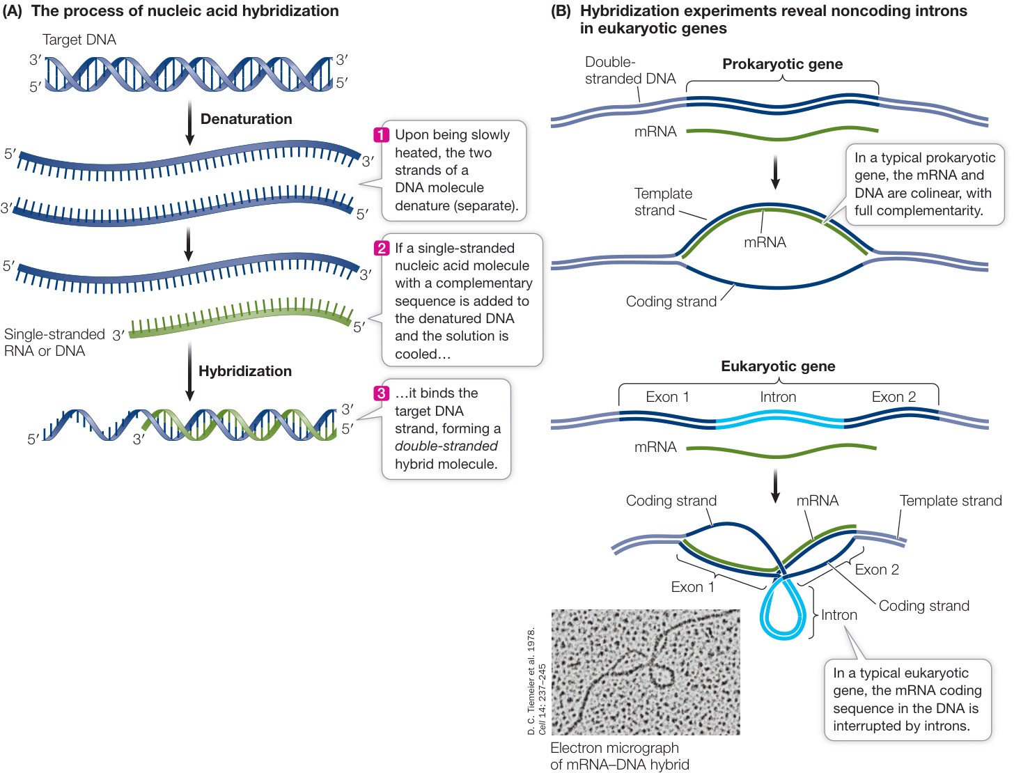
Figure 14.7 Nucleic Acid Hybridization and Introns (A) Base pairing permits the detection of a sequence that is complementary to a probe. (B) Hybridization experiments show that there are introns in eukaryotic genes but generally not in prokaryotic genes.
the probe is labeled in some way so that its binding to a specific target sequence can be detected. The double-stranded hybrids can also be viewed by electron microscopy.
Noncoding sequences called introns often appear between genes in eukaryotic chromosomes
Differences between prokaryotic and eukaryotic transcription are revealed when mRNA probes from prokaryotes and eukaryotes are incubated with their respective chromosomal DNAs:
- In prokaryotes (Figure 14.7B, top) there is usually a 1:1 linear complementarity between the base sequence of the mRNA at the ribosome and that of the chromosomal DNA.
- In eukaryotes (Figure 14.7B, bottom) one or more non-hybridized DNA loops are often observed sticking out of the mRNA–DNA hybrid, indicating that there are stretches of DNA sequence that do not have a complementary sequence in the mRNA that is translated at the ribosome.
The discovery that some stretches of eukaryotic DNA sequence are not present in the mRNA that reaches the ribosome for translation initiated the question of whether this “extra” DNA actually gets transcribed. Does transcription somehow skip these sequences, or are they transcribed and then edited out of the mRNA transcript before it arrives at the ribosome? To find out, an experiment can be conducted in which the initial mRNA transcript in the cell nucleus—the precursor mRNA, or pre-mRNA (see Figure 14.4)—is hybridized with chromosomal DNA. This experiment shows full, linear, loop-free hybridization between the pre-mRNA and the template DNA, allowing us to conclude that the noncoding, intervening regions of DNA, known as introns, do get transcribed but are then spliced out of the pre-mRNA in the nucleus. The remaining sequences (exons) make up the mRNA that reaches the ribosome. The step of splicing out the introns is one of the steps in RNA processing (Figure 14.8).
Introns interrupt, but do not scramble, the DNA sequence of a gene. The base sequences of the exons in the template strand, if joined and taken in order, form a continuous sequence that is complementary to that of the mature mRNA. In some cases the separated exons often encode different functional regions, or domains, of the protein. For example, the globin polypeptides that make up stop site) to terminate translation. The two introns—noncoding sequences of DNA containing almost 1,000 bp among them—are initially transcribed but are spliced out of the pre-mRNA transcript during processing.
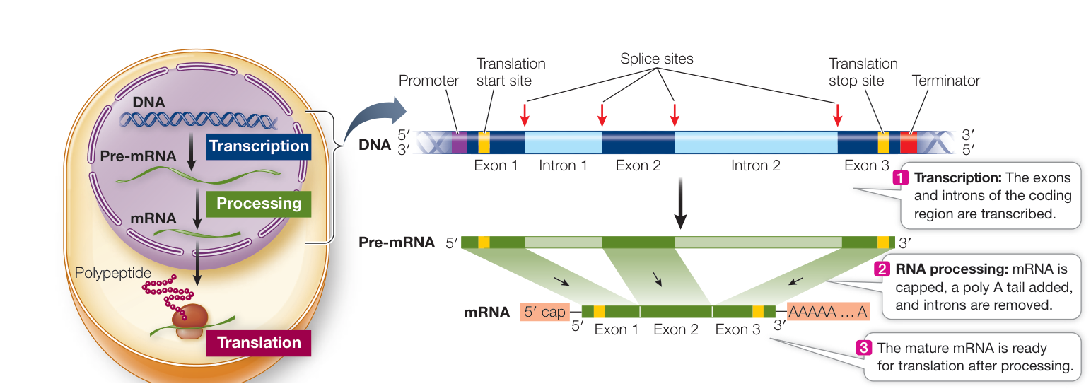
Figure 14.8 Transcription of a Eukaryotic Gene The -globin gene diagrammed here is about 1,700 base pairs (bp) long. The three exons contain the sequence encoding 147 amino acids plus a signal (translation
hemoglobin each have two domains: one for binding to a nonprotein pigment called heme, and another for binding to the other globin subunits. These two domains are encoded by different exons in the globin genes. Most (but not all) eukaryotic genes contain introns, and in rare cases, introns are also found in prokaryotes. Introns can be numerous and large, though the average human gene has only eight, each with an average length of 3,300 bp. The largest human gene encodes a muscle protein called titin; it has 365 exons, which together code for 24,000–36,000 amino acids.
Pre-mRNA processing prepares the mRNA transcript for translation
The transcript of a eukaryotic gene is modified in several ways before it leaves the nucleus: both ends of the pre-mRNA are modified, and the introns are removed.
MODIFICATION AT BOTH ENDS Two steps in the processing of pre-mRNA take place in the nucleus, one at each end of the molecule (Figure 14.9):
- A 5’ cap is added to the 5’ end of the pre-mRNA as it is transcribed. The 5’ cap, which is a chemically modified molecule of guanosine triphosphate (GTP), facilitates the binding of mRNA to the ribosome for translation, and it protects the mRNA from being digested by ribonucleases that break down RNAs.
- A poly A tail is added to the 3’ end of the pre-mRNA at the end of transcription. Transcription ends downstream of the protein-coding region in DNA. In eukaryotes there is usually a “polyadenylation” sequence (AAUAAA) near the 3’ end of the pre-mRNA, downstream of the protein-coding region. This sequence acts as a signal for an enzyme to cut the pre-mRNA. Immediately after this cleavage, another enzyme adds 100 to 300 adenine nucleotides (the poly A tail) to the 3’ end of the pre-mRNA. This tail helps in the export of mature mRNA from the nucleus and is important for mRNA recognition and stability in the cytosol.
SPLICING TO REMOVE INTRONS Within the nucleus, a process called RNA splicing removes the introns and splices the exons together. You can follow the steps of this important process in Fig-ure 14.10. There are specific sequences that are commonly found in introns (consensus sequences) that allow the splicing machinery to identify and precisely remove introns from pre-mRNAs. The splicing machinery includes proteins and RNA–protein complexes called small nuclear ribonucleoproteins (snRNPs) that together recognize and cut out each intron.
Molecular studies of human genetic diseases have provided insights into RNA splicing. For example, people with the genetic disease beta thalassemia have a defect in the production of one of the hemoglobin subunits. These people suffer from severe anemia because they have an inadequate amount of hemoglobin in their red blood cells. In some cases, the genetic mutation that causes the disease occurs at an intron consensus sequence, where the splicing machinery binds to the RNA (see Figure 14.10, step 1) in the -globin gene. Consequently, -globin pre-mRNA is spliced incorrectly, and the resulting mRNA encodes a nonfunctional polypeptide.
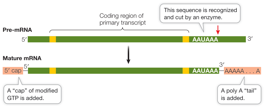
Figure 14.9 Processing the Ends of Eukaryotic Pre-mRNA Modifications at each end of the pre-mRNA transcript—the 5’ cap and the poly A tail—are important for mRNA function.
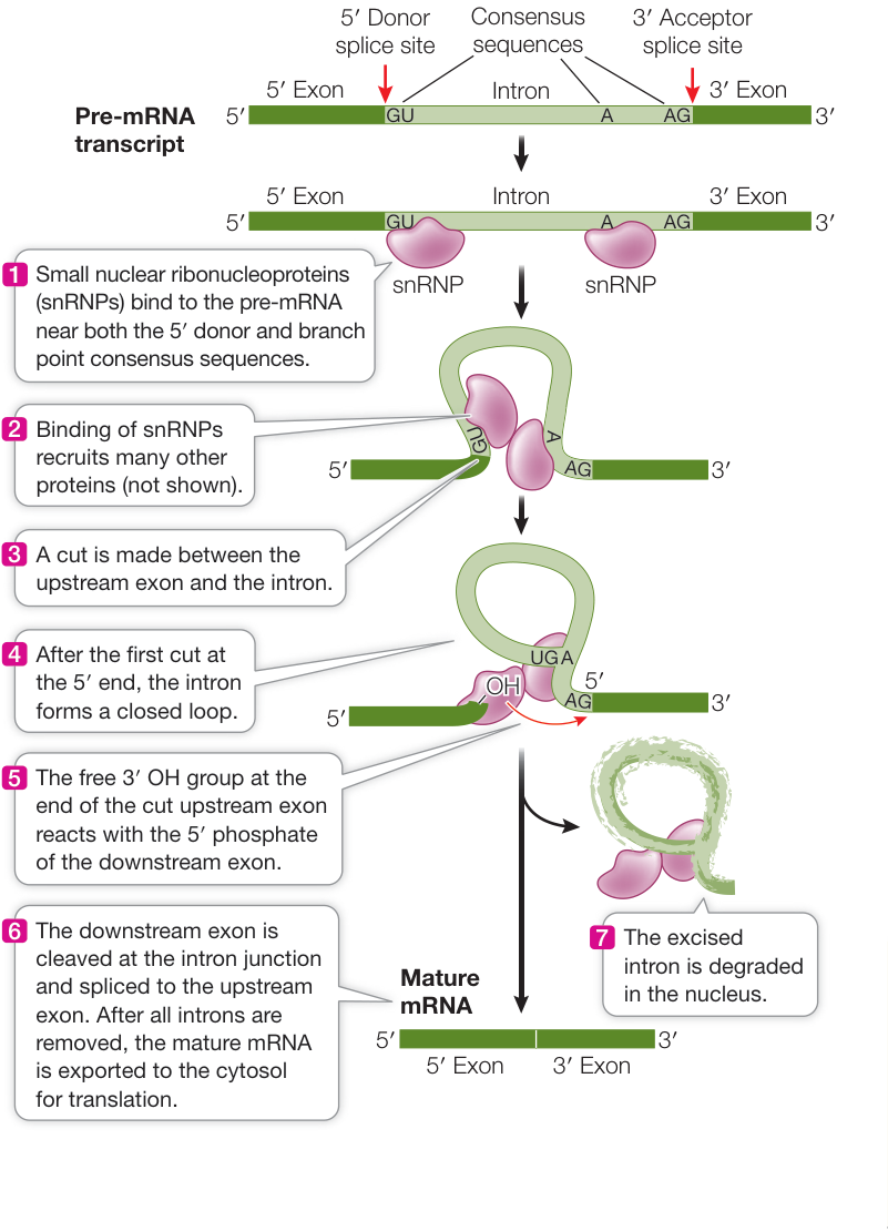
Figure 14.10 RNA Splicing The binding of snRNPs to consensus sequences bordering the introns on the pre-mRNA results in a series of proteins binding and cutting the pre-mRNA with great precision. Animation 14.2 RNA Splicing
After processing is completed in the nucleus, the mature mRNA has a polypeptide-coding region that will be translated by ribosomes, a 5’ untranslated region (upstream of the polypeptide-coding region), and a 3’ untranslated region (downstream of the polypeptide-coding region). The mature mRNA moves out into the cytosol through the nuclear pores. A protein bound to the 5’ nucleotide cap during processing is recognized by a receptor at the nuclear pore, stimulating export. Unprocessed or incompletely processed pre-mRNAs remain in the nucleus.
Recap and Assess
Most eukaryotic genes contain noncoding sequences called introns, which are removed from the pre-mRNA transcript in the nucleus. The mature mRNA transcript with 5’ cap and 3’ tail is then exported through a nuclear pore to ribosomes in the cytosol where translation takes place.
- How and why is the pre-mRNA transcript modified at the 5’ and 3’ ends during mRNA processing?
- Outline the steps involved in RNA splicing. What are the consequences if it does not happen correctly?
- A eukaryotic gene that is 1,440 base pairs long codes for a polypeptide that is 192 amino acids long. However, only 576 base pairs encode the polypeptide. Discuss the discrepancy.
Transcription and posttranscriptional events produce an mRNA that is ready to be translated into a sequence of amino acids in a polypeptide. We turn now to the relationship between mRNA sequence and amino acid sequence.
The Genetic Code KEY CONCEPT 14.5 Determines the Protein Sequence Encoded by an mRNA
Learning Objectives
14.5.1 Use the genetic code table to determine the amino acid sequence encoded by an mRNA sequence.
14.5.2 Define redundancy in the genetic code.
14.5.3 Distinguish between sense and nonsense, or stop, codons.
The genetic code is the informational key by which a sequence of mRNA nucleotides corresponding to a gene is translated into the sequence of amino acids composing the protein expressed by that gene. That is, the genetic code specifies which amino acids will be used to build a protein. You can think of the code as consisting of a series of sequential, non-overlapping, three-letter “words.” The three “letters” represent three adjacent nucleotide bases in the mRNA polynucleotide. Each three-letter “word” is called a codon, and each codon specifies a particular amino acid. Each codon is complementary to the corresponding triplet of bases in the DNA molecule from which it was transcribed. In short, the genetic code relates codons to their specifically encoded amino acids.
The genetic code is redundant but not ambiguous
Molecular biologists “broke” the genetic code in the early 1960s. The problem they addressed was perplexing: how could more than 20 “code words” be written with an “alphabet” consisting of only four “letters”? In other words, how could four bases (A, U, G, and C) code for 20 different amino acids?
The Genetic Code Determines the Protein Sequence Encoded by an mRNA
A triplet code, based on three-letter codons, was considered likely. Since there are only four letters (A, G, C, and U), a one-letter code clearly could not unambiguously encode 20 amino acids; it could encode only four of them. A two-letter code could have only unambiguous codons—still not enough. But a triplet code could have codons, more than enough to encode the 20 amino acids.
Marshall W. Nirenberg and J. Heinrich Matthaei, at the U.S. National Institutes of Health, made the first decoding breakthrough in 1961 when they realized that the code would be easier to break if they were working with a very simple, known mRNA sequence rather than with a complex natural mRNA molecule. They set out to synthesize mRNA molecules consisting of just one type of nucleotide base; poly U mRNA, for example, consisted of just uracil nucleotides. Nirenberg and Matthaei’s goal was to then identify, through a translation process conducted in a test tube, the polypeptide that the artificial messenger encoded. Their experiment, which is laid out in Investigating Life: Deciphering the Genetic Code, led to the identification of the first codons. Soon after this experiment, the rest of the code was identified. This was a major achievement, linking the information in DNA (the gene) to its expression in a protein (the phenotype). The understanding that the amino acids for each protein are spelled out via codons not only led to our understanding of the fundamentals of genetics and mutation, but to investigations into the genetic underpinnings of disease, such as the development of resistance in MRSA described in the opening of this chapter.
The complete genetic code is shown in Figure 14.11. Notice that there are many more codons than there are different amino acids in proteins. Proteins are built from just 20 amino acids, but all possible combinations of the four available “letters” (the bases) yield 64 () different three-letter codons. Why are there more codons than amino acids? One reason is that there is more than one codon for almost all amino acids. For example, leucine is represented by six different codons (see Figure 14.11). Only methionine and tryptophan are represented by just one codon each. Thus we say that the genetic code is redundant (or degenerate). Also, a few select codons serve functions other than coding for amino acids. AUG, for example, not only codes for methionine but is also the start codon, the initiation signal for translation. Sixty one codons encode amino acid information and are termed sense codons. Three codons (UAA, UAG, UGA) do not encode amino acid information and are termed nonsense or stop codons, as they act as termination signals for translation. When the translation machinery reaches one of these codons, translation stops and the polypeptide is released from the translation complex.
Don’t confuse a redundant code with an ambiguous code. If the code were ambiguous, a single codon could specify two (or more) different amino acids, and there would be doubt about which amino acid should be incorporated into a growing polypeptide chain. Redundancy in the code simply means there is more than one clear way to say “Put leucine here.” The genetic code is not ambiguous: a given amino acid may be encoded by more than one codon, but a codon can code for only one amino acid.
The genetic code is (nearly) universal
One of the most amazing discoveries following the deciphering of the genetic code in bacteria was that the genetic code is essentially identical across all the species on our planet. Thus the code must be an ancient one that has been maintained intact throughout the evolution of living organisms. Exceptions are known: for example, the code for mitochondrial DNA (see Key Concept 12.5) and for chloroplast DNA differs slightly from that used by prokaryotes and for the nuclear DNA of eukaryotic cells; and in one group of protists, UAA and UAG code for glutamine rather than for stop codons. The significance of these differences is not yet clear. What is clear is that the exceptions are exceedingly few, and involve only a few codons when present.
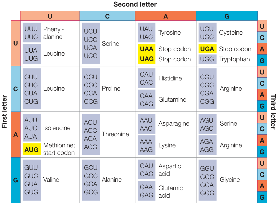
Figure 14.11 The Genetic Code Genetic information is encoded in mRNA in three-letter units—codons—made up of nucleoside monophosphates with the bases uracil (U), cytosine (C), adenine (A), and guanine (G) and is read in a 5’-to-3’ direction on mRNA. To decode a codon, find its first letter in the left column, then read across the top to its second letter, then read down the right column to its third letter. The amino acid the codon specifies is given in the corresponding row. For example, AUG codes for methionine, and GUA codes for valine.
Investigating Life: Deciphering the Genetic Code
Experiment
Original Paper: M. Nirenberg and J. H. Matthaei. 1961. The dependence of cell-free protein synthesis in E. coli upon naturally occurring or synthetic polyribonucleotides. Proc Natl Acad Sci USA 47: 1588–1602.
Nirenberg and Matthaei used a test tube protein synthesis system to determine the amino acids specified by synthetic mRNAs of known compositions.
HYPOTHESIS An artificial mRNA containing only one repeating base will direct the synthesis of a protein containing only one repeating amino acid.
METHOD
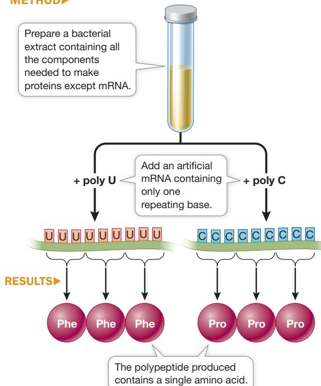
CONCLUSION Poly U contains codons for phenylalanine. Poly C contains codons for proline.
Work with the Data
After the relationship between DNA and proteins was established, genetic evidence pointed to triplets of nucleotides on RNA specifying each amino acid. The race was on to identify which triplet coded for which amino acid. Test tube systems were developed in which cell extracts were made and protein synthesis occurred. Marshall Nirenberg, a scientist at the U.S. National Institutes of Health, and J. Heinrich Matthaei, a postdoctoral fellow from Germany, made a synthetic RNA consisting of the base uracil only (called poly U, codon UUU) and tested it in 20 tubes. Each test tube was supplied with all 20 amino acids, but in each one a different amino acid was tagged with a radioactive marker. In each tube the same polypeptide was made: a protein consisting of the amino acid phenylalanine bonded repeatedly to itself. However, only in the test tube with radiolabeled phenylalanine was the resulting protein radioactive.
QUESTIONS
- Poly U, an artificial mRNA, was added to a test tube with radiolabeled phenylalanine and all the other components for protein synthesis (“complete system”). Other test tubes differed from the complete system as indicated in the table. Samples were tested for incorporation of radioactivity into the resulting protein, with the results in Table A. Explain the results for each of the conditions.
Table A
| Condition | Counts/minute (units of radioactivity) |
|---|---|
| Complete system | 29,500 |
| Minus poly U mRNA | 70 |
| Minus ribosomes | 52 |
| Minus ATP | 83 |
| Plus RNase (hydrolyzes RNA) | 120 |
| Plus DNase (hydrolyzes DNA) | 27,600 |
| Complete system minus all amino acids except radioactive phenylalanine | 31,700 |
- Poly U (red dots) was added to the test tubes and then samples were tested for protein synthesis by radioactive amino acid incorporation after various times (and were compared with results of a control run, in which no RNA was added, indicated by blue dots). The results are shown in the graph. What do these data show about the dependence of protein synthesis on added RNA?
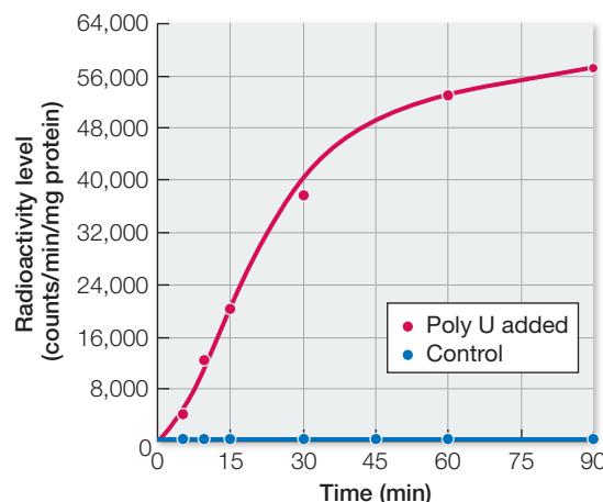
- The experiment described in Question 2 was repeated with different amino acids; the results are in Table B. Explain these results in terms of the codon specificity of poly U.
Table B
| Radioactive amino acid(s) | Counts/minute/mg protein (radioactivity level) |
|---|---|
| Phenylalanine | 38,300 |
| Glycine, alanine, serine, aspartic acid, glutamic acid | 33 |
| Leucine, isoleucine, threonine, methionine, arginine, histidine, valine, lysine, tyrosine, proline, tryptophan | 899 |
| Cysteine | 113 |
Connect the Concepts As discussed in Key Concept 12.5, some cytoplasmic organelles, notably the mitochondria and chloroplasts, contain small numbers of genes that are remnants of the genomes of the prokaryotes that eventually gave rise to these organelles. The evolutionary process of endosymbiosis that is responsible for the assimilation of these organelles into eukaryotic cells is discussed in Key Concept 26.1.
A common genetic code suggests that it originated early in the evolution of life and is shared by descent from these early species. The common code has profound implications for genetic engineering, as we will see in Chapter 18, since it means that the code for a human gene is the same as that for a bacterial gene. It is therefore impressive, but not surprising, that a human gene can be quite easily expressed in E. coli via laboratory manipulations, since these cells speak the same “molecular language.”
The codons illustrated in the Nirenberg and Matthaei experiment in Investigating Life: Deciphering the Genetic Code are mRNA codons. The base sequence of the template DNA strand that is transcribed to produce the mRNA is complementary and antiparallel to these codons. Thus, for example, 3’-AAA-5’ in the template DNA strand corresponds to phenylalanine (which is encoded by the mRNA codon 5’-UUU-3’). In contrast, the coding strand of DNA has the same sequence as the mRNA (but with T’s instead of U’s). By convention, DNA sequences are usually shown beginning with the 5’ end of the coding sequence.
You might be thinking that in a long DNA molecule with many protein-coding regions, one strand is the coding strand for all the genes and the other strand is the template strand. In fact, some genes are transcribed from one strand and some genes are transcribed from the other. There is strand switching along a long DNA molecule in terms of the roles of the two strands. So while it is incorrect to say that one strand is “the” coding strand for all genes in a particular DNA molecule, it is correct to say that for a given gene one strand is coding and the other is template.
Recap and Assess
The genetic code relates the information in mRNA (as a linear sequence of codons) to protein (a linear sequence of amino acids).
- Explain why the genetic code has triplets (e.g., AUA) of nucleotides, rather than singlets (e.g., A) or doublets (e.g., AU).
- Explain what it means to say the genetic code is redundant.
- A short mRNA is translated into the following peptide: methionine-tryptophan-tryptophan-glycine-tryptophan. What are the possible sequences of mRNAs (from start to stop) that could encode this peptide?
Now that we understand the rules by which mRNA encodes protein information, let’s see how the cell is able to synthesize proteins from mRNA molecules using the genetic code.
The Coding Sequence in mRNA Is Translated into Proteins by Ribosomes
Learning Objectives
14.6.1 Describe initiation, elongation, and termination of translation.
14.6.2 Explain how tRNAs are able to translate mRNA codons into the corresponding amino acids.
14.6.3 Predict the consequences of an aminoacyl-tRNA synthetase attaching the wrong amino acid to a tRNA.
14.6.4 Describe each step experienced by a tRNA, starting with an uncharged tRNA in the cytosol and ending with the release of the uncharged tRNA from a ribosome.
14.6.5 Explain the biological significance of polysomes.
Transcription uses complementary base pairing to synthesize an mRNA molecule from a DNA template strand. The information in both molecules is equivalent—a sequence of nucleotides. In contrast, translation uses mRNA to synthesize a polypeptide; nucleotide information encodes amino acid information, as we showed with the genetic code. How is this translation accomplished? How is the genetic code “read” by the translation machinery? The answer is a special kind of RNA molecule called transfer RNA (tRNA) that can (1) read mRNA codons and (2) bring the corresponding amino acid to the ribosome.
Once the tRNAs “decode” the mRNA and deliver the appropriate amino acids, components of the ribosome catalyze the formation of peptide bonds between amino acids. Let’s now look at how the tRNAs read codons and get the appropriate amino acids into the ribosome.

Animation 14.4 Translation
A transfer RNA carries a specific amino acid and binds to a specific mRNA codon
There is at least one specific tRNA molecule for each of the 20 amino acids. Each tRNA has three functions that are fulfilled by its structure and base sequence (Figure 14.12):
- tRNAs bind to particular amino acids. Each tRNA binds to a specific enzyme that attaches it to only 1 of the 20 amino acids. The covalent attachment to an amino acid takes place at the 3’ end of the tRNA. When it is carrying an amino acid, the tRNA is said to be “charged.”
- tRNAs bind to mRNA. At about the midpoint on the tRNA polynucleotide chain there is a triplet of bases called the anticodon, which is complementary to the mRNA codon for the particular amino acid that the tRNA carries. For example, the mRNA codon for arginine is 5’-CGG-3’, and the complementary tRNA anticodon is 3’-GCC-5’. Like the two strands of DNA, the codon and anticodon bind together via noncovalent hydrogen bonds.
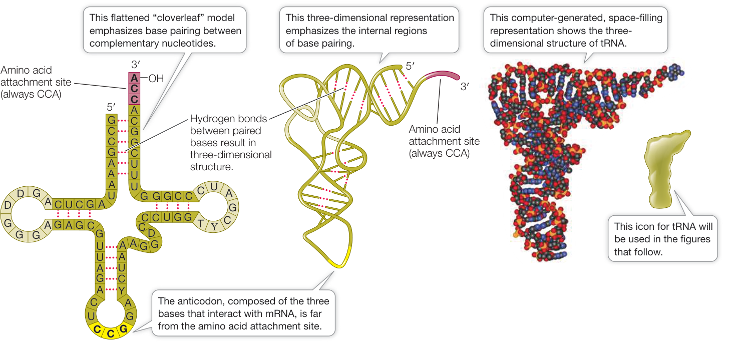
Figure 14.12 Transfer RNA The stem and loop structure of a tRNA molecule is well suited to its functions: binding to amino acids, binding to mRNA molecules, and interacting with ribosomes. (“Cloverleaf” model and three-dimensional representation after H. Shi and P. B. Moore. 2000. RNA 6: 1091–1105; tRNA space-filling representation by David McIntyre based on data from PDB ID: 1EHZ. H. Shi and P. B. Moore. 2000. RNA 6: 1091–1105.)
- tRNAs interact with ribosomes. The ribosome has several sites on its surface that just fit the three-dimensional structure of a tRNA molecule. Interaction between the ribosome and the tRNA is noncovalent.
Recall that 61 different codons encode the 20 amino acids in proteins (see Figure 14.11). Does this mean that the cell must produce 61 different tRNA species, each with a different anticodon? No. The cell gets by with about two-thirds of that number of tRNA species because the interaction between the bases at the third position of the codon–anticodon interaction, which is the 3’ end of the codon and the 5’ end of the anticodon, is not as specific as it is for other base pairs. This phenomenon is called wobble base pairing—base pairing that does not strictly follow the standard rules (i.e., A with U, and G with C). Wobble base pairing occurs only at the third position of the codon–anticodon interaction. Sometimes an unusual purine base, inosine (I), which can pair with A, C, and U, is found at the third position. For example, the presence of inosine in the tRNA with the anticodon 3’-UAI-5’ allows it to recognize and bind to the three isoleucine codons: AUU, AUC, and AUA. Wobble base pairing also occurs for some normal bases in the third position; for example, G in the third position of the anticodon can pair with C or U in the codon. Wobble base pairing explains many of the patterns observed in the genetic code (see Figure 14.11); you may have noticed that all codon pairs that start with the same two bases but end in C or U always code for the same amino acid—such codons can be read by tRNAs with a G in their third position. It is important to recognize that wobble base pairing allows fewer tRNAs to read the code than would be required with strict base pairing, but it does not allow the genetic code to be ambiguous. That is, each mRNA codon can bind only to tRNAs that carry a specific amino acid.
Each tRNA is specifically attached to an amino acid
The charging of each tRNA with its correct amino acid is achieved by a family of enzymes known as aminoacyl-tRNA synthetases. Each enzyme is specific for one amino acid and for its corresponding tRNA. The reaction uses ATP, forming a high-energy bond between the amino acid and the tRNAs (Figure 14.13). The energy in this bond is later used in the formation of peptide bonds between amino acids in a growing polypeptide chain.
The specificity between the tRNA and its corresponding amino acid is essential. For example, tRNAcys (with anticodon ACA), which is the tRNA that carries cysteine, always has cysteine added to it by its synthetase to form cys-tRNAcys. A clever experiment by Seymour Benzer and his colleagues at Purdue University demonstrated the importance of this specificity. They took the cys-tRNAcys molecule and chemically modified the cysteine, converting it into alanine (to form ala-tRNAcys). Which component—the amino acid or the tRNA—would be recognized when this hybrid charged tRNA was put into a protein synthesizing system? The answer was the tRNA. Everywhere in the synthesized protein where cysteine was supposed to be, alanine appeared instead. The cysteine-specific tRNA had delivered its cargo (alanine) to every mRNA codon for cysteine. This experiment showed that the protein synthesis machinery recognizes the anticodon of the charged tRNA, not the amino acid attached to it.
The ribosome is the workbench for translation
The ribosome is the molecular workbench where the task of translation is accomplished. Its structure enables it to hold mRNA and charged tRNAs in the correct positions, thus allowing a polypeptide chain to be assembled efficiently. A given ribosome does not specifically produce just one kind of protein. A ribosome can use any mRNA and all species of charged tRNAs, and thus can be used to make many different polypeptide products. Ribosomes can be used over and over again, and there are thousands of them in a typical cell.
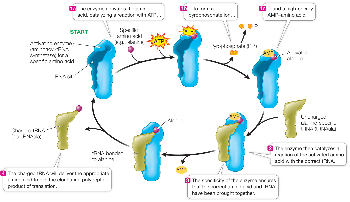
Figure 14.13 Charging a tRNA Molecule An aminoacyl-tRNA synthetase activates a specific amino acid and charges a specific tRNA with that amino acid.
Although ribosomes are small relative to other cellular structures, their mass of several million daltons makes them large in comparison with charged tRNAs. Each ribosome consists of two subunits, a large one and a small one (Figure 14.14). The two subunits and several dozen other molecules interact noncovalently. In fact, when hydrophobic interactions between the proteins and RNAs are disrupted, the ribosome falls apart. The two subunits separate and all the RNAs and proteins separate from one another. If the disrupting agent is removed, the complex structure self-assembles perfectly! This is amazing: think of throwing the pieces of a jigsaw puzzle up in the air and having them fit together when they land. The ribosome reflects the high specificity of molecular machines in a cell composed of many molecules.
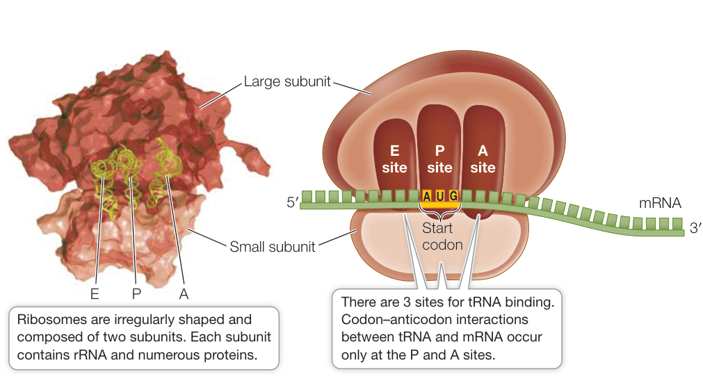
Figure 14.14 Ribosome Structure Each ribosome consists of a large and a small subunit. The subunits remain separate when they are not being used for protein synthesis. (Left by David McIntyre based on data from PDB 1GIX and 1G1Y. M. M. Yusupov et al. 2001. Science 292: 883–896.) Q: The ribosome consists of several dozen proteins and several RNA molecules, held together noncovalently. What are the chemical forces involved? How can these forces be disrupted and the molecules separated from one another?
In eukaryotes, the large subunit consists of three different molecules of ribosomal RNA (rRNA) and about 45 different protein molecules, arranged in a precise configuration. The small subunit consists of 1 rRNA molecule and about 32 different protein molecules. The exact number of proteins present in each subunit varies a little across species. The ribosomes of prokaryotes are somewhat smaller than those of eukaryotes, and their ribosomal proteins and RNAs are different. Mitochondria and chloroplasts also contain ribosomes, which have features similar to those of prokaryotes (see Chapter 5).
There are three sites to which a tRNA can bind on the large subunit of the ribosome, designated A, P, and E (see Figure 14.14). The mRNA and ribosome move in relation to one another, and as they do so, an initially charged tRNA traverses these three sites in order:
- The A (aminoacyl-tRNA) site is where the charged tRNA anticodon binds to the mRNA codon, thus lining up the correct amino acid to be added to the growing polypeptide chain.
- The P (peptidyl-tRNA) site is where the tRNA carrying the growing peptide chain resides.
- The E (exit) site is where the uncharged tRNA, having given up its amino acid, resides before being released from the ribosome into the cytosol to pick up another amino acid and begin the process again.
The ribosome has a fidelity function that ensures that the mRNA-tRNA interactions are accurate; that is, that a charged tRNA with the correct anticodon (e.g., 3’-UAC-5’) forms hydrogen bonds with the appropriate codon in the mRNA (e.g., 5’-AUG-3’). The rRNA of the small ribosomal subunit plays a role in validating the three-base-pair match. If hydrogen bonds have not formed between all three base pairs, the tRNA must be the wrong one for that mRNA codon, and the incorrect tRNA is ejected from the A site of the ribosome.
Translation takes place in three steps
Translation is the process by which the information in mRNA (derived from DNA) is used to specify and link a specific sequence of amino acids, producing a polypeptide. Like transcription, translation occurs in three steps: initiation, elongation, and termination.
INITIATION The translation of mRNA begins with the formation of an initiation complex, which consists of a charged tRNA and a small ribosomal subunit, both bound to the mRNA (Figure 14.15).
In prokaryotes, the rRNA of the small ribosomal subunit first binds to a complementary ribosome binding site (AGGAGG; known as the
Shine-Dalgarno sequence) on the mRNA. This sequence is less than ten bases upstream of the actual start codon but lines up the start codon so that it is adjacent to the P site of the large subunit:
rRNA 3’…..U C C U C C…..(P site)…..5’
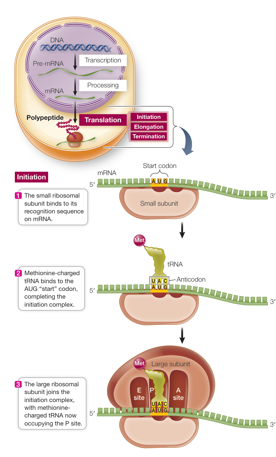
Figure 14.15 The Initiation of Translation Translation begins with the formation of an initiation complex. In prokaryotes, the small ribosomal subunit binds to the Shine-Dalgarno sequence to begin the process, whereas in eukaryotes it binds to the 5’ cap.
Eukaryotes load the mRNA onto the ribosome somewhat differently: the small ribosomal subunit binds to the 5’ cap on the mRNA and then moves along the mRNA until it reaches the start codon.
Recall that the mRNA start codon in the genetic code is AUG (see Figure 14.11). The anticodon (UAC) of a methionine-charged tRNA binds to this start codon by complementary base pairing to complete the initiation complex. Thus the first amino acid in a polypeptide chain is always methionine. However, not all mature proteins have methionine as their N-terminal amino acid. In many cases, the initial methionine is removed by an enzyme after translation.
After the methionine-charged tRNA has bound to the mRNA, the large subunit of the ribosome joins the complex. The methionine-charged tRNA now lies in the P site of the ribosome, and the A site is aligned with the second mRNA codon. These ingredients—mRNA, two ribosomal subunits, and methionine-charged tRNA—are assembled by a group of proteins called initiation factors.
ELONGATION A charged tRNA now enters the open A site of the large ribosomal subunit. If it does not have the correct anticodon, corresponding to the codon in the A site, it exits. At some point, a charged tRNA whose anticodon is complementary to the A site codon of the mRNA enters the A site (Figure 14.16). The large subunit then catalyzes two reactions:
- It breaks the bond between the tRNA and its amino acid in the P site.
- It catalyzes the formation of a peptide bond between the amino acid that has just been released from the P site tRNA and the one attached to the tRNA in the A site.
Because the large ribosomal subunit performs these two actions, it is said to have peptidyl transferase activity. In this way, methionine (the amino acid in the P site) becomes the N terminus of the new protein. The second amino acid is now bound to methionine but remains attached to its tRNA at the A site.
How does the large ribosomal subunit catalyze peptide bond formation? Harry Noller and his colleagues at the University of California at Santa Cruz did a series of experiments and found that:
- if they removed almost all of the proteins from the large subunit, it still catalyzed peptide bond formation.
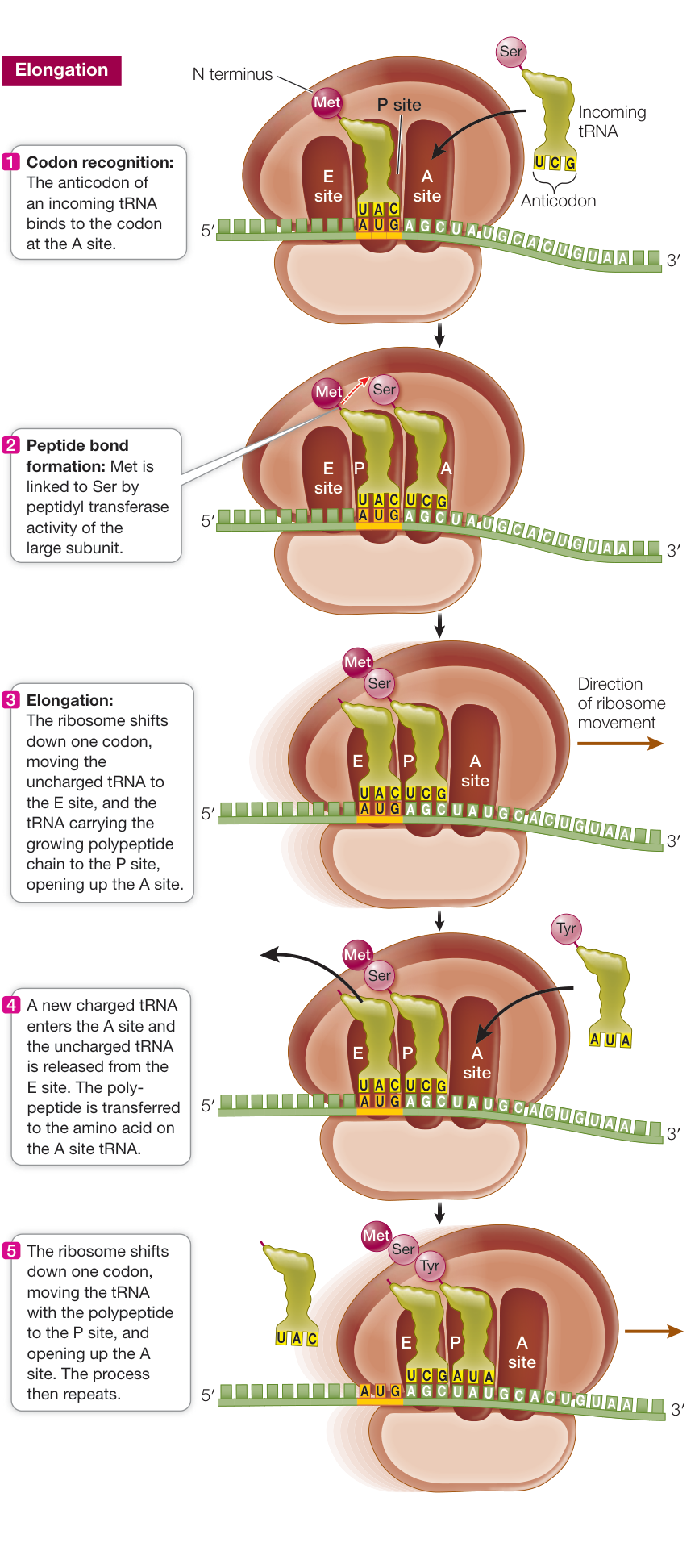
Figure 14.16 The Elongation of Translation The polypeptide chain elongates as the mRNA is translated.
- if the rRNA was extensively modified, peptidyl transferase activity was destroyed.
The experiment showed that rRNA is the catalyst. The purification and crystallization of ribosomes have allowed scientists to examine ribosome structure in detail, and the catalytic role of rRNA in peptidyl transferase activity has been confirmed. These findings support the hypothesis that RNA, and catalytic RNA in particular, evolved before DNA (see Key Concept 4.3).
Connect the Concepts As discussed in Key Concept 4.3, the folded, three-dimensional surface of an RNA molecule can be just as specific as that of a protein, and may thus take on a catalytic function. The “RNA world” hypothesis, which proposed that RNA serves as a catalyst for its own replication, was boosted by the discovery of ribozymes, catalytic RNAs that can speed up biological reactions, including those that involve their own nucleotides.
After the first tRNA releases its methionine, it moves to the E site and is then dissociated from the ribosome, returning to the cytosol to become charged with another methionine. The second tRNA, now bearing a dipeptide (a two-amino acid chain), is shifted to the P site as the ribosome moves one codon along the mRNA in the 5’-to-3’ direction. The elongation process continues, and the polypeptide chain grows, as these steps are repeated. Follow the process in Figure 14.16. All of these steps are assisted by ribosomal proteins called elongation factors.
TERMINATION The elongation cycle ends, and translation is terminated, when a stop codon—UAA, UAG, or UGA—enters the A site (Figure 14.17). Stop codons do not correspond with any amino acids and thus do not bind any tRNAs. Rather, they bind a protein release factor, which allows hydrolysis of the bond between the polypeptide chain and the tRNA in the P site. The newly completed polypeptide thereupon separates from the ribosome. Its C terminus is the last amino acid to join the chain. Its N terminus, at least initially, is methionine, as a consequence of the AUG start codon. In its amino acid sequence, it contains information specifying its conformation, as well as its ultimate cellular destination.
Table 14.3 lists the nucleic acid signals for initiation and termination of transcription and translation.
Polysome formation increases the rate of protein synthesis
Several ribosomes can work simultaneously at translating a single mRNA molecule, producing multiple polypeptides at the same time. As soon as the first ribosome has moved far enough from the 5’ end of the mRNA, a second initiation complex can form, then a third, and so on. An assemblage consisting of a strand of mRNA with its beadlike ribosomes and their growing polypeptide chains is called a polyribosome, or polysome (Figure 14.18). Cells that are actively synthesizing proteins contain large numbers of polysomes and few free ribosomes or ribosomal subunits.
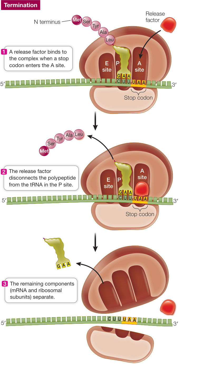
Figure 14.17 The Termination of Translation Translation terminates when the A site of the ribosome encounters a stop codon on the mRNA. Q: What happens if there is not a stop codon in the mRNA?
Table 14.3 Signals that Start and Stop Transcription and Translation
| Transcription | Translation | |
|---|---|---|
| Initiation | Promoter sequence in DNA | AUG start codon in the mRNA |
| Termination | Terminator sequence in DNA | UAA, UAG, or UGA in the mRNA |
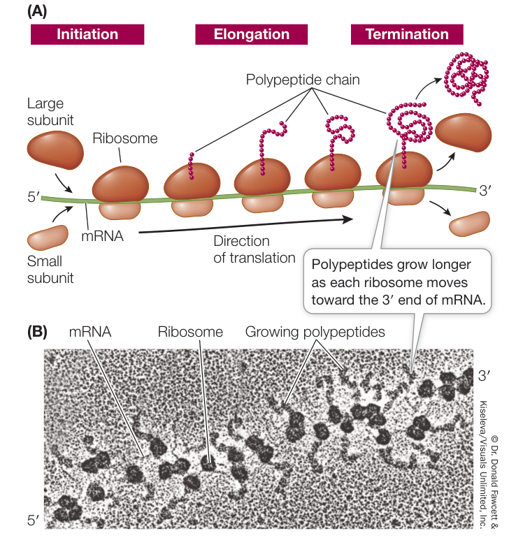
Figure 14.18 A Polysome (A) A polysome consists of multiple ribosomes and their growing polypeptide chains moving along an mRNA molecule. (B) An electron micrograph of a polysome.
Recap and Assess
A key step in protein synthesis is the attachment of an amino acid to its proper tRNA, which is facilitated by a family of specific activating enzymes called aminoacyl-tRNA synthetases. tRNAs are the molecules that read mRNA codons and bring the appropriate amino acid to the growing polypeptide. Translation of the genetic information from mRNA into protein occurs at the ribosome in three stages: initiation, elongation, and termination. Multiple ribosomes may act on a single mRNA to make multiple copies of the protein that it encodes.
- What are the roles of rRNA molecules in the ribosome?
- Suppose the following mature mRNA sequence encodes a single
polypeptide: 5’ cap-UCGAUCAUGCCCGGGUUAAGAUAUUUAAAUAGAAUAAAAA…AAA-3’
- Is this mRNA from a prokaryotic or a eukaryotic cell?
- Write out the sequence of the complementary DNA strand from which this mRNA was synthesized, assuming no introns were present.
- Write out the amino sequence of the translated polypeptide.
- A charging enzyme inadvertently adds arginine to tRNAser. What will happen if this tRNA is used during translation?
The polypeptide chain that is released from the ribosome is not necessarily a functional protein. Let’s look at some of the posttranslational changes that can affect the fate and function of a polypeptide.
Polypeptides Can Be Modified and Transported during or after Translation
Learning Objectives
14.7.1 Outline the steps by which a newly synthesized protein is moved into the nucleus or exported outside the cell.
14.7.2 Justify the claim that posttranslational modification of a protein is ultimately directed by the DNA sequence of the gene encoding the protein.
The site of a polypeptide’s function may be far away from its point of synthesis. This is especially true for eukaryotes. The polypeptide may be moved into an organelle, or even out of the cell. In addition, polypeptides are often modified by the addition of new chemical groups that have functional significance. In this key concept we examine these posttranslational aspects of protein synthesis.
How are proteins directed to their cellular destinations?
As a polypeptide chain emerges from the ribosome it may simply fold into its three-dimensional shape and perform its cellular role locally in the cytosol. However, if a newly formed polypeptide is meant to do its work elsewhere, a signal sequence (or signal peptide)—a short stretch of amino acids attached to the polypeptide—will tell the polypeptide where in the cell it belongs. Proteins destined for different locations have different signals.
Protein synthesis always begins on free ribosomes, and the “default” location for a protein is the cytosol. In the absence of a signal sequence, the protein will remain in the same cellular compartment in which it was synthesized. Some proteins contain signal sequences that “target” them to the nucleus, mitochondria, plastids, or peroxisomes (Figure 14.19). A signal sequence binds to a specific receptor protein at the surface of the organelle. Once it has bound, the targeted protein moves into the organelle. For example, here is a nuclear localization signal (NLS):
-Pro-Pro-Lys-Lys-Lys-Arg-Lys-Val-
How do we know this signal sequence directs the protein to the nucleus? The function of this NLS peptide was established using experiments like the one illustrated in Figure 14.20. Proteins were made in the laboratory with or without the peptide, and then tested by injecting them into cells. Only proteins with the NLS were found in the nucleus.
If a polypeptide carries a signal of about 20 hydrophobic amino acids at its N terminus, it will be directed to the rough endoplasmic reticulum (RER) for further processing (see Figure 14.19). Note that this is not a specific sequence of amino acids, just a generally hydrophobic sequence at the N terminus that is first translated. Translation will pause, and the ribosome will bind to a receptor at the RER membrane. Once the polypeptide—ribosome complex is bound, translation will resume, and as elongation continues, the protein will traverse the RER membrane.
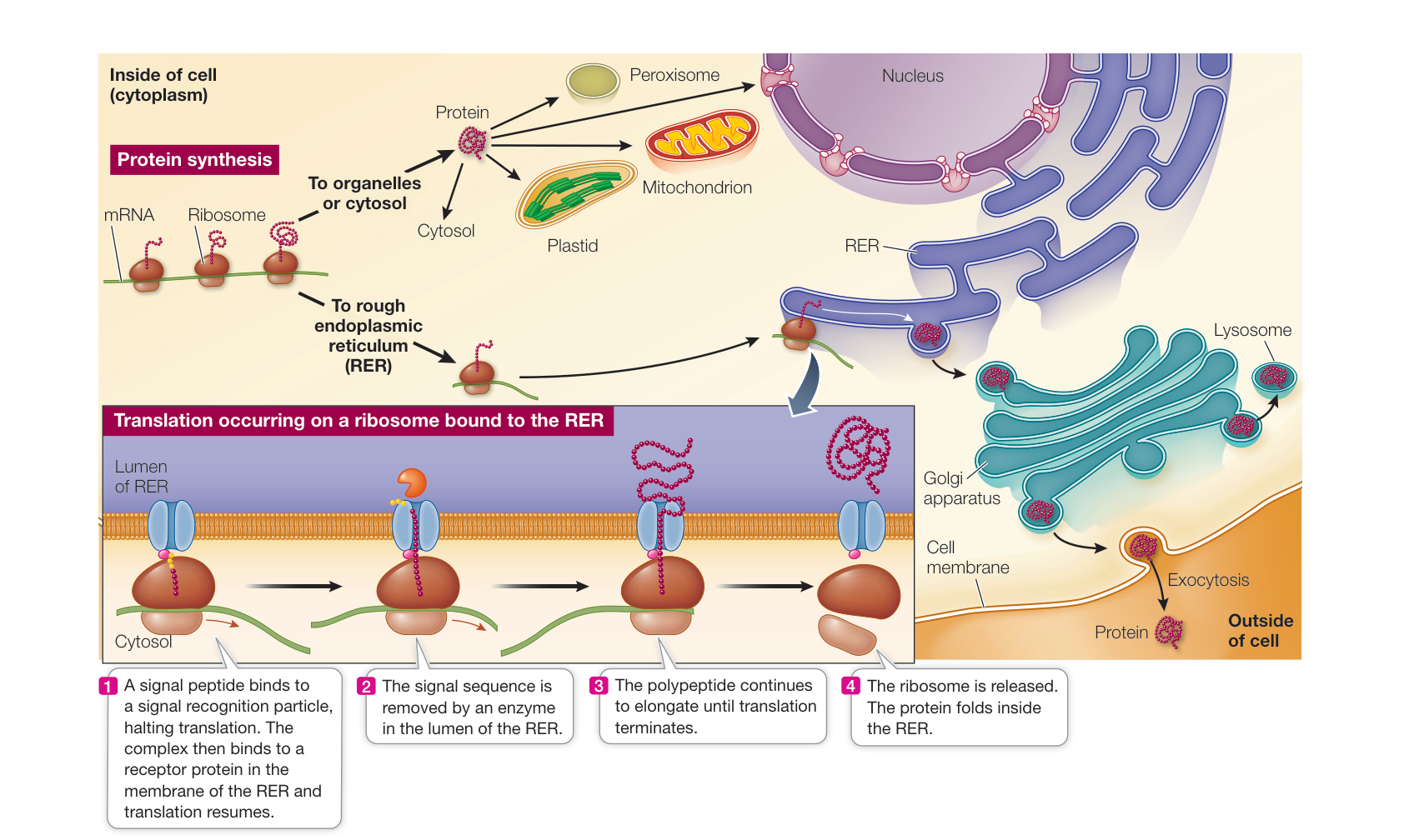
Figure 14.19 Destinations for Newly Translated Polypeptides in a Eukaryotic Cell Signal sequences on newly synthesized cytosolic polypeptides bind to specific receptor proteins on the outer membranes of the organelles to which they are “addressed.” Once the protein has bound to it, the receptor forms a channel in the membrane, and the protein enters the organelle. Hydrophobic sequences at the amino end of a polypeptide may halt translation, which then resumes after binding of the ribosome to the RER. The polypeptide either enters the membrane or the lumen of the RER. Q: What happens to a protein that has no amino acid sequence “address”?
Such proteins may be retained in the lumen (the inside of the RER) or in the membrane of the RER, or they may move elsewhere within the endomembrane system (Golgi apparatus, lysosomes, and cell membrane). If the proteins lack specific signals or modifications (discussed shortly) that specify destinations within the endomembrane system, they are usually secreted from the cell via vesicles that fuse with the cell membrane.
The importance of signals is shown by Inclusion-cell (I-cell) disease, an inherited disease that causes death in early childhood. People with this disease have a mutation in the gene encoding a Golgi enzyme that adds specific sugars to proteins destined for the lysosomes. These sugars act like signal sequences; without them, enzymes that are essential for the hydrolysis of various macromolecules cannot reach the lysosomes, where the enzymes are normally active. Without these enzymes, the macromolecules accumulate in the lysosomes, and this lack of cellular recycling has drastic effects, resulting in early death.
Mitochondria and chloroplasts make some of their own proteins and import others
Both organelles contain the entire protein synthesis machinery, including a specific RNA polymerase, and their own specialized ribosomes that differ from the ones in the cytoplasm. The genetic coding capacity of the organelle DNA is small, however:
- Mitochondrial DNA encodes mitochondrial rRNAs, a few tRNAs, and 12 to 20 proteins, most of them involved in the respiratory chain.
- Chloroplast DNA encodes chloroplast rRNAs, some tRNAs, and about 40 proteins, many of which are involved in photosynthesis.
These organelles contain dozens more proteins, all of which are imported from the nuclear–cytoplasm protein synthesis system. In some cases (e.g., rubisco in the chloroplast) an organelle protein has several subunits, some of which are made locally inside the organelle and others of which are imported from the nucleus–cytoplasm system. As you can imagine, putting this together requires coordination between the two protein synthesis systems.
Many proteins are modified after translation
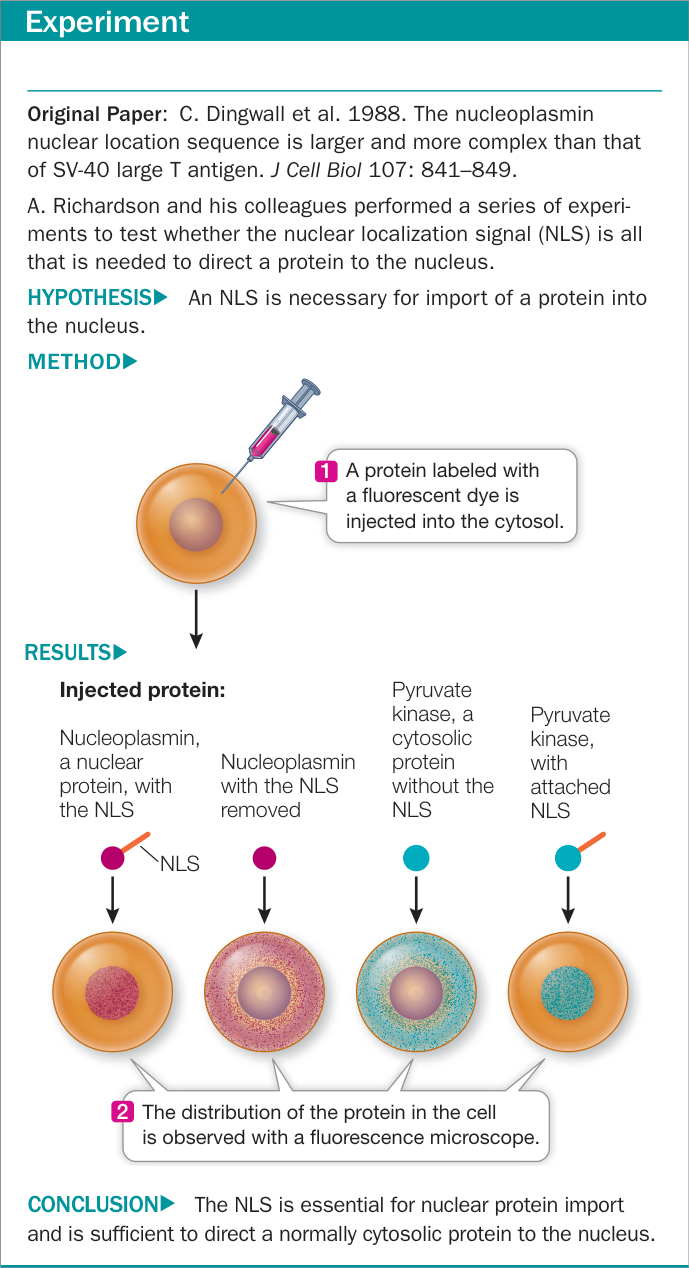
Figure 14.20 Testing the Signal
mRNA on the ribosomes, because polypeptides are often modified in any of several ways after translation (Figure 14.21). These modifications are essential to the final functioning of the protein:
- Proteolysis is the cutting of a polypeptide chain, a reaction catalyzed by enzymes called proteases (also called peptidases or proteinases). Cleavage of the signal sequence from the growing polypeptide chain in the RER is an example of proteolysis (see Figure 14.19). Some proteins are actually made from polyproteins (long polypeptides) that are cut into final products by proteases. These proteases are essential to some viruses, including human immunodeficiency viruses (HIVs), because the large viral polyprotein cannot fold properly unless it is cut. Certain drugs used to treat acquired immune deficiency syndrome (AIDS) work by inhibiting the HIV protease, thereby preventing the formation of proteins needed for viral reproduction.
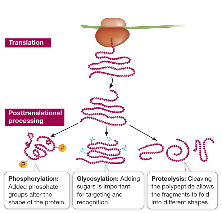
Figure 14.21 Posttranslational Modifications of Proteins Most polypeptides must be modified after translation in order to become functional proteins.
- Glycosylation is the addition of sugars to proteins to form glycoproteins. In both the RER and the Golgi apparatus, resident enzymes catalyze the addition of various sugars or short sugar chains to certain amino acid R groups on proteins. One such type of “sugar coating” is essential for directing proteins to lysosomes, as mentioned earlier. Other types are important in the conformation of proteins and their recognition functions at the cell surface (see Key Concept 6.2). Other attached sugars help stabilize extracellular proteins, or proteins stored in vacuoles in plant seeds.
- Phosphorylation is the addition of phosphate groups to proteins, and is catalyzed by protein kinases. The charged phosphate groups change the conformation of a protein, often exposing the active site of an enzyme or the binding site for another protein. We have seen the important role of phosphorylation in cell signaling (see Chapter 7) and the cell cycle (see Chapter 11).
Recap and Assess
Signal sequences in polypeptides direct them to their appropriate destinations inside or outside the cell. Many polypeptides are modified after translation.
- How do signal sequences determine where a protein will go after it is made?
- The genetic code determines the amino acid sequence of a protein. Is there a “code” that determines posttranslational modifications such as phosphorylation or glycosylation? How might this relate to the genetic code?
All of the processes we have just described result in a functional protein, but only if the amino acid sequence of that protein is correct. If the sequence is not correct, cellular dysfunction may result. Changes in the DNA—mutations—are a major source of errors in amino acid sequences. This is the subject of the next chapter.
Investigating Life: QA How does knowledge of the genetic code help us understand the actions of some antibiotics?
MRSA can be treated if detected early. Sequencing the DNA of the bacteria can be used to detect mutations and predict the severity of the infection. Antibiotics such as tetracyclines, which target bacterial protein synthesis, are effective in some strains. Tetracyclines kill bacteria by interrupting translation, binding to the small subunit of bacterial ribosomes so that charged tRNA cannot bind to the A site on the ribosome. Tetracyclines do not kill eukaryotic cells, because eukaryotic ribosomes have different proteins and RNAs that do not offer a binding site for tetracyclines.
Whether tetracyclines will continue to offer a viable treatment for MRSA, however, is in doubt. Some strains of MRSA are already resistant to tetracyclines, having acquired genes that convey resistance through bacterial conjugation. The DNA of S. aureus has been altered through acquisition of genes from a plasmid so important that it has been given its own name, the resistome. As with methicillin resistance, the alleles of the resistome encode a ribosomal protein with an altered amino acid sequence that consequently does not bind the antibiotic.
Can you think of other ways to target protein synthesis in MRSA?
Future directions
While the expression of protein-coding genes has been well worked out, recent analyses of eukaryotic genomes show that a lot of transcription remains unaccounted for. International cooperation among laboratories in huge projects such as ENCODE (Encyclopedia of DNA Elements) has revealed, for example, that up to 70% of human DNA is transcribed at some point in some cells. With only 2% to 3% of DNA consisting of protein-coding genes, the question immediately arises as to what purpose the rest of the RNAs serve. A small fraction are tRNAs and rRNAs, and you will see in Chapter 16 that some of these noncoding RNAs are small (< 200 nucleotides) and have roles in regulating the expression of protein-coding genes. The roles of the longer RNAs (> 200 nucleotides) are much less clear. In humans, more than 25,000 of the longer RNAs have been identified. They have much in common with mRNAs: they are transcribed using the same RNA polymerase, undergoing poly A addition, capping at the 5’ and 3’ ends, and splicing. However, the longer RNAs are not as long as mRNAs and are not translated at the ribosome. Some never leave the nucleus, where they may be involved in pre-mRNA events such as transcription and splicing. Others actually arrive at the ribosome, where they may regulate the translation of particular mRNAs. These longer untranslated RNAs are found in many organisms, indicating that they must have important roles that have been selected for in evolution.
Courtesy of the National Institute of Allergy and Infectious Diseases (NIAID)
You should be able to relate each summary to the adjacent figures. If you go to this visual summary in Achieve, you can follow links to figures, animations, activities, and simulations that will help you consolidate the material.
Visual Summary
Genes Code for Proteins
- ■ Model organisms have characteristics that make them ideal for laboratory work.
- ■ Experiments on metabolic enzymes led to the one-gene, one-enzyme hypothesis. We now know there is a one-gene, one-polypeptide relationship.
- ■ A mutant that has a nonfunctional enzyme in a biosynthetic pathway can be rescued by adding pathway intermediates occurring after the blocked step.

Figure 14.2 1. 1. List the characteristics of a model organism. 2. 2. Individuals with PKU lack an enzyme that converts phenylalanine to tyrosine. Do you expect the levels of phenylalanine and tyrosine in individuals with PKU to be higher or lower than those in individuals without PKU? Why is the inability to synthesize tyrosine from phenylalanine not lethal in humans?
Information Flows from Genes to Proteins
- The central dogma of molecular biology states how information usually flows in cells, from DNA to DNA, DNA to RNA and RNA to protein.
Questions
- What three types of RNA molecules are involved in the flow of information from DNA to protein, and what are their primary functions?
- Telomerase is an enzyme that uses an RNA template to synthesize DNA to extend telomeres. How does this enzyme fit into the central dogma?
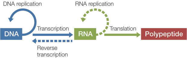
Figure 14.3
DNA Is Transcribed to Produce RNA
- RNA polymerases read in the 3’-to-5’ direction, synthesize in the 5’-to-3’ direction, and do not need a primer to begin synthesis.
- Transcription begins at a promoter sequence and ends at a terminator sequence.
Questions
- What are the substrates for the RNA polymerase during transcription?
- List the ways in which RNA polymerase differs from DNA polymerase.
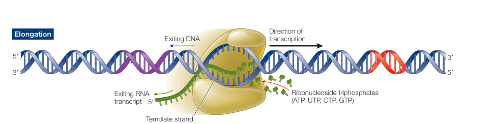
Figure 14.6
Eukaryotic Pre-mRNA Transcripts Are Processed prior to Translation
- Prokaryotic mRNAs are not processed prior to being translated.
- Eukaryotic mRNAs have a 5’ cap and a 3’ poly A tail added, and their introns are spliced out.
- If a change in DNA sequence were to alter an intron splice site, what effect would that have on the resulting protein?
- If an mRNA is not capped during processing, what are its possible fates?
- When a cytosolic mRNA from a eukaryote is hybridized to nuclear DNA, in regions where the RNA hybridizes to the DNA, some loops appear that represent regions of DNA that do not pair with the mRNA. Explain the regions of the DNA sequence in the gene that do and do not hybridize to the mRNA.
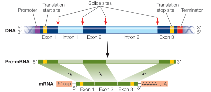
Figure 14.8
- Coding sequence in mRNA is read as a series of codons, starting at the AUG start codon.
- The genetic code is nearly universal and specifies which amino acids will be included in the polypeptides encoded by mRNA.
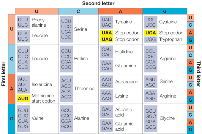
Figure 14.11 Questions
- Assuming that the ribosome starts translation at the start codon, write down the amino acid sequence of the protein that will be translated from the following mRNA: 5’-GCUUAGCAUGUUUCAAAGAGGUUGACUGAAU-AAAAAAAAAAAA-3’.
- What is the difference between sense codons and stop (or nonsense) codons?
The Coding Sequence in mRNA Is Translated into Proteins by Ribosomes
- Each codon is translated into an amino acid by a specific tRNA by codon—anticodon base pairing.
- Translation initiates at the 5’ end of the mRNA and begins at an AUG codon.
- Translation elongation involves the ribosome reading one codon at a time as it progresses down the mRNA.
- Charged tRNAs carrying a single amino acid enter the A site, then move to the P site, where they carry the entire polypeptide, then become uncharged and move to the E site, where finally they exit the ribosome.
- Termination occurs when the ribosome reaches a stop codon.
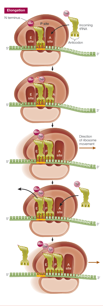
Figure 14.16 1. How is the genetic code “read” by a cell during translation? 2. When are the two subunits of a ribosome together? 3. Does a ribosome begin translation at the first nucleotide in an mRNA? If not, where does it begin?
Polypeptides Can Be Modified and Transported during or after Translation
- Some proteins have signal sequences that direct them to particular locations inside or outside the cell.
- Many proteins are modified post-translationally by mechanisms such as proteolysis, glycosylation, and phosphorylation.
Questions
- What effect will removing a signal sequence have on a polypeptide?
- Explain why polypeptides have a methionine amino acid at their amino end when they are first synthesized, but often have a different amino acid later on when they are fully functional.
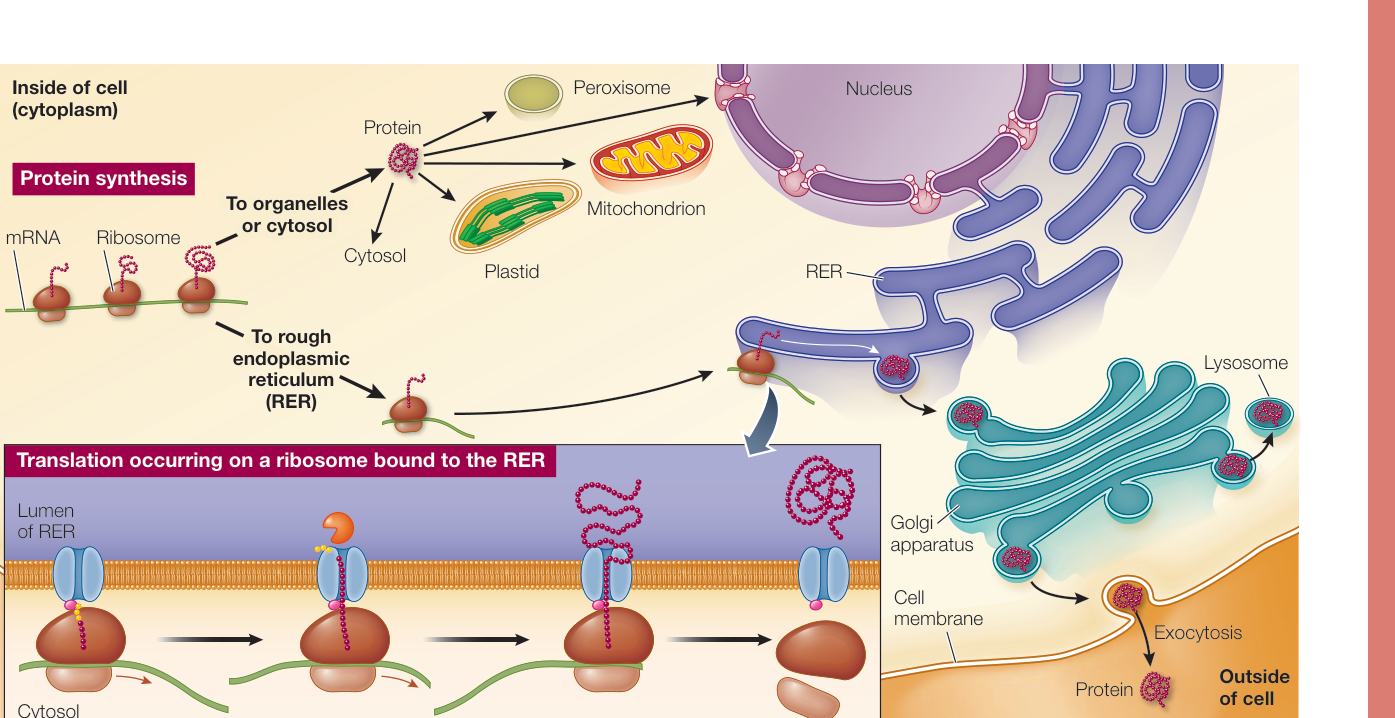
Figure 14.19