Chapter 21Evolution of Genes and Genomes
Courtesy of Dan Higgins and Douglas Jordan/CDC
Key Concepts
- 21.1 DNA Sequences Record the History of Gene Evolution
- 21.2 Genomes Reveal Both Neutral and Selective Processes of Evolution
- 21.3 Changes in the Size and Arrangement of Genomes Can Result in New Features
- 21.4 Changes in Gene Expression Often Shape Evolution
- 21.5 Molecular Evolution Has Many Practical Applications
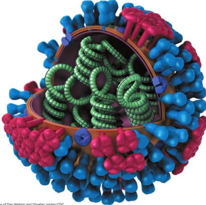
An artist’s view of an influenza virus The spikes on the surface represent proteins that elicit an immune response in humans.
Investigating Life: Evolutionary Theory Helps Us Make Better Flu Vaccines
Combat in World War I ended in November 1918. But the death toll from four years of war was soon surpassed by the casualties of a massive influenza epidemic that killed more than 50 million people worldwide—more than twice the number of World War I combat deaths.
The 1918–1919 pandemic was noteworthy because the death rate among young adults—who are usually less likely to die from influenza than are the elderly or the very young—was 20 times higher than in flu epidemics before or since. Why was that particular virus so deadly, especially to typically hardy individuals? The 1918 flu strain triggered an especially intense reaction in the human immune system. This overreaction meant that people with strong immune systems were likely to be more severely affected.
Usually we can count on our immune system to fight viruses, and the immune response is the basis of vaccination. Since 1945, programs to administer flu vaccines have helped keep the number and severity of influenza outbreaks in check. Last year’s vaccine, however, will probably not be effective against this year’s virus. New strains of flu virus are evolving continuously, producing genetic variation in the population. If these viruses did not evolve, we would become resistant to them and annual vaccination would become unnecessary. But because the viruses do evolve, biologists must develop a new and different flu vaccine each year.
The vertebrate immune response is initiated when the immune system recognizes proteins on the viral surface. Changes in viral surface proteins can allow the virus to escape immune detection. Virus strains with the greatest number of changes to their surface proteins are most likely to avoid detection and infect their hosts, and thus have an advantage over other strains. Biologists observe evolution in action by following changes in influenza virus proteins from year to year, and with this knowledge can produce more effective vaccines.
We learn a great deal about the molecular basis of evolution by examining rapidly evolving organisms such as viruses. Molecular studies of evolution, in turn, are put to practical uses, such as the development of better strategies for combating deadly diseases.
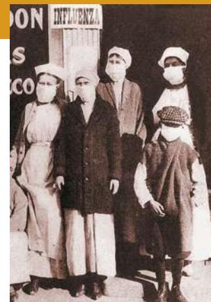
Courtesy of the Office of the Public Health Service Historian
QA Why was the 1918–1919 flu pandemic worse than any before or since? (Find the answer on page 490.)
DNA Sequences Record the History of Gene Evolution
Learning Objectives
21.1.1 Analyze and compare DNA and protein sequences across species by using matrices of similarities and differences.
21.1.2 Explain why counts of nucleotide or amino acid differences between two sequences often underestimate the actual number of changes that have occurred between the sequences.
An organism’s genome is the full set of genes it contains, as well as any noncoding regions of the DNA (or in the case of some viruses, RNA). Most of the genes of eukaryotic organisms are found on chromosomes in the nucleus, but genes are also present in chloroplasts and mitochondria. In organisms that reproduce sexually, both males and females contribute nuclear genes, but mitochondrial and chloroplast genes are usually transmitted only via the cytoplasm of one of the two gametes (often from the female parent).
Evolution results from changes to gene sequences, location, and expression
Genomes must be replicated to be transmitted from parents to offspring. DNA replication does not occur without error, however. Mistakes in DNA replication—mutations—provide much of the raw material for evolutionary change. Even though the replication error rate per nucleotide is very small, most genomes are so large that a few errors are usually made every time a genome is replicated. These errors are actually essential for the long-term survival of life, because mutations are the initial source of the genetic variation that permits species to evolve in response to changes in their environment.
A particular allele of a gene will not be passed on to successive generations unless an individual carrying that allele survives and reproduces. The allele must function in combination with many other genes in the genome or it will quickly be selected against. Moreover, the degree and timing of a gene’s expression are affected by its location in the genome. Evolutionary changes result from mutations to gene sequences, from gene duplications and rearrangements, and from changes in the timing, degree, and location of gene expression.
A genome, then, is not simply a random collection of genes in a random order along chromosomes. Rather, it is a complex set of integrated genes, regulatory sequences, and structural elements, interspersed with vast stretches of noncoding DNA that may have little direct function. The sequences of genes, as well as their location and expression, are subject to evolutionary change, as are the extent and location of noncoding DNA. All of these changes can affect the phenotype of an organism.
Biologists have now sequenced the complete genomes of a large number of organisms, including humans. The information in these sequences is helping us understand how and why organisms differ, how they function, and how they have evolved.
Evolution of genomes results in biological diversity
The field of molecular evolution investigates the mechanisms and consequences of the evolution of macromolecules—particularly nucleic acids (DNA and RNA) and proteins. Molecular evolutionists study relationships between the structures of genes and proteins and the functions of organisms. They also examine molecular variation to reconstruct evolutionary history and to study the mechanisms and consequences of evolution. Students of this field ask questions such as: What does molecular variation tell us about a gene’s function? Why do the genomes of different organisms vary in size? What evolutionary forces shape patterns of variation among genomes? And a crucial question from an evolutionary perspective: How do genomes acquire new functions? Investigations into the evolution of particular nucleic acids and proteins are instrumental in reconstructing the evolutionary histories of genes. Ultimately, molecular evolutionary biologists hope to explain the molecular basis of biological diversity.
The evolution of nucleic acids and proteins depends on genetic variation introduced by mutations. One of several ways genes evolve is by means of nucleotide substitutions (the incorporation of point mutations in populations). In genes that encode proteins, nucleotide substitutions sometimes result in amino acid replacements that can change the charge, the structure, and other chemical and physical properties of the encoded protein. These changes in a protein molecule often affect the way that protein functions in the organism.
Evolutionary changes in genes and proteins can be identified by comparing nucleotide or amino acid sequences from different organisms. The longer two sequences have been evolving separately, the more differences they accumulate (bearing in mind that different genes in the same species evolve at different rates). Determining how long ago changes in nucleotide or amino acid sequences occurred is a useful step toward inferring their causes. Knowledge of the pattern and rate of evolutionary change in a given macromolecule is useful in reconstructing the evolutionary history of groups of organisms.
To compare genes or proteins from different organisms, biologists need a way to identify homologous parts of macromolecules. (Recall from Key Concept 20.1 that homologous features are those shared by two or more species that have been inherited from a common ancestor.) Homologous parts of a protein can be identified by their homologous amino acid sequences. And since nucleotide sequences encode amino acid sequences, the concept of homology extends down to the level of individual nucleotide positions. Therefore one of the first steps in studying the evolution of genes or proteins is to align homologous positions in the amino acid or nucleotide sequence of interest.
Genes and proteins are compared through sequence alignment
Once the nucleotide or amino acid sequences of molecules from different organisms have been determined, they can be compared. Homologous positions can be identified only if we first pinpoint the locations of deletions and insertions that have occurred in the molecules of interest in the time since the organisms diverged from a common ancestor. A simple hypothetical example illustrates this sequence alignment technique. In Figure 21.1 we compare two amino acid sequences from homologous proteins in different organisms. The two sequences at first appear to differ in both the number and identity of their amino acids. If we insert a gap after the first amino acid in sequence 2 (after leucine), however, the similarities in the two sequences become obvious. This gap represents the occurrence of one of two evolutionary events: an insertion of an amino acid in the longer protein or a deletion of an amino acid in the shorter protein. Having adjusted for this insertion or deletion event, we can see that the two sequences differ by only one amino acid at position 6 (serine or phenylalanine).
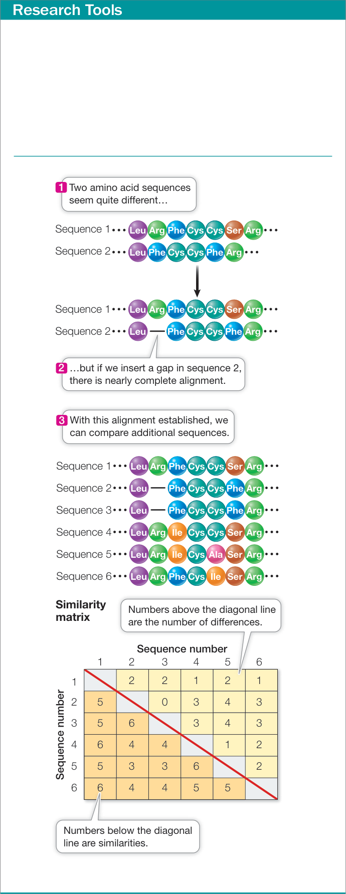
Figure 21.1 Amino Acid Sequence Alignment Amino acid sequence alignment is a way of arranging protein sequences to identify regions of homology between the sequences. Gaps are inserted between the amino acid residues to align similar residues in columns. Differences (number of amino acid differences plus insertion or deletion events) and similarities (number of identical amino acids) between each pair of aligned sequences are then summarized in a similarity matrix. Homologous DNA sequences can be aligned in a similar manner.
Activity 21.1 Amino Acid Sequence Alignment
Adding a single gap—that is, identifying a deletion or an insertion—aligns the two sequences in Figure 21.1. Additional sequences can now be added to the alignment in a similar manner. Longer amino acid sequences, and those that have diverged more extensively, require more elaborate adjustments. Explicit models (incorporated into computer algorithms) have been developed to account for the relative probabilities of deletions, insertions, and particular amino acid replacements.
Having aligned the sequences, we can compare them by counting the number of nucleotides or amino acids that differ between them. Summing the numbers of the same and different amino acids in each pair of sequences allows us to construct a similarity matrix, which gives us a measure of the minimum number of changes that have occurred since the divergence of each pair of organisms (see Figure 21.1).
Activity 21.2 Similarity Matrix Construction
Models of sequence evolution are used to calculate evolutionary divergence
The sequence comparison procedure illustrated in Figure 21.1 gives a simple count of the number of similarities and differences between the proteins of two species. In the context of two aligned DNA sequences, we can count the number of differences at homologous nucleotide positions, and this count indicates the minimum number of nucleotide changes that must have occurred since the two sequences diverged from a common ancestral sequence.
Although it is useful in determining a minimum number of changes between two DNA sequences, the count provided by sequence alignment almost certainly underestimates the actual number of changes that have occurred since the sequences diverged. Any given change counted in a similarity matrix of DNA sequences may result from multiple substitution events that occurred at a given nucleotide position over time. As illustrated in Figure 21.2, any of the following events may have occurred at a given nucleotide position that would not be revealed by a simple count of similarities and differences between two DNA sequences:
- Multiple substitutions. More than one change has occurred at a given position between the ancestral sequence and at least one of the observed sequences.
- Coincident substitutions. At a given position, different substitutions have occurred between the ancestral sequence and each observed sequence.
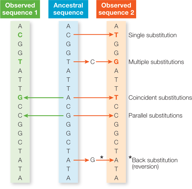
Figure 21.2 Multiple Substitutions Are Not Reflected in Pairwise Sequence Comparisons Two observed sequences descended from a common ancestral sequence (center) have undergone a series of substitutions. Although the two observed sequences differ by only three nucleotides (colored letters), these three differences result from a total of nine substitutions (arrows).
- Parallel substitutions. The same substitution has occurred independently between the ancestral sequence and each observed sequence.
- Back substitutions (also called reversions). In a variation on multiple substitutions, after a change at a given position, a subsequent substitution has changed the position back to the ancestral state.
To correct for undercounting of substitutions, molecular evolutionists have developed mathematical models that describe how DNA (and protein) sequences evolve. These models take into account the relative rates of change from one nucleotide to another. For
Tuna
Rice
Acidic side chains
- D Aspartic acid
- E Glutamic acid
Basic side chains
- H Histidine
- K Lysine
- R Arginine
Hydrophobic side chains
F Phenylalanine
I Isoleucine
L Leucine
M Methionine
V Valine
Y Tyrosine
W Tryptophan
A Alanine
Other
- C Cysteine
- P Proline
- Q Glutamine
- N Asparagine
- S Serine
- T Threonine
- G Glycine
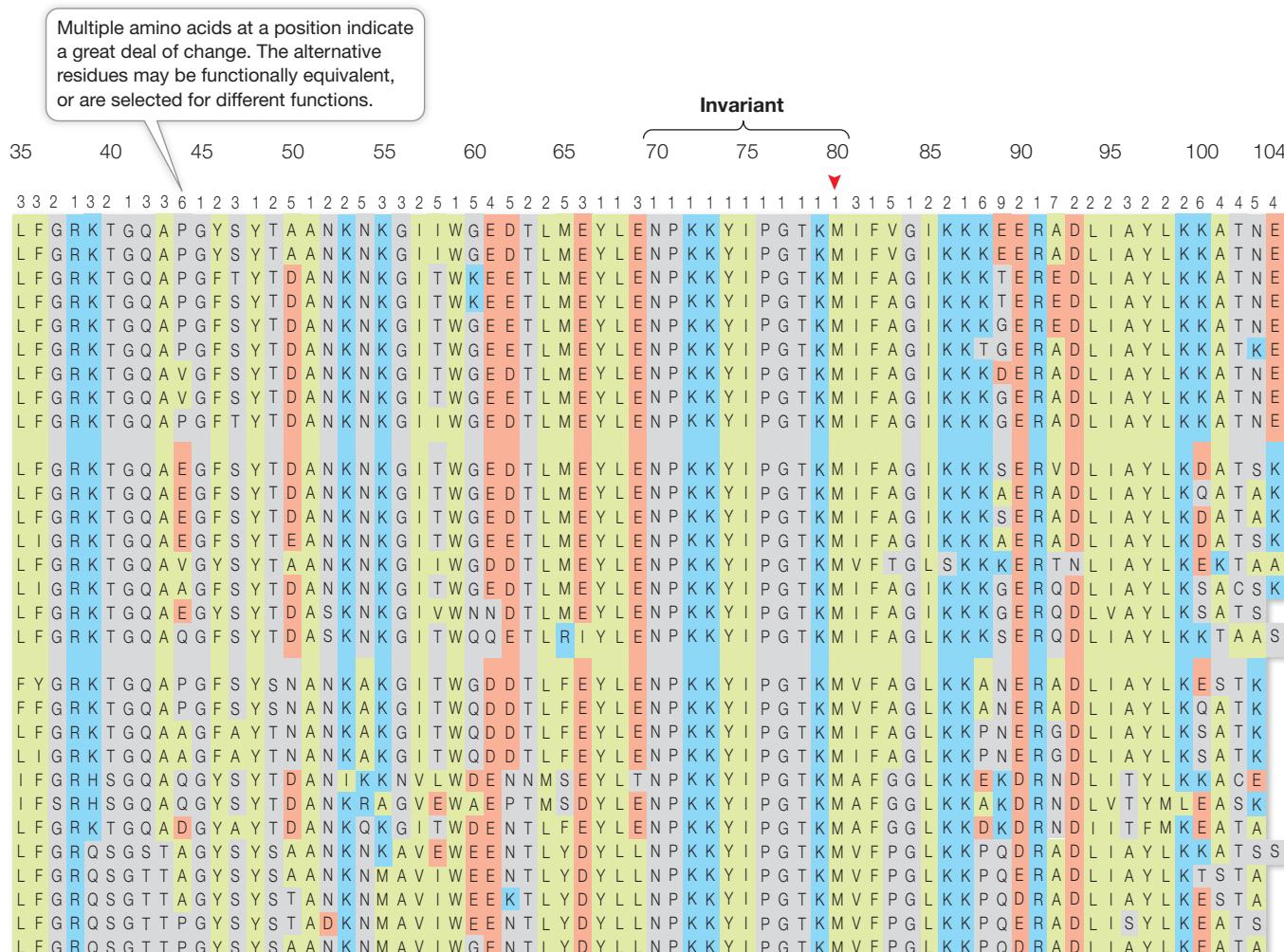
The number 1 indicates an invariant position in the cytochrome c molecule (i.e., all the organisms have the same amino acid in this position). Such a position is probably under strong purifying selection.
Amino acids at positions marked by red arrowheads have side chains that interact with the heme group.
| Position in sequence | 1 | 5 | 10 | 15 | 20 | 25 | 30 | |
|---|---|---|---|---|---|---|---|---|
| Number of amino acids at the position | 1 | 3 | 5 | 5 | 5 | 5 | 5 | |
| Human, chimpanzee | G D V E K G K K I F I M K C S Q C H T V E K G G K H K T G P N L H G | |||||||
| Rhesus monkey | G D V E K G K K I F I M K C S Q C H T V E K G G K H K T G P N L H G | |||||||
| Horse | G D V E K G K K I F V Q K C A Q C H T V E K G G K H K T G P N L H G | |||||||
| Donkey | G D V E K G K K I F V Q K C A Q C H T V E K G G K H K T G P N L H G | |||||||
| Cow, pig, sheep | G D V E K G K K I F V Q K C A Q C H T V E K G G K H K T G P N L H G | |||||||
| Dog | G D V E K G K K I F V Q K C A Q C H T V E K G G K H K T G P N L H G | |||||||
| Rabbit | G D V E K G K K I F V Q K C A Q C H T V E K G G K H K T G P N L H G | |||||||
| Gray whale | G D V E K G K K I F V Q K C A Q C H T V E K G G K H K T G P N L H G | |||||||
| Gray kangaroo | G D V E K G K K I F V Q K C A Q C H T V E K G G K H K T G P N L N G | |||||||
| Chicken, turkey | G D I E K G K K I F V Q K C S Q C H T V E K G G K H K T G P N L H G | |||||||
| Pigeon | G D I E K G K K I F V Q K C S Q C H T V E K G G K H K T G P N L H G | |||||||
| Pekin duck | G D V E K G K K I F V Q K C S Q C H T V E K G G K H K T G P N L H G | |||||||
| Snapping turtle | G D V E K G K K I F V Q K C A Q C H T V E K G G K H K T G P N L N G | |||||||
| Rattlesnake | G D V E K G K K I F T M K C S Q C H T V E K G G K H K T G P N L H G | |||||||
| Bullfrog | G D V E K G K K I F V Q K C A Q C H T C E K G G K H K V G P N L Y G | |||||||
| Tuna | G D V A K G K K T F V Q K C A Q C H T V E N N G G K H K V G P N L W G | |||||||
| Dogfish | G D V E K G K K V F V Q K C A Q C H T V E N N G G K H K T G P N L S G | |||||||
| Samia cynthia (moth) | G N A E N G K K I F V Q R C A Q C H T V E A G G K H K V G P N L H G | |||||||
| Tobacco hornworm moth | G N A D N G K K I F V Q R C A Q C H T V E A G G K H K V G P N L H G | |||||||
| Screwworm fly | G D V E K G K K I F V Q R C A Q C H T V E A G G K H K V G P N L H G | |||||||
| Drosophila (fruit fly) | G D V E K G K K L F V Q R C A Q C H T V E A G G K H K V G P N L H G | |||||||
| Baker’s yeast | G S A K K G A T L F K T R C E L C H T V E K G G P H K V G P N L H G | |||||||
| Candida krusei (yeast) | G S A K K G A T L F K T R C A E C H T I E A G G P H K V G P N L H G | |||||||
| Neurospora crassa (mold) | G D S K K G A N L F K T R C A E C H — E — N L T Q K I G P A L H G | |||||||
| Wheat | G N P D A G A K I F K T K C A Q C H T V D A G A — H K Q G P N L H G | |||||||
| Sunflower | G D P T T G A K I F K T K C A Q C H T V E K G A — H K Q G P N L N G | |||||||
| Mung bean | G D S K S G E K I F K T K C A Q C H T V D K G A — H K Q G P N L N G | |||||||
| Rice | G N P K A G E K I F K T K C A Q C H T V D K G A — H K Q G P N L N G | |||||||
| Sesame | G D V K S G E K I F K T K C A Q C H T V D K G A — H K Q G P N L N G |
Gaps indicate insertion and/or deletion events.
example, transitions (changes between the two purines, A ↔︎ G, or between the pyrimidines, C ↔︎ T) are typically more frequent than transversions (a purine is replaced by a pyrimidine, or vice versa). These models also include parameters such as the different rates of substitution across different parts of a gene and the proportions of each nucleotide present in a given sequence. Once such parameters have been estimated, the model is used to correct for multiple substitutions, coincident substitutions, parallel substitutions, and back substitutions. The revised estimate accounts for the total number of substitutions likely to have occurred between two sequences, which is almost always greater than the observed number of differences.
As sequence information becomes available for more and more genes in an ever-expanding database, sequence alignments can be extended across multiple homologous sequences, and the minimum number of insertions, deletions, and substitutions can be summed across homologous genes of an entire group of organisms. Similar databases have been constructed for homologous proteins. Figure 21.3 shows aligned data for cytochrome c protein sequences in 33 species of animals, plants, and fungi. Such information is used extensively in determining evolutionary relationships among species.
Experimental studies examine molecular evolution directly
Although molecular evolutionists are often interested in naturally evolved genes and proteins, molecular evolution can also be observed directly in the laboratory. Increasingly, evolutionary biologists are studying evolution experimentally. Because substitution rates are related to generation time rather than to absolute time, most of these experiments use unicellular organisms or viruses with short generations. Viruses, bacteria, and unicellular eukaryotes (such as the yeasts) can be cultured in large populations in the laboratory, and many of these organisms can evolve rapidly. In the case of some RNA viruses, the natural substitution rate may be as high as 1 substitution per 1,000 nucleotides per generation. Therefore in a virus of a few thousand nucleotides, one or more substitutions are expected (on average) every generation, and these changes can easily be determined by sequencing the entire viral genome (because of its small size). Generation time may be only tens of minutes (rather than years or decades, as in many animals), so biologists can directly observe substantial molecular evolution in a controlled population over the course of days, weeks, or months.
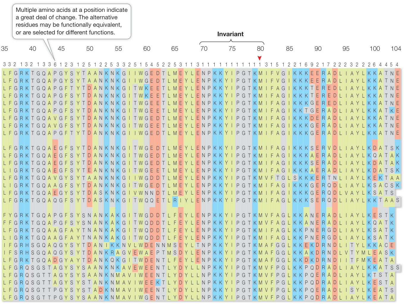
Figure 21.3 Amino Acid Sequences of Cytochrome c The amino acid sequences shown in the table were obtained from analyses of the enzyme cytochrome c from 33 species of plants, fungi, and animals. Note the lack of variation across the sequences at positions 70–80, suggesting that this region is under strong purifying selection and that changing its amino acid sequence would impair the protein’s function. The molecular models at the upper left are created from these sequences and show the three-dimensional structures of tuna and rice cytochrome c. Alpha helixes are in red, and the molecule’s heme group is shown in yellow. (Based on table by Graphic Presentation Services, Inc. in R. E. Dickerson. 1972. Sci Am 226: 58–72.)
Experimental molecular evolutionary studies are used for a wide variety of purposes and have greatly expanded the ability of evolutionary biologists to test evolutionary concepts and principles. Biologists now routinely study evolution in the laboratory, and as we will see later in this chapter, they can use in vitro evolutionary techniques to produce novel molecules that perform new functions with industrial and pharmaceutical uses.
Recap and Assess
The genomes of all organisms evolve over time. Evolutionary changes can be detected by aligning and then comparing the nucleic acid and protein sequences of different species. Experimental studies of molecular evolution under controlled conditions allow biologists to study many processes of evolution directly.
- Align the following sequences. Then create a matrix that compares both the number of identical nucleotides and the number of differences (including insertion and deletion events). Sequence a: AATGCAGGGTATACG Sequence b: ATTCAGGGTATACC Sequence c: ATTGCAGCGTATAACC Sequence d: ATTGCAGGGTATACG
- Explain why a simple count of nucleotide differences between two sequences often underestimates the actual number of nucleotide substitutions since the sequences diverged. Use an example based on the comparison of your alignment of Sequences a and b from Question 1.
We have seen that molecular evolutionists can directly observe the evolution of genomes over time, compare the genomes of different organisms, and reconstruct the changes that have occurred during their evolution. Let’s turn now to the question of how genomes change, and examine some of the consequences of those changes.
Genomes Reveal Both Neutral and Selective Processes of Evolution
Learning Objectives
- 21.2.1 Explain how substitutions may be neutral or selective in a given organism.
- 21.2.2 Provide a mathematical explanation for why the rate of evolution of neutral mutations is independent of population size.
- 21.2.3 Evaluate gene sequences of protein-coding genes to identify positions that are evolving under purifying versus positive selection, and apply this knowledge to a biological problem.
Most natural populations harbor far more genetic variation than we would expect to find if genetic variation were influenced by natural selection alone. This discovery, combined with the knowledge that many mutations do not change molecular function, stimulated the development of the neutral theory of molecular evolution.
Much of molecular evolution is neutral
To discuss the evolution of genes, we need to consider the specific types of mutations that are possible. A nucleotide substitution is a change in a single nucleotide in a DNA sequence (a type of point mutation; see Key Concept 15.1). Many nucleotide substitutions have no effect on phenotype, even if the change occurs in a gene that encodes a protein, because most amino acids are specified by more than one codon. A substitution that does not change the encoded amino acid is known as a synonymous substitution (also called a silent substitution) (Figure 21.4A). Synonymous substitutions do not affect the functioning of a protein (although they may have other effects, such as changes in mRNA stability or translation rates) and are therefore less likely than other types of substitutions to be influenced by natural selection.
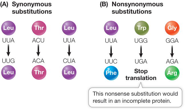
Figure 21.4 When One Nucleotide Changes (A) Synonymous substitutions do not change the amino acid specified and do not affect protein function. Such substitutions are less likely than other substitutions to be influenced by natural selection, although they contribute greatly to the buildup of neutral genetic variation in a population. (B) Nonsynonymous substitutions do change the amino acid sequence and are likely to have an effect (often deleterious, but sometimes beneficial) on protein function. Such nucleotide substitutions are targets for natural selection.
A nucleotide substitution that does change the amino acid sequence encoded by a gene is known as a nonsynonymous substitution (a missense substitution changes an amino acid, and a nonsense substitution produces a new stop codon) (Figure 21.4B). In general, nonsynonymous substitutions are likely to be deleterious to the organism. But not every amino acid replacement alters a protein’s shape and charge (and hence its functional properties). Therefore some nonsynonymous substitutions are selectively neutral, or nearly so. Another possibility is that a nonsynonymous substitution alters a protein in a way that confers an advantage to the organism and is therefore favored by natural selection.
Synonymous substitutions accumulate over time in protein-coding genes at a much higher rate than nonsynonymous substitutions do. This is true even though the underlying mutation rates for both types of substitutions are equal (or at least similar). The reason is simple: as most nonsynonymous substitutions are harmful to the function of a gene, they are removed from the population over time through natural selection. Therefore substitution rates are higher at nucleotide positions that do not change the amino acid being expressed (Figure 21.5). The rate of substitution is even higher in pseudogenes, which are copies of genes that are no longer functional (and so are not subject to strong selection).
Insertions, deletions, and rearrangements of DNA sequences are all mutations that may affect a larger portion of the gene or genome than do point mutations. Insertions and deletions of nucleotides in a protein-coding sequence interrupt its reading frame, unless they occur in multiples of three nucleotides (the length of one codon). Rearrangements may merely change the order of whole genes along chromosomes, or they may rearrange functional domains among individual genes.
When biologists began to examine the details of genetic variation of populations, they soon discovered many gene variants that had little or no effect on function. This gave rise to new ideas about how these neutral variants arise and spread in populations. Motoo Kimura proposed the neutral theory in 1968. He suggested that, at the molecular level, the majority of variants found in most populations are selectively neutral. That is, most gene variants confer neither an advantage nor a disadvantage on their bearers. Therefore these neutral variants must accumulate through genetic drift rather than through positive selection.
We saw in Key Concept 19.2 that genetic drift of existing gene variants tends to be greatest in small populations. However, the rate of fixation of new neutral mutations by genetic drift is independent of population size. To see why this is so, consider a population of size and a neutral mutation rate of (mu) per gamete per generation at a particular locus. The number of new mutations would be, on average, , because gene copies are available to mutate in a population of diploid organisms. The probability that a given mutation will be fixed by drift alone is its frequency, which equals for a newly arisen mutation. We can multiply these two terms to get the rate of fixation of neutral mutations () in a given population of individuals:
But notice that the two effects of population size () cancel one another out:
Therefore the rate of fixation of neutral mutations depends only on the neutral mutation rate and is independent of population size. Any given mutation is more likely to appear in a large population than in a small one, but any mutation that does appear is more likely to become fixed in a small population. The rate of fixation of neutral mutations is thus equal to the mutation rate in populations of any size (i.e., ).
As long as the underlying mutation rate is constant, genes and proteins evolving in different populations should diverge from one another in neutral changes at a constant rate. The rate of evolution of particular genes and proteins is indeed often relatively constant over time, and therefore can be used as a molecular clock to calculate evolutionary divergence times between species (see Key Concept 20.3).
Neutral theory does not imply that most mutations have no effect on the individual organism, even though much of the genetic variation present in a population is the result of neutral evolution. Many mutations are rarely or never observed in populations because they are lethal or strongly detrimental, and the individuals that carry them are quickly removed from the population through natural selection. Similarly, because mutations that confer a selective advantage tend to be quickly fixed in populations, they also do not result in significant variation at the population level. Nonetheless, if we compare homologous proteins from different populations or species, some amino acid positions will remain constant under purifying selection, others will vary through neutral genetic drift, and still others will differ as a result of positive selection for change. How can these evolutionary processes be distinguished?
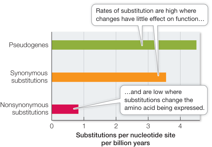
Figure 21.5 Rates of Substitution Differ Rates of nonsynonymous substitution are typically much lower than rates of synonymous substitution, and much lower than substitution rates in pseudogenes. This pattern reflects stronger stabilizing selection in functional genes than in pseudogenes.
Positive and purifying selection can be detected in the genome
As we have just seen, substitutions in a protein-coding gene can be either synonymous or nonsynonymous, depending on whether or not they change the resulting amino acid sequence of the protein. The relative rates of synonymous and nonsynonymous substitutions are expected to differ in regions of genes that are evolving neutrally, or evolving under positive selection for change, or staying unchanged under purifying selection.
- If a given amino acid in a protein can be one of many alternatives (without changing the protein’s function), then an amino acid replacement is neutral with respect to the fitness of an organism. In this case, the rates of synonymous and nonsynonymous substitutions in the corresponding DNA sequences are expected to be very similar, so the ratio of the two rates should be close to 1.
- If a given amino acid position is under positive selection for change, the observed rate of nonsynonymous substitutions is expected to exceed the rate of synonymous substitutions in the corresponding DNA sequences.
- If a given amino acid position is under purifying selection, then the observed rate of synonymous substitutions is expected to be much higher than the rate of nonsynonymous substitutions in the corresponding DNA sequences.
The evolution of lysozyme illustrates how and why particular codons in a gene sequence might be under different modes of selection. The enzyme lysozyme is found in almost all animals. It is produced in the tears, saliva, and milk of mammals and in the albumen (whites) of bird eggs. Lysozyme digests the cell walls of bacteria, rupturing and killing them. As a result, it plays an important role as a first line of defense against invading bacteria. Most animals defend themselves against bacteria by digesting them, which is probably why most animals have lysozyme. Some animals also use lysozyme in the digestion of food.
Among mammals, a mode of digestion called foregut fermentation has evolved twice. In mammals with this mode of digestion, the foregut—consisting of part of the esophagus and/or stomach—has been converted into a chamber in which bacteria break down ingested plant matter by fermentation. Foregut fermenters can obtain nutrients from the otherwise indigestible cellulose that makes up a large proportion of plant tissue. Foregut fermentation evolved independently in ruminants (a group of hoofed mammals that includes cattle) and in certain leaf-eating monkeys, such as langurs. We know that these evolutionary events were independent because both langurs and ruminants have close relatives that are not foregut fermenters.
In both mammalian foregut-fermenting lineages, lysozyme has been modified to play a new, nondefensive role. The modified lysozyme enzyme ruptures some of the bacteria that live in the foregut, releasing nutrients metabolized by the bacteria, which the mammal then absorbs. How many changes in the lysozyme molecule were needed to allow it to perform this function amid the digestive enzymes and acidic conditions of the mammalian foregut? To answer this question, Caro-Beth Stewart and her colleagues compared the lysozyme-coding sequences in foregut fermenters with those in several of their nonfermenting relatives. They determined which amino acids differed and which were shared among the species (Figure 21.6), as well as the rates of synonymous and nonsynonymous substitutions in lysozyme genes across the evolutionary history of the sampled species.
For many of the amino acid positions of lysozyme, the rate of synonymous substitution in the corresponding gene sequence was much higher than the rate of nonsynonymous substitution. This observation indicates that many of the amino acids that make up lysozyme are evolving under purifying selection. In other words, there is selection against change in the lysozyme protein at these positions, and the encoded amino acids must therefore be critical for lysozyme function. At other positions, several different amino acids function equally well, and the corresponding codons have similar rates of synonymous and nonsynonymous substitutions.
The most striking finding was that amino acid replacements in lysozyme happened at a much higher rate in the lineage leading to langurs than in any other primate lineage. The high rate of nonsynonymous substitution in the langur lysozyme gene shows that lysozyme went through a period of rapid change in adapting to the foreguts of langurs. Moreover, the lysozymes of langurs and cattle exhibit five instances of convergent evolution, a process whereby organisms that are not closely related independently evolve similar traits as a result of having to adapt to similar environments or ecological niches. More specifically, the lysozymes of langurs and cattle share five convergent amino acid replacements, all of which lie on the surface of the lysozyme molecule, well away from the enzyme’s active site. Two of these shared replacements are changes from arginine to lysine, which make the protein more resistant to degradation by the stomach enzyme pepsin. By understanding the functional significance of amino acid replacements, biologists can explain the observed changes in amino acid sequences in terms of changes in the functioning of the protein.
Connect the Concepts As explained in Key Concept 20.1, not all similar traits are evidence of relatedness. Similar traits may evolve independently in different lineages through the phenomenon of convergent evolution.
A large body of fossil, morphological, and molecular evidence shows that langurs and cattle do not share a recent common ancestor. However, langur and ruminant lysozymes share several amino acids that neither mammal shares with the lysozymes of its own closer relatives. The lysozymes of these two mammals have converged on some of the same amino acids despite their very different ancestry. The amino acids they share give these lysozymes the ability to lyse the bacteria that ferment plant material in the foregut.
The ability to test for positive, purifying, and neutral selection has allowed biologists to distinguish functional from nonfunctional genes, and to understand how genes are evolving within given species.
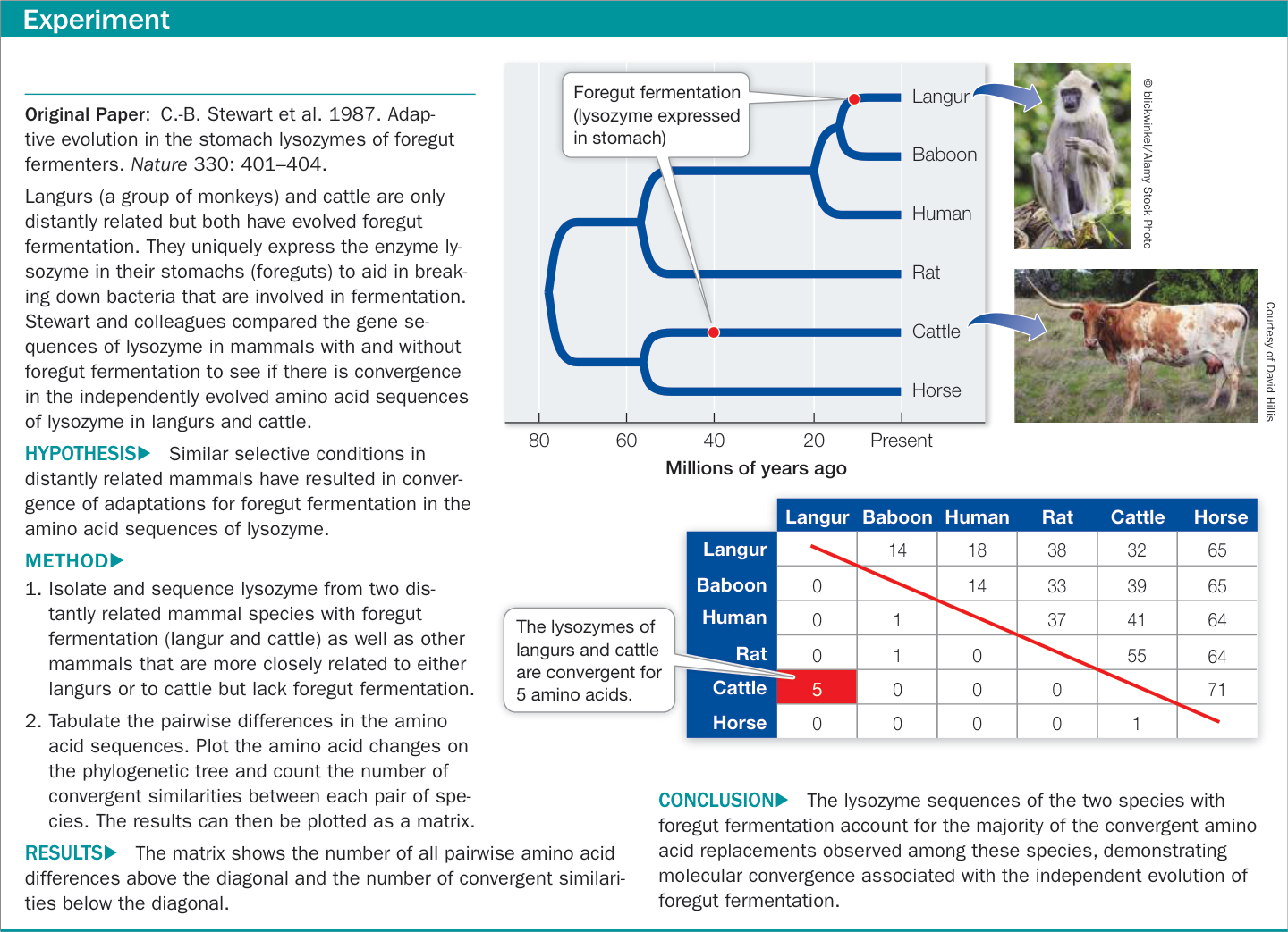
Figure 21.6A Convergent Molecular Evolution Figure 21.6A Convergent Molecular Evolution Figure 21.6B Convergent Molecular Evolution
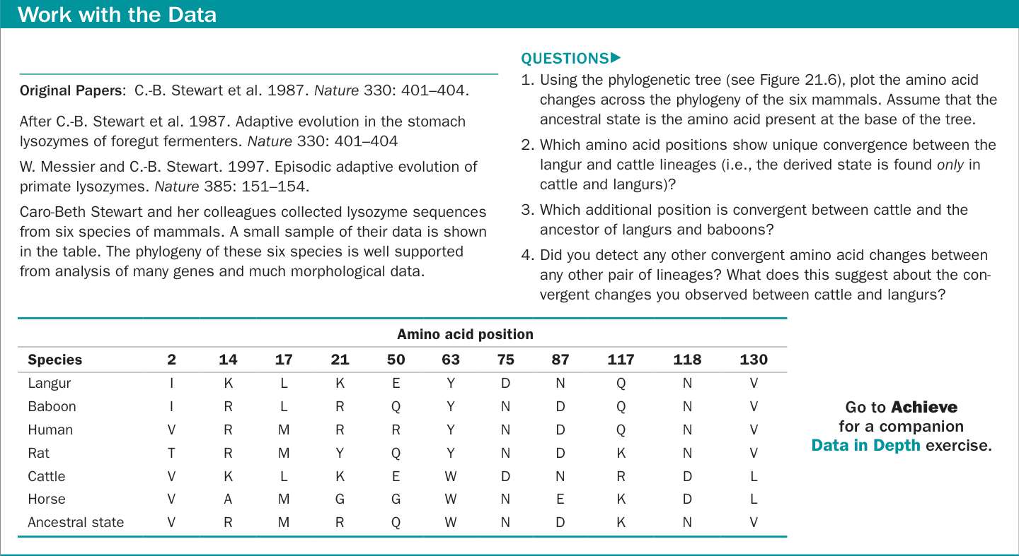
Figure 21.6B Convergent Molecular Evolution
Recap and Assess
The neutral theory of molecular evolution provides an explanation for the relatively constant rate of molecular change seen in many species. By examining the relative rates of synonymous and non-synonymous substitutions in genes over time, biologists can distinguish the evolutionary mechanisms acting on individual genes, and understand how they are changing through time.
- Over evolutionary history, many groups of organisms that inhabit caves have lost the organs of sight. For example, although surface-dwelling crayfishes have functional eyes, several crayfish species that are restricted to underground habitats lack eyes. Opsins are light-sensitive proteins known to have an important function in vision (see Chapter 44), and opsin genes are expressed in eye tissues. Opsin genes are present in the genomes of eyeless, cave-dwelling crayfishes. Two alternative hypotheses that might explain the presence of opsin genes in an eyeless organism are (a) the genes were inherited from an eyed ancestor but are no longer functional; or (b) the genes have a new function other than vision. How would you investigate these two hypotheses using the sequences of the opsin genes in various species of crayfishes?
- Why is the rate of fixation of neutral mutations independent of population size? Use mathematical expressions to demonstrate why this is true.
- An investigator compared many gene sequences encoding surface proteins from influenza viruses sampled over time and collected the data shown in the table.
| Codon position | Synonymous substitutions | Nonsynonymous substitutions |
|---|---|---|
| 12 | 0 | 7 |
| 15 | 1 | 9 |
| 61 | 0 | 12 |
| 80 | 7 | 0 |
| 137 | 12 | 1 |
| 156 | 24 | 2 |
| 165 | 3 | 4 |
| 226 | 38 | 3 |
- Which positions encode amino acids that are likely to have changed as a result of positive selection? Explain your answer.
- Which positions encode amino acids that are likely to have changed as a result of purifying selection? Explain your answer.
(Hint: To calculate rates of each substitution type, consider the number of synonymous and nonsynonymous substitutions present relative to the number of possible substitutions of each type. There are approximately three times as many possible nonsynonymous substitutions as there are synonymous substitutions.)
Based on data in GenBank under accession numbers AF008656 to AF008909, as analyzed in W. M. Fitch et al. 1997. Proc Natl Acad Sci U.S.A. 94: 7712–7718 © 1997. National Academy of Sciences, U.S.A.; R. M. Bush et al. 1999. Science 286: 1921–1925.
Although nucleotide substitutions within coding sequences result in many important changes to organisms, genomes evolve through many additional processes. In particular, additions and deletions of noncoding sequences, gene duplications, and movement of genes between genomes can have major evolutionary consequences.
Changes in the Size and Arrangement of Genomes Can Result in New Features
Learning Objectives
- 21.3.1 Explain the role of noncoding DNA in the evolution of genome size.
- 21.3.2 Compare and contrast the evolutionary advantages and disadvantages of sexual reproduction.
- 21.3.3 Describe how genes can move among different lineages via lateral gene transfer.
- 21.3.4 Using a phylogenetic tree of a gene family, identify gene duplications across the history of a group of species.
- 21.3.5 Describe how a duplicated gene provides opportunities for the evolution of new functions.
Several evolutionary processes result in changes to the size and organization of genomes, which can lead to acquisition of major new characteristics in populations. These genome-scale processes often result in larger and more rapid evolutionary changes than do single point mutations.
Genome sizes among organisms differ by orders of magnitude
We know that genome size varies tremendously among organisms. Across broad taxonomic categories, there is some correlation between genome size and organismal complexity. The genome of the tiny bacterium Mycoplasma genitalium has only 470 genes. Homo sapiens, by contrast, has about 20,500 protein-coding genes. Figure 21.7 shows the number of genes from a sample of organisms whose genomes have been fully sequenced, arranged by their evolutionary relationships. As this figure reveals, however, a larger genome does not always indicate greater complexity (compare rice with the other plants, for example). It is not surprising that more complex genetic instructions are needed for building and maintaining a large multicellular organism than a small single-celled bacterium. What is surprising is that some organisms, such as lungfishes, some salmonders, and lilies, have about 40 times as much DNA as humans do (Figure 21.8). Structurally, a lungfish or a lily is not 40 times more complex than a human. So why does genome size vary so much?
Differences in genome size are not so great if we take into account only the portion of DNA that actually encodes proteins. The organisms with the largest total amounts of nuclear DNA (some ferns and flowering plants) have 80,000 times as much DNA as do the bacteria with the smallest genomes, but no species has more than about 100 times as many protein-coding genes as a bacterium. Therefore much of the variation in genome size lies not in the number of functional genes, but in the amount of noncoding DNA (see Figure 21.8).
Why do the cells of most eukaryotic organisms have so much noncoding DNA? Does this noncoding DNA have a function? Although it does not encode proteins, noncoding DNA can alter the expression of the genes surrounding it. The degree or timing of gene expression can vary dramatically depending on a gene’s location relative to noncoding sequences that regulate gene expression. Other regions of noncoding DNA consist of pseudogenes (nonfunctional copies of former genes). Pseudogenes are often carried in the genome because the cost of doing so is very small. Occasionally these pseudogenes become the raw material for the evolution of new genes with novel functions. Other noncoding sequences function in maintaining chromosomal structure. Still others consist of transposable elements (also called transposons; see Key Concept 17.2) that spread through populations because they reproduce faster than the host genome.
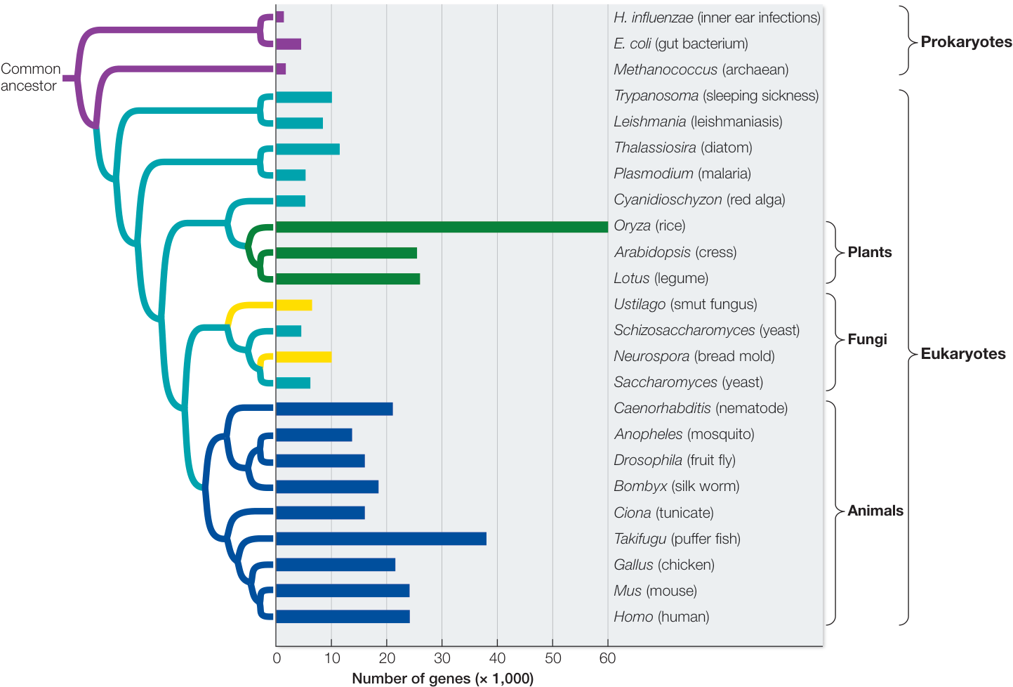
Figure 21.7 Evolution of Gene Number This figure shows the number of genes from a sample of organisms whose genomes have been fully sequenced, arranged by their evolutionary relationships. Bacteria and archaea (purple branches) typically have fewer genes than most eukaryotes. Among eukaryotes, multicellular organisms with tissue organization (plants and animals; dark green and blue branches, respectively) have more genes than single-celled organisms (aqua branches) or multicellular organisms that lack pronounced tissue organization (yellow branches). (Data from T. R. Gregory et al. 2007. Nucleic Acids Res 35 [Database issue]: D332–D338.)
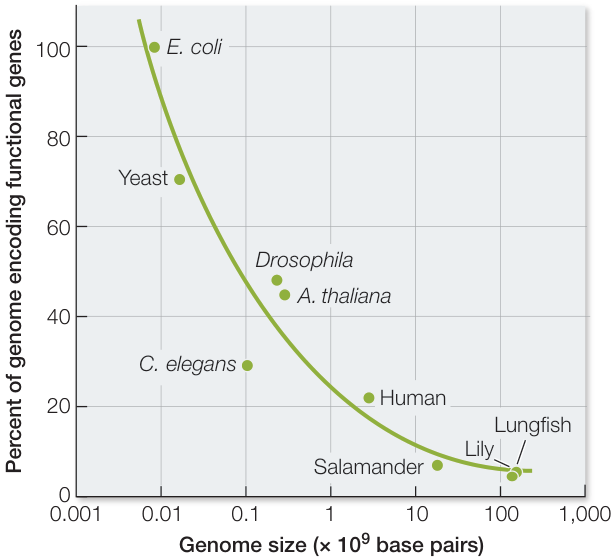
Figure 21.8 A Large Proportion of DNA Is Noncoding Most of the DNA of bacteria and yeasts encodes RNAs or proteins, whereas a large percentage of the DNA of multicellular species is noncoding. (Data from T. R. Gregory et al. 2007. Nucleic Acids Res 35 [Database issue]: D332–D338.)
It may be that the proportion of noncoding DNA is related primarily to population size. Noncoding sequences that are only slightly deleterious to the organism are likely to be purged by selection most efficiently in species with large population sizes. In species with small populations, the effects of genetic drift can overwhelm selection against noncoding sequences that have small deleterious consequences. Therefore selection against the accumulation of noncoding sequences is most effective in species with large populations, and thus species such as bacteria and yeasts have relatively little noncoding DNA compared with species with small populations (see Figure 21.8).
Sex results in the recombination of genomes
In asexually reproducing organisms, each new individual is genetically identical to its parent unless there has been a mutation. When organisms reproduce sexually, however, offspring differ from their parents because of crossing over and independent assortment of chromosomes during meiosis, as well as the combination of genetic material from two different gametes, as described in Key Concept 12.4. Sexual recombination generates an endless variety of genotype combinations that increase the evolutionary potential of populations—a long-term advantage of sex. Although some species may reproduce asexually most of the time, most asexual species have some means of achieving genetic recombination.
The evolution of meiosis and sexual recombination was a crucial event in the history of life. Exactly how these processes arose is puzzling, however, because in the short term, sex has at least three striking disadvantages:
- Recombination breaks up adaptive combinations of genes.
- Sex reduces the rate at which females pass genes on to their offspring.
- Dividing offspring into separate genders greatly reduces the overall reproductive rate.
To see why this last disadvantage exists, consider an asexual female that produces the same number of offspring as a sexual female. Let’s assume that both females produce two offspring, but that 50% of the sexual female’s offspring will be males (and thus contribute only sperm). In this next () generation, both asexual females will produce two more offspring each, but there is only one sexual female to produce offspring. Thus the effective reproductive rate of the asexual lineage is twice that of the sexual lineage.
The evolutionary problem is to identify the advantages of sex that can overcome short-term disadvantages. Several hypotheses have been proposed to explain the existence of sex, none of which is mutually exclusive. One hypothesis is that sexual recombination facilitates repair of damaged DNA, because breaks and other errors in DNA on one chromosome can be repaired by copying the intact sequence from the homologous chromosome.
Another advantage of sexual reproduction is that it permits the elimination of deleterious mutations. As explained in Key Concept 13.4, DNA replication is not perfect. Errors are introduced in every generation, and most of these errors result in lower fitness. Asexual organisms have no mechanism to eliminate deleterious mutations. Hermann J. Muller noted that the accumulation of deleterious mutations in a nonrecombining genome is like a genetic ratchet.
The mutations accumulate—“ratchet up”—at each replication. A mutation occurs and is passed on when the genome replicates, then two new mutations occur in the next replication, so three mutations are passed on, and so on. Over time, the least-mutated class of individuals is lost from the population as new mutations occur. Deleterious mutations cannot be eliminated except by the death of the lineage or a rare back mutation. The accumulation of deleterious mutations in lineages that lack genetic recombination is known as Muller’s ratchet. In sexual species, by contrast, genetic recombination produces some individuals with more of these deleterious mutations and some with fewer. The individuals with fewer deleterious mutations are more likely to survive. Thus sexual reproduction allows natural selection to eliminate particular deleterious mutations from the population over time.
Still another advantage of sex is the great variety of genetic combinations it creates in each generation. Sexual recombination does not directly influence the frequencies of alleles; rather, it generates new combinations of alleles on which natural selection can act. It expands variation in a character influenced by alleles at many loci by creating new genotypes. For example, genetic variation can be a defense against pathogens and parasites. Most pathogens and parasites have much shorter life cycles than their hosts and can rapidly evolve counteradaptations to host defenses. Sexual recombination can give the host’s defenses a chance to keep up.
Lateral gene transfer can result in the gain of new functions
The tree of life is usually visualized as a branching diagram, with each lineage diverging into two (or more) lineages over time, from one common ancestor to the millions of species that are alive today. Ancestral lineages divide into descendant lineages, resulting in speciation events that are diagrammed on the tree of life. Species’ genomes are altered as well by processes that result in lateral gene transfer—the horizontal movement of individual genes, organelles, or fragments of genomes from one lineage to another. Some species may pick up fragments of DNA directly from the environment. A virus may pick up some genes from one host and transfer them to a new host when the virus becomes integrated into the new host’s genome. Hybridization between species also results in the lateral transfer of large numbers of genes.
Lateral gene transfer can be highly advantageous to a species that incorporates novel genes from a distant relative. Genes that confer antibiotic resistance, for example, are commonly transferred among different species of bacteria. Lateral gene transfer is another way, in addition to mutation and recombination, in which species can increase their genetic variation. That genetic variation then provides the raw material on which selection acts, resulting in evolution.
A phylogenetic tree constructed from a single laterally transferred genome fragment is likely to reflect only the evolutionary history of that fragment, rather than the overall organismal phylogeny (see Key Concept 20.1). Most biologists prefer to build trees from large samples of genes or their products, so that the underlying species tree (as well as any lateral gene transfer events) can be reconstructed. Depictions of lateral gene transfer events on the underlying species tree are known as reticulations.
The degree to which lateral gene transfer events occur in various parts of the tree of life is a matter of considerable current investigation and debate. Lateral gene transfer appears to be relatively uncommon among most eukaryotic lineages, although the two major endosymbioses that gave rise to mitochondria and chloroplasts can be viewed as lateral transfers of entire bacterial genomes to the eukaryotic lineage. Some groups of eukaryotes, most notably some plants, are subject to relatively high levels of hybridization among closely related species. Hybridization leads to the exchange of many genes among recently separated lineages of plants. The greatest degree of lateral transfer, however, appears to occur among bacteria. Many bacterial genes have been transferred repeatedly among lineages of bacteria, to the point that relationships among bacterial species are often hard to decipher. Nonetheless, the broad relationships of the major groups of bacteria can still be determined (as we will discuss in Part Six of this book). Lateral transfer of genes also makes it difficult to identify the boundaries of bacterial species, which is one reason why fewer bacterial species have been named than are known to exist.
Many new functions arise following gene duplication
Gene duplication is yet another way in which genomes can acquire new functions. When a gene is duplicated, one copy of that gene is potentially freed from having to perform its original function. The initially identical copies of a duplicated gene can have any one of four subsequent fates:
Both copies of the gene may retain their original function (which can result in a change in the amount of gene product that is produced by the organism).
Both copies of the gene may retain the ability to produce the original gene product, but the expression of the genes may diverge in different tissues or at different times in development.
- One copy of the gene may be incapacitated by the accumulation of deleterious substitutions and become a nonfunctional pseudogene, or may be eliminated from the genome altogether.
- One copy of the gene may retain its original function while the second copy accumulates enough substitutions that it can perform a different function.
How often do gene duplications arise, and which of these four outcomes is most likely? Investigators have found that rates of gene duplication are fast enough for a yeast or Drosophila population to acquire several hundred duplicate genes over the course of a million years. They have also found that most of the duplicated genes in these organisms are very young. Many extra genes are lost from a genome within 10 million years (which is rapid on an evolutionary time scale).
Some genes may be duplicated many times, resulting in large numbers of related pseudogenes scattered throughout the genome. In the human genome, the functional copy of the ribosomal protein gene RPL21 is located on chromosome pair 13, but pseudogenes derived from it are found on most of the other chromosome pairs (Figure 21.9). Although not all genes are represented by pseudogenes, there are nearly as many known pseudogenes in the human genome as there are functional protein-coding genes.
Although many extra genes disappear rapidly, some duplication events lead to the evolution of genes with new functions. Several successive rounds of duplication and mutation may result in a gene family: a group of homologous genes with related functions, often arrayed in tandem along a chromosome. An example of this process is provided by the globin gene family (Figure 21.10). The globins were among the first proteins to be sequenced and compared. Comparisons of their amino acid sequences strongly suggest that the different globins arose via gene duplications. These comparisons also allow us to estimate how long the globins have been evolving separately, because differences among these proteins have accumulated with time.
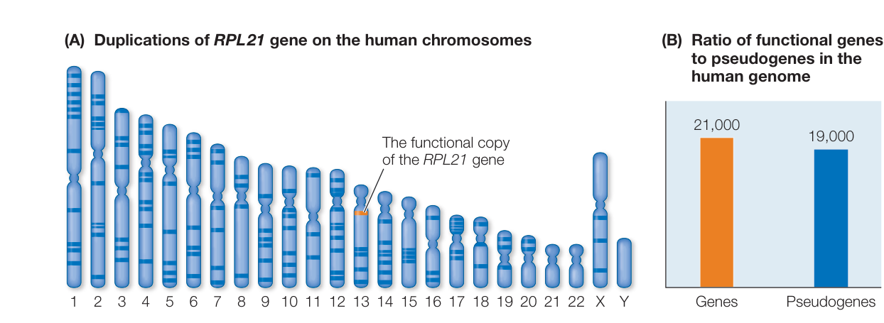
Figure 21.9 Some Functional Genes Are Duplicated Many Times as Nonfunctional Pseudogenes (A) The functional gene that encodes ribosomal protein RPL21 is located on human chromosome 13 (indicated in orange). In addition, there are many nonfunctional pseudogenes of RPL21 in the human genome, produced through repeated duplication events (indicated in blue). (B) Although RPL21 represents a relatively extreme example of pseudogene duplication, there are almost as many known pseudogenes in the human genome as there are functional genes. (After M. Gerstein and D. Zheng. 2006. Sci Am 295: 48–55; Courtesy of Lucy Reading-Ikkanda.)
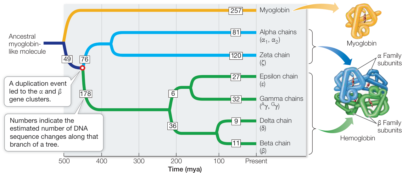
Figure 21.10 A Globin Family Gene Tree This gene tree suggests that the -globin (blue) and -globin (green) gene clusters diverged about 450 million years ago (mya) (open circle), soon after the origin of the vertebrates. (After W-H Li and D. Graur. 1991. Fundamentals of Molecular Evolution. Sinauer Associates: Sunderland, MA.)
Q: When did the gene duplication event that gave rise to the delta and beta chains occur?
Hemoglobin, a tetramer (four-subunit molecule) consisting of two -globin and two -globin polypeptide chains, carries oxygen in blood. Myoglobin, a monomer, is the primary storage protein in muscle. Myoglobin’s affinity for is much higher than that of hemoglobin, but hemoglobin has evolved to be more diversified in its roles. Hemoglobin binds in the lungs or gills, where the concentration is relatively high, transports it to deep body tissues, where the concentration is low, and releases it in those tissues. With its more complex tetrameric structure, hemoglobin is able to carry four molecules of , as well as hydrogen ions and carbon dioxide, in the blood.
To estimate the time of the globin gene duplication that gave rise to the - and -globin gene clusters, we can create a gene tree—a phylogenetic tree that describes the evolutionary history of particular genes or gene families, in this case the gene sequences that encode the various globins (see Figure 21.10). The rate of molecular evolution of globin genes has been estimated from other studies, using the divergence times of groups of vertebrates that are well documented in the fossil record. These studies indicate an average rate of divergence for globin genes of about 1 nucleotide substitution every 2 million years. By applying this rate to the globin gene tree, we can estimate the divergence time of the two globin gene clusters at about 450 million years ago.
Many gene duplications affect only one or a few genes at a time, but entire genomes are duplicated in polyploid organisms (which include many plants). When all the genes are duplicated, there are massive opportunities for new functions to evolve. That is exactly what appears to have happened in the evolution of vertebrates. The genomes of the jawed vertebrates appear to have four diploid sets of many major genes, which led biologists to conclude that two genome-wide duplication events occurred in the ancestor of these species. These duplications have allowed considerable specialization of individual vertebrate genes, many of which are now highly tissue-specific in their expression.
Some gene families evolve through concerted evolution
Although the members of the globin gene family have diversified in form and function, the members of many other gene families do not evolve independently of one another. For instance, almost all organisms have many copies (up to thousands) of the ribosomal RNA genes. Ribosomal RNA (rRNA) is the principal structural element of ribosomes and, as such, has a primary role in protein synthesis. Every living species needs to synthesize proteins, often in large amounts (especially during early development). Having many copies of the rRNA genes ensures that organisms can rapidly produce many ribosomes and thereby maintain a high rate of protein synthesis.
Like all portions of the genome, ribosomal RNA genes evolve, and differences accumulate in the rRNA genes of different species. But within any one species, the multiple copies of the rRNA genes are very similar, both structurally and functionally. This similarity makes sense because, ideally, every ribosome in a species should synthesize proteins in the same way. In other words, within a given species, the multiple copies of these rRNA genes are evolving in concert with one another, a phenomenon called concerted evolution.
How does concerted evolution occur? Two different mechanisms appear to be responsible. The first of these is unequal crossing over. When DNA is replicated during meiosis in a diploid species, the homologous chromosome pairs align and recombine by crossing over (see Key Concept 11.5). In the case of highly repeated genes, however, it is easy for genes to become displaced in alignment, since so many copies of the same genes are present on the chromosomes
(Figure 21.11A). The end result is that one chromosome may gain extra copies of the repeat and the other chromosome may have fewer copies of the repeat. If a new substitution arises in one copy of the repeat, it can spread to new copies (or be eliminated) through unequal crossing over. Thus over time, a novel substitution will either become fixed or it will be lost entirely. In either case, all copies of the repeat will remain very similar to one another.
The second mechanism that produces concerted evolution is biased gene conversion. This mechanism can be much faster than unequal crossing over and has been shown to be the primary mechanism for concerted evolution of rRNA genes. DNA strands are frequently broken and repaired (see Key Concept 13.4). At many times during the cell cycle, the genes for ribosomal RNA are clustered close together. If damage occurs to one of the genes, a copy of the rRNA gene on another chromosome may be used to repair the damaged copy, and the sequence that is used as a template can thereby replace the original sequence (Figure 21.11B). In many cases, this repair system appears to be biased in favor of using particular sequences as templates for repair, and thus the favored sequence rapidly spreads across all copies of the gene. In this way, changes may appear in a single copy and then rapidly spread to all the other copies.
Regardless of the mechanism responsible, the net result of concerted evolution is that the copies of a highly repeated gene do not evolve independently of one another. Mutations still occur, but once they arise in one copy, they either spread rapidly across all the copies or are lost from the genome completely. This process allows the products of each copy to remain similar over time in both sequence and function.
Recap and Assess
The noncoding portion of eukaryotic genomes is more variable in size across species than is the coding portion. Sexual recombination produces new combinations of genes and facilitates the elimination of deleterious mutations from populations. Lateral gene transfer can result in the transfer of genetic functions between distantly related species, whereas duplication can lead to the evolution of new functions. Some highly repeated genes undergo concerted evolution, which maintains uniform functionality.
- Suggest and contrast two hypotheses for the wide diversity of genome sizes among different organisms.
- Contrast three disadvantages and three advantages of sexual reproduction compared with asexual reproduction. Why do you think that most species have some means of achieving genetic recombination among individuals?
- What are some potential advantages of lateral gene transfer to the organisms that gain new genes by this mechanism?
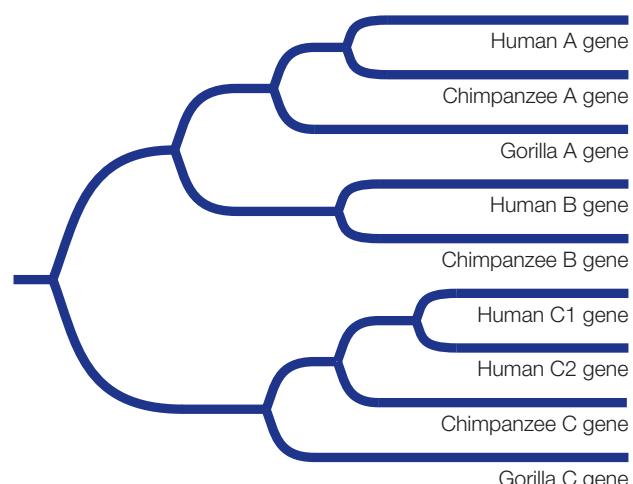
- Consider the following tree of a gene family sampled from the complete genomes of humans, chimpanzees, and gorillas. All members of the gene family are included in the tree. How many gene duplications and losses are likely to have occurred in the history of this gene family? Diagram their locations on the tree.
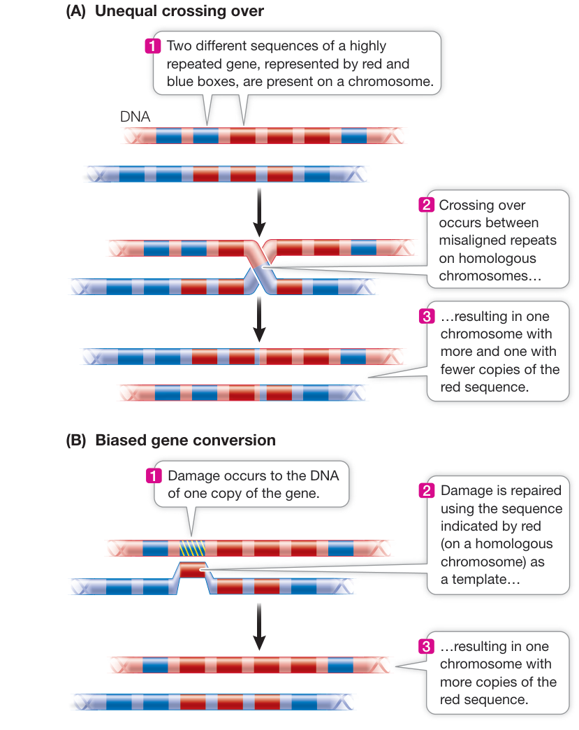
Figure 21.11 Concerted Evolution Two mechanisms can produce concerted evolution of highly repeated genes. (A) Unequal crossing over results in deletions and duplications of a repeated gene. (B) Biased gene conversion can rapidly spread a new variant across multiple copies of a repeated gene.
- Why is gene duplication considered important for long-term evolutionary change?
- Describe the pattern of concerted evolution among highly repeated genes, and diagram the processes that lead to concerted evolution.
We have considered how changes in gene structure and number can produce evolutionary change. We will next consider how phenotypic transformations can also be produced by changes in the amount, timing, and location of gene expression during development.
Changes in Gene Expression Often Shape Evolution
Learning Objectives
21.4.1 Describe factors that determine cell fate during development, using body segmentation in animals as an example.
21.4.2 Defend the claim “Genes controlling development are highly conserved across animals,” using a specific example.
21.4.3 Provide examples of morphological differences among species that result from changes in the amount, timing, and location of gene expression.
21.4.4 Summarize how a mutation in a developmental gene led to a reduction in leg number in insects compared with other arthropods.
Although a genome contains all of the genes needed to form a complete organism, these genes need to be expressed at specific times and in specific tissues. Given that every cell in the body contains the same genetic information, how are the right genes turned on to form a head at the anterior end of an animal, or a tail at the posterior end? Why don’t heads or tails form elsewhere in the developing embryo? How do insects develop antennae on their head, but legs on their thorax? As biologists began to study and answer these questions, they realized that changes in gene expression might account for much of the evolution of diverse body forms we observe across living organisms. Thus was born the science of evo-devo, or evolutionary developmental biology.
A cascade of transcription factors establishes body segmentation in animals
Before we examine how changes in gene expression can account for evolution of body forms, we need to consider how body axis orientation and segmentation are controlled during development. This has been especially well studied in the fruit fly Drosophila melanogaster, but many of the genes that control developmental processes are conserved across a diversity of organisms, including ourselves.
The insect body is made up of segments that differ from one another. The adult fly has an anterior head (composed of several fused segments), three different thoracic segments, and eight abdominal segments at the posterior end. Each segment gives rise to different body parts: for example, antennae and eyes develop from head segments, wings from the thorax, and so on.
The life cycle of Drosophila from fertilized egg to adult takes about 2 weeks. The egg hatches into a larva, which then forms a pupa, which finally is transformed into the adult fly. By the time a larva appears—about 24 hours after fertilization—there are recognizable segments. Although the thoracic and abdominal segments all look similar, the fates of the cells to become different adult segments are already determined.
As in other organisms, fertilization in Drosophila leads to a rapid series of mitoses. However, the first 12 nuclear divisions do not result in the formation of new cells. A multinucleate embryo forms instead of a multicellular embryo. With no cell membranes to cross, gene inducers known as morphogens can diffuse easily within the embryo. Many of these morphogens affect transcription in the cell nuclei.
Genetic studies of Drosophila revealed a sequential pattern (cascade) of gene expression that results in the determination of each segment within 24 hours after fertilization. Three classes of genes are involved:
- Maternal effect genes set up the major axes (anterior–posterior and dorsal–ventral) of the embryo.
- Segmentation genes determine the boundaries and polarity of each of the segments.
- Hox genes determine what organ will be made at a given location.
MATERNAL EFFECT GENES Like the eggs and early embryos of many other organisms, Drosophila eggs and larvae are characterized by unevenly distributed mRNAs and proteins, which are the products of specific maternal effect genes. These genes are transcribed in the cells of the mother’s ovary, and the mRNAs are passed to the egg. These mRNAs and proteins establish the anterior–posterior axis and the dorsal–ventral axis of the developing embryo. Where some of these proteins are in highest concentrations in the embryo, transcription of additional genes is stimulated. For example, products of maternal effect genes stimulate the hunchback gene to be transcribed in the anterior end of the embryo, and the resulting gradient of Hunchback protein (itself a transcription factor) establishes the head region. A gradient of other proteins concentrated in the posterior region of the embryo establishes the anterior–posterior axis of the developing embryo.
The events that involve maternal effect genes begin before fertilization and continue after it, during the multinucleate stage, which lasts a few hours. At this stage the embryo looks like a bunch of indistinguishable nuclei under the light microscope. But the fates of the individual nuclei and the cells they will occupy have already begun to be determined. After the anterior and posterior ends have been established, the next step in pattern formation in fruit flies is the determination of segment number and locations.
SEGMENTATION GENES The number, boundaries, and polarity of the Drosophila larval segments are determined by proteins encoded by the segmentation genes. These genes are expressed when there are about 6,000 nuclei in the embryo (about 3 hours after fertilization). Three classes of segmentation genes act one after the other to regulate finer and finer details of the segmentation pattern:
- Gap genes organize broad areas along the anterior–posterior axis. Mutations in gap genes result in gaps in the body plan—the omission of several consecutive larval segments.
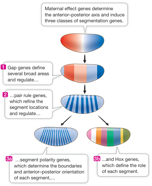
Figure 21.12 A Gene Cascade Controls Pattern Formation in the Drosophila Embryo Maternal effect genes induce gap, pair rule, and segment polarity genes. Hox genes then define the role of each segment. By the end of this cascade, a group of nuclei at the anterior of the embryo, for example, is determined to become the first head segment in the adult fly.
- Pair rule genes divide the embryo into units of two segments each. Mutations in pair rule genes result in embryos missing every other segment.
- Segment polarity genes determine the boundaries and anterior–posterior organization of the individual segments. Mutations in segment polarity genes can result in segments in which posterior structures are replaced by reversed (mirror-image) anterior structures.
The expression of the genes is sequential (Figure 21.12). The products of the gap genes active pair rule genes, and the pair rule gene products activate segment polarity genes. By the end of this part of the cascade, nuclei throughout the embryo “know” which segment they will be part of in the adult fly. The next set of genes in the cascade determines the form and function of each segment.
HOX GENES Hox genes encode a family of transcription factors that are expressed in different combinations along the length of the embryo, and help determine cell fates within each segment. Expression of certain Hox genes leads to the development of antennae in the head segment, whereas other Hox genes are expressed in the thorax to make wings, and so on. The Drosophila Hox genes occur in two clusters in the genome, in the same order as the segments whose function they determine (Figure 21.13). Hox genes are shared by all animals, and mutations in these genes have been linked to major changes in body structure among different animal groups.
How do we know that the Hox genes determine segment identity, and what structures are expressed in each segment? A clue comes from homeotic mutations—mutations that result in the replacement of one organ by another. For example, a mutation in the Hox gene Antennapedia causes legs to grow on the head of Drosophila in place of antennae (Figure 21.14). When another Hox gene, bithorax, is mutated, an extra pair of wings grows in a thoracic segment where wings do not normally occur. So the normal (wild-type) functions of the Hox genes must be to “tell” a segment what organ to form. Hox genes encode transcription factors and have a conserved 180-base-pair sequence called the homeobox (from which the genes get their name). The homeobox encodes a 60-amino acid sequence called the homeodomain. The homeodomain recognizes and binds to a specific DNA sequence in the promoters of its target genes, “turning on” the formation of specific structures.
Connect the Concepts As discussed in Key Concept 16.2, cell differentiation often is mediated by changes in gene expression resulting from the activation or deactivation of various transcription factors.
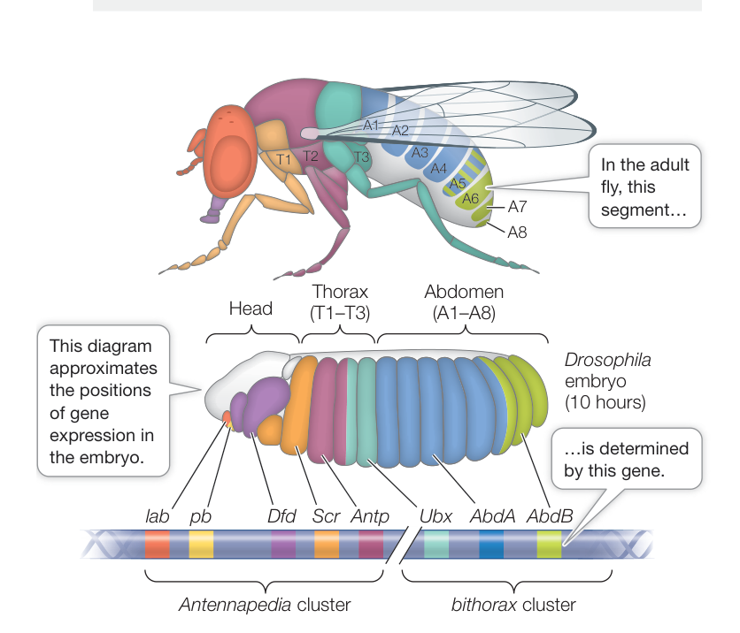
Figure 21.13 Hox Genes in Drosophila Determine Segment Identity Two clusters of Hox genes on chromosome 3 (bottom) determine segment function in the adult fly (top). These genes are expressed in the embryo (center) long before the structures of the segments actually appear. (Top after M. Peifer et al. 1987. Genes Dev 1: 891–898; bottom after S. D. Hueber et al. 2010. PLOS ONE 5: e10820. doi:10.1371/journal.pone.0010820.)
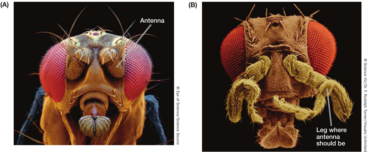
Figure 21.14 A Homeotic Mutation Replaces One Structure with Another (A) Normal development of antennae on the head of a fruit fly. (B) A homeotic mutation of the Hox gene Antennapedia, which controls formation of legs during development, causes legs, rather than antennae, to grow on the head.
A common “toolkit” of genetic mechanisms controls gene expression in development
About a dozen major kinds of eyes are found among animals, including the camera-like eyes of humans and the compound eyes of insects. Although the eyes of insects and vertebrates evolved independently, a remarkable discovery showed that common developmental pathways are involved in the embryonic formation of eyes in both groups.
More than a century ago, a mutation was described in Drosophila called eyeless, with a phenotype of—you guessed it—no eyes. This mutation remained a laboratory curiosity for many decades until its molecular nature was elucidated in the 1990s. Biologists isolated the protein product of the wild-type version of the eyeless gene, which they determined was a transcription factor that controls the expression of genes responsible for eye development. How did they know this? By making recombinant DNA constructs that allowed the wild-type eyeless gene to be expressed in different embryonic tissues of transgenic flies, the biologists were able to produce flies with extra eyes on various body parts such as the legs, the antennae, and under the wings.
The big surprise came when the biologists sequenced the eyeless gene and used computers to query a database of genes with known sequences. The eyeless gene sequence was similar to that of the Pax6 gene in mice; Pax6, when mutated, leads to the development of abnormally small eyes. Could the extremely different eyes of flies and mice be variations on a common developmental theme? To test for functional similarity between the insect and mammalian genes, the biologists repeated their experiments on flies, but using the mouse Pax6 gene instead of the fly eyeless gene. Once again, eyes developed at various sites on the transgenic flies. Thus a gene whose expression normally leads to the development of a mammalian eye now led to the development of the very different insect eye.
Fruit flies and mice last shared a common ancestor more than a half-billion years ago. Yet the eyeless and Pax6 genes contain sequences that are highly conserved, not only in these two species but in others as well. Biologists call such genes homologous, meaning that they evolved from a gene present in a common ancestor (see Key Concept 20.1).
In recent years a large number of homologous genes (the regulatory “toolkit”) have been shown to control development in distantly related species. For example, the fruit fly homeotic genes such as Antennapedia and bithorax are similar to mouse (and human) genes that play similar developmental roles. This discovery indicates that the positional information controlled by these genes has been conserved, even as the structures formed at each position have changed. Remarkably, these genes are arranged along a chromosome in both fruit fly and mouse in the same order as they are expressed along the anterior–posterior axis of the embryos (Figure 21.15). These and other examples have led biologists to the idea that certain developmental mechanisms are controlled by specific sequences composing a genetic toolkit, the contents of which have been changed and reshuffled over the course of evolutionary time to produce the amazing diversity of plants, animals, and other organisms that we see today.
Developmental modules based on a common set of genetic instructions can evolve separately within a species because genetic switches control how the toolkit is used. These switches turn genes on or off, or regulate the degree and timing of gene expression. Multiple switches control each gene, creating different expression patterns in different locations. In this way, elements of the genetic toolkit can be involved in multiple developmental processes and still allow individual modules to develop and evolve independently.
During evolution, changes in the functions of genetic switches have led to changes in the forms and functions of organisms. To illustrate this, let’s look at the development of wings in Drosophila and other insects. Drosophila species are members of the insect group Diptera, which means “two wings”—that is, they have a single pair of wings, whereas most insects have two pairs of wings (i.e., four wings). The single pair of wings of dipterans develops on the second thoracic segment, where the Hox gene Antennapedia (Antp) is expressed. Antp is also expressed in the third thoracic segment, but in that segment a pair of balancing organs called halteres develops in dipterans. A critical difference between thoracic segments 2 and 3 is that another Hox gene, Ultrabithorax (Ubx), is expressed along with Antp in segment 3 (Figure 21.16). Ubx represses Antp function in dipterans. If Ubx is inactivated by mutation, a second pair of wings forms in thoracic segment 3, as is typical of many other insect groups. Thus some major morphological differences among groups of animals can result from relatively small changes in gene expression.
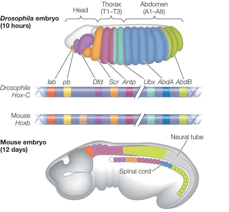
Figure 21.15 Regulatory Genes Show Similar Expression Patterns Homologous genes encoding similar transcription factors are expressed in similar patterns along the anterior–posterior axes of both insects and vertebrates. The mouse (and human) Hox genes are present in multiple copies; this prevents a single mutation from having drastic effects. (Top after S. D. Hueber et al. 2010 PLOS ONE 5: e10820. doi:10.1371/journal.pone.0010820; bottom after S. B. Carroll. 1995. Nature 376: 479–485.)
The amount, timing, and location of gene expression control many morphological features
The modularity of development means that the molecular pathways for developmental processes such as organ formation operate independently of one another. For instance, the Antennapedia mutant fly shown in Figure 21.14 grew legs where antennae should be, but the rest of the segments formed structures normally. Modularity allows the timing and location of a developmental process to change without changing the whole organism. Major changes can result from differences in the amount, timing, and location of gene expression.
HETEROMETRY A striking example of heterometry (“different measure”) is seen in beak development in Darwin’s finches, in which the beaks of some species are large and robust for cracking seeds and in other species are thin and long for probing for food (see Figure 22.8). When he visited the Galápagos archipelago, Charles Darwin wrote that “seeing this gradation and diversity of structure in one small intimately related group of birds, one might really fancy that from an original paucity of birds in this archipelago, one species had been taken and modified for different ends.”
Darwin had no idea of the genetic basis for such modification. Now we do. Beak shapes are evident when birds hatch from their eggs, so they must be determined during development. The beak develops from tissues at the anterior of the embryo that will form the facial bones. Cell divisions in this embryonic tissue are controlled by signaling proteins, one of which is called bone morphogenetic protein 4 (BMP4); another is the protein calmodulin. If BMP4 is present early and in large amounts, the beak becomes broad and deep. If calmodulin is present early and in large amounts, the beak grows longer and thinner (Figure 21.17). Thus beak structure is affected by changes in protein production.
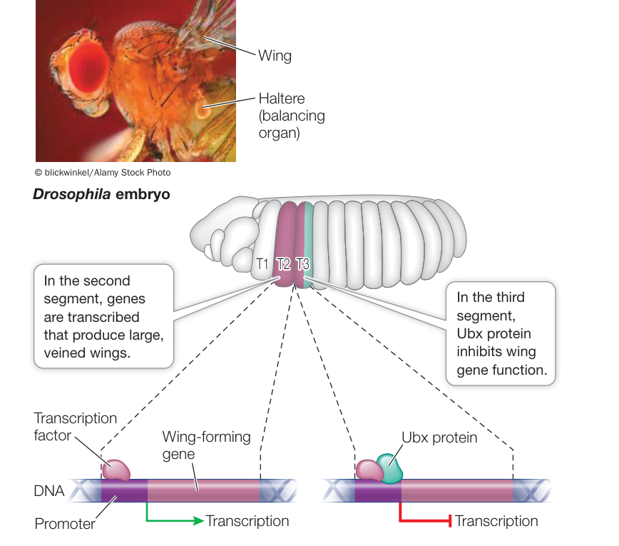
Figure 21.16 Segments Differentiate under Control of Genetic Switches The binding of a single protein, Ultrabithorax (Ubx), determines whether a thoracic segment produces full wings or halteres. (After S. D. Hueber et al. 2010. PLOS ONE 5: e10820. doi:10.1371/journal.pone.0010820.) Q: What would happen if a butterfly embryo were genetically engineered so that Ubx was expressed in all its thoracic segments?
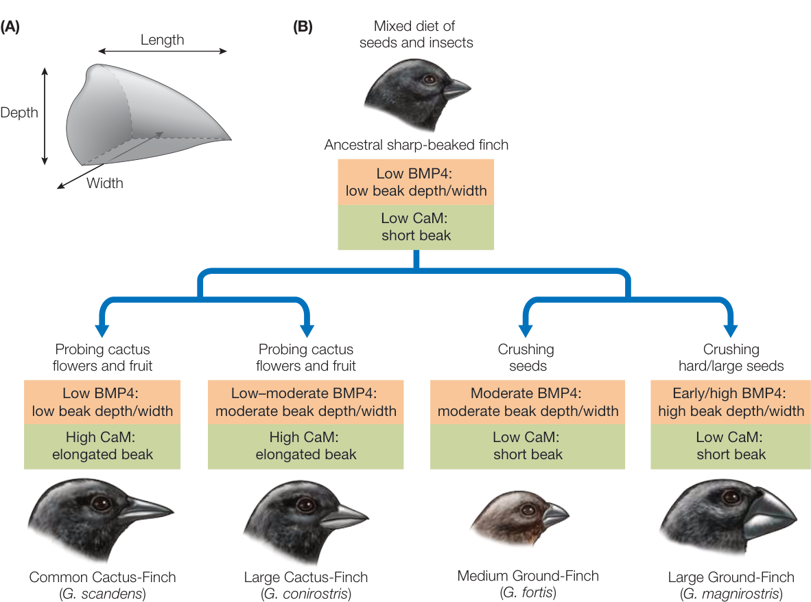
Figure 21.17 Heterometry and the Beaks of Finches (A) Bird beaks can be measured in three dimensions (length, width, and depth) for the purpose of comparing different species. (B) Among Darwin’s finches, mutations resulting in different patterns of BMP4 and calmodulin (CaM) expression may be responsible for the different species’ beak sizes and shapes, which are suited for exploiting different types of food. The four species shown here are all in the genus Geospiza. (After A. Abzhanov et al. 2004. Science 305: 1462–1465.)
HETEROCHRONY The evolution of the giraffe neck provides an example of heterochrony (“different time”). Giraffes, like most mammals, have seven cervical (neck) vertebrae. So giraffes did not get longer necks by adding more vertebrae. Instead, the cervical vertebrae of giraffes are much longer than those of other mammals (Figure 21.18).
Bone growth in mammals is the result of the proliferation of cartilage-producing cells called chondrocytes. Bone growth is stopped by a genetic signal that results in apoptosis, or cell death, of chondrocytes and calcification of the bone matrix. In giraffes this signaling process is delayed in the cervical vertebrae, so these vertebrae grow longer. Thus the evolution of longer necks resulted from changes in the timing of expression of the genes that control bone formation.
HETEROTOPY Spatial differences in the expression of a developmental gene are known as heterotopy (“different place”), exemplified by the different development of feet in ducks and chickens. The feet of all bird embryos have webs of skin that connect the toes. This webbing is retained in adult ducks (and other aquatic birds) but not in adult chickens (and other nonaquatic birds). The loss of webbing is controlled by the BMP4 signaling protein, which we noted is also involved in beak development (another example of use of a common genetic toolkit to produce different kinds of change).
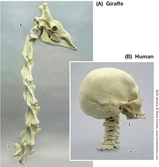
Figure 21.18 Heterochrony in the Development of a Longer Neck There are seven vertebrae in the neck of a giraffe (A) and human (B) (not to scale). But the vertebrae of the giraffe are much longer (25 cm vs. 1.5 cm in the human) because during development, growth continues for a longer period of time. This timing difference is called heterochrony.
BMP4 protein induces the cells that produce webbing to undergo apoptosis and thus eliminate the webbing between the toes. The hindlimbs of both duck and chicken embryos express the BMP4 gene in the webbing between the toes. However, duck and chicken embryos differ in expression of the Gremlin gene, which encodes a protein that inhibits BMP4 expression (Figure 21.19). In ducks, but not in chickens, Gremlin is expressed in the webbing cells and Gremlin protein inhibits BMP4 expression. With no BMP4 protein to stimulate apoptosis, a webbed foot develops. If chick hindlimbs are experimentally exposed to Gremlin during development, the adult chicken will have ducklike webbed feet.
Mutations in developmental genes can cause major morphological changes
Sometimes a major developmental change is caused by an alteration in the regulatory molecule itself rather than by a change in where, when, or how much it is expressed. The gene that controls the number of legs in arthropods offers an excellent example. Arthropods all have head, thoracic, and abdominal regions with variable numbers of segments. Insects, such as Drosophila, have three pairs of legs on their three thoracic segments, whereas centipedes have many legs on both thoracic and abdominal segments. All arthropods express a gene called Distal-less (Dll) that controls segmental leg development. In insects, Dll expression is repressed in abdominal segments by the Hox gene Ultrabithorax (Ubx). Ubx is expressed in the abdominal segments of all arthropods, but it has different effects in different species. In centipedes, Ubx is co-expressed with Dll to promote the formation of legs. During the evolution of insects, a mutation in the Ubx gene sequence resulted in a modified Ubx protein that represses Dll expression in abdominal segments (Figure 21.20). A phylogenetic tree of arthropods shows that this change in Ubx occurred in the ancestor of insects, at the same time that abdominal legs were lost.
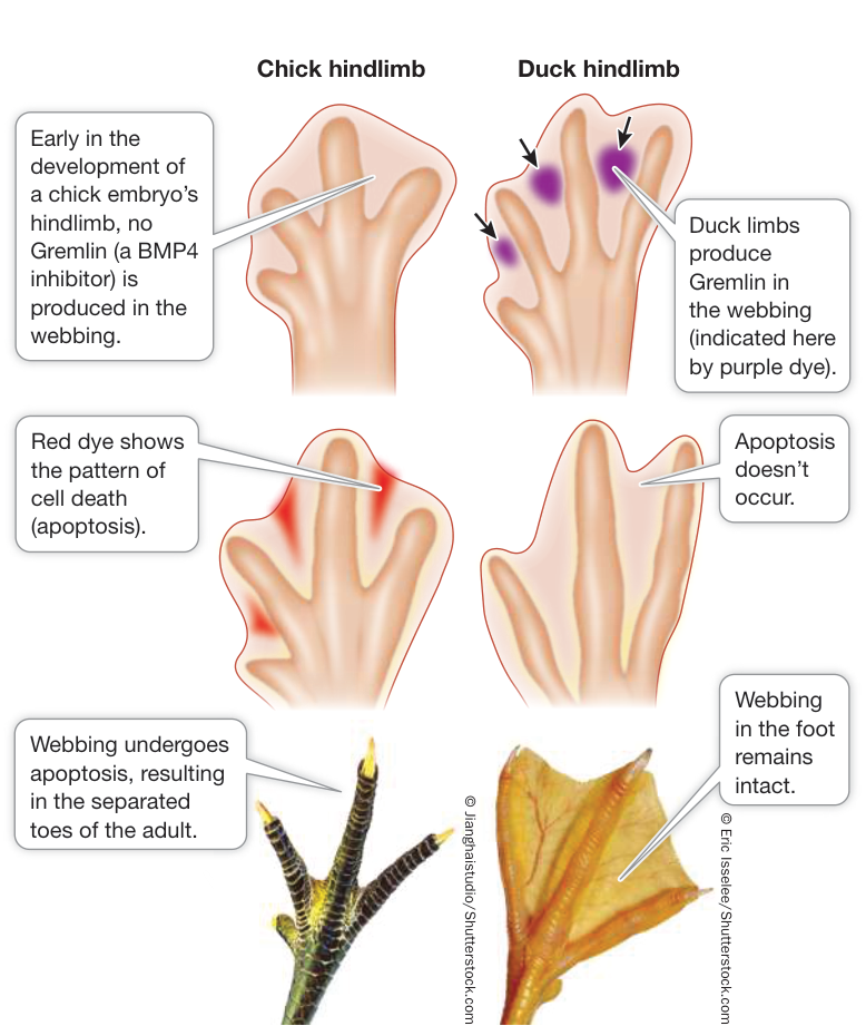
Figure 21.19 Heterotopy in Gremlin Expression Correlates with Changes in Hindlimb Structure The left column shows foot development in a chicken; the right column shows foot development in a duck. Gremlin protein in the webbing of the duck foot inhibits BMP4 signaling, thus preventing the embryonic webbing from undergoing apoptosis. Q: What do you predict would happen if you blocked the expression of Gremlin in the feet of duck embryos?
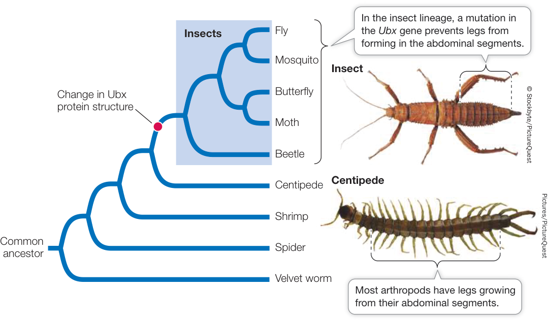
Figure 21.20 A Mutation in a Hox Gene Changed the Number of Legs in Insects In the insect lineage (blue box) of the arthropods, a change to the Ubx gene resulted in a protein that inhibits the Dll gene, which is required for legs to form. Because insects express this modified Ubx gene in their abdominal segments, no legs grow from these segments. Other arthropods, such as centipedes, produce an unmodified Ubx protein and do grow legs from their abdominal segments. (After R. Galant and S. Carroll. 2002. Nature 415: 910–913.)
Recap and Assess
A genetic toolkit consisting of highly conserved regulatory genes governs pattern formation in multicellular organisms. Genetic toolkit genes act independently among modules of a developing embryo. The expression of these developmental genes can differ among species in various ways, often resulting in major morphological differences. They can differ in amount of expression (heterometry), in the timing of expression (heterochrony), or in the location of expression (heterotopy). A mutation in a gene controlling a developmental pathway can result in major changes in morphology.
- Using the story of the eye-determining genes Pax6 and eyeless, support the claim that genes controlling development are highly conserved.
- In a series of experiments on chick embryos, researchers applied different concentrations of BMP4 to the embryos’ beak growth region. They measured the size of the beak cartilage at a later stage of development. Based on the data in the table, what can you conclude about the role of BMP4 in beak growth?
| Amount of BMP4 | Cartilage diameter (mm) |
|---|---|
| None (control) | 0.5 |
| 0.1 unit | 0.7 |
| 0.3 unit | 1.0 |
| 1.0 unit | 1.8 |
Data from P. Wu et al. 2004. Science 305: 1465–1466.
- How have diverse body forms in arthropods evolved by means of mutations in developmental genes?
Our understanding of molecular evolution has many practical applications across biology, and new ones are being developed every day. We now consider a few of these applications to medicine and industry.
Molecular Evolution Has Many Practical Applications
Molecular Evolution Has Many Practical Applications
Learning Objectives
- 21.5.1 Describe how studies of molecular evolution are used to understand protein function.
- 21.5.2 Explain how the methods of in vitro evolution correspond to natural evolutionary processes.
- 21.5.3 Evaluate evolutionary relationships among viral sequences to understand the transmission of a viral disease.
Studies of molecular evolution have practical applications throughout biology, from understanding basic aspects of biological function to studies of human health.
Molecular sequence data are used to determine the evolutionary history of genes
A gene tree can show the evolutionary relationships of a single gene in different species, or it can trace the evolution of members of a gene family (as in Figure 21.10). The methods for constructing a gene tree are the same as those we described in Key Concept 20.2 for building phylogenetic trees of species. The process involves identifying differences between genes and using those differences to reconstruct the evolutionary history of the genes. Gene trees are often used to construct phylogenetic trees of species, but the two types of trees are not necessarily equivalent. Processes such as gene duplication can give rise to differences between the phylogenetic trees of genes and species. From a gene tree, biologists can reconstruct the history and timing of gene duplication events and learn how gene diversification has resulted in the evolution of new protein functions.
All the genes of a particular gene family have similar sequences because they have a common ancestry. As we discussed in Key Concept 20.1, features that are similar as a result of common ancestry are said to be homologous. When discussing gene trees, however, we usually need to distinguish between two forms of homology. Homologous genes that are found in different species and whose divergence we can trace to the speciation events that gave rise to those species are called orthologs. Homologous genes in the same or different species that are related through gene duplication events are called paralogs. When we examine a gene tree, the questions we wish to address determine whether we should compare orthologous or paralogous genes. If we wish to reconstruct the evolutionary history of the species that contain the genes, then our comparison should be restricted to orthologs (because they will reflect the history of speciation events). If we are interested in the changes in function that have resulted from gene duplication events, however, then the appropriate comparison is among paralogs (because they will reflect the history of gene duplication events). If our focus is on the diversification of a gene family through both processes, then we will want to include both paralogs and orthologs in our analysis.
Figure 21.21 depicts a gene tree for the members of a gene family called engrailed (its members encode transcription factors that regulate development). At least three gene duplications have occurred in this family, resulting in up to four different engrailed genes (En) in some vertebrate species (such as the zebrafish, Danio rerio). All of the engrailed genes are homologs because they have a common ancestor. Gene duplication events have generated paralogous engrailed genes (En1 and En2) in some lineages of vertebrates. We could compare the orthologous sequences of the En1 group of genes to reconstruct the history of the bony vertebrates (i.e., all the vertebrate species in Figure 21.21 except the lamprey), or we could use the orthologous sequences of the En2 group of genes and expect the same answer (because there is only one history of the underlying speciation events). All bony vertebrates have both groups of engrailed genes because the two groups arose from a gene duplication event in the common ancestor of bony vertebrates. If we wanted to focus on the diversification that occurred as a result of this duplication, then the appropriate comparison would be between the paralogous genes of the En1 versus En2 groups.
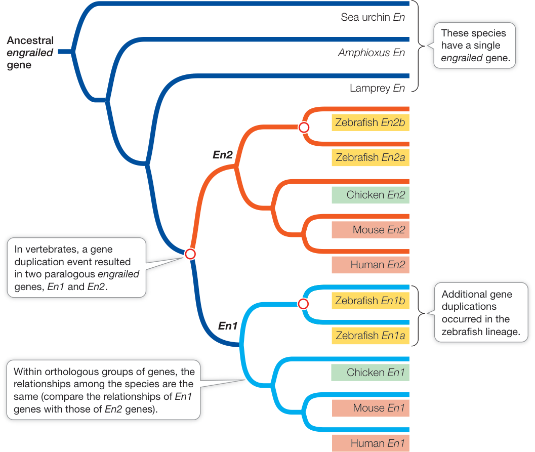
Figure 21.21 Phylogeny of the engrailed Genes The engrailed genes are homologous because they share a common ancestor. Speciation events have generated orthologous engrailed genes, and gene duplication events (open circles) have generated paralogous engrailed genes among bony vertebrates. (After A. Force et al. 1999. Genetics 151: 1531–1545.) Q: How many gene duplication events occurred just within the zebrafish lineage?
Gene evolution is used to study protein function
Earlier in this chapter we discussed the ways in which biologists can detect regions of genes that are under positive selection for change. What are the practical uses of this information? Consider the evolution of the family of genes encoding voltage-gated sodium channels. Sodium channels have many functions, including the control of muscle contractions as well as nerve impulses in the nervous system. Sodium channels can be blocked by various toxins, such as tetrodotoxin (TTX), a neurotoxin present in the tissues of some puffer fishes and several other animals. A human who eats those tissues of a puffer fish that contain TTX can become paralyzed and die because the toxin blocks sodium channels and prevents muscles and nerves from functioning.
Connect the Concepts As described in detail in Key Concept 43.2, voltage-gated ion channels are stimulated to open or close by a change in the electric charge difference across a cell membrane, and thus let various ions into or out of cells.
But puffer fishes have sodium channels too, so why doesn’t the TTX cause paralysis in the puffer fishes themselves? The sodium channels of puffer fishes (and of other animals that sequester TTX, such as the rough-skinned newt shown in Figure 19.20) have evolved to become resistant to the toxin. Nucleotide substitutions in the puffer fish genome have resulted in changes in the proteins that make up the sodium channels, and those changes prevent TTX from binding to the sodium channel pore.
Several different substitutions that result in TTX resistance have evolved in the various duplicated sodium channel genes of the many species of puffer fishes. Many other changes that have nothing to do with the evolution of TTX resistance have occurred in these genes as well. Biologists who study the function of sodium channels can learn a great deal about how the channels work (and about neurological diseases that are caused by mutations in the sodium channel genes) by understanding which changes have been selected for TTX resistance. They do this by comparing the rates of synonymous and nonsynonymous substitutions across the genes in various lineages that have evolved TTX resistance. In a similar manner, molecular evolutionary principles are used to understand function and diversification of function in many other proteins.
In vitro evolution is used to produce new molecules
As biologists studied the relationships among selection, evolution, and function in macromolecules, they realized that molecular evolution could be employed in a controlled laboratory environment to produce new molecules with novel and useful functions. Thus were born the applications of in vitro evolution.
Living organisms produce thousands of compounds that humans have found useful. The search for such naturally occurring compounds, which can be used for pharmaceutical, agricultural, or industrial purposes, has been termed “bioprospecting.” These compounds are the result of millions of years of molecular evolution across millions of species of living organisms. Biologists can imagine molecules that could have evolved but, lacking the right combination of selection pressures and opportunities, have not. For instance, we might want to have a molecule that binds a particular environmental contaminant so that the contaminant can be isolated and extracted from the environment. But if the contaminant is synthetic (i.e., not produced naturally), it is unlikely that any living organism will have evolved a molecule with the function we desire. This problem was the inspiration for the field of in vitro evolution.
The principles of in vitro evolution are based on the principles of molecular evolution that we have learned from the natural world. Consider the evolution of a new RNA molecule that was produced in the laboratory using the principles of mutation and selection. This molecule’s intended function was to join two other RNA molecules. That is, the lab created a ribozyme—an RNA molecule capable of catalyzing a biochemical reaction. The process started with a large pool of random RNA sequences ( different sequences, each about 300 nucleotides long), which were then selected for any ligase activity (Figure 21.22). None were very effective ligases, but some were slightly better than others. The best of the ribozymes were selected and reverse-transcribed into cDNA (using the enzyme reverse transcriptase). The cDNA molecules were then amplified using the polymerase chain reaction (PCR; see Figure 13.20).
Connect the Concepts Key Concept 17.1 describes how complementary DNA (cDNA) is made by complementary base-pairing to mRNA isolated from cells.
PCR amplification is not perfect, and it introduced many new mutations into the pool of sequences. These sequences were then transcribed back into RNA molecules using RNA polymerase II, and the process was repeated. The ligase activity of the RNAs evolved quickly; after ten rounds of in vitro evolution, it had increased by about 7 million times. Similar techniques have since been used to create a wide variety of molecules with novel enzymatic and binding functions.
Molecular evolution is used to study and combat diseases
Many of the most problematic human diseases are caused by living, evolving organisms that present a moving target for modern medicine. Historically, viral diseases such as influenza have been among the greatest factors in human mortality, and new viral diseases are constantly emerging in human populations, such as the human immunodeficiency virus (HIV) that causes AIDS. The control of these and many other human diseases depends on techniques that can track the evolution of pathogenic organisms over time.
Advances in transportation over the past century have allowed humans to move around the world with unprecedented speed and frequency. Unfortunately, this mobility has allowed pathogens to be transmitted among human populations, and between humans and other animals, at increasing rates. Cross-species transmission of viruses has led to the global emergence of many “new” diseases.
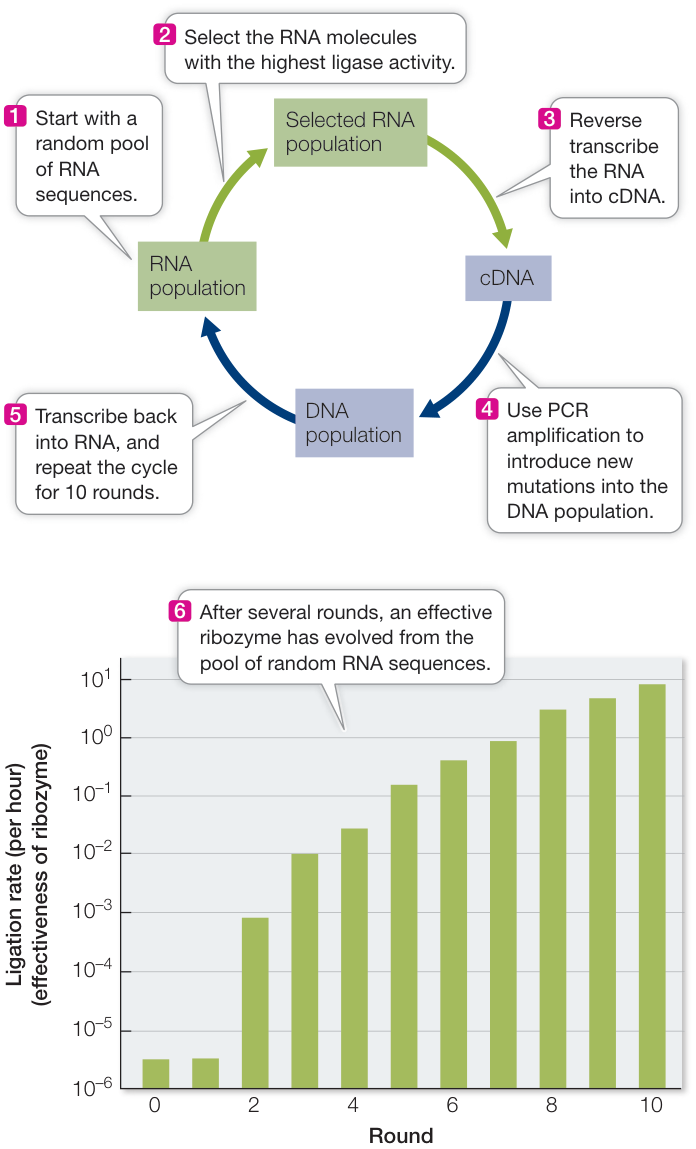
Figure 21.22 In Vitro Evolution Starting with a large pool of random RNA sequences, David Bartel and Jack Szostak of Massachusetts General Hospital produced a new ribozyme through rounds of mutation and selection for the ability to ligate (join) RNA sequences. (After D. P. Bartel and J. W. Szostak. 1993. Science 261: 1411–1418.)
In the opening of this chapter we described the devastating influenza pandemic of 1918–1919. Michael Worobey and his colleagues used evolutionary analyses of influenza genomes to explore the reasons why this particular flu epidemic was so much more severe than any before or since (Investigating Life: Why Was the 1918–1919 Influenza Pandemic So Severe?). They reconstructed the evolutionary history of influenza viruses in birds, pigs, horses, and humans and found that influenza viruses have repeatedly been transferred from birds to the various mammals. Most cross-species transfers have been accompanied by a major epidemic in the new host. The 1918–1919 epidemic occurred immediately after such a transfer between hosts. Human immune systems had no experience with the newly acquired virus, and the strong resulting reaction caused many of the strongest symptoms and deaths. Now that cross-species transmission has been associated with major flu epidemics, health workers around the world are constantly monitoring flu cases for indications of new cross-species transmissions. When emerging viruses are identified early on, efforts to control and eradicate them have a good chance of being successful. So far, our understanding of molecular evolution has prevented a repeat of the devastating flu pandemic of 1918–1919.
Investigating Life: Why Was the 1918–1919 Influenza Pandemic So Severe?
Experiment
Original Paper: T. M. Tumpey et al. 2005. Characterization of the reconstructed 1918 Spanish influenza pandemic virus. Science 310: 77–80.
M. Worobey et al. 2014. A synchronized global sweep of the internal genes of modern avian influenza virus. Nature 508: 254–257.
As conveyed in the story that opens this chapter, the 1918–1919 influenza pandemic killed more than 50 million people worldwide—many more than the number of World War I–related combat deaths. Unlike flu epidemics before or since, the flu viruses that circulated in the 1918–1919 epidemic triggered an especially intense reaction in the human immune system. What was different about the flu viruses circulating at that time?
Terrence Tumpey and his colleagues at the Centers for Disease Control and Prevention isolated and sequenced a complete flu virus from biopsies of 1918 victims of the pandemic. Michael Worobey of the University of Arizona and his colleagues then analyzed and compared the genomes of flu viruses that have been collected from 1918 to the present, from birds, pigs, horses, and humans. The viruses are largely transmitted within each of these host groups, but rarely jump between host groups. Worobey and his colleagues reconstructed the phylogeny of the viruses to determine if the 1918 virus had recently “jumped” into human populations from another species.
HYPOTHESIS The 1918–1919 influenza epidemic was so severe because the 1918 virus had recently moved into human populations from another species.
METHOD
- Sequence the genomes of influenza viruses isolated from samples of birds, pigs, horses, and humans throughout the past century.
- Reconstruct a phylogenetic tree based on sequences of the polymerase protein PA from the influenza virus genomes. Repeat this analysis for each of the genes in the genome. (Note that only the results based on PA are shown here.)
- Determine the rate of evolution of the influenza viruses in each species, and use this information to conduct a molecular clock analysis to date the origins of each cross-species transmission.
RESULTS Species-specific rates of the polymerase protein PA from the genome of influenza viruses in each host species:
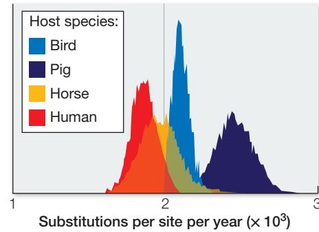
Time-calibrated phylogeny of the PA gene from influenza:
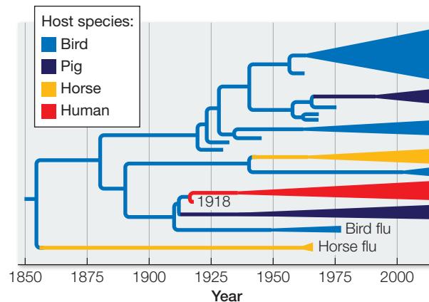
Viruses represented by blue lineages were isolated from birds; green lineages from pigs; orange lineages from horses; and red lineages from humans. The triangles represent many closely related sequences sampled from the respective species in the time period indicated; the width of the triangle is proportional to the number of sequences analyzed. The 1918 human influenza virus is labeled “1918.”
CONCLUSION What would you conclude about the 1918–1919 influenza pandemic from these results? Explore the possibilities in the following Work with the Data.
Work with the Data
Michael Worobey and his colleagues were able to reconstruct many of the cross-species transmissions of influenza virus that have occurred over the past 150 years, and to determine a likely reason that the 1918–1919 pandemic in humans was so severe. Can you replicate their conclusions?
QUESTIONS
- All the influenza viruses shown in the tree in the experiment are thought to have arisen in one group of host animals. Which group of animals do you think is the original source of the virus, and why?
- How many cross-species transmissions are shown on the tree? What hosts were involved (as source and recipient) of each transmission event?
- What do you think is a likely explanation for the severity of the 1918–1919 influenza pandemic in humans?
- Influenza was known in humans well before the 1918–1919 pandemic. Why do you think that no human influenza viruses are indicated on the tree before about 1918?
Studies of the genomes of many infectious agents have advanced our understanding and treatment of the diseases they cause. For example, rodent-borne hantaviruses were identified as the source of widespread respiratory illnesses, and the virus (and its host) that causes severe acute respiratory syndrome (SARS) was identified using evolutionary comparisons of genes. Studies of the origins, the timing of emergence, and the global diversity of many human pathogens depend on the principles of molecular evolution, as do the efforts to develop and use effective vaccines against these pathogens. For example, the techniques used to develop modern polio vaccines, as well as the methods used to track their effectiveness in human populations, rely on molecular evolutionary approaches.
In the future, understanding of molecular evolution will become even more critical to the identification of human (and other) diseases. Once biologists have collected data on the genomes of enough organisms, it will be possible to identify an infection by sequencing a portion of the infecting organism’s genome and comparing this sequence with other sequences on an evolutionary tree. At present it is difficult to identify many common viral infections (those that cause “colds,” for instance). As genomic databases and evolutionary trees increase, however, automated methods of sequencing and rapid phylogenetic comparison of the sequences will allow us to identify and treat a much wider array of human illnesses.
Recap and Assess
Studies of molecular evolution have provided biologists with new tools for understanding the functions of macromolecules and how those functions can change over time. These tools can be used to develop synthetic molecules and to identify and combat human diseases.
- Describe a specific example of how gene evolution can be used to study protein function.
- List the key elements of in vitro evolution. How do these elements correspond to natural evolutionary processes?
- How are principles of evolutionary biology used to identify emerging diseases?
Now that we have discussed how organisms and biological molecules evolve, we are ready to consider the process of speciation. Speciation is what leads to the branching events on the tree of life, and is the process that results in the millions of species that constitute biodiversity.
Investigating LIFE
QA Why was the 1918–1919 flu pandemic worse than any before or since?
Many different strains of influenza virus circulate among human populations and other vertebrate hosts each year, but only a few of those strains survive and produce descendants. One of the ways in which influenza strains differ is in the configuration of proteins on their surface. These surface proteins are the targets of recognition by the host immune system. When changes occur in the surface proteins of an influenza virus, the host immune system may no longer recognize the invading virus, so that virus is more likely to replicate successfully. Those virus strains with the greatest number of changes to their surface proteins are most likely to escape detection by the host immune system, and are therefore most likely to spread among the host population and result in future flu epidemics. In other words, there is positive selection for change in the surface proteins of influenza viruses.
As demonstrated by data presented in Investigating Life: Why Was the 1918–1919 Influenza Pandemic So Severe?, the 1918–1919 flu epidemic occurred shortly after the cross-species transfer of an avian flu virus into human populations. For humans, this was a new virus, different than any influenza viruses that had circulated before in human populations. As human immune systems had no previous exposure to this virus, they had not yet developed effective immune defenses. The virus overstimulated human immune systems, which led to secondary complications and death. Today investigators carefully monitor influenza viruses circulating in human populations around the world. Early detection of new cross-species transmissions from birds or pigs into human populations is the key to their elimination before they can result in new global human influenza pandemics.
Future directions
By comparing the survival and proliferation rates of influenza virus strains that have different gene sequences coding for surface proteins, biologists can study the adaptation of the viruses over time. If biologists can predict which of the currently circulating flu virus strains are most likely to escape host detection, they can then identify the strains that are most likely to be involved in upcoming influenza epidemics and can target those strains for vaccine production.
How can biologists make such predictions? By determining the ratio of synonymous to nonsynonymous substitutions in genes that encode viral surface proteins, biologists can detect which codon changes (i.e., mutations) are under positive selection. They can then assess which of the currently circulating flu strains show the greatest number of changes in these positively selected codons. It is these flu strains that are most likely to survive, proliferate, and lead to the flu epidemics of the future, so they are the logical targets for new vaccines. This practical application of evolutionary theory leads to more effective flu vaccines—and thus fewer illnesses and influenza-related deaths each year.
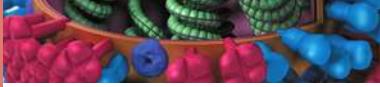
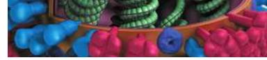
Courtesy of Dan Higgins and Douglas Jordan/CDC
You should be able to relate each summary to the adjacent figures. If you go to this visual summary in Achieve, you can follow links to figures, animations, activities, and simulations that will help you consolidate the material.
Visual Summary
DNA Sequences Record the History of Gene Evolution
- ■ Sequence alignments of proteins or nucleic acids from different organisms allow us to compare the sequences and identify homologous positions.
- ■ The minimum number of changes between sequences can be calculated from a similarity matrix.
- ■ Models of sequence evolution can be used to account for changes that cannot be observed directly.
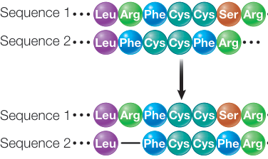
Figure 21.1 Questions
- What biological processes might lead us to hypothesize a gap in a set of aligned gene sequences?
- What are four types of nucleotide substitutions that might lead us to underestimate the total number of changes between two sequences that are compared in a similarity matrix?
Genomes Reveal Both Neutral and Selective Processes of Evolution
- ■ Nonsynonymous substitutions of nucleotides result in amino acid replacements in proteins, whereas synonymous substitutions do not. Rates of synonymous substitution are typically higher than rates of nonsynonymous substitution in protein-coding genes (a result of stabilizing selection).
- ■ Much of the change in nucleotide sequences over time is a result of neutral evolution. The rate of fixation of neutral mutations is independent of population size and is equal to the mutation rate. Specific codons within a given gene sequence can be under different modes of selection.
- ■ Positive selection for change in a protein-coding gene may be detected by a higher rate of nonsynonymous than synonymous substitutions. The reverse is true of purifying selection.
- ■ Common selective constraints can lead to convergent evolution of amino acid sequences in distantly related species.
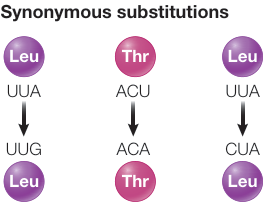
Figure 21.4
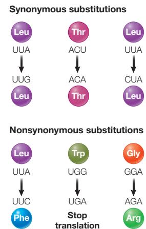
Questions
- Why do we expect stabilizing selection to result in higher rates of synonymous than nonsynonymous substitutions?
- How can biologists distinguish protein-coding genes that are evolving under positive selection from those evolving under stabilizing selection?
- Which position in codons (first, second, or third) would you expect to evolve the fastest? Why?
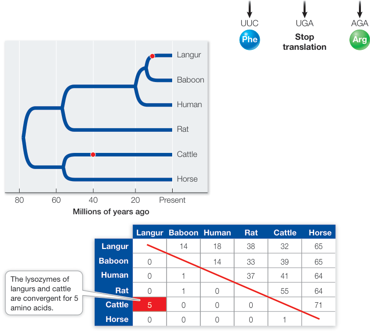
Figure 21.6
- The total size of genomes varies much more widely across multi-cellular organisms than does the number of functional genes.
- Despite its short-term disadvantages, sexual reproduction generates countless genotype combinations that increase genetic variation in populations. In the absence of genetic recombination (as in some asexual organisms), deleterious mutations accumulate with each replication—a phenomenon known as Muller’s ratchet.
- Lateral gene transfer can result in the rapid acquisition of new functions from distantly related species.
- Gene duplications can result in increased production of the gene’s product, in divergence of the duplicated gene’s expression, in pseudogenes, or in new gene functions. Several rounds of gene duplication can give rise to multiple genes with related functions, known as a gene family.
- Some highly repeated genes undergo concerted evolution, in which the multiple copies within the genome maintain their similarity, even as the genes diverge among species.
Questions
- How would you support the statement that large genomes do not necessarily result in more complex organisms?
- How does sexual recombination help keep deleterious mutations from accumulating in a population?
- In what organisms is lateral gene transfer most common?
- What is an example of a new function that arose following gene duplication?
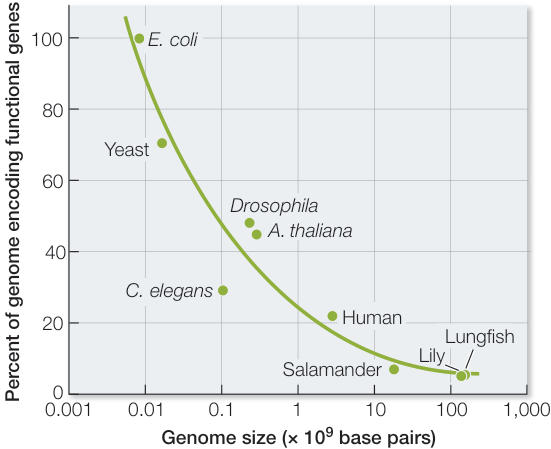
Figure 21.8
Changes in Gene Expression Often Shape Evolution
- A cascade of transcriptional activation sets up the axes of the embryo, the development of the body segments, and the determination of cell fate in each segment. Hox genes help determine cell fate in the embryos of animals.
- Genes encoding transcription factors and other regulatory proteins that govern pattern formation in the developing bodies of multicellular organisms compose what is called a genetic toolkit. These regulatory genes have been highly conserved throughout evolution.
- Developmental genes can be expressed in a modular fashion in different amounts (heterometry), in different timing (heterochrony), or in different locations (heterotopy). Mutations to genes that regulate development often result in major morphological changes.
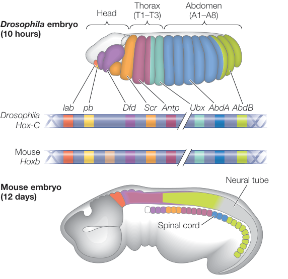
Figure 21.15
Questions
- What is an example of a homeotic mutation?
- Give examples of heteromery, heterochrony, and heterotopy.
- How have changes in body segment development been important in the evolution of arthropod appendages?
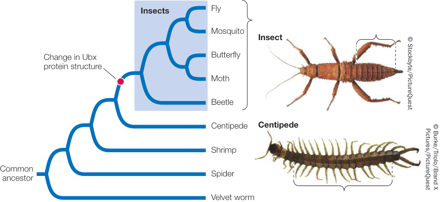
Figure 21.20
Molecular Evolution Has Many Practical Applications
- ■ Protein function can be studied by examining gene evolution. Detection of positive selection can be used to identify molecular changes that have resulted in functional changes.
- ■ In vitro evolution is used to produce synthetic molecules with particular desired functions.
- ■ Many diseases are identified, studied, and combated through molecular evolutionary investigations.
Questions
- Why is positive selection associated with functional change?
- What are the similarities and differences between in vitro evolution and natural selection?
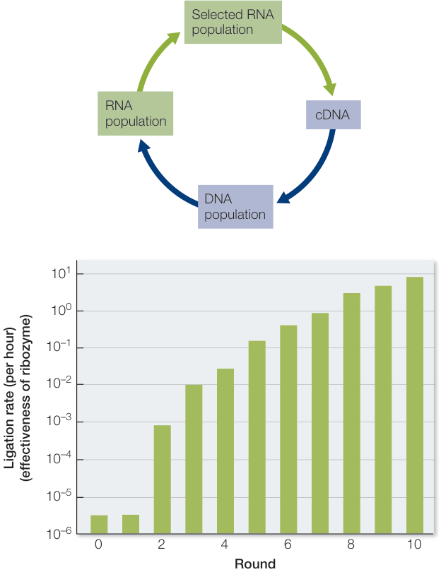
Figure 21.22