Chapter 24Bacteria, Archaea, and Viruses
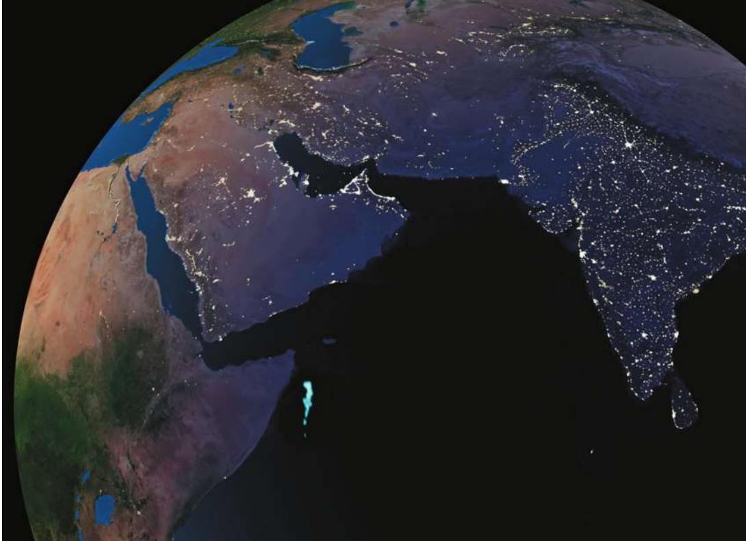
“Milky seas” data prepared by Steven Miller of the Naval Research Laboratory, Monterey, CA. Background data (Earth at Night image) courtesy Marc Imhoff (NASA/GSFC) and Christopher Elvidge (NOAA/NGDC); image by Craig Mayhew (NASA/GSFC) and Robert Simmon (NASA/GSFC)
Key Concepts
- 24.1 Bacteria and Archaea Represent the First Split in the Tree of Life
- 24.2 Prokaryotic Diversity Reflects the Ancient Origins of Life
- 24.3 Ecological Communities Depend on Prokaryotes
- 24.4 Viruses Have Multiple Evolutionary Origins
The milky blue area near the Horn of Africa represents a large, bioluminescent bloom of bacteria, as seen from space.
Investigating Life: Bacteria Light Up the Sea
One night in January of 1995, the British merchant vessel Lima was off the coast of Somalia, near the Horn of Africa. This area is infamous for bands of pirates, so the crew was keeping a watchful eye on the seas when they spotted an eerie, whitish glow on the horizon. It was directly in their path, and there was no way to avoid it. Was this strange sight the result of some strange trick of piracy?
Within 15 minutes of first sighting the glow, the Lima was surrounded by shining waters for as far as her crew could see. The ship’s log recorded that “it appeared as though the ship was sailing over a field of snow or gliding over the clouds.” Fortunately for the crew, the glow had nothing to do with pirates.
For centuries, mariners in this part of the world had reported occasional “milky seas” in which the sea surface produced a strange glow at night, extending from horizon to horizon. Scientists up to that point had never been able to confirm the reality or the cause of such phenomena. It was well established, however, that many organisms emit light by bioluminescence, a complex, enzyme-catalyzed biochemical reaction that emits light but not heat.
What kind of organism could cause the vast expanse of bioluminescence observed by the Lima? Some marine organisms emit flashes of light when they are disturbed, but they could not have produced the sustained and uniform glow seen in milky seas. The only organisms known to produce the level of sustained bioluminescence consistent with milky seas are certain prokaryotes, such as bacteria of the genus Vibrio. Using information supplied by the Lima, biologists scanned satellite images of the Indian Ocean for the specific light wavelengths emitted by Vibrio. The satellite images clearly identified thousands of square kilometers of Vibrio-produced milky seas.
Vibrio’s bioluminescence requires a critical concentration of a specific chemical signal produced by the bacteria, so at low densities, free-living Vibrio populations do not glow. But as a colony establishes itself on phytoplankton, the bacteria’s population density increases and concentrations of the luminescence signal build up. Eventually bacterial density becomes high enough for the huge colony to produce visible light. Such chemical-induced action among bacterial cells is referred to as quorum sensing.
QA How and why do bacteria communicate with one another to assess their density? (Find the answer on page 557.)
Bacteria and Archaea Represent the First Split in the Tree of Life
Learning Objectives
24.1.1 Compare and contrast the features of bacteria, archaea, and eukaryotes.24.1.2 Describe how archaea and bacteria each contributed to the evolution of eukaryotes.24.1.3 Draw a phylogenetic tree demonstrating the effects of lateral gene transfer.You may think that you have little in common with a bacterium. But all multicellular eukaryotes, including you, share many attributes with the prokaryotes—organisms that lack a nucleus (see Key Concept 5.1). For example, all organisms, whether eukaryotes or prokaryotes,
have cell membranes and ribosomes (see Chapter 5 and 6).
have a common set of metabolic pathways, such as glycolysis (see Chapter 9).
replicate DNA semiconservatively (see Chapter 13).
use DNA as the genetic material to encode proteins, and use similar genetic codes to produce those proteins by transcription and translation (see Chapter 14).
These shared features support the hypothesis that all living organisms share a common ancestor. If life had multiple origins, there would be little reason to expect all organisms to use overwhelmingly similar genetic codes or to share structures as distinctive as ribosomes. Furthermore, similarities in the DNA sequences of genes that are shared by all organisms confirm the monophyly of life.
The earliest split in the tree of life gave rise to Bacteria and ArchaeaAlthough all living things share many features, major differences have evolved across the diversity of life. Biologists have now sequenced the genomes of many living organisms, and these genomes allow us to reconstruct the details of evolutionary history. These studies clarify that the earliest split in the tree of life resulted in two major groups: Bacteria and Archaea (Figure 24.1).
Although the earliest division of life gave rise to two major lineages, biologists usually refer to three major domains of life: Bacteria, Archaea, and Eukarya (which includes all plants, animals, and fungi). But if you examine Figure 24.1, you will see that the third domain, Eukarya, arose through phylogenetic contributions from both bacteria and archaea. (Note that we use lowercase when referring to members of these domains and initial capitals when referring to the domains themselves.) Most genes of eukaryotes are more closely related to those of archaea, but several endosymbiotic events involving bacteria also contributed to making eukaryotes a distinctive new lineage. So biologists are beginning to view eukaryotes as a specialized group of archaea that developed some important new features (including a cell nucleus and mitochondria, at least partly through contributions from bacteria). We call all the organisms that lack these specializations prokaryotes (Greek, “before the kernel,” or before the evolution of a cellular nucleus).
In Chapter 20 we discussed the concept of monophyly (a group that includes an ancestor and all of its descendants) and the importance of monophyly in biological taxonomy. However, the reticulations of bacterial and archaeal lineages near the base of the tree of life make the recognition of strictly monophyletic groups of life’s primary domains impractical. Prokaryotes are clearly not a monophyletic group (because the group excludes some of their descendants—the eukaryotes), but the name “prokaryotes” is nonetheless a useful way of talking about all organisms that are not eukaryotes. In a similar manner, when biologists talk about “archaea” or “bacteria,” they are typically excluding their eukaryotic descendants, so neither of those groups are strictly monophyletic either. These complications in our taxonomic terminology are unavoidable, given the reticulations that occur among the major lineages near the base of the tree of life. We will use “bacteria” and “archaea” to refer to the two major groups of prokaryotes, and “eukaryotes” to refer to the lineage that emerged from combinations of some specific bacteria and archaea.
All prokaryotic organisms are unicellular, although they may form large, coordinated colonies or communities consisting of many individuals. Eukaryotes, by contrast, include both unicellular as well as many multicellular life forms. As we saw in Chapter 5, prokaryotic cells differ from eukaryotic cells in some important ways:
- Prokaryotic cells do not divide by mitosis. Instead, after replicating their DNA, prokaryotic cells divide by their own method, binary fission (see Key Concept 11.1).
- The organization of the genetic material differs. The DNA of the prokaryotic cell is not organized within a membrane-enclosed nucleus. DNA molecules in prokaryotes are often circular. Many (but not all) prokaryotes have only one main chromosome and are effectively haploid, although many have additional smaller DNA molecules, called plasmids (see Key Concept 12.6).
- Prokaryotes have none of the membrane-enclosed cytoplasmic organelles that are found in most eukaryotes. However, the cytoplasm of a prokaryotic cell may contain a variety of infoldings of the cell membrane and photosynthetic membrane systems that are not found in eukaryotes.
Although the study and classification of eukaryotic organisms go back centuries, much of our knowledge of the evolutionary relationships of prokaryotes is extremely recent. Not until the final quarter of the twentieth century did advances in molecular genetics and biochemistry enable the research that revealed the deep-seated distinctions between archaea and bacteria.
The two prokaryotic groups differ in significant waysA glance at Table 24.1 will show you that there are major differences (most of which cannot be seen even under an electron microscope) between archaea and bacteria. Archaea share several features with eukaryotes, but they also retain some ancestral similarities with bacteria. The basic unit of an archaeon (the term for a single archaeal organism) or bacterium (a single bacterial
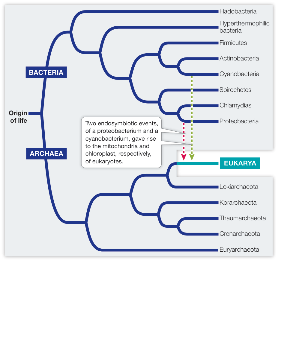
Figure 24.1 The Major Groups of the Living World This phylogenetic tree of Bacteria and Archaea shows their relationships to each other and to their descendants, Eukarya. The relationships among the many clades of bacteria, most of which are not shown here, are incompletely resolved at this time.
Table 24.1 Characteristics of Bacteria, Archaea, and Eukarya
| Characteristic | Bacteria | Archaea | Eukarya |
|---|---|---|---|
| Membrane-enclosed nucleus | Absent | Absent | Present |
| Membrane-enclosed organelles | Few | Absent | Many |
| Peptidoglycan in cell wall | Present | Absent | Absent |
| Membrane lipids | Ester-linked Unbranched | Ether-linked Branched | Ester-linked Unbranched |
| Ribosomes a | 70S | 70S | 80S |
| Initiator tRNA | Formylmethionine | Methionine | Methionine |
| Operons | Yes | Yes | Rare |
| Plasmids | Yes | Yes | Rare |
| Number of RNA polymerases b | One | One | Three |
| Ribosomes sensitive to chloramphenicol and streptomycin | Yes | No | No |
| Ribosomes sensitive to diphtheria toxin | No | Yes | Yes |
a70S ribosomes are smaller than 80S ribosomes.
bThe structure of archaeal RNA polymerase is similar to that of eukaryotic polymerases.
organism) is the prokaryotic cell. Each single-celled prokaryote contains a full complement of genetic and protein-synthesizing systems, including DNA, RNA, and all the enzymes needed to transcribe and translate genetic information into proteins. The prokaryotic cell also contains at least one system for generating the ATP it needs.
Genetic studies clearly indicate that all forms of life share a single common ancestor. As we noted earlier, most of the eukaryotic genome shares a more recent common ancestor with certain groups of archaea than with bacteria (see Figure 24.1). However, the mitochondria of eukaryotes and the chloroplasts of photosynthetic eukaryotes (such as plants) originated through endosymbioses with bacteria. Some biologists prefer to view the origin of eukaryotes as a fusion of two equal partners (one ancestor that was related to modern archaea and another that was more closely related to modern bacteria). Others view the divergence of the early eukaryotes from specific groups of archaea as an event separate from the later endosymbioses. In either case, most eukaryotic genes are more closely related to those of specific groups of archaea, whereas other genes (especially genes related to mitochondria and chloroplasts) are most closely related to those of bacteria.
Connect the Concepts The origin of mitochondria and chloroplasts by endosymbiosis is described in Key Concepts 5.5 and 26.1.
Biologists estimate that the last common ancestor of all living organisms lived about 3 billion years ago. We can deduce that it had DNA as its genetic material, and that its machinery for transcription and translation produced RNAs and proteins, respectively. This ancestor likely had a circular chromosome. All living organisms are the products of billions of years of mutation, natural selection, and genetic drift, and they are all well adapted to present-day environments. The earliest prokaryotic fossils, which date back at least 3.5 billion years, indicate that there was considerable diversity among the prokaryotes even during those earliest days of life.
The small size of prokaryotes has hindered our study of their evolutionary relationships
Until about 300 years ago, nobody had even seen an individual prokaryote. Most prokaryotes remained invisible to humans until the invention of the first simple microscope. Prokaryotes are so small, however, that even the best light microscopes don’t reveal much about them. It took advanced microscopic equipment and modern molecular techniques to open up the microbial world. (Microscopic organisms—both prokaryotes and eukaryotes—are often collectively referred to as “microbes.”)
Before DNA sequencing became practical, taxonomists based prokaryote classification on observable phenotypic characters such as shape, color, motility, nutritional requirements, and sensitivity to antibiotics. One of the characters most widely used to classify prokaryotes is the structure of their cell walls.
The cell walls of almost all bacteria contain peptidoglycan, a cross-linked polymer of amino sugars that produces a firm, protective, meshlike structure around the cell. Peptidoglycan is a substance unique to bacteria; its absence from the cell walls of archaea is a key difference between the two prokaryotic domains. Peptidoglycan is also an excellent target for combating pathogenic (disease-causing) bacteria because it has no counterpart in eukaryotic cells. Antibiotics such as penicillin and ampicillin, as well as other agents that specifically interfere with the synthesis of peptidoglycan-containing cell walls, tend to have little, if any, effect on the cells of humans and other eukaryotes.
The Gram stain is a technique that can be used to separate most types of bacteria into two distinct groups. A smear of bacterial cells on a microscope slide is soaked in a violet dye and treated with iodine; it is then washed with alcohol and counterstained with a red dye called safranin. Gram-positive bacteria retain the violet dye and appear blue to purple (Figure 24.2A). The alcohol washes the violet stain out of Gram-negative bacteria, which then pick up the safranin counterstain and appear pink to red (Figure 24.2B). For most bacteria, the effect of the Gram stain is determined by the chemical structure of the cell wall:
- A Gram-negative cell wall usually has a thin peptidoglycan layer, which is surrounded by a second, outer membrane quite distinct in chemical makeup from the cell membrane (see Figure 24.2B). Together the cell wall and the outer membrane are called the cell envelope. The space between the cell membrane and the outer membrane (known as the periplasmic space) contains proteins that are important in digesting some materials, transporting others, and detecting chemical gradients in the environment.
- A Gram-positive cell wall usually has about five times as much peptidoglycan as a Gram-negative cell wall. Its thick peptidoglycan layer is a meshwork that may serve some of the same purposes as the periplasmic space of the Gram-negative cell envelope.
Shape is another phenotypic character that is useful for the basic identification of bacteria. The three most common shapes are spheres, rods, and spiral forms (Figure 24.3). Many bacterial names are based on these shapes. A spherical bacterium is called a coccus (plural cocci). Cocci may live singly or may associate in two-or three-dimensional arrays such as chains, plates, blocks, or clusters of cells. A rod-shaped bacterium is called a bacillus (plural bacilli). A spiral bacterium (shaped like a corkscrew) is called a spirillum (plural spirilla). Bacilli and spirilla may be single, form chains, or gather in regular clusters. Among the other bacterial shapes are long filaments and branched filaments.
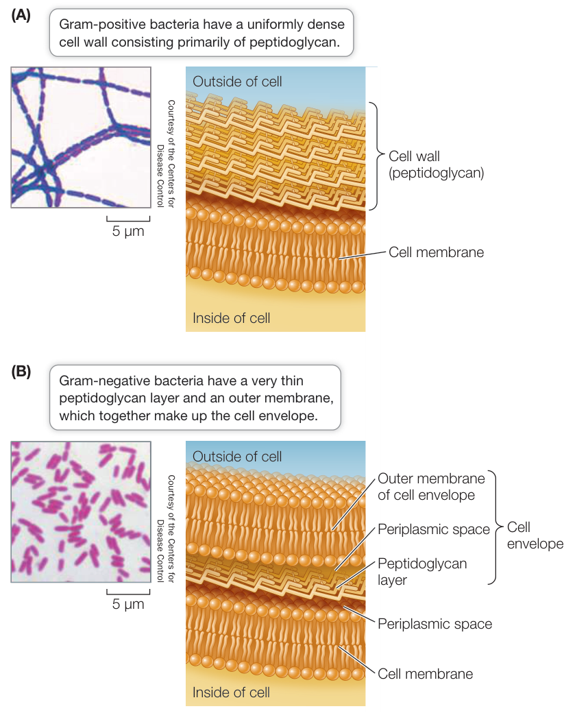
Figure 24.2 The Gram Stain and the Bacterial Cell Wall When treated with Gram-staining reagents, the cell walls of bacteria react in one of two ways. (A) Gram-positive bacteria have a thick peptidoglycan cell wall that retains the violet dye and appears deep blue or purple. (B) Gram-negative bacteria have a thin peptidoglycan layer that does not retain the violet dye, but picks up the counterstain and appears pink to red.
Less is known about the shapes of archaea because many of these organisms have never been seen. Many archaea are known only from samples of DNA from the environment. However, the species whose morphologies are known include cocci, bacilli, and even triangular and square species. Some flattened species grow on surfaces, arranged like sheets of postage stamps.
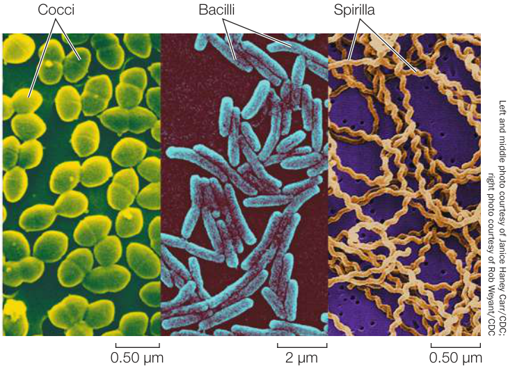
Figure 24.3 Bacterial Cell Shapes This composite, colorized micrograph shows the three most common bacterial shapes. Spherical cells are called cocci; those pictured are a species of Enterococcus from the mammalian gut. Rod-shaped cells are called bacilli; these Escherichia coli also reside in the gut. The helix-shaped spirilla are Leptospira interrogans, a human pathogen.
The nucleotide sequences of prokaryotes reveal their evolutionary relationships
Analyses of the nucleotide sequences of ribosomal RNA (rRNA) genes provided the first comprehensive evidence of evolutionary relationships among prokaryotes. Comparisons of rRNA genes are often used to identify microbes. For several reasons, rRNA is particularly useful for phylogenetic studies and identification purposes:
- rRNA was present in the common ancestor of all life and is therefore evolutionarily ancient.
- No free-living organism lacks rRNA, so rRNA genes can be compared across the tree of life.
- rRNA plays a critical role in translation in all organisms, so lateral transfer of rRNA genes among distantly related species is unlikely.
- rRNA has evolved slowly enough that gene sequences from even distantly related species can be aligned and analyzed.
Although studies of rRNA genes reveal much about the evolutionary relationships of prokaryotes, they don’t always reveal the entire evolutionary history of these organisms. In some groups of prokaryotes, analyses of multiple gene sequences have suggested several different phylogenetic patterns. How could such differences among different gene sequences arise? Studies of whole prokaryotic genomes have revealed that even distantly related prokaryotes sometimes exchange genetic material.
Lateral gene transfer can lead to discordant gene trees
As noted earlier, prokaryotes reproduce by binary fission. If we could follow these divisions back through evolutionary time, we would be tracing the complete tree of life. At a much broader scale, these divisions of organisms lead to splits among the major evolutionary lineages, or species of life (represented in highly abbreviated form in Appendix A). Because binary fission is an asexual process that replicates whole genomes, we would expect phylogenetic trees of prokaryotes constructed from most gene sequences (see Chapter 21) to reflect these same relationships.
Even though binary fission is an asexual process, there are other processes—including transformation, conjugation, and transduction—that allow the transfer of genetic information between some prokaryotes without reproduction. Thus prokaryotes can transfer and recombine their DNA with that of other individuals (this is sex in the genetic sense of the word), but this genetic exchange is not directly linked to reproduction, as it is in most eukaryotes.
Connect the Concepts Prokaryotic exchange of genetic material by conjugation and transformation is described in Key Concept 12.6 and Chapter 18, respectively.
From early in evolution to the present day, some genes have been moving “sideways” from one prokaryotic species to another, a phenomenon known as lateral gene transfer (sometimes called horizontal gene transfer). Lateral gene transfers are well documented among closely related species, and some have been documented even across the domains of life.
Consider, for example, the genome of Thermotoga maritima, a bacterium that can survive extremely high temperatures. By comparing the 1,869 gene sequences of T. maritima with sequences encoding the same proteins in other species, investigators found that some of this bacterium’s genes have their closest relationships not with the genes of other bacterial species, but with the genes of archaea that live in similar extreme environments.
When genes involved in lateral transfer events are sequenced and analyzed, the resulting gene trees will not match the organismal tree in every respect (Figure 24.4). The individual gene trees will vary because the history of lateral transfer events is different for each gene. Biologists can reconstruct the underlying organismal phylogeny by comparing multiple genes (to produce a consensus tree) or by concentrating on genes that are unlikely to be involved in lateral gene transfer events. For example, genes that are involved in fundamental cellular processes (such as the rRNA genes discussed in the previous section) are unlikely to be replaced by the same genes from other species because functional, locally adapted copies of these genes are already present.
What kinds of genes are most likely to be involved in lateral gene transfer? Genes that result in a new adaptation that confers higher fitness on a recipient species are most likely to be transferred repeatedly among species. For example, genes that produce antibiotic resistance are often transferred among bacterial species on plasmids, especially under the strong selection pressure such as that imposed by modern antibiotic medications. Improper or overly frequent use of antibiotics can select for resistant strains of bacteria that are much harder to treat. This selection for antibiotic resistance explains why informed physicians have become more careful in prescribing antibiotics.
It is debatable whether lateral gene transfer has seriously complicated our attempts to resolve the tree of prokaryotic life. Recent work suggests that it has not. Lateral gene transfer rarely creates problems at higher taxonomic levels, even though it may complicate our understanding of the relationships among individual species. Some species clearly obtain some of their genes from otherwise distantly related species, so evolutionary histories of individual genes may differ within a single organism. But it is now possible to make nucleotide sequence comparisons involving entire genomes, and these studies are revealing a stable core of crucial genes that are uncomplicated by lateral gene transfer. Gene trees based on this stable core more accurately reveal the organismal phylogeny (see Figure 24.4). The problem remains, however, that only a very small proportion of the prokaryotic world has been described and studied.
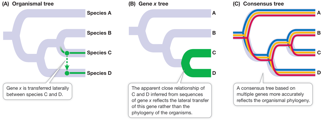
Figure 24.4 Lateral Gene Transfer Complicates Phylogenetic Relationships (A) The phylogeny of four hypothetical prokaryotic species, two of which have been involved in a lateral transfer of gene x. (B) A tree based only on gene x shows the phylogeny of the laterally transferred gene, rather than the organismal phylogeny. (C) A consensus tree based on multiple genes is more likely to reflect the true organismal phylogeny, especially if those genes come from a stable core of genes involved in fundamental processes. Q: Why are multiple lateral gene transfers between the same two branches on a phylogeny expected to be rare, at least compared with similarities inherited through the stable core?
The great majority of prokaryotic species have never been studied
Most prokaryotes have defied all attempts to grow them in pure culture, causing biologists to wonder how many species, and possibly even major clades, we might be missing. A window onto this problem was opened with the introduction of a new way of examining nucleic acid sequences. When biologists are unable to work with the whole genome of a single prokaryotic species, they can instead examine genomes collected from an environmental sample (such as a scoop of sediment from the seafloor). This technique is known as environmental genomics.
Biologists now routinely isolate gene sequences, or even whole genomes, from environmental samples such as soil and seawater. Comparing such sequences with previously known ones has revealed that an extraordinary number of the sequences represent new, previously unrecognized species. Biologists have described only about 10,000 species of bacteria and only a few hundred species of archaea (see Figure 1.12). The results of some environmental genomic studies suggest that there may be millions—perhaps hundreds of millions—of prokaryotic species. Other biologists put the estimate much lower, arguing that the high dispersal ability of many bacterial species greatly reduces endemism (i.e., the number of species restricted to a particular geographic area). Only the magnitude of these estimates differs, however; all sides agree that we have just begun to uncover Earth’s prokaryotic diversity.
Recap and Assess
Bacteria and archaea are the two major lineages that arose from the first split in the tree of life. Eukaryotes evolved from a group of archaea that captured and incorporated bacteria through endosymbiosis. Mitochondria and chloroplasts are among the cellular structures of eukaryotes that are derived from endosymbiosis of bacteria. Although only about 10,000 species of bacteria and a few hundred species of archaea have been formally described, genetic evidence suggest there are many millions of species.
- What findings support the separation of Bacteria and Archaea into separate domains?
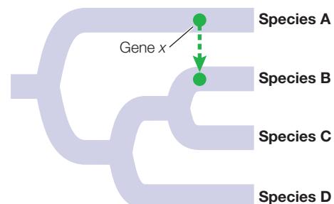
- The figure below shows an organismal tree in which gene x has undergone a lateral transfer event. Draw the phylogenetic tree you would expect based on gene x, as well as the phylogenetic tree you would expect based on a consensus of non-transferred genes.
- How did eukaryotes arise through evolutionary contributions from both archaea and bacteria?
Despite the challenges of reconstructing prokaryotic phylogeny, taxonomists are starting to establish evolutionary classification systems for these organisms. With a full understanding that new information requires periodic revisions in these classifications, we next apply a current system of classification to organize our survey of prokaryotic diversity.
Prokaryotic Diversity Reflects the Ancient Origins of Life
Learning Objectives
- 24.2.1 Interpret a phylogenetic tree of major lineages of bacteria and archaea and use it to explain the origins of eukaryotes.
- 24.2.2 Discuss two lines of evidence that support the origin of life in a high-temperature environment.
- 24.2.3 Explain the role of bacteria in changing the oxygen content of Earth’s atmosphere.
The prokaryotes were alone on Earth for a very long time, adapting to new environments and to changes in existing environments. They have survived to this day, in massive numbers and incredible diversity, and they are found nearly everywhere. In numbers of individuals, prokaryotes are far more abundant than eukaryotes. Individual prokaryotes in the oceans number more than —more than the number of stars in the universe. Closer to home, the individual bacteria living in your intestinal tract outnumber all the humans who have ever lived.
Given our still-fragmentary knowledge of prokaryotic diversity, it is not surprising that there are many different hypotheses about the relationships of the major groups of prokaryotes. In this book we use a classification system that is supported by nucleotide sequence data. We discuss a few of the major bacterial groups that have the broadest phylogenetic support and have received the most study, including hadobacteria, hyperthermophilic bacteria, firmicutes, actinobacteria, cyanobacteria, spirochetes, chlamydias, and proteobacteria (see Figure 24.1). Many other major groups of bacteria are known but are less thoroughly studied. We then describe the archaea, whose great diversity is just beginning to be fully understood and appreciated.
Two early-branching lineages of bacteria live at very high temperatures
Several lineages of bacteria and archaea are extremophiles: they thrive under extreme conditions that would kill most other organisms. The hadobacteria, for example, are thermophiles (Greek, “heat lovers”). The group’s name is derived from Hades, the ancient Greek name for the underworld. Hadobacteria of the genus Deinococcus are resistant to radiation and can degrade nuclear waste and other toxic materials. They can also survive extremes of cold as well as hot temperatures. Another hadobacterium, Thermus aquaticus, was the source of the thermally stable DNA polymerase that was critical for the development of the polymerase chain reaction. Thermus aquaticus was originally isolated from a hot spring, but it can be found wherever hot water occurs (including in many residential hot-water heaters).
The hyperthermophilic bacteria are another major group of extremophiles. Genera such as Aquifex live near volcanic vents and in hot springs, sometimes at temperatures near the boiling point of water. Some species of Aquifex need only hydrogen, oxygen, carbon dioxide, and mineral salts to live and grow. Species of the genus Thermotoga live deep underground in oil reservoirs as well as in other high-temperature environments.
Biologists have hypothesized that high temperatures characterized the ancestral conditions for life, given that most environments on early Earth were much hotter than those of today. Reconstructions of ancestral bacterial genes have supported this hypothesis by showing that the ancestral sequences functioned best at elevated temperatures. The presence of multiple lineages of extremophiles at the base of the bacterial tree (see Figure 24.1) also provides support for the origin of life in a high-temperature environment.
Firmicutes include some of the smallest cellular organisms
The firmicutes are also known as low-GC Gram-positive bacteria. The first part of this description (low-GC) derives from the relatively low ratio of G-C to A-T nucleotide base pairs in their DNA. The second part (Gram-positive) is less accurate: some of the firmicutes are in fact Gram-negative, and some have no cell wall at all. Despite these differences, phylogenetic analyses of DNA sequences support the monophyly of this bacterial group.
One group of firmicutes can produce resting structures called endospores (Figure 24.5). When a key nutrient such as nitrogen or carbon becomes scarce, the bacterium replicates its DNA and encapsulates one copy, along with some of its cytoplasm, in a tough endospore wall heavily thickened with peptidoglycan and surrounded by a spore coat. The parent cell then breaks down, releasing the endospore. Endospore production is not a reproductive process, as the endospore merely replaces the parent cell. The endospore, however, can survive harsh environmental conditions that would kill the parent cell, such as high or low temperatures or drought, because it is dormant—its normal metabolic activity is suspended. Later, if it encounters favorable conditions, the endospore becomes metabolically active and divides, forming new cells that are like the parent cell. Members of this endospore-forming group include the many species of Clostridium and Bacillus. Some of their endospores can be reactivated after more than 1,000 years of dormancy. There are even credible claims of reactivation of Bacillus endospores that are millions of years old.
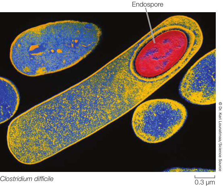
Figure 24.5 A Structure for Waiting Out Bad Times Under harsh conditions, some firmicutes can replicate their DNA and encase it in an endospore. The parent cell then breaks down, and the endospore survives in a dormant state until conditions improve.
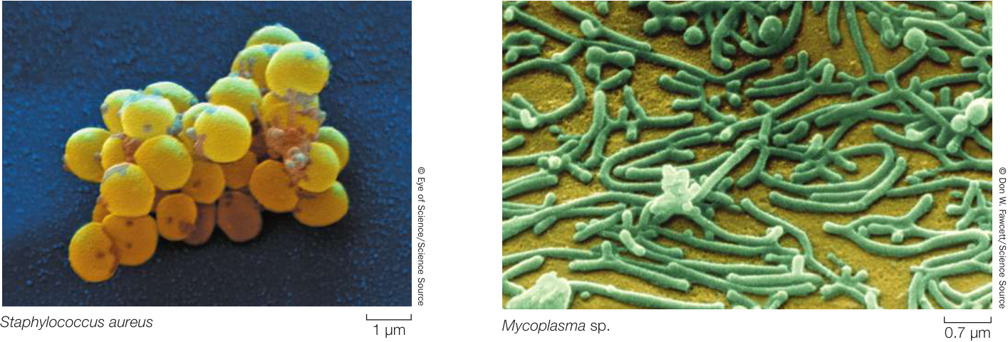
Figure 24.6 Staphylococci “Grape clusters” are the usual arrangement of these firmicutes, which are often the cause of skin or wound infections. Figure 24.7 Tiny Cells With about one-fifth as much DNA as E. coli has, mycoplasmas are among the smallest known bacteria.
Endospores of Bacillus anthracis are the cause of anthrax. Anthrax is primarily a disease of cattle and sheep, but it can be fatal in humans. When the endospores sense macrophage (a type of white blood cell that digests cellular debris and foreign substances in mammalian blood), they reactivate and release toxins into the bloodstream. Bacillus anthracis has been used as a bioterrorism agent because it is relatively easy to transport large quantities of its endospores and release them among human populations, where they may be inhaled or ingested.
Members of the genus Staphylococcus—the staphylococci (Figure 24.6)—are abundant on the human body surface; they are responsible for boils and many other skin problems. Staphylococcus aureus is the best-known human pathogen in this genus; it is present in 20% to 40% of normal adults (and in 50%–70% of hospitalized adults). In addition to causing skin diseases, S. aureus can cause respiratory, intestinal, and wound infections.
Another interesting group of firmicutes, the mycoplasmas, lack cell walls, although some have a stiffening material outside the cell membrane. The mycoplasmas are among the smallest cellular organisms known (Figure 24.7). The smallest mycoplasmas have a diameter of about 0.2 µm. They are small in another crucial sense as well: they have less than half as much DNA as most other prokaryotes. It has been speculated that the DNA in a mycoplasma, which codes for fewer than 500 proteins, may be close to the minimum amount required to encode the essential properties of a living cell.
Actinobacteria include major pathogens as well as valuable sources of antibiotics
Actinobacteria, also known as high-GC Gram-positive bacteria, have a higher ratio of G-C to A-T nucleotide base pairs than do the firmicutes (low-GC Gram-positives). These bacteria develop an elaborately branched system of filaments (Figure 24.8) that resembles the filamentous growth habit of fungi, albeit at a smaller scale. Some actinobacteria reproduce by forming chains of spores at the tips of the filaments. In species that do not form spores, the branched, filamentous growth ceases and the structure breaks up into typical cocci or bacilli, which then reproduce by binary fission.
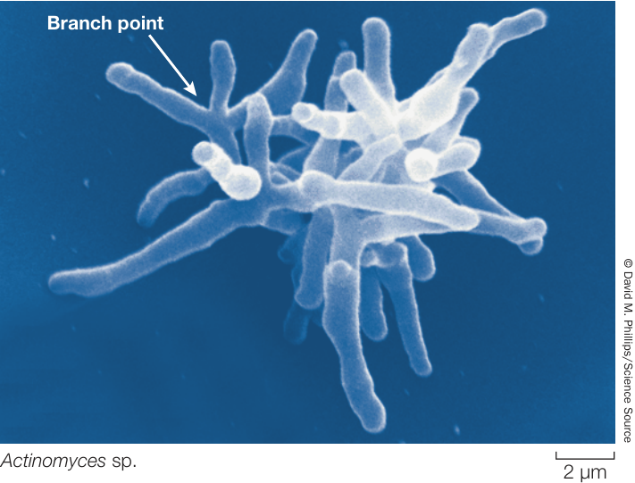
Figure 24.8 Actinobacteria Often Produce Branching Filaments The tangled, branching filaments seen in this scanning electron micrograph are typical of this medically important bacterial group.
The actinobacteria include several medically important bacteria. Mycobacterium tuberculosis causes tuberculosis, which kills 2 million people each year. Genetic data suggest that this bacterium may have been infecting our ancestors for almost 3 million years, making it the oldest known human bacterial pathogen. The genus Streptomyces produces streptomycin as well as hundreds of other antibiotics. We derive most of our antibiotics from actinobacteria.
Cyanobacteria were the first photosynthesizers
Cyanobacteria, sometimes called blue-green bacteria because of their pigmentation, are photosynthetic. They use chlorophyll a for photosynthesis and release oxygen gas (); many species also fix nitrogen (discussed in Key Concept 24.3). The production of oxygen by these bacteria transformed the atmosphere of early Earth, eventually leading to the oxygen-rich atmosphere we know today.
Cyanobacteria carry out the same type of photosynthesis that is characteristic of eukaryotic photosynthesizers. They contain elaborate and highly organized internal membrane systems called photosynthetic lamellae. As mentioned in Key Concept 24.1, the chloroplasts of photosynthetic eukaryotes are derived from an endosymbiotic cyanobacterium. So all photosynthesis is either directly from cyanobacteria, or from eukaryotes with chloroplasts that are derived from cyanobacteria.
Cyanobacteria may live free as single cells or associate in multicellular colonies. Depending on the species and on growth conditions, these colonies may range from flat sheets one cell thick to filaments to spherical balls of cells. Some filamentous colonies of cyanobacteria differentiate into three specialized cell types: vegetative cells, spores, and heterocysts (Figure 24.9). Vegetative cells photosynthesize, spores are resting stages that can survive harsh environmental conditions and eventually develop into new filaments, and heterocysts are cells specialized for nitrogen fixation. All of the known cyanobacteria with heterocysts fix nitrogen. Heterocysts also have a role in reproduction: when filaments break apart to reproduce, the heterocyst may serve as a breaking point.
Spirochetes move by means of axial filaments
Spirochetes are Gram-negative, motile bacteria characterized by a unique structure known as an axial filament, which is composed of multiple internal flagella running through the periplasmic space (Figure 24.10A). The cell body is a long cylinder coiled into a helix. The internal flagella begin at either end of the cell and overlap in the middle. Motor proteins connect the axial filament to the cell wall, enabling the corkscrew-like movement of the bacterium. Many spirochetes are parasites of humans; a few are pathogens, including those that cause syphilis (Figure 24.10B) and Lyme disease. Others live free in mud or water.
Chlamydias are extremely small parasites
Chlamydias are among the smaller bacteria ( in diameter). They are obligate parasites—that is, they can live only as parasites in the cells of other organisms. It was once believed that their obligate parasitism resulted from an inability to produce ATP—that chlamydias were “energy parasites.” However, genome sequencing indicates that chlamydias have the genetic capacity to produce at least some ATP. They can augment this capacity by using an enzyme called a translocase, which allows them to take up ATP from the cytoplasm of their host in exchange for ADP from their own cells.
These tiny, Gram-negative cocci are unique among prokaryotes because of a complex life cycle that involves two different
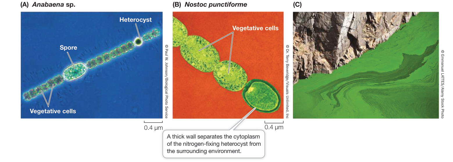
Figure 24.9 Cyanobacteria (A) Some cyanobacteria form filamentous colonies containing three cell types. (B) Heterocysts are specialized for nitrogen fixation and may serve as a breaking point when filaments reproduce. (C) This pond in Canada has experienced eutrophication: phosphorus and other nutrients generated by human activity have accumulated, feeding an immense green mat (commonly referred to as “pond scum”) that is made up of several species of free-living cyanobacteria. Media Clip 24.1 Cyanobacteria Life12e.com/mc24.1
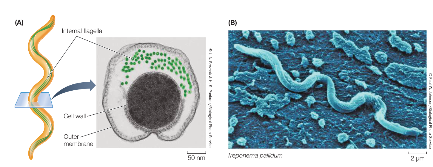
Figure 24.10 Spirochetes Get Their Shape from Axial Filaments (A) A spirochete from the gut of a termite, seen in cross section, shows the internal flagella that compose the axial filament, which these helical prokaryotes use to produce a corkscrew-like movement. (B) This spirochete species causes syphilis in humans.
forms of cells, elementary bodies and reticulate bodies (Figure 24.11). Various strains of chlamydias cause eye infections (especially trachoma), sexually transmitted diseases, and some forms of pneumonia in humans.
The proteobacteria are a large and diverse group
By far the largest bacterial group, in terms of numbers of described species, is the proteobacteria. The proteobacteria include many species of Gram-negative photoautotrophs (see Key Concept 24.3) that use light-driven reactions to metabolize sulfur, as well as dramatically diverse bacteria that bear no phenotypic resemblance to the photoautotrophic species. Genetic and morphological evidence indicates that the mitochondria of eukaryotes were derived from a proteobacterium by endosymbiosis. Among the proteobacteria are some nitrogen-fixing genera, such as Rhizobium, and other bacteria that contribute to the global nitrogen and sulfur cycles. Escherichia coli, one of the most studied organisms on Earth, is a proteobacterium. So, too, are many of the most famous human pathogens, such as Yersinia pestis (which causes bubonic plague), Vibrio cholerae (cholera), and Salmonella typhimurium (gastrointestinal disease) (Figure 24.12).
The bioluminescent Vibrio shown at the opening of this chapter are also members of this group. There are many potential applications of the genes that encode bioluminescent proteins in
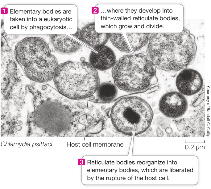
Figure 24.11 Chlamydias Change Form Elementary bodies and reticulate bodies are the two cell forms of the chlamydia life cycle.
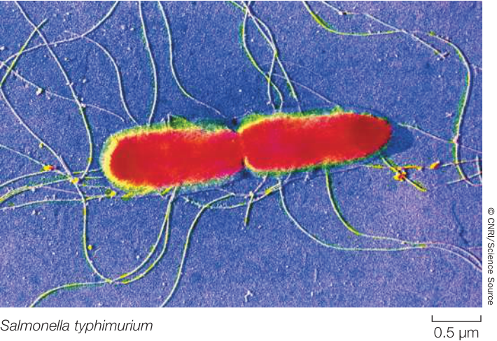
Figure 24.12 Proteobacteria Include Many Familiar Bacteria These conjugating (see Key Concept 12.6) cells of Salmonella typhimurium are exchanging genetic material. This pathogen causes a wide range of gastrointestinal illnesses in humans.
Media Clip 24.2 A Swarm of Salmonella Life12e.com/mc24.2 bacteria. Already, these genes are being inserted into the genomes of other species in which the resulting bioluminescence is used as a marker of gene expression. Futuristic proposals for making use of bioluminescence in bioengineered organisms include crop plants that glow when they become water-stressed and need to be irrigated, and glowing trees that could light highways at night in place of electric lights.
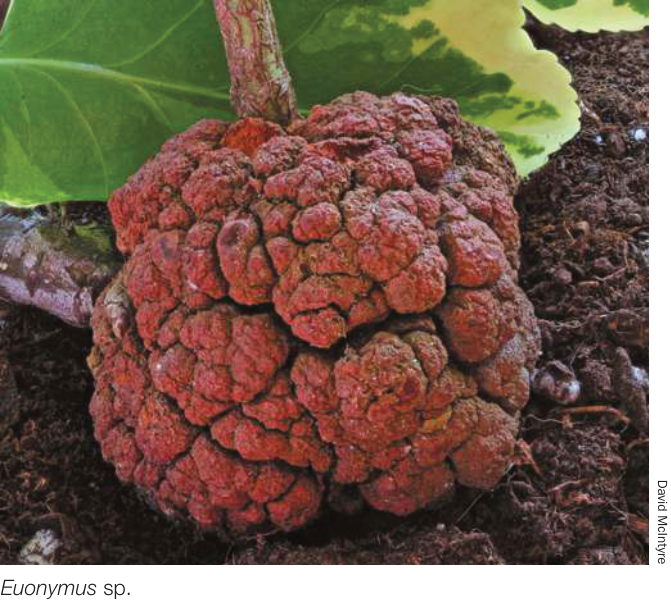
Figure 24.13 Crown Gall Crown gall, a type of tumor shown here growing on the stem of a Euonymus shrub, is caused by the proteobacterium Agrobacterium tumefaciens.
Although fungi cause most plant diseases, and viruses cause others, about 200 known plant diseases are of bacterial origin. Crown gall, with its characteristic tumors (Figure 24.13), is one of the most striking. The causal agent of crown gall is Agrobacterium tumefaciens, a proteobacterium that harbors a plasmid used in recombinant DNA studies as a vehicle for inserting genes into new plant hosts.
Gene sequencing enabled biologists to differentiate Archaea from Bacteria
The original identification of Archaea as a group distinct from Bacteria was based on phylogenetic relationships determined from rRNA gene sequences. This separation was supported when biologists sequenced the first complete archaeal genome, which consisted of 1,738 genes—more than half of which were unlike any genes ever found in Bacteria.
Archaea are known for living in extreme habitats such as those with high salinity (salt content), low oxygen concentrations, high temperatures, or high or low pH (Figure 24.14). Many archaea are not extremophiles, however—they are common in soil, in many aquatic environments, and in the guts of animals, for example.
Recent studies are revealing many new lineages of archaea, a few of which are shown in Figure 24.1: Euryarchaeota, Crenarchaeota, Thaumarchaeota, Korarchaeota, and Lokiarchaeota.
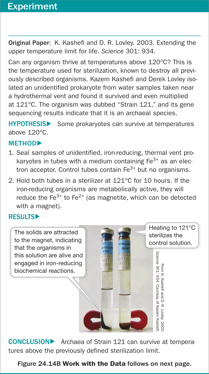
Figure 24.14A What Is the Highest Temperature Compatible with Life? Figure 24.14A What Is the Highest Temperature Compatible with Life?
The Lokiarchaeota were the first discovered members of the Asgard archaea—a group of particular interest because recent studies of their genomes show that they include the closest known prokaryotic relatives of eukaryotes.
Two characteristics shared by all archaea are the absence of peptidoglycan in their cell walls and the presence of lipids of distinctive composition in their cell membranes (see Table 24.1). The unusual lipids in the membranes of archaea are found in no bacteria or eukaryotes. Most lipids in bacterial and eukaryotic membranes contain unbranched long-chain fatty acids connected to glycerol molecules by ester linkages:
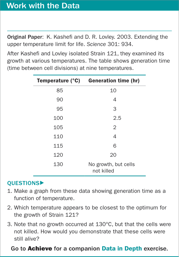
Figure 24.14B What Is the Highest Temperature Compatible with Life?
In contrast, some lipids in membranes of archaea contain long-chain hydrocarbons connected to glycerol molecules by ether linkages:
In addition, the long hydrocarbon chains in the lipids of archaea are branched. One class of archaeal lipids contains glycerol at both ends of the hydrocarbons (Figure 24.15). These lipids form a lipid monolayer structure that is unique to archaea. They still fit into a biological membrane because they are twice as long as the typical lipids in the bilayers of other membranes. Lipid monolayers and bilayers are both found among the archaea.
Archaea live in extremely diverse environments
Most known crenarchaeotes are either thermophilic, acidophilic (acid loving), or both. Members of the genus Sulfolobus live in hot sulfur springs at temperatures of 70°C to 75°C. They become metabolically inactive at 55°C (131°F). Hot sulfur springs are also extremely acidic. Sulfolobus grows best in the range from pH 2 to pH 3, but some members of this genus readily tolerate pH values as low as 0.9. Most acidophilic thermophiles maintain an internal pH of 5.5 to 7 (close to neutral) in spite of their acidic environment. These and other crenarchaeotes thrive where very few other organisms can even survive (Figure 24.16).
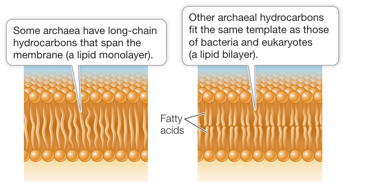
Figure 24.15 Membrane Architecture in Archaea The long-chain hydrocarbons of many archaeal lipids have glycerol molecules at both ends, so that the membranes they form consist of a lipid monolayer. In contrast, the membranes of other archaea, bacteria, and eukaryotes consist of a lipid bilayer.
Some species of euryarchaeotes are methanogens: they produce methane () by reducing carbon dioxide as the key step in their energy metabolism. All of the methanogens are obligate anaerobes (see Key Concept 24.3). Comparison of their rRNA gene sequences has revealed a close evolutionary relationship among these methanogenic species, which were previously assigned to several different groups of bacteria.
Methanogenic euryarchaeotes release approximately 2 billion tons of methane gas into Earth’s atmosphere each year, accounting for 80% to 90% of the methane that enters the atmosphere, including that produced in many animal digestive systems (including our own). Approximately one-third of this methane comes from methanogens living in the guts of ruminants such as cattle, sheep, and deer, and another large fraction comes from methanogens living in the guts of termites and cockroaches. Methane is increasing in Earth’s atmosphere by about 1% per year and contributes to the greenhouse effect. Part of that increase is due to increases in cattle and rice production and the methanogens associated with both.
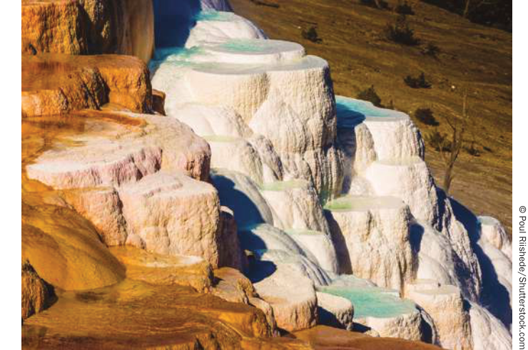
Figure 24.16 Some Crenarchaeotes Like It Hot Thermophilic crenarchaeotes can thrive in the intense heat of volcanic hot sulfur springs such as these in Yellowstone National Park.
Another group of euryarchaeotes, the extreme halophiles (salt lovers), live exclusively in very salty environments. Because they contain pink carotenoid pigments, these archaea are sometimes easy to see (Figure 24.17). Extreme halophiles grow in the Dead Sea and in brines of all types. The reddish pink spots that can occur on pickled fishes are colonies of halophilic archaea. Few other organisms can live in the saltiest homes that the extreme halophiles occupy—most would “dry” to death, losing too much water to the hypertonic environment. Extreme halophiles have been found in lakes with pH values as high as 11.5. These are the most alkaline environments inhabited by living organisms, and almost as alkaline as household ammonia.
Some of the extreme halophiles have a unique system for trapping light energy and using it to form ATP—without using any form of chlorophyll—when oxygen is in short supply. They use the pigment retinal (also found in the vertebrate eye) combined with a protein to form a light-absorbing molecule called bacteriorhodopsin.
Another member of Euryarchaeota, Thermoplasma, has no cell wall. It is thermophilic and acidophilic, its metabolism is aerobic, and it lives in coal deposits. Its genome of 1,100,000 base pairs is among the smallest (along with that of the mycoplasmas) found in any free-living organism, although some parasitic organisms have even smaller genomes.
Many of the known archaea are crenarchaeotes or euryarchaeotes, but studies of extreme environments have identified several lineages that are not closely related to either of these major groups. For example, the korarchaeotes are known only from DNA isolated directly from hot environments. The thaumarchaeotes were originally found in hot environments as well but have since been found to be common in marine surface waters, where they oxidize ammonia and appear to play an important role in the nitrogen cycle.
The lokiarchaeotes were discovered in 2015 by sequencing environmental samples from near a hydrothermal vent called Loki’s Castle deep in the Arctic Ocean. An organism’s genome was detected and sequenced from this deep-sea sample. Sequence analysis revealed that the organism was a distinct lineage of archaea, which was named Lokiarchaeum. Of particular interest was the finding that the Lokiarchaeum genome contains a large number of genes with cell membrane-related functions—genes and functions that had previously been known to occur only in eukaryotes.
After the discovery of Lokiarchaeum, biologists began to look for related archaea in other aquatic environments (both marine and freshwater) and discovered many new lineages of archaea with affinities to eukaryotes. Together, these close relatives of eukaryotes are known as the Asgard archaea (named after Asgard, one of the nine worlds of Norse mythology). The genomes of the Asgard archaea include many additional genes that were formerly thought to be restricted to eukaryotes, including genes that function in eukaryotes in controlling cell shape and cytoskeleton formation. These findings suggest that some of the properties that have long been associated exclusively with eukaryotes probably first arose within the Asgard archaea.
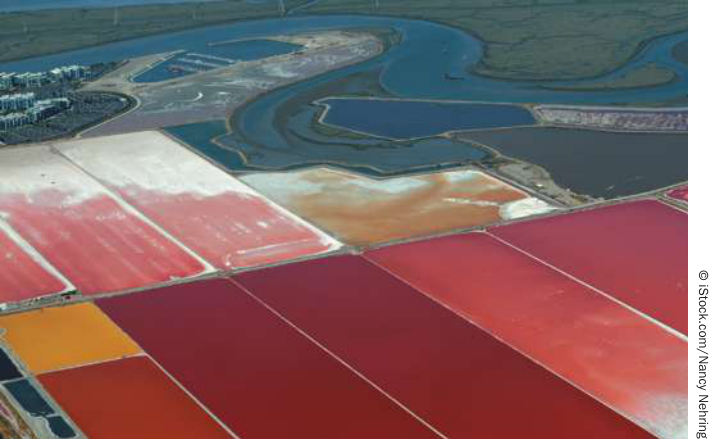
Figure 24.17 Extreme Halophiles Highly saline environments such as these commercial seawater evaporating ponds in San Francisco Bay are home to extreme halophiles. The archaea are easily visible here because of the rich red coloration of their carotenoid pigments.
Recap and Assess
Bacteria and archaea are highly diverse groups that survive in almost every imaginable habitat on Earth. Many prokaryotes can survive and even thrive in habitats where no eukaryotes can live, including extremely hot, acidic, or saline conditions. Eukaryotes are most closely related to the Asgard archaea, although endosymbioses of bacteria within eukaryotic cells contributed to the evolution of eukaryotic organelles.
- Consider the differences between prokaryotes and eukaryotes shown in Table 24.1. Why might eukaryotes be more like archaea in some features and more like bacteria in others?
- Given that all species of life have evolved for the same amount of time since their common origin, how would you respond to someone who characterizes prokaryotes as “primitive”? Include at least two examples of major groups of prokaryotes to support your answer.
- How have bacteria changed Earth’s atmosphere over the past 3 billion years?
- What two lines of evidence can you use to support the idea that eukaryotes evolved from a lineage of the Asgard archaea?
Prokaryotes are found almost everywhere on Earth and live in a wide variety of ecosystems. In the next key concept we examine the contributions of prokaryotes to the functioning of those ecosystems.
Ecological Communities Depend on Prokaryotes
Learning Objectives
24.3.1 Describe what biofilms are, how they are formed, and why they present problems for humans.
24.3.2 Explain how bacteria communicate through quorum sensing.
24.3.3 Explain the critical role of prokaryotes in the nitrogen cycle.
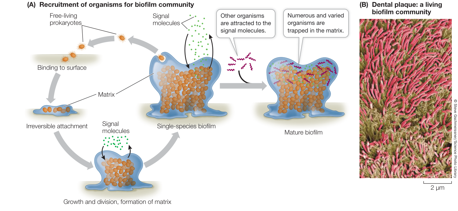
Figure 24.18 Forming a Biofilm (A) Free-living prokaryotes readily attach themselves to surfaces and form films that are stabilized and protected by a surrounding matrix. Once the population is large enough, the developing biofilm can send out chemical signals that attract other microorganisms. (B) Scanning electron micrography reveals a biofilm of dental plaque. The bacteria (red) are embedded in a matrix consisting of proteins from both bacterial secretions and saliva. (A after D. Monroe. 2007. PLOS Biol 5: e307.)
Prokaryotic cells do not usually live in isolation. Rather, they live in communities of many different species, often including microscopic eukaryotes. Whereas some microbial communities are harmful to humans, others provide important services. They help us digest our food, break down municipal waste, and recycle organic matter and chemical elements in the environment.
Many prokaryotes form complex communities
Some microbial communities form layers in sediments, and others form clumps a meter or more in diameter. Many microbial communities tend to form dense biofilms. Upon contacting a solid surface, the cells bind to that surface and secrete a sticky, gel-like polysaccharide matrix that traps other cells (Figure 24.18). Once a biofilm forms, the cells become more difficult to kill.
Biofilms are found in many places, and in some of those places they cause problems for humans. The material on our teeth that we call dental plaque is a biofilm. Pathogenic bacteria are difficult for the immune system—and modern medicine—to combat once they form a biofilm, which may be impermeable to antibiotics. Biofilms may form on just about any available surface, including contact lenses and artificial joint replacements. They foul metal pipes and cause corrosion, a major problem in steam-driven electricity generation plants. Fossil stromatolites—large, rocky structures made up of alternating layers of fossilized biofilm and calcium carbonate—are among the oldest remnants of life on Earth (see Figure 23.9A).
Some biologists are studying the chemical signals that prokaryotes use to communicate with one another and that trigger density-linked activities such as biofilm formation. One example of this type of communication—called quorum sensing—is found in the bioluminescent Vibrio that are shown in the opening photo of this chapter. How does quorum sensing work? As demonstrated in Investigating Life: How Do Bacteria Communicate with One Another?, individual Vibrio bacteria can excrete a signal that is detected by other individuals, and this signal then functions to turn on the genes that produce luciferase—an enzyme that produces bioluminescence when it is active.
Microbiomes are critical to the health of many eukaryotes
Although only a few bacterial species are pathogens, popular notions of bacteria as “germs” and fear of the consequences of infection cause many people to assume that most bacteria are harmful. Increasingly, however, biologists are discovering that the health of humans (as well as that of most other eukaryotes) depends in large part on the health of our microbiomes: the communities of bacteria and archaea that live in and on our bodies. Other communities of microbes live in close association with other multicellular organisms.
Every surface of your body is covered with diverse communities of bacteria (Figure 24.19). A recent study identified more than 1,000 species of bacteria that live on human skin. Inside your body, your digestive system teems with bacteria. When these communities are disrupted, they must be restored before the body can function normally.
Biologists are discovering that many complex health problems are linked to the disruption of our microbiomes. These diverse
Investigating Life: How Do Bacteria Communicate with One Another?
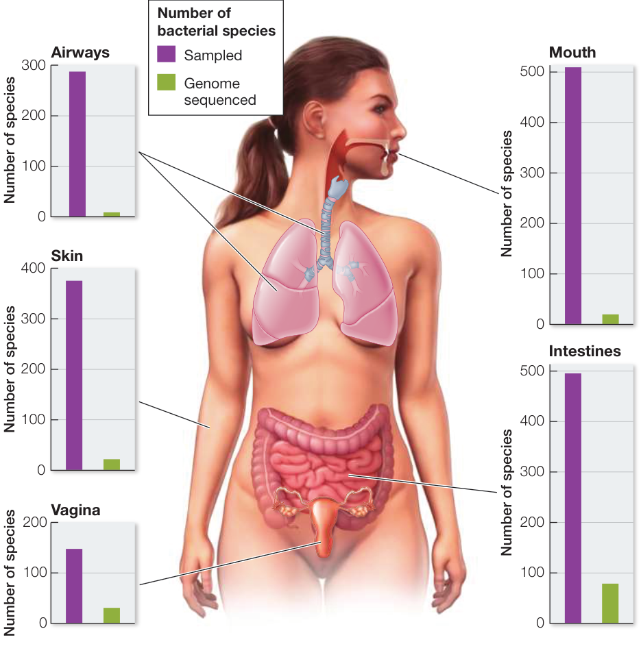
Figure 24.19 The Body’s Microbiome Is Critical to the Maintenance of Health Surveys of the human microbiome have shown that this community includes thousands of diverse bacterial species that are adapted to grow in or on various parts of the body. Although we now know that the composition of this microbiome is closely associated with many aspects of human health, most of the component species are poorly characterized and remain largely unstudied by biologists. What has become clear is that, although the “subcommunities” in different parts of the body share similarities, each is a site-specific assemblage of many distinctive species. (After Y. K. Lee and S. K. Mazmanian. 2010. Science 330: 1768–1773.)
microbial communities affect the expression of our genes and play a critical role in the development and maintenance of a healthy immune system. When our microbiomes contain an appropriate community of beneficial species, our bodies function normally. But these communities are strongly affected by our life experiences, by the food we eat, by the medicines we take, and by our exposure to various environmental toxins. The recent rapid increase in the rate of autoimmune diseases in humans—diseases in which the immune system begins to attack the body—has been linked to the changing diversity and composition of our microbiomes.
Experiment
Original Paper: M. B. Miller and B. L. Bassler. 2001. Quorum sensing in bacteria. Annu Rev Microbiol 55: 165–199.
Bonnie Bassler and her colleagues at Princeton University investigated how Vibrio fischeri bacteria communicate with one another. These bacteria produce bioluminescence when they are present in sufficiently high densities. In a normal V. fischeri bacterium, the following pathway produces bioluminescence when a bacterial colony becomes dense enough to produce sufficient signal:

The fact that the bacteria emit light only when they are present in high densities suggests that the signal is used to communicate among nearby bacteria, alerting one another to their presence. But how can we tell that the signal produced by one bacterium is being received by another?
HYPOTHESIS V. fischeri can transmit the signal molecule from one individual to another, thus communicating about the presence of other nearby bacteria.
METHOD
- Select two mutant strains of V. fischeri incapable of emitting light on their own. In strain A, a mutation for the signal receptor renders the bacteria unable to detect the signal molecule, so they do not bioluminesce:

- Strain B bacteria have a mutated signal gene, so they do not produce a signal molecule, although the receptor and the luciferase gene are normal:

- Incubate samples of V. fischeri on agar petri plates, as follows:
- Plate 1: Strain A only
- Plate 2: Strain B only
- Plate 3: Half of plate with strain A, half with strain B
RESULTS
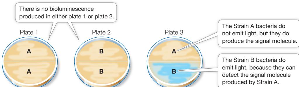
CONCLUSION Vibrio bacteria can detect the signal molecule produced by other nearby bacteria and use this information to detect the presence of other bacteria.
The early acquisition of an appropriate microbiome is critical for lifelong health. Normally, a human infant acquires much of its microbiome at birth, from the microbiome in its mother’s vagina. Other components of the microbiome are also acquired from the mother, especially through breast feeding. Recent studies have shown that babies born by cesarean section, as well as babies that are bottle-fed on artificial milk formula, typically acquire microbes from a wider variety of sources. Many of the bacteria acquired in this way are not well suited for human health. Biologists have discovered that the incidence of many autoimmune diseases is much higher in people who were born by cesarean section and in those who were fed on formula as infants, compared with individuals who were born vaginally and breast-fed as infants. The difference appears to be related to the composition of the individual’s original microbiome.
Humans use some of the metabolic products—especially vitamins and K—produced by the microbiome living in the large intestine. Communities of bacteria line our intestines with a dense biofilm that is in intimate contact with the mucosal lining of the gut. This biofilm facilitates nutrient transfer from the intestine into the body, functioning like a specialized “tissue” that is essential to our health. This biofilm has a complex ecology that scientists have just begun to explore in detail—including the possibility that the species composition of an individual’s gut microbiome may contribute to obesity.
Animals harbor a variety of microbes in their digestive tracts, many of which play important roles in digestion. Cattle depend on prokaryotes to break down plant material. Like most animals, cattle cannot produce cellulase, the enzyme needed to start the digestion of the cellulose that makes up the bulk of their plant food. However, bacteria living in a special section of the gut, called the rumen, produce enough cellulase to process the daily diet for the cattle.
A small minority of bacteria are pathogens
The late nineteenth century was a productive era in the history of medicine—a time when bacteriologists, chemists, and physicians proved that many diseases are caused by microbial agents. During this time, the German physician Robert Koch laid down a set of four rules for establishing that a particular microorganism causes a particular disease:
- The microorganism is always found in individuals with the disease.
- The microorganism can be taken from the host and grown in pure culture.
- A sample of the culture produces the same disease when injected into a new, healthy host.
- The newly infected host yields a new, pure culture of microorganisms identical to those obtained in the second step.
These rules, called Koch’s postulates, were important tools in a time when it was not widely understood that microorganisms cause disease. Although modern medical science has more powerful diagnostic tools, Koch’s postulates remain useful. For example, physicians were taken aback in the 1980s when stomach ulcers—long accepted and treated as the result of excess stomach acid—were shown by Koch’s postulates to be caused by the bacterium Helicobacter pylori (Figure 24.20).
For an organism to be a successful pathogen, it must:
- arrive at the body surface of a potential host;
- enter the host’s body;
- evade the host’s defenses;
- reproduce inside the host; and
- infect a new host.
Failure to complete any of these steps ends the disease cycle of a pathogenic organism. Yet in spite of the many defenses available to potential hosts, some bacteria are very successful pathogens. Pathogenic bacteria are often surprisingly difficult to combat, even with today’s arsenal of antibiotics. One source of this difficulty is their ability to form biofilms.
For the host, the consequences of a bacterial infection depend on several factors. One is the invasiveness of the pathogen: its ability to multiply in the host’s body. Another is its toxigenicity: its ability to produce toxins (chemical substances that are harmful to the host’s tissues). Corynebacterium diphtheriae, the agent that causes diphtheria, has low invasiveness and multiplies only in the throat, but its toxigenicity is so great that the entire body is affected. In contrast, Bacillus anthracis, which causes anthrax, has low toxigenicity but is so invasive that the entire bloodstream ultimately teems with the bacteria.
There are two general types of bacterial toxins: exotoxins and endotoxins. Endotoxins are released when certain bacteria grow or lyse (burst). Endotoxins are lipopolysaccharides (complexes consisting of a polysaccharide and a lipid component) that form part of the outer bacterial membrane. Endotoxins are rarely fatal to the host; they normally cause fever, vomiting, and diarrhea. Among the endotoxin producers are some strains of the proteobacteria Salmonella and Escherichia.
Exotoxins are soluble proteins released by living, multiplying bacteria. They are highly toxic—often fatal—to the host. Human diseases induced by bacterial exotoxins include tetanus (Clostridium tetani), cholera (Vibrio cholerae), and bubonic plague (Yersinia pestis). Anthrax is caused by three exotoxins produced by Bacillus anthracis. Botulism is caused by exotoxins produced by Clostridium botulinum; these exotoxins are among the most poisonous ever discovered. The lethal dose for humans of one exotoxin of C. botulinum is about one-millionth of a gram. Nonetheless, much smaller doses of this exotoxin, marketed under various trade names (e.g., Botox®), are used to treat muscle spasms and for cosmetic purposes (temporary wrinkle reduction in the skin).
© Sage Publications
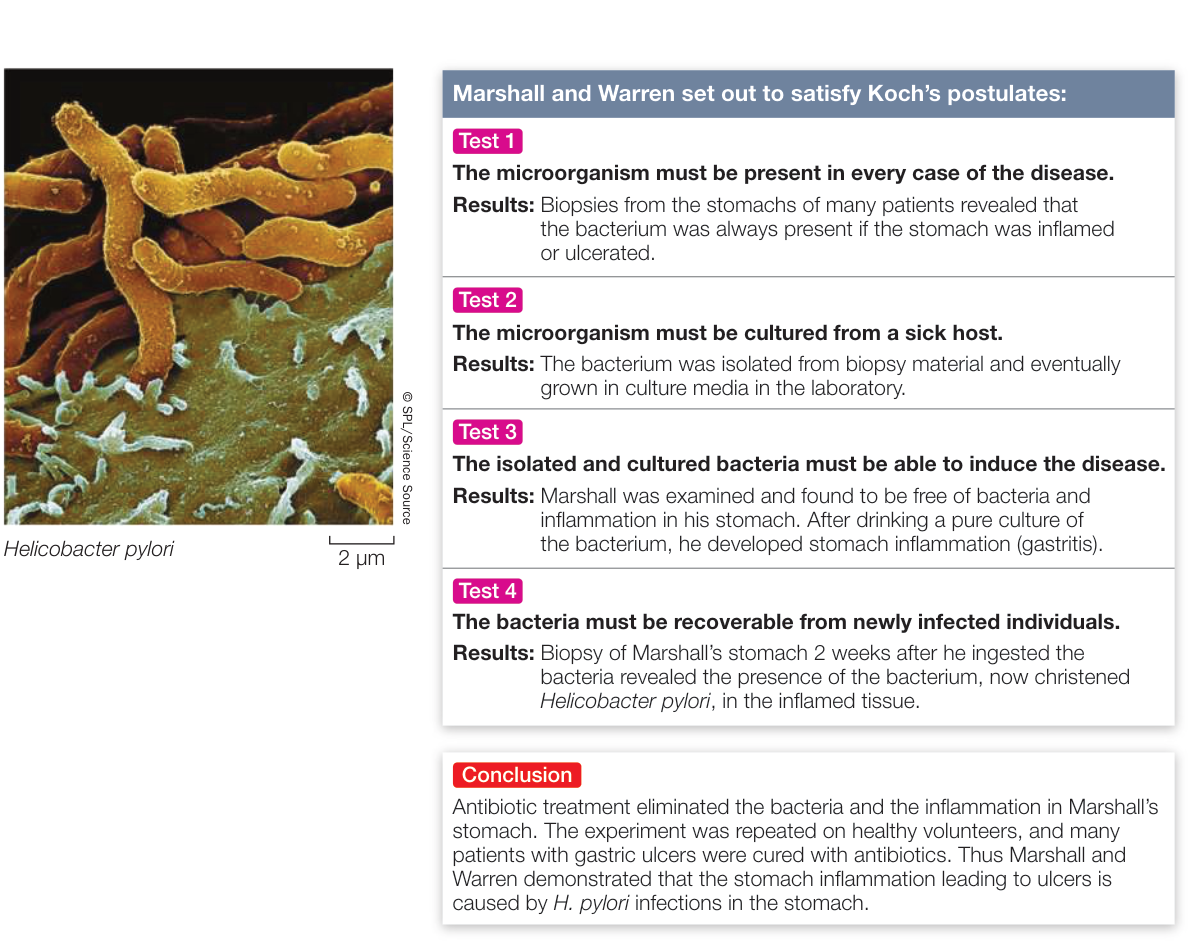
Figure 24.20 Satisfying Koch’s Postulates Robin Warren and Barry Marshall of the University of Western Australia won the 2005 Nobel Prize in Physiology or Medicine for showing that ulcers are caused not by the action of stomach acid but by infection with the bacterium Helicobacter pylori. (After B. J. Marshall and J. R. Warren. 1983. Lancet 321: 1273–1275; B. J. Marshall et al. 1985. Med J Aust 142: 436–439.)
Prokaryotes have amazingly diverse metabolic pathways
Archaea and bacteria outdo the eukaryotes in terms of metabolic diversity. Although they are much more diverse in size and shape, eukaryotes draw on fewer metabolic mechanisms for their energy needs. In fact, much of the eukaryotes’ energy metabolism is carried out in organelles—mitochondria and chloroplasts—that are endosymbiotic descendants of bacteria. The long evolutionary history of prokaryotes, during which they have had time to explore a wide variety of habitats, has led to the extraordinary diversity of their metabolic “lifestyles”—their use or nonuse of oxygen, their energy sources, their sources of carbon atoms, and the materials they release as waste products.
ANAEROBIC VERSUS AEROBIC METABOLISM Some prokaryotes can live only by anaerobic metabolism because oxygen is poisonous to them. These oxygen-sensitive organisms are called obligate anaerobes. By definition, an anaerobe does not use oxygen as an electron acceptor for its respiration. Other prokaryotes, called facultative anaerobes, can alternate between an anaerobic mode of metabolism (such as fermentation) and an aerobic mode (such as cellular respiration) as conditions dictate. Aerotolerant anaerobes cannot conduct cellular respiration, but they are not damaged by oxygen when it is present. At the other extreme from the obligate anaerobes, some prokaryotes are obligate aerobes, unable to survive for extended periods in the absence of oxygen. Obligate aerobes require oxygen for cellular respiration.
NUTRITIONAL CATEGORIES All living organisms face the same nutritional challenges: they must synthesize energy-rich compounds such as ATP to power their life-sustaining metabolic reactions, and they must obtain carbon atoms to build their own organic molecules. Biologists recognize four broad nutritional categories of organisms: photoautotrophs, photoheterotrophs, chemoautotrophs, and chemoheterotrophs. Prokaryotes are represented in all four groups (Table 24.2).
Photoautotrophs perform photosynthesis. They use light as their energy source and carbon dioxide () as their carbon source. The cyanobacteria, like green plants and other photosynthetic eukaryotes, use chlorophyll a as their key photosynthetic pigment and produce oxygen gas () as a by-product of noncyclic electron transport.
There are other photoautotrophs among the bacteria, but these organisms use bacteriochlorophyll as their key photosynthetic pigment, and they do not produce . Instead, some of these photosynthesizers produce particles of pure sulfur, because hydrogen sulfide (), rather than , is their electron donor for photophosphorylation. Many proteobacteria fit into this category. Bacteriochlorophyll molecules absorb light of longer wavelengths than the chlorophyll molecules used by other photosynthesizing organisms. As a result, bacteria using this pigment can grow in water under fairly dense layers of algae, using light of wavelengths that are not absorbed by the algae (Figure 24.21).
Photoheterotrophs use light as their energy source but must obtain their carbon atoms from organic compounds made by other organisms. Their “food” consists of organic compounds such as carbohydrates, fatty acids, and alcohols. For example, compounds released from plant roots (as in rice paddies) or from decomposing photosynthetic bacteria in hot springs are taken up by photoheterotrophs and metabolized to form building blocks for other compounds. Sunlight provides the ATP necessary for metabolism through photophosphorylation.
Chemoautotrophs obtain their energy by oxidizing inorganic substances, and they use some of that energy to fix carbon (convert inorganic carbon into organic carbon-based molecules). Some chemoautotrophs use reactions identical to those of the typical photosynthetic cycle, but others use alternative pathways for carbon fixation. Some bacteria oxidize ammonia or nitrite ions to form nitrate ions. Others oxidize hydrogen gas, hydrogen sulfide, sulfur, and other materials. Many archaea are chemoautotrophs.
Finally, chemoheterotrophs obtain both energy and carbon atoms from one or more complex organic compounds that have been synthesized by other organisms. Most known bacteria and archaea are chemoheterotrophs—as are all animals and fungi and many protists.
Although most chemoheterotrophs rely on the breakdown of organic compounds for energy, some chemoheterotrophic prokaryotes obtain their energy by breaking down inorganic substances. Organisms that obtain energy from oxidizing inorganic substances (both chemoautotrophs as well as some chemoheterotrophs) are also known as lithotrophs (Greek, “rock consumers”).
Prokaryotes play important roles in element cycling
The metabolic diversity of the prokaryotes makes them key players in the cycles that keep elements moving through ecosystems.
Many prokaryotes are decomposers: organisms that metabolize organic compounds in dead organic material and return the products to the environment as inorganic substances. Prokaryotes, along with fungi, return tremendous quantities of carbon to the atmosphere as carbon dioxide, thus carrying out a key step in the carbon cycle.
The key metabolic reactions of many prokaryotes involve nitrogen or sulfur. For example, some bacteria carry out respiratory electron transport without using oxygen as an electron acceptor. These organisms use oxidized inorganic ions such as nitrate, nitrite, or sulfate as electron acceptors.
Table 24.2 How Organisms Obtain Their Energy and Carbon
| Nutritional category | Energy source | Carbon source |
|---|---|---|
| Photoautotrophs (some bacteria, some eukaryotes) | Light | Carbon dioxide |
| Photoheterotrophs (some bacteria) | Light | Organic compounds |
| Chemoautotrophs (some bacteria, many archaea) | Inorganic substances | Carbon dioxide |
| Chemoheterotrophs (some bacteria, some archaea, most eukaryotes) | Usually organic compounds; sometimes inorganic substances | Organic compounds |
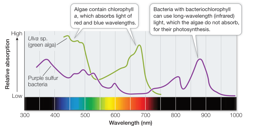
Figure 24.21 Bacteriochlorophyll Absorbs Long-Wavelength Light The green alga Ulva contains chlorophyll, which absorbs no light of wavelengths longer than 750 nanometers (nm). Purple sulfur bacteria, which contain bacteriochlorophyll, can conduct photosynthesis using longer infrared wavelengths. As a result, these bacteria can grow under layers of algae. (After F. Haxo and L. R. Blinks. 1950. J Gen Physiol 33: 389–422; S. Mehrabi et al. 2001. Biomol Eng 18: 49–56.)
Examples include the denitrifiers, which release nitrogen to the atmosphere as nitrogen gas (). These normally aerobic bacteria, mostly species of the genera Bacillus and Pseudomonas, use nitrate () as an electron acceptor in place of oxygen if they are kept under anaerobic conditions:
Denitrifiers play a key role in the cycling of nitrogen through ecosystems. Without denitrifiers, which convert nitrate ions back into nitrogen gas, all forms of nitrogen would leach from the soil and end up in lakes and oceans, making life on land much more difficult.
Nitrogen fixers convert atmospheric nitrogen gas into a chemical form (ammonia) that is usable by the nitrogen fixers themselves as well as by other organisms:
All organisms require nitrogen in order to build proteins, nucleic acids, and other important compounds. Nitrogen fixation is thus vital to life as we know it. This all-important biochemical process is carried out by a wide variety of archaea and bacteria (including cyanobacteria) but by no eukaryotes, so we depend on these prokaryotes for our very existence.
Connect the Concepts For descriptions of the role of nitrogen in plant nutrition and in the global nitrogen cycle, see Key Concepts 34.3 and 56.4.
Ammonia is oxidized to nitrate in soil and in seawater by chemoautotrophic bacteria called nitrifiers. Bacteria of two genera, Nitrosomonas and Nitrosococcus, convert ammonia () to nitrite ions (), and Nitrobacter oxidizes nitrite to nitrate (), the form of nitrogen most easily used by many plants. What do the nitrifiers get out of these reactions? Their metabolism is powered by the energy released by the oxidation of ammonia or nitrite. For example, by passing the electrons from nitrite through an electron transport system, Nitrobacter can make ATP and, using some of this ATP, can also make NADH. With this ATP and NADH, the bacterium can convert and into glucose.
We have already seen the importance of the cyanobacteria in the cycling of oxygen: in ancient times, the oxygen generated by their photosynthesis converted Earth’s atmosphere from an anaerobic to an aerobic environment (see Key Concept 23.2). Other prokaryotes—both bacteria and archaea—contribute to the cycling of sulfur. Deep-sea hydrothermal vent ecosystems depend on chemoautotrophic prokaryotes that are incorporated into large communities of crabs, mollusks, and giant worms, all living at a depth of 2,500 meters—below any hint of sunlight. These bacteria obtain energy by oxidizing hydrogen sulfide and other substances released in the near-boiling water flowing from volcanic vents in the ocean floor.
Recap and Assess
Many prokaryotes are beneficial and even necessary to other forms of life. Most animals, including humans, depend on a complex community of prokaryotes—a microbiome—to maintain health, especially of the immune and digestive systems. Prokaryotes play critical roles in cycling many elements through Earth’s ecosystems, including carbon, nitrogen, and oxygen. Some bacteria are pathogenic—the direct causes of diseases. Finding cures for pathogenic diseases entails understanding how pathogens enter and reproduce in the body.
- How do biofilms form, and why are they of special interest to researchers?
- Why would elimination of all bacteria from a human gut be problematic from a health standpoint?
- Why is nitrogen metabolism in prokaryotes vital to other organisms?
Before moving on to discuss the diversity of eukaryotic life, it is appropriate to consider another category of life that includes some pathogens: the viruses. Although they are not cellular, viruses are numerically among the most abundant forms of life on Earth. Their effects on other organisms are enormous. Where did viruses come from, and how do they fit into the tree of life? Biologists are still working to answer these questions.
Viruses Have Multiple Evolutionary Origins
Learning Objectives
24.4.1 Explain why viruses are difficult to place within the tree of life.
24.4.2 Contrast the “escaped component” and “reduced parasite” hypotheses for the origin of viruses, and name major groups of viruses that best support each hypothesis.
24.4.3 Explain why phage therapy is an active area of research.
Some biologists do not think of viruses as living organisms, primarily because they are not cellular and must depend on cellular organisms for basic life functions such as replication and metabolism. But viruses are derived from the cells of living organisms. They use the same essential forms of genetic information storage and transmission as do cellular organisms. Viruses infect all cellular forms of life—both prokaryotes and eukaryotes. They replicate, mutate, evolve, and interact with other organisms, often causing serious diseases in their hosts. Finally, viruses clearly evolve independently of other organisms, so it is almost impossible not to treat them as a part of life.
Viruses are abundant in many environments. In some freshwater and marine ecosystems, they can occur at densities of up to 10 million viruses per milliliter of water. Biologists estimate that there are approximately individual virus particles on Earth—about 1,000 times the number of cellular organisms on the planet. Viruses have an enormous effect on the ecology of the oceans. Every day, about one-half of the bacteria in the oceans are killed by viruses. Huge marine blooms of bacteria, such as the Vibrio bloom shown in the opening of this chapter, do not last for long because viral blooms soon follow the initial bacterial bloom. As the viruses increase, they begin to kill bacteria faster than the bacteria can reproduce.
Although viruses are found nearly everywhere on Earth and play an important role in many ecosystems, many aspects of their ecology and evolution are still poorly known. For example, several factors make virus phylogeny difficult to resolve. The tiny size of many virus genomes restricts the phylogenetic analyses that can be conducted to relate viruses to cellular organisms. Their rapid mutation rate, which results in rapid evolution of virus genomes, tends to cloud evolutionary relationships over long periods. There are no known fossil viruses (viruses are too small and delicate to fossilize), so the paleontological record offers no clues to virus origins. Finally, viruses are highly diverse (Figure 24.22). Several lines of evidence support the hypothesis that viruses have evolved repeatedly within each of the major groups of life. The difficulty in resolving deep evolutionary relationships of viruses makes a phylogeny-based classification difficult. Instead, viruses are placed in one of several functionally similar groups on the basis of the structure of their genomes (for example, whether the genomes are composed of RNA or DNA, and are double-or single-stranded). Most of these defined groups are not thought to represent monophyletic taxa, however.
Many RNA viruses probably represent escaped genomic components of cellular life
Although viruses are now obligate parasites of cellular species, many viruses may once have been cellular components involved in basic cellular functions—that is, they may be “escaped” components of cellular life that now evolve independently of their hosts.
NEGATIVE-SENSE SINGLE-STRANDED RNA VIRUSES An example of a likely “escaped” biochemical function comes from viruses with genomes composed of single-stranded negative-sense RNA: RNA that is the complement of the mRNA needed for protein translation. Many of these negative-sense single-stranded RNA viruses have only a few genes, including one for an RNA-dependent RNA polymerase that allows them to make mRNA from their negative-sense RNA genome. Modern cellular organisms cannot generate mRNA in this manner (at least in the absence of viral infections), but scientists speculate that single-stranded RNA genomes may have been common in the distant past, before DNA became the primary molecule for genetic information storage.
A self-replicating RNA polymerase gene that began to replicate independently of a cellular genome could conceivably acquire a few additional protein-coding genes through recombination with its host’s DNA. If one or more of these genes were to foster the development of a protein coat, the virus might then survive outside the host and infect new hosts. It is believed that this scenario has been repeated many times independently across the tree of life, given that many of the negative-sense single-stranded RNA viruses that infect organisms from bacteria to humans are not closely related to one another. In other words, negative-sense single-stranded RNA viruses do not represent a distinct taxonomic group, but rather exemplify a particular process of cellular escape that probably happened many different times.
Familiar examples of negative-sense single-stranded RNA viruses include the viruses that cause measles, mumps, rabies, and influenza (see Figure 24.22A).
POSITIVE-SENSE SINGLE-STRANDED RNA VIRUSES The genome of another type of single-stranded RNA virus is composed of positive-sense RNA. Positive-sense genomes are already set for translation; no replication of the genome to form a complement strand is needed before protein translation can take place. Positive-sense single-stranded RNA viruses (see Figure 24.22B) are the most abundant and diverse class of viruses. Most of the viruses that cause diseases in crop plants are members of this group. These viruses kill patches of cells in the leaves or stems of plants, leaving live cells amid a patchwork of discolored dead tissue (giving them the name of mosaic, or mottle, viruses) (Figure 24.23). Other viruses in this group infect bacteria, fungi, and animals. Human diseases caused by positive-sense single-stranded RNA viruses include polio, hepatitis C, and the common cold. As is true of the other functionally defined groups of viruses, these viruses appear to have evolved multiple times across the tree of life from different groups of cellular ancestors.
RNA RETROVIRUSES The RNA retroviruses are best known as the group that includes the human immunodeficiency viruses
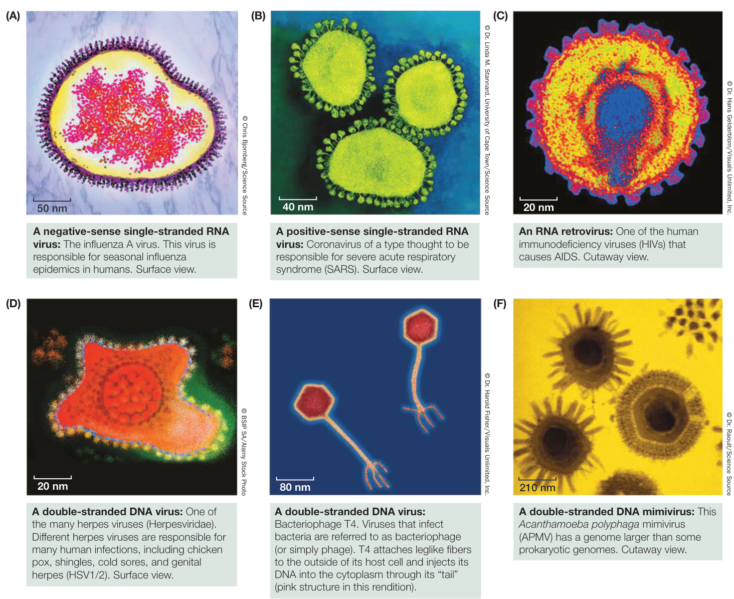
Figure 24.22 Viruses Are Diverse Relatively small genomes and rapid evolutionary rates make it difficult to reconstruct phylogenetic relationships among viruses. Instead, viruses are classified largely by general characteristics of their genomes. The images here are computer artists’ reconstructions based on cryoelectron micrographs.
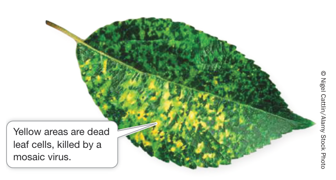
Figure 24.23 Mosaic Viruses Are a Problem for Agriculture Mosaic, or mottle, viruses are the most diverse class of viruses. This leaf is from an apple tree infected with a mosaic virus.
(HIVs; see Figure 24.22C). Like the previous two categories of viruses, RNA retroviruses have genomes composed of single-stranded RNA and probably evolved as escaped cellular components.
Retroviruses are so named because reverse transcription (see Key Concept 14.2) is a necessary component of their reproduction. When the retrovirus enters the nucleus of its vertebrate host, viral reverse transcriptase produces complementary DNA (cDNA) from the viral RNA genome and then replicates the single-stranded cDNA to produce double-stranded DNA. Another virally encoded enzyme called integrase catalyzes the integration of the new piece of double-stranded viral DNA into the host’s genome. The viral genome is then replicated along with the host cell’s DNA. The integrated retroviral DNA is known as a provirus.
Retroviruses are only known to infect vertebrates, although genomic elements that resemble portions of these viruses are a component of the genomes of a wide variety of organisms, including bacteria, plants, and many animals. Several retroviruses are associated with the development of various forms of cancer, as cells infected with these viruses are likely to undergo uncontrolled replication.
As retroviruses become incorporated into the genomes of their hosts, many become nonfunctional copies that are no longer expressed as functional viruses. These sequences may provide a record of ancient viral infections that plagued our ancestors. Humans, for example, carry about 100,000 fragments of endogenous retroviruses in our genome. These fragments make up about 8% of our DNA—a considerably larger fraction of our genome than the fraction that comprises all of our protein-coding genes (about 1.2% of our genome).
DOUBLE-STRANDED RNA VIRUSES Double-stranded RNA viruses may have evolved repeatedly from single-stranded RNA ancestors—or perhaps vice versa. These viruses, which are not closely related to one another, infect organisms from throughout the tree of life. Many plant diseases are caused by double-stranded RNA viruses. Other viruses of this type cause many cases of infant diarrhea in humans.
Some DNA viruses may have evolved from reduced cellular organisms
Another class of viruses has a double-stranded DNA genome (see Figure 24.22D–F). This group is also almost certainly polyphyletic (with many independent origins). Many of the common phage that infect bacteria are double-stranded DNA viruses, as are the viruses that cause smallpox and herpes in humans.
Some biologists think that at least some of the DNA viruses may represent highly reduced parasitic organisms that have lost their cellular structure as well as their ability to survive as free-living species. For example, the mimiviruses, which are some of the largest DNA viruses (see Figure 24.22F), have a genome in excess of a million base pairs of DNA that encode more than 900 proteins. This genome is similar in size to the genomes of many parasitic bacteria and about twice as large as the genome of the smallest bacteria (Figure 24.24). Phylogenetic analyses of these DNA viruses suggest that they have evolved repeatedly from cellular organisms. Furthermore, recombination among different viruses may have allowed the exchange of various genetic modules, further complicating the history and origins of these viruses.
Viruses can be used to fight bacterial infections
Although some viruses cause devastating diseases, other viruses have been used to fight disease. Most bacterial diseases are treated today with antibiotics. Antibiotics were first discovered in the 1930s, but they were not widely used to treat bacterial diseases until the 1940s. So antibiotics were not yet available during World War I, when bacterial infections plagued the battlefields. Battlefield wounds were often infected by bacteria, and in the absence of antibiotics, these infections often led to the loss of limbs and lives. While trying to find a way to combat this problem, a physician named Felix d’Herelle discovered the first evidence of viruses that attack bacteria. He named these viruses bacteriophage, or “eaters of bacteria.” Herelle extracted bacteriophage from the stool of infected patients. He then used these extracts to treat patients with deadly bacterial infections, including dysentery, cholera, and bubonic plague. This practice became known as phage therapy. After the war, phage therapy was widely used among the general public to treat bacterial infections of the skin and intestines.
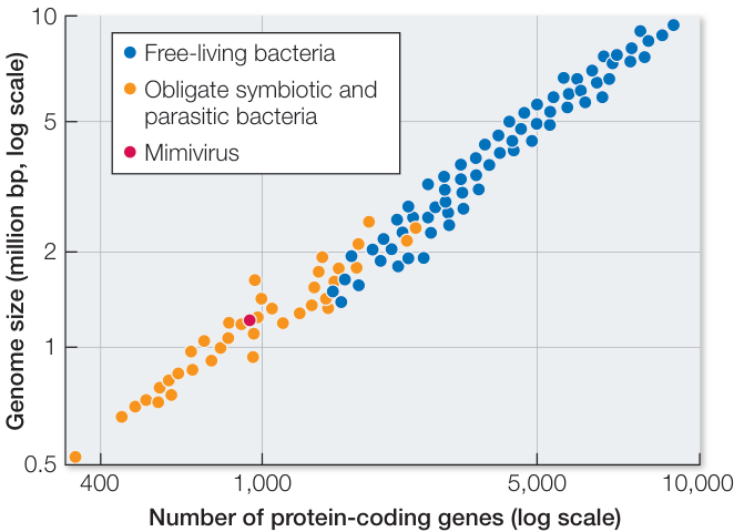
Figure 24.24 Mimiviruses Have Genomes Similar in Size to Those of Many Parasitic Bacteria The genome of Acanthamoeba polyphaga mimivirus contains 1,181,404 base pairs and encompasses 911 protein-coding genes. This observation is consistent with the hypothesis that this virus evolved from a parasitic bacterium. (After S. J. Giovannoni et al. 2005. Science 309: 1242–1245.) Q: Why do parasitic bacteria generally have smaller genomes and fewer protein-coding genes than do free-living bacteria?
Phage therapy was mostly replaced by the use of antibiotics in the 1930s and 1940s as physicians grew concerned about treating patients with live viruses. Phage therapy continued to be used in the Soviet Union but largely disappeared from western medical practice. Today, however, many antibiotics are losing their effectiveness as bacterial pathogens evolve resistance to these drugs. Phage therapy is once again an active area of research, and it is likely that bacteriophage will become increasingly important as weapons against bacterial diseases. One advantage that bacteriophage may have over antibiotics is that, like bacteria, bacteriophage can evolve. As bacteria evolve resistance to a strain of bacteriophage, biologists can select for new strains of bacteriophage that retain their effectiveness against the pathogens. In this way, biologists are using their understanding of evolution to combat the problem of antibiotic-resistant bacteria.
It appears that the enormous diversity of viruses is, at least in part, a result of their multiple origins from many different cellular organisms. It may be best to view viruses as spin-offs from the various branches on the tree of life—sometimes evolving independently of cellular genomes, sometimes recombining with them. One way to think of viruses is as the “bark” on the tree of life: certainly an important component all across the tree, but not quite like the main branches.
Recap and Assess
Viruses are highly diverse cellular forms of life that appear to have evolved independently from many different cellular organisms. Major groups of viruses are characterized by the nature of their genomes. Some viruses appear to have evolved from escaped components of cellular organisms, whereas other viruses may have evolved from parasitic cellular ancestors. Although some viruses are responsible for serious diseases, other viruses can be used to combat bacterial infections.
- How does the size of viral genomes limit phylogenetic analyses of viruses?
- What are the two main hypotheses of viral origins?
- How can viruses be used to treat some human diseases?
Investigating Life
Q&A How and why do bacteria communicate with one another to assess their density?
As we explored in Investigating Life: How Do Bacteria Communicate with One Another?, Vibrio bacteria send a chemical signal that is received by other bacteria. This signal is needed to produce bioluminescence. As Vibrio populations increase in density, the bioluminescence becomes strong enough to be visible to humans and other animals. But how is bioluminescence an advantage to the bacteria? What is its function?
Although marine Vibrio are able to live independently, they truly thrive inside the guts of fishes and other marine animals. Inside a fish, Vibrio cells attach themselves to food particles, including phytoplankton, and are often expelled into the ocean as waste. How can they get back into their preferred environment? The bioluminescent glow produced by a dense colony of free-living Vibrio growing on phytoplankton attracts fishes, which consume the phytoplankton and thus ingest the bacteria—which gets the bacteria into a new host fish.
The bioluminescent Vibrio described in Investigating Life: How Do Bacteria Communicate with One Another?, V. fischeri, have evolved a symbiotic relationship with the Hawaiian bobtail squid (Euprymna scolopes). Chemicals produced by a developing squid embryo specifically “recruit” V. fischeri from the surrounding seawater. The bacteria then preferentially migrate to specific tissues that develop into a bioluminescent “light organ” in the belly of the adult squid (Figure 24.25). The tiny (about 3 cm long) adult squid feed while floating near the sea surface at night. The soft glow produced by the bioluminescent bacteria mimics the moonlight above, so the squid are less visible to potential predators coming at them from below.
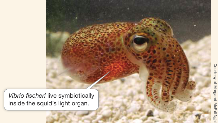
Figure 24.25 Bioluminescent Bacterial Symbionts Vibrio bacteria within the light organ emit bioluminescence downward as the Hawaiian bobtail squid floats near the ocean surface to feed. At night, this allows the squid to blend in with the moonlight or starlight rather than becoming a target for a predator from below. Figure 24.25 Bioluminescent Bacterial Symbionts Vibrio bacteria within the light organ emit bioluminescence downward as the Hawaiian bobtail squid floats near the ocean surface to feed. At night, this allows the squid to blend in with the moonlight or starlight rather than becoming a target for a predator from below.
Future directions
Research that helps biologists understand how bacteria communicate, and allows us to disrupt or change communication signals among bacteria, is critical for developing treatment strategies against persistent bacterial infections. In the case of health-threatening bacteria that produce problematic biofilms, researchers hope to find ways to block the quorum-sensing signals that lead to the production of the matrix polysaccharides, thus preventing pathogenic biofilms from forming.


Steven Miller, Naval Research Laboratory, Monterey, CA
You should be able to relate each summary to the adjacent figures. If you go to this visual summary in Achieve, you can follow links to figures, animations, activities, and simulations that will help you consolidate the material.
Visual Summary
Bacteria and Archaea Represent the First Split in the Tree of Life
- The earliest split in the tree of life is between Bacteria and Archaea. Eukarya branched off from Archaea, having evolved specializations (such as a nucleus and organelles, some of which resulted from endosymbiosis with bacteria) that differentiate the eukaryotes from the prokaryotes.
- Bacteria and archaea can be differentiated by their cell walls. The cells walls of bacteria contain peptidoglycan, which is a target for the action of many antibiotics.
- Although lateral gene transfer sometimes complicates our understanding of prokaryotic evolutionary history, prokaryotes are now principally classified by the nucleotide sequences of their genomes.
Questions
- What evidence links the origin of eukaryotes more strongly with archaea than with bacteria?
- How is peptidoglycan relevant to the function of antibiotics?
- How does lateral gene transfer complicate the study of bacterial phylogeny?

Figure 24.4
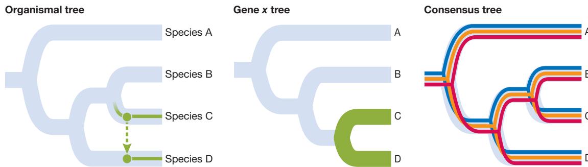
Prokaryotic Diversity Reflects the Ancient Origins of Life
- Prokaryotes are the most numerous organisms on Earth, but only a small fraction of prokaryotic diversity has been characterized to date.
- Life on Earth likely originated in a high-temperature environment.
- Biotic communities depend on the actions of bacteria, which provide such important ecological services such as nitrogen fixation and oxygen production through photosynthesis.
- Archaea live in highly diverse environments and include many extremophiles and methanogens.
Questions
- What lines of evidence support the origin of life in a hot environment?
- How have cyanobacteria been critical in changing Earth’s environment over the past 3 billion years?
- What genes present in Asgard archaea were formerly thought to be found only in eukaryotic genomes?
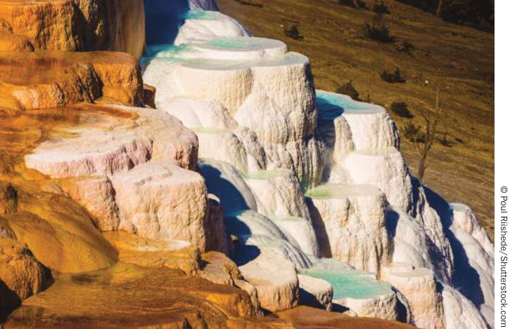
Figure 24.16
Ecological Communities Depend on Prokaryotes
- ■ Microbiomes are communities of prokaryotes that live in and on the bodies of multicellular organisms and are often important to the health of the hosts. Bacteria use processes such as quorum sensing to recruit members to their communities.
- ■ Koch’s postulates establish the criteria by which an organism may be classified as a pathogen. Relatively few bacteria—and no archaea—are known to be pathogens.
- ■ Prokaryotic metabolism is diverse. Prokaryotes fall into four broad nutritional categories: photoautotrophs, photoheterotrophs, chemoautotrophs, and chemoheterotrophs.
- ■ Prokaryotes play key roles in the cycling of elements such as nitrogen, oxygen, sulfur, and carbon.
Questions
- How are biofilms important to human health?
- When and where do newborn humans typically acquire their microbiomes?
- Which group—bacteria, archaea, or eukaryotes—contains no organisms that use light as an energy source?
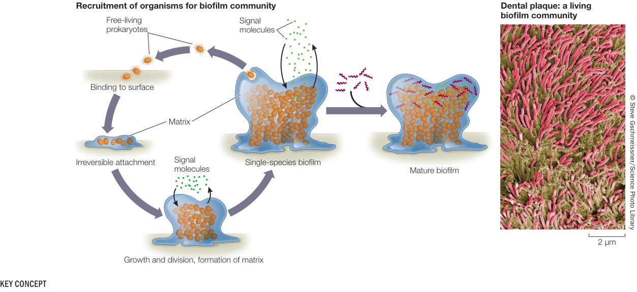
Figure 24.18
Viruses Have Multiple Evolutionary Origins
- ■ Viruses have evolved many times from many different groups of cellular organisms. They are placed in groups according to the structure of their genomes, but these groups are not thought to represent monophyletic taxa.
- ■ Viruses manipulate host genetic mechanisms in various ways to replicate themselves. Retroviruses, for example, use reverse transcriptase to produce complementary DNA from their viral RNA genome and then insert their genetic material into the host’s genome.
- ■ Some viruses cause serious diseases in cellular organisms, but bacteriophage can be used to treat bacterial infections, in a process known as phage therapy.
Questions
- What molecules encode viral genomes?
- How did endogenous retroviruses end up in vertebrate genomes?
- Why did phage therapy fall out of use, and why is it once again of interest?
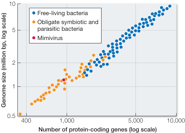
Figure 24.24