Chapter 29Animal Origins and the Evolution of Body Plans
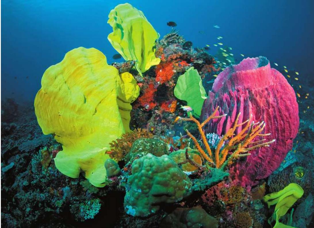
© readbrickstock.com/Alany/StockPhoto
Key Concepts
29.1 Diverse Body Plans Allow Animals to Move and Feed in Many Ways
29.2 Animal Life Cycles Involve Trade-Offs
29.3 The Root of the Animal Tree Provides Clues to Early Animal Diversification
Many of the major animal lineages, including sponges, cnidarians, and chordates, can be seen together in this coral reef.
Investigating Life: A Mysterious Animal Discovered in an Aquarium
In 1883 the zoologist Franz Schulze noticed something unusual in his Austrian laboratory: transparent, flattened organisms were crawling on the sides of his saltwater aquarium. These organisms, which Schulze had collected accidentally along with the sponges that were his primary interest, appeared to be animals, but they were unlike any animals previously described—especially since they continually changed shape as they moved.
Further examination revealed that the new organisms were indeed animals. Structurally, however, they were among the simplest animals that Schulze—or anyone else—had ever observed, being made up of only four types of cells. He named the new species Trichoplax adhaerens, which means “sticky hairy plate.” For decades, however, most biologists dismissed Schulze’s findings, insisting that the transparent organisms must be larval forms of other, well-known animals.
In the 1960s more detailed studies confirmed the distinctive nature of Trichoplax. Even then, this odd animal continued to be known almost exclusively from aquariums. Finally, in the early 2000s biologists located natural populations of T. adhaerens growing on hard surfaces in tropical and subtropical coastal regions. A few additional closely related species have been discovered (although most have not yet been formally named). Collectively, these species are known as placozoans (Greek, “flat animals”).
The more biologists have studied Trichoplax, the odder this animal appears. It has the smallest genome of any animal studied to date. The mature stages lack body symmetry and have no mouth, gut, or nervous system. Is Trichoplax a relict representative of a group of animals that appeared early in animal evolution?
Which groups of organisms are at the root of the animal tree has been a subject of considerable investigation and debate. Several possible hypotheses of relationships have been explored with genomic analyses. The structural simplicity of Trichoplax is now considered by most biologists to be an evolutionary reversal from a more complex body form. Most genomic studies point to other groups of animals as forming the earliest split with the remaining species.
This chapter explores the earliest branches on the animal tree and shows how a few fundamental “body plans” have been modified to yield the remarkable variety of animal forms described in this and the following two chapters.
Q&A Which animal groups are involved in the earliest split in the animal tree? (Find the answer on page 665.)
Diverse Body Plans Allow Animals to Move and Feed in Many Ways
Diverse Body Plans Allow Animals to Move and Feed in Many Ways
Learning Objectives
29.1.1 Explain how differences in body symmetry, body cavities, segmentation, appendages, and nervous systems are functionally related to movement and feeding in animals.
29.1.2 Describe two important functions of nervous systems that allow many animals to have better control over mobility in the water, land, and air.
How do we recognize an organism as an animal? The answer may seem obvious for many familiar animals, but less so for groups such as sponges, which were once thought to be plants.
Some of the general characteristics we associate with animals include:
- Multicellularity. In contrast to the bacteria, archaea, and most protists (see Chapters 24 and 25), all animals are multicellular. Animal life cycles feature complex patterns of development from a single-celled zygote into a multicellular adult.
- Heterotrophic metabolism. In contrast to most plants, all animals are heterotrophs. Animals are able to synthesize very few organic molecules from inorganic chemicals, so they must obtain the necessary organic molecules from their environment.
- Internal digestion. Although the fungi are also heterotrophs (see Chapter 28), animals and fungi digest their food differently. Whereas fungi digest food outside their bodies, most animals use internal processes to break down materials from their environment into the organic molecules they need. Most animals
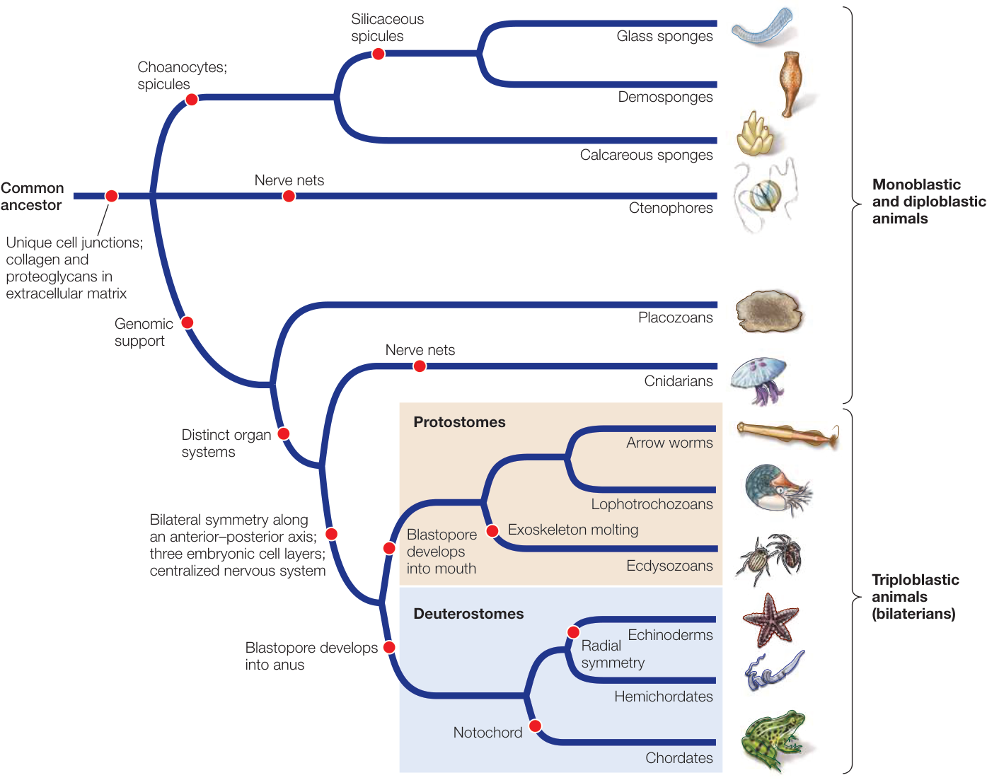
Figure 29.1 A Phylogenetic Tree of the Animals This tree presents the best-supported current hypotheses of the evolutionary relationships among major groups of animals. The traits highlighted by red circles are explained in this and the following two chapters.
Q: Based on this tree, which of the depicted traits evolved multiple times among animals, and in which lineages?
ingest food into an internal gut that is continuous with the outside environment and in which digestion takes place.
- Movement and nervous systems. In contrast to the majority of plants and fungi, most animals can move their bodies. This movement is often coordinated through a well-developed nervous system, which also typically functions as a sensory system. Animals must move to find food or bring food to them. Muscle tissue and nervous systems are unique to animals, and many animal body plans are specialized for movement and detection of prey.
Although these general features help us recognize animals, none is diagnostic for all animals. Some animals do not move, at least during certain life stages, and some plants and fungi do have limited movement. Some animals lack a nervous system, and the diffuse nerve nets (which lack a coordinating brain) of ctenophores and cnidarians are distinct and independently evolved from the central nervous systems (which have a coordinating brain) of bilaterians. Some animals lack a gut, and many multicellular organisms are not animals. So on what basis do we group all animals together in a single clade?
Animal monophyly is supported by gene sequences and cellular morphology
The most convincing evidence that all the organisms considered to be animals share a common ancestor comes from phylogenetic analyses of their gene sequences. Many complete animal genomes are now available, and more are being sequenced each year. Analyses of these genomes and of many individual gene sequences have shown that the animals are indeed monophyletic. The best-supported phylogenetic tree for the major animal groups is shown in Figure 29.1.
Surprisingly few morphological features are shared across all animal species. The morphological synapomorphies (shared, derived traits) that are present are evident primarily at the cellular level. They include a common set of extracellular matrix molecules, including collagen and proteoglycans, and several unique features of the junctions between animal cells. Although some animals in a few groups lack one of these cellular characteristics, it is believed that these traits were possessed by the ancestor of all animals and were subsequently lost in those groups.
Similarities in the organization and function of Hox and other developmental genes provide additional evidence of developmental mechanisms shared by a common animal ancestor. The Hox genes specify body pattern and axis formation, leading to developmental similarities across animals.
Connect the Concepts Hox genes and their role in animal development and evolution are discussed in Chapter 21.
The common ancestor of animals may have been a colonial flagellated protist similar to existing colonial choanoflagellates (Figure 29.2A), which have similarities to the multicellular sponges (Figure 29.2B). Why did these early animal ancestors begin to form multicellular colonies? One hypothesis is that multicellular colonies are more efficient than are single cells at capturing their prey. Experiments with living species of choanoflagellates show that they spontaneously form multicellular colonies in response to signaling compounds that are found on certain species of planktonic bacteria they eat (Figure 29.3).
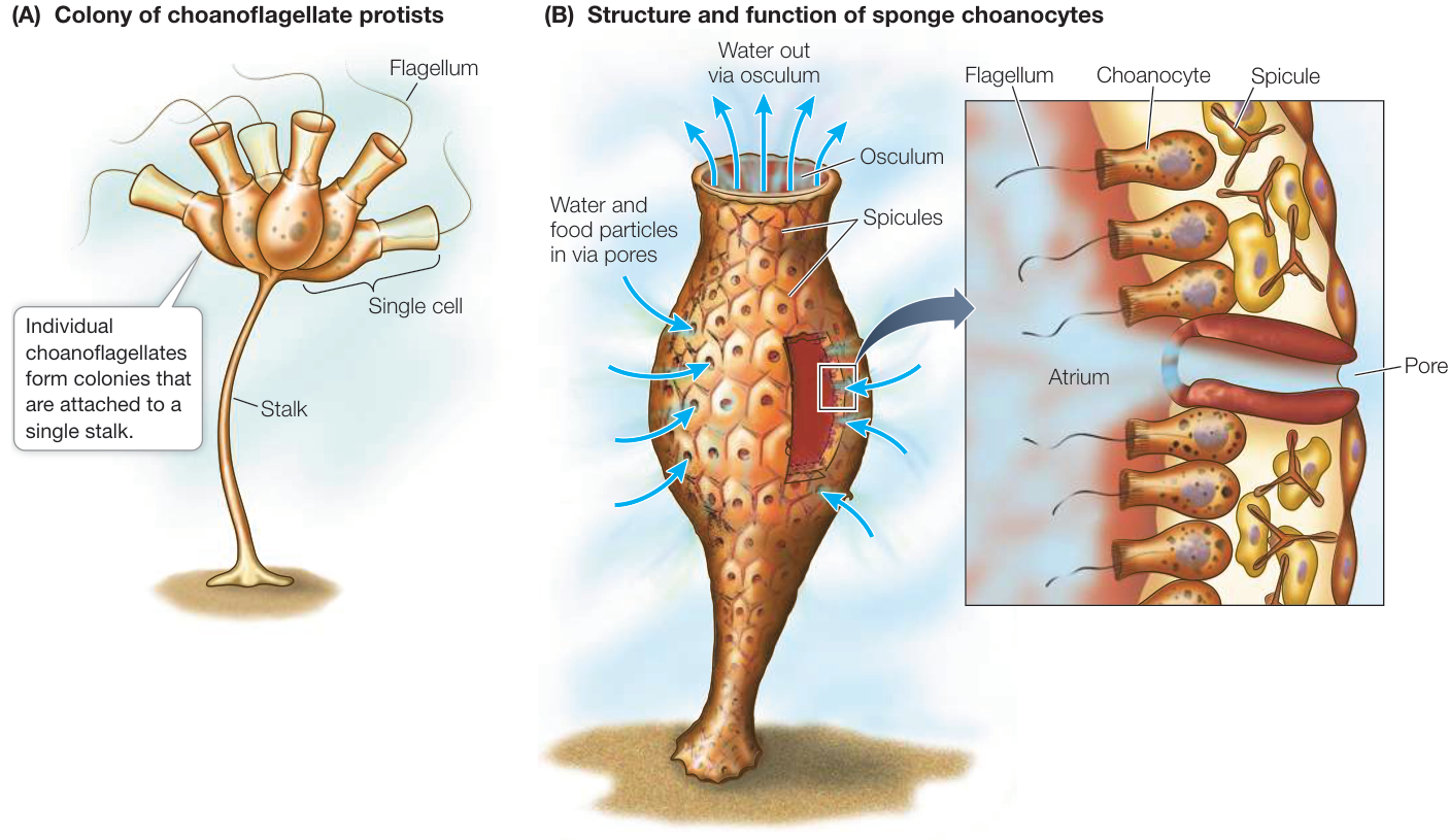
Figure 29.2 Choanocytes in Sponges Resemble Choanoflagellate Protists (A) The similarity of choanoflagellate protist colonies to sponge choanocytes supports an evolutionary link between this protist lineage and the animals. (B) A sponge moves food-containing water through its body by beating the flagella of its choanocytes. Water enters the sponge through small pores and passes into water canals or an open atrium, where the choanocytes capture food particles from the water. The spicules are supportive, skeletal structures.
Once multicellular colonies were formed in early animal lineages, it is likely that certain cells in the colony began to be specialized—some for movement, others for nutrition, others for reproduction, and so on. Once this functional specialization had begun, cells could continue to differentiate. Coordination among groups of cells could have improved by means of specific regulatory and signaling molecules that guided differentiation and migration of cells in developing embryos. Such coordinated groups of cells eventually evolved into the larger and more complex organisms that we call animals.
Nearly 80% of the 1.8 million named species of living organisms are animals, and millions of additional animal species await discovery. Evidence about the evolutionary relationships among animal groups can be found in fossils, in patterns of embryonic development, in the morphology and physiology of living animals, in the structure of animal proteins, and in gene sequences. Increasingly, studies of higher-level relationships have come to depend on genomic sequence comparisons, as genomes are ultimately the source of all inherited trait information.
A few basic developmental patterns differentiate major animal groups
The early cell divisions of an embryo are known as cleavage. Several different patterns of cleavage exist among animals. Although these patterns can be useful for characterizing major animal groups, genomic analyses have shown that many changes have occurred in cleavage patterns throughout animal evolution.
Connect the Concepts The various patterns of embryonic cleavage and animal development are described in more detail in Key Concept 42.1.
Cleavage patterns are influenced by the configuration of the yolk, the acellular nutritive material that nourishes the growing embryo. The eggs of many animal groups contain a small amount of yolk that is evenly distributed throughout the egg cytoplasm. In some of these groups, the zygote and its descendant cells divide completely and evenly in a pattern known as radial cleavage. Radial cleavage is thought to be the ancestral condition for the bilaterian animals, as it is widely distributed among the major lineages. Spiral cleavage—a complicated permutation of radial cleavage—is found among many lophotrochozoans (a group that includes earthworms and clams). Lophotrochozoans with spiral cleavage are thus sometimes known as spiralians. The early branches of the ecdysozoans (molting animals, such as insects and nematodes) have radial cleavage, but most ecdysozoans have an idiosyncratic cleavage pattern that is neither radial nor spiral in organization.
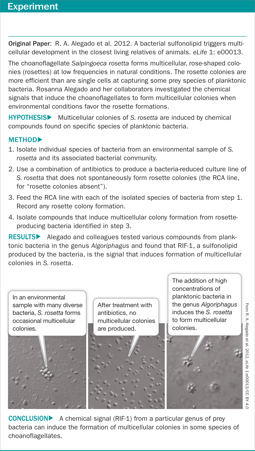
Figure 29.3 What Induces Choanoflagellates to Form Multicellular Colonies? Figure 29.3 What Induces Choanoflagellates to Form Multicellular Colonies?
Distinct layers of cells form during the early development of most animals. These cell layers differentiate into specific organs and organ systems as development continues. The embryos of diploblastic animals have two cell layers: an outer ectoderm and an inner endoderm. Embryos of triploblastic animals have, in addition to ectoderm and endoderm, a third distinct cell layer, mesoderm, which lies between the ectoderm and the endoderm. The existence of three cell layers in embryos is a synapomorphy of the triploblastic animals (which form a clade), whereas the diploblastic animals (ctenophores, placozoans, and cnidarians, which are not a clade) exhibit the ancestral condition (see Figure 29.1). Some biologists also consider sponges to be diploblastic, but since they do not have clearly differentiated tissue types or embryonic cell layers, they are usually considered to be monoblastic.
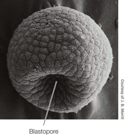
Figure 29.4 Gastrulation Illuminates Evolutionary Relationships The blastopore is clear in this scanning electron micrograph of a sea urchin gastrula. Because sea urchins (echinoderms) are deuterostomes, this blastopore will eventually become the anal end of the animal’s gut.
During early development in many animals, in a process known as gastrulation, a hollow ball one cell thick indents to form a cup-shaped structure. The opening of the cavity formed by this indentation is called the blastopore (Figure 29.4). The overall pattern of gastrulation immediately after formation of the blastopore divides the triploblastic animals into two major groups:
- In the protostomes (Greek, “mouth first”), the mouth arises from the blastopore, and the anus forms later.
- In the deuterostomes (“mouth second”), the blastopore becomes the anus, and the mouth forms later.
Although the developmental patterns of animals are more varied than suggested by this simple dichotomy, sequencing data indicate that the protostomes and deuterostomes represent distinct animal clades. Together these two groups are known as the bilaterians (named for their usual bilateral symmetry), and they account for the vast majority of animal species.
The general structure of an animal, the arrangement of its organ systems, and the integrated functioning of its parts are referred to as its body plan. As we discussed in Chapter 21, the regulatory and signaling genes that govern many major aspects of development (including body symmetry, body cavities, segmentation, and appendages) are widely shared among the different animal groups. Although the myriad animal body plans cover a wide range of morphologies, they can be seen as variations on five key features:
- The symmetry of the body
- The structure of the body cavity
- The segmentation of the body
- The existence and location of external appendages that are used for sensing, feeding, locomotion, mating, and other functions
- The development of the nervous system
Each of these features affects how an animal interacts with its environment.
Most animals are symmetrical
The overall shape of an animal can be described by its symmetry. An animal is said to be symmetrical if it can be divided along at least one plane into similar halves. Animals that have no plane of symmetry are said to be asymmetrical. Placozoans and many sponges are asymmetrical, but most other animals have some kind of symmetry, which is governed by the expression of regulatory genes during development.
In animals with radial symmetry, body parts are arranged around one main axis at the body’s center (Figure 29.5A). Ctenophores (comb jellies) and many cnidarians (such as sea anemones and jellyfishes) are radially symmetrical, as are adult echinoderms. A perfectly radially symmetrical animal can be divided into similar halves by any plane that contains the main axis. However, most radially symmetrical animals—including the adults of echinoderms such as sea stars—are slightly modified, so that only some planes can divide them into identical halves. Some radially symmetrical animals are sessile (they remain fixed in one place) or drift with water currents. Others move about slowly but can move equally well in any direction.
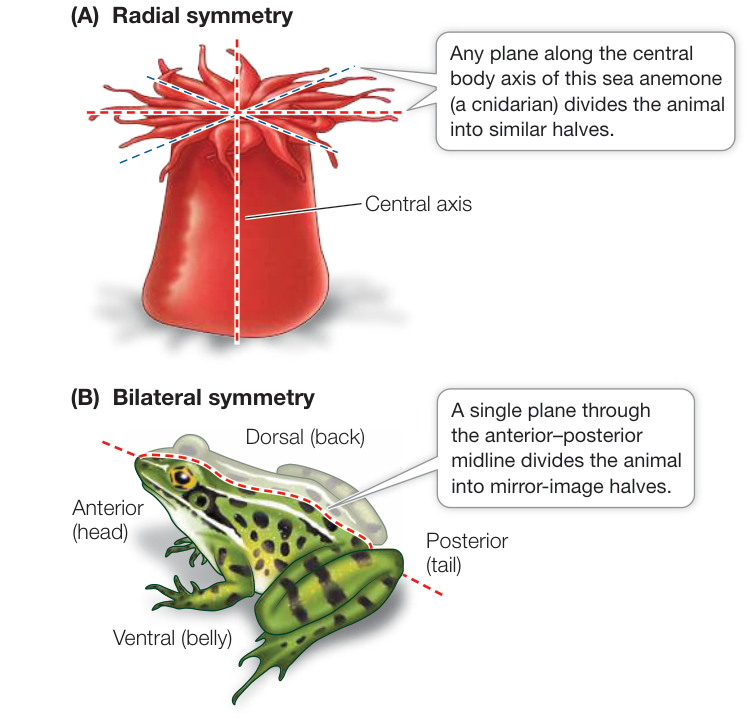
Figure 29.5 Body Symmetry Most animals are either (A) radially symmetrical or (B) bilaterally symmetrical.
Bilateral symmetry is characteristic of animals that have a distinct front end, which typically precedes the rest of the body as the animal moves. A bilaterally symmetrical animal can be divided into mirror-image (left and right) halves by a single plane that passes through the midline of its body. This plane runs from the front, or anterior, end of the body, to the rear, or posterior, end (Figure 29.5B). A plane at right angles to the midline divides the body into two dissimilar sides. The back of a bilaterally symmetrical animal is its dorsal surface; the underside is its ventral surface.
Bilateral symmetry is strongly correlated with cephalization (Greek kephalos, “head”), which is the concentration of sensory organs and nervous tissues at the anterior end of the animal. Cephalization has been favored by natural selection because the anterior end of a bilaterally symmetrical animal typically encounters new environments first.
The structure of the body cavity influences movement
The body plans of triploblastic animals can be divided into three types based on the presence and structure of an internal, fluid-filled body cavity:
- Acoelomate animals such as flatworms lack an enclosed, fluid-filled body cavity. Instead, the space between the gut (derived from endoderm) and the muscular body wall (derived from mesoderm) is filled with masses of cells called mesenchyme (Figure 29.6A). These animals typically move by beating cilia.
- Pseudocoelomate animals have a body cavity called a pseudocoel, a fluid-filled space in which many of the internal organs are suspended. A pseudocoel is enclosed by muscles (mesoderm) only on its outside; there is no inner layer of mesoderm surrounding the internal organs (Figure 29.6B).
- Coelomate animals have a body cavity, the coelom, that develops within the mesoderm. It is lined with a layer of muscular tissue called the peritoneum, which also surrounds the internal organs. The coelom is thus enclosed on both the inside and the outside by mesoderm (Figure 29.6C).
The structure of an animal’s body cavity strongly influences the ways in which it can move. The body cavities of many animals function as hydrostatic skeletons. Fluids are relatively incompressible, so when the muscles surrounding a fluid-filled body cavity contract, fluids shift to another part of the cavity. If the body tissues around the cavity are flexible, fluids squeezed out of one region can cause another region to expand. The moving fluids can thus move specific body parts. (You can see how a hydrostatic skeleton works by watching a snail emerge from its shell.) A coelomate animal has better control over the movement of the fluids in its body cavity than a pseudocoelomate animal does. An animal that has longitudinal muscles (running along the length of the body) as well as circular muscles (encircling the body cavity) has even greater control over its movement.
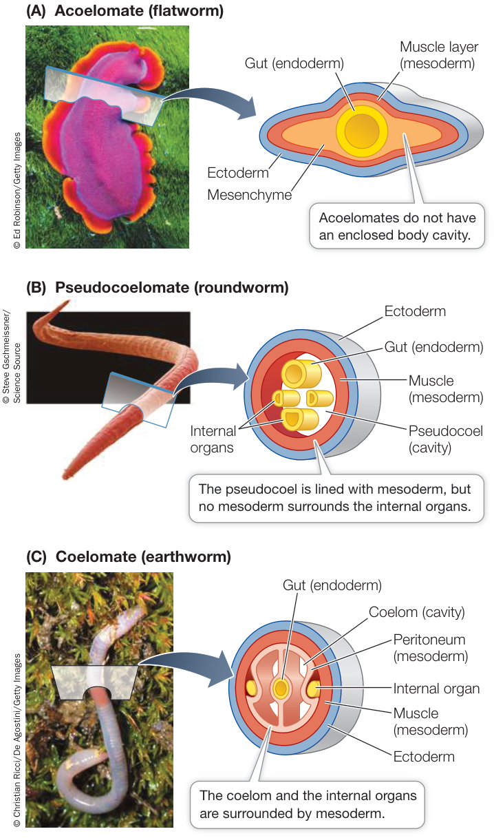
Figure 29.6 Animal Body Cavities (A) Acoelomates do not have an enclosed body cavity. (B) Pseudocoelomates have a body cavity enclosed by mesoderm only on its outside. (C) Coelomates have a body cavity that is enclosed by mesoderm on both its inside and its outside. Activity 29.1 Animal Body Cavities
In terrestrial environments, the hydrostatic function of fluid-filled body cavities applies mostly to relatively small, soft-bodied organisms. Most larger animals (as well as many smaller ones) have hard skeletons that provide protection and facilitate movement. Muscles are attached to those firm structures, which may be inside the animal or on its outer surface (in the form of a shell or cuticle).
Segmentation facilitates specialization
Segmentation—the division of the body into segments—is seen in many animal groups. Segmentation facilitates specialization of different body regions. It also allows an animal to alter the shape of its body in complex ways and to control its movements precisely. If an animal’s body is segmented, muscles in each individual segment can change the shape of that segment independently of the others. In only a few segmented animals is the body cavity separated into discrete compartments, but even partly separated compartments allow better control of movement. Segmentation occurs in several groups of protostomes and deuterostomes.
In some animals, such as annelids (earthworms and their relatives), similar body segments are repeated many times (Figure 29.7A). In other animals, including most arthropods, segments differ strikingly from one another (Figure 29.7B). The dramatic evolutionary radiation of the arthropods (including the insects, spiders, centipedes, and crustaceans) was based on changes in a segmented body plan that features muscles attached to the inner surface of an external skeleton, including a variety of external appendages that move these animals. In some animals, distinct body segments are not apparent externally—for example, the segmented vertebrae of vertebrates, including humans. Nonetheless, muscular segmentation is clearly visible in humans with well-defined, muscular bodies (Figure 29.7C).
Appendages have many uses
Getting around under their own power allows animals to obtain food, to avoid predators, and to find mates. Even some sedentary species, such as sea anemones, have larval stages that use cilia to swim, thus increasing the animal’s chances of finding a suitable habitat.
Appendages that project externally from the body greatly enhance an animal’s ability to move around. Many echinoderms, including sea urchins and sea stars, have large numbers of tube feet that allow them to move slowly across the substrate. Animals whose appendages have become modified into limbs are capable of highly controlled, more rapid movement. The presence of jointed limbs has been a prominent factor in the evolutionary success of the arthropods and the vertebrates. In four independent instances—among the arthropod insects and among the vertebrate pterosaurs, birds, and bats—body plans emerged in which limbs were modified into wings, allowing these animals to use powered flight.
Appendages also include many structures that are not used for locomotion. Many animals have antennae, which are specialized appendages used for sensing the environment. Other appendages (such as claws and the mouthparts of many arthropods) are adaptations for capturing prey or chewing food. In some species, appendages are used for reproductive purposes, such as sperm transfer or egg incubation.
Nervous systems coordinate movement and allow sensory processing
The bilaterian animals have a well-coordinated central nervous system. More diffuse nervous systems, called nerve nets, are present in some other animals, such as ctenophores and cnidarians.
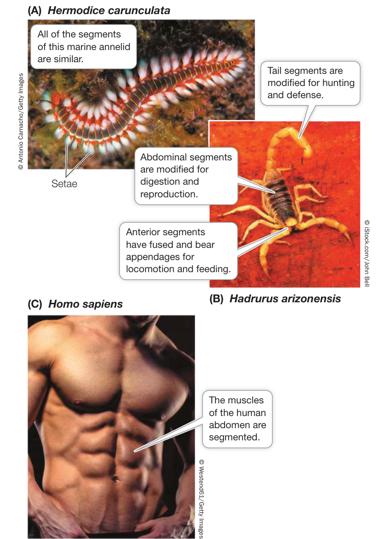
Figure 29.7 Segmentation (A) All of the segments of this marine fireworm, an annelid, are similar. Its appendages are tipped with bristles (setae) that are used for locomotion and (in this species) for protection—the setae contain a noxious toxin. (B) Segmentation allows the evolution of differentiation among the segments. The segments of this scorpion, an arthropod, differ in their form, their function, and the appendages they bear. (C) Muscular segmentation is clearly visible in the abdomen of this body builder.
Nervous systems appear to be completely absent in a few animal groups, such as sponges and placozoans.
The central nervous system of bilaterians coordinates the actions of muscles, which allows coordinated movement of appendages and body parts. This coordination of muscles permits highly effective and efficient movement on land, in water, or through the air. The central nervous system is also essential for processing the sensory information gathered from a wide variety of sensory systems. Many animals have sensory systems for detecting light, for forming images of their environment (sight), for mechanical touch, for detecting movement, for detecting sounds (hearing), for detecting electrical fields, and for chemical detection (e.g., taste and smell). These sensory systems allow animals to find food, and the ability of animals to move allows them to capture or collect food from their environment. These same abilities also allow most animals to move to avoid potential predators or to search for suitable mates. Most animals can also assess the suitability of different environments and move appropriately in response to that information.
Animals use diverse forms of movement to feed
As we have already noted, animals are heterotrophs, or “other-feeders.” Although some animals rely on photosynthetic endosymbionts to nourish them (see Investigating Life: Can Corals Reacquire Dinoflagellate Endosymbionts Lost to Bleaching? in Chapter 25), most animals must expend energy to obtain an outside source of nutrition, otherwise known as food.
The need to locate food has favored the evolution of sensory structures that provide animals with detailed information about their environment as well as nervous systems that can receive, process, and coordinate that information. Furthermore, in order to acquire food, animals must either move through the environment to where food is located, or move the environment and the food it contains to them. Animals that move from one place to another are motile; animals that stay in one place are sessile.
The principal feeding strategies that animals use fall into five broad categories:
- Filter feeders (or suspension feeders) strain small organisms from their environment.
- Herbivores eat plants or parts of plants.
- Predators capture and eat other animals.
- Parasites live in or on other, generally much larger, organisms, from which they obtain energy and nutrients.
- Detritivores feed on dead organic material.
Each of these strategies can be found in many different animal groups, and none of them is limited to a single group. Individuals of some species employ more than one of these feeding strategies, and some animals employ different feeding strategies at different stages of their life cycle. The constant and ongoing need to obtain food, the variety of nutrient sources available in a given environment, and the necessity of competing with other animals to obtain food mean that a variety of feeding strategies can be found among all the major animal groups.
Filter feeders capture small prey
Air and water often contain small organisms and organic molecules that are potential food for animals. Moving air and water may carry those items to an animal that positions itself in a good location. Other animals can move through the environment, filtering out prey items as they move. In either case, filter feeders use some kind of straining device to filter the food from the environment.
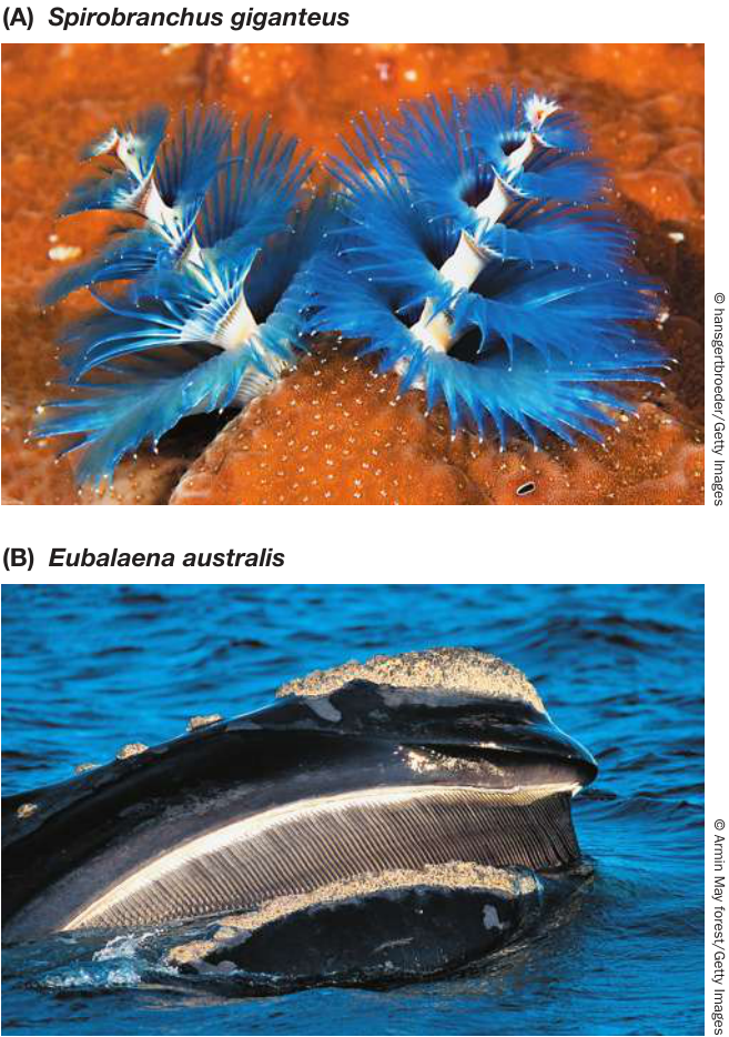
Figure 29.8 Filter-Feeding Strategies (A) Sessile marine filter feeders such as this Christmas tree worm, a polychaete (annelid), allow the ocean currents to bring their food—plankton—to them. (B) The southern right whale is a motile filter feeder that strains small crustaceans such as krill from the oceans for its food.
Many sessile aquatic animals rely on water currents to bring prey to them (Figure 29.8A). Some sessile filter feeders (such as sponges; see Figure 29.2B) expend energy to move water past their food-capturing devices. Motile filter feeders move their bodies to the nutrient source. Flamingos, for example, use their serrated beaks to filter small organisms out of the muddy mixture they pick up as they wade through shallow water. Baleen whales—the largest animals that have ever lived—are filter feeders that strain tiny crustaceans from the water column as they swim (Figure 29.8B).
Herbivores eat plants
An individual plant has many different structures—leaves, wood, roots, sap, flowers, fruits, nectar, and seeds—that animals can consume. Not surprisingly, then, many different kinds of herbivores—animals that feed only on plants—may feed on a single kind of plant, consuming different parts of the plant or feeding on the
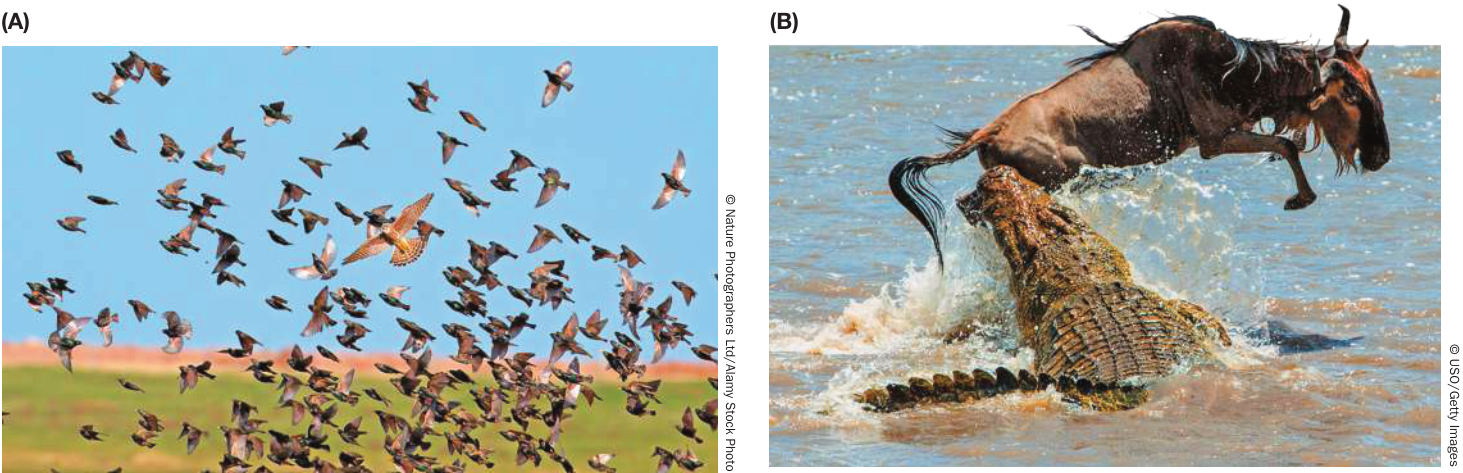
Figure 29.9 Active and Sit-and-Wait Predators (A) The appendages (legs and wings) of the Merlin (Falco columbarius), along with its strong beak, are adaptations to the life of an active predatory hunter. (B) A crocodile typically sits motionless in the water, waiting in one spot for unsuspecting prey to walk within striking range.
same part in different ways. Whereas an individual animal that is captured by a predator is likely to die, herbivores often feed on plants without killing them.
Animals do not need to expend energy subduing and killing plants in order to feed on them. However, plant matter can be difficult to digest and can pose special challenges to terrestrial herbivores because the dominant land plants tend to have several different kinds of tissues, many of which are tough or fibrous. Herbivorous animals typically have long, complex guts to accomplish the tasks involved in digesting plants. Animals also must expend energy to detoxify plants’ defensive chemicals.
Connect the Concepts The structure and function of animal guts, including the differences in the guts of herbivores and carnivores, are explained in Key Concept 49.3.
Predators and omnivores capture and subdue prey
Predators possess features that enable them to capture and subdue other animals (referred to as their prey). Many vertebrate predators have sensitive sensory organs that enable them to locate prey, as well as sharp teeth or claws that allow them to capture and subdue prey (Figure 29.9). Predators may stalk and pursue their prey or wait (often camouflaged) for their prey to come to them.
Omnivores (“all-devouring”) are animals, such as raccoons and humans, that eat both plants and other animals. The diets of some omnivores differ at different life stages; many songbirds, for example, eat fruit or seeds as adults but feed insects to their young.
Parasites live in or on other organisms
Parasites obtain nutrients from another organism—a host—by living on or within the host. Some animal parasites consume parts of the host itself (such as ticks that suck body fluids); others highjack nutrients the host would otherwise consume (such as tapeworms that may live in the intestines of mammals). Most animal parasites are much smaller than their hosts, and many parasites can consume parts of their host without killing it. To set up residence within a host, a parasite must first overcome the host’s defenses. Parasites often have complex life cycles that rely on multiple hosts, as you will see in Key Concept 29.2.
Parasites that live inside their hosts are called endoparasites, and these are often morphologically very simple. Endoparasites often function without a digestive system, absorbing their food directly from the host’s gut or body tissues. Many flatworms are endoparasites of humans and other mammals, as we will describe in Chapter 30.
Parasites that live outside their hosts are called ectoparasites; they are generally more complex morphologically than endoparasites. Ectoparasites have digestive tracts and mouthparts that enable them to pierce the host’s tissues or suck on the host’s body fluids. Fleas and ticks are ectoparasitic arthropods that feed on many vertebrates, including humans.
Detritivores live on the remains of other organisms
Detritivores feed on the dead bodies or waste products of other organisms, organic matter known as detritus. Detritivores (sometimes called decomposers) perform an important ecosystem function by breaking down dead organic matter and returning the nutrients it contains to the environment in a form that can be used by other organisms. Detritivores are common in any soil with high organic content, as well as on the ocean floor. Well-known detritivores include earthworms and other annelids, millipedes, and many insects and crustaceans.
Variations in body symmetry, body cavity structure, life cycles, patterns of development, and survival strategies differentiate the many animal species. Table 29.1 summarizes the living members of the major animal groups.
Table 29.1 Summary of Living Members of the Major Animal Groups
| Group | Approximate number of living species described | Major subgroups, other names, and notes |
|---|---|---|
| Sponges | 8,500 | Demosponges, glass sponges, calcareous sponges |
| Ctenophores | 250 | Comb jellies |
| Placozoans | 2 | Additional species have been discovered but not yet formally named |
| Cnidarians | 12,500 | Anthozoans: Corals, sea anemones, sea pens Hydrozoans: Hydras, hydroids Scyphozoans: Jellyfishes |
| Orthonectids | 45 | Microscopic wormlike parasites of marine invertebrates; relationships uncertain |
| Rhombozoans | 125 | Tiny (0.5–7 mm) parasites of cephalopods; relationships uncertain |
| PROTOSTOMES | ||
| Arrow worms | 180 | Glass worms |
| LOPHOTROCHOZOANS | ||
| Bryozoans | 5,500 | Moss animals |
| Flatworms | 30,000 | Free-living flatworms; flukes and tapeworms (all parasitic); monogeneans (ectoparasites of fishes) |
| Gastrotrichs | 800 | “Hairy backs” |
| Rotifers and relatives | 3,800 | Rotifers, spiny-headed worms, jaw worms |
| Ribbon worms | 1,200 | Proboscis worms |
| Brachiopods | 450 | Lampshells |
| Phoronids | 10 | Sessile marine filter feeders |
| Annelids | 19,000 | Polychaetes (generally marine; may not be
monophyletic) Clitellates: earthworms, freshwater worms, leeches |
| Mollusks | 117,000 | Chitons Bivalves: Clams, oysters, mussels Gastropods: Snails, slugs, limpets Cephalopods: Squid, octopuses, nautiloids |
| ECDYSOZOANS | ||
| Nematodes | 25,000 | Roundworms |
| Horsehair worms | 350 | Gordian worms |
| Kinorhynchs | 180 | Mud dragons |
| Loriciferans | 30 | Brush heads |
| Priapulids | 20 | Penis worms |
| Onychophorans | 180 | Velvet worms |
| Tardigrades | 1,200 | Water bears |
| ARTHROPODS | ||
| Chelicerates | 114,000 | Horseshoe crabs, sea spiders, arachnids (scorpions, harvestmen, spiders, mites, ticks) |
| Myriapods | 12,000 | Millipedes, centipedes |
| Crustaceans | 67,000 | Crabs, shrimps, lobsters and crayfishes, barnacles, copepods |
| Hexapods | 1,056,000 | Insects, springtails, two-pronged bristletails, proturans |
| DEUTEROSTOMES | ||
| Xenoturbellids | 5 | Secondarily simple marine worms; relationships uncertain |
| Acoels | 400 | Very small (mostly <2 mm) flattened marine worms; relationships uncertain |
| Echinoderms | 7,500 | Crinoids (sea lilies, feather stars), brittle stars, sea stars, sea daisies, sea urchins, sea cucumbers |
| Hemichordates | 120 | Acorn worms, pterobranchs |
| CHORDATES | ||
| Tunicates | 2,800 | Sea squirts (ascidians), thaliaceans, larvaceans |
| Lancelets | 35 | Cephalochordates |
| Vertebrates | 71,000 | Hagfishes, lampreys, cartilaginous fishes, ray-finned fishes, coelacanths, lungfishes, amphibians, reptiles (including birds), mammals |
Recap and Assess
We generally recognize animals as multicellular heterotrophs with internal digestion, muscular systems that allow movement, and nervous systems. Animals are thought to be monophyletic because they share several derived traits, especially among their gene sequences. Major developmental differences also provide evidence of their evolutionary relationships. The body plans of animals are variations on patterns of symmetry, body cavity structure, segmentation, appendages, and nervous systems. Most animals either move through the environment to where food is located or move the environment and the food it contains to them.
- What general features distinguish animals from other major groups of living organisms?
- Differentiate among the members of each of the following sets of
related terms:
- radial symmetry/bilateral symmetry
- protostome/deuterostome
- diploblastic/triploblastic
- Why is bilateral symmetry strongly associated with cephalization, the concentration of sensory organs in an anterior head?
- Explain several ways in which body cavities, segmentation, and centralized nervous systems improve control over movement.
- How can you distinguish among filter feeders, predators, and parasites—all of which may feed on other animals?
- What adaptations are necessary for animals that eat plants? What adaptations are needed for a predatory lifestyle?
As an animal grows from a single-celled zygote into a larger, more complex adult, its body structure, its diet, and the environment in which it lives may all change. In the next key concept we describe some animal life cycles and discuss why they are so varied.
Animal Life Cycles Involve Trade-Offs
Learning Objectives
- 29.2.1 Formulate a hypothesis that accounts for animal species that lose metamorphosis and evolve direct development.
- 29.2.2 Distinguish the similarities and differences between coloniality and multicellularity.
- 29.2.3 Explain the concept of “trade-offs” by comparing two distinct life cycles of animal species.
The life cycle of an animal encompasses its embryonic development, birth, growth to maturity, reproduction, and death. During its life an individual animal ingests food, grows, interacts with other individuals of the same and other species, and reproduces.
Many animal life cycles feature specialized life stages
In some groups of animals, newborns look much like miniature versions of the adults (a pattern called direct development). Newborns of most animal species, however, differ dramatically from adults. Many animal species have a life stage called a larva (plural larvae), which is an immature form that the animal takes early in its life before assuming an adult form. Some of the most striking life cycle changes are found among insects such as beetles, flies, moths, butterflies, and bees, which undergo radical change (called complete metamorphosis) between their larval and adult stages (Figure 29.10). In these animals, one stage may be specialized for feeding and the other for reproduction. Adults of many moth species, for example, do not eat. In some animal species, individuals eat during all life cycle stages, but what they eat changes with the stage. For example, butterfly larvae (known as caterpillars) eat leaves and flowers, whereas most adult butterflies eat only nectar. Having different life cycle stages that are specialized for different activities may increase the efficiency with which an animal performs those activities and reduce the competition for food between juveniles and adults.
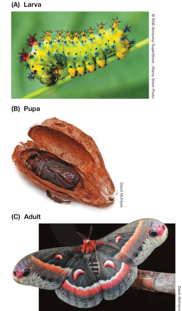
Figure 29.10 A Life Cycle with Complete Metamorphosis (A) The larval stage (caterpillar) of the cecropia moth (Hyalophora cecropia) is specialized for feeding. (B) The pupa is the stage during which the transformation to the adult form occurs. (C) The adult moth is specialized for dispersal and reproduction and does not feed.
Most animal life cycles have at least one dispersal stage
At some time during their lives, most animals move, or are moved, so few animals die exactly where they were born. Movement of organisms away from a parent organism or from an existing population is called dispersal.
Animals that are sessile as adults typically disperse as eggs or larvae. Most sessile marine animals discharge their eggs and sperm into the water, where fertilization takes place. A larva soon hatches and floats freely in the plankton, where it filters small food items from the water. Many animals that live on the seafloor, including polychaete worms and mollusks, have a radially symmetrical larval form known as a trochophore (Figure 29.11A). Other animals, such as crustaceans, have a bilaterally symmetrical larval form called a nauplius (Figure 29.11B). Both types of larvae feed for some time in the plankton and may travel long distances before settling on the ocean floor and transforming into adults.
Other animal species that are motile as adults disperse when they are mature. A caterpillar, for example, may spend its entire larval stage feeding on a single plant, but after it metamorphoses into a flying adult—a butterfly or moth—it may fly to and lay eggs on other plants located far from the one where it spent its caterpillar days. In some species, individuals disperse during several different life cycle stages.
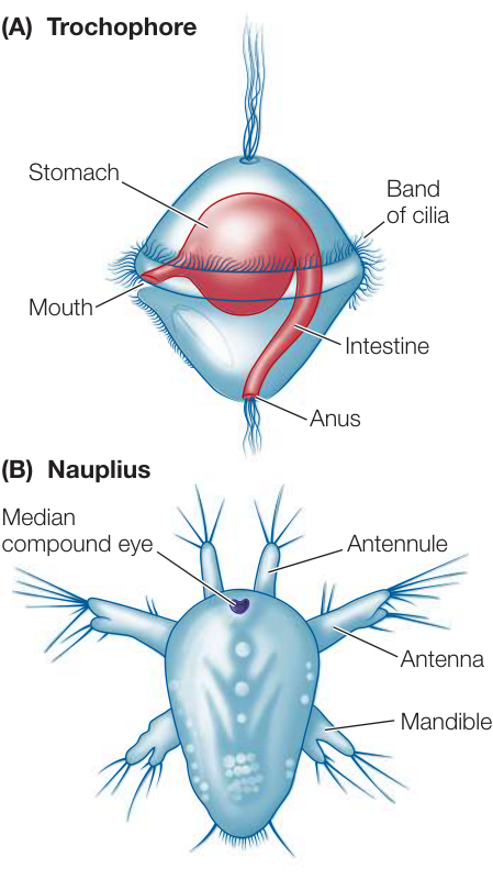
Figure 29.11 Planktonic Larval Forms of Marine Animals (A) The trochophore (“wheel-bearer”) is a distinctive larval form found in several marine animal clades with spiral cleavage, most notably the polychaete worms and the mollusks. (B) This nauplius larva will mature into a crustacean with a segmented body and jointed appendages.
Parasite life cycles facilitate dispersal and overcome host defenses
Animals that live as endoparasites are bathed in the nutritious tissues of their host or in the digested food that fills their host’s digestive tract. Thus they may not need to exert much energy to obtain food, but to survive they must overcome the host’s defenses. Furthermore, either they or their offspring must disperse to new hosts while their host is still living, because they die when their host dies.
The fertilized eggs of some parasites are voided with the host’s feces and later ingested directly by other host individuals. Most parasite species, however, have complex life cycles involving one or more intermediate hosts and several larval stages (Figure 29.12). Some intermediate hosts transport individual parasites directly between other host species. Others house and support the parasite until another host ingests them. Complex life cycles may thus facilitate the transfer of individual parasites among hosts.
Some animals form colonies of genetically identical, physiologically integrated individuals
Most people tend to view the distinction between individuals and populations as clear-cut. In several groups of animals, however, asexual reproduction without fission leads to the formation of colonies composed of many physiologically integrated individuals. At first appearance, these colonies may look much like a single integrated organism. The individuals in the colony are clonal copies of one another, so they are genetically homogeneous.
Coloniality has arisen several times among animal groups, with widely varying levels of integration and specialization among the individuals. In some species, colonies are composed of loosely connected but integrated individuals that all function alike (Figure 29.13). In other colonial species, the individuals may become specialized for different functions, just as different cell types in multicellular organisms do. The Portuguese man-of-war (a cnidarian; see Figure 29.20) is an example of such a colonial animal, as it is composed of many individuals of four different specialized body forms, all integrated and functioning together. The individuals in the colony are themselves multicellular, however, unlike the cells of a single multicellular organism.
No life cycle can maximize all benefits
A common saying, “a jack-of-all-trades is master of none,” suggests why there are constraints on the evolution of life cycles. The characteristics an animal has in any one life cycle stage may improve its performance in one activity but reduce its performance in another—a situation known as a trade-off. An animal that is good at filtering small food items from the water, for example, is unlikely to be good at capturing large prey. Similarly, energy devoted to building protective structures such as shells cannot be used for growth.
Some major trade-offs can be seen in animal reproduction. Some animals produce large numbers of small eggs, each with a small energy store (Figure 29.14A). Other animals produce a small number of large eggs, each with a large energy store (Figure 29.14B).
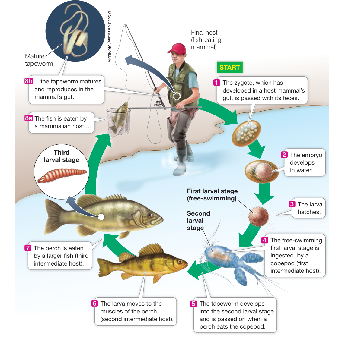
Figure 29.12 Reaching a New Host by a Complex Route The broad fish tapeworm Diphyllobothrium latum must pass through the bodies of a copepod (a type of crustacean) and at least one fish before it can reinfect its primary host, a mammal. Such complex life cycles assist the parasite’s colonization of new host individuals, but they also provide opportunities for humans to break the cycle with hygienic measures (such as thoroughly cooking food to kill the parasites).
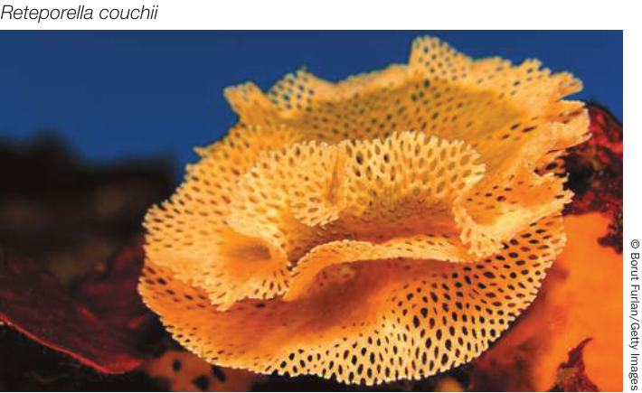
Figure 29.13 Colonial Animals This bryozoan colony consists of many asexually reproducing, genetically homogeneous, physiologically interacting individuals. The colony looks much like a single individual with many parts, but in fact is a group of individuals acting together.
With a fixed amount of energy available for reproduction, a female animal can produce many small eggs or a few large eggs, but she cannot produce many large eggs. Thus there is a trade-off between the number of offspring produced and the energy resources each offspring receives from its mother.
The larger the energy store in an egg, the longer an offspring can develop before it must either find its own food or be fed by its parents. Birds of all species lay relatively small numbers of relatively large eggs, but incubation periods vary. In some species, eggs hatch when the young are still helpless (Figure 29.15A). Such altricial young must be fed and cared for until they can feed themselves; parents can provide for only a small number of altricial offspring. In contrast, some bird species incubate their eggs longer, and the hatchlings are developed to the point that they are able to forage for themselves almost immediately (Figure 29.15B). The young of such species are called precocial.
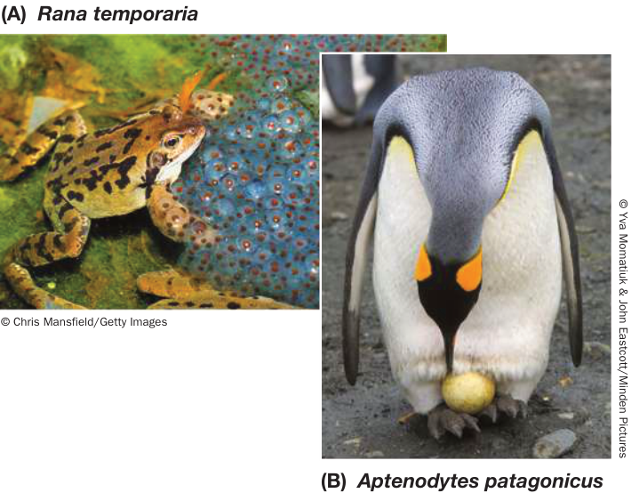
Figure 29.14 Many Small or Few Large Allocation of energy to eggs requires trade-offs. (A) This European common frog has divided her reproductive energy among a large number of small eggs. (B) This King Penguin has invested all of her reproductive energy in one large egg. Q: What are the respective advantages and disadvantages of these two reproductive strategies?
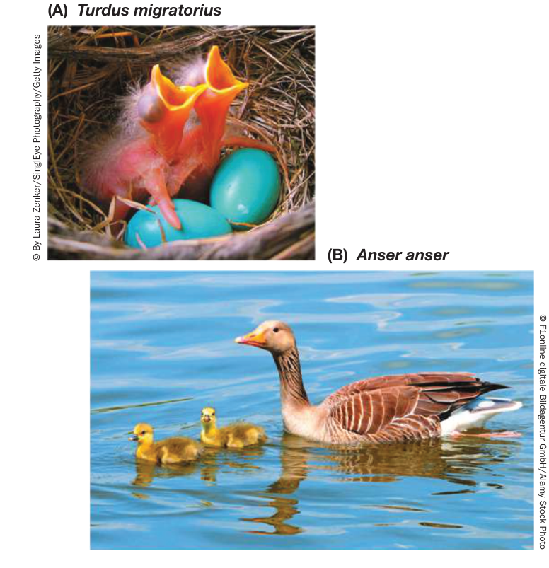
Figure 29.15 Helpless or Independent (A) The altricial young of the American Robin are essentially helpless when they hatch. Their parents feed and care for them for several weeks. (B) Graylag Goose hatchlings are precocial, ready to swim and feed independently almost immediately after hatching.
Recap and Assess
Many animals have life cycle stages that differ from one another morphologically. In some animals, the larval form is a dispersal stage; in other animals, the adults are more likely to disperse than are larvae. In several groups of organisms, asexual reproduction without fission leads to coloniality.
- Why can’t a life cycle evolve that maximizes all aspects of feeding and reproduction simultaneously?
- How does a colony of unicellular animals differ from a multicellular animal?
In the remainder of this chapter and in Chapters 30 and 31, we will become acquainted with the major animal groups and learn how the general characteristics of body form and life history strategies we have just described apply to each of them.
The Root of the Animal Tree Provides Clues to Early Animal Diversification
The Root of the Animal Tree Provides Clues to Early Animal Diversification
Learning Objectives
29.3.1 Apply knowledge of phylogenetic relationships among major groups of animals to describe the evolution and loss of major structural features.
29.3.2 Compare the life histories of sponges, ctenophores, placozoans, and cnidarians and discuss their similarities and differences.
Looking at Figure 29.1, you can see that the protostome and deuterostome animals together comprise a monophyletic group known as Bilateria. Some major traits that support the monophyly of bilaterians (in addition to genomic analyses) are strong bilateral symmetry, the presence of three distinct cell layers in embryos (triploblasty), and a centralized nervous system. There are, however, four animal groups—the sponges, ctenophores, placozoans, and cnidarians—that are not bilaterians.
The root of the animal tree is still debated
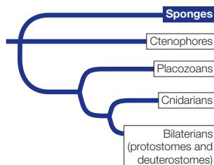
Biologists have proposed at least three different lineages as sister group of the remaining animals. Traditionally, sponges have been considered the sister group of other animals, based in large part on the similarity of some sponges to choanoflagellates (see Figure 29.2) and on the fact that sponges lack a nervous system. Placozoans also have very few tissue types, and also have no nervous system, so they have also been placed by most biologists as branching off near the base of the animal tree. However, as we explore in Investigating Life: Reconstructing Animal Phylogeny from Protein-Coding Genes, some recent genomic studies suggest that the first split in the animal tree may have been between the lineage that leads to ctenophores and the rest of the animals.
Recent studies of the many genes that encode nervous systems indicate that these genes were duplicated and specialized separately in ctenophores, cnidarians, and bilaterians. This finding suggests that nervous systems evolved independently in these three groups in response to similar selection pressures associated with multicellularity, movement, and prey capture. Therefore the nervous systems of these groups are no longer considered homologous, or as evidence for a close evolutionary relationship. Most biologists now agree that the sponges and ctenophores were the first two lineages to split from the remaining animals, although there is still disagreement about which of these two lineages split first. The placozoans were then the next group to split from the remaining animals, followed by the split that gave rise to the cnidarians (see Figure 29.1).
Investigating Life: Reconstructing Animal Phylogeny from Protein-Coding Genes
Experiment
Original Paper: C. W. Dunn et al. 2008. Broad phylogenomic sampling improves resolution of the Animal Tree of Life. Nature 452: 745–749.
Several breakthroughs in our understanding of animal phylogeny have occurred in recent years as the sequences of genes and proteins have been compared across species. Casey Dunn and his colleagues compared sequences from many different proteins across a wide variety of animal groups to reconstruct their phylogeny.
QUESTIONS What was the earliest split in the animal tree? Are ctenophores, sponges, or placozoans the sister group of other animals?
METHOD
Collect mRNA from major groups of animals and choanoflagellate outgroups.
Reverse transcribe the mRNA into cDNA.
Sequence the cDNA, translate the genes into the amino acid sequences of the proteins, and align sequences of homologous proteins.
Reconstruct the phylogeny of the animal groups from the aligned protein sequences.
RESULTS The tree below represents just a sample of the major lineages examined by Dunn and colleagues.
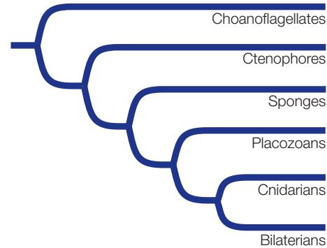
CONCLUSION According to this data set, the earliest split in the animal tree was between ctenophores and the remaining animals.
Work with the Data
The table below includes a small sample of the data collected and analyzed by Dunn and his colleagues. Use these representative data to reconstruct a tree of the represented species. In the original paper, Dunn and colleagues reported on 11,234 amino acid positions among 77 species of animals. Twenty-seven of these amino acid positions for 11 of those species are shown in the table. (See Table 3.2 for the one-letter amino acid abbreviations.)
QUESTIONS
Construct a phylogenetic tree of these 11 species using the parsimony method. Use the outgroup (a choanoflagellate) to root your tree. Assume that all changes from one amino acid to another are equally likely. See Key Concept 20.2 for a review of the parsimony method of reconstructing phylogenetic trees.
How many character state changes (i.e., changes from one amino acid to another) occur along each branch on your tree?
Which characters support the ctenophores as the sister group of the remaining animals?
Which group on your tree represents the bilaterian animals? The protostomes? The deuterostomes?
| Species | Character state (amino acid at position) | ||||||||||||||||||||||||||
|---|---|---|---|---|---|---|---|---|---|---|---|---|---|---|---|---|---|---|---|---|---|---|---|---|---|---|---|
| 1 | 2 | 3 | 4 | 5 | 6 | 7 | 8 | 9 | 10 | 11 | 12 | 13 | 14 | 15 | 16 | 17 | 18 | 19 | 20 | 21 | 22 | 23 | 24 | 25 | 26 | 27 | |
| Clam | Y | S | T | G | L | H | E | N | Y | A | R | A | M | R | I | A | L | T | I | V | K | L | S | I | V | I | L |
| Earthworm | Y | A | T | G | L | H | E | N | Y | P | H | A | M | R | I | A | L | T | I | V | K | L | S | I | V | M | L |
| Tardigrade | Y | A | T | G | L | H | E | H | Y | K | R | A | M | R | V | A | T | S | I | V | R | L | N | L | V | L | L |
| Fruit fly | F | A | T | G | L | H | E | N | Y | K | R | A | M | R | I | A | L | S | I | V | S | L | D | L | V | L | L |
| Sea urchin | Y | A | T | G | L | L | E | N | Y | P | N | A | M | R | I | A | L | T | V | I | R | Q | N | L | T | V | K |
| Human | W | A | A | G | L | R | E | H | Y | P | K | A | I | R | I | S | V | T | V | I | R | Q | N | L | T | V | K |
| Chicken | W | A | A | G | L | R | E | H | Y | P | R | A | I | R | I | A | V | T | V | I | R | Q | N | L | T | V | K |
| Lancelet | Y | A | T | G | L | R | E | H | Y | P | K | A | M | R | I | A | V | T | V | I | R | L | N | L | T | V | K |
| Sponge | Y | G | L | S | L | R | P | N | F | P | K | S | M | S | V | A | L | T | V | I | R | Q | N | L | V | I | L |
| Ctenophore | Y | G | L | G | Q | D | P | N | F | P | K | S | M | S | V | A | L | T | V | I | R | Q | N | L | V | I | L |
| Choanoflagellate (outgroup) | Y | G | L | G | Q | D | P | N | F | P | K | S | F | S | V | A | L | T | V | I | R | Q | N | L | V | I | L |
Whichever of these groups was first to diverge, biologists think that the common ancestor of animals was very different from all animals that are alive today. An early split on the animal tree does not imply that sponges or ctenophores look anything like the ancestral animal. For example, all living ctenophores are quite closely related to one another, and they have been evolving for as long as all other animal lineages—ever since animals last shared a common ancestor more than 700 million years ago. Thus many traits of modern ctenophores (as well as modern sponges) are highly derived and specialized.
Molecular analyses of genomes place the earliest splits in animal phylogeny at more than 700 million years ago, well before the first large multicellular animals appear in the fossil record about 565 million years ago (see Figure 23.12). The first animals are thought to have had relatively few cells and to have been quite small. Then large multicellular animals, along with some of their characteristics (such as nervous systems), appear to have evolved several times in different animal lineages. As Earth warmed and oxygen concentrations increased in the oceans near the beginning of the Cambrian, several groups of animals evolved larger bodies and greater tissue complexity.
Sponges are loosely organized animals
Although they have some specialized cells, sponges have no distinct embryonic cell layers and no true organs. Early naturalists classified sponges as plants because they were sessile and lacked body symmetry.
Sponges have hard skeletal elements called spicules, which may be small and simple or large and complex. Three major groups of sponges, which separated soon after the split between sponges and the rest of the animals, are distinguished by their spicules. Members of two groups (demosponges and glass sponges) have skeletons composed of silicaceous spicules made of hydrated silicon dioxide (Figure 29.16A,B). These spicules have greater flexibility and toughness than synthetic glass rods of similar length. Members of the third group, the calcareous sponges, take their name from their calcium carbonate spicules (Figure 29.16C).
The body plan of all sponges—even large ones, which may reach a meter or more in length—is an aggregation of cells built around a water canal system. Sponges bring water into their bodies by beating the flagella of their specialized feeding cells, the choanocytes (see Figure 29.2B). Water, along with any food particles it contains, enters the sponge by way of small pores and passes into the water canals or a central atrium, where choanocytes capture food particles.
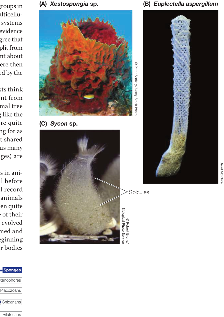
Figure 29.16 Sponge Diversity (A) The majority of sponge species are demosponges, such as this barrel sponge. The system of pores and water canals (see Figure 29.2B) that is typical of the sponge body plan is apparent in this photograph. (B) The supporting structures of both demosponges and glass sponges are silicaceous spicules, seen here in the skeleton of a glass sponge. (C) The spicules of calcareous sponges are made of calcium carbonate.
A skeleton of simple or branching spicules, often combined with a complex network of elastic fibers, supports the body of most sponges. Sponges also have an extracellular matrix, composed of collagen, adhesive glycoproteins, and other molecules, that holds the cells together. Most sponges are filter feeders. A few species are carnivores that trap prey on hook-shaped spicules that protrude from the body surface.
Most of the 8,500 species of sponges are marine animals. Only about 50 species of sponges live in fresh water. Sponges come in a wide variety of sizes and shapes that are adapted to different movement patterns of water. Sponges living in intertidal or shallow subtidal environments are subject to strong wave action and attach firmly to the substrate. Most sponges that live in slowly flowing water are flattened and are oriented at right angles to the direction of current flow, allowing them to intercept water and the food it contains as it flows past them.
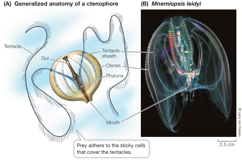
Figure 29.17 Comb Jellies Feed with Tentacles (A) The body plan of a typical ctenophore. The long, sticky tentacles sweep through the water, efficiently harvesting small prey. (B) This comb jelly, photographed in Sydney Harbor, Australia, has short tentacles. Comb jellies get their name from the ctenes—rows or “combs” of cilia used for movement.
Sponges reproduce both sexually and asexually. In most species, a single individual produces both eggs and sperm, but individuals do not self-fertilize. Water currents carry sperm from one individual to another. Sponges also reproduce asexually by budding and fragmentation.
Activity 29.2 Sponge and Diploblast Classification
Ctenophores are radially symmetrical and diploblastic
Ctenophores, also known as comb jellies, were until recently thought to be most closely related to the cnidarians (jellyfishes, corals, and their relatives). But ctenophores lack most of the Hox genes found in cnidarians and bilaterians, and recent studies of their genomes have indicated that ctenophores were among the earliest lineages to split from the remaining animals (see Investigating Life: Reconstructing Animal Phylogeny from Protein-Coding Genes).
Ctenophores have a radially symmetrical, diploblastic body plan. The two cell layers are separated by an inert, gelatinous extracellular matrix called mesoglea. Ctenophores, unlike sponges, have a complete gut, meaning they have different openings for food entry and waste elimination. Food enters through a mouth, and wastes are eliminated through two anal pores.
Ctenophores move by beating cilia rather than by muscular contractions. Most of the 250 known species have eight comblike rows of cilia-bearing plates, called ctenes (Figure 29.17). The feeding tentacles of ctenophores are covered with cells that discharge adhesive material when they contact prey. After capturing its prey, a ctenophore retracts its tentacles to bring the food to its mouth. In some species, the entire surface of the body is coated with sticky mucus that captures prey. Most ctenophores eat small planktonic organisms, although some eat other ctenophores. Ctenophores are common in open seas and can become abundant in protected bodies of water, where large populations may inhibit the growth of other organisms.
Ctenophore life cycles are uncomplicated. Gametes are released into the body cavity and then discharged through the mouth or the anal pores. Fertilization takes place in open seawater. In nearly all species, the fertilized egg develops directly into a miniature ctenophore, which gradually grows into an adult.
Media Clip 29.4 Ctenophores Life12e.com/mc29.4
Placozoans are abundant but rarely observed
As we discussed in the opening of this chapter, placozoans are (like sponges) structurally very simple animals with only four distinct cell types (Figure 29.18A). Individuals in the mature, asymmetrical life stage are usually observed adhering to surfaces (such as the glass of aquariums, where they were first discovered, or to rocks and other hard substrates in nature). Their structural simplicity—they have no mouth, gut, or nervous system—initially led biologists to suspect they might be the sister group of all other animals. Most phylogenetic analyses have not supported this hypothesis, however, and some aspects of the placozoans’ structural simplicity may be secondarily derived. They are generally considered to have a diploblastic body plan, with upper and lower epithelial (surface) layers that sandwich a layer of contractile fiber cells.
Recent studies have found that placozoans have a pelagic (open-ocean) life stage that is capable of swimming (Figure 29.18B), but the life history of placozoans is incompletely known. Most studies have focused on the larger adherent stages that can be found in aquariums, where they appear after being inadvertently collected with other marine organisms. The transparent nature and small size of placozoans make them very difficult to observe in nature. Nonetheless, it is known that placozoans can reproduce both asexually as well as sexually, although the details of their sexual reproduction are poorly understood. They are found in warm coastal seas around the world.
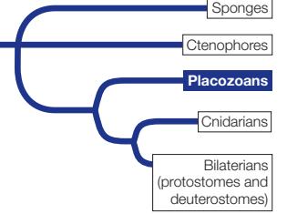
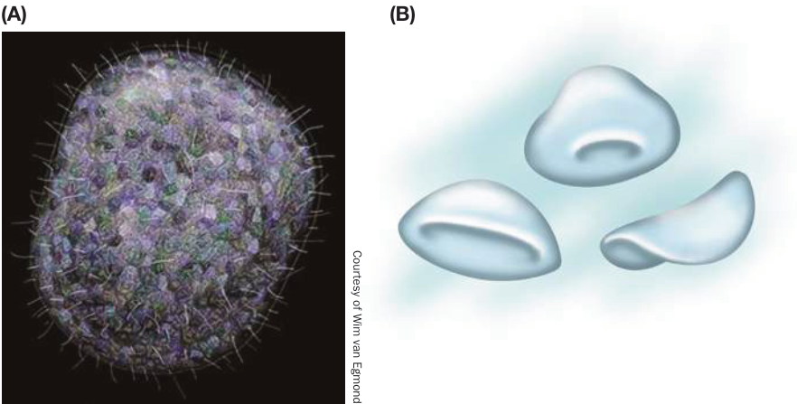
Figure 29.18 Placozoan Simplicity (A) As seen in this artist’s rendition, adult placozoans are tiny (1–2 mm across), flattened, asymmetrical animals. (B) Recent studies have found a symmetrical, weakly swimming pelagic stage of placozoan to be abundant in many warm tropical and subtropical seas. (B after V. B. Pearse and O. Voigt. 2007. Integr Comp Biol 47(5): 677–692. Drawing by J. Keller and C. Patton, based on observations by V.B.P.)
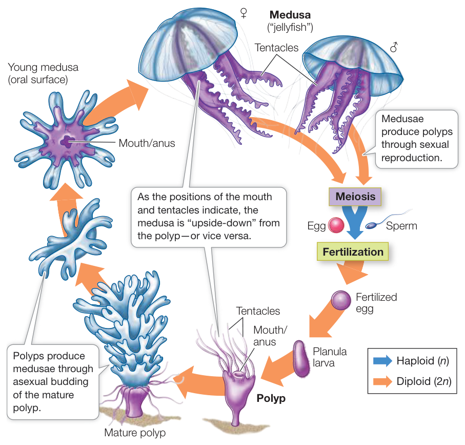
Figure 29.19 The Life Cycle of Most Cnidarians Has Two Stages The life cycle of a scyphozoan (jellyfish) exemplifies the typical cnidarian body forms: the sessile, asexual polyp and the motile, sexual medusa. Some species of cnidarians have life cycles that lack polyps or medusae. (Adapted from F. M. Bayer and H. B. Owre. 1968. The Free-Living Lower Invertebrates, Macmillan Publishing Co.: New York.) Animation 29.1 Life Cycle of a Cnidarian
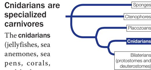
The cnidarians (jellyfishes, sea anemones, sea pens, corals, and hydrozoans) make up the largest and most diverse group of non-bilaterian animals. The mouth of a cnidarian is connected to a blind sac called the gastrovascular cavity (a cnidarian thus does not have a complete gut). The gastrovascular cavity functions in digestion, circulation, and gas exchange, and it also acts as a hydrostatic skeleton. The single opening serves as both mouth and anus.
The life cycle of many cnidarians has two distinct stages, one sessile and the other motile (Figure 29.19), although one or the other of these stages is absent in some groups. In the sessile polyp stage, a cylindrical stalk is attached to the substrate. The motile medusa (plural medusae) is a free-swimming stage shaped like a bell or an umbrella. It typically floats with its mouth and feeding tentacles facing downward.
Mature polyps produce medusae by asexual budding. Medusae then reproduce sexually, producing eggs or sperm by meiosis and releasing the gametes into the water. A fertilized egg develops into a free-swimming, ciliated larva called a planula, which eventually settles to the bottom and develops into a polyp.
Cnidarians are specialized predators adapted for capturing and subduing relatively large and complex prey. As we noted at the start of this key concept, recent genomic studies suggest that the nerve nets found in cnidarians are largely independently derived from those found in ctenophores, as well as from the nervous systems found in bilaterians. The tentacles of cnidarians are covered with specialized cells that contain stinging organelles called nematocysts, which inject toxins into their prey (Figure 29.20). Some cnidarians, including many corals and anemones, gain additional nutrition from photosynthetic endosymbionts that live in their tissues.
Cnidarians have cells containing muscle fibers whose contractions enable the animals to move, as well as simple nerve nets that integrate the body’s activities. Their bodies also contain specialized structural molecules (collagen, actin, and myosin). Yet cnidarians, like ctenophores, are largely made up of inert mesoglea. Most species have low metabolic rates and can survive in environments where they encounter prey only infrequently.
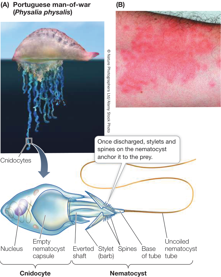
Figure 29.20 Nematocysts Are Potent Weapons (A) The tentacles of the Portuguese man-of-war, a hydrozoan, are rife with specialized cells called cnidocytes. These cells contain stinging organelles called nematocysts, which inject toxins into their prey. The Portuguese man-of-war is a colonial organism, composed of many physiologically integrated individuals with specialized functions. (B) Nematocysts of the Portuguese man-of-war cause painful reactions when they come into contact with human skin. Media Clip 29.5 Stunning Siphonophore: Colonial Hydrozoans Life12e.com/mc29.5
Of the roughly 12,500 living cnidarian species, all but a few live in the oceans (Figure 29.21). The smallest cnidarians can barely be seen without a microscope. One small group, known as myxozoans, consists of tiny parasites, usually with a two-host life cycle that includes a fish and an annelid worm or a bryozoan. The largest known jellyfish is 2.5 meters in diameter, and some colonial hydrozoans (which include the Portuguese man-of-war; see Figure 29.20) can reach lengths in excess of 30 meters. Here we describe the three clades of cnidarians that contain the most species: anthozoans, scyphozoans, and hydrozoans.
ANTHOZOANS Members of the anthozoan clade include sea anemones, sea pens, and corals. Sea anemones (see Figure 29.21A), all of which are solitary, are widespread in both warm and cold ocean waters. Sea pens (see Figure 29.21B), by contrast, are colonial. Each colony consists of two or more different kinds of polyps. The primary polyp has a lower portion
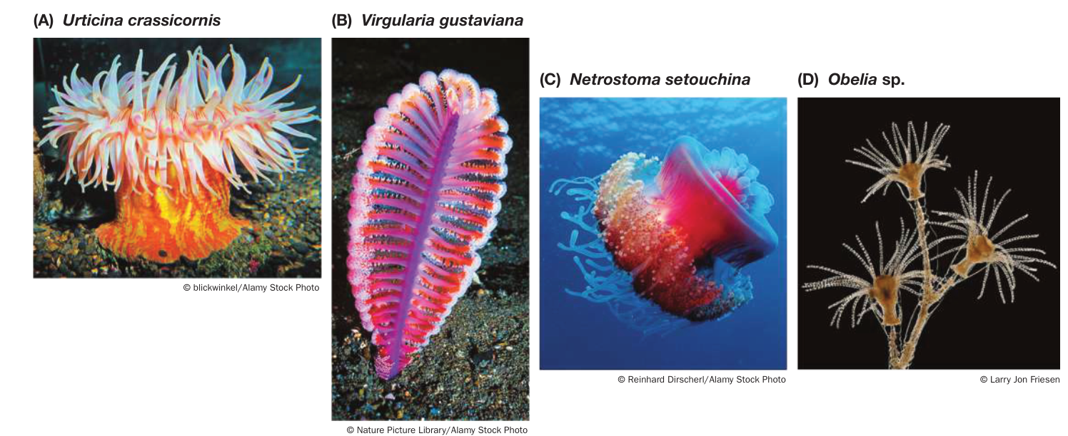
Figure 29.21 Diversity among Cnidarians (A) Sea anemones are sessile, living attached to marine substrates. Water currents carry prey into the nematocyst-studded tentacles. (B) The sea pen is a colonial cnidarian that lives in soft bottom sediments and projects polyps above the substrate. (C) This jellyfish illustrates the complexity of a scyphozoan medusa. (D) Polyp colonies of this hydrozoan grow on rocks of intertidal pools.
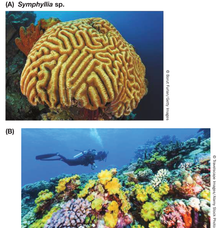
Figure 29.22 Corals (A) The descriptive common name of this Caribbean coral is “brain coral.” (B) Many different coral species form the Great Barrier Reef off the coast of Australia.
anchored in the bottom sediment and a branched upper portion that projects above the substrate. Along the upper portion, the primary polyp produces smaller secondary polyps by budding. Some of these secondary polyps differentiate into feeding polyps; in some species, other secondary polyps differentiate to circulate water through the colony.
The common names of coral groups—brain corals, staghorn corals, and organ pipe corals, among others—often describe their appearance (Figure 29.22A). Corals are sessile and colonial. The polyps of most species form a skeleton by secreting a matrix of organic molecules on which they then deposit calcium carbonate. As the colony grows, old polyps die, but their calcium carbonate skeletons remain. Living corals form a layer on top of a growing bank of skeletal remains, eventually forming chains of islands and reefs. The Great Barrier Reef along the northeastern coast of Australia is a system of coral formations more than 2,000 kilometers long, which is about the distance from New York City to St. Louis (Figure 29.22B). A single coral reef in the Red Sea has been calculated to contain more material than all the buildings in the major cities of North America combined.
Corals flourish at shallow depths in clear, nutrient-poor tropical waters. They grow well in such environments because unicellular photosynthetic dinoflagellates live endosymbiotically within their cells. These dinoflagellates provide the corals with products of photosynthesis; the corals, in turn, provide the dinoflagellates with nutrients and a place to live. This endosymbiotic relationship explains why reef-forming corals are restricted to clear surface waters, where light levels are high enough to support photosynthesis.
Coral reefs throughout the world are threatened by rising CO2 levels (which result in increased ocean temperatures and acidification of ocean waters through the formation of carbonic acid). Polluted runoff from development on adjacent shorelines is an additional threat to corals. Warmer temperatures lead to the loss of coral endosymbionts (known as coral bleaching), and acidification can cause coral skeletons to dissolve. An overabundance of nitrogen from fertilizer in runoff is advantageous to algae, which overgrow and eventually smother the corals.
Connect the Concepts Investigating Life: Can Corals Reacquire Dinoflagellate Endosymbionts Lost to Bleaching? in Chapter 25 describes an experiment in which coral endosymbionts were lost and replaced by new endosymbionts in response to changing environmental conditions.
SCYPHOZOANS The several hundred species of scyphozoans are all marine. The mesoglea of their medusae is thick and firm, giving rise to their common name of jellyfishes (or sea jellies). The medusa rather than the polyp dominates the life cycle of scyphozoans. An individual medusa is male or female, releasing eggs or sperm into the open sea. A fertilized egg develops into a small planula larva that quickly settles on a substrate and develops into a small polyp. This polyp feeds and grows and may produce additional polyps by budding. After a period of growth, the polyp begins to bud off small medusae, which feed, grow, and transform into adult medusae (see Figures 29.19 and 29.21C).
HYDROZOANS The polyp typically dominates the life cycle of hydrozoans, but some species have only medusae and others have only polyps. Most hydrozoans are colonial. A single planula larva eventually gives rise to a colony of many polyps, all interconnected and sharing a continuous gastrovascular cavity (Figure 29.23). Within such a colony, some polyps have tentacles with many nematocysts; they capture prey for the colony. Other individuals lack tentacles and are unable to feed, but are specialized for the asexual production of medusae. Still others are fingerlike and defend the colony with their nematocysts.
Some small groups of parasitic animals may be the closest relatives of bilaterians
Two small groups of tiny marine parasites are listed in Table 29.1 but are not depicted in the phylogeny in Figure 29.1: the orthonectids and the rhombozoans. Recent genomic analyses suggest that these groups may be among the closest surviving relatives of the bilaterians, although their exact phylogenetic placement is uncertain. Both groups are highly reduced parasites that lack many of the structures that traditionally have been used to study animal relationships. As their genomes become more completely known, the relationships of these two groups to other animals should become clearer. Two other small groups (also listed in Table 29.1 but not shown in Figure 29.1) have been proposed as also falling just outside the bilaterians: the xenoturbellids and acoels. Some genomic analyses, however, suggest that these animals are actually highly specialized deuterostomes.
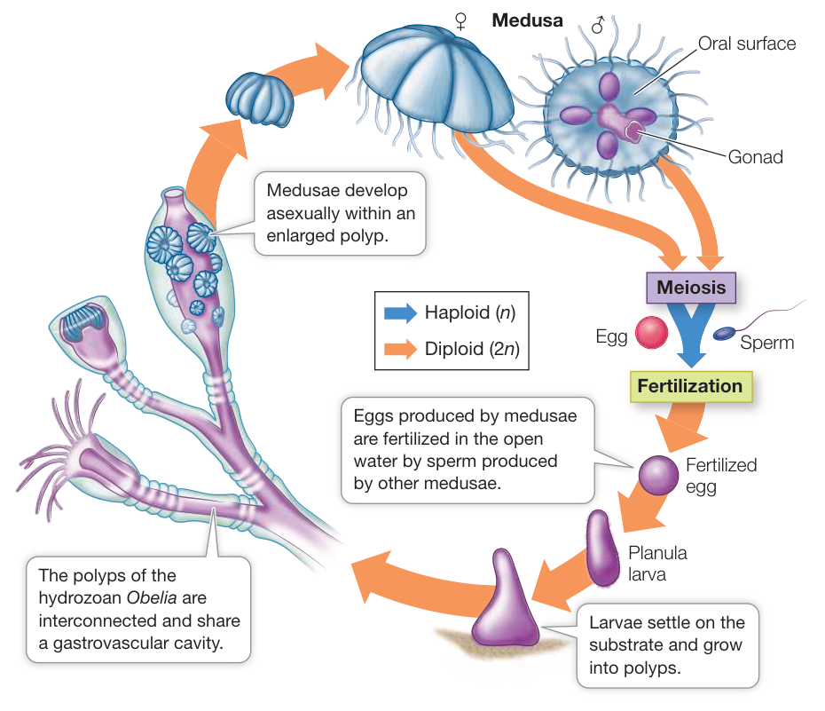
Figure 29.23 Many Hydrozoans Are Colonial The polyps in a hydrozoan colony may differentiate to perform specialized tasks. In the species whose life cycle is diagrammed here, the medusa is the sexual reproductive stage, producing eggs and sperm in organs called gonads. (Adapted from F. M. Bayer and H. B. Owre. 1968. The Free-Living Lower Invertebrates, Macmillan Publishing Co.: New York.)
Recap and Assess
Early animal diversification led to five major lineages: sponges, ctenophores, placozoans, cnidarians, and bilaterians. Sponges and ctenophores split first from the remaining animals, followed closely by the placozoans. The earliest animals are thought to have been small and to have consisted of relatively few cells per individual. Large multicellular animals, with greater tissue differentiation, arose independently several times, giving rise to the five major groups of modern animals. Major components of nervous systems arose independently in three of these lineages (ctenophores, cnidarians, and bilaterians).
- What is the basis for the claim that animal nervous systems evolved independently in ctenophores, cnidarians, and bilaterians?
- Why are sponges and placozoans considered to be animals even though they lack the complex body structures found in most other animal groups?
- The discoveries that placozoans in their pelagic stages are abundant in warm seas and that the mature stage settles on smooth surfaces suggest how these organisms might be collected and surveyed. What sampling procedures might you use to discover whether placozoans occur at a particular location along a coast?
Investigating Life: QA Which animal groups are involved in the earliest split in the animal tree?
Three major hypotheses have been proposed in recent decades about the earliest split in the animal tree. Given the structural simplicity of sponges and their similarity to some animal outgroups (especially choanoflagellates), for many years biologists considered sponges to form the sister group of all other animals. When placozoans were first described, their structural simplicity also led other biologists to suspect that they, rather than sponges, were the sister group of other animals. In contrast, the ctenophores share some superficial similarities with cnidarians and were traditionally considered to be more closely related to them, rather than splitting earlier at the base of the animal tree. However, as biologists collected complete genomes from all these lineages, it became clearer that the simplicity of placozoans derives from a mix of ancestral retained features and secondarily derived losses of complexity. For example, although placozoans, with only four cell types and no true organs, are structurally less complex than ctenophores, this structural simplicity is now thought to represent an evolutionary reversal in the placozoan lineage. In contrast, some of the “advanced” features of ctenophores (such as their nervous systems) were gained through independent gene duplications and specializations compared with the nervous systems of cnidarians and bilaterians. As we explored in Investigating Life: Reconstructing Animal Phylogeny from Protein-Coding Genes, phylogenetic analyses of many genes suggest that ctenophores are the sister group of all other animals; other analyses of large numbers of genes support the sponges as the sister group of all other animals. Which answer is correct should become clearer as additional complete genomes are collected from sponges and ctenophores.
Future directions
As biologists collect more genomes from new species of animals, it is becoming possible to reconstruct the various gene duplications and changes that have led to the major structural and functional differences among animal groups. Whether sponges or ctenophores are the sister group of other animals, it is clear that there have been multiple independent origins of animal nervous systems. Having several independent origins of nervous systems is a boon for understanding how nervous systems arise and evolve. The understanding and study of these genomes are just beginning, but already complete genomes are shedding considerable light on the origins of animal complexity.

Visual Summary
© redbrickstock.com/Alamy Stock Photo
You should be able to relate each summary to the adjacent figures. If you go to this visual summary in Achieve, you can follow links to figures, animations, activities, and simulations that will help you consolidate the material.
Diverse Body Plans Allow Animals to Move and Feed in Many Ways
- ■ Differences in their patterns of early development characterize two major clades of triploblastic animals, the protostomes and the deuterostomes.
- ■ Animal body plans can be described in terms of symmetry, body cavity structure, segmentation, type of appendages, and type of nervous system.
- ■ On the basis of their body cavity structure, animals can be described as acoelomates, pseudo-coelomates, or coelomates.
- ■ Segmentation takes many forms and improves control of movement, especially if the animal also has appendages.
Questions
- What major difference in early development distinguishes protostomes from deuterostomes?
- Which major groups of animals exhibit radial symmetry?
- How do body cavities differ in acoelomates, pseudo-coelomates, and coelomates?
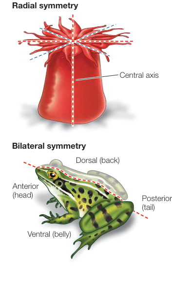
Figure 29.5
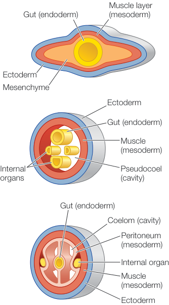
Figure 29.6
Animal Life Cycles Involve Trade-Offs
- ■ The stages of an animal’s life cycle may be specialized for different activities. An immature stage whose morphology is dramatically different from that of the adult stage is called a larva.
- ■ Most animal life cycles have at least one dispersal stage.
- ■ A characteristic of an animal or a life cycle stage may improve the animal’s performance in one activity but reduce its performance in another, a situation known as a trade-off.
- ■ Parasites have complex life cycles that may involve one or more hosts and several larval stages.
- ■ In some groups of animals, asexual reproduction without fission leads to the formation of colonies composed of many genetically homogeneous, physiologically integrated individuals.
Questions
- How can specialized stages of life cycles facilitate dispersal?
- How might specialized stages of life cycles allow animals to reduce competition between juveniles and adults?
- How can complex life cycles with specialized stages help parasites move among multiple hosts?
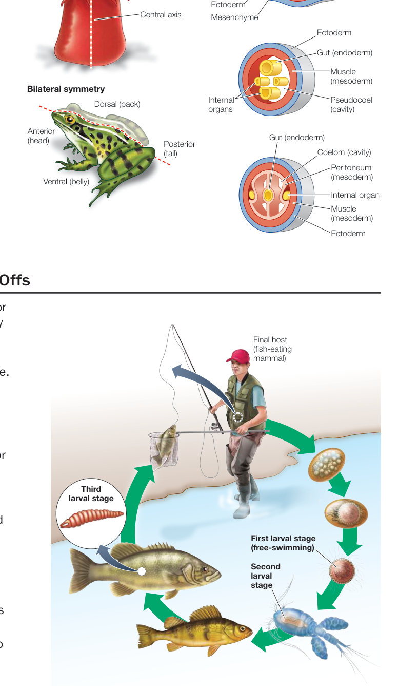
Figure 29.12
- ■ Several large, multicellular groups diverged early in animal evolution, including sponges, ctenophores, placozoans, cnidarians, and bilaterians. The ancestral animal was unlike any of the modern groups and was likely small and composed of relatively few cells and no differentiated tissues.
- ■ Complex nervous systems developed several times in animals, including in ctenophores, cnidarians, and bilaterians.
- ■ The life cycle of most cnidarians has two distinct stages: a sessile polyp stage and a motile medusa. A fertilized egg develops into a free-swimming larval planula, which settles to the bottom and develops into a polyp.
Questions
- Which two groups are considered the most likely candidates to be the sister group of the remaining animals?
- Which animal groups lack a nervous system?
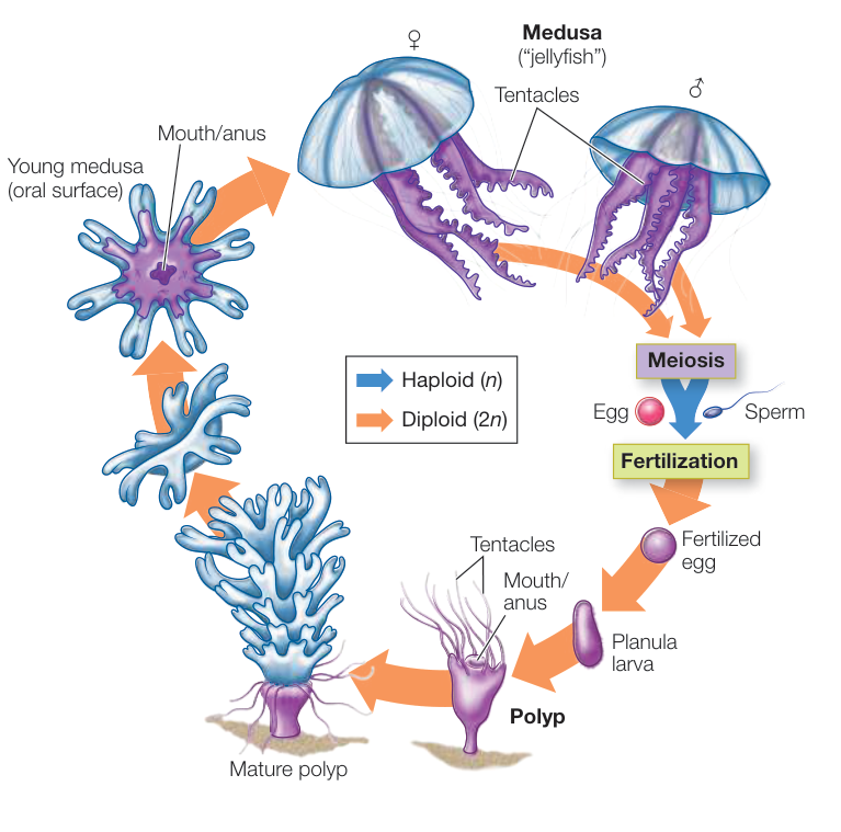
Figure 29.19