Chapter 35Regulation of Plant Growth
Key Concepts
35.1 Plant Development Occurs Progressively as Plants Mature
35.2 Gibberellins and Auxin Have Diverse Effects but a Generally Similar Mechanism of Action
35.3 Other Plant Hormones Have Diverse Effects
35.4 Photoreceptors Initiate Developmental Responses to Light
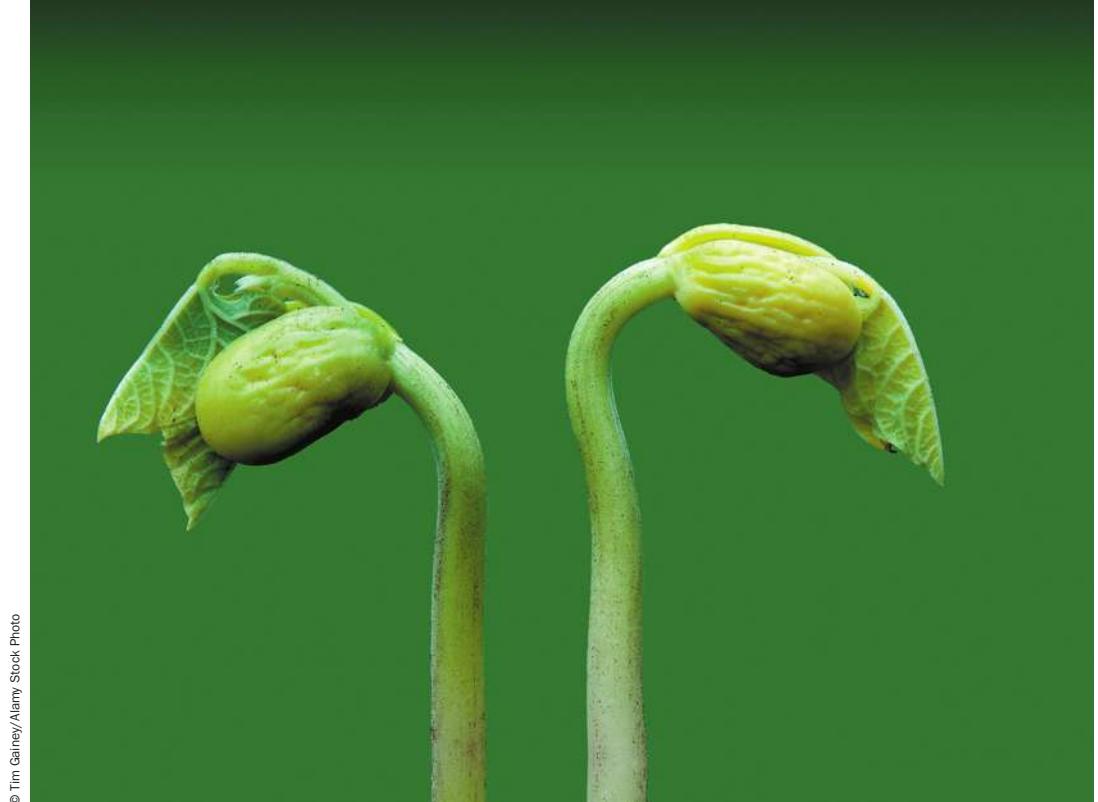
The root, stem, and leaf develop from a plant embryo within a seed. Here, two seedlings are shown opening their first leaves.
© Tim Gaines/Alemy Stock Photo
Investigating Life: A Nobel Prize for a Plant Biologist
In their constant search for ways to help farmers produce more food for a growing population, biologists have developed cereal crops whose physiology allows them to produce more grain per plant (resulting in higher yields). The drawback of this approach is that the sheer weight of the load of seeds may cause the stem to bend over. The problem is made worse when fertilizer added to the soil stimulates plants to grow taller. Harvesting seeds on the ground is very difficult; think of how hard it would be to pick up seeds one by one, when some have already sprouted.
During World War II, Japan, an island nation with a limited amount of land suitable for farming, was blockaded and could not import food or other supplies. Food was rationed and many people were hungry, but there were no major famines in Japan during that period. How were the Japanese able to produce enough grain to feed their population? The answer to this question lay in the fields: the Japanese had bred genetic strains of rice and wheat with short, strong stems, growing in thickness rather than height, that could bear high yields of grain without bending over. An agricultural advisor to the American army that temporarily occupied Japan in 1945 sent samples of the grains to the United States.
A decade later, the American plant geneticist Norman Borlaug, who was working in Mexico at the time, began making genetic crosses between the Japanese wheat and other varieties that had genes conferring rapid growth, adaptability to varying climates, and resistance to fungal diseases. The results were “semi-dwarf” wheat varieties that gave record yields. The varieties were grown first in Mexico, and later in India and Pakistan during the 1960s. At about the same time and using a similar strategy, scientists in the Philippines developed semi-dwarf rice with equally spectacular results. People who had lived on the edge of starvation now produced enough food. Countries that had relied on food from other countries were now able to grow more than enough grain, and export the surplus. The development of these semi-dwarf grains began what was called the “green revolution.” Borlaug was awarded the Nobel Peace Prize for his research on wheat, which is estimated to have saved a billion lives.

© Micheline Pelletier Decaux/Getty Images
Qanta How is plant growth controlled, and what changes in growth patterns made the new strains of wheat and rice successful? (Find the answer on page 799.)
Plant Development Occurs Progressively as Plants Mature
Learning Objectives
- 35.1.1 Describe how plant cells increase in size by taking in water.
- 35.1.2 Explain how expression of a transcription factor can establish the identity of a cell.
- 35.1.3 Hypothesize as to why retaining the ability to undergo redifferentiation is advantageous to plants.
- 35.1.4 List the ways a seed can maintain dormancy, and describe with examples how each method might be overcome to achieve germination.
- 35.1.5 Construct a diagram that shows how a genetic screen can be used to identify the genes in the development of plant signal transduction pathways.
As organisms move through their life cycles, they change continually. A gardener who plants a seed may talk about the resulting plant as if it were a single thing, but the size and structure of the seed, seedling, leafy plant, and fruit-bearing plant are always different. Development is the term used to describe the process by which a plant or other organism changes, taking on the successive forms that characterize its life cycle. Plant development occurs through a combination of growth, an increase in size of the body and its organs, and differentiation, the process by which cells become different from one another. Because plants are rooted in place, their survival depends on their ability to cope with the environment around them. They can’t run away or put on a coat as we can. Their remarkable ability to produce new organs and reshape their bodies throughout their lives plays a significant role in their ability to thrive in a variety of environmental circumstances.
Cell expansion increases the size of a plant cell and gives it shape
The size of a plant increases when cells expand. Cell expansion often leads to cell division, which partitions the existing material into smaller units, so increases in size and cell number are highly correlated. Plant cells increase in size by taking in water in what is called the stress-relaxation cycle (Figure 35.1). The cycle begins when pressure from the cell contents (turgor pressure) acts on the cell wall. This physical stress alters the structure of the material that makes up the cell wall. Some of the changes that occur in the cell wall are reversed when the pressure decreases, but some of the changes are not reversible—much as when someone pulls on a swimsuit that is too small and the material becomes permanently stretched out. Once the structure of the cell wall changes, the wall “relaxes.” Relaxation of the wall causes the internal turgor pressure to fall—because the forces between the cell and its wall are equal and opposite. Having a lower internal pressure can result in more water entering the cell, continuing the cycle.
The rate of cell wall expansion can be regulated by changing the chemical composition of the wall. Some plant hormones increase the rate of cell expansion by making the wall easier to extend (“looser”). That is, they change the composition of the wall such that a given amount of pressure results in a larger expansion of the cell. These hormones promote growth.
Growing plant cells must synthesize new wall material that adds to the thickness of their walls; otherwise their walls would continually become thinner. Most plant cell walls are composed of long filaments called cellulose microfibrils that are embedded in a network of polysaccharides and proteins. Cellulose is a polymer composed of repeating glucose units joined in a straight chain
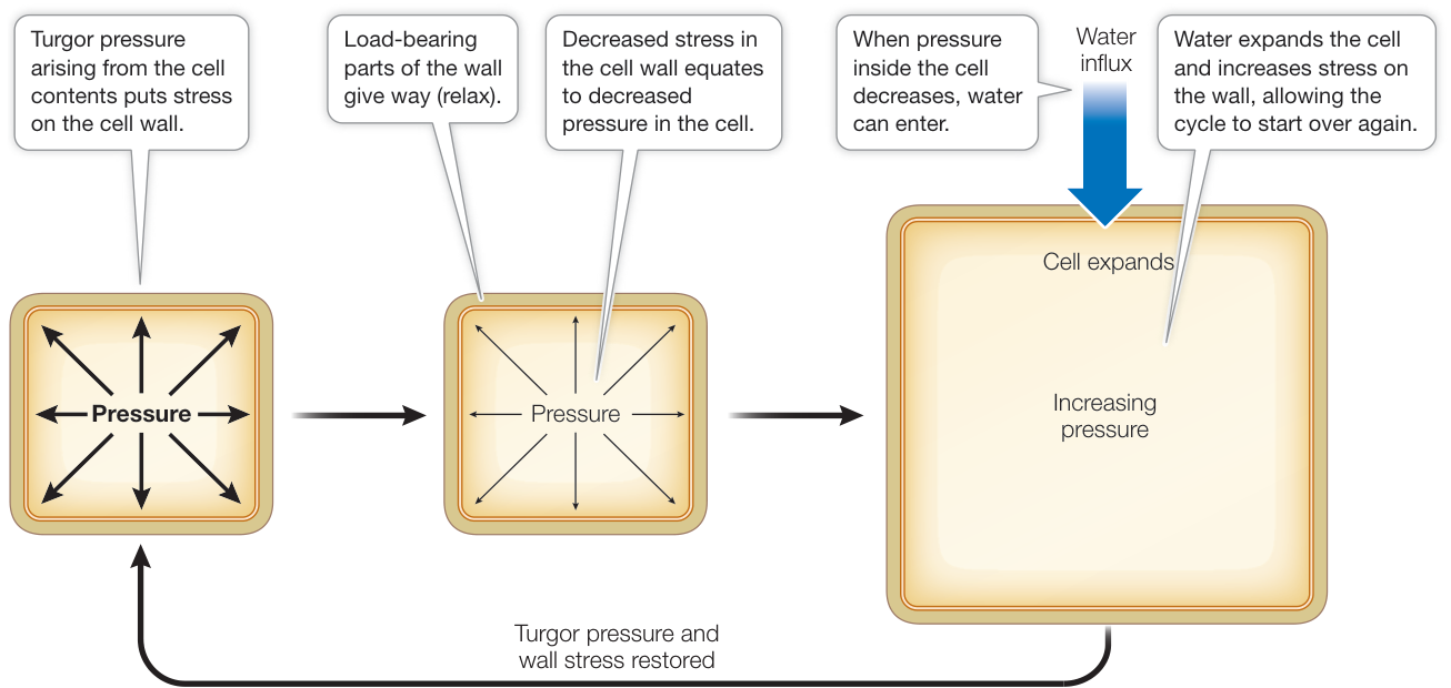
Figure 35.1 Plant Cell Expansion Occurs via the Stress-Relaxation Cycle (After D. J. Cosgrove. 2015. J Exp Bot 67: 463–476.)
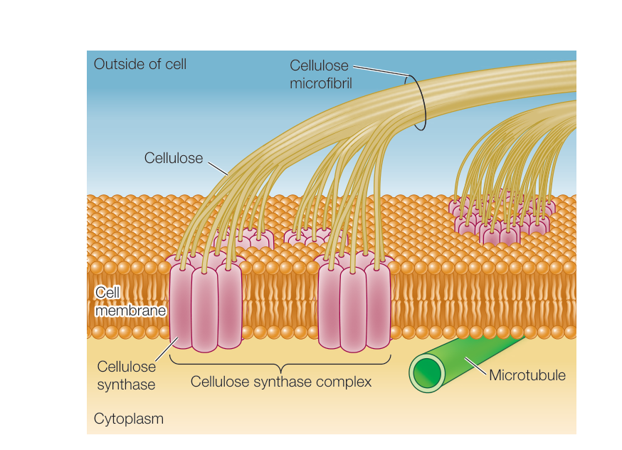
Figure 35.2 Cellulose Synthase Complexes Cellulose synthase complexes form chains of glucose (glucan) that come together in the cell wall space to form cellulose microfibrils.
(see Figure 3.19). Cellulose microfibrils are synthesized by protein complexes called cellulose synthases that sit in the cell membrane, receiving subunits from the cytoplasm and releasing microfibril chains toward the cell wall (Figure 35.2). The cellulose synthase complexes synthesize multiple chains at a time, and these chains pack together to form a single microfibril. Cellulose microfibrils provide strength and rigidity to the wall.
If plant cells lacked a stiff restraint, they would grow as spheres and plants might resemble beach balls. Neither plants nor plant cells are entirely spherical, however. Epidermal cells on the leaf can be shaped like pieces of a jigsaw puzzle, while cells in an elongating stem are often cylindrical. Nonspherical shapes arise because the plant has the ability to regulate the extent of growth in one area versus another. Typically, the wall in one part of a cell is softer or easier to loosen than the wall in other parts of the cell. These differences give an asymmetry to cell expansion that affects the overall shape of a plant.
Whether a cell continues to expand in the same manner or changes its direction of expansion is of central importance to the development of the organism. Changes in the direction of expansion are responsible for the formation of branches and complex shapes. The direction of maximal cell expansion usually can be predicted by the arrangement of cortical microtubules, cytoskeletal proteins that are anchored to the cell membrane. Cortical microtubules are linked to the protein complexes that synthesize cellulose, and the orientation of the cortical microtubules appears to direct the orientation in which cellulose microfibrils are laid down in the cell wall. For elongated cells in a growing stem, both the microtubules and cellulose microfibrils wrap around the narrow axis of the cell, like hoops in a wine barrel (Figure 35.3). Cellulose microfibrils resist expansion, resulting in there being more growth in length than in width. The orientation of microtubules is highly regulated by the proteins that bind to them and gives the plant a mechanism for controlling the shape of its body.
Cell fates become progressively more restricted during development
As plants mature, cells not only get larger, but also acquire different characteristics. A zygote is a single cell that gives rise to an embryo and, from that, an organism. The cells of an early embryo are totipotent (toti, “all,” + potent, “capable”), meaning they have the potential to differentiate into any cell type, including more embryonic cells. As the embryos of many organisms mature, cells become progressively restricted in their potential, eventually being limited to producing just one cell type. When a cell has taken on the characteristics of a single specific cell type, we say that it has differentiated. That differentiated state reflects the cell fate.
While plant cells undergo progressive changes in their identity as the plant develops, plant cells are remarkable in the extent to which they remain capable of redifferentiation. It is possible to cut sections of mature leaves or roots and grow them in culture. Culturing plant cells causes them to lose their prior differentiated state and form a callus (plural calli)—a cluster of dividing cells. Given appropriate hormonal treatments, callus cells can redifferentiate to form a whole new plant. In 1958 Frederick Steward at Cornell University showed that if he isolated cells from a carrot root and maintained them in a suitable nutrient medium, he could induce them to divide and give rise to masses of calli, which no longer had the cell-type characteristics that they started with. These callus cells could be maintained in culture indefinitely. Furthermore, if the callus cells were provided with the right chemical cues, they could develop into embryos and eventually into complete new plants (Figure 35.4). The new plants in Steward’s experiments were genetically identical to the cells from which they came—that is, they were clones of the original carrot plant. The ability to produce clones is evidence that the somatic cells in a plant have all the genetic information needed to become any cell in the plant. The next section explores the mechanisms by which distinct cell fates are specified.
Changes in gene expression underlie cell fate determination and differentiation
A central component of development is the ability to form distinct cell types. The cells in human eyes contain proteins that allow us to see light, while intestinal cells produce digestive enzymes; if eyes produced digestive enzymes, they would not work very well! The same is true for plants. Epidermal cells in the root contain proteins that span the cell membrane and promote uptake of nutrients from the soil, while leaves contain proteins that promote photosynthesis. Given that each cell in a multicellular organism generally contains the same genes, how do the differences between cell types arise? The answer is that the distinction between cells largely arises from the proteins they contain.
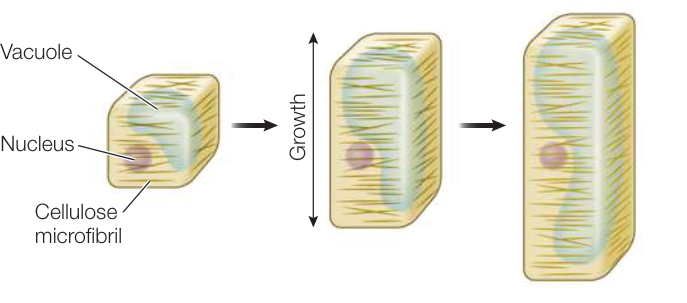
Figure 35.3 The Orientation of Cellulose Microfibrils Affects the Direction of Cell Expansion Transverse cellulose microfibrils restrict lateral expansion, resulting in an elongated shape.
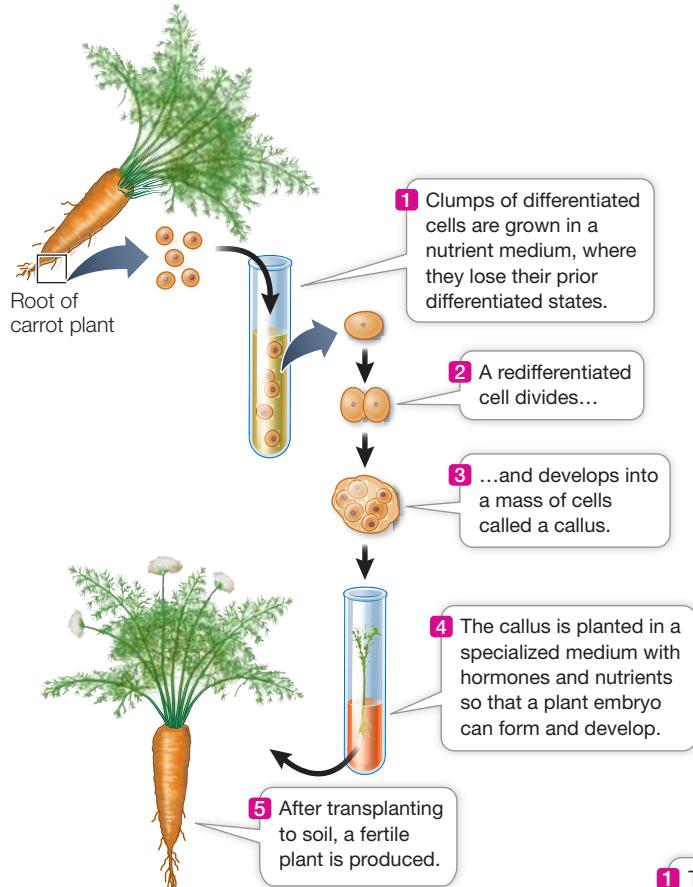
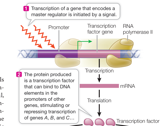
Figure 35.4 Differentiated Plant Cells Can Be Totipotent When differentiated plant cells are removed from the plant and placed in medium with nutrients and specific growth hormones, they can redifferentiate into callus and then be induced to generate an entire new plant. (After F. C. Steward. 1958. Am J Bot 45: 705–709.)
Transcription factors, a class of proteins that bind to regulatory sequences in DNA, play a central role in regulating gene expression (Figure 35.5). If a transcription factor affects the expression of other transcription factors, a cascade is generated, amplifying the effect. Some transcription factors that sit at the head of such cascades are called master regulators because their expression can cause a cell to produce or repress so many proteins that the cell takes on a new cell fate. Development can thus be seen as a question of how cells come to express different transcription factors, particularly the master regulators that cause a cell to differentiate into its final cell fate.
An example of plant transcription factors that act to specify cell fate is found in the PLETHORA (PLT) family. Stem cells are cells that both maintain themselves and give rise to the cells that produce differentiated cell types. In plants, stem cells can be found in the meristems: think of the vascular cambium that maintains itself while producing cells that form the xylem and phloem, or of the divisions that take place in the center of the shoot apical meristem that give rise to the cells that form the stem and leaves. In the root meristem, stem cells surround the quiescent center, a small group of cells that rarely divide (Figure 35.6A). If the root meristem did not contain stem cells, the cell production that leads to regular growth of the root would stop. PLETHORA genes encode transcription factors that are required for the formation and maintenance of stem cells. Experiments examining mutant plants that have lost the function of multiple PLETHORA proteins have demonstrated that PLETHORA proteins confer the stem cell identity. Plants lacking functional copies of PLT1 and PLT2 have short roots, while embryos lacking PLT1, PLT2, and PLT3 form no root at all (Figure 35.6B). In contrast, when high levels of PLT2 protein are induced in the shoot apical meristem, the meristem gives rise to a structure that resembles a root (Figure 35.6C)! A mutation in which one type of organ is replaced by another is called a homeotic mutation.
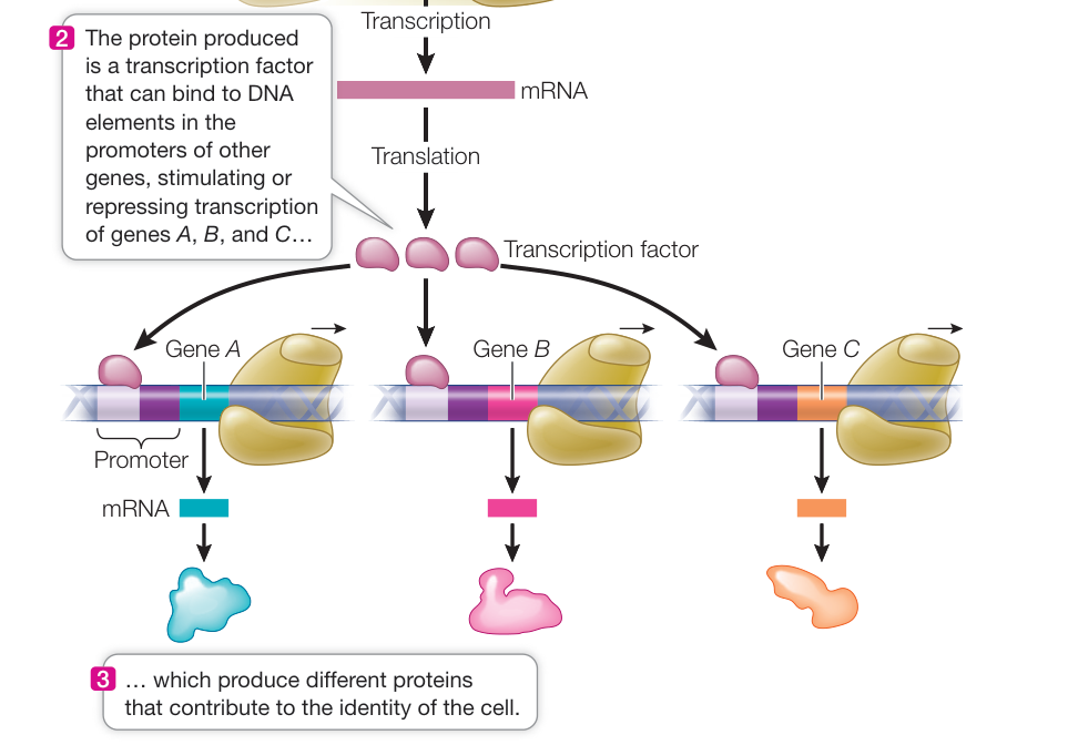
Figure 35.5 Coordinating Gene Expression A single signal causes the synthesis of a transcription factor that acts on many genes.
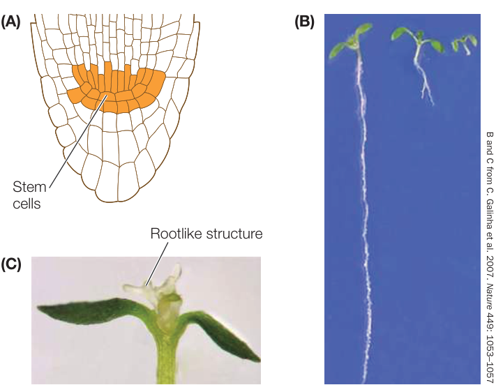
Figure 35.6 PLETHORA Expression Maintains Root Stem Cells Loss of PLETHORA (PLT) function results in failure to maintain the stem cells in the root. (A) Diagram showing the stem cells and the quiescent center in the root meristem. (B) A wild-type root is shown on the left; a plt1,plt2 double mutant in the center; and a plt1,plt2,plt3 triple mutant on the right. When stem cells are not maintained, the root in the mutant plants does not continue to elongate. (Wild-type plant gene names are written in uppercase, mutant gene names in lowercase.) (C) Inducing expression of PLETHORA 2 in the shoot apical meristem results in the formation of a rootlike structure there.
Genetic screens have increased our understanding of molecular pathways that regulate development
A method that has been particularly helpful in identifying factors that regulate development is called a genetic screen. If a mutation in a specific gene disrupts a developmental process, then the product of that gene must be involved in that process. In a similar way, genetic studies can be used to analyze signal transduction pathways in plants: if proper signaling does not occur in a mutant plant, then the mutant gene must be involved in the signal transduction process. Mapping the mutant gene and identifying its function is a starting point for understanding the signaling pathway. Arabidopsis thaliana has been a major model organism for plant biologists investigating signal transduction.
One technique for identifying the genes involved in a plant signal transduction pathway is illustrated in Figure 35.7. This genetic screen involves creating a large, random collection of mutant plants and identifying those individuals that are likely to have a defect in the pathway of interest. Plant genes can be randomly mutated in a variety of ways, including treatment with a chemical mutagen or the random insertion of transposons (see Key Concept 17.2) in the genome. After treatment, the plants are grown and then examined for a specific phenotype, usually a characteristic that is influenced by the pathway of interest. Once mutant plants have been selected, their genotypes are compared with those of wild-type plants. A. thaliana mutants with altered developmental patterns have provided a wealth of new information about the mechanisms of hormone and receptor action.
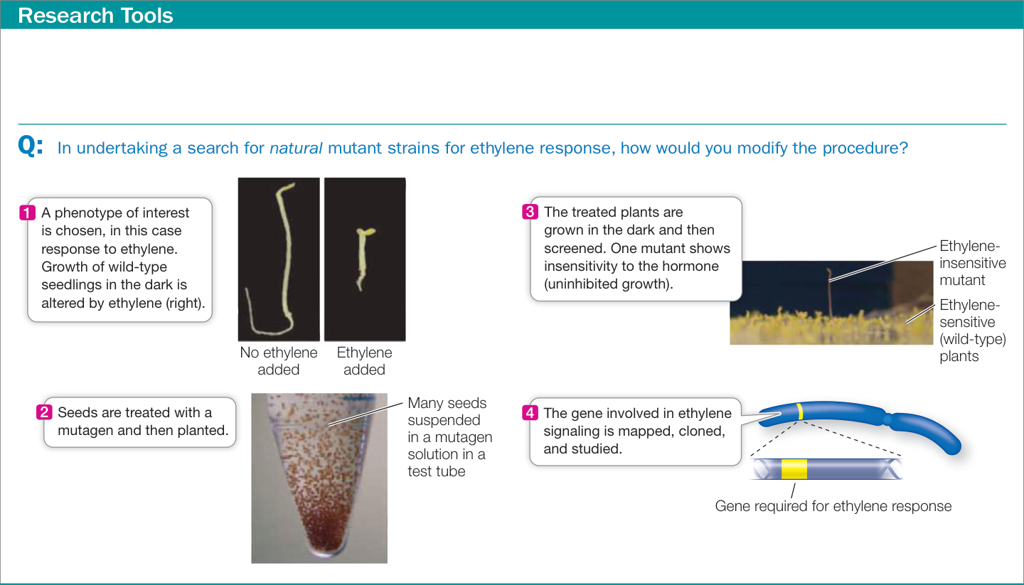
Figure 35.7 A Genetic Screen Genetics of the model plant Arabidopsis thaliana can be used to identify the steps of a signal transduction pathway. If a mutant strain does not respond to a hormone (in this case, ethylene), the corresponding wild-type gene must be essential for the pathway (in this case, ethylene response). This method has been instrumental to scientists in understanding plant growth regulation. (All photos from J. M. Alonso and J. R. Ecker. 2006. Nat Rev Genet 7: 524–536.)
In early development, the seed germinates and forms a growing seedling
To see how some of the factors discussed so far come together, let’s consider the first stages of plant development, beginning with the seed. The events of plant reproduction and development that lead to the formation of seeds are discussed in Chapter 36. A seed is the structure that contains the early embryo. Unlike most animal embryos, plant seeds may be held in “suspended animation,” with the development of the embryo halted, for long periods. If development stops even when external conditions (such as water supply) are adequate for development, the seed is said to be dormant.
DORMANCY Seed dormancy may last for weeks, months, or years. Plants use several mechanisms to maintain dormancy:
- Exclusion of water or oxygen from the embryo by an impermeable seed coat
- Mechanical restraint of the embryo by a tough seed coat
- Chemical inhibition of germination by growth regulators
- Photodormancy: some seeds need a period of light or dark before they can germinate
- Thermodormancy: some seeds need high or low temperatures to germinate
Dormancy can be broken by conditions that overcome these mechanisms. For example, the seed coat may be damaged by passage through an animal’s digestive system, or heavy rains may wash away chemical inhibitors. There are some unusual methods to overcome dormancy. One example is the breaking of dormancy by components of smoke. Emmenanthe penduliflora is a common plant in dry chaparral of the southwestern United States, an area that is prone to wildfires. These plants germinate rapidly after a fire. Jon Keeley of Occidental College in Los Angeles found that dormancy in seeds of this plant is broken not by heat but by smoke—in particular, by the nitrogen oxides found in smoke. Other molecules in smoke have been identified that regulate seed germination.
Plant biologists distinguish between seed dormancy, which prevents germination under conditions that are suitable for plant growth, and seed quiescence, which occurs when a seed fails to germinate because conditions are unfavorable for growth. Some seeds may remain quiescent, yet viable, for centuries; botanists have germinated a 1,300-year-old lotus seed recovered from a dry lake bed in China.
Seed dormancy and quiescence are common, so they must provide selective advantages for plants. Dormancy ensures that the seed will germinate at a time suitable for the plant to complete its life cycle. For example, some seeds require exposure to a long cold period (winter) before they germinate in the spring; this ensures that the plant has the entire growing season to mature and set new seeds. Dormancy and quiescence also help seeds survive droughts or long-distance dispersal, allowing plants to colonize new territory.
GERMINATION Seeds begin to germinate, or sprout, when dormancy is broken and environmental conditions are satisfactory.
The first step in germination is the uptake of water, called imbibition (from imbibe, “to drink in”). Before germination, a seed contains very little water: only 5%–15% of its weight is water, compared with 80%–95% in most other plant parts. Seeds also contain polar macromolecules, such as cellulose and starch, which attract and bind water molecules. Consequently a seed has a very negative water potential (see Chapter 33) and will take up water if the seed coat is permeable to water. The force exerted by imbibing seeds, which expand several-fold in volume, demonstrates the magnitude of their water potential; for example, imbibing cocklebur seeds can exert a pressure of up to 1,000 atmospheres.
As a seed takes up water, it undergoes metabolic changes: enzymes are activated upon hydration, RNA and then proteins are synthesized, the rate of cellular respiration increases, and other metabolic pathways are activated. In many seeds, cell division is not initiated during the early stages of germination. Instead, growth results solely from the expansion of small, preformed cells.
As germination proceeds, starch, proteins, and lipids that are stored in the seed are hydrolyzed to provide metabolic energy and chemical building blocks—carbohydrates, amino acids, and lipid monomers—for the growing embryo. These reserves are stored in the cotyledons (the first leaf or leaves of the embryo) or in the endosperm (the non-embryonic storage tissue of the seed). Germination is completed when the radicle (embryonic root) emerges from the seed coat. The plant is then called a seedling.
If the seed germinates underground, the new seedling must elongate rapidly (in the right direction!) and cope with a period of life in darkness or dim light. Photoreceptors that sense light and specialized cells that sense gravity direct this stage of development and prepare the seedling for growth in the light.
The pattern of early shoot development varies among the flowering plants. Figure 35.8 shows the shoot development patterns of monocots and eudicots. In monocots, the growing shoot is protected by a sheath of cells called the coleoptile as it pushes its way through the soil. In eudicots, the shoot is protected by the cotyledons.
Several hormones and photoreceptors help regulate plant growth
The description just provided of the early stages of plant development illustrates some of the environmental cues that influence plant growth. When part of a plant, such as a leaf cell, senses a change in its environment, signal transduction pathways that result in the synthesis and activation of specific plant hormones may be stimulated. The resulting hormones function as signals that can act locally or, in many cases, travel to other parts of the plant where they can trigger pathways that affect plant growth. In most cases these changes involve alterations in the expression of specific genes.
Perhaps the most important environmental cue for a plant is light: the source of energy for photosynthesis. Plants have an abundance of photoreceptors that detect changes in the quality and direction of light as well as the timing of light availability (day length). Photoreceptors are often proteins associated with pigments. Light acts directly on photoreceptors, which in turn regulate developmental processes that need to be responsive to light, such as the many changes that occur as a seedling emerges from the soil.
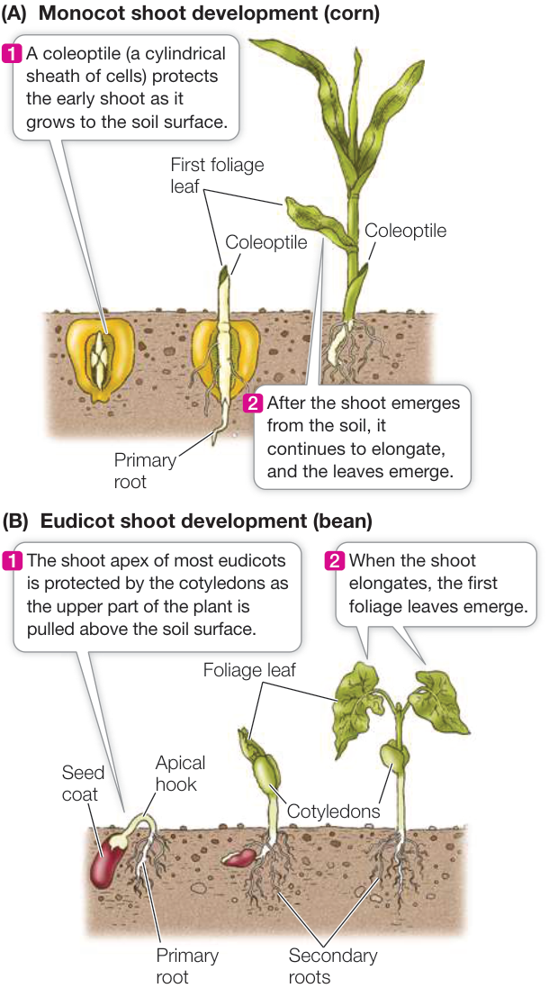
Figure 35.8 Patterns of Early Shoot Development (A) In grasses and some other monocots, growing shoots are protected by a coleoptile until they reach the soil surface. (B) In most eudicots, the growing point of the shoot is protected within the cotyledons. - - Activity 35.1 Monocot Shoot Development - - Activity 35.2 Eudicot Shoot Development
Recap and Assess
Plants grow and produce new organs throughout their lives, a feature that allows them to acclimate to changing environmental conditions. Cell expansion occurs when the internal pressure of the cell leads to structural changes in the wall that reduce that pressure, allowing more water to enter. Cells differentiate into distinct cell types largely as a result of changes in gene expression. Early embryonic development in many plants occurs within seeds, which may remain dormant for a long period. Water uptake (imbibition) and germination occur when external cues in the environment are present.
- Describe how cell expansion occurs in the context of the stress-relaxation cycle.
- Explain how the activity of a transcription factor can determine cell type. What is it that the transcription factor does?
- Under what conditions would it be advantageous for a species to have seed dormancy broken by fire?
- How would a genetic screen be used to identify a plant gene involved in seed dormancy?
You have now seen the early stages of plant growth and development, and how the environment influences these processes. We turn now to the subject of plant hormones, which are central to the internal regulation of development.
Gibberellins and Auxin Have Diverse Effects but a Generally Similar Mechanism of Action
Learning Objectives
- 35.2.1 Provide evidence for the role of gibberellins in various plant growth processes.
- 35.2.2 Provide evidence for the role of auxin in plant growth and development.
- 35.2.3 Explain the mechanisms that result in shoots growing toward the light.
Hormones are chemical signals that act at very low concentrations at target sites often distant from where they are produced. Most plant hormones differ from their counterparts in animals in that each plant hormone plays multiple regulatory roles (Table 35.1). Several plant hormones regulate the growth and development of plants from seedling to adult (Table 35.2). Other hormones (for example, jasmonic acid and salicylic acid) are involved in plant defenses against herbivores and microorganisms, as we will discuss in Chapter 37.
Early in the twentieth century, two plant hormones were discovered: gibberellins (there are several active forms) and auxin (there are several forms, but one predominates). Initially, the discoveries came from observations of natural phenomena:
- Gibberellins: In rice plants, a disease caused by the fungus Gibberella fujikuroi resulted in plants that grew overly tall and spindly.
Table 35.1 Comparison of Plant and Animal Hormones
| Characteristic | Plant hormones | Animal hormones |
|---|---|---|
| Size, chemistry | Peptides, small organic molecules | Peptides, proteins, small molecules |
| Site of synthesis | Usually at many locations | Specialized glands or cells |
| Site of action | Local or distant | Usually distant, transported |
| Effects | Often diverse | Often specific |
| Regulation | By biochemical feedback | By central nervous system, ions, or feedback |
Table 35.2 Plant Growth Hormones
*See Chapter 36.
- Auxin: Biologists (including Charles and Francis Darwin) and indoor gardeners noted that seedlings would bend toward the light when placed near a light source.
A chemical substance was then isolated that could cause each phenomenon:
- Gibberellic acid (see Table 35.2) made by the G. fujikuroi fungus caused rice plants to overgrow. In addition, the short (dwarf) pea plants that Gregor Mendel worked with during his groundbreaking discoveries on the nature of inheritance (see Chapter 12) were found to carry a loss-of-function mutation in a gene that encodes an enzyme involved in gibberellin biosynthesis (Figure 35.9A). Wild-type plants that do not have this mutation make more gibberellin and grow taller.
- Indole-3-acetic acid (auxin, see Table 35.2) applied asymmetrically to the growing tips of seedlings caused cell elongation on the side away from the light, which resulted in the shoot bending toward the light. Furthermore, the appearance of mutant plants that do not make auxin could be restored to that of wild-type plants by adding auxin. Arabidopsis thaliana plants that do not make auxin are short (and have smaller leaves and other defects); supplying them with auxin reverses those characteristics and restores a wild-type appearance (Figure 35.9B).
Note that the phenotype involved—short stature, or dwarfism—is similar in both mutant plants even though different hormones are involved.
The approaches we have outlined—observation, hormone isolation, hormone treatment, and analysis of mutant plants—are just some of the methods used to identify plant hormones and understand their roles in plant development. Plant biologists have also studied hormones using chemical inhibitors and using plant transformation experiments that alter hormone levels or the plants’ responses to hormones.
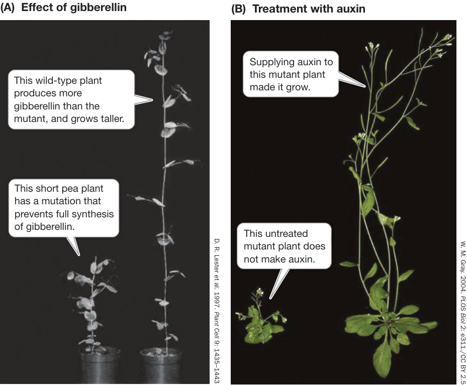
Figure 35.9 Hormones Reverse a Mutant Phenotype (A) The short (dwarf, left) pea plants that Mendel studied are now thought to have arisen because of a loss-of-function mutation in a gene that encodes an enzyme involved in gibberellin synthesis. (B) The short phenotype of this Arabidopsis thaliana mutant was reversed in the plant on the right by supplying auxin.
Gibberellins have many effects on plant growth and development
The functions of gibberellins can be inferred from the effects of experimentally decreasing gibberellins or blocking their action at various points in plant development. Such experiments reveal that gibberellins have multiple roles in regulating plant growth.
STEM ELONGATION The effects of gibberellins on wild-type plants are not as dramatic as those seen on dwarf plants. However, gibberellins are indeed active in wild-type plants, because inhibitors of gibberellin synthesis cause a reduction in stem elongation. Such inhibitors can be put to practical uses. For example, plants such as chrysanthemums that are grown in greenhouses tend to get tall, but leggy plants do not appeal to consumers. Flower growers spray such plants with gibberellin synthesis inhibitors to control their height. Some wheat crops are similarly sprayed to keep them short, so they do not fall over when they produce grain; the result is chemically produced semi-dwarfs similar to the genetically produced varieties described in the opening story of this chapter. In some plants, such as cabbage, the normal growth habit is to be a squat, leafy head near the ground. When environmental signals are right, however, the plant “bolts,” quickly producing a tall stem with flowers—a response that can be mediated by gibberellins.
FRUIT GROWTH Gibberellins and other hormones regulate the growth of fruits. Grapevines that produce seedless grapes develop smaller fruits than varieties that produce seed-bearing grapes. Biologists wanting to explain this phenomenon removed seeds from immature seeded grapes and found that this prevented normal fruit growth, suggesting that the seeds are sources of a growth regulator. Biochemical studies showed that developing seeds produce gibberellins, which diffuse into the immature fruit tissue. Spraying young seedless grapes with a gibberellin solution causes them to grow as large as seeded ones, and this is now a standard commercial practice (Figure 35.10).
MOBILIZATION OF SEED RESERVES Early in seed germination, hydrolytic enzymes are produced to break down stored reserves of starch, proteins, and lipids. Just after imbibition in germinating seeds of barley and other cereals, the embryo secretes gibberellins. The hormones diffuse through the endosperm to a surrounding tissue called the aleurone layer, which lies underneath the seed coat. The gibberellins trigger a cascade of events in the aleurone layer, causing it to synthesize and secrete enzymes that hydrolyze proteins and starch stored in the endosperm (Figure 35.11).
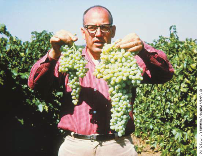
Figure 35.10 Gibberellins and Fruit Growth Spraying developing seedless grapes with gibberellins (right) increases their size compared with untreated fruit (left).
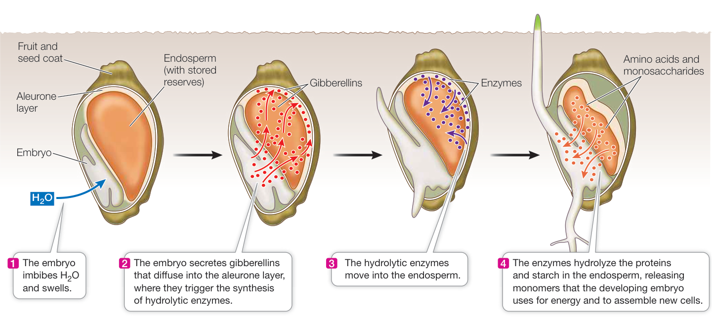
Figure 35.11 Embryos Mobilize Their Reserves During seed germination in cereal grasses, gibberellins trigger a cascade of events that result in the conversion of starch and protein reserves into monomers that can be used by the developing embryo.
Q: In brewing beer, stored starch in barley seeds is converted to maltose. How do you think this is done in the brewery?
These observations have practical importance: in the beer brewing industry, gibberellins are used to enhance the “malting” (germination) of barley and the breakdown of its endosperm, producing sugar that is fermented to alcohol.
Another hormone, abscisic acid (ABA) (see Table 35.2) plays an antagonistic role with gibberellins in seed dormancy and germination. ABA levels are high in dormant seeds and fall off during germination. Thus ABA plays a role in maintaining seed dormancy, whereas gibberellins function to break dormancy and promote germination.
Auxin plays a role in differential plant growth
Auxin was first discovered for its role in the bending of plants toward light. Subsequent research has shown that auxin is involved in many other aspects of plant growth and development.
IDENTIFYING AUXIN AND ITS TRANSPORT Auxin (from the Greek “to increase”) was discovered in the context of phototropism: a response to light in which plant stems bend toward a light source. This was a familiar observation by biologists and home gardeners when the ever-curious Charles Darwin and his son Francis investigated this phenomenon and concluded that a signal was made in the shoot apex and diffused down the shoot in a polar (unidirectional) fashion, stimulating cell elongation.
The Darwins worked with canarygrass (Phalaris canariensis) seedlings grown in the dark. While underground, a young grass seedling is protected by a coleoptile (see Figure 35.8). The coleoptiles of grasses are phototropic—they grow toward the light (Figure 35.12). To find the light-receptive region of the coleoptile, the Darwins “blindfolded” the coleoptiles of dark-grown canarygrass seedlings in various places and then illuminated them from one side (Investigating Life: The Darwins’ Phototropism Experiment). The coleoptile grew toward the light whenever its tip was exposed. If the top millimeter or more of the coleoptile was covered, however, the coleoptile showed no phototropic response. The Darwins concluded that the tip contains the photoreceptor that responds to light. The actual bending toward the light, however, takes place in a growing region a few millimeters below the tip. Therefore, the Darwins reasoned, some type of signal must travel from the tip of the coleoptile to the growing region.
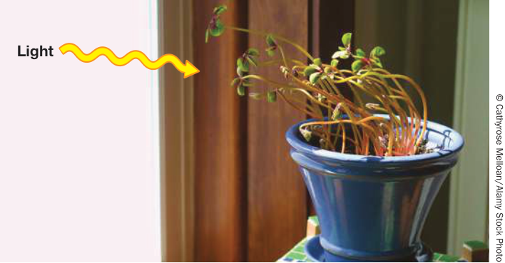
Figure 35.12 Phototropism Plants grow toward the light by elongating more on the side away from the light, and less on the side toward to the light. This difference in elongation generates a curve.
Investigating Life: The Darwins’ Phototropism Experiment
Experiment
Original Paper: C. R. Darwin assisted by F. Darwin. 1880. The Power of Movement in Plants. Chapter IX: Sensitiveness of plants to light: Its transmitted effects. John Murray: London.
Charles Darwin and his son Francis wanted to know how plants bend toward the light. They grew canarygrass seedlings (coleoptiles) in the dark. To discover what part of the coleoptile responds to light, they covered up (“blindfolded”) different regions of each coleoptile and then exposed the seedlings to light from one side. The Darwins discovered that the tip of the seedling senses the light and that growth occurs below the tip. Their observations led them to hypothesize the existence of a growth-promoting signal produced by the coleoptile tip.
HYPOTHESIS Only part of the coleoptile senses the light that triggers phototropism.
METHOD
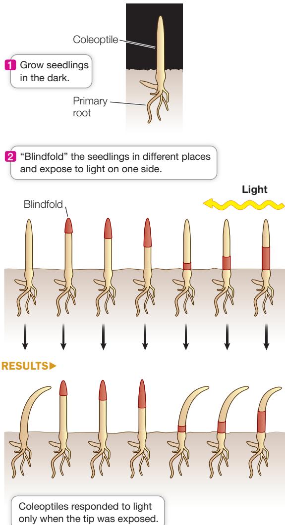
CONCLUSION The coleoptile tip senses light and then transmits a growth signal to the tissue away from the light source.
Work with the Data
Fascinated by experiments showing that light induces plants to bend toward it and that this is due to increased growth on the “dark side” of the stem, Charles Darwin and his son Francis performed additional experiments on canarygrass (Phalaris canariensis) seedlings grown in the dark to learn more about how the seed germinates in the soil. Their conclusion that the light-sensing mechanism might reside in the coleoptile was validated when they repeated their experiments with the coleoptile tip cut off: bending did not occur. Because it was possible that the cut plants did not bend because they had been irreversibly damaged in some way, the Darwins repeated their initial experiment with undamaged seedlings whose coleoptile tips had been covered with a thin glass tube cap that was blacked out with India ink. In this case the plants did not bend. The Darwins’ conclusion that the tip of the coleoptile senses the light and transmits a message to tissue below the tip to grow was initially controversial but ultimately led to the identification of auxin, the first plant growth hormone.
QUESTIONS
- The figure shows a drawing by the Darwins of the bending of coleoptiles after 8 hours of light exposure. From which direction was the light shone?
Fig. 181

- The Darwins reported:
“Seven cotyledons [Note: The Darwins used the term ”cotyledon” for what is now called a coleoptile.] had their tips cut off for lengths varying between 0.1 and 0.16 of an inch, and these, when left exposed all day to a lateral light, remained upright. In another set of 7 cotyledons, the tips were cut off for a length of only about 0.05 of an inch (1.27 mm) and these became bowed towards a lateral light, but not nearly so much as the many other seedlings in the same pots.”
What do these data indicate about the possible role of the tip and about the possibility that injury in cutting blocks the bending response?
- The Darwins described their further experiments:
“The summits of nine cotyledons, differing somewhat in height, were enclosed for rather less than half their lengths in uncoloured or transparent tubes; and these were then exposed before a southwest window on a bright day for 8 h. All of them became strongly curved towards the light, in the same degree as the many other free seedlings in the same pots; so that the glass-tubes certainly did not prevent the cotyledons from bending towards the light. Nineteen other cotyledons were, at the same time, similarly enclosed in tubes thickly painted with Indian ink. On five of them, the paint, to our surprise, contracted after exposure to the sunlight, and very narrow cracks were formed, through which a little light entered; and these five cases were rejected. Of the remaining 14 cotyledons, the lower halves of which had been fully exposed to the light for the whole time, 7 continued quite straight and upright; 1 was considerably bowed to the light, and 6 were slightly bowed, but with the exposed bases of most of them almost or quite straight.”
What do these data indicate about the role of the tip? Can you explain why there was slight bending in six of the coleoptiles that were covered with painted tubes?
Others showed that placing the coleoptile tip (the source of the growth signal) on a decapitated coleoptile led to bending, even when a block of gelatin separated the tip and coleoptile:
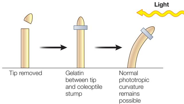
In these experiments, the soluble growth signal moved from the coleoptile tip into the gelatin and then down into the decapitated coleoptile; later the growth signal was isolated from such gelatin blocks and identified as indole-3-acetic acid (see Table 35.2), the major form of auxin.
MECHANISM OF AUXIN TRANSPORT The movement of auxin down a coleoptile is an example of polar (directional) transport. Polar transport of auxin (Figure 35.13) depends on four biochemical processes:
- Diffusion across a cell membrane. Polar molecules (in the chemical sense) diffuse across cell membranes less readily than nonpolar molecules (see Key Concept 6.3).
- Membrane protein asymmetry. Some proteins that transport auxin are located in specific portions of the cell membrane (see Key Concept 6.4 for a discussion of transport proteins). In the stem, most of the transport proteins that allow auxin to efflux (move out of the cell) are located at the basal (bottom) end of the cell.
- Proton pumping/chemiosmosis. A proton pump (see Figure 33.4) moves from the cytoplasm to the cell wall, thereby increasing the intracellular pH and decreasing the pH in the cell wall. Proton pumping also sets up an electrochemical gradient (see Key Concept 9.3), which provides potential energy to drive the transport of auxin by the carriers just mentioned.
- Ionization of a weak acid. The main form of auxin, indole-3-acetic acid, is a weak acid (see Key Concept 2.4):
When the pH is low, this reaction is driven to the right, and HA (non-ionized auxin) is the predominant form. When the pH is higher, there is more (ionized auxin).
The polar auxin transporters are distributed such that the hormone predominately moves down along the longitudinal (up and down) axis of the shoot. However, lateral (side-to-side) redistribution of auxin occurs and is responsible for directional plant growth. This was shown in early experiments that followed the Darwins’ when a coleoptile tip containing the growth hormone was placed asymmetrically on the decapitated coleoptile. The asymmetrical distribution of growth hormone down the coleoptile resulted in excess growth on that side, and bending away from it, even in the absence of light:
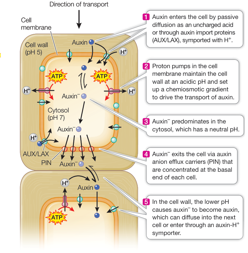
Figure 35.13 Polar Transport of Auxin Proton pumps set up a chemiosmotic gradient directing ionized auxin ( ) toward the basally placed active transport carriers for auxin, which leads to a net movement of auxin in a basal direction.
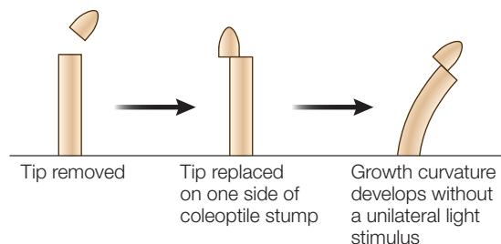
Movement of auxin to the shaded side of the plant could be explained if auxin transporters on the lit side relocate within cells such that auxin exits the cells laterally (sideways, toward the shaded side). There is evidence that supports this hypothesis.
This lateral movement of auxin explains the bending of canarygrass seedlings toward light that the Darwins observed. When light strikes a canarygrass coleoptile on one side, auxin at the tip moves laterally toward the shaded side. The asymmetry is maintained as auxin moves down the coleoptile, so that in the growing region below, the auxin concentration is highest on the shaded side. Cell elongation is thus stimulated on the shaded side, causing the coleoptile to grow toward the light (Figure 35.14A).

Figure 35.14 Plants Respond to Light and Gravity Phototropism (A) and gravitropism (B) occur in shoot apices in response to a redistribution of auxin.
Light is not the only signal that can cause the redistribution of auxin. Auxin moves to the lower side of a shoot that has been tipped sideways, causing more rapid growth in the lower side and hence an upward bending of the shoot. Such growth in a direction determined by gravity is called gravitropism (Figure 35.14B). The upward gravitropic response of shoots is defined as negative gravitropism; that of roots, which bend downward, is positive gravitropism. Gravitropism in roots also involves differential growth caused by lateral movement of auxin, but the details of the mechanism differ between the root and the shoot.
Auxin affects plant growth in several ways
Like the gibberellins, auxin has many roles in plant development. Auxin affects the vegetative and reproductive growth of plants in several ways.
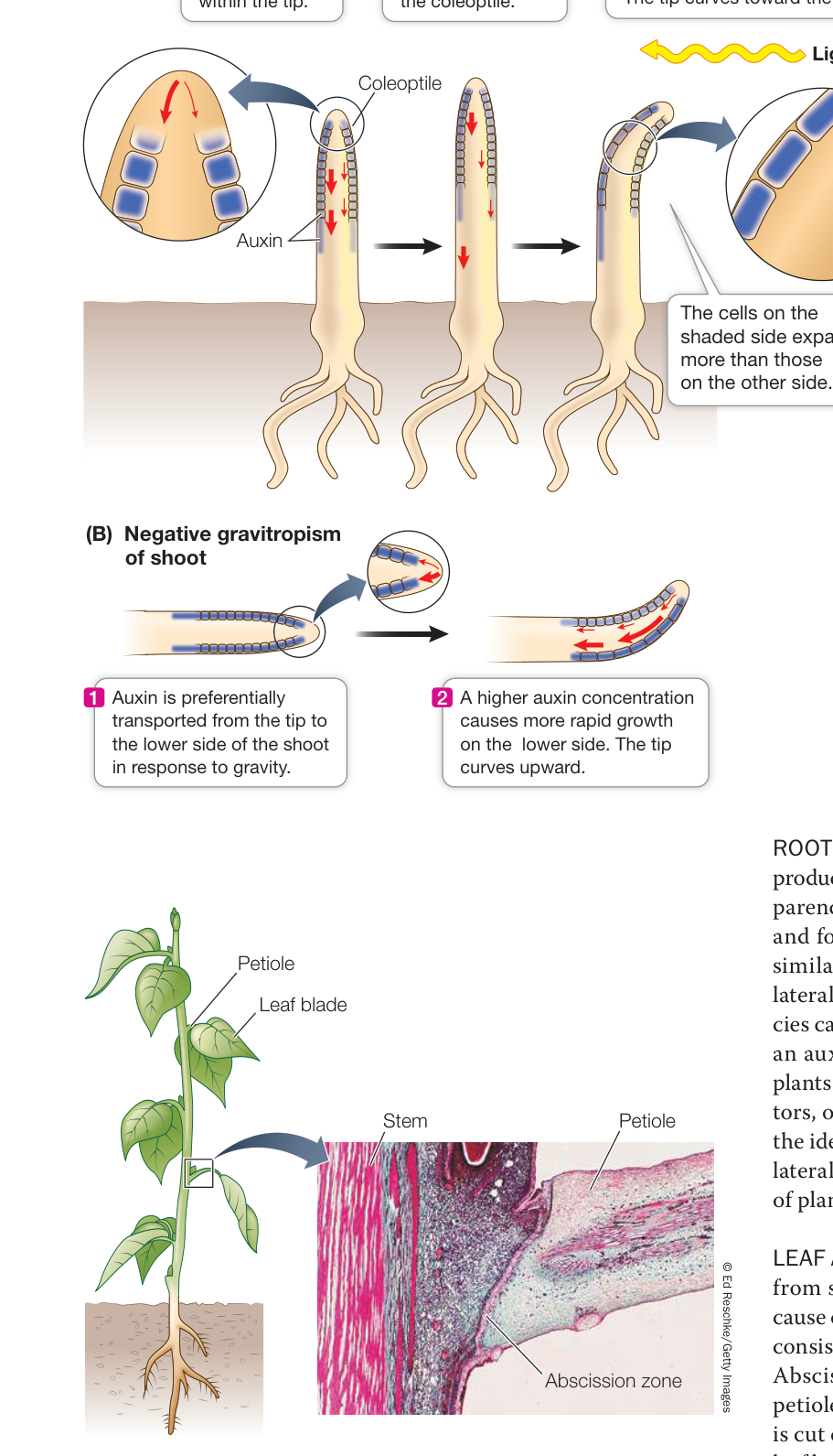
Figure 35.15 Changes Occur When a Leaf Is about to Fall The breakdown of cells in the abscission zone of the petiole causes the leaf to fall.
ROOT INITIATION Cuttings from the shoots of some plants can produce roots and develop into entire new plants. For this to occur, parenchyma cells in the interior of the shoot must redifferentiate and form an apical meristem of a new root. These changes are similar to those that take place in the pericycle of a root when a lateral root forms (see Figure 32.12). Shoot cuttings of many species can be made to develop roots by dipping the cut surfaces into an auxin solution. These observations together with data from plants carrying mutations that affect auxin synthesis, auxin receptors, or downstream factors in auxin response pathways support the idea that the plant’s own auxin plays a role in the initiation of lateral roots. Commercial preparations that enhance the rooting of plant cuttings typically contain synthetic auxins.
LEAF ABSCISSION Auxin inhibits the detachment of old leaves from stems. This detachment process, called abscission, is the cause of leaves falling from some trees in the autumn. Many leaves consist of a blade and a petiole that attaches the blade to the stem. Abscission results from the breakdown of a specific part of the petiole, the abscission zone (Figure 35.15). If the blade of a leaf is cut off, the petiole falls from the plant more rapidly than if the leaf had remained intact. If the cut surface is treated with an auxin solution, however, the petiole remains attached to the plant, often longer than an intact leaf would have. The timing of leaf abscission in nature appears to be determined in part by a decrease in the movement through the petiole of auxin produced in the blade.
APICAL DOMINANCE Auxin helps maintain apical dominance, a phenomenon in which apical (terminal) buds inhibit the growth of axillary buds (see Figure 32.1), resulting in the growth of a single main stem with minimal branching. Apical dominance can be demonstrated by an experiment with a young seedling. If the plant remains intact, the stem elongates and the axillary buds remain inactive. Removal of the apical bud—the major site of auxin production—results in growth of the axillary buds. If the cut surface of the stem is treated with auxin, however, the axillary buds do not grow. The apical buds of branches also exert apical dominance: the axillary buds on the branch are inactive unless the apex of the branch is removed. That is why gardeners prune shrubs to encourage branching.
FRUIT DEVELOPMENT Fruit development normally depends on prior fertilization of the ovule (egg), but in many species treatment of an unfertilized ovary with auxin or gibberellins causes parthenocarpy—fruit formation without fertilization. Parthenocarpic fruits form spontaneously in some cultivated varieties of plants, including seedless grapes, bananas, and some cucumbers.
CELL EXPANSION Cell expansion causes plant growth. As discussed in Key Concept 35.1, the cell wall plays a key role in controlling the rate and direction of plant cell growth. Auxin acts on cell walls to regulate this process.
Cell expansion is driven mostly by the uptake of water, which enters the cytoplasm of the cell and accumulates in its central vacuole (Figure 35.16). Growth of the vacuole accounts for most of the increase in volume of a growing cell, and the vacuole often makes up more than 90% of the volume of a mature cell. As the vacuole expands, it presses the cytoplasm against the cell wall, and the wall resists this force (the basis of turgor pressure). The cell wall is an extensively cross-linked network of polysaccharides and proteins, dominated by cellulose microfibrils. If the cell is to expand, some adjustments must be made in the wall structure to allow the wall to “give” under turgor pressure. Think of a balloon (the cell surrounded by a membrane) inside a box (the cell wall). How does the cell wall “box” loosen to allow expansion?
The acid growth hypothesis explains auxin-induced cell expansion (see Figure 35.16). The hypothesis holds that auxin increases the pumping of protons () from the cytoplasm into the cell wall, lowering the pH of the wall and activating enzymes called expansins. Expansins cause polysaccharides in the cell wall to adhere less strongly to the cellulose microfibrils, allowing the microfibrils to slip past one another. Expansins loosen the cell wall, making it easier to stretch as the cell expands. Several lines of evidence support the acid growth hypothesis. For example, adding acid to the cell wall to lower the pH stimulates cell expansion even in the absence of auxin. Conversely, when a buffer is used to prevent the wall from becoming more acidic, auxin-induced cell expansion is blocked. The acid growth hypothesis applies to most species; in some plants, auxin stimulates secretion of new cell wall components quickly enough to account for even rapid changes in growth rate.
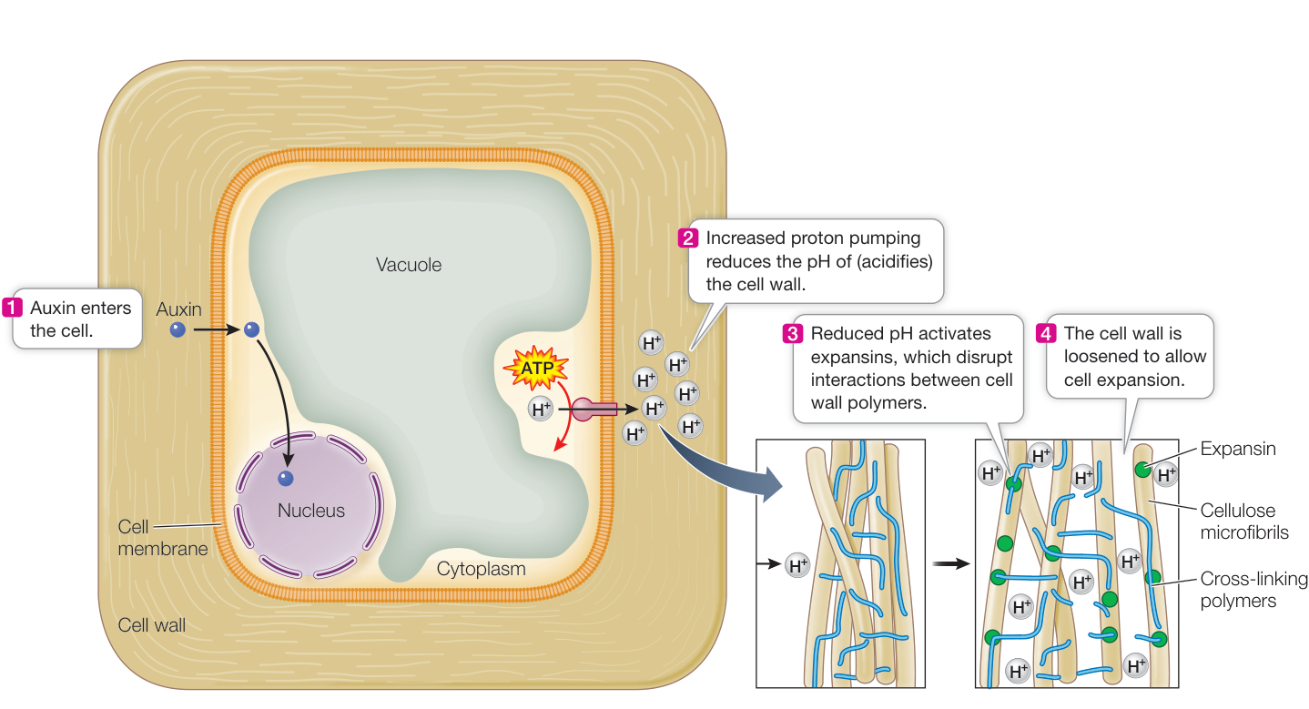
Figure 35.16 Auxin Increases Cell Expansion The plant cell wall is an extensive network of cross-linked polymers. Auxin induces wall loosening by activating a proton pump that reduces pH in the cell wall.
Q: Where else in the plant cell are proton pumps important?
Animation 35.3 Auxin Affects Cell Walls
Auxin and gibberellins trigger the destruction of specific target proteins
The molecular mechanisms underlying both auxin and gibberellin action have been worked out with the help of genetic screens (see Figure 35.7). Biologists started by identifying mutant plants whose growth and development are insensitive to the hormones—that is, plants that are not affected by added hormone. Such mutant plants fall into two general categories:
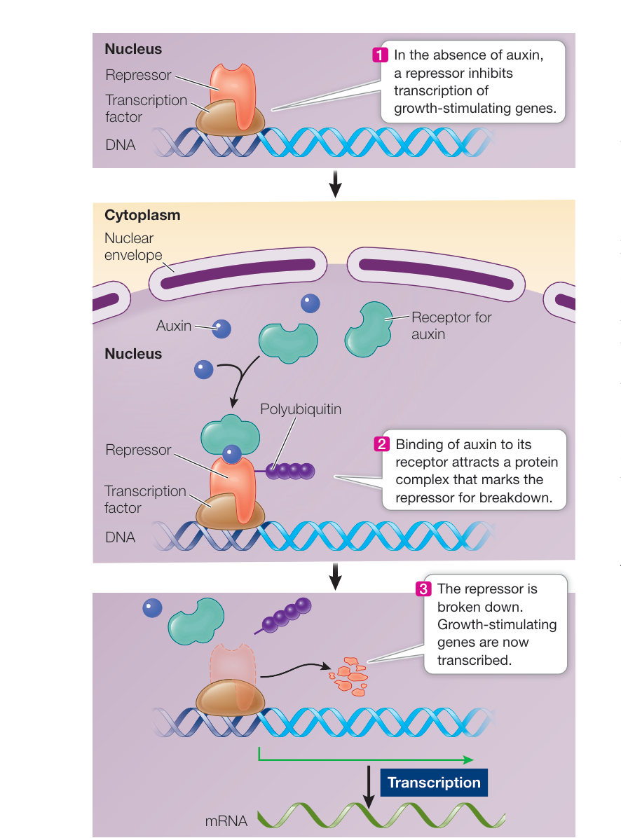
Figure 35.17 Auxin Causes Proteins to be Marked for Degradation The auxin signal transduction pathway shown here is similar to that of gibberellins in that both hormones affect transcription by causing proteins that regulate transcription to be degraded. Q: Are the molecules (receptor, repressor, transcription factor, ubiquitin, proteasome) the same or different in auxin versus gibberellin signaling? Media Clip 35.1 Gibberellin Binding to Its Receptor Life12e.com/mc35.1 1. 1. What would the result be, in terms of the synthesis of hydrolytic enzymes, if germinating barley seeds were treated with an inhibitor of mRNA translation? What would the result be if the seeds were treated with an inhibitor of proteasome function? What would the result be if the two inhibitors were applied to coleoptiles treated with auxin? 2. 2. Explain why, even though auxin moves away from the lighted side of a coleoptile tip, the coleoptile grows toward the light. 3. 3. Corn stunt spiroplasma (a bacterium) causes a significant reduction in the growth rate of infected corn plants. Diseased plants take on a dwarfed form. Since their appearance is reminiscent of a genetically dwarfed corn, you suspect that the bacterium may inhibit the synthesis of gibberellins by corn plants. Describe two experiments you might conduct to test this hypothesis, only one of which requires chemical measurement.
- Excessively tall plants. These plants resemble wild-type plants given an excess of hormone and grow no taller when given extra hormone. They grow tall even when treated with inhibitors of hormone synthesis. Their hormone response is always “on,” even in the absence of the hormone. In such cases, it is presumed that the normal allele for the mutant gene codes for an inhibitor of the hormone signal transduction pathway. In wild-type plants that pathway is “off,” but in the mutant plants the pathway is “on” and the plant grows tall.
- Dwarf plants. These plants resemble dwarf plants that are deficient in hormone synthesis (see Figure 35.9), but they do not respond to added hormone. In these mutant plants the hormone response is always “off,” regardless of the presence of the hormone.
By examining the proteins that were affected by these mutations, the steps leading from hormone receptor to response were worked out (Figure 35.17).
Both auxin and gibberellins act by removing a repressor protein (here called a target protein) that affects transcription of some genes. The specific target proteins that are degraded vary between the two hormones. As the concentration of hormone within a cell increases, the hormone promotes formation of a large protein complex that includes the hormone, the hormone receptor, the target protein that will be broken down, and an enzyme complex that adds a label (polyubiquitin) to the target protein. Addition of polyubiquitin to a target protein results in the marked protein being broken down in the proteasome (see Figure 16.24). The part of this complex that recognizes (binds to) the target protein is called an F-box protein. Whereas animal genomes have few F-box-containing proteins, plant genomes have hundreds, an indication that this type of gene regulation is common in plants.
Recap and Assess
Gibberellins are plant hormones that affect stem growth, fruit size, seed germination, and many other aspects of plant development; the effects vary from species to species. Auxin regulates cell expansion and thus mediates phototropism and gravitropism; it also plays roles in apical dominance, leaf abscission, fruit development, and root initiation. The molecular mechanisms by which gibberellins and auxin convey their effects on plants are similar: both hormones affect gene transcription by causing specific proteins that regulate transcription to be degraded.
As you have seen, auxin and gibberellin can lead to the degradation of transcriptional regulators, with the specific proteins that are degraded varying depending on the type of cell. Thus a single hormone can have many different effects. In the next key concept, you will learn about other important plant hormones that also have multiple effects.
Other Plant Hormones Have Diverse Effects
Learning Objectives
- 35.3.1 Use information on the functions of cytokinins to predict how different aspects of plant growth will be affected by cytokinins and by various combinations of cytokinins and auxin.
- 35.3.2 Describe the function of ethylene in fruit ripening, and explain its importance in the cut-flower and food-production industries.
- 35.3.3 Identify some effects of brassinosteroids on plant growth.
The discoveries of gibberellins and auxin led to the isolation of additional classes of plant hormones (see Table 35.2). These hormones have multiple effects, and in many cases they interact with one another.
Cytokinins are active from seed to senescence
Like bacteria and yeasts, plant cells such as parenchyma cells can be grown in a liquid or solidified growth medium containing sugars and salts. The cells will divide continuously until they run out of nutrients. In the early days of plant cell culturing, scientists experimented with many supplements to determine the optimal chemical environment for growth. The best supplement was coconut milk, the fluid that surrounds the developing embryo in coconut fruit. Investigators suspected that a molecule in the fluid must stimulate plant cell division.
A clue to the identity of the molecule came when Folke Skoog at the University of Wisconsin tested various pure substances that might substitute for coconut milk. DNA was among the substances tested, and it did not work; however, heating DNA at high pressure in an autoclave produced a mixture that strongly promoted plant cell division. A derivative of adenine called kinetin was identified as the active ingredient. Because it stimulated cell division (cytokinesis), it was called a cytokinin.
Kinetin does not exist in cells, but it gave scientists a hint as to what type of molecule might be the active ingredient in coconut milk. An adenine derivative called zeatin was extracted from corn endosperm, and since then more than 150 different cytokinins have been isolated, and most are derivatives of adenine.
Cytokinins (see Table 35.2) have several different effects, in many cases interacting with auxin:
Adding an appropriate combination of auxin and cytokinins to a growth medium induces rapid proliferation of cultured plant cells.
Cytokinins can cause certain light-requiring seeds to germinate even when kept in constant darkness.
In plant cell cultures, a high cytokinin-to-auxin ratio promotes the formation of shoots; a low ratio promotes the formation of roots.
Cytokinins usually inhibit the elongation of stems, but they cause lateral swelling of stems and roots (the fleshy roots of radishes are an extreme example).
Cytokinins stimulate axillary buds to grow into branches; the auxin-to-cytokinin ratio controls the extent of branching (bushiness) of a plant. A lower ratio promotes more branching.
Cytokinins delay the senescence of leaves. If leaf blades are detached from a plant and placed in water or a nutrient solution, they quickly turn yellow and show other signs of senescence. If instead they are placed in a solution containing a cytokinin, they remain green and senesce much more slowly. Roots contain abundant cytokinins, and cytokinin transport to the leaves delays senescence (Figure 35.18).
Ethylene is a gaseous hormone that hastens leaf senescence and fruit ripening
Whereas the cytokinins delay senescence, another plant hormone promotes it: the gas ethylene (see Table 35.2), which is sometimes called the senescence hormone. Ethylene can be produced by all parts of the plant, and like all plant hormones, it has several effects.
Back when streets were lit by gas rather than by electricity, leaves on trees near street lamps dropped earlier than those on trees farther from the lamps. We now know why: ethylene, a combustion product of the illuminating gas, caused the early abscission. Whereas auxin delays leaf abscission, ethylene strongly promotes it; thus the balance of auxin and ethylene controls abscission.
FRUIT RIPENING By promoting senescence, ethylene also speeds the ripening of fruit. As a fruit ripens, it loses chlorophyll and its cell walls break down; ethylene promotes both of these processes. Ethylene also causes an increase in its own production. Thus once ripening begins, more and more ethylene forms, and because it is a gas, it diffuses readily throughout the fruit and even to neighboring fruits on the same or other plants. The old saying “one rotten apple spoils the barrel” is true. That rotten apple is a rich source of ethylene, which speeds the ripening and subsequent rotting of the other fruit in a barrel or other confined space.
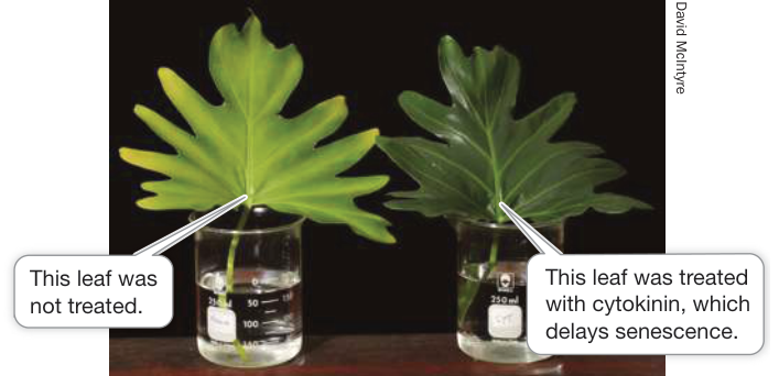
Figure 35.18 Cytokinin Delays Leaf Senescence
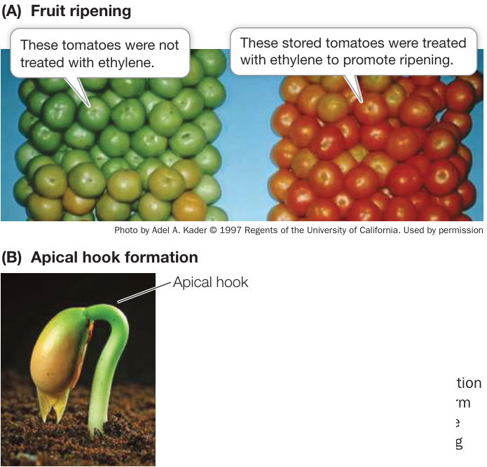
Figure 35.19 Ethylene Promotes Fruit Ripening and Formation of an Apical Hook (A) Tomato fruits ripen when gassed with ethylene. (B) Formation of an apical hook gives a seedling a firm structure that can push up through the soil. The hook opens after the seedling reaches the surface of the soil.
Farmers used to poke holes in developing figs to make them ripen faster. We now know that wounding causes an increase in ethylene production by the fruit, and that the raised ethylene level promotes ripening in many fruits, including apples, bananas, melons, apricots, and tomatoes (Figure 35.19A). Today commercial shippers and storers of such fruit hasten ripening by adding ethylene to storage chambers. This use of ethylene is the single most important use of a natural plant hormone in agriculture and commerce. Ripening can also be delayed by the use of “scrubbers” and adsorbents that remove ethylene from the atmosphere in fruit storage chambers.
As flowers senesce, their petals may abscise, decreasing their value in the cut-flower industry. Growers and florists often immerse the cut stems of ethylene-sensitive flowers in dilute solutions of silver thiosulfate before sale. Silver salts inhibit ethylene action by interacting directly with the ethylene receptor—thus they delay senescence, keeping flowers “fresh” for longer. An alternative product, used to delay fruit ripening and preserve cut flowers, is 1-methylcyclopropene, a gas that also binds to ethylene receptors and blocks their function.
STEM GROWTH Ethylene is active at other stages of plant development as well. In some species, including deepwater rice, higher concentrations of ethylene build up around the stems of plants that are submerged in water, promoting rapid elongation of those stems. Increased levels of ethylene may arise because the rate of diffusion of this gas is slower in water than in air, because of changes in the rate of ethylene synthesis, or a combination of the two.
The effects of ethylene on seedling development illustrate the interactions that occur among plant hormones. The hypocotyl of many eudicot seedlings forms an apical hook that protects the delicate shoot apex while the stem grows through the soil (Figure 35.19B). As in phototropic and gravitropic responses, the apical hook is maintained through an auxin gradient, which promotes the elongation of cells on the outer surface of the hook. Once the seedling breaks through the soil surface and is exposed to light, the auxin level on the inside of the hook increases and the hook unfolds, raising the shoot apex and the expanding leaves into the sunlight.
There is evidence that ethylene controls the formation of the auxin gradient during seedling development. Treatment of dark-grown seedlings of some species with ethylene results in what is called the “triple response”: an exaggeration of the apical hook and a thickening and shortening of the hypocotyl and root (this response was exploited in a genetic screen for ethylene-response mutant plants; see Figure 35.7). It has been shown that ethylene affects both auxin synthesis and transport during apical hook development.
Brassinosteroids are plant steroid hormones
In animals, steroid hormones such as cortisol and estrogen are formed from cholesterol (see Figure 39.2B). Initially, biologists isolated a plant steroid hormone from the pollen of rape, a member of the Brassicaceae (mustard family). When applied to various plant tissues, this brassinosteroid (see Table 35.2) stimulated cell elongation, pollen tube elongation, and vascular tissue differentiation, but it inhibited root elongation. Since then, dozens of chemically related, growth-affecting brassinosteroids have been found in most plants.
Mutant plants that either do not make brassinosteroids or have defects in brassinosteroid reception and signal transduction are usually dwarf, infertile, and slow to develop (Figure 35.20). These effects can be reversed by adding small amounts of brassinosteroids, indicating that brassinosteroids are true hormones. These hormones have diverse effects, which vary among plants. Brassinosteroids can:
- promote xylem differentiation.
- promote growth of pollen tubes during reproduction.
- promote seed germination.
- promote apical dominance and leaf senescence.
- enhance cell elongation and cell division in shoots.
The signaling pathway for these plant steroids differs sharply from those for steroid hormones in animals. In animals, steroids diffuse through the cell membrane and bind to receptors in the cytoplasm. In contrast, the receptor for brassinosteroids is an integral protein in the cell membrane.
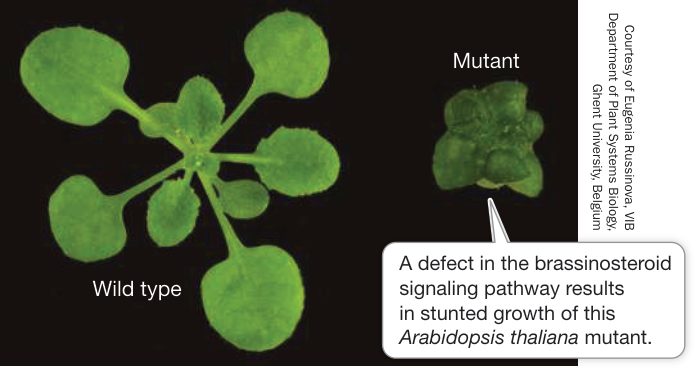
Figure 35.20 Brassinosteroids Promote Growth
Recap and Assess
Cytokinins, ethylene, and brassinosteroids are hormones that work in concert with auxin and gibberellins to mediate plant development. They have various effects on different plants, ranging from stem growth to fruit ripening. They also interact in their effects with other hormones. Ethylene is remarkable in that it is a gas.
- A plant has an extremely bushy growth habit. What can you say about the likely auxin-to-cytokinin ratio in this plant?
- Some supermarkets sell plastic bags that are impregnated with activated charcoal, which binds gases. The bags are designed to keep fruit fresh. How do they work?
- A plant shows rapid leaf senescence. Which hormone or hormones are most likely active in this plant? Which are most likely inactive?
A plant’s response to light—the energy source for photosynthesis—is crucial to its survival. You saw how the Darwins’ pioneering investigations of phototropism led to the discovery of auxin. Let’s now look more closely at how plants sense and respond to light.
Photoreceptors Initiate Developmental Responses to Light
Learning Objectives
35.4.1 Describe and evaluate evidence for the existence of blue-light receptors in plants.
35.4.2 Explain the process and conditions under which conversion of the and forms of phytochrome occur.
35.4.3 Explain how a photoreceptor works by describing which part interacts with light and how that interaction leads to a change in cell signaling.
Light is possibly the most important of the many environmental cues to which plants respond. Much has been learned about the receptors that plants use to sense light, and we focus on those receptors here.
What are plant photoreceptors?
Plants respond to two aspects of light: (1) its quality—that is, the wavelengths of light that can be absorbed by molecules in the plant; and (2) its quantity—that is, the intensity and duration of light exposure.
In Chapter 10 you learned about the role of light in photosynthesis: how chlorophyll and other pigments absorb light at certain wavelengths (quality), and how light intensity affects photosynthetic rate (quantity). Here we discuss how light affects plant development. Earlier in this chapter we described phototropism and how auxin mediates a plant stem’s bending toward light. In addition to phototropism, light influences seed germination, shoot elongation, the initiation of flowering, and many other important aspects of plant development. Several photoreceptors take part in these processes. Blue-light receptors such as cryptochromes and phototropins mediate the effects of higher-intensity blue light, and phytochrome mediates the effects of red light.
Phototropins and cryptochromes are blue-light receptors
Charles and Francis Darwin showed that the apical tip of a growing coleoptile receives light as a signal and then redistributes auxin to stimulate cell elongation below the tip on the shaded side. You may recall from Chapter 10 that an action spectrum involves exposing plants to different wavelengths of light to determine which wavelengths are most effective in driving a given process (e.g., photosynthesis). For photosynthesis, such studies showed that the most effective wavelengths are those absorbed by chlorophylls (see Figure 10.5). When an action spectrum was obtained for phototropism of coleoptiles, blue light (peak 436 nanometers [nm]) was found to be the most effective at inducing the coleoptile to curve (Figure 35.21). What is the blue-light-absorbing receptor/pigment? Biologists have used a genetic approach to answer this question, once again employing the model plant Arabidopsis thaliana.
Researchers recovered blue-light-insensitive mutant A. thaliana plants from a genetic screen and identified the gene for a blue-light receptor protein located in the cell membrane called phototropin. Phototropin protein has a flavin mononucleotide associated with it that absorbs blue light, leading to a change in the shape of the protein. This change exposes an active site for a protein kinase, which in turn initiates a signal transduction cascade that ultimately results in the stimulation of cell elongation by auxin. Phototropin is also involved in chloroplast movements in relation to light, and in the light-induced opening of stomata.
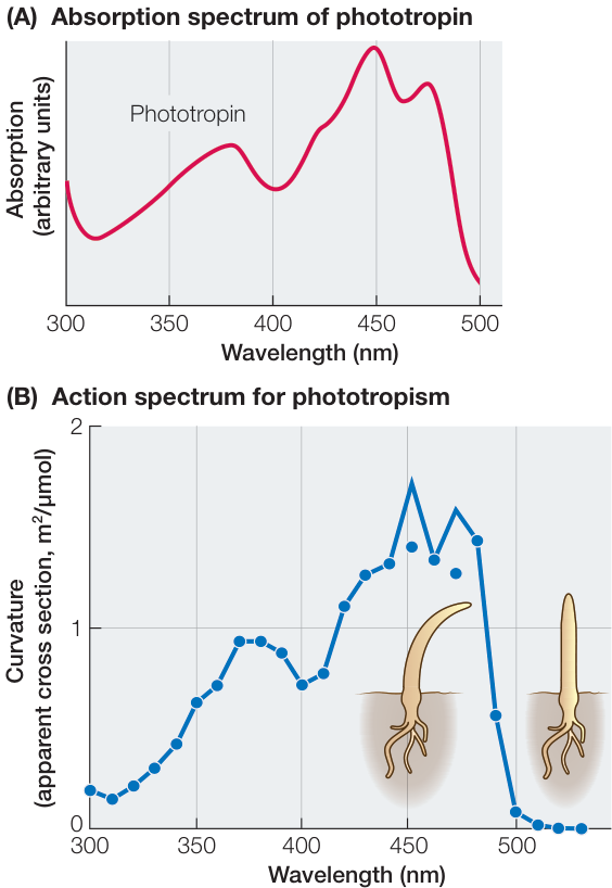
Figure 35.21 Action Spectrum for Phototropism The absorption spectrum for phototropin (A) is similar to the action spectrum for the bending of a coleoptile toward light (B). Maximum bending occurs when coleoptiles are exposed to blue light. (A after W. R. Briggs and J. M. Christie. 2002. Trends Plant Sci 7: 204–210; B after T. Baskin and M. lino. 1987. Photochem Photobiol 46: 127–136.)
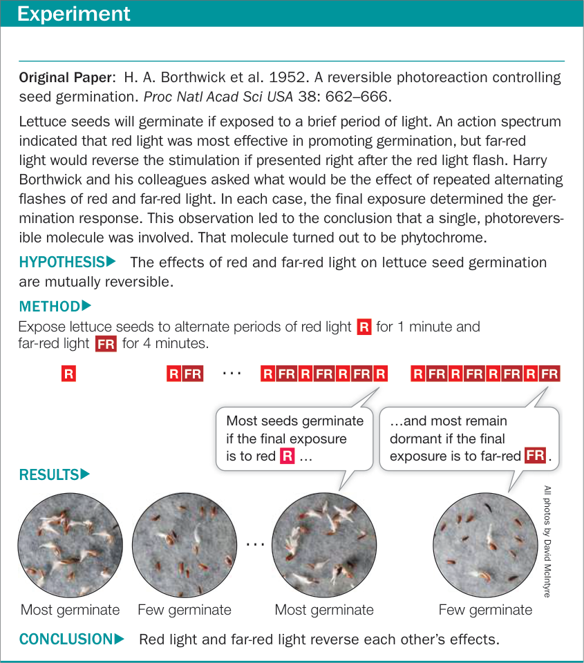
Figure 35.22A Sensitivity of Seeds to Red and Far-Red Light
Another class of blue-light receptors is the cryptochromes, which absorb blue and ultraviolet light. When activated by light, these yellow pigments induce seedlings to grow shorter hypocotyls and larger cotyledons, and increase synthesis of a red-purple pigment called anthocyanin, along with other effects. Like phototropin, cryptochromes contain pigments called chromophores (see the next section) that can absorb light, resulting in a conformational change in the photoreceptor protein.
Phytochromes mediate the effects of red and far-red light
Photomorphogenesis refers to several physiological and developmental events in plants that are controlled by light. For example:
A bean seedling germinating belowground has an elongated stem, a pale yellow, folded leaf, and a hook that protects the first leaves (see Figures 35.8 and 35.19B)—it is etiolated. As the seedling reaches the surface of the soil, it undergoes several light-induced changes: the apical hook straightens, the rudimentary leaves unfold, and chlorophyll is made, so that photosynthesis can begin. Even very dim light will induce these changes.
Lettuce seeds spread on the soil will germinate only in response to light. Even just a flash of dim light will suffice.
Adult cocklebur plants flower when they are exposed to long nights. If there is a brief light flash in the middle of the night, they do not flower.
Action spectra of these processes show that they are induced by light with wavelengths in the range of 650–680 nm, which we see as red light. This indicates that plants must have a photoreceptor pigment that absorbs this part of the red light spectrum and initiates photomorphogenesis.
What is especially remarkable about these red light responses is that they are reversible by far-red light (710–740 nm). For example, if lettuce seeds are exposed to brief, alternating periods of red and far-red light in close succession, they respond only to the final exposure. If it is red, they germinate; if it is far-red, they remain dormant (Figure 35.22). This reversibility of the effects of red and far-red light regulates many other aspects of plant development, including flowering and seedling growth.
The basis for the effects of red and far-red light resides in the photoreceptor pigment protein in the cytoplasm of plants called phytochrome. Phytochrome exists in two interconvertible “isoforms,” or states. The molecule undergoes a conformational change upon absorbing light at particular wavelengths. The default or “ground” state, which absorbs principally red light, is called Pr. When Pr absorbs a photon of red light it is converted into Pfr. Pfr is the active form of phytochrome—the form that triggers important biological processes in various plants (Figure 35.23).
The part of phytochrome that absorbs red and far-red light is a covalently attached pigment called a chromophore (Figure 35.24A). The chromophore of Pr preferentially absorbs red light; when it does so, it changes conformation and the phytochrome is converted to the Pfr form. When the chromophore of Pfr absorbs far-red light, the phytochrome is converted back to the Pr form. If you know organic chemistry, this reaction is a familiar cis-trans isomerization. In addition to being affected by these rapid light-mediated changes, the form can also slowly revert to the form, an event that typically occurs gradually. Because this reversion does not require light, it is called dark reversion.
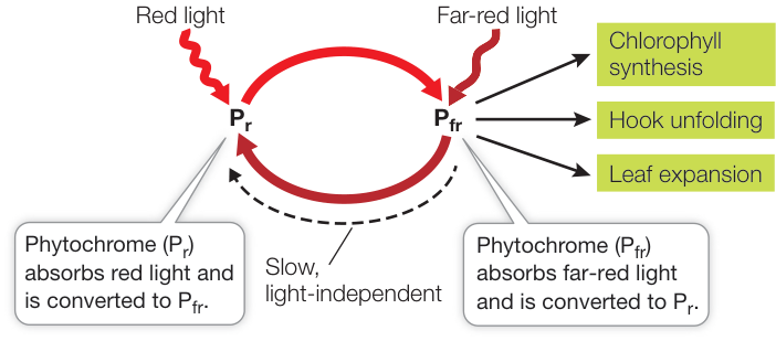
Figure 35.23 Phytochrome Is a Photoreceptor Pigment Protein
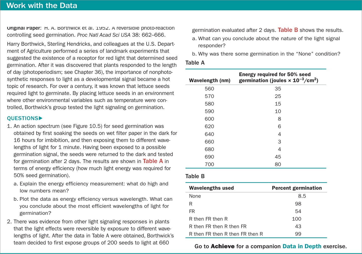
Figure 35.22B Sensitivity of Seeds to Red and Far-Red Light O i i l P H A B th i k t l 1952 A ibl h t ti nm (red, R) for 1 minute, and then some batches to light at 700 nm (far-red, FR) for 4 minutes. The seeds were returned to the dark and Figure 35.22B Sensitivity of Seeds to Red and Far-Red Light
The absorption spectra for and (Figure 35.24B) correlate with their action spectra. As you have seen, phytochrome affects seed germination, shoot development after etiolation, and flowering. In A. thaliana there is a gene family that encodes five slightly different phytochromes, each functioning in different photomorphogenic responses.
For a plant in nature, the ratio of red to far-red light determines whether a phytochrome-mediated response will occur:
- In daylight the ratio is about 1.2:1; because there is more red than far-red light, the form predominates.
- In the shade of other plants, the ratio is as low as 0.13:1, and phytochrome is mostly in the form. The low ratio of red to far-red light in the shade results from absorption of red light by chlorophyll in the leaves overhead, so less of the red light gets through to the plants below.
For some plants growing in the shade, the low ratio stimulates cell elongation and the plants grow upward toward the sunlight. The shade cast by other plants also prevents germination of seeds that require red light to germinate. The reflective properties of the soil can also affect the red to far-red ratio—and thus plant behavior. For example, cotton seedlings grow more slowly on soils (such as clay) that reflect more red than far-red light.
Phytochrome stimulates gene transcription
How does phytochrome, or more specifically , work? Phytochrome has two subunits (Figure 35.25), each composed of a protein chain and a chromophore. Gene transcription is stimulated when is converted to the isoform. When absorbs red light, the chromophore changes shape, causing change in the conformation of the protein itself, from the form to the form. Conversion to the form (see Figure 14.18) results in movement of from the cytoplasm to the nucleus. Once in the nucleus, binds to some transcription factors and induces their degradation. This action can stimulate expression of genes that were formerly repressed, and repress expression of genes that were formerly stimulated. The effect is quite large: in A. thaliana, phytochrome affects an amazing 2,500 genes (10% of the entire genome!) by either increasing or decreasing their expression. Some of these genes are related to hormones. For example, when is formed in seed germination, genes for gibberellin synthesis are activated and genes for gibberellin breakdown are repressed. As a result, gibberellins accumulate and seed reserves are mobilized.
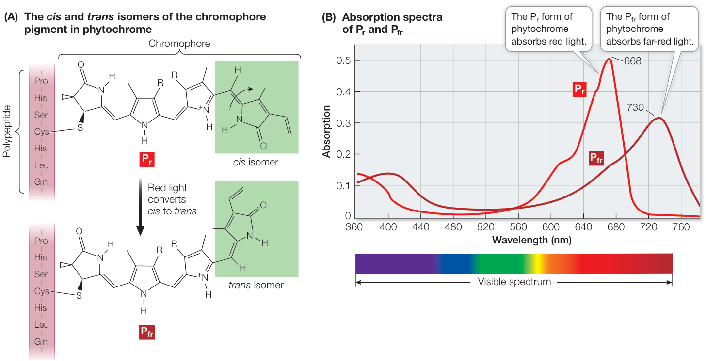
Figure 35.24 Phytochrome Exists in Two Forms Absorption spectra of phytochrome reveal two interconvertible forms, corresponding to the cis and trans isomers of phytochrome’s chromophore. The Pr form absorbs red light; the Pfr form absorbs far-red light.
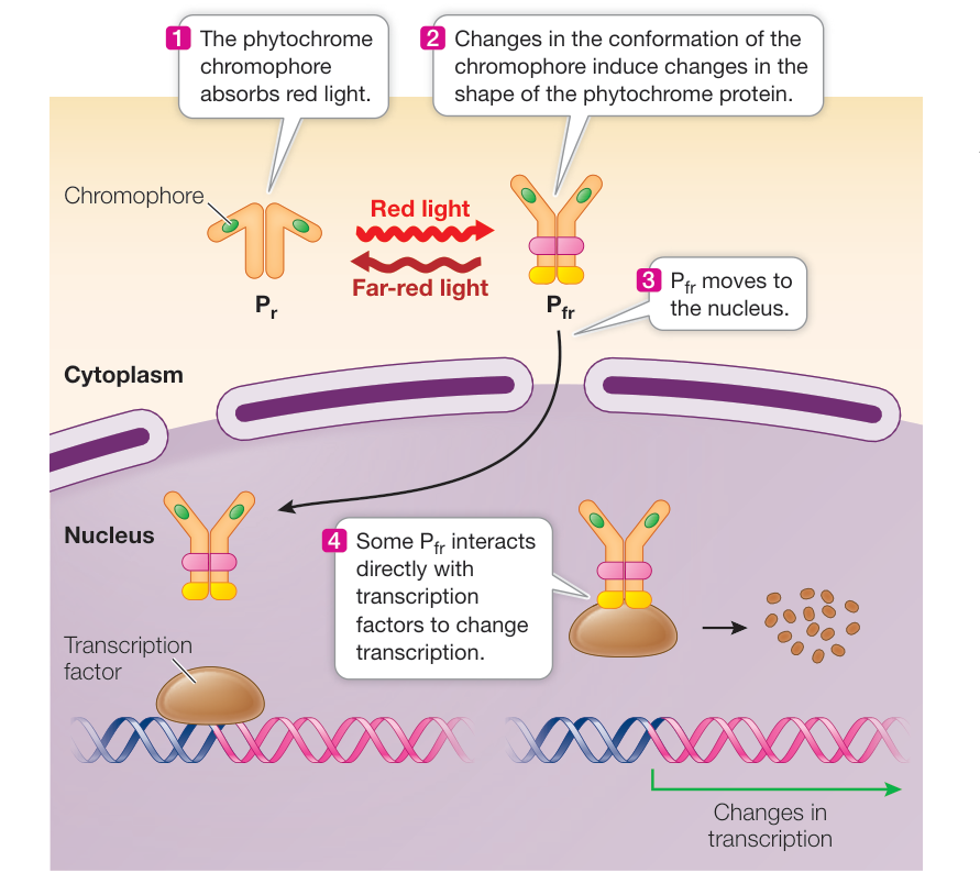
Figure 35.25 Phytochrome Stimulates Gene Transcription Phytochrome is composed of two polypeptide chains, each with a chromophore. This pair of polypeptides undergoes a conformational change upon absorbing light. When phytochrome absorbs red light, it converts to the form, which activates transcription of phytochrome-responsive genes.
Circadian rhythms are entrained by light reception
The timing and duration of biological activities in living organisms are governed in all eukaryotes and some prokaryotes by what is commonly called a “biological clock”—an oscillator within cells that alternates back and forth between two states at roughly 12-hour intervals. The major outward manifestations of this clock are known as circadian rhythms (Latin circa, “about,” + dies, “day”). Think of your own life: in all probability you sleep at night and are awake during the day. The circadian rhythms of animals are discussed in Key Concept 51.2. In plants, circadian rhythms influence, for example, the opening (during the day) and closing (at night) of stomata in A. thaliana, and the raising toward sunlight (during the day) and lowering (at night) of leaves in bean plants. From these two examples, it is obvious that circadian rhythms are ecologically useful adaptations, in that they relate the plant’s physiology to its environment.
Two qualities characterize circadian rhythms, as well as other regular biological cycles: the period is the length of one cycle, and the amplitude is the magnitude of the change over the course of a cycle. The circadian rhythms of plants have several noteworthy characteristics:
- The period of a circadian rhythm is remarkably insensitive to temperature, although lowering the temperature may drastically reduce the amplitude.
- Circadian rhythms are highly persistent; they may continue for days, even in the absence of environmental cues, such as light–dark periods.
- Circadian rhythms can be entrained, within limits, by light–dark cycles that do not exactly correspond to
24 hours. That is, the period of a rhythm can be made to coincide (within limits) with that of the light–dark cycle to which the organism is exposed.
Consider what happens when a person abruptly moves across many time zones: what was the night becomes the day, and gradually the person’s sleep–wakefulness circadian rhythm entrains to the new environmental cues. Similar entrainment occurs in plants adapting to day length as the seasons progress during the year. The action spectrum for plant entrainment indicates that phytochrome (and to a lesser extent, blue-light receptors) is very likely involved. At sundown phytochrome is mostly in the active form. But as the night progresses, gradually gets converted back to the inactive form. By dawn phytochrome is mostly in the state, but as daylight begins, it rapidly converts to . The switch to the state resets the plant’s biological clock. However long the night, the clock is still reset at dawn every day. Thus while the total period measured by the clock is consistent, the clock adjusts to changes in day length over the course of the year.
Recap and Assess
Light controls several physiological and developmental events in plants, a process called photomorphogenesis. Pigment photoreceptors such as phototropin, cryptochromes, and phytochrome mediate the effects of light on plant growth and development. Phytochrome exists in two interconvertible states; conversion from one state to the other is controlled by the ratio of red to far-red light. Circadian rhythms are influenced by light reception.
- What is the evidence for blue-light receptors in plants?
- Why does red light affect seed germination differently than far-red light does?
Investigating Life
Q&A How is plant growth controlled, and what changes in growth patterns made the new strains of wheat and rice successful?
In normal wheat plants, gibberellins stimulate stem elongation. But in the semi-dwarf plants, a mutation affects the signal transduction mechanism for gibberellins so that the stem cells do not respond to it and growth is reduced. In rice, the mutation is in the gene for an enzyme in the biochemical pathway for the synthesis of gibberellins. Without the hormone, the stem does not elongate. The lives of countless people have been saved by intentional disruptions of hormone signaling. These high-yielding crops respond to added fertilizer with greatly increased growth and production of grain compared with conventional varieties of wheat and rice.
Future directions
Unlike animal hormones, which are made in one tissue and often have specific effects on a limited range of targets, plant hormones are widespread in their sites of synthesis and their effects. It is not surprising that with several hormones in a given tissue at a particular time, they interact to produce a physiological effect, a phenomenon called crosstalk. For example, as a seed germinates underground, the growth of the embryonic stem, or hypocotyl (see Figure 35.19B), is stimulated by auxin and gibberellins, and inhibited by ethylene and brassinosteroids. Studies on model plants such as Arabidopsis thaliana are revealing proteins common to the signaling pathways of these hormones that have a role in their interacting effects.
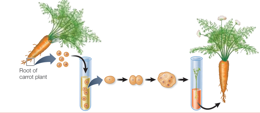
Figure 35.4
Visual Summary
- ■ Genetic screens can be used to identify the factors that carry out specific processes in plants.
Questions
- What anatomical feature of plants allows them to create new organs continually throughout their lives?
- How is the identity of a cell determined?
- Why might the remarkable degree of developmental flexibility that plant cells exhibit be particularly advantageous?
- Construct a flow chart that illustrates how a genetic screen can be used to identify the genes involved in the development of plant signal transduction pathways.
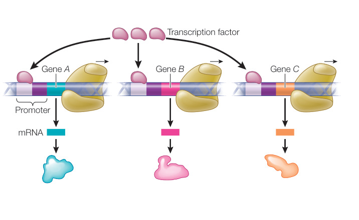
Figure 35.5 KEY CONCEPT
Gibberellins and Auxin Have Diverse Effects but a Similar Mechanism of Action
- ■ The location of auxin transport proteins in cells results in directional movement of auxin called polar auxin transport.
- ■ Phototropism occurs when perception of light leads to lateral redistribution of auxin. More auxin on the shaded sided leads to more elongation on that side, causing the plant to grow toward the light.
- ■ Gibberellins have many effects, including cell elongation, fruit growth, and mobilization of seed storage polymers during germination.
- ■ Both auxin and gibberellins affect gene expression by causing proteins that affect transcription of specific genes to be marked for degradation.
Questions
- An experiment has been performed in which a section of a stem is removed from a plant, a block of agar containing auxin is placed on the top side, and a block of agar that does not contain auxin is placed on the bottom of the stem segment. In this case, the auxin moves from the top to the bottom. If the same experiment is performed, but the piece of stem tissue is inverted such that the side that grew closer to the roots is on top, auxin that is applied to the top of the system does not move down. Explain these results.
- Construct a flow diagram showing the steps by which gibberellins promote seed germination.
- The effect that many plant hormones have on a cell varies depending on the type of cell. Keeping in mind that there are many different F-box proteins in plants, and that each F-box protein targets a different subset of target proteins for degradation, come up with a hypothesis as to how auxin can have different effects on different types of cells.
Investigating Life: The Darwins’ Phototropism Experiment
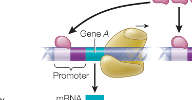
Figure 35.13
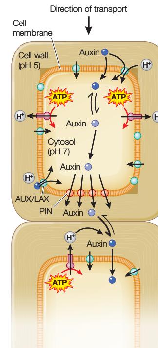
Other Plant Hormones Have Diverse Effects
- ■ Cytokinins promote plant cell division and delay leaf senescence, among other effects. Cytokinins often work in opposition to auxin, particularly when controlling the formation of shoots versus roots.
- ■ Ethylene is a gaseous plant hormone. Ethylene stimulates its own production, resulting in an autocatalytic cycle that causes fruit to ripen all at once.
- ■ Brassinosteroids are plant steroid hormones. Among the responses they affect are cell elongation, pollen tube elongation, vascular tissue differentiation, and root elongation.
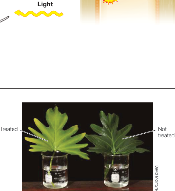
Figure 35.18
Questions
- Plants can often be regenerated from callus tissue by placing the callus first on media containing auxin, and then on media rich in cytokinins. What happens at each stage of this process to allow formation of a complete plant?
- Explain why one rotten apple spoils the whole barrel.
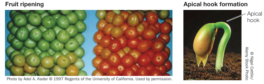
Figure 35.19
Photoreceptors Initiate Developmental Responses to Light
- ■ Photoreceptors such as phototropin and phytochrome are proteins that contain small organic molecules called chromophores that can absorb photons of particular wavelengths. Absorbing light results in a change in the conformation of the protein.
- ■ Phytochrome is a photoreceptor that has two interconvertible states, and . The fraction that is in the active () state depends on the ratio of red to far-red light to which the plant is exposed.
- ■ When the chromophore of phytochrome absorbs red light, the conformation of the photoreceptor changes to the active form, which moves to the nucleus and influences transcriptional activity affecting many genes.
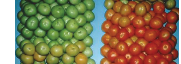
Figure 35.23
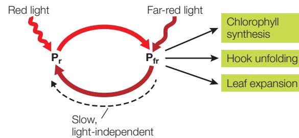
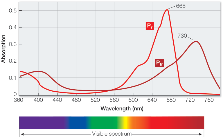
Figure 35.24