Chapter 48Circulatory Systems
Key Concepts
48.1 Circulatory Systems Are Transport Systems
48.2 Vertebrate Circulatory Systems Evolved from Single to Double Circuits
48.3 Heart Function Depends on Properties of Cardiac Muscle
48.4 Circulatory System Functions Depend on Blood and Blood Vessels
48.5 Circulation Is Controlled by Hormonal and Neuronal Signals
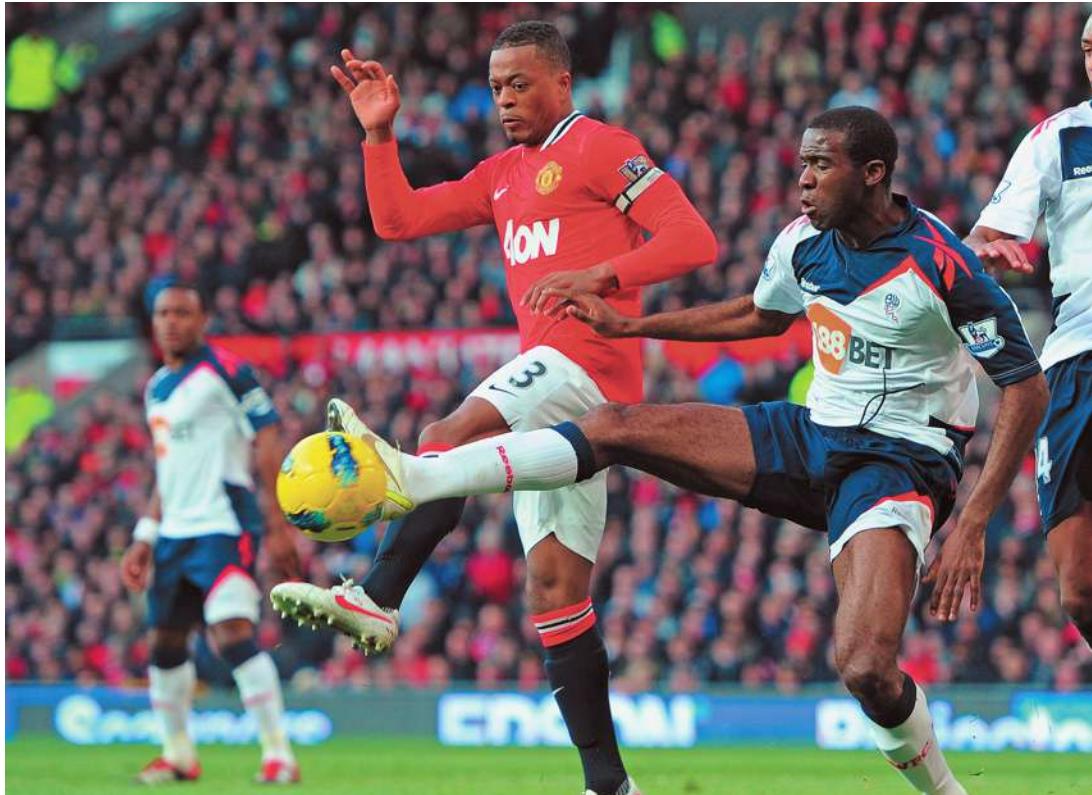
Fabrice Muamba (on the right) was a lucky survivor of cardiac arrest caused by hypertrophic cardiomyopathy, the most common cause of death among young athletes. What causes this condition, and can it be prevented or treated?
Investigating Life: Athletes with Big Hearts
Fabrice Muamba is a much-loved and now retired professional soccer player. On March 17, 2012, during a championship quarterfinal match, 23-year-old Fabrice suddenly collapsed on the field. He was immediately attended to by team physicians and a cardiologist who happened to be a spectator at the match. Fabrice received numerous defibrillator shocks, cardiopulmonary resuscitation, and was rushed to a hospital, but still his heart stopped for 78 minutes. Remarkably he survived, but many young, fit athletes have died from cardiac arrest during physical exertion. Sudden cardiac arrest is the most common cause of death for athletes. In most cases the cause is not a heart attack, but a condition known as hypertrophic cardiomyopathy (HCM).
Your heart is a muscular pump. At rest it beats an average of 60–70 times per minute. With each beat, it circulates about 70 milliliters of blood through your body. Without adding the increases due to work or exercise, that is 300 liters per hour, 7,200 liters per day, 2.6 million liters per year—no time-outs.
Heart failure is the leading cause of death in the United States, accounting for some one-fourth (about 600,000) of the deaths each year. Heart failure most commonly results from blockage of the blood vessels called coronary arteries that supply the heart muscle with blood, and the risk for blockage of these vessels increases with age. HCM has a different cause, originating with gene mutations that affect the contractile proteins of the heart. About 1,000 such mutations have been identified, and they exist in about 1 in 500 people. Most people with one of these mutations live normally, so why are they so dangerous in athletes? The mutations decrease the efficiency of the contractions of the heart muscle, so the heart has to work much harder. Especially in athletes, the heart gets bigger to compensate for its inefficiency. The walls of the heart can get so thick that they impede the blood from leaving the heart, and the thick walls can interfere with the signals that coordinate the contractions of the heart muscle. The result is cardiac arrest. Assessment of HCM risk is improving, and many afflicted athletes are choosing to retire rather than taking a chance of dying from cardiac arrest.
QA Are there potential therapies that could treat HCM? (Find the answer on page 1092.)
Circulatory Systems Are Transport Systems
Learning Objectives
48.1.1 List the diverse functions of circulatory systems.
48.1.2 Explain why some animals can function without a circulatory system.
48.1.3 Describe the differences between hemolymph, blood plasma, and interstitial fluid.
48.1.4 Explain why a closed circulatory system can be more efficient than an open circulatory system.
A circulatory system consists of a muscular pump (heart), a fluid (blood), and a series of conduits (blood vessels) through which the fluid is pumped throughout the body. Heart, blood, and vessels are also known collectively as a cardiovascular system (Greek kardia, “heart,” + Latin vasculum, “vessel”). The function of a circulatory system is to transport things around the body. Preceding chapters discussed how circulatory systems transport heat, hormones, respiratory gases, blood cells, platelets, and cells and molecules of the immune system. Succeeding chapters will add nutrients and waste products to that list. In this key concept we describe the general types of circulatory systems found in animals.
Some animals do not have a circulatory system
Single-celled organisms serve all of their needs through direct exchanges with the environment. Such organisms are found mostly in aquatic or very moist terrestrial environments. Similarly, many multicellular aquatic organisms are small or thin enough that all of their cells are close to the external environment (see Figure 47.1A). Such species may not have a circulatory system because nutrients, respiratory gases, and wastes can diffuse directly between the cells of their bodies and the environment.
Connect the Concepts Key Concept 47.1 explains how Fick’s law of diffusion describes all of the physical factors that control rates of diffusion.
The internal cells of some larger aquatic multicellular animals without a circulatory system are served by highly branched central cavities called gastrovascular systems that bring the external environment into the animal. All the cells of a sponge are in contact with, or very close to, the water that surrounds the animal and circulates through its central cavity (see Figure 47.1B). Very small animals without a circulatory system can maintain high levels of metabolic activity, but larger animals without a circulatory system such as sponges, jellyfishes, and flatworms tend to be inactive, slow, or even sedentary. Large, active animals require circulatory systems.
Circulatory systems can be open or closed
The cells of large, mobile animals are supported by extracellular fluid. All requirements of cells—oxygen, fuel, nutrients, essential molecules—come from that fluid, and the waste products of cell metabolism go into it. Circulatory systems have muscular chambers, or hearts, that move the extracellular fluid through the body.
In open circulatory systems, extracellular fluid is the same as the fluid in the circulatory system and is called hemolymph. This fluid leaves the vessels of the circulatory system, percolates between cells and through tissues, and then flows back into the heart or vessels of the circulatory system to be pumped out again. In contrast, closed circulatory systems completely contain the circulating fluid (blood) in a continuous system of vessels. Blood cells and large molecules stay within the system, but water and low-molecular-weight solutes leak out of the smallest vessels, which are highly permeable.
In animals with a closed circulatory system, extracellular fluid refers to both the fluid in the circulatory system and the fluid outside it. The fluid in the circulatory system is the blood plasma; the extracellular fluid outside the circulatory system is the interstitial fluid. A 70-kilogram person has a total extracellular fluid volume of about 14 liters. Less than one-fourth of it—about 3 liters—is the blood plasma.
Connect the Concepts Key Concept 38.2 and Figure 38.4 describe the different extracellular fluid compartments that constitute the internal environment that supports all of the metabolic needs of the cells of the body.
Open circulatory systems move extracellular fluid
Open circulatory systems are found in arthropods, mollusks, and some other invertebrate groups. In these systems a heart moves the hemolymph through vessels leading to different regions of the body. The fluid leaves the vessels to filter through the tissues before returning to the heart. In the arthropod shown in Figure 48.1A, the fluid returns directly to the heart through openings called ostia. Ostia have valves that allow hemolymph to enter the relaxed heart but prevent it from flowing in the reverse direction when the heart contracts. In a mollusk such as a clam, open vessels collect hemolymph from different regions of the body and return it to the heart (Figure 48.1B).
Lest you think that open circulatory systems are inefficient and can support only sluggish lifestyles such as those of mollusks, remember that locusts swarming over crops, yellow jackets buzzing around your picnic, and scorpions dashing across the desert all have open circulatory systems.
Closed circulatory systems circulate blood through a system of blood vessels
In closed circulatory systems, blood vessels separate blood from interstitial fluid. Blood is pumped through this vascular system by one or more hearts, and cellular and macromolecular components of the blood never leave the vessels. Closed circulatory systems characterize vertebrates and some invertebrate groups, among them annelids.
A simple example of a closed circulatory system is that of the earthworm (Figure 48.1C). One large ventral blood vessel carries blood from the worm’s anterior end to its posterior end. Smaller vessels branch off and transport the blood to even smaller vessels serving the tissues in each body segment. In the smallest vessels, respiratory gases, nutrients, and metabolic wastes diffuse between the blood and interstitial fluid. The blood then flows from these vessels into larger vessels that lead into one large, muscular dorsal vessel that carries the blood from the posterior to the anterior end of the body. Five pairs of muscular vessels connect the large dorsal and ventral vessels in the anterior end, thus completing the circuit. The dorsal vessel and the five connecting vessels serve as hearts for the earthworm; their contractions keep the blood circulating. The direction of circulation is determined by one-way valves in the dorsal vessel.
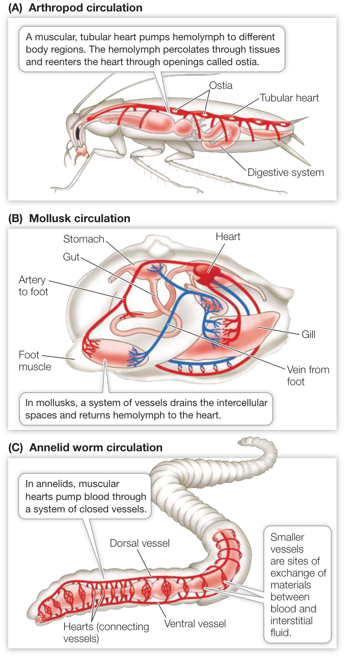
Figure 48.1 Circulatory Systems Arthropods, illustrated here by a grasshopper (A), and mollusks, illustrated by a clam (B), have open circulatory systems. Hemolymph is pumped by a tubular heart and directed to different regions of the body through vessels that open into the intercellular environment. (C) Annelids, illustrated here by an earthworm, contain the cellular and macromolecular elements of their blood in the vessels of a closed circulatory system that includes muscular hearts. Earthworms exchange respiratory gases across their skin. The circulation distributes those gases between the skin and the deeper tissues.
Closed circulatory systems have several advantages compared with open systems:
- Fluid can flow more rapidly through vessels than through intercellular spaces and can therefore transport things to and from tissues more rapidly.
- By changing the diameter (and hence the resistance) of specific vessels, closed systems can control the flow of blood to selective tissues and organs to match their needs.
- Specialized cells and large molecules that aid in transporting hormones, nutrients, and oxygen can be kept in the vessels but can drop their cargo in the tissues where it is needed.
With all of these “advantages” of closed circulatory systems, you might wonder how some species with open circulatory systems can sustain such high levels of activity. In the case of insects, the answer is clear: insects do not depend on their circulatory systems for respiratory gas exchange (see Figure 47.4).
Recap and Assess
Circulatory systems consist of a pump and an open or closed set of vessels through which a fluid transports oxygen, nutrients, wastes, and a variety of other substances. Flow to and from tissues in closed systems of vessels can be more rapid and can be directed to specific tissues according to their needs.
- Describe three ways that circulatory systems support exercising muscles.
- A sponge is neither small nor thin. How can it survive without a circulatory system?
- Explain the importance of extracellular fluid in large mobile animals.
- How does a closed circulatory system facilitate the fight-or-flight response?
Our overview of the open and closed systems found among invertebrates introduced some basic concepts about circulatory systems. Next we turn to describing the closed circulatory systems of vertebrates.
Vertebrate Circulatory Systems Evolved from Single to Double Circuits
Learning Objectives
48.2.1 Diagram the single circuit of the circulatory system of fishes.
48.2.2 Describe how a lungfish distributes oxygen to its tissues in both water and air environments.
48.2.3 Explain how and why the circulatory systems of ectothermic reptiles are unique in their control of pulmonary and systemic circulation.
48.2.4 Describe how the avian and mammalian four-chambered hearts maintain separate but linked pulmonary and systemic circuits operating at different pressures.
Vertebrates have a closed circulatory system that includes a heart with multiple chambers. When heart chambers contract, they squeeze the blood, putting it under pressure and causing it to flow. One-way valves prevent backflow of blood as the heart chambers cycle between contraction and relaxation. Blood flows out of the heart and into vessels called arteries that carry blood away from the heart. Arteries branch into smaller arterioles that feed blood into capillary beds. Capillaries are tiny, thin-walled vessels where materials are exchanged between blood and the interstitial fluid. Small vessels called venules drain capillary beds. The venules join to form the larger veins that ultimately deliver blood back to the heart.
We can trace the evolutionary history of vertebrate circulatory systems by comparing the circulatory systems of fishes, lungfishes, amphibians, reptiles (including crocodilians), birds, and mammals (Figure 48.2). These comparisons show that as circulatory systems became more complex, they evolved from a single circuit (heart, gills, body, heart) to a double circuit (heart, lungs, heart, body, heart). Through this progression of vertebrates, the blood flowing to the gas exchange organs (gills or lungs; see Figure 47.3) becomes increasingly separated from the blood flowing to the rest of the body.
Fishes have a four-chambered heart that pumps blood through the gills to be oxygenated and then throughout the tissues of the body and back to the heart—a single circuit. In birds and mammals, blood is pumped from the heart to the lungs and back to the heart in a pulmonary circuit, and then from the heart to the rest of the body and back to the heart in a systemic circuit. In amphibians and ectothermic reptiles (i.e., all reptiles except birds) we see various adaptations that incompletely separate the blood flow into pulmonary and systemic circuits.
Circulation in fishes is a single circuit
Since fishes are phylogenetically the oldest vertebrates, their circulatory systems reflect the most primitive vertebrate circulatory systems. Frequently it is stated that fish have a two chambered heart. That is wrong, the fish heart has four chambers that are connected in series (see Figure 48.2A). Blood returning from all parts of the body collects in a sinus venosus that feeds into the muscular atrium. The atrium pumps blood into the more muscular chamber, the ventricle. Contraction of the ventricle pushes blood into the last chamber, the bulbus arteriosus, a highly elastic chamber. The pressure imparted to the blood by the ventricle stretches the bulbus arteriosus, and its elastic recoil dampens the blood pressure oscillations generated by the beating of the heart. The arterial blood leaving the bulbus arteriosus under pressure flows through the gills, where respiratory gases are exchanged. Blood leaving the gills collects in a large dorsal artery, the aorta, which distributes blood to smaller arteries and arterioles leading to all the organs and tissues of the body. In the tissues, blood flows through beds of tiny capillaries, collects in venules and veins, and eventually returns to the sinus venosus of the heart. The unidirectional flow of blood in this circuit is enabled by one-way valves between the sinus venosus and the atrium, between the atrium and the ventricle, and between the ventricle and the bulbus arteriosus.
Most of the pressure imparted to the blood by the contraction of the ventricle is dissipated as a result of resistance to flow in the many narrow spaces in the gill lamellae (see Figure 47.5). Therefore blood leaving the gills and entering the aorta is under low pressure, limiting the capacity of the fish circulatory system to supply the tissues with oxygen and nutrients. In spite of this limitation on arterial blood pressure, some species, such as tunas and marlins, can swim at remarkably high speeds for considerable distances.
The evolutionary transition from breathing water to breathing air had important consequences for the vertebrate circulatory system. An example of how the system changed to serve a primitive lung can be seen in the African lungfishes.
Lungfishes evolved a gas-breathing organ
Lungfishes have descended from a lineage that predates the rise of tetrapod vertebrates. Present-day lungfishes are exposed periodically to water with low oxygen content or to situations in which their aquatic environment dries up. The adaptation that enables them to survive these conditions is an outpocketing of the gut just behind the mouth cavity. This outpocketing forms a lung structure that runs most of the length of the animal. Air is gulped into this lung and oxygen is picked up by the blood flowing through the many capillaries in its walls.
How does the lungfish circulatory system take advantage of this new organ? In fishes the gills are arranged on supportive pairs of gill arches (see Figure 47.5A,B). In most fishes the gills on all the gill arches are similar. Deoxygenated blood from the heart flows through thin gill filaments, where it picks up oxygen, and then flows into the aorta, which distributes the oxygenated blood throughout the body. The circulatory anatomy in lungfishes is quite different. Of the six pairs of gill arches, only the posterior three have fine gill filaments that enable the blood to take up oxygen from the water. The blood leaving these three pairs of gill arches can flow into the dorsal aorta or into a pulmonary artery to the lung. Thus even if the blood has picked up little oxygen in the gills, it can pick up considerable oxygen in the lung from the air that the fish has gulped. The oxygenated blood from the lung returns to the atrium of the heart. From the atrium, the blood flows into the ventricle, which pumps it into the bulbous arteriosus. From there, the oxygenated blood mostly flows into the anterior three pairs of gill arches. These gills do not have fine filaments for gas exchange; instead they are modified to be low-resistance vessels that enable the oxygenated blood to flow directly into the dorsal aorta.
But doesn’t the oxygenated blood from the lung completely mix with the deoxygenated blood from the systemic circulation? The answer is no, because the lungfish heart partially, but effectively, separates the flow of blood into pulmonary and systemic circuits. The atrium is partially divided by a tissue fold (see Figure 48.2B). The left side receives oxygenated blood from the lung, and the right side receives deoxygenated blood from the body via the sinus venosus. The ventricle and the bulbous arteriosus also have tissue folds and septa that keep the oxygenated and deoxygenated bloodstreams mostly separate. As a result, the oxygenated blood from the lung goes mostly to the low-resistance, high-throughput anterior gill arches leading to the dorsal aorta, and deoxygenated blood goes mostly to the posterior gill arches that have functional gill filaments. The blood leaving those posterior gill arches can flow to the dorsal aorta or to the lung. Thus the dorsal aorta can receive oxygenated blood either from the lung (via the anterior gill arches) or from the gill filaments of the posterior gill arches.
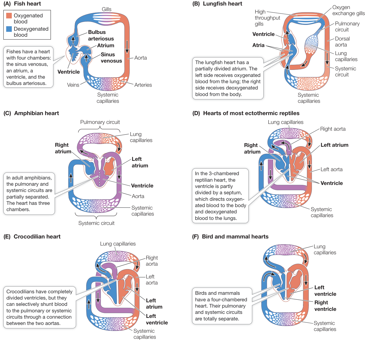
Figure 48.2 Comparing Circulatory Systems of Vertebrates (B) Lungfish heart (A) The general fish heart has a single muscular ventricle that pumps blood first through the gills and then through the systemic blood vessels. (B) Lungfish hearts can pump blood through the gills or to a lung that the fish fills by gulping air. (C) Amphibians have three chambered hearts that incompletely separate blood flow to lungs and to the rest of the body. (D) Most ectothermic reptilian hearts have an incomplete separation of left and right ventricles and they have two aortas, thus they can either send blood returning to the right ventricle from the systemic circulation to the lungs if they are breathing or back into the systemic circuit if they are not breathing. (E) The crocodilian heart has fully divided ventricles, but communication between their left and right aortas can shunt blood from the pulmonary to the systemic circuits. (F) Bird and mammal hearts have fully divided pulmonary and systemic circuits.
We can conclude that the lungfish lung evolved as a means of supplementing oxygen uptake from the gills. When the water is oxygenated, the lungfish can obtain oxygen through its gills; but in oxygen-depleted water, it can depend on getting oxygen from its lung. Associated modifications of the lungfish vascular system set the stage for the evolution of separate pulmonary and systemic circulations in all tetrapod vertebrates.
Amphibians have partial separation of systemic and pulmonary circulation
In adult amphibians, a single ventricle pumps blood to the lungs and the rest of the body, but two atria receive blood returning to the heart (see Figure 48.2C). The left atrium receives oxygenated blood from the lungs, and the right atrium receives deoxygenated blood from the body. Because both atria deliver blood to the same ventricle, the oxygenated and deoxygenated blood could mix, in which case blood going to the tissues would not carry a full load of oxygen. Mixing is limited, however, because, as in lungfishes, anatomical features of the ventricle direct the flow of deoxygenated blood from the right atrium primarily to the pulmonary circuit and the flow of oxygenated blood from the left atrium primarily to the aorta. Partial separation of pulmonary and systemic circulation has the advantage of allowing blood destined for the tissues to sidestep the large pressure drop that occurs in the gas exchange organ. Blood leaving the amphibian heart for the tissues moves directly to the aorta, and hence to the body, at a higher pressure than if it had first flowed through the lungs as it does in fishes.
Some amphibians have another adaptation for oxygenating their blood: they can pick up a considerable amount of oxygen in blood flowing through small blood vessels in their skin.
Reptiles have exquisite control of pulmonary and systemic circulation
As described in Chapter 31, the reptiles include turtles, snakes, lizards, crocodilians, and birds (see Figures 31.21 and 31.22). Crocodilians and birds have circulatory systems with two completely separated ventricles. All other reptiles have ventricles that are not completely separated into left and right chambers. Are incompletely separated ventricles in reptiles a disadvantage or an adaptation?
Consider the behavior, ecology, and physiology of ectothermic reptiles. Many are active, powerful, fast animals, but their activity comes in bursts that are interspersed with long periods of inactivity. At these times the animals’ metabolic rates are much lower than the resting metabolic rates of the endothermic birds and mammals. So enormous is the range of metabolic demand in ectothermic reptiles that they do not need to breathe continuously. Some species are accomplished divers and spend long periods under water, where they cannot breathe air.
When these animals are not breathing, it would be a waste of energy for them to pump blood through their lungs. Thus they pump blood to the lungs and the rest of the body when they are breathing, but when they are not breathing, they bypass the pulmonary circuit and pump blood just to the body. How do they do this?
In ectothermic reptiles with a three-chambered heart—that is, the turtles, snakes, and lizards—the ventricle is partially divided into left and right halves by a septum (see Figure 48.2D). Oxygenated blood from the lungs enters the left side of the ventricle through the left atrium. Deoxygenated blood from the body enters the right side of the ventricle through the right atrium. These species have two aortas, left and right. The left aorta is positioned so that it receives oxygenated blood from the left side of the ventricle. The right aorta, however, is positioned so that it can receive blood from either the right or left side of the ventricle.
When the animal is breathing air, the resistance in the pulmonary circuit is lower than the resistance in the systemic circuit, so blood from the right side of the ventricle tends to flow into the pulmonary artery rather than the right aorta. When the animal is not breathing, pulmonary vessels constrict, resistance in the pulmonary circuit goes up, and blood from the right side of the ventricle tends to flow into the right aorta. As a result, blood from both sides of the ventricle flows through the two aortas to the systemic circuit.
Crocodilians, like birds, have two completely separated ventricles. Unlike birds, they have two aortas, one originating in each ventricle. But there is a connection between the two aortas just as they leave the heart, and this connection enables them to alter the proportions of blood going to their pulmonary and systemic circuits (see Figure 48.2E). When a crocodilian is breathing and resistance in the pulmonary circuit is low, backpressure from the stronger left ventricle closes the valve between the right ventricle and the right aorta, forcing all of the blood from the right ventricle to flow into the pulmonary circuit. When the animal stops breathing, pulmonary vessels constrict, resistance in the pulmonary circuit rises, and pressure in the right ventricle overcomes the backpressure in the right aorta from the left ventricle. As a result, the blood from the right ventricle flows into the right aorta. This ability of all ectothermic reptiles to direct blood to their pulmonary or systemic circuits is highly adaptive for their lifestyle of intermittent breathing.
Birds and mammals have fully separated pulmonary and systemic circuits
The four-chambered hearts of birds and mammals have completely separate pulmonary and systemic circuits (see Figure 48.2F). Separate circuits have several advantages for these active animals with continuously high metabolic rates:
- Oxygenated and deoxygenated blood cannot mix; therefore the systemic circuit always receives blood with the highest oxygen content.
- Respiratory gas exchange is maximized because the blood with the lowest oxygen content and highest content is sent to the lungs.
- Separate systemic and pulmonary circuits can operate at different pressures.
The tissues of birds and mammals have high nutrient demands and thus a very high density of blood vessels, requiring the heart to generate a high blood pressure to perfuse all the vessels of the systemic circuit. The pulmonary circuit of these animals receives a blood flow equal to that of the systemic circuit, but the lungs have far fewer blood vessels. Thus the pulmonary circuit of birds and mammals can function at lower pressures, and the four-chambered heart makes that possible. Additionally, the high pressure in the systemic circuit increases the ability to direct blood to tissues that need it the most, through controlled changes in resistance in arterioles.
Recap and Assess
The closed circulatory system of vertebrates has evolved from a single circuit in fishes to separate pulmonary and systemic circuits in birds and mammals. A key step was the evolution of a primitive lung in a lineage of air-breathing fishes. Further adaptations increasingly separated the flows of oxygenated and deoxygenated blood in separate circuits, as seen in amphibians and reptiles. Ectothermic reptiles retained the ability to direct blood to either the pulmonary or systemic circuit, but birds have complete division of those two circuits, as do mammals, allowing the circuits to function at different pressures.
- The bulbous arteriosus of fishes is less muscular than the ventricle but has more elastic fibers. Given the location of the bulbous arteriosus in the fish circulatory system, what could be its function?
- A lungfish brooding in stagnant water can increase the oxygen content in the water immediately surrounding its eggs. How is this possible?
- Explain how and why ectothermic reptiles shunt blood from the pulmonary circuit when not breathing.
- The ductus arteriosus in a human fetus connects the dorsal aorta with the pulmonary artery, allowing blood to bypass the fluid-filled lungs. This connection normally closes at birth. What are the consequences if it does not?
We turn now to the structure and function of the mammalian heart, focusing in particular on human cardiac function.
Heart Function Depends on Properties of Cardiac Muscle
Learning Objectives
- 48.3.1 Trace the path of blood through both sides of the mammalian heart, naming the major blood vessels and heart valves.
- 48.3.2 Explain how heart valves function.
- 48.3.3 Describe and diagram the cardiac cycle in terms of left ventricular volume and pressure, aortic pressure, and heart sounds.
- 48.3.4 Explain the differences between pacemaker potentials and ventricular muscle potentials and their ionic bases.
In this key concept we describe in detail the structure and function of the mammalian heart, using the human heart as the example. We first trace the paths of blood flow through the heart and the body, and then we examine the unique electrical properties of cardiac muscle that result in the rhythmic contractions of the heart.
The mammalian heart has four chambers—a right and a left atrium and a right and a left ventricle (Figure 48.3). The right atrium receives blood from the systemic circuit, and the left atrium receives blood from the pulmonary circuit. The right ventricle pumps blood through the pulmonary circuit, and the left ventricle pumps blood through the systemic circuit.
One-way valves between the atria and ventricles, the atrioventricular (AV) valves, prevent backflow of blood into the atria when the ventricles contract. The right AV valve is called the tricuspid valve because it has three leaves. The left AV valve is called the bicuspid valve because it has two leaves. The bicuspid valve is also called the mitral valve because it has the shape of a religious headdress called a miter. There are also one-way valves between the ventricles and the arteries leaving the heart. The pulmonary valve goes to the lungs, and the aortic valve goes to the aorta. These two valves are also called semilunar valves because their separate leaves are shaped like half-moons.
Blood flows from right heart to lungs to left heart to body
The superior (upper) vena cava and the inferior (lower) vena cava (see Figure 48.3) are the large veins that return deoxygenated blood from the upper and lower body to the right atrium. The veins of the heart itself also drain into the right atrium. From the right atrium, the blood flows through the tricuspid valve into the right ventricle. The filling of the ventricle is mostly passive flow while the heart is relaxed between beats. At the end of this phase of passive ventricular filling, the atrium contracts and adds a little more blood to the ventricular volume. The right ventricle then contracts, causing the tricuspid valve to close and pumping the blood into the pulmonary artery leading to the lungs.
Pulmonary veins return the oxygenated blood from the lungs to the left atrium, from where the blood enters the left ventricle through the bicuspid valve. As on the right side of the heart, most left ventricular filling is passive, and is only topped off when the atria contract.
The walls of the ventricles are powerful muscles that contract around the blood with a wringing motion starting from the bottom. When pressure in the left ventricle is high enough to push open the aortic valve, blood rushes into the aorta to begin its circulation throughout the body. In Figure 48.3 observe that the walls of the left ventricle are thicker than those of the right ventricle. The left ventricle has to propel blood through many more kilometers of blood vessels than does the right ventricle, and must therefore push against more resistance, even though both ventricles pump the same volume of blood.
It is the left ventricle that creates the condition of hypertrophic cardiomyopathy (HCM) that you learned about at the beginning of this chapter. Mutations that reduce the efficiency of contraction of the cardiac muscle result in an increased workload of the left ventricle, so the ventricular muscle compensates by getting bigger (hypertrophy). When the heart is repeatedly stressed by high levels of activity, as is common in athletes, the hypertrophy can become extreme (Figure 48.4). The consequences can be blockage of blood flowing into the aorta and disruption of the signals that coordinate the contractions of the ventricular muscles.
Both sides of the heart contract at the same time. Contraction of the two atria, followed by contraction of the two ventricles and then relaxation, is the cardiac cycle. The cardiac cycle is divided into two phases: systole (pronounced sís-toll-ee), when the ventricles contract, and diastole (die-ás-toll-ee), when the ventricles relax (Figure 48.5). At the very end of diastole (step 1 in Figure 48.5), just before the ventricles contract, the atria contract and top off the volume of blood in the ventricles.
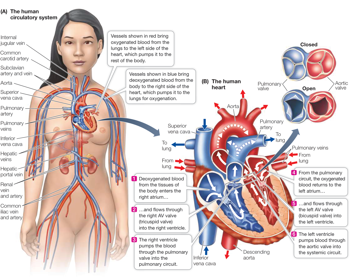
Figure 48.3 The Human Heart and Circulation (A) The major blood vessels of the body either carry blood away from the heart (arteries) or return blood to the heart (veins). (B) In the human heart, blood flows from the right side of the heart to the lungs, back to the left side of the heart, and then out to the body. The atrioventricular valves prevent blood from flowing back into the atria when the ventricles contract. The pulmonary and aortic valves prevent blood from flowing back into the ventricles from the arteries when the ventricles relax. Activity 48.2 The Human Heart
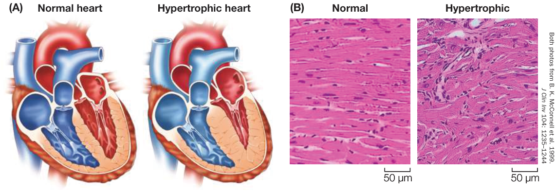
Figure 48.4 Hypertrophic Cardiomyopathy (A) Comparison of a normal and a hypertrophic heart. (B) Micrographs of the ventricular heart muscle fibers of a normal and a hypertrophic heart. The organization of the muscle fibers is irregular, causing inefficient contractions.
The sounds of the cardiac cycle—the “lub-dup” heard through a stethoscope—are created by the heart valves slamming shut. The closing and opening of these valves are simple mechanical events resulting from pressure differences on the two sides of the valves. As the ventricles begin to contract (step 2 in Figure 48.5), the pressure in them rises above the pressure in the atria, so the
AV valves close (“lub”). When the ventricles begin to relax (step 4 in Figure 48.5), the high pressures in the aorta and pulmonary artery close the aortic and pulmonary valves (“dup”).
Defective valves that do not close completely produce turbulent blood flow and the sounds known as heart murmurs. For example, if an AV valve does not close completely, blood will flow back into the atrium with a “whoosh” at the beginning of systole.
Blood pressure changes associated with the cardiac cycle can be measured in the large artery in your arm by using an inflatable
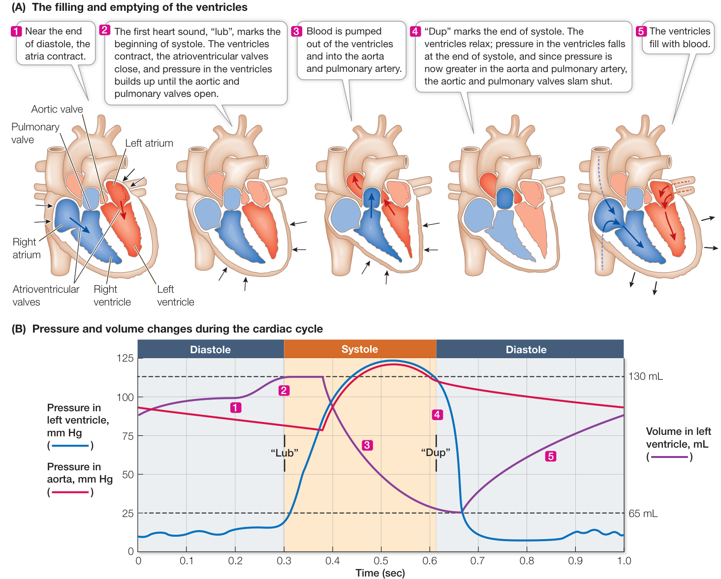
Figure 48.5 The Cardiac Cycle (A) The rhythmic contraction (systole) and relaxation (diastole) of the ventricles is called the cardiac cycle. (B) During diastole, the ventricles fill and pressure is low. During systole, the ventricular pressure rises and the ventricles empty. The representation here shows pressure and volume changes for the left ventricle only. The dashed lines represent the maximum and minimum volumes of blood in the left ventricle.
Q: If these curves were for the right ventricle instead of the left ventricle, how would they differ and how would they be the same?
Animation 48.1 The Cardiac Cycle pressure cuff and a pressure gauge, together called a sphygmomanometer, and a stethoscope (Figure 48.6). This method measures the minimum pressure necessary to compress an artery so blood does not flow through it at all (the systolic pressure) and the maximum pressure that allows continuous flow through the artery (the diastolic pressure). A conventional blood pressure reading is expressed as the systolic pressure placed over the diastolic pressure. Healthy values for a young adult might be 120 millimeters of mercury (mm Hg) during systole and 70 mm Hg during diastole, or 120/70. Modern blood pressure measuring devices such as you might encounter in a drugstore are automated so that the stethoscope is unnecessary.
The heartbeat originates in the cardiac muscle
Some cardiac muscle cells are pacemaker cells that initiate action potentials without stimulation from the nervous system. These specialized muscle cells do not contract, but they generate rhythmic sequences of action potentials that spread to neighboring cardiac muscle cells that do contract. Since cardiac muscle cells are in electrical contact with one another through gap junctions, the action potentials from the pacemaker cells spread rapidly through a large mass of cardiac muscle, causing it to contract in unison. Coordinated contraction is essential for pumping blood effectively.
The primary pacemaker of the heart is a group of modified cardiac muscle cells, the sinoatrial node, located at the junction of the superior vena cava and right atrium (see Figure 48.9). The resting membrane potentials of these cells are less negative than those of other cardiac muscle cells and are not stable; instead they gradually become even less negative until they reach threshold for initiating an action potential. The action potentials of pacemaker cells are very different from those of neurons and other muscle cells (see Figure 43.9). They are slower to rise; they are broader; and they are slower to return to resting potential (Figure 48.7A). These properties of pacemaker cells are due to the ion channels in their membranes.
Pacemaker potentials involve , , and channels (Figure 48.7B). As discussed in Key Concept 43.2, when or channels open, positive charges flow into the cell and the membrane potential becomes less negative. When channels open, positive charges flow out of the cell and the membrane potential becomes more negative. Because the channels of pacemaker cells are open more of the time than are those of other cardiac muscle cells, the pacemaker resting potential is less negative. The action potential of pacemaker cells is due to voltage-gated channels rather than to voltage-gated channels as in neurons, skeletal muscle, and the contractile cardiac muscle cells. These channels open and close more slowly than voltage-gated channels, explaining the rounded shape of pacemaker action potentials.
The unstable resting potential of pacemaker cells is due to the behavior of cation channels. As in neurons and skeletal muscle cells, the rise of the action potential is followed by the opening of voltage-gated channels. The increased permeability of the cells to brings the membrane potential back to a negative level. But that restoration of a negative membrane potential causes a unique class of voltage-gated channels to open. At the same time, the voltage-gated channels that restored the negative membrane potential are gradually closing. The opening channels and closing channels cause the membrane potential to drift upward (become less negative) (see Figure 48.7B).
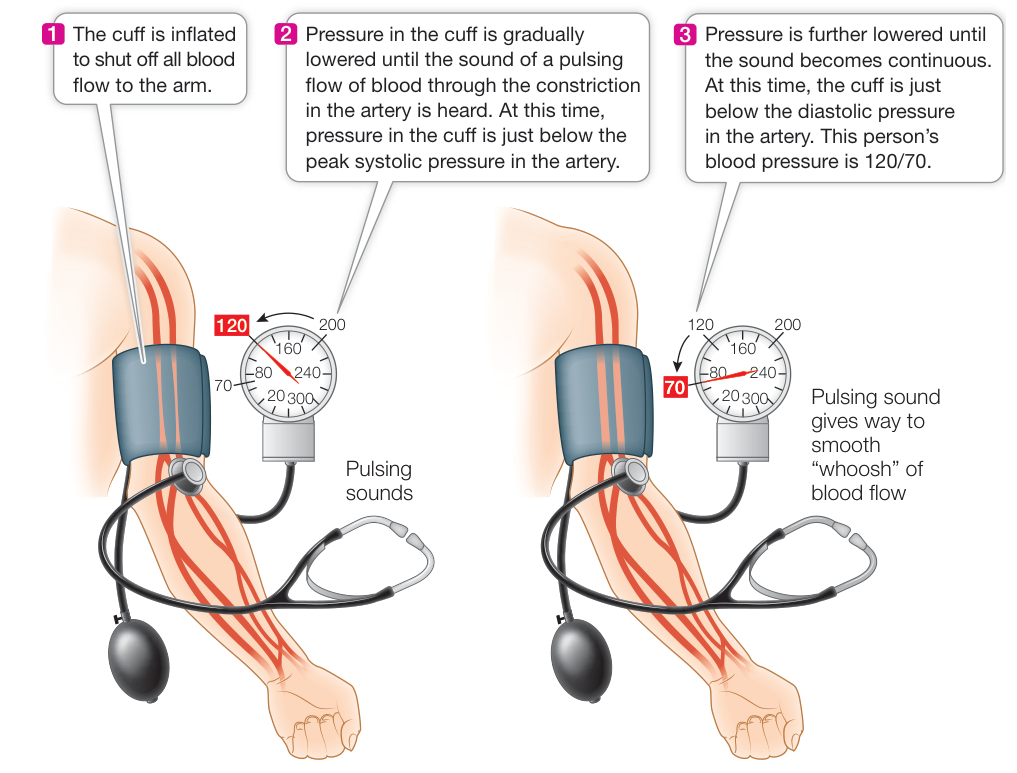
Figure 48.6 Measuring Blood Pressure Blood pressure in the major artery of the arm can be measured with a device called a sphygmomanometer, which combines an inflatable cuff and a pressure gauge. A stethoscope or an automatic sensor is used to detect sounds created by the blood vessels in response to changes in pressure during the cardiac cycle.
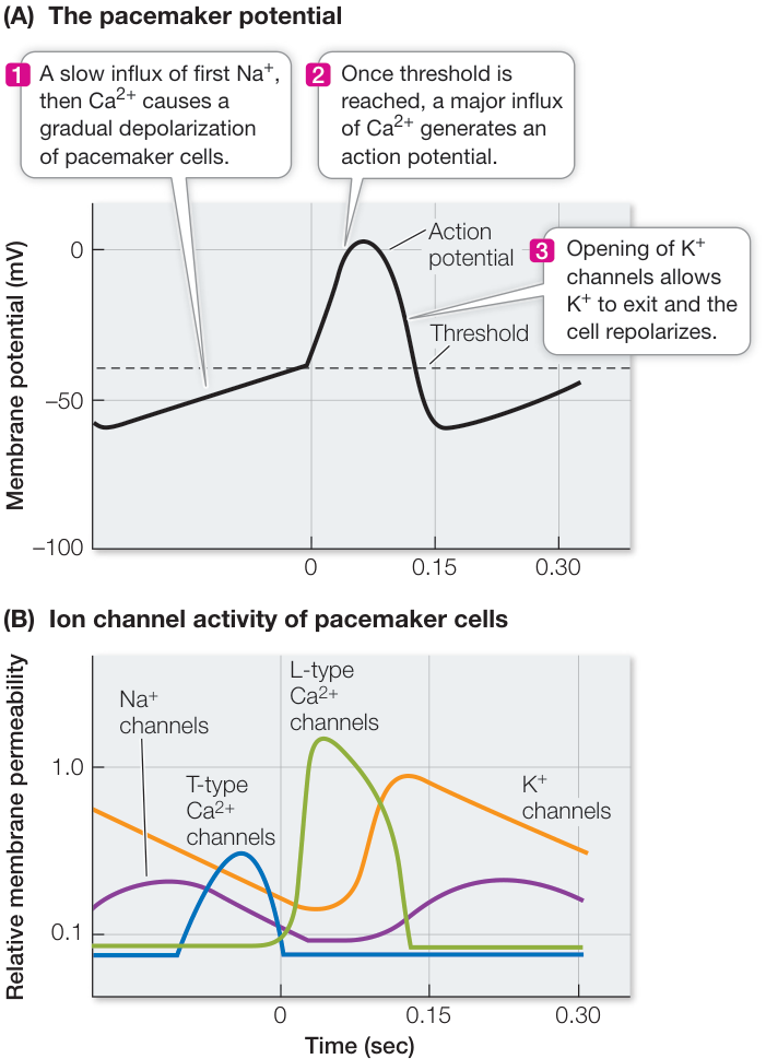
Figure 48.7 The Pacemaker Potential (A) The resting potential of the pacemaker cells of the sinoatrial node gradually drifts upward between action potentials. The pacemaker action potential is slow to rise and is broad. These characteristics are due to properties of , , and two types of channels. (B) At rest, sinoatrial pacemaker cells are more permeable to than are neurons or other muscle cells. During rest, the channels gradually close, but voltage-gated T-type channels open. When a threshold is reached, the L-type channels open, generating the action potential. Opening of the channels helps the membrane potential return to its most negative value. (From E. P. Widmaier et al. 2008. Vander’s Human Physiology, 11th Edition. McGraw-Hill: New York.)
The gradual rise in membrane potential closes the channels that allow to move into the cell, but as the membrane becomes less negative, some channels open, causing the membrane potential to continue its gradual rise. These channels are called T-types because they open transiently. Eventually the rising membrane potential reaches threshold for the major voltage-gated channels to open. These channels are called L-types because their opening is long-lasting. It is the opening of these L-type channels that generates the pacemaker action potential. The intricate interaction of these ion channels through their effects on membrane potential causes the rhythmic generation of action potentials that characterizes pacemaker cells.
The autonomic nervous system controls the heartbeat (speeds it up or slows it down) by influencing the rate at which the membrane potentials of pacemaker cells drift upward (Figure 48.8). Norepinephrine (NE) released onto pacemaker cells by sympathetic nerves increases the permeability of the channels and the channels. The result is that the resting potential of the pacemaker cells drifts up more rapidly, the interval between action potentials is decreased, and the heart beats faster. Conversely, the parasympathetic neurotransmitter acetylcholine (ACh) has opposite effects. ACh increases the permeability of channels so that the membrane potential becomes even more negative following an action potential and rises more slowly. ACh also decreases the permeability of the channels so that the rate of rise of the membrane potential slows, the interval between pacemaker action potentials lengthens, and the heart slows down.
A conduction system coordinates the contraction of heart muscle
A normal heartbeat begins with an action potential in the sinoatrial node (Figure 48.9). This action potential spreads rapidly throughout the electrically coupled cells of the atria, causing them to contract in unison. Because there are no gap junctions between the cells of the atria and those of the ventricles, the action potential does not spread directly to the ventricles. Therefore the ventricles do not contract in unison with the atria.
How does the action potential move from the atria to the ventricles? Situated at the junction of the atria and the ventricles is a nodule of modified cardiac muscle cells—the atrioventricular node—which is stimulated by the depolarization of the atria. With a slight delay, the AV node generates action potentials that are conducted to the ventricles via the bundle of His, which consists of modified cardiac muscle fibers that do not contract but do conduct action potentials. These fibers course down the septum between the left and right ventricles and divide into right and left bundle branches. The branches of conducting fibers run to the tips of the ventricles and then spread throughout the ventricular muscle mass as Purkinje fibers. This system of conducting fibers ensures that the cardiac action potential spreads rapidly and evenly throughout the ventricular muscle mass, starting at the very bottom of the ventricles. The short delay in the spread of the action potential imposed by the atrioventricular node ensures that the atria contract before the ventricles do, so that the blood passes progressively from the atria to the ventricles to the arteries.
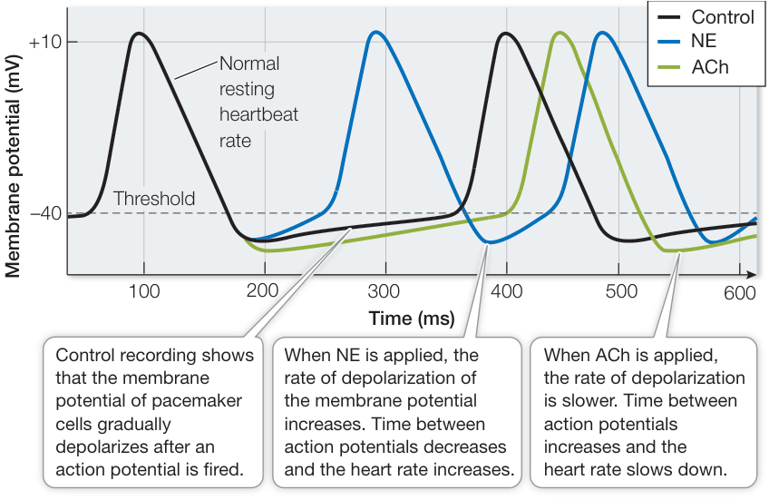
Figure 48.8 The Autonomic Nervous System Controls Heart Rate Neurotransmitters from the two divisions of the autonomic nervous system either speed up (norepinephrine, NE) or slow down (acetylcholine, ACh) the rate at which the pacemaker membrane potential drifts upward, thereby controlling the rate at which pacemaker cells fire action potentials.
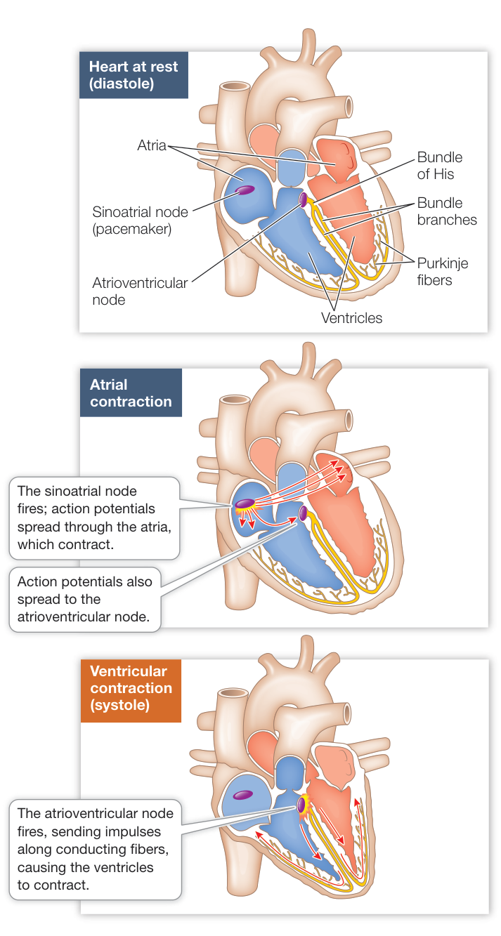
Figure 48.9 The Heartbeat Pacemaker cells in the sinoatrial node initiate the heartbeat by firing action potentials that spread through the electrically coupled atrial muscle. The atrial action potential spreads to the atrioventricular node which, with a delay, conducts it through the bundle of His and Purkinje fibers to the cells of the ventricles.
HCM can disrupt conduction
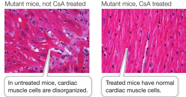
Returning to the case of HCM introduced at the beginning of this chapter, consider in Figure 48.9 how HCM can impair the function of the left ventricle. First, in many cases the septum between the ventricles is greatly modified by the hypertrophy, and this can disrupt the conducting fibers that run down through the septum. Second, the thickening of the septum near the aorta can block the entrance to the aorta and therefore impede blood flow and the emptying of the ventricle. How can these effects be prevented? In Investigating Life: Silencing Mutant Myosin Genes we describe a recent study on mice that have been genetically engineered to carry mutations of a gene for one of the myosin chains of cardiac muscle that is associated with a large percentage of human HCM cases. These mice develop the pathologies that are seen in humans who inherit the genes causing HCM. The investigators found that by using RNA interference to silence the mutated genes, they could prevent the development of the HCM pathology.
Connect the Concepts The use of RNA interference to block gene expression at the mRNA level is discussed in Key Concept 18.4.
Electrical properties of ventricular muscles sustain heart contraction
Ventricular muscle fibers can contract for about 300 milliseconds—much longer than skeletal muscle fibers. As in neuronal and skeletal muscle action potentials, the rising phase of the ventricular muscle cell action potentials is due to the opening of voltage-gated channels. Unlike neurons and skeletal muscle fibers, however, ventricular muscle cells remain depolarized for a long time due to sustained opening of voltage-gated channels (Figure 48.10). Like other muscle, cardiac muscle is stimulated to contract when is available to bind with troponin (see Figure 46.6). As long as remains in the sarcoplasm, the ventricular muscle cells continue to contract.
To terminate systole and allow the ventricles to fill again, must be cleared from the sarcoplasm of the ventricular muscle cells. pumps in the sarcoplasmic reticulum membrane and the cell membrane actively transport ions out of the sarcoplasm and into the sarcoplasmic reticulum or into the interstitial fluid. Thus in the sarcoplasm is maintained at a low level until the next action potential triggers another round of release and muscle contraction. The rate of cycling of into and out of the sarcoplasm puts limits on the heart rate and strength of contraction of the ventricle.
The ECG records the electrical activity of the heart
Electrical events in the cardiac muscle during the cardiac cycle are recorded by electrodes placed on the surface of the body. Such a recording is an electrocardiogram, or ECG or EKG reflecting the Greek spelling (kardia). The ECG is an important tool for diagnosing heart problems (Figure 48.11A).
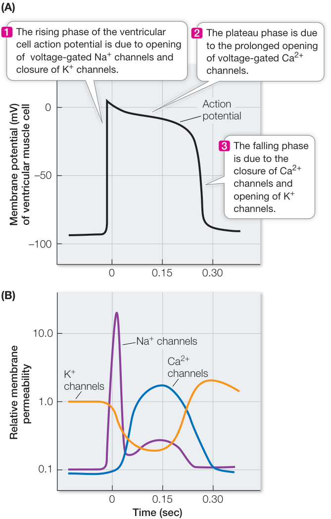
Figure 48.10 The Action Potential of Ventricular Muscle Fibers (A) The three phases of the action potential of ventricular muscle fibers are due to the opening and closing of voltage-gated channels. (B) At the initiation of the action potential, voltage-gated channels open rapidly but briefly. At the same time, but more slowly, channels are closing and channels are opening. The open channels sustain the depolarization. Repolarization occurs when the channels close, and the slow opening of the channels also contributes to the repolarization. (After D. E. Mohrman and L. J. Heller. 2002. Cardiovascular Physiology, Fifth Edition. Langer Medical Books/McGraw-Hill: New York.) Q: Why is the broadening of the ventricular muscle action potential important?
Investigating Life: Silencing Mutant Myosin Genes
Experiment
Original Paper: J. Jiang et al. 2013. Allele-specific silencing of mutant Myh6 allele in mice suppresses hypertrophic cardiomyopathy. Science 342: 111–114.
Hypertrophic cardiomyopathy (HCM) is caused by mutations in myosin genes. A similar mutation has been engineered into mice, and they develop HCM. In addition, it was shown that treatment of these mice with cyclosporine A (CsA) exacerbates their development of HCM. Since the HCM model mice are heterozygous for the mutated gene (as are most afflicted humans), and since there are multiple myosin genes, the question arose as to whether or not the selective silencing of the mutant gene would allow the normal alleles to correct the disorganization of the cardiac muscle due to the mutant allele.
HYPOTHESES
- Mice genetically engineered to carry a mutation in the myosin gene most commonly responsible for HCM in humans will show the HCM phenotype, and it will be exacerbated by treatment with CsA.
- Silencing expression of the mutant gene will prevent the development of the HCM phenotype.
METHOD
- Using genetic engineering techniques, generate a mouse model that carries a mutation in one of its myosin genes. The heterozygotes are viable, but the homozygotes are not.
- Using the wild-type mice as a comparison, measure left ventricular wall thickness in mutant mice with and without treatment with CsA to exacerbate the development of left ventricular wall hypertrophy.
- Develop an interference RNA (RNAi) that will silence expression of the mutant gene in vitro.
- Transfect mice heterozygous for the mutation with the RNAi construct and at a later time assess them for hypertrophy of their left ventricular walls. Some mice are also treated with CsA, which exacerbates the development of hypertrophy in the mutant mice.
- Calculate the means and standard deviations for each cohort of mice. Then do non-paired t-tests to see if there are significant differences.
RESULTS Data in the graph are simulated based on reported means and standard deviations.
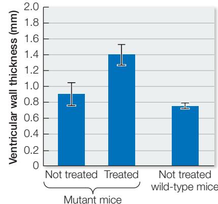
CONCLUSION The mice carrying the mutation show a significant () increase in left ventricular wall thickness. The mutant mice treated with CsA show a larger increase in left ventricular wall thickness () in comparison with the mutant mice not treated with CsA. Thus the mutation has the expected effect of promoting the development of HCM symptoms, and the effect is exacerbated by treatment with CsA.
Work with the Data
The second hypothesis posed in the Investigating Life experiment is that treatment of the mutant mice with interference RNA (RNAi) targeted to the mutant myosin gene will reduce the development of the HCM phenotype. The data are listed in the table. All values are for left ventricular wall thickness (mm) of individual mice and are simulated based on the reported means and standard deviations.
| CsA treated | Not CsA treated | RNAi and CsA treated | RNAi but not CsA treated |
|---|---|---|---|
| 1.60 | 0.96 | 0.87 | 0.63 |
| 1.34 | 0.98 | 0.82 | 0.68 |
| 1.50 | 1.01 | 0.87 | 0.85 |
| 1.39 | 0.89 | 0.91 | 0.65 |
| 1.50 | 0.86 | 0.74 | 0.68 |
| 1.44 | 0.91 | 0.83 | |
| 1.42 | 0.92 |
QUESTIONS
- Comparing mutant mice that did not receive CsA, did the treatment with RNAi significantly reduce their left ventricular wall thickness?
- Comparing mutant mice that did receive CsA, did the treatment with RNAi significantly reduce their left ventricular wall thickness?
The action potentials that sweep through the muscles of the atria and ventricles before they contract are massive, localized electrical events causing electric currents to flow throughout the body. Electrodes placed at different locations on the skin detect those currents at different times and register a voltage difference between them. The appearance of the ECG depends on the placement of the electrodes. Electrodes placed on the right wrist and left ankle produced the normal ECG shown in Figure 48.11B. The wave patterns of the ECG are designated P, Q, R, S, and T, each letter representing a particular event in the cardiac muscle, as shown in the figure.
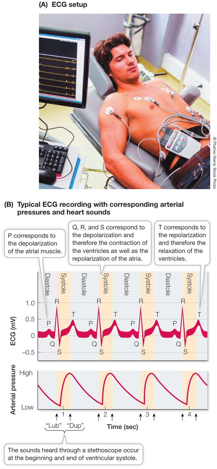
Figure 48.11 The Electrocardiogram (A) An electrocardiogram (abbreviated as ECG or EKG) is used to monitor heart function. Electrodes attached to the person record an ECG that is amplified and displayed on a monitor. (B) Variations from the normal pattern shown here can be used to diagnose heart problems. Q: Abnormal ECG patterns are characteristic of HCM hearts. If there were an occasional or a long-lasting block of the AV node, what would you expect to see in the ECG?
Recap and Assess
The mammalian heart has two atria and two ventricles. The right atrium and ventricle serve the pulmonary circuit, and the left atrium and ventricle serve the systemic circuit. Valves that are open and shut by pressure differences prevent the backflow of blood. Modified cardiac muscle tissue in the right atrium functions to spontaneously generate pacemaker action potentials. Other modified cardiac muscle tissue between the atria and ventricles and throughout the ventricles conducts those signals and coordinates the heart contraction. Broad action potentials in ventricular muscle reflect cycling in the ventricular muscle cells and make sustained contractions possible. The depolarization and repolarization of cardiac muscle during the cardiac cycle create electrical events that can be recorded by electrodes placed on the skin to generate an ECG.
- Valves of the heart can become stiff and calcified, limiting the flow of blood through them in a condition called stenosis. What would be the consequences of aortic valve stenosis and of pulmonary valve stenosis?
- Digitalis is a drug that causes an increase in concentration in the cardiac muscle cells. Why is it used to treat patients with a weakened heart?
- Heart valve stenosis impedes blood flow, whereas heart valve prolapse allows blood to flow backward (regurgitation). Both of these conditions can be detected as heart murmurs when a stethoscope is used to listen to heart sounds. When in the cardiac cycle could a heart murmur indicate aortic valve stenosis, and when could it indicate aortic valve prolapse? Explain your answer.
Next we consider the composition of the blood and the characteristics of the vessels through which blood circulates around the body, illustrating once again how structure serves function. We also consider the role of the lymphatic vessels that return interstitial fluid to the blood.
Blood is a connective tissue. Like all connective tissues, it consists of cells suspended in an extracellular matrix. Different blood cell types serve different functions, and the extracellular matrix has a complex composition that is highly regulated. Blood is a fluid tissue circulating through a system of closed vessels. The properties of the different classes of vessels reflect their functions.
Blood cells can be separated from the fluid matrix, called blood plasma, by centrifugation (Figure 48.12). If a sample of blood is spun in a centrifuge, all the cells move to the bottom of the tube, leaving the clear, straw-colored blood plasma on top. The packed-cell volume, or hematocrit, is the percentage of the blood volume made up by red blood cells (RBCs). Normal hematocrit is about 42% for women and 46% for men, but these values can vary considerably. The hematocrits of people living and working at high elevations are usually greater than in people living and working at low elevations, because exposure to low oxygen stimulates the production of more RBCs. Between the blood plasma and the packed RBCs there is a very thin layer—less than 1% of the blood volume—called the buffy coat that contains most of the white blood cells and platelets. Platelets are pinched-off fragments of a class of bone marrow cells. Here we consider the RBCs and the platelets. White blood cells, or leukocytes, are cells of the immune system, discussed in Chapter 40.
Red blood cells transport respiratory gases
Most blood cells are RBCs—also called erythrocytes. Mature RBCs are biconcave, flexible discs that lack nuclei but are packed with hemoglobin. Their function is to transport respiratory gases (see Key Concept 47.4). The shape of RBCs gives them a large surface area for gas exchange, and their flexibility enables them to squeeze through narrow capillaries. Men have 4.5–6.0 million RBCs per microliter of blood, and women have 3.5–5.0 million.
RBCs, as well as all the other cellular components of blood, are generated by stem cells in the bone marrow, particularly in the ribs, breastbone, pelvis, vertebrae, and the long bones of the limbs. RBC production is controlled by a hormone, erythropoietin (EPO), released by cells in the kidneys in response to low oxygen—hypoxia. Many tissues respond to hypoxia by expressing a transcription factor called hypoxia-inducible factor 1 (HIF-1). When the kidneys become hypoxic and express HIF-1, one of the actions of the transcription factor is to activate the gene encoding erythropoietin. Increased circulating erythropoietin extends the lives of mature RBCs and stimulates production of new RBCs in the bone marrow. For the discovery of HIF and its mechanisms of action, William G. Kaelin Jr., Sir Peter J. Ratcliffe, and Gregg L. Semenza of Harvard, Oxford, and Johns Hopkins Universities respectively were awarded the 2019 Nobel Prize in Physiology or Medicine.
Under normal conditions, your bone marrow produces about 2 million RBCs every second. Developing RBCs divide many times while still in the bone marrow, and during this time they produce hemoglobin. When hemoglobin makes up about 25% of the volume of the immature RBC, the nucleus is extruded from the cell and other cell organelles are also lost. This process is almost complete when the newly mature RBC squeezes between the endothelial cells of blood vessels in the bone marrow and enters the
Circulatory System Functions Depend on Blood and Blood Vessels
Learning Objectives
- 48.4.1 Describe the features, life cycle, and function of a red blood cell.
- 48.4.2 Describe the blood-clotting process as a flow diagram.
- 48.4.3 Explain the role of arterioles in the control of blood flow.
- 48.4.4 Explain the forces that cause water to leave and reenter the capillaries.
- 48.4.5 Describe the factors that affect the return of blood in veins to the heart.
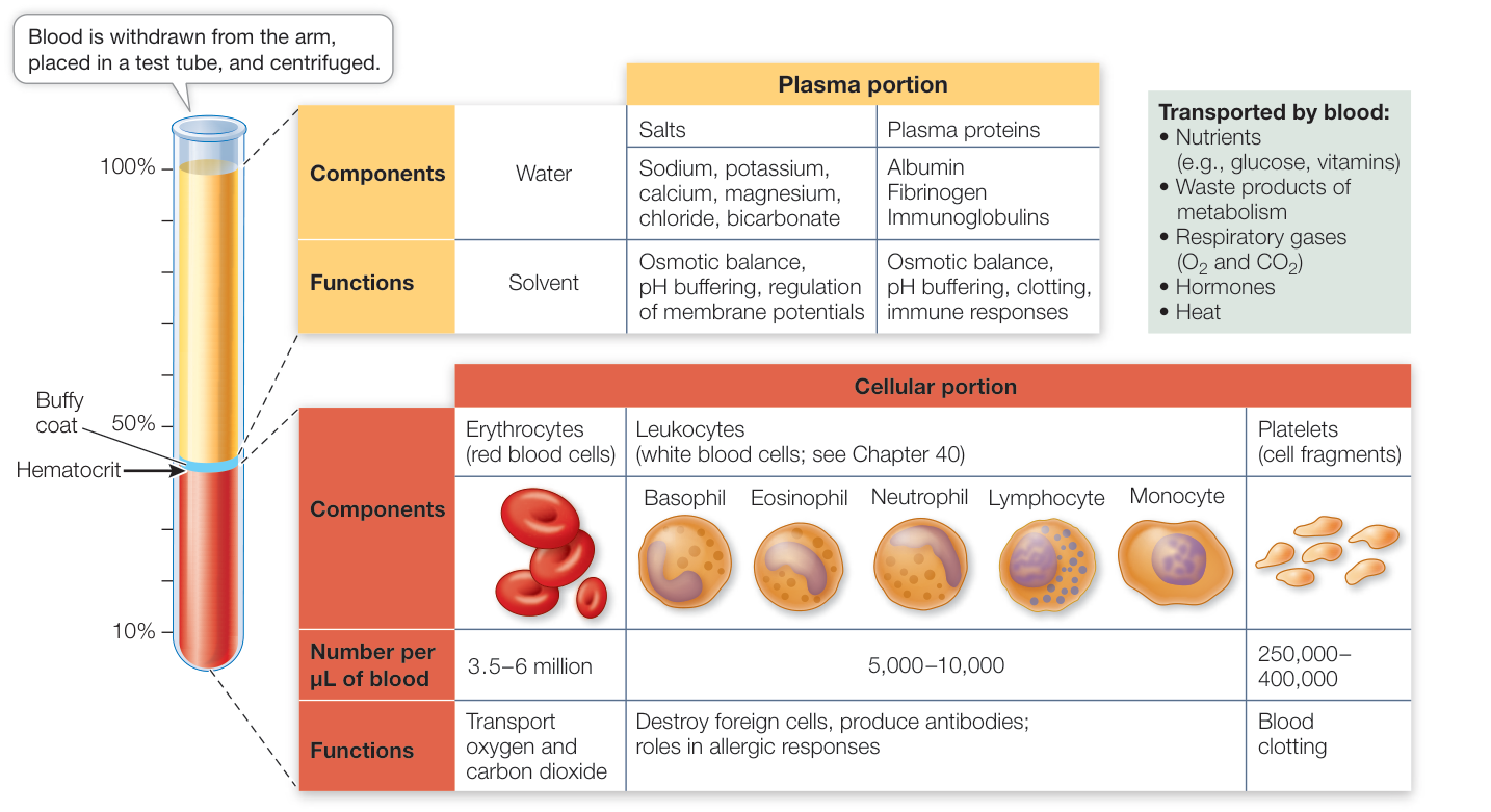
Figure 48.12 The Composition of Blood Blood consists of a complex aqueous solution (the blood plasma) and numerous cell types and cell fragments. The hematocrit (arrow) is a measure of the volume of red blood cells as a percentage of total blood volume. At the surface of the packed cells is the thin buffy coat that contains leukocytes and platelets. Major classes of leukocytes are shown, but a more complete list along with their functions can be found in Key Concept 40.1.
circulation. Loss of nuclei from the RBCs occurs in most mammalian species, but the RBCs of a few mammals and of all other vertebrates are nucleated.
Each RBC circulates for about 120 days. As it gets older, its membrane becomes less flexible and more fragile, so older RBCs are more likely to rupture as they bend to fit through narrow capillaries. RBCs are particularly squeezed in the spleen, an organ that sits near the stomach in the upper left side of the abdominal cavity. The spleen has many sinuses (cavities) that serve as reservoirs for RBCs. To get into the sinuses, however, the RBCs must squeeze between spleen cells. When old RBCs are ruptured by this squeezing, their remnants are taken up and degraded by macrophages (a class of white blood cells that ingest debris and foreign materials; see Key Concept 40.3).
Platelets are essential for blood clotting
Besides producing erythrocytes and leukocytes, the bone marrow stem cells described in Key Concept 40.1 also produce cells called megakaryocytes. Megakaryocytes are large cells that remain in the bone marrow and release platelets into the circulation. A platelet is a tiny fragment of a cell without cell organelles, but packed with enzymes and chemicals necessary for its function: sealing leaks in blood vessels and initiating blood clotting (Figure 48.13).
Damage to a blood vessel exposes collagen fibers. An encounter with collagen fibers activates platelets. The platelet swells, becomes irregularly shaped and sticky, and releases chemicals that activate other platelets and initiate the clotting of blood. The sticky platelets also form a plug at the damaged site.
Blood clotting requires many steps and many clotting factors, most of which are circulating in the blood in an inactive form. The absence of any one of these proteins can impair clotting, resulting in excessive bleeding. Because the liver produces most of the clotting factors, liver diseases such as hepatitis and cirrhosis can result in excessive bleeding. People with hemophilia experience uncontrolled bleeding because of a genetic inability to produce a clotting factor.
Blood clotting factors participate in a cascade of chemical activations of other substances circulating in the blood. The cascade begins with damage to a blood vessel or other tissue that exposes the blood to proteins such as collagen that are normally separated from the blood by endothelial cells lining the blood vessels. This exposure activates platelets and initiates the clotting factor cascade. The end result of this cascade is to convert an inactive circulating enzyme, prothrombin, to its active form, thrombin. Thrombin is a protease that acts on molecules of fibrinogen, a blood plasma protein, to create insoluble threads of fibrin. The fibrin threads form a meshwork that binds platelets, seals the vessel, and provides a scaffold for the formation of scar tissue (see Figure 48.13).
Arteries withstand high pressure, arterioles control blood flow
The walls of the large arteries have many extracellular collagen and elastin fibers that enable them to withstand the high blood pressures generated by the heart (Figure 48.14A). These elastic tissues have another important function: they are stretched during systole, and thereby store some of the energy imparted to the blood by the heart. Elastic recoil during diastole returns this energy to the blood by squeezing it and pushing it forward. As a result, even though pressure in the arteries pulsates with the beating of the heart, the flow of blood is smoother than it would be through a system of rigid pipes.
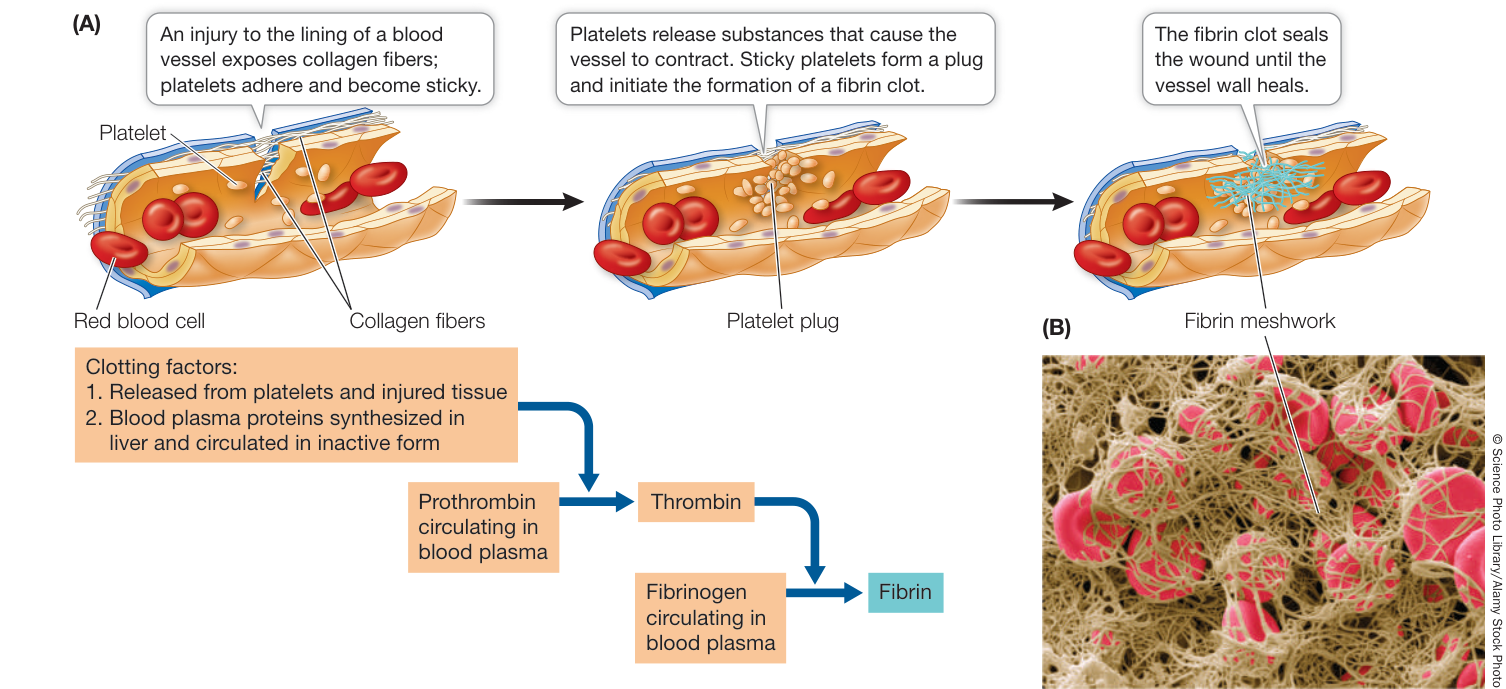
Figure 48.13 Blood Clotting (A) Damage to a blood vessel initiates a cascade of events that produce a fibrin meshwork. (B) As the meshwork forms, red blood cells are enmeshed in the fibrin threads, forming a clot, as shown in this color-enhanced electron micrograph.
Contraction of smooth muscle cells in the walls of the arteries constricts the vessels, and relaxation of the smooth muscle dilates them. When the diameter of a vessel changes, its resistance to blood flow changes, and the amount of blood flowing through it changes as a result. Neural and hormonal mechanisms act on smooth muscle cells in the walls of the arteries and arterioles, controlling the flow of blood through these vessels. The arterioles are referred to as resistance vessels because their resistance can vary to control the blood flow to specific tissues.
Materials are exchanged in capillary beds by filtration, osmosis, and diffusion
Beds of capillaries lie between arterioles and venules (Figure 48.14B). Most cells are not more than a few cell diameters away from a capillary. The needs of cells are served by the exchange of materials between blood and interstitial fluid across the capillary walls. Capillary walls are thin and permeable to water and many solutes. Also, blood flows slowly through capillaries, allowing time for exchange.
It may seem strange that blood flows through the large arteries rapidly at high pressures, but when it reaches the small capillaries the pressure and rate of flow decrease (Figure 48.14C). When you constrict the opening of a garden hose, the pressure in the hose and the velocity of the water spraying out increase. But keep in mind that the arteries branch into many arterioles that serve huge numbers of capillaries. Even though each capillary has a diameter so small that red blood cells pass through in single file, there are so many capillaries that their total cross-sectional area is much greater than that of any other class of vessel. As a result, all the capillaries together have a much greater capacity for blood than do the arterioles.
Capillary walls consist of a single layer of endothelial cells (Figure 48.15). Capillaries are permeable to water, some ions, and some small molecules but not to large molecules such as proteins, and certainly not to red blood cells. At the arterial (high-pressure) end, blood pressure squeezes water and small solutes out through spaces between the cells of the capillary walls into the surrounding intercellular space.
How is the blood volume maintained if fluid is continuously leaking out of the capillaries? An answer to this question was put forth more than 100 years ago by the physiologist E. H. Starling. Starling suggested that water movement across capillary walls is a result of two opposing forces, which are now known as Starling’s forces:
- Blood hydrostatic pressure squeezes water and small solutes out of the capillaries.
- Blood osmotic pressure pulls water back into the capillaries.
Blood hydrostatic pressure is high at the arterial end of a capillary bed and steadily drops as blood moves toward the venous end (Figure 48.16). Blood osmotic pressure is due to the large protein molecules that cannot leave the capillaries, and it is relatively constant along the capillaries. As long as the blood hydrostatic pressure is above the blood osmotic pressure, fluid leaves the capillaries. At the venule end of most capillaries, blood hydrostatic pressure falls below blood osmotic pressure, so fluid returns to the capillaries. The actual numbers for a normal capillary bed in a resting person suggest that there would be a slight net loss of fluid to the intercellular spaces. This lost fluid, about 4 liters per day, percolates between cells as the interstitial fluid before it returns to the venous blood via the lymphatic system, which we discuss later in this key concept.
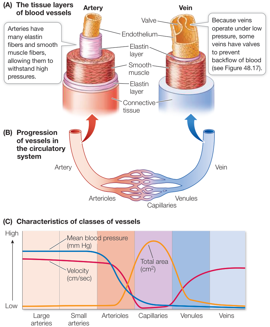
Figure 48.14 Anatomy of Blood Vessels (A) The different anatomical characteristics of arteries and veins match their functions. (B) Blood from the arterial system feeds into capillary beds where exchanges with the interstitial fluid occur. The venous system returns the blood to the heart. (C) The cross-sectional area encompassed by each vessel type is graphed along with the pressure and velocity of the blood within it.
Recent research suggests that in addition to large protein molecules in the blood, bicarbonate ions () may also contribute significantly to pulling water back into the capillaries. The produced by cellular metabolism diffuses into the endothelial cells lining the capillaries, where it is converted into and released into the blood plasma. When an individual is at rest, the increasing concentration can cause the blood osmotic pressure at the venous end to be 30 mm Hg higher than at the
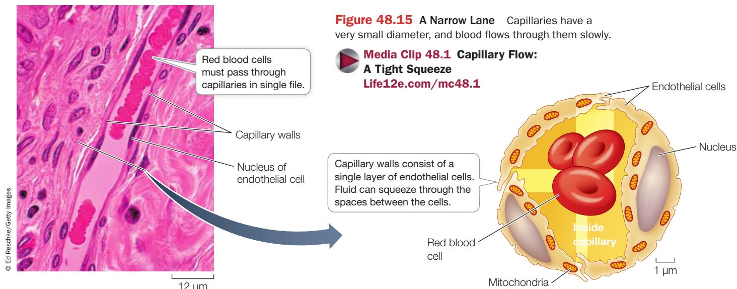
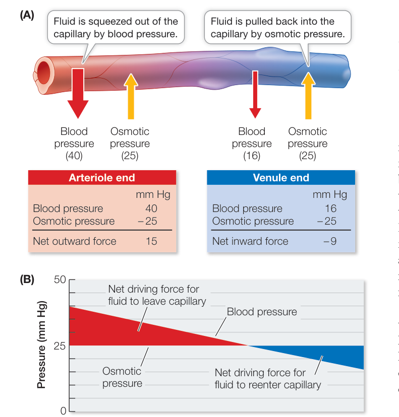
Figure 48.16 Starling’s Forces Starling’s model explains fluid balance in capillary beds. (A) When blood hydrostatic pressure is greater than blood osmotic pressure, fluid leaves the capillaries; when blood hydrostatic pressure falls below blood osmotic pressure, fluid returns to the capillaries. (B) The balance of these two forces changes over the capillary bed as blood hydrostatic pressure falls.
Blood flow through veins that are above the level of the heart is assisted by gravity. Below the level of the heart, however, venous return is against gravity. The most important force propelling blood from these regions is the squeezing of the veins by the contractions of surrounding skeletal muscles. As muscles contract, the veins are compressed and blood is squeezed through them. Blood flow may be temporarily obstructed during a prolonged muscle contraction, but when muscles relax, blood is free to move again. One-way valves in the veins of the extremities prevent backflow of blood. Thus when veins are squeezed, blood is propelled forward toward the heart (Figure 48.17).
When a person is sitting, gravity causes blood accumulation in the veins of the lower body and exerts backpressure on the capillary beds. This backpressure shifts the balance between blood hydrostatic pressure and osmotic pressure, causing increased loss of fluid to the intercellular spaces. That is why your feet swell during a long airline flight.
arterial end, and during strenuous exercise this difference can be higher. Thus it appears that and are factors that pull water back into the capillaries.
All capillaries are permeable to , , and small ions. Lipid-soluble substances readily pass through the capillary walls. Water and small solutes pass through intercellular spaces in the capillary wall, and through holes called fenestrations in the endothelial cells. More permeable and less selective capillaries are found in the digestive tract, where nutrients are absorbed, and in the kidneys, where wastes are filtered. Capillary endothelial cells also contain transporters that can facilitate the passage of specific molecules, such as glucose and lactate.
The capillaries of the brain are a special case, being rather impermeable and wrapped by glia. Not much can pass through them other than lipid-soluble substances (including alcohol and anesthetics). This high selectivity of brain capillaries is known as the blood-brain barrier. Even in the brain, however, there are specific regions where the capillaries are more permeable, enabling the brain to detect non-lipid-soluble hormones.
Blood flows back to the heart through veins
The pressure of the blood flowing from capillaries to venules is low and insufficient to propel blood back to the heart. The walls of veins are more expandable than the walls of arteries, and blood tends to accumulate in veins. Sixty percent of your total blood volume may be in your veins when you are resting. Because of their capacity to stretch and store blood, veins are called capacitance vessels.
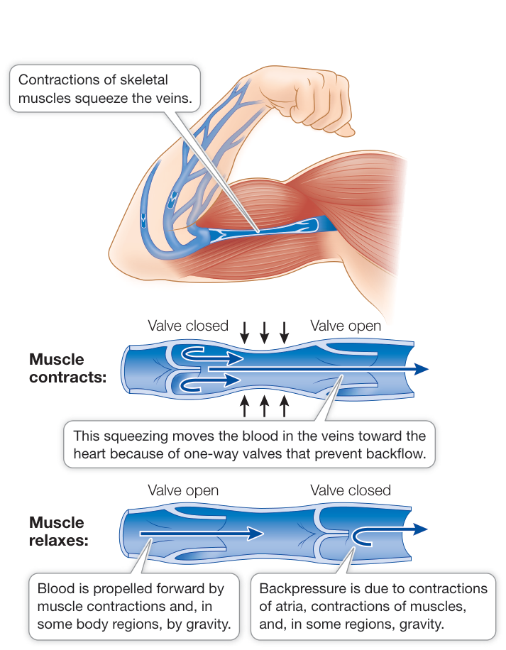
Figure 48.17 One-Way Flow Veins of the extremities have valves that prevent blood from flowing backward, and contractions of skeletal muscle help move blood toward the heart.
Because of the one-way valves in the veins of the legs, the contractions of leg muscles act as auxiliary vascular pumps when an animal walks or runs and facilitate the return of blood to the heart from the lower body. As a greater volume of blood is returned to the heart, the heart contracts more forcefully and its pumping action is enhanced. The heartbeat gets stronger because of a property of cardiac muscle cells described by the Frank–Starling law: if the cardiac muscle cells are stretched, as they are when the volume of returning blood increases, they contract more forcefully.
The actions of breathing also help return venous blood to the heart. The muscles involved in inhalation create negative pressure that pulls air into the lungs (see Figure 47.11), and this negative pressure also pulls blood toward the chest, increasing venous return to the right atrium. In addition, some of the largest veins closest to the heart contain smooth muscle that contracts at the onset of exercise. Contraction of veins can rapidly increase venous return and stimulate the heart in accord with the Frank–Starling law, increasing cardiac output.
Lymphatic vessels return interstitial fluid to the blood
The interstitial fluid contains water and other small molecules, but no RBCs, and less protein than found in blood plasma. A separate system of vessels—the lymphatic system—returns interstitial fluid to the blood. Each capillary bed contains at least one blind-ended lymph capillary.
Connect the Concepts The lymphatic system was introduced in conjunction with the immune system in Key Concept 40.1.
Interstitial fluid that enters the lymphatic vessels is called lymph. Fine lymphatic capillaries merge into progressively larger vessels and ultimately into the right and left lymphatic ducts that empty into large veins at the base of the neck (see Figure 40.3). The left lymphatic duct, also known as the thoracic duct, carries most of the lymph from the lower part of the body and is larger than the right lymphatic duct. Lymphatic vessels, like veins, have one-way valves that keep the lymph flowing toward the lymphatic ducts. Therefore lymph, like blood, is propelled toward the heart by skeletal muscle contractions and breathing movements. Lymph nodes along the major lymphatic vessels are a major site of lymphocyte production and of the phagocytic action that removes microorganisms and other foreign materials from the circulation.
Vascular disease is a killer
As mentioned at the beginning of this chapter, cardiovascular disease is responsible for about one-fourth of all deaths each year in the United States, and the same is true in Europe. The immediate cause of most of these deaths is not a defect in heart muscle as in cases of HCM, but is heart attack or stroke—both of which are usually the result of a disease called arteriosclerosis (“hardening of the arteries”) that begins many years before symptoms are detected.
Healthy arteries have a smooth internal lining of endothelial cells (Figure 48.18A) which can be damaged by chronic high blood pressure, smoking, a high-fat diet, or microorganisms. Deposits called plaque begin to form at sites of endothelial damage, initiating an early stage of arteriosclerosis called atherosclerosis.
First, the damaged endothelial cells attract certain white blood cells to the site. These cells are then joined by smooth muscle cells migrating from the deeper layers of the arterial wall. Lipids, especially cholesterol, are deposited in these cells, so that the developing plaque becomes fatty. Fibrous connective tissue made by the invading smooth muscle cells in the plaque, along with deposits of calcium, gradually makes the artery wall less elastic—hence “hardening of the arteries.” Plaque deposits narrow the artery and causes turbulence in the blood flow. Blood platelets stick to the plaque (see Figure 48.13) and initiate formation of an intravascular blood clot, a thrombus (plural thrombi) that can block the artery (Figure 48.18B).
The blood supply to the heart muscle flows through the coronary arteries, which are highly susceptible to atherosclerosis. As these arteries narrow, blood flow to the heart muscle decreases, causing the symptoms of chest pain and shortness of breath during mild exertion. A person with atherosclerosis is at high risk of forming a thrombosis, can totally block the vessel, causing a myocardial infarction (heart attack).
A piece of a thrombus that breaks loose, called an embolus (plural emboli), is likely to become lodged in a vessel of smaller diameter, blocking its flow (an embolism). Arteries already narrowed by plaque formation are likely places for an embolism. An embolism in an artery in the brain causes the cells fed by that artery to die. This event is a stroke. The specific damage resulting from a stroke, such as memory loss, speech impairment, or paralysis, depends on the location of the blocked artery in the brain.
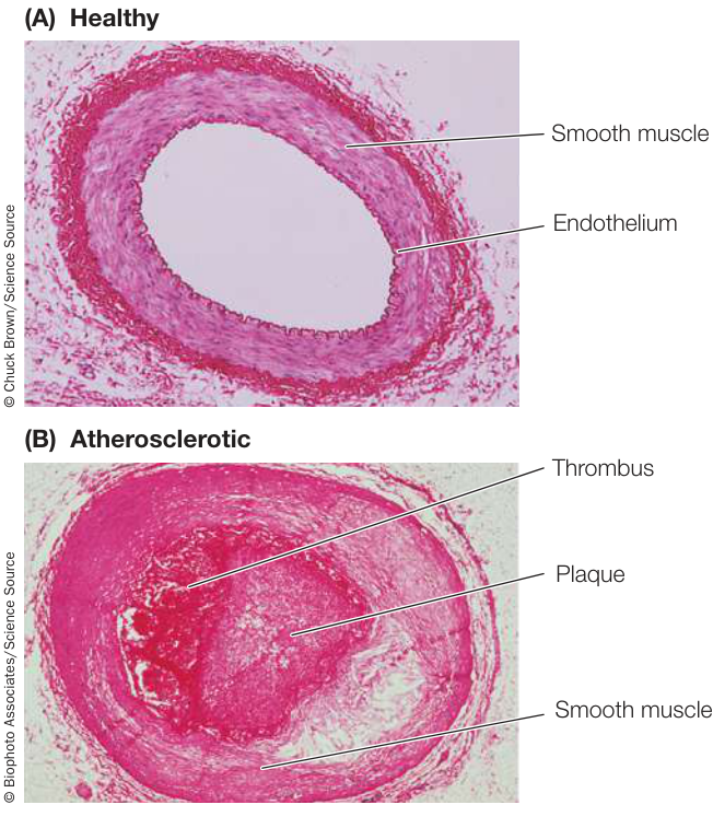
Figure 48.18 Atherosclerotic Plaque (A) A healthy, clear artery. (B) An atherosclerotic artery, clogged with plaque and a thrombus.
Circulation Is Controlled by Hormonal and Neuronal Signals
Important risk factors for developing atherosclerosis are your genetic predisposition and your age. Environmental risk factors also play a large role. These include high-fat and high-cholesterol diets, smoking, and a sedentary lifestyle. Certain untreated medical conditions such as hypertension (high blood pressure), obesity, and diabetes are also risk factors for atherosclerosis. For those who have a genetic predisposition to atherosclerosis, it is even more important to minimize environmental risk factors. Changes in diet and behavior and treatment of predisposing medical conditions can prevent and reverse early atherosclerosis and help fend off this silent killer.
Recap and Assess
Blood is a fluid tissue with cellular components that play roles in transport of respiratory gases, immune system function, and blood clotting. The properties of the arteries, arterioles, capillaries, venules, and veins reflect their functions. Exchanges between the blood and interstitial fluids occur in the smallest of those vessels, the capillaries. These exchanges are governed by Starling’s forces and the movement of into the blood. Lymphatic vessels return interstitial fluid to the blood. Blockage of arteries by plaque, thrombi, or emboli are major causes of heart disease and stroke.
- Why do athletes train at high elevations?
- Why is the blood-clotting process called a cascade?
- Why are arterioles called resistance vessels and veins called capacitance vessels, and what do these terms imply about function?
- In advanced starvation, the body breaks down its blood plasma proteins for energy. At this stage of disease, fluid accumulates in the abdomen and the extremities. What is the connection between these two consequences of starvation?
Every tissue in the body requires an adequate flow of oxygen-saturated blood. Blood flow depends on the maintenance of an appropriate blood pressure, and the distribution of blood flow throughout the body depends on control of the resistance in the blood vessels supplying different tissues.
48.5
Circulation Is Controlled by Hormonal and Neuronal Signals
Learning Objectives
48.5.1 Describe the mechanisms underlying autoregulation of local blood flow and how it influences mean arterial pressure (MAP).
48.5.2 Explain the role of the autonomic nervous system in regulating MAP.
48.5.3 Explain how an increase or a decrease in the firing rate of baroreceptors causes a change in MAP.
48.5.4 Discuss the roles of positive and negative feedback in controlling blood pressure.
When we investigate how a physiological process is regulated, we start by identifying the critical components of that process, how they can be controlled, and the information used to govern that control. Because blood flow depends on pressure, we can identify the mean arterial pressure (MAP) as a critical variable of the circulatory system.
Pressure in the aorta oscillates between systole and diastole, and the duration of diastole is usually twice as long as that of systole (see Figure 48.5). Therefore a simple way of estimating MAP is to take the diastolic pressure and add to it one-third of the difference between systolic and diastolic pressure. However, to understand the physiological mechanisms that control MAP, we have to consider the factors that determine it. MAP is determined by the cardiac output (CO) and the resistance to flow in the blood vessels, or total peripheral resistance (TPR):
Since CO is equal to the heart rate (HR) times how much blood the heart pumps with each beat (stroke volume [SV]), the critical relationships can be expressed as:
HR, SV, and TPR are controlled by neural and hormonal mechanisms at both the local and systemic levels.
At the local level, each tissue controls its own blood flow through autoregulatory mechanisms that alter the diameter and therefore the resistance of the arterioles serving it. The collective autoregulatory actions in the arterioles in all tissues of the body influence TPR and therefore MAP. If many arterioles suddenly dilate, TPR goes down and MAP falls. If many arterioles constrict, TPR goes up and MAP goes up. Therefore changes in MAP provide information about changing needs of the body. In addition, as blood flows through capillary beds, its composition changes—its content goes up and its content goes down. Thus blood composition also provides information the body uses to regulate the circulatory system.
The nervous and endocrine systems respond to changes in MAP and blood composition by changing breathing rate, heart rate, stroke volume, and peripheral resistance to match the metabolic needs of the body.
Autoregulation matches local blood flow to local need
The amount of blood that flows through a capillary bed is controlled by contraction or relaxation of the smooth muscle of the arteries and arterioles feeding that bed. Figure 48.19 illustrates the flow of blood in a typical capillary bed. Blood flows into the bed from an arteriole. Smooth muscle “cuffs,” or precapillary sphincters, on the arteriole can shut off the supply of blood to the capillary bed. When the precapillary sphincters are relaxed and the arteriole is open, blood flow through the capillaries increases.
Autoregulation depends on the sensitivity of the smooth muscle to its local chemical environment. Low concentrations and high concentrations cause the smooth muscle to relax, thus increasing the supply of blood, which brings in more and carries away —a response known as hyperemia, which means “excess blood.” Increases in other by-products of metabolism, such as lactic acid, hydrogen ions, potassium, and adenosine (all of which increase in exercising muscle), also promote hyperemia. Hence activities that increase the metabolism of a tissue also induce hyperemia in that tissue. Regional autoregulation, however, sets up a dangerous positive-feedback loop (Figure 48.20). A fall in MAP decreases blood flow to tissues. Decreased local blood flow generates autoregulatory decreases in resistance in the arterioles serving those tissues, contributing to decreased TPR that further decreases MAP. Systemic regulatory mechanisms must counter the positive feedback caused by autoregulation.
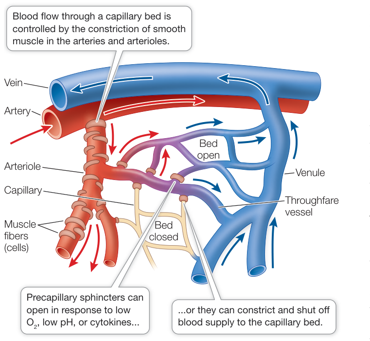
Figure 48.19 Local Control of Blood Flow Low concentrations or high levels of metabolic by-products cause the smooth muscle of the arteries and arterioles to relax, thus increasing the supply of blood to the capillary bed.
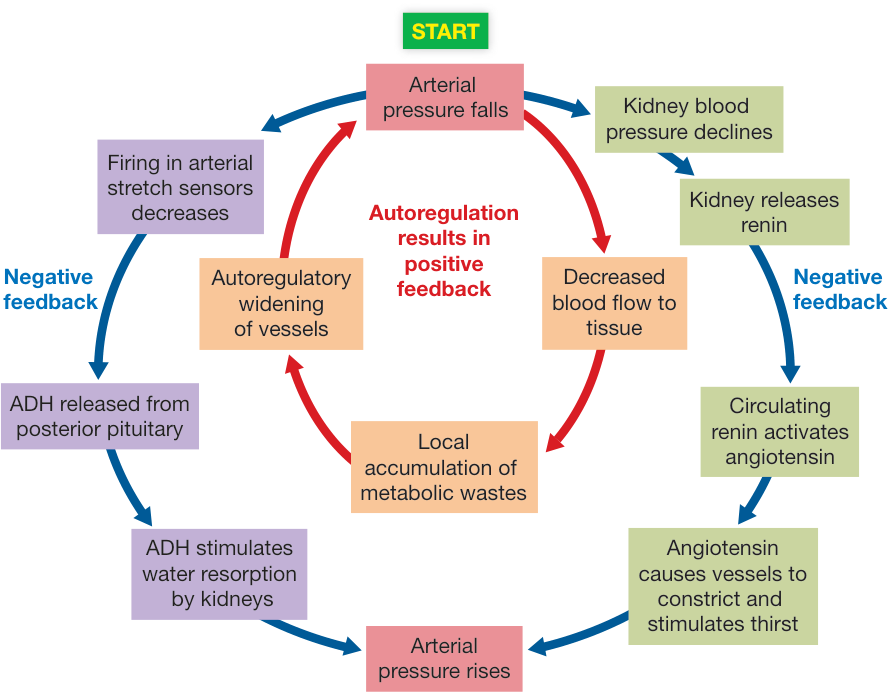
Figure 48.20 Control of Blood Pressure through Local and Systemic Mechanisms A drop in arterial pressure reduces blood flow to tissues, resulting in local accumulation of metabolic wastes. This change in the extracellular environment stimulates autoregulatory opening of the arterioles. A fall in central blood pressure is prevented by negative-feedback mechanisms (including the release of antidiuretic hormone, ADH) that constrict arterioles in less essential tissues and stimulate maintenance of blood volume and blood pressure.
Arterial pressure is regulated by hormonal and neural mechanisms
Control and regulation of the circulation of the blood begin with the local autoregulatory mechanisms that alter the resistance of arteries and arterioles feeding capillary beds. The demands of the capillary beds influence MAP and blood composition. Both of these provide information for the control of systemic endocrine and neural responses that return blood pressure and composition to normal. Thus circulatory functions are matched to the regional and to the overall needs of the body.
Arteries and arterioles are innervated by the autonomic nervous system, particularly the sympathetic division. The sympathetic postganglionic neurotransmitter norepinephrine binds to receptors in smooth muscle in blood vessels in the gut and other tissues not essential for “fight or flight” and causes these vessels to constrict, resulting in reduced blood flow through them and an elevation in MAP. As we discussed earlier in this chapter, increased sympathetic activity increases heart rate, and by increasing the strength of the cardiac muscle contraction, it also increases stroke volume. Thus the actions of sympathetic activation increase MAP.
Hormones also play a role in regulating arterial pressure. Epinephrine has actions similar to those of norepinephrine and is released from the adrenal medulla during massive sympathetic activation stimulated by a fall in arterial pressure or by activation of the fight-or-flight response to a dangerous threat. Another hormone, angiotensin, is activated when blood pressure in the kidneys falls (see Figure 48.20). In response to the fall in blood pressure, the kidneys release an enzyme, renin, into the circulation. Renin and another enzyme in the lungs converts a circulating protein, angiotensinogen into the active hormone angiotensin. Angiotensin has several actions: constricting arterioles located in peripheral tissues, stimulating thirst, and increasing release of another hormone aldosterone from the adrenal gland. Aldosterone increases reabsorption. All of these responses protect or increase blood volume and therefore central blood pressure necessary to maintain adequate blood flow to essential organs such as the heart, brain, and kidneys.
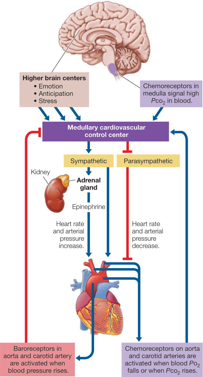
Figure 48.21 Regulating Cardiac Output The autonomic nervous system controls heart rate in response to information about blood pressure and blood composition originating in baroreceptors and chemoreceptors shown at the bottom of the figure. Information from these sensors goes to the cardiovascular control center in the medulla, where it is integrated with other information. The medullary center generates responses in the sympathetic and parasympathetic nervous systems that control cardiac output.
The autonomic nervous system activity that controls heart rate and constriction of blood vessels originates in a cardiovascular control center in the medulla. Many inputs converge on this central integrative network and influence the commands it issues via parasympathetic and sympathetic nerves (Figure 48.21).
Of special importance is incoming information about changes in blood pressure from baroreceptors (stretch receptors) in the walls of the large arteries leading to the brain—the aorta and the carotid arteries.
Increased activity in baroreceptors of the large arteries signals rising blood pressure and inhibits sympathetic nervous system signaling to arteries and arterioles while increasing parasympathetic signaling to the heart’s pacemaker. As a result, the heart slows and arterioles in peripheral tissues dilate, reducing blood pressure. If pressure in the large arteries falls, the activity of the baroreceptors decreases, stimulating sympathetic output to the heart’s pacemaker. As a result, the heart beats faster and the arterioles in peripheral tissues constrict, increasing blood pressure.
Another hormone that helps stabilize blood pressure is antidiuretic hormone (ADH), also called vasopressin, which is secreted by the posterior pituitary in response to a fall in the activity of the baroreceptors, signaling a fall in MAP. ADH causes the kidneys to reabsorb more water and thereby maintain blood volume and increase blood pressure (see Figure 50.14). Increased activity of the baroreceptors inhibits the release of ADH, and as a result the kidneys excrete more water, reducing blood volume and contributing to a fall in arterial pressure (see Figure 48.20).
Other information that causes the cardiovascular control center to increase heart rate and blood pressure comes from chemoreceptors in the medulla, aorta, and the carotid arteries. The information from the chemoreceptors, from the baroreceptors, and from the kidney constitutes negative-feedback signals that serve to regulate MAP at optimal levels in spite of greatly changing demands of the body.
Connect the Concepts Key Concept 47.5 discusses the mechanisms for sensing and concentrations in the blood. The medullary chemoreceptors are activated by increases in levels and decreases in pH, and the carotid and aortic bodies are activated by falling levels. Chemoreceptors send signals to the cardiovascular regulatory center as well as to the respiratory regulatory center.
Recap and Assess
The delivery of blood to specific regional tissues is controlled locally by autoregulatory mechanisms that dilate or constrict arterioles. These local actions are translated into alterations in central blood pressure and composition. Those changes generate neural and hormonal negative-feedback signals that mediate corrective cardiovascular adjustments.
- Why is autoregulation in Figure 48.20 described as a positive-feedback loop?
- Distinguish between the roles of the two divisions of the autonomic nervous system (ANS) in regulating MAP?
- Baroreceptor neurons have a range of firing rates depending on their degree of stretch, and at rest they are firing at the midpoint of that range. Why is this adaptive?
- Explain how local autoregulatory controls of blood flow can stimulate hormonal adjustments of MAP.
Investigating Life: QA Are there potential therapies that could treat HCM?
Many organs of your body can shut down for a while and you can recover—but not your heart. Cardiac arrest lasting only minutes can result in death. But why should the heart of a healthy, physically fit, top-performing athlete stop? Just as physical conditioning results in making skeletal muscles bigger and stronger, it does the same for the heart. The heart responds to increased demand by getting bigger and stronger. However, if an individual has mutations in genes responsible for proteins involved in contractions of cardiac muscle, that muscle will be less efficient. To compensate for that inefficiency, the heart gets bigger than a normal heart would when repeatedly challenged by excessive exercise. Hypertrophy of the walls of the left ventricle can have two serious consequences: (1) The opening of the aorta is at the top of the heart next to the septum that divides the two ventricles. Enlargement of that septum can block the entrance to the aorta, so the cardiac output falls. (2) The bundle of His and the left and right bundle branches that convey the pacemaker potential to the ventricular muscle course down through the septum. Hypertrophy of the septum can interfere with conduction of these critical signals. If advanced conditions of HCM are discovered, there are surgical procedures that remove some of the heart muscle to increase blood flow.
Future directions
The mutations responsible for the development of HCM are not rare, but most people who carry them live their entire lives without even knowing they are at risk for HCM. Why? Most people do not place the excessive demands on their hearts that athletes do. So what can be done to reduce the risk of death by HCM? Preventative measures such as genetic screening could allow carriers of the mutations to make decisions about whether or not to pursue extreme physical activity. Physical examinations including ECGs, sonograms, and even magnetic resonance imaging (MRI) would enable diagnosis and recommendations to decrease levels of physical exertion. The work presented in Investigating Life: Silencing Mutant Myosin Genes suggests a potential future treatment through gene therapy. If safe reagents could be developed to silence the mutant genes, and those reagents were applied before severe HCM developed, they could have a protective benefit.

© Andrew Yates/Stringer/Getty Images
You should be able to relate each summary to the adjacent figures. If you go to this visual summary in Achieve, you can follow links to figures, animations, activities, and simulations that will help you consolidate the material.
Visual Summary
Circulatory Systems Are Transport Systems
- ■ Circulatory systems consist of muscular pumps (hearts) that move extracellular fluid through vessels to serve tissues of the body. These systems can be open or closed.
- ■ Circulatory systems transport nutrients, respiratory gases, heat, signaling molecules, immune system elements, and metabolic wastes throughout the body.
Questions
- What are the relationships and differences between extracellular fluid, hemolymph, blood, and interstitial fluid?
- What advantages does a closed circulatory system have for an active species? Why are insects an exception?
- How does an animal with a closed circulatory system respond to changing demands for oxygen in its different tissues?
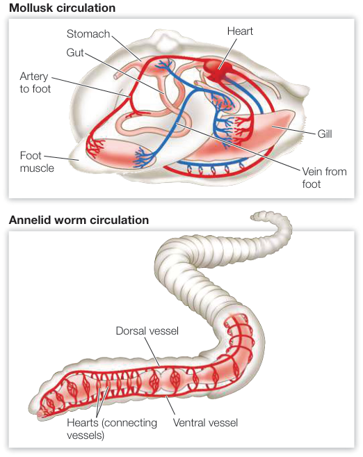
Figure 48.1
Vertebrate Circulatory Systems Evolved from Single to Double Circuits
- ■ Vertebrate hearts pump blood through arteries and arterioles to capillaries, the site of exchange between blood and interstitial fluid. Blood returns to the heart through venules and veins.
- ■ Vertebrate circulatory systems evolved from single circuits in fishes to partially or completely separate pulmonary circuits and systemic circuits in amphibians, reptiles, and mammals.
- ■ Ectothermic reptiles shunt blood from the pulmonary to the systemic circuit when they are not breathing.
- ■ Pulmonary and systemic circuits are completely separate in birds and mammals and operate at different pressures.
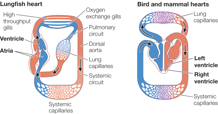
Figure 48.2 Questions
- Explain how a lungfish can breathe in water or in air.
- How does blood flow change in an ectothermic reptile when it isn’t breathing?
- Trace the flow of blood through a mammalian circulatory system.
Heart Function Depends on Properties of Cardiac Muscle
- ■ The ventricles of the mammalian heart fill mostly passively during diastole and pump blood into the pulmonary and systemic circuits during systole. Valves in the heart prevent the backflow of blood.
- ■ The heartbeat is due to interactions of unique , , and channels that generate rhythmic action potentials in cardiac muscle pacemaker cells.
- ■ Action potentials in the sinoatrial node cause atrial muscle to contract and the atrioventricular node to fire.
- ■ Action potentials from the atrioventricular node are conducted throughout the ventricular muscle by the bundle of His and Purkinje fibers.
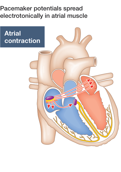
Figure 48.9 Pacemaker potentials spread electrotonically in atrial muscle
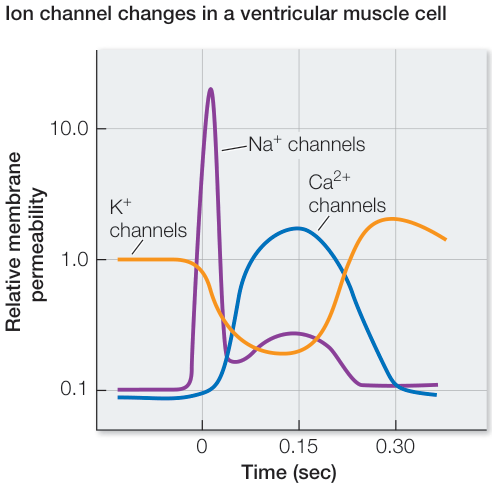
Figure 48.10 Ion channel changes in a ventricular muscle cell
- ■ Sustained contraction of ventricular muscle cells is due to channels.
- ■ The electrocardiogram (ECG or EKG) reflects electrical events in the heart.
Questions
- Explain the heart sounds (“lub”/“dup”) in terms of events occurring in the heart.
- Explain how the different functions of pacemaker cells and ventricular muscle cells are due to their unique ion channels.
- How are the contractions of the atria and the ventricles coordinated?
Circulatory System Functions Depend on Blood and Blood Vessels
- ■ Blood consists of plasma containing protein and ionic solutes, cells of the immune system (leukocytes), red blood cells carrying oxygen, and platelets that form blood clots.
- ■ The functions of arteries as resistance vessels and of veins as capacitance vessels are reflected in their structural differences.
- ■ Extracellular fluid filters out of capillaries where blood pressure is higher than osmotic pressure and flows back into capillaries where blood pressure is lower than osmotic pressure.
- ■ Valves in veins and in lymphatic ducts enable muscle contraction to propel blood back to the heart and interstitial fluid back to the circulation, respectively.
- ■ Buildup of plaque and of thrombi (blood clots) in arteries can block blood flow, causing heart attack and stroke.
Questions
- How do platelets initiate blood clotting?
- Capillaries are permeable to water, and blood is under pressure, so why doesn’t interstitial fluid accumulate in tissues?
- What is the progression of a healthy cardiac artery with a smooth endothelial lining to one that causes a myocardial infarction?
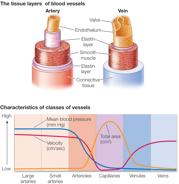
Figure 48.14
Circulation Is Controlled by Hormonal and Neuronal Signals
- ■ To meet local needs, blood flow through capillary beds is controlled by local autoregulatory mechanisms.
- ■ Mean arterial pressure (MAP) depends on cardiac output and total peripheral resistance (TPR).
- ■ Baroreceptors provide negative-feedback information on MAP.
- ■ Chemoreceptors provide negative-feedback information on composition of the blood.
- ■ Antidiuretic hormone (ADH, also called vasopressin) and angiotensin act to raise MAP.
Questions
- What neural and hormonal mechanisms raise MAP?
- How does cardiovascular regulation respond to variations in local autoregulation of capillary beds?
- How do chemoreceptors and baroreceptors contribute to the regulation of MAP?
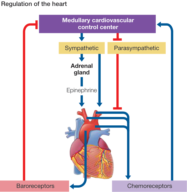
Figure 48.21