Amino acids and the peptide bond
Nearly every structural computation starts from the same three atoms in each residue, named N, CA, and
C in a structure file. Backbone angles are measured from them, a contact map uses each CA, and a
structure predictor's job is to place them. Because the peptide bond joining residues is flat and rigid, a protein's thousands of coordinates
reduce to about two angles per residue.
The amino acid
Proteins are linear polymers of amino acids, and it is the distinct sequence of component amino acids that determines the ultimate three-dimensional structure of the protein. The sequence of a protein is often referred to as its primary structure.
Amino acids are small molecules that contain an amino group (NH2), a carboxyl group (COOH), and a hydrogen atom attached to a central alpha (α) carbon. In addition, amino acids also have a side chain (or R group) attached to the α-carbon. It is this group, and only this group, that distinguishes one amino acid from another. Furthermore, the side chain confers the specific chemical properties of the amino acid.
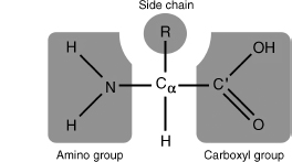
The structure of a prototypical amino acid. The chemical groups bound to the central alpha (α) carbon are highlighted in gray. The R-group represents any of the possible 20 amino acid side chains.
There are 20 amino acids that can be incorporated into a protein via the genetic code. This set of 20 amino acids is considered "standard" in that it is common to all observed organisms. Modified forms of these 20 amino acids do exist in proteins, but these are the product of modifications that occur subsequent to protein synthesis.
The amino and carboxyl groups are a base and an acid, so lesson 1.3 predicts their state in water:
At the pH levels typically found in cells (usually pH 7.0–7.4), both the carboxyl and amino groups of amino acids are ionized: the carboxyl group has lost a hydrogen ion:
and the amino group has gained a hydrogen ion:
Thus amino acids show both acid and basic properties.
A free amino acid in water therefore carries a plus and a minus charge at once and is neutral overall, a form called a zwitterion. The α carbon, bonded to four different groups, is also a chiral center; only the L form is used in proteins, and glycine, with two hydrogens on Cα, is not chiral at all (lesson 1.6).
From amino acids to a chain of residues
The process consists of joining the –OH component of the –COOH group (carboxyl end) of the left amino acid with a hydrogen atom of the NH2– group (amino end) of the right amino acid, creating a CN covalent bond and freeing one molecule of water. Such polymers, consisting of the concatenation of amino acid residues (residues, because of the subtraction of H2O) are commonly referred to as polypeptides.
The amino group of an adding amino acid interacts in a condensation reaction with the carboxyl group of the last amino acid in the growing chain, forming a peptide bond.
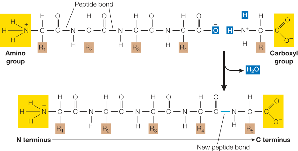
Peptide bonds are formed by condensation reactions in which the amino group of one amino acid reacts with the carboxyl group of another amino acid, releasing a molecule of water.
Just as a sentence begins with a capital letter and ends with a period, polypeptide chains have a beginning and an end. The "capital letter" marking the beginning of a polypeptide is the amino group of the first amino acid added to the chain and is known as the N terminus. The "period" is the carboxyl group of the last amino acid added; this is the C terminus.
Chains are written and numbered N to C, so residue 1 carries the free amino group. Condensation also removes charges: the amino and carboxyl groups that join become a neutral peptide bond, so only the chain's two ends keep theirs, and every other charge on a protein sits on a side chain.
[The chain] may be viewed as an alternating sequence of a fixed "spacer" components, –CO–NH–, called peptide groups, and of variable components –HCRj–, where Rj is a side chain (the carbon atom is called an α-carbon to distinguish it from the carbon in the peptide group). Essentially, the polypeptide sequence has a backbone of the type (CαCN)n and the residue sequence R1R2R3… completely describes it. This description is called the primary structure.
That repeating N–Cα–C is exactly what a structure file lists. Each residue's atoms come backbone first
(N CA C O), then the side chain, with atoms named by Greek letters counting outward from Cα: β, γ, δ, ε,
ζ, written B, G, D, E, Z.
from primer import structure

ubq = structure("1UBQ")[0]["A"]          # model 0, chain A
for res in list(ubq)[:3]:
    print(res.id[1], res.get_resname(), " ".join(atom.get_id() for atom in res))
1 MET N CA C O CB CG SD CE
2 GLN N CA C O CB CG CD OE1 NE2
3 ILE N CA C O CB CG1 CG2 CD1
Methionine's side chain reads CB CG SD CE, a β-carbon, a γ-carbon, a δ-sulfur, and an ε-carbon.
Isoleucine branches at its β-carbon into CG1 and CG2. There are no hydrogens: X-ray diffraction rarely
locates them.
The peptide bond is flat
The specific characteristics of the peptide bond have important implications for the three-dimensional structures that can be formed by polypeptides. The peptide bond is planar and quite rigid. Therefore, the polypeptide chain has only a rotational freedom about the bonds formed by the α-carbons. These bonds have been termed the Phi (Φ) and Psi (ψ) angles. However, rotational freedom about the Φ (Cα–N) and ψ (Cα–C) angles is limited by steric hindrance between the side chains of the residues and the peptide backbone. Consequently, the possible conformations of a given polypeptide chain are quite limited.
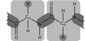
The peptide bond. Two peptide units (amino acid residues) are shown shaded in light gray. The peptide bond between them is shaded in dark gray.
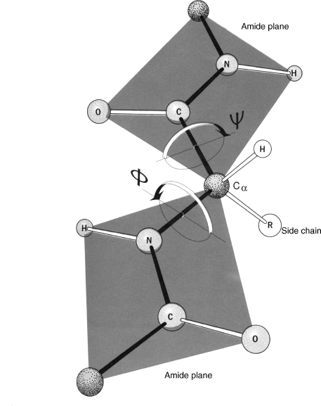
Two planar peptide bonds about a central alpha carbon, shown as a ball-and-stick model. Rotation is only possible about the Φ (Cα–N) and ψ (Cα–C′) angles. Arrows about the two angles show the direction that is considered positive rotation. Here both angles are approximately 180°.
Two characteristics of the peptide bond are especially important in the three-dimensional structures of proteins:
- In the C–N bond, the adjacent α carbons (Cα–C–N–Cα) are not free to rotate fully, which limits the folding of the polypeptide chain.
- The oxygen bound to the carbon (C=O) carries a slight negative charge (δ⁻), whereas the hydrogen bound to the nitrogen (N–H) is slightly positive (δ⁺). This asymmetry of charge favors hydrogen bonding within the protein molecule itself and between molecules.
Why can't the C–N bond rotate? On paper it is a single bond, but the nitrogen's lone pair is shared into the neighboring C=O,A lone pair is two outer-shell electrons that belong to one atom and aren't in a bond; the amide nitrogen has one. Here those electrons spread over N, C, and O, the same averaging that lesson 1.1 called resonance in the carboxylate group. giving the bond partial double-bond character, and double bonds don't rotate (lesson 1.1). Bond lengths show it. In a high-resolution structure of ribonuclease A, the peptide C–N bond is clearly shorter than the N–Cα single bond and close to a C=N double bond (about 1.27 Å):
import numpy as np

rnase = structure("7RSA")[0]["A"]
residues = [r for r in rnase if r.id[0] == " "]      # skip waters and ligands
pairs = list(zip(residues, residues[1:]))
c_n  = [b["N"] - a["C"] for a, b in pairs]            # peptide bond (Bio.PDB atoms subtract to a distance)
n_ca = [r["N"] - r["CA"] for r in residues]           # a true single bond
c_o  = [r["C"] - r["O"] for r in residues]            # a true double bond
print(f"C–N (peptide) {np.mean(c_n):.3f} Å")
print(f"N–Cα (single) {np.mean(n_ca):.3f} Å")
print(f"C=O (double)  {np.mean(c_o):.3f} Å")
C–N (peptide) 1.318 Å
N–Cα (single) 1.468 Å
C=O (double)  1.236 Å
Twisting the bond would break the shared electrons, which costs about 20 kcal/mol, some 30 times the thermal energy kT. So six atoms stay in one plane: Cα of residue i, its C and O, the N and H of residue i+1, and that residue's Cα.
The backbone of ubiquitin's first six residues (carbon grey, nitrogen blue, oxygen red). Rotate until you look along one C=O edge-on: each C=O, the N after it, and the two Cα atoms on either side line up in one plane. The planes meet at the Cα atoms, which act as the joints.
Open 1UBQ in Mol*The torsion around the peptide bond is called ω (omega). The plane allows two values: 180°, trans, with the two Cα atoms on opposite sides, and 0°, cis, with them on the same side, where they and their side chains bump. Trans wins overwhelmingly. The exception is the bond before a proline, whose ring puts a carbon on the nitrogen instead of a hydrogen, so both forms are crowded and about 5% of them are cis.
from Bio.PDB.vectors import calc_dihedral
import matplotlib.pyplot as plt

def omega(a, b):
    """Torsion Cα(i)–C(i)–N(i+1)–Cα(i+1), in degrees."""
    atoms = [a["CA"], a["C"], b["N"], b["CA"]]
    return np.degrees(calc_dihedral(*(x.get_vector() for x in atoms)))

omegas = [(a, b, omega(a, b)) for a, b in pairs]
for a, b, w in omegas:
    if abs(w) < 30:
        print(f"cis: {a.get_resname()} {a.id[1]} – {b.get_resname()} {b.id[1]}, ω = {w:.1f}°")

fig, ax = plt.subplots(figsize=(6.4, 2.6))
ax.hist([abs(w) for *_, w in omegas], bins=np.arange(0, 181, 5), color="#2a78d6")
ax.set_xlabel("|ω| (degrees)")
ax.set_ylabel("peptide bonds")
plt.show()
cis: TYR 92 – PRO 93, ω = 3.1°
cis: ASN 113 – PRO 114, ω = 0.7°
Ribonuclease A's two cis bonds are both into prolines (Pro 93 and Pro 114); every other bond sits within a few degrees of 180°.
The peptide bond is ideally, although not in reality, planar and rigid, and the rotation is only allowed by the φ (Cα–N) and ψ (Cα–C′) torsion angles. Given a sequence of n amino acids, we are interested in finding all torsion angles (φ1, ψ1, φ2, ψ2, …, φn, ψn) for all peptide units to determine the three-dimensional structure of the protein backbone.
This is a change of representation: from three coordinates per atom (about 24 numbers per residue for the backbone) to two joint angles per residue, the way a robot arm is described by its joint angles. Unit 3 maps which (φ, ψ) pairs are allowed.
Writing a protein down
The precise sequence of amino acids in a polypeptide chain held together by peptide bonds constitutes the primary structure of a protein. The single-letter abbreviations for amino acids are used to record the amino acid sequence of a protein. Here, for example, are the first 20 amino acids (out of a total of 124) in the protein ribonuclease from a cow:
KETAAAKFERQHMDSSTSAA
A sequence is a string over a 20-letter alphabet, read N to C, and describes one chain. A protein built from
several chains, like hemoglobin with two α and two β chains, needs one string per distinct chain.
The standard text format is FASTA: a header line starting with >, then the residues on one or more
lines. Here is hemoglobin as the Protein Data Bank distributes it:
import urllib.request
from primer import fasta, uniprot

text = urllib.request.urlopen("https://www.rcsb.org/fasta/entry/4HHB").read().decode()
print(text)
for header, seq in fasta(text):
    print(f"{len(seq):>4} residues  {header}")
>4HHB_1|Chains A, C|Hemoglobin subunit alpha|Homo sapiens (9606)
VLSPADKTNVKAAWGKVGAHAGEYGAEALERMFLSFPTTKTYFPHFDLSHGSAQVKGHGKKVADALTNAVAHVDDMPNALSALSDLHAHKLRVDPVNFKLLSHCLLVTLAAHLPAEFTPAVHASLDKFLASVSTVLTSKYR
>4HHB_2|Chains B, D|Hemoglobin subunit beta|Homo sapiens (9606)
VHLTPEEKSAVTALWGKVNVDEVGGEALGRLLVVYPWTQRFFESFGDLSTPDAVMGNPKVKAHGKKVLGAFSDGLAHLDNLKGTFATLSELHCDKLHVDPENFRLLGNVLVCVLAHHFGKEFTPPVQAAYQKVVAGVANALAHKYH

 141 residues  4HHB_1|Chains A, C|Hemoglobin subunit alpha|Homo sapiens (9606)
 146 residues  4HHB_2|Chains B, D|Hemoglobin subunit beta|Homo sapiens (9606)
The header 4HHB_1|Chains A, C|… says chains A and C share the α sequence: four chains, two distinct
sequences. Headers are free text laid out however their source chose, so code should split off the header
and assume nothing about it. Compare UniProt's header for the same α chain:
header, alpha = fasta(uniprot("P69905"))[0]
print(header)
print(len(alpha), alpha[:12] + "…")
pdb_alpha = next(seq for h, seq in fasta(text) if "alpha" in h)
print("UniProt without its first residue equals the PDB chain:", alpha[1:] == pdb_alpha)
sp|P69905|HBA_HUMAN Hemoglobin subunit alpha OS=Homo sapiens OX=9606 GN=HBA1 PE=1 SV=2
142 MVLSPADKTNVK…
UniProt without its first residue equals the PDB chain: True
UniProt's sequence is one residue longer. Every protein is made starting with methionine, and mature hemoglobin has lost it, so residue 1 of the crystal structure is residue 2 in UniProt. Unit 6 covers these numbering offsets.
Check yourself
A PDB file lists one residue's atoms as N CA C O CB OG. Without a name, what can you say about it? What
would N CA C O alone mean?
Reasoning
It has one side-chain carbon (CB) carrying an oxygen at the γ position (OG): a –CH₂–OH side chain,
which is serine. (Threonine would have OG1 and CG2, since its β-carbon branches.) Four atoms N CA C O
with no CB would be glycine, the only residue without a β-carbon, or any residue whose side chain wasn't
seen in the experiment, which happens with floppy surface side chains. Check the residue name before
concluding.
The peptide MKV is made by a ribosome. Which residue carries the free amino group, how many peptide bonds
and waters were involved, and what is its charge at pH 7?
Reasoning
Met is at the N-terminus and carries the free –NH₃⁺; Val carries the free –COO⁻. Three residues have two peptide bonds, each made by a condensation that released one water, so two waters. At pH 7 the ends contribute +1 and −1 and lysine's side chain +1, so the net charge is +1.
Suppose the C–N peptide bond rotated as freely as N–Cα does. How would that change the search problem of predicting a backbone, and what would happen to the regular hydrogen-bonding patterns of helices and sheets?
Reasoning
Each residue would gain a third free torsion. With k choices per angle, the number of backbone states would grow from about to , exponentially more. The C=O and N–H groups that form a helix's or sheet's hydrogen bonds would no longer be held in a fixed relation to each other, so the repeating geometry that lets them line up residue after residue would be much less likely. The flat peptide plane both shrinks the search and makes regular secondary structure possible.
In a 3.5 Å crystal structure you find an alanine–glycine peptide bond with ω = 5°. Is that a discovery or a warning sign? (A structure's resolution is roughly the finest detail its data can show, so 3.5 Å is blurry next to 7RSA's 1.26 Å; unit 6 covers this properly.)
Reasoning
Most likely a warning sign. Cis bonds not followed by a proline are a small fraction of a percent of all peptide bonds, and at 3.5 Å the density barely defines the plane's orientation, so a modeling error is more likely. Check the electron density, or a higher-resolution structure of the same protein, before believing it. Genuine non-proline cis bonds do exist, usually in active sites.