Meet the twenty
An experienced structural biologist expects …LVILAG… in a sequence to hide from water and …GPDG… to be
a turn. That comes from knowing the twenty side chains individually, along with the few that don't fit the
usual classes. The goal is to guess which residues of a sequence end up buried, then test the guess on a
real protein.
Three classes, loosely
The 20 standard amino acids can be loosely grouped into classes based on the chemical properties conferred by their side chains. Three classes are commonly accepted: hydrophobic, polar, and charged. Within these classes, additional subclassifications are possible, for example, aromatic or aliphatic, large or small, and so on. The figure below provides one possible amino acid classification.
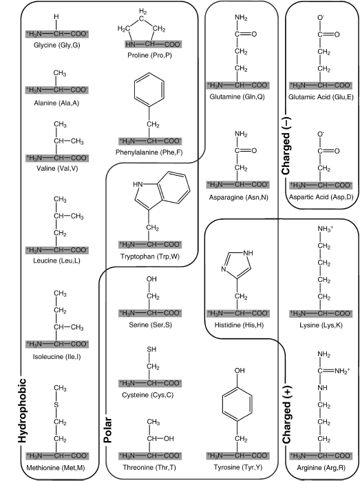
The 20 "standard" amino acids used in proteins, grouped on the basis of the properties of their side chains. The shared amino acid structure is shaded gray. Each amino acid is labeled with its full name, followed by its three-letter and one-letter abbreviations. This classification groups amino acids on the basis of the form that predominates at physiological conditions (note that their amino and carboxyl groups are charged under these conditions). Although this classification is a useful guideline, it does not convey the full complexity of side chain properties. For example, tryptophan and histidine do not fall clearly into a single grouping. Tryptophan is somewhat polar due to the nitrogen in its five-membered ring but has a hydrophobic six-membered ring at the end of its side chain. Histidine can be neutral polar and/or positively charged under physiological conditions.
The side chains (or R groups) of amino acids contain functional groups that are important in determining the three-dimensional structure and thus the function of the protein. The 20 amino acids are grouped and distinguished by their side chains:
- Five amino acids have electrically charged (ionized, +1 or −1) side chains at pH levels typical of living cells. These side chains attract water (are hydrophilic) and attract oppositely charged ions of all sorts.
- Five amino acids have polar side chains (δ⁺ and δ⁻). They tend to form hydrogen bonds with water and other polar substances. They are also hydrophilic.
- Seven amino acids have side chains that are nonpolar hydrocarbons and are thus hydrophobic. In the watery environment of the cell, these hydrophobic groups may cluster together in the interior of the protein.
Three amino acids, cysteine, glycine, and proline, are special cases, although the side chains of the latter two are generally hydrophobic.
- The cysteine side chain, which has a terminal –SH group, can react with another cysteine side chain in an oxidation reaction to form a covalent bond. Such a bond, called a disulfide bridge or disulfide bond (–S–S–), helps determine how a polypeptide chain folds. Note that most cysteines in a protein are not involved in disulfide bridges.
- The glycine side chain consists of a single hydrogen atom. It is small enough to fit into tight corners in the interiors of protein molecules where larger side chains could not fit. The small side chain allows the protein to be more flexible at the locations where glycine occurs.
- Proline possesses a modified amino group that lacks a hydrogen atom and instead forms a covalent bond with the hydrocarbon side chain, resulting in a ring structure. This limits both its hydrogen-bonding ability and its ability to rotate around the α carbon. So proline is often found where a protein bends or loops.
Aliphatic side chains are open chains of carbon and hydrogen (alanine, valine, leucine, isoleucine), and aromatic ones contain a flat six-membered ring with alternating single and double bonds, like benzene, whose electrons are shared around the whole ring (phenylalanine, tyrosine, tryptophan).
The five charged residues are aspartate, glutamate, lysine, arginine, and histidine; the five polar are serine, threonine, asparagine, glutamine, and tyrosine; the seven hydrophobic are alanine, valine, leucine, isoleucine, methionine, phenylalanine, and tryptophan. The figure above draws the lines differently: it puts cysteine with the polar residues and lets tryptophan and histidine straddle two groups. The residues where the two groupings disagree are the ones to watch. Sort and filter the twenty here, and pick one to turn it in 3D.
Code
from primer import structure

EXPECTED = {"GLY": 4, "ALA": 5, "SER": 6, "CYS": 6, "PRO": 7, "VAL": 7, "THR": 7, "ILE": 8, "LEU": 8,
            "ASN": 8, "ASP": 8, "GLN": 9, "GLU": 9, "LYS": 9, "MET": 8, "HIS": 10, "PHE": 11, "ARG": 11,
            "TYR": 12, "TRP": 14}

# One well-ordered example of each amino acid from protein kinase A (1ATP): complete side chain, lowest B factor.
best = {}
for res in structure("1ATP")[0]["E"]:
    name = res.get_resname()
    if res.id[0] != " " or len(res) != EXPECTED.get(name):
        continue
    b = sum(a.get_bfactor() for a in res) / len(res)
    if name not in best or b < best[name][0]:
        best[name] = (b, [[a.get_id(), a.element, *(round(float(x), 2) for x in a.coord)] for a in res])

from primer import display_widget
display_widget("amino-explorer", residues={k: v[1] for k, v in best.items()},
               source="Coordinates from protein kinase A, PDB 1ATP")
Twenty individuals
A one-letter code is the first letter of the name unless another residue took it first, which is why phenylalanine is F ("Fenylalanine") and tryptophan W ("tWo rings").
Hydrophobic: A, V, L, I, M
These are carbon and hydrogen, plus one sulfur in methionine, with nothing that can hydrogen-bond to water, so the hydrophobic effect (lesson 1.5) pushes them into the core. Alanine is a single methyl: small, mildly hydrophobic, at home almost anywhere, and the strongest helix former. Valine and isoleucine branch at the β-carbon, right next to the backbone, and that crowding favors extended β-strands. Leucine branches one carbon further out and prefers helices; it is the most common residue in proteins, about one in ten. Methionine is an unbranched chain with a sulfur in the middle that has no hydrogen to donate. Every protein is made starting with methionine, because its codon, AUG, is the start signal, though it is often removed later.
Aromatic: F, W, Y
Flat, rigid rings that stack against each other in cores. Phenylalanine is a benzene ring and purely hydrophobic. Tyrosine adds an –OH to that ring, so the ring buries while the OH looks for a hydrogen-bond partner, and tyrosines often sit at the edge of the core. Its OH has pKa ≈ 10, so it is neutral at pH 7. Tryptophan is the largest side chain, an indole ring (a benzene ring fused to a five-membered ring that carries one N–H), and the rarest residue, about one in a hundred. It is usually buried, and membrane proteins collect tryptophans where lipid meets water.
Polar: S, T, N, Q
Neutral side chains with O–H or amide groups that donate and accept hydrogen bonds. They sit comfortably on the surface, and when buried they nearly always have a partner. Serine (–CH₂OH) is small, common in loops, and a site where kinases attach phosphate. (A kinase is an enzyme that moves a phosphate group, –PO₃²⁻, onto a protein's –OH, which switches the protein's behavior; lesson 3.5.) Threonine is serine with an extra methyl, branched at Cβ like valine, and is also phosphorylated. Asparagine (N, "asparagiNe") and glutamine (Q, "Q-tamine") end in amides; asparagine is common in turns. Don't confuse these neutral amides with the charged acids D and E, which differ from them by one atom.
Charged: D, E, K, R, and H
Aspartate (D) and glutamate (E) are carboxylates with pKa ≈ 4, negative at pH 7 (lesson 1.3). Both bind metal ions such as calcium; glutamate is a strong helix former and nearly always on the surface. Lysine (K) is four CH₂ groups ending in –NH₃⁺ (pKa ≈ 10.5). Its long stem can lie along a hydrophobic patch with only the charged tip in water. Cells attach ubiquitin to lysines to mark a protein, and ubiquitin's own lysines (K48, K63) are where chains of ubiquitins link. Arginine (R) ends in a flat guanidinium group, one carbon bonded to three nitrogens that share a single positive charge (pKa ≈ 12.5). It is positive at any pH a cell sees, makes several hydrogen bonds at once, and grips negatively charged phosphates: those of DNA, of phosphorylated residues, and of ATP, the small molecule that carries energy in cells and ends in a tail of three phosphates. Histidine (H) is an imidazole ring (five atoms, two of them nitrogen) with pKa ≈ 6, so its charge flips with small changes around it. It moves protons in active sites and binds metals: hemoglobin carries oxygen on four hemes, flat iron-holding ring molecules, and each heme's iron is held by a histidine (lesson 3.6).
Special: G, P, C
Their effect is on the backbone or on chemistry more than on water. Glycine has no side chain, so its backbone can take shapes no other residue can (unit 3). It turns up in tight turns and flexible loops, it is the only achiral residue, and it is every third residue of collagen, the rope-like protein of skin and tendon. Proline's ring bonds back to its own backbone nitrogen, which fixes φ near −65° and removes the N–H. It breaks α-helices except in their first turn and allows the cis peptide bonds of lesson 2.1. Cysteine is a thiol with pKa ≈ 8.3. Pairs form disulfides in proteins that work outside cells, like ribonuclease A's four, and free cysteines bind zinc and other metals. On the Kyte–Doolittle hydropathy scale (lesson 2.3) it is the fifth most hydrophobic residue.
Reading a sequence for burial
Rules of thumb for a water-soluble protein:
| Residues | Usually | Watch for |
|---|---|---|
| I, L, V, F, M, W | buried | some sit on the surface in patches that bind partners |
| A, C, Y, T, G | either | small or two-faced side chains go wherever there's room |
| S, N, Q, H, P | surface, turns, loops | buried ones hydrogen-bond to something |
| D, E, K, R | surface | a buried one is almost always paired in a salt bridge |
Two spacing patterns help, both from unit 3's regular backbone shapes (lesson 2.3 turns them into tools): the α-helix, a spiral with 3.6 residues per turn, and the β-strand, a nearly straight, stretched-out run. In a helix, residues 3 or 4 apart face the same side, so a buried helix face reads as hydrophobic letters at i, i+3, i+4, i+7. In a β-strand, neighbors point to opposite sides, so a strand at the protein's edge alternates buried and exposed.
Try it on ubiquitin. Mark the residues you think are buried (less than 20% of their surface exposed), then check against the crystal structure. Decide before turning on "Color by class".
Code
from Bio.PDB.SASA import ShrakeRupley
from Bio.PDB.DSSP import residue_max_acc
from primer import sequence

model = structure("1UBQ")[0]
chain = model["A"]
for res in [r for r in chain if r.id[0] != " "]:
    chain.detach_child(res.id)                     # waters out: we want exposure to bulk solvent
ShrakeRupley().compute(model, level="R")
max_asa = residue_max_acc["Wilke"]                 # Tien et al. 2013, theoretical maximum per residue
rsa = [round(r.sasa / max_asa[r.get_resname()], 3) for r in chain]
display_widget("burial-game", "The lab at the end of this unit has you compute these numbers yourself.",
               sequence=sequence(chain), rsa=rsa, name="ubiquitin", threshold=0.2)
The lab at the end of this unit has you compute these numbers yourself.
The usual misses are the exceptions in the table: a buried lysine (K27), a buried threonine or two, and hydrophobic residues left on the surface. Lesson 2.4 explains each kind.
Check yourself
A colleague's sequence has the stretch …KDEGSPNGKE… and asks whether it's likely in the core. What do you
say?
Reasoning
Almost certainly not. It's dense with charged residues (K, D, E, K, E), which need water or a partner, and
it contains glycine, proline, and asparagine, the residues of turns and loops. Read it as a surface loop
that is probably flexible. A core stretch would look like …VILAGLFV….
In a buried part of a protein, a valine is mutated to aspartate. Another protein has a surface lysine mutated to arginine. Which mutation is more likely to destabilize its protein, and why?
Reasoning
The buried V→D. It puts a negative charge in the oily core, where nothing solvates it, unless a positive partner happens to sit next to it. That costs several kcal/mol, comparable to a protein's whole stability margin (unit 4). It also removes a hydrophobic side chain that was packing against its neighbors. K→R on the surface swaps one long positive side chain for another, both in water. It's usually close to neutral for stability, though it can matter for function if that lysine was binding something or was a site of modification.
One classification calls cysteine a special case, another puts it with the polar residues, and the Kyte–Doolittle scale ranks it the fifth most hydrophobic residue. Which is right?
Reasoning
Each answers a different question. Chemically, cysteine's thiol is special: it can form a covalent disulfide or bind a metal, which no other residue does. The S–H bond is slightly polar, which is why a classification by functional group calls it polar. But sulfur is not very electronegative (lesson 1.2), so the thiol barely hydrogen-bonds with water, and a scale of how much a side chain prefers an oily environment ranks it as hydrophobic. In structures, free cysteines are often buried. Cysteine, tyrosine, tryptophan, histidine, and lysine are the residues any three-class scheme fits worst.
Why might replacing a glycine in a tight turn with alanine, which only adds a methyl group, destabilize a protein, even though alanine is "harmless" almost everywhere else?
Reasoning
Because the glycine may be doing something only glycine can. Tight turns often need backbone angles that put the β-carbon of any non-glycine residue in a clash with its own backbone. Glycine has no β-carbon, so it can take those angles. Alanine's methyl brings the clash back and forces the turn into a different, strained shape. The cost here is about backbone geometry, not hydrophobicity, which is why glycine sits in the "special" group. Unit 3's Ramachandran plot shows exactly which angles are at stake.