Beyond the soluble globule
A hydrophobic core, a polar surface, one stable fold: everything so far describes water-soluble globular proteins. They dominate the PDB because they crystallize most easily, and prediction methods inherit that bias. Three kinds of protein break the pattern: proteins that live in membranes, long fibers, and chains that never fold on their own. Structure files also hold atoms that belong to none of the 20 amino acids, such as metal ions, cofactors, and chemical groups added after the chain is made.
Three environments
One method of protein classification sidesteps structural organization in favor of biochemical properties. Here, proteins are classified into three major groups: globular, membrane, and fibrous.
Globular proteins exist in an aqueous environment and thus fold as compact structures with hydrophobic cores and polar surfaces. This class of proteins is well represented in the PDB, partly because these structures are the easiest to determine experimentally.
Membrane proteins exhibit many of the same characteristics as globular proteins, but they are distinguished in two important ways. First, they exist in an environment completely different from typical globular proteins: the cell membrane. In the interior of the cell membrane, the protein is surrounded by a hydrophobic environment. Thus, the regions of the protein within the cell membrane must have a hydrophobic surface in order to be stable. Second, experimental structure determination of membrane proteins is more difficult than the determination of the structure of globular proteins. However, the structures that have been determined show that they use the same secondary structural elements and follow the same general folding principles as globular proteins.
Fibrous proteins differ markedly from both membrane and globular proteins. These proteins are often constructed of repetitive amino acid sequences that form simple, elongated fibers. Their repetitive design is well suited to the structural roles they often play in organisms. Some fibrous proteins consist of a single type of regular secondary structure that is repeated over very long sequences. Others are formed from repetitive atypical secondary structures.
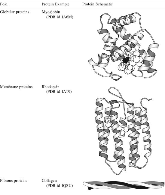
One example of each class: myoglobin (globular), rhodopsin (membrane), and collagen (fibrous).
Membrane proteins were rare in the PDB when that was written. Cryo-electron microscopy has since added thousands of them, though they remain a small share of all entries.
Membrane proteins
Membrane proteins are very diverse. About one-fourth of the protein-coding genes in the eukaryotic genome (that of organisms like us, whose cells have a nucleus) encode membrane proteins. Integral membrane proteins are at least partly embedded in the phospholipid bilayer (phospholipids are the two-tailed, fat-like molecules that make up the bilayer of lesson 2.3). Like phospholipids, these proteins have both hydrophilic and hydrophobic regions. Stretches of amino acids with hydrophilic side chains give certain regions of the protein a polar character; these regions interact with water and stick out into the aqueous environment on either side of the membrane. Stretches of amino acids with hydrophobic side chains give other regions of the protein a nonpolar character; these interact with the fatty acids in the interior of the phospholipid bilayer, away from water.
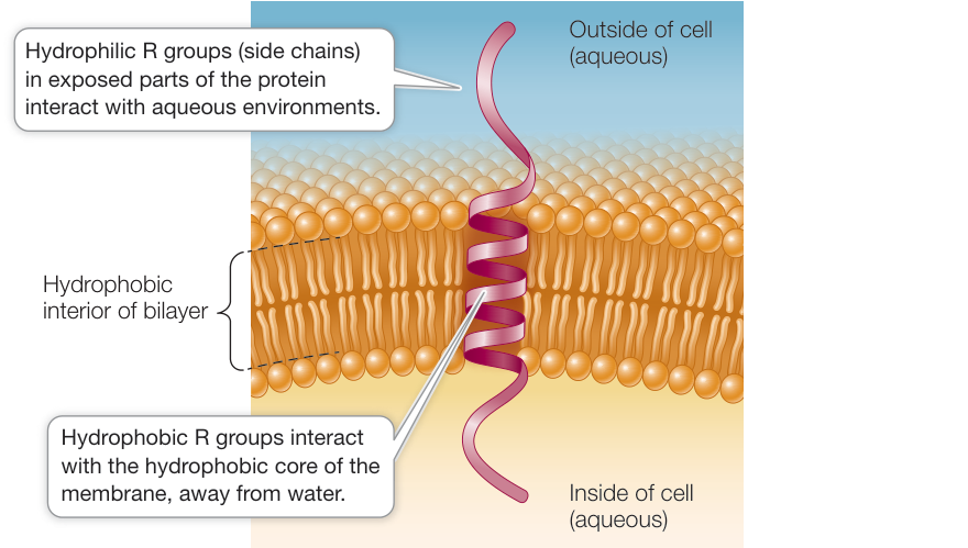
An integral membrane protein is held in the membrane by where its hydrophilic and hydrophobic side chains are.
Peripheral membrane proteins lack exposed hydrophobic groups and are not embedded in the bilayer. Anchored membrane proteins are covalently attached to fatty acids or other lipid groups, which insert into the bilayer. An integral protein that extends all the way through the bilayer and protrudes on both sides is a transmembrane protein.
The hydrophobic core of a lipid bilayer is about 30 Å thick. An α-helix rises 1.5 Å per residue, so a helix that crosses it needs about 20 residues, and in a sequence that shows up as a stretch of roughly 20 hydrophobic residues in a row. That is how transmembrane helices are predicted from sequence. Two other patterns recur at the membrane's edges: a belt of tryptophan and tyrosine side chains where the lipid tails meet the head groups, and more lysine and arginine on the cytoplasmic side (the inside of the cell) than on the outside (the "positive-inside rule"). The other transmembrane architecture is the β-barrel, found in the outer membranes of bacteria, mitochondria, and chloroplasts.
The inside-out surface is easy to measure. Count the residues that are mostly exposed (more than 30% of their surface reachable by solvent, in the structure as deposited) and ask what fraction of them are hydrophobic:
from Bio.PDB.SASA import ShrakeRupley
from primer import structure

# Largest possible exposed surface of each residue type (Ų), for turning areas into fractions.
MAX_ASA = {"ALA": 129, "ARG": 274, "ASN": 195, "ASP": 193, "CYS": 167, "GLN": 225, "GLU": 223, "GLY": 104,
           "HIS": 224, "ILE": 197, "LEU": 201, "LYS": 236, "MET": 224, "PHE": 240, "PRO": 159, "SER": 155,
           "THR": 172, "TRP": 285, "TYR": 263, "VAL": 174}
HYDROPHOBIC = {"ALA", "VAL", "LEU", "ILE", "MET", "PHE", "TRP", "CYS"}

def exposed_hydrophobic(pdb_id, chain_id):
    chain = structure(pdb_id)[0][chain_id]
    for res in [r for r in chain if r.id[0] != " "]:      # drop waters and ligands
        chain.detach_child(res.id)
    ShrakeRupley().compute(chain, level="R")
    exposed = [r for r in chain if r.get_resname() in MAX_ASA and r.sasa / MAX_ASA[r.get_resname()] > 0.3]
    return len(exposed), sum(r.get_resname() in HYDROPHOBIC for r in exposed) / len(exposed)

for pdb_id, name in [("1UBQ", "ubiquitin (soluble)"), ("1A6M", "myoglobin (soluble)"),
                     ("1C3W", "bacteriorhodopsin (membrane, 7 helices)")]:
    n, frac = exposed_hydrophobic(pdb_id, "A")
    print(f"{name:42} {n:3} exposed residues, {frac:.0%} hydrophobic")
ubiquitin (soluble)                         44 exposed residues, 11% hydrophobic
myoglobin (soluble)                         72 exposed residues, 17% hydrophobic
bacteriorhodopsin (membrane, 7 helices)     82 exposed residues, 51% hydrophobic
Half of bacteriorhodopsin's exposed residues are hydrophobic, against one in six for myoglobin and one in nine for ubiquitin. Its surface faces lipid tails, not water.
Bacteriorhodopsin (1C3W), a light-driven proton pump, colored by residue class. Hydrophobic residues (amber) cover the band of surface that sits in the bilayer; polar and charged residues gather at the two ends. The second view shows its seven transmembrane helices from N-terminus (blue) to C-terminus (red). Sticks are non-protein molecules: retinal, the light-absorbing group, inside, and pieces of lipid on the surface.
Open 1C3W in Mol*Fibrous proteins: collagen and coiled coils
Collagen, the most abundant protein in mammals, is three chains wound into a triple helix. Each chain has the repeating sequence Gly-X-Y, often Gly-Pro-Hyp, where Hyp is hydroxyproline, a proline with an OH group added after the chain is made. Every third residue faces the crowded center of the triple helix, where only glycine fits. Each chain is a left-handed polyproline-II-like helix, and the three twist around each other in a right-handed superhelix.
A collagen-like peptide (1CAG), three chains of Pro-Hyp-Gly repeats. In the second view, glycines (violet) line the narrow axis. Each chain has one glycine replaced by alanine (red), which mimics the mutations that cause brittle bone disease: the extra methyl group doesn't fit, and the triple helix loosens around it.
Open 1CAG in Mol*Coiled coil proteins consist of two or more α-helices that wind around one another with a slight left-handed superhelical twist and are characterized by a heptad repeat pattern in which residues in the first and fourth position are hydrophobic, and residues in the fifth and seventh position are predominantly charged or polar.
The heptad positions are labeled abcdefg, so the hydrophobic ones are a and d. With 3.6 residues per turn, positions 7 apart come back to nearly the same side of the helix (7 × 100° = 700°, 20° short of two full turns), so a and d form a hydrophobic stripe that winds slowly around the helix. Two helices pack their stripes together. In the "leucine zipper" of GCN4, a yeast protein that binds DNA as a pair of helices (2ZTA), the d positions are leucines 5, 12, 19, and 26.
Disordered proteins
While most proteins achieve a stable structure, a large number of proteins are intrinsically unstructured, in whole or part. Unstructured proteins appear to be particularly important in cell signaling and regulation. Often, the function of these proteins depends on achieving an ordered structure only under certain conditions or while engaged in specific interactions. It is therefore appropriate to add a special class of "intrinsically unstructured" to the available classifications of protein folds.
An irregular loop is not the same as a disordered region. An ordered loop has one conformation, the same in every copy of the protein, with clear electron density and well-defined coordinates. A disordered region is a moving ensemble: in a crystal structure its atoms are missing because there is no density to place them in, and in an NMR ensemble its models disagree. Roughly a third of eukaryotic proteins contain a disordered stretch longer than 30 residues. These regions are rich in charged residues, proline, glycine, and serine, and poor in the large hydrophobic residues that would form a core.
p53, a "tumor suppressor" that halts the division of cells with damaged DNA, is the standard example. Its DNA-binding domain is folded, but its N-terminal region, the part that switches on target genes, is disordered until it meets a partner. The structure below shows residues 17–29 of p53 bound to the protein MDM2. On binding, p53 forms a short helix that lays three hydrophobic side chains (Phe 19, Trp 23, Leu 26) into a groove on MDM2. This is called coupled folding and binding, and it was the starting point for MDM2 inhibitors designed to mimic those three side chains.
MDM2 (1YCR, chain A, under a grey surface) with the p53 peptide (chain B) as a helix in its groove. Phe 19, Trp 23, and Leu 26 (red sticks) fill the pocket. Alone in solution, this stretch of p53 has no fixed structure.
Open 1YCR in Mol*Structure predictors flag disorder on their own. AlphaFold reports its confidence per residue as pLDDT (0–100) and stores it in the B-factor column of its PDB files. For p53 the folded domains come out confident, and the rest is predicted with low confidence, often as long loose ribbons that are not a real conformation:
import numpy as np
from Bio.PDB import PDBParser
from primer import alphafold

model = PDBParser(QUIET=True).get_structure("p53", alphafold("P04637"))
plddt = np.array([r["CA"].get_bfactor() for r in model[0]["A"]])
print(f"{len(plddt)} residues: {np.mean(plddt < 50):.0%} with pLDDT < 50, {np.mean(plddt > 90):.0%} with pLDDT > 90")
confident = np.flatnonzero(plddt > 70) + 1
runs = np.split(confident, np.flatnonzero(np.diff(confident) > 1) + 1)
print("confident stretches (pLDDT > 70, at least 6 residues):",
      ", ".join(f"{r[0]}–{r[-1]}" for r in runs if len(r) >= 6))
393 residues: 30% with pLDDT < 50, 52% with pLDDT > 90
confident stretches (pLDDT > 70, at least 6 residues): 20–25, 94–182, 187–292, 325–355
One short confident stretch, residues 20–25, sits inside the disordered N-terminal region. It is the helix that p53 forms on MDM2 in the structure above. The predictor has learned that this segment is helical, even though the isolated protein doesn't hold the helix.
AlphaFold's model of p53 colored by pLDDT. The DNA-binding domain (about 94–292) and the tetramerization domain (about 325–355) are blue (confident). The N-terminal 90 residues and the C-terminal tail are mostly orange and yellow: the predictor is saying "I don't know", which for this protein is the right answer.
Open AF-P04637 in Mol*Cofactors and metal ions
Proteins can associate with small molecules or metal atoms (covalently or noncovalently) to diversify their functional and structural capabilities. Many enzymes employ such molecules as cofactors, which assist them in chemical catalysis. Other proteins require these molecules or atoms for proper tertiary structure formation. For example, the zinc-finger motif, a structural motif important for the interaction of some proteins with DNA, cannot form without the interaction of a zinc atom with specific cysteine, or cysteine and histidine, residues.
The cofactor is a separate molecule with its own name in the structure file (HETATM records, residue names such as HEM, ZN, MN, ATP), not part of the polypeptide sequence. Examples so far: the heme in hemoglobin and myoglobin, which is what actually binds oxygen (lesson 3.6); the two manganese ions holding ATP's phosphates in protein kinase A (lesson 3.4); the calcium in calmodulin's EF-hands. In the Zif268 zinc fingers below, each zinc sits about 2 Å from two cysteine sulfurs and two histidine nitrogens. Without it, a 28-residue finger is too small to bury a hydrophobic core and does not fold.
Zif268 (1ZAA): three zinc fingers wrapped in the major groove of DNA. Each finger is a β-hairpin and a helix around one zinc ion (sphere). The second view shows the first finger's zinc with its ligands Cys 7, Cys 12, His 25, and His 29.
Open 1ZAA in Mol*Modifications after synthesis
The chemical properties of the 20 standard amino acids can be extended through side chain modification. In some cases, such modification can be indispensable to the proper formation of tertiary structures. For example, the protein collagen contains a modified proline residue, hydroxyproline, which greatly stabilizes the protein fold. In other cases, residue modification has no effect on fold, but rather extends the functional repertoire of the protein.
Molecules, such as carbohydrates and lipids, can be attached to the protein via covalent bonds with specific residues. Carbohydrate modifications are often seen on proteins that function extracellularly and play a role in protein localization. Lipid modifications can help anchor a protein in the cell membrane.
- Glycosylation is the addition of sugars to proteins to form glycoproteins. Some attached sugars are important in the conformation of proteins and their recognition functions at the cell surface; others help stabilize extracellular proteins.
- Phosphorylation is the addition of phosphate groups to proteins, and is catalyzed by protein kinases. The charged phosphate groups change the conformation of a protein, often exposing the active site of an enzyme or the binding site for another protein.
- Proteolysis is the cutting of a polypeptide chain, a reaction catalyzed by enzymes called proteases. Some proteins are made as long precursors that are cut into their final products; the protease of HIV does this for the virus's proteins, and drugs that inhibit it are used to treat AIDS.
None of these change the gene or the amino-acid sequence a predictor reads. Phosphorylation adds a group carrying about two negative charges at pH 7 to a serine, threonine, or tyrosine. N-linked sugars attach to asparagine in the sequence pattern Asn-X-Ser/Thr (X not proline). Lipids such as myristate (a 14-carbon fatty-acid chain) attach to an N-terminal glycine; protein kinase A itself is anchored this way. And ubiquitin (1UBQ) is a modification too: its C-terminal Gly 76 is linked to a lysine of a target protein, and chains of ubiquitins joined through ubiquitin's own Lys 48 mark the target for destruction.
In a structure file a modified residue gets its own residue name (TPO for phosphothreonine, SEP for phosphoserine, HYP for hydroxyproline) and a MODRES record naming the standard residue it came from. Protein kinase A in 1ATP is phosphorylated twice, and the phosphate on Thr 197, in a loop near the active site, holds that loop in its active shape:
from Bio.PDB import NeighborSearch
from primer import fetch

print("\n".join(line[:60] for line in fetch("1ATP").read_text().splitlines() if line.startswith("MODRES")))
model = structure("1ATP")[0]
search = NeighborSearch(list(model.get_atoms()))
pthr = model["E"][("H_TPO", 197, " ")]
for name in ("O1P", "O2P", "O3P"):
    for atom in search.search(pthr[name].coord, 3.3):
        res = atom.get_parent()
        if res is not pthr and res.get_resname() != "HOH":
            print(f"phosphate {name} … {res.get_resname()} {res.id[1]} {atom.get_id():4} {pthr[name] - atom:.2f} Å")
MODRES 1ATP TPO E  197  THR  PHOSPHOTHREONINE
MODRES 1ATP SEP E  338  SER  PHOSPHOSERINE
phosphate O1P … ARG 165 NH1  3.21 Å
phosphate O1P … HIS 87 NE2  2.74 Å
phosphate O1P … ARG 165 NH2  2.87 Å
phosphate O2P … LYS 189 NZ   3.05 Å
phosphate O3P … THR 195 OG1  2.69 Å
phosphate O3P … ARG 165 NH1  2.78 Å
Four residues hold the phosphate's negative charge: Arg 165, which sits next to the catalytic Asp 166; His 87, on the αC helix of the other lobe; Lys 189; and Thr 195. One modification ties the two lobes and the catalytic loop together.
Check yourself
A sequence has a stretch of 22 consecutive residues that are all L, I, V, F, A, or G except one serine. Where in a cell would you expect this part of the protein to be, and why does its length matter?
Reasoning
In a membrane, as a transmembrane α-helix. A stretch that hydrophobic would be badly out of place on a soluble protein's surface, and at 1.5 Å per residue, 22 residues make a helix about 33 Å long, just enough to cross the bilayer's ~30 Å hydrophobic core. A single serine is tolerable if its hydroxyl hydrogen-bonds to the helix backbone or a neighboring helix. Only a structure or experiment can confirm it, but this is how transmembrane prediction works.
A crystal structure has no coordinates for residues 1–45, and the AlphaFold model of the same protein shows those residues as a long, straight ribbon with pLDDT around 30. Should you trust the AlphaFold coordinates for those residues? What experiment would tell you whether the region becomes ordered?
Reasoning
No. Both sources say the region has no single structure: no electron density in the crystal, and very low confidence from the predictor. The ribbon is a placeholder, not a conformation. The region may still be functional, perhaps folding when it binds a partner, like p53's N-terminus on MDM2. To test that, determine a structure of the complex, or use NMR, which can see a disordered segment become ordered when the partner is added.
A zinc finger's two cysteines are mutated to serines. Serine differs from cysteine only by O in place of S. Predict what happens to the finger's structure and its DNA binding.
Reasoning
The finger unfolds and stops binding DNA. Serine's hydroxyl is a poor zinc ligand compared with cysteine's thiolate (the thiol with its proton removed, –S⁻), so the zinc is lost, and the finger is too small to hold a hydrophobic core without the metal. The secondary structure, a hairpin and helix that need the zinc to stay together, falls apart, and the recognition helix no longer sits in the major groove.