Cells, proteins, and scale
A red blood cell picks up oxygen in your lungs and releases it in your tissues. The oxygen rides on hemoglobin, a protein small enough that thousands of copies could fit across the cell. Each copy has four binding sites. Oxygen attaches, comes off, and attaches again as the molecule moves through water and collides with its neighbors. Change the arrangement of a few atoms around those sites and you can change how readily the protein lets oxygen go.
This is the scale at which we will work. To understand a protein, we need to picture the molecule itself and the surroundings that keep pushing on it. Begin with a cell, then keep zooming until individual atoms become visible in the model.
A small volume that maintains a difference
A cell encloses a watery interior in a membrane. Inside are dissolved salts, small molecules, and large molecules such as DNA, RNA, and proteins. These large molecules are chains assembled from smaller units. A protein's units are amino acids; their order and chemistry determine what the chain can do.
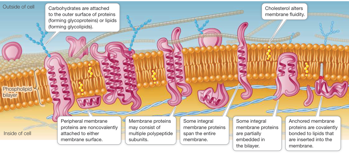
The membrane lets the cell maintain conditions different from those outside. This would be impossible if everything crossed it freely. Sodium ions, for example, cannot readily pass through the membrane's oily interior. Proteins embedded in the membrane provide selective routes across it. Some make channels; others use energy to move substances against a concentration difference. Maintaining those differences is part of being alive.
A bacterial cell is commonly a few micrometers long. Your cells are generally larger. They also have internal compartments surrounded by membranes, called organelles. One is the nucleus, which contains most of the cell's DNA. Cells with a nucleus are eukaryotic; bacteria and archaea lack a nucleus and are called prokaryotic. We will need this distinction occasionally, but the protein chemistry is shared: human and bacterial proteins use the same standard set of twenty amino acids.
The cytoplasm is the material inside the cell membrane, excluding the nucleus in a eukaryotic cell. Its fluid portion is the cytosol. Picture water crowded with molecules of many sizes, all in thermal motion. A protein rotates, bends, and repeatedly collides with water and other molecules. A structure viewer freezes one arrangement so that we can inspect it; the molecule in the cell keeps moving.
Crossing the membrane
A major membrane ingredient is the phospholipid. One end, the head, interacts favorably with water. The other end consists of hydrocarbon tails, long groups made mostly of carbon and hydrogen. Water interacts with these tails differently, and exposing a large area of them to water is unfavorable. We call the head hydrophilic and the tails hydrophobic; lesson 1.5 develops the reason.
Put many phospholipids in water and they can assemble into a bilayer. The tails face inward toward one another, while the heads face the water on either side. The membrane is only about 4 nm thick, with an oily interior roughly 3 nm thick. It is a fluid sheet, with molecules moving sideways within it.
The drawing exaggerates individual molecules so that you can see the arrangement. Follow a path from one watery side to the other: you must cross the tails. A charged particle loses favorable interactions with water along that path, making passage costly. Small nonpolar molecules such as oxygen cross much more readily. Water itself can cross slowly, and water channels greatly increase its passage in many cells. The membrane's selectivity comes from chemistry before any protein adds further control.
A protein that spans the membrane must fit this environment. The part exposed to the tails usually has a nonpolar exterior. A water-filled channel through that protein can have a very different lining. Knowing where a protein sits already gives us a prediction about which chemical groups face outward.
What a protein can accomplish by binding
Imagine a molecule approaching a protein. At long range it encounters electric fields and water. Up close, the shapes must fit: an atom cannot occupy a space already occupied by another atom. If the approaching molecule fits and makes favorable contacts, it may remain attached for a while. This is binding. The bound partner is called a ligand.
Hemoglobin binds oxygen at an iron-containing group called heme. An enzyme binds molecules involved in a reaction and helps them react faster. An enzyme is a catalyst: after a complete reaction cycle, it is available to do the job again. Its binding site arranges chemical groups so that the difficult steps of the reaction become easier. A receptor binds a signal molecule and changes the behavior of the cell, often through a change in the receptor's shape.
These jobs demand more than a geometrically suitable hole. Put the wrong charges in a pocket and a molecule that fits can bind poorly. Make the protein too rigid and a required movement may become impossible. We study atomic positions because they let us ask what can approach, what can bind, and what can move next.
We will return to a few proteins often enough to recognize them:
| Protein | Where it works | A job to keep in mind |
|---|---|---|
| Ubiquitin | cytosol and nucleus | Attaches to other proteins as a tag; some arrangements of these tags direct protein destruction |
| Hemoglobin | inside red blood cells | Binds oxygen for transport from lungs to tissues |
| Protein kinase A | within cells | Transfers a phosphate group from ATP to a protein, changing the recipient's behavior |
| Ribonuclease A | secreted into the gut | Cuts RNA during digestion |
ATP is a small molecule used in many reactions that require chemical energy. A phosphate group contains phosphorus bonded to oxygens. We will learn to recognize both in structures; for now, notice that kinase A has to bring two partners into a suitable arrangement for a chemical transfer.
Location also changes the chemistry of a protein's own chain. Two cysteine amino acids can join through their sulfur atoms, making a disulfide bond that cross-links the chain. Such bonds are common in secreted proteins, including ribonuclease A. The cytosol usually favors the unlinked form of cysteine. Lesson 1.6 explains the electron transfer behind this difference. Even before inspecting a protein's fold, its surroundings tell us which kinds of cross-link to expect.
Put numbers on the picture
A micrometer, µm, is one millionth of a meter. A nanometer, nm, is one thousandth of a micrometer. An ångström, Å, is one tenth of a nanometer. Protein coordinates are usually given in ångströms because an ordinary covalent bond is about 1–2 Å long.
| Object or distance | Approximate size |
|---|---|
| C–C single bond | 1.5 Å = 0.15 nm |
| Water molecule | 3 Å = 0.3 nm across |
| Small compact protein | a few nm across |
| Membrane | 4 nm thick |
| Bacterial cell | a few µm long |
| Many animal cells | 10–30 µm across |
Suppose a protein 4 nm across were enlarged to the size of a 4 cm walnut. Every length has grown by a factor of ten million. A water molecule is now about 3 mm across, and a 2 µm bacterium is 20 m long. There is room for a great many proteins inside a cell, yet every protein is continually touching water molecules large enough to interact with particular parts of its surface.
Move from a cell down to a bond. At each scale, compare two objects before zooming further. Ordinary light microscopy resolves details roughly 200 nm apart; individual atomic positions require other kinds of measurement, including X-ray diffraction, NMR, and electron microscopy.
Molecular mass gives another way to estimate size. A dalton, Da, is approximately the mass of a hydrogen atom; 1 kDa is a thousand daltons. An amino acid incorporated into a protein is called a residue. Protein residues average roughly 110 Da, so a 30 kDa chain contains around 270 residues.
We can check that estimate against ubiquitin. structure("1UBQ") loads its deposited atomic model.
The [0]["A"] selects the first model and chain A; the lab will unpack that organization.
from Bio.SeqUtils import molecular_weight
from primer import sequence, structure

ubq = structure("1UBQ")[0]["A"]
seq = sequence(ubq)
mass = molecular_weight(seq, seq_type="protein")  # daltons
heavy = sum(1 for atom in ubq.get_atoms() if atom.get_parent().id[0] == " ")  # protein atoms, no water
print(f"ubiquitin: {len(seq)} residues, {mass:,.0f} Da = {mass / 1000:.1f} kDa")
print(f"{mass / len(seq):.0f} Da per residue; {heavy} atoms in the file, {heavy / len(seq):.1f} per residue")
ubiquitin: 76 residues, 8,565 Da = 8.6 kDa
113 Da per residue; 602 atoms in the file, 7.9 per residue
Ubiquitin has 76 residues and a mass of about 8.6 kDa. Its crystal structure lists 602 protein atoms, all of them heavier than hydrogen. The molecule also has hundreds of hydrogens, which this experiment did not locate individually. A file's atom count and a molecule's atom count are different quantities.
To turn mass into length, make an explicit approximation: replace the folded protein with a sphere of density 1.35 g/cm³. Divide mass by density to get volume, then solve for the radius.
import math

M = 30_000  # a typical 30 kDa protein, in daltons (grams per mole)
residues = M / (mass / len(seq))
atoms = residues * heavy / len(seq)
volume = M / 1.35 / 6.022e23 * 1e21  # cm³ per mole → per molecule → nm³ (1 cm³ = 10²¹ nm³)
diameter = 2 * (3 * volume / (4 * math.pi)) ** (1 / 3)
print(f"30 kDa: about {residues:.0f} residues, {atoms:,.0f} non-hydrogen atoms ({2 * atoms:,.0f} with hydrogens)")
print(f"volume {volume:.0f} nm³; as a sphere, {diameter:.1f} nm across")
30 kDa: about 266 residues, 2,109 non-hydrogen atoms (4,217 with hydrogens)
volume 37 nm³; as a sphere, 4.1 nm across
The 30 kDa protein comes out about 4 nm across. This is a useful estimate for a compact protein; a long rod of the same mass would have a very different largest dimension. Notice the cube root. Doubling the mass only multiplies a compact protein's diameter by , about 1.26.
Start with the space-filling view of ubiquitin. The light-blue spheres are oxygen atoms from water molecules located in the crystal; many more waters surround a protein in solution. Switch to the cartoon to trace the chain through the packed atoms. Both views describe the same coordinates.
Open 1UBQ in Mol*Check yourself
A compact protein has eight times the mass of another protein of the same density. How do their diameters compare? What assumption would fail for a long, thin protein?
Reasoning
Volume grows eightfold, so diameter grows by . This assumes comparable compact shapes. A thin rod can gain mass mainly by growing longer, making its length grow much faster than the cube-root estimate.
A membrane-spanning helix advances 0.15 nm along its axis per residue. Estimate how many residues are needed to cross a 3 nm oily region, and predict the chemistry of the side chains facing the lipids.
Reasoning
A perpendicular crossing needs about residues. The lipid-facing side chains should be mostly nonpolar. A tilted helix needs a longer segment, and side chains facing a water-filled pore can be polar. The environment applies to each surface, not simply to the protein as a whole.
Two proteins contain several cysteines. One has covalent links between most of its cysteine sulfurs; the other has none. Which would you first suspect is secreted, and why is this only a clue?
Reasoning
The cross-linked protein is the stronger candidate. Secreted proteins commonly form disulfides, while the cytosol generally favors unlinked cysteines. But cysteines also have other jobs, and their positions may prevent pairing. Location constrains chemistry without uniquely determining it.
A viewer shows a protein as a smooth ribbon with a few blue water spheres nearby. Which visible features are molecules, and which are drawing conventions?
Reasoning
The protein and waters are molecules. The ribbon is a trace that helps us follow the protein chain; no ribbon material fills the space between atoms. Sphere size and color are chosen conventions. The listed water oxygens represent only waters that could be located, so the apparent empty space around the protein is not empty in solution.