Folding: Anfinsen, Levinthal, and funnels
Structure prediction maps a sequence to coordinates. That only makes sense if the structure is determined by the sequence, and not by how a cell happened to fold the chain. An experiment on ribonuclease showed that it is. The next question, how a chain finds that fold in milliseconds when trying every shape would outlast the universe, led to the energy-landscape picture behind today's search and sampling methods.
Sequence determines structure
If a protein is heated slowly and moderately, the heat energy will disrupt only the weak interactions, causing the secondary and tertiary structure to break down. A protein whose structure is disrupted is said to be denatured. A comparison of native (untreated) and denatured proteins shows major differences:
- Native proteins are compact; denatured proteins have a larger volume.
- Native proteins exist in one, preferred shape; denatured proteins can take many shapes.
- Native proteins have hydrogen bonds that stabilize the structure internally; denatured proteins have hydrogen bonds on the exterior, to water.
You can't "unboil" an egg after it has been hard-boiled; the egg proteins are irreversibly denatured. Amazingly, in some cases a protein can return to its normal tertiary structure when it cools, demonstrating that all the information needed to specify the unique shape of a protein is contained in its primary structure. This was first shown (using chemicals instead of heat to denature the protein) by biochemist Christian Anfinsen for the protein ribonuclease. High concentrations of polar substances such as urea can disrupt the hydrogen bonding that is crucial to protein structure, and urea was used in that experiment.
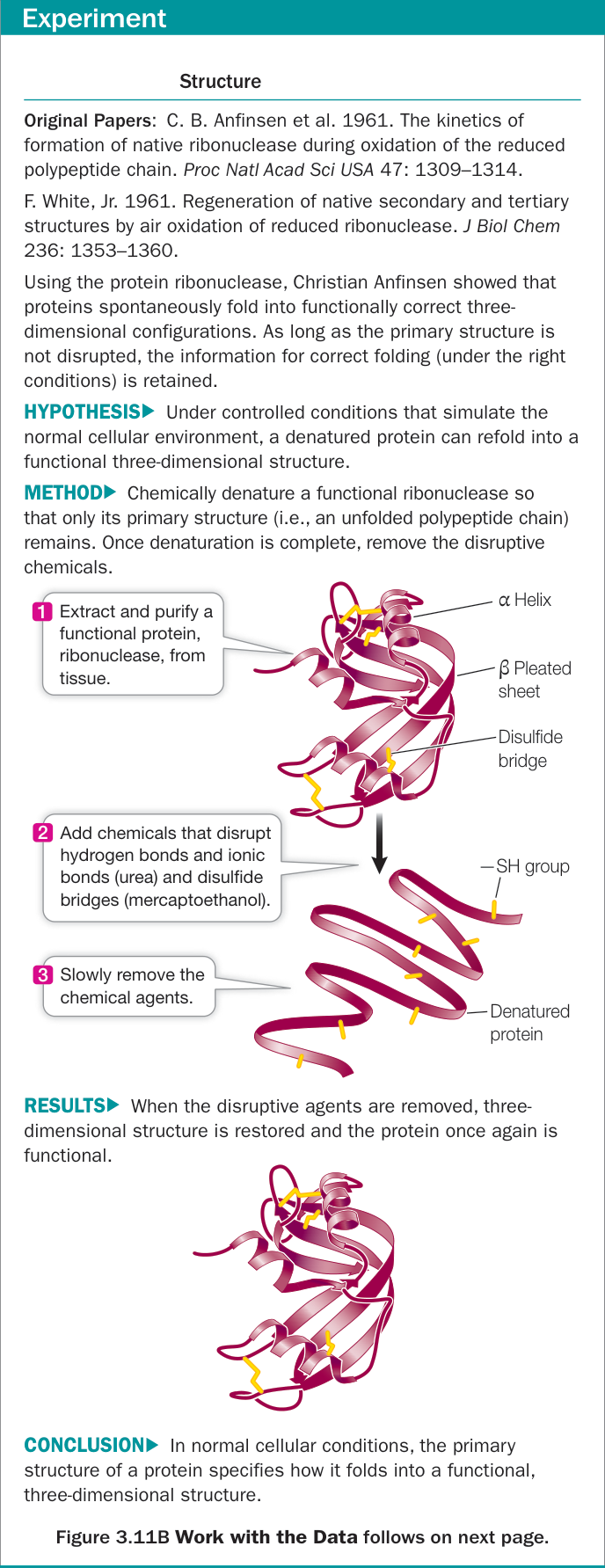
Ribonuclease A is an enzyme that cuts RNA, so its activity, how fast it cuts, is a direct test of whether the chain is folded correctly. It has 124 residues, and its eight cysteines are paired in four disulfide bonds (lesson 1.6). Urea unfolds the chain, and β-mercaptoethanol, a reducing agent, breaks the disulfides by handing each sulfur a hydrogen back, leaving free thiols (–SH). When both are removed slowly, oxygen from the air re-oxidizes the thiols (takes those hydrogens off again, so pairs of sulfurs can rejoin), and close to 100% of the activity returns: of all the ways eight cysteines could pair, the chain re-formed the right four.
Ribonuclease A (7RSA, 1.26 Å) with its eight cysteines in amber and the four disulfides measured: 26–84, 40–95, 58–110, and 65–72, each about 2.0 Å from sulfur to sulfur. The partners are far apart in sequence and close together only in the folded chain.
Open 7RSA in Mol*The control experiment is what makes the result convincing. Remove the reducing agent first, so that disulfides form while urea still keeps the chain unfolded, and only then remove the urea. The product is "scrambled" ribonuclease with about 1% of normal activity. The first cysteine can pair with any of 7 others, the next unpaired one with any of 5, and so on, so there are ways to make four disulfides, and random pairing gets about 1 molecule in 105 right. Adding a trace of β-mercaptoethanol, which lets disulfides break and re-form, converts scrambled molecules back to native ones over about ten hours. The disulfides that last are the ones the folded chain brings together.
Anfinsen's work demonstrated a key concept in protein folding: the primary structure of the protein contains all of the information required for it to acquire the correct fold. In other words, proteins are capable of self-assembly. Anfinsen also proposed that the native fold of a protein corresponded to its lowest potential energy state and that protein folding was thus a process by which a protein chain spontaneously "falls" from the higher energy unfolded state to the lower energy folded state.
Stated precisely, this is the thermodynamic hypothesis: under physiological conditions (solvent, pH, temperature, any bound cofactors), the native structure is the state of lowest free energy, and the sequence determines it. It earned Anfinsen a share of the 1972 Nobel Prize in Chemistry, and it is the premise that makes predicting structure from sequence a well-posed problem.
What the hypothesis does not promise
The hypothesis is about equilibrium, for a chain that can reach it. Cells show where that condition fails.
Within a living cell, a polypeptide chain is sometimes in danger of binding the wrong substance. There are two major situations when this can occur:
- Just after a protein is made. When a protein has not yet folded completely, it can present a surface that binds the wrong molecule.
- Following denaturation. Certain conditions, such as moderate heat, can cause some proteins in a living cell to denature without killing the organism. Before the protein can refold, it may present a surface that binds the wrong molecule. In these cases, the inappropriate binding may be irreversible.
Many cells have a special class of proteins, called chaperones, that protect the three-dimensional structures of other proteins. They bind to their partner proteins just as they are being made and also when they become denatured. Typically, a chaperone protein has a cagelike structure that pulls in a polypeptide, causes it to fold into the correct shape, and then releases it.
If a protein has changes in amino acid sequence such that regions that ordinarily are on the inside of the protein (hydrophobic) are now on the outside, the protein may adopt a flat, beta configuration instead of a three-dimensional tertiary structure. These flat sheets act like fibrous proteins and aggregate, forming amyloids. Amyloids are characteristic of a rare brain disease called spongiform encephalopathy (made from fragments of a protein called a prion), and amyloids of a different protein are a hallmark of the brains of people with Alzheimer's disease.
Chaperones don't encode the structure. They keep partly folded chains from sticking to each other and give misfolded ones another chance, so the destination is still set by the sequence. Amyloid is the uncomfortable case: at high concentration, many sequences have an aggregated state that is more stable than the native fold, so "lowest free energy" holds only for an isolated chain. Three more exceptions recur in structural work. The serpins, a family of proteins that block proteases (enzymes that cut other proteins), fold into an active form that is metastable; a more stable "latent" form exists, and the protein works because it is spring-loaded. Intrinsically disordered proteins (lesson 3.5) have no single dominant structure, and many fold only when they bind a partner. And proteins begin folding while still being made, one end first, and some need cofactors or modifications before the native state is stable at all. A predicted structure is best read as the dominant state of one chain under ordinary conditions.
Marginal stability and two-state folding
Many small proteins fold cooperatively: a molecule is either essentially folded (N) or essentially unfolded (U), and partly folded intermediates are too rare to detect. Folding is then a two-state equilibrium, and lesson 4.2 applies directly:
Typical stabilities are – kcal/mol. At 7 kcal/mol and 25 °C, about one molecule in 130,000 is unfolded at any moment; at 3 kcal/mol it is one in 160. A mutation changes the stability by , often 0.5–4 kcal/mol, and cutting a buried leucine down to alanine commonly costs 2–5 kcal/mol by leaving a cavity in the core. A protein with 4 kcal/mol to spare can lose all of it to one mutation. Many disease mutations work this way: the protein is made correctly and then doesn't stay folded.
import numpy as np
import matplotlib.pyplot as plt

R = 1.987e-3
T = np.linspace(280, 370, 300)

def fraction_folded(dH, Tm, T):
    """Two-state folding with constant dH (no heat-capacity term): dG_unf = dH (1 - T/Tm)."""
    dG = dH * (1 - T / Tm)
    return 1 / (1 + np.exp(-dG / (R * T)))

wild_type = dict(dH=80.0, Tm=340.0)                  # kcal/mol, K
dG_37 = wild_type["dH"] * (1 - 310 / wild_type["Tm"])
print(f"wild type at 37 C: dG_unf = {dG_37:.1f} kcal/mol")

fig, ax = plt.subplots()
ax.plot(T - 273.15, fraction_folded(T=T, **wild_type), label="wild type")
for ddG in (2.0, 4.0):
    # A destabilizing mutation lowers dG_unf by ddG at 37 C; keep dH and solve for the new Tm.
    Tm = 1 / (1 / wild_type["Tm"] + ddG / (wild_type["dH"] * 310))
    ax.plot(T - 273.15, fraction_folded(wild_type["dH"], Tm, T), label=f"mutant, ΔΔG = {ddG:.0f} (Tm {Tm - 273.15:.0f} °C)")
ax.axvline(37, color="#888888", lw=0.8, ls="--")
ax.set_xlabel("temperature (°C)")
ax.set_ylabel("fraction folded")
ax.legend()
plt.show()
wild type at 37 C: dG_unf = 7.1 kcal/mol
The dashed line is body temperature. Losing 4 kcal/mol moves this protein's melting point down by about 18 °C. At 37 °C the wild type is unfolded about one moment in 100,000, and the mutant nearly 1% of the time.
Levinthal's paradox and the folding funnel
Though Anfinsen's description of folding explained why proteins fold, it did not describe exactly how the process takes place. Cyrus Levinthal demonstrated the remaining difficulties in protein folding with a simple thought experiment that measured the time a protein chain would take to find its lowest energy conformation through exhaustive search of all possible conformations. He reported that the universe would likely come to an end before the protein would fold! However, most proteins are known to fold in fractions of a second. This conflict between theory and experiment became known as Levinthal's paradox, demonstrating that the search for a protein's native state must not be random, but rather directed in some way such that the protein can fold rapidly.
The numbers are easy to reproduce. Give each residue's backbone three possible conformations. A 100-residue chain has of them, and trying one every s, about as fast as a bond can rotate, an exhaustive search takes s. The universe is about s old.
Change the chain length, the conformations per residue, and the sampling rate, and compare an exhaustive search with the age of the universe and with how fast real proteins fold. No physically plausible setting makes random search work.
The paradox is about the search space, and it is the same space any structure-prediction program faces:
The search space for most structural problems is continuous. Structures are represented generally by atomic Cartesian coordinates (or internal angular coordinates) that are continuous variables. Thus, there are infinite search spaces for algorithms attempting to assign atomic coordinate values. Many simplifications can be applied (such as lattice models for 3D structure), but these are attempts to manage the inherent continuous nature of these problems. When reduced representations, such as pseudoatoms or lattice models, are applied, they become more difficult to relate to the underlying physics that governs the interactions.
A sequence lives in a discrete space with 20 choices per position. A conformation has real-valued dihedral angles at every residue. Cutting each angle into a few allowed regions already gives exponentially many states, finer grids make it worse, and the continuous space can't be enumerated at all. The lab for this unit uses a lattice model small enough to enumerate exactly. What resolves the paradox is the shape of the landscape:
The need for directed folding led to the current leading model for protein folding, initially proposed by Peter Wolynes and colleagues, in which the folding "energy landscape" is funnel-shaped. The edges of the funnel correspond to high potential energy unfolded states, and as the protein folds it is directed down the funnel into lower energy states until it reaches its native fold. The difficulty with this model is how the protein chain avoids getting stuck in "dips" in the funnel that are low energy, but not the native (lowest energy) fold. The answer appears to be found in the evolutionary process itself. It appears the protein sequences have evolved not only to form their native structures but also to have smooth energy landscapes that lack significant dips. Further, random polypeptide sequences will almost never fold into an ordered structure; an important property of natural protein sequences is that they have been selected by the evolutionary process to achieve a reproducible, stable structure.
Larger proteins (> 100 residues) appear to fold based on the same principles, but their complexity leads to slower folding, often with clear transition states on the way to the native state, and a more modular folding pattern. For these proteins, the more complex folding landscape introduces a new risk: the partly folded protein could start to interact improperly with other proteins, leading to the formation of harmful protein aggregates.
Compare the three landscapes below with the Metropolis walker of lesson 4.2. On a golf course, which is flat everywhere except one narrow hole, nothing points toward native and the walker can only stumble in. On a funnel, nearly every downhill step is also a step toward native. A rugged funnel adds the "dips", or kinetic traps.
A two-dimensional energy landscape (darker is lower) with the native basin circled at the center. The walker makes Metropolis moves at the temperature you set, and the readout counts the steps it takes to reach native. Try each landscape at kT = 0.6. On the rugged funnel, compare kT = 0.3, where the walker sticks in a dip, with kT = 1.0, which climbs out of dips but holds the native basin less tightly.
In two dimensions the golf course already costs the walker about ten times as many steps as the funnel (roughly 8,000 against 600 at kT = 0.6). Every added dimension multiplies the gap, and a protein has hundreds of them; that growth is Levinthal's paradox.
What computers minimize
For tertiary structure, the interactions between R groups (the amino acid side chains) and between R groups and the environment are key:
- Covalent disulfide bridges can form between specific cysteine side chains, holding a folded polypeptide in place.
- Hydrogen bonds between side chains also stabilize folds in proteins.
- Hydrophobic side chains can aggregate together in the interior of the protein, away from water, folding the polypeptide in the process. Close interactions between the hydrophobic side chains are stabilized by van der Waals forces.
- Ionic bonds can form between positively and negatively charged side chains, forming salt bridges between amino acids. Salt bridges can be near the surfaces of polypeptides or buried deep within a protein, away from water.
A protein folds into its final shape in a way that maximizes all the interactions noted and minimizes inappropriate interactions, such as two positively charged residues being near one another, or a hydrophobic residue being near water.
A molecular-mechanics force field turns that list into a function of the coordinates, a sum of simple terms whose parameters are fit to experiments and quantum calculations:
The bonded terms hold bond lengths, bond angles, and torsions near their ideal values. The pair sum has a Lennard-Jones term, for van der Waals attraction and steric clashes, and a Coulomb term for charges and partial charges, with hydrogen bonds and salt bridges emerging from the two. (Here is in Å, in units of the electron charge, and 332 converts to kcal/mol.) The hydrophobic effect has no pairwise term; it lives in a solvation term added to stand in for water. Given , energy minimization is gradient descent to the nearest local minimum and never crosses a barrier. Molecular dynamics integrates at a temperature, which samples the Boltzmann distribution. Docking searches a ligand's six rigid-body degrees of freedom plus its torsions on a scoring function, an empirical relative of . Classical structure prediction searched for the global minimum of a free-energy estimate over the continuous space above. Deep-learning predictors learn the sequence-to-structure map from data instead, and still relax their outputs with a force field.
Check yourself
In the control experiment, why did forming the disulfides in urea give about 1% activity rather than 0%, and why did a trace of reducing agent rescue it?
Reasoning
In urea the chain is unfolded, so cysteines meet nearly at random and each of the 105 possible pairings forms with similar probability. About 1 in 105, roughly 1%, gets all four right. The trace of β-mercaptoethanol lets disulfides break and re-form. Once the urea is gone, the chain is pulled toward its native fold, wrong disulfides strain it and break, and the native pairing, which fits the lowest free-energy fold, accumulates. The outcome is set by the folded state, not by the order in which bonds formed.
When reduced ribonuclease re-oxidizes in air, disulfide bonds re-form steadily from the start, but enzyme activity lags well behind: half the disulfides are back long before half the activity is. What does the lag say about the disulfides that form first?
Reasoning
Many of the early disulfides are wrong pairings. A bond counts toward "disulfides formed" as soon as any two cysteines join, but the enzyme is active only once all four correct pairs are in place and the chain has its native fold. The lag is the time it takes wrongly paired molecules to break and re-form bonds, the same reshuffling that rescues scrambled ribonuclease, until the native pairing wins.
A protein with kcal/mol at 37 °C carries a mutation with kcal/mol (destabilizing). Estimate the fraction unfolded before and after.
Reasoning
At 310 K, kcal/mol. Before: , about one molecule in 17,000 unfolded. After: kcal/mol, so , and about 8% of the molecules are unfolded at any moment. That 8% is exposed to proteases and aggregation, a disease mechanism invisible in the folded structure.
An energy minimization of a predicted structure removes all its steric clashes and lowers its energy by 200 kcal/mol, yet the structure is still the wrong fold. How is that possible?
Reasoning
Minimization is local. It slides downhill to the nearest minimum, and clash removal produces huge energy drops because Lennard-Jones repulsion grows as . A wrong fold has its own local minimum, and getting from it to the right fold means moving whole strands or helices through regions of much higher energy. Gradient descent never crosses those barriers. A low energy after minimization means the structure is locally reasonable, not that it is the global minimum.