Motifs, domains, and folds
Helices and strands pack against each other into a chain's overall shape, its fold. Fold recognition, structure comparison, and classification databases all work with that shape, and with the units it splits into: motifs of a few elements and domains that fold on their own. Finding domains is a graph-partitioning problem, and a short brute-force search solves it for a three-domain enzyme.
Four levels of structure
The tertiary structure of a protein is defined as the global three-dimensional structure of its polypeptide chain. There are a wide variety of ways in which the various helix, sheet, and loop elements can combine to produce a complete structure. These combinations are brought about largely through interactions between the side chains of the constituent amino acid residues of the protein. Thus, at the level of tertiary structure the side chains play a much more active role in creating the final structure. In contrast, backbone interactions are primarily responsible for the generation of secondary structure (particularly in the case of helices and sheets). The final three-dimensional tertiary structure of a protein is commonly referred to as its fold.
Tertiary structure is a macromolecule's definitive three-dimensional shape, often including a buried interior as well as a surface that is exposed to the environment. A complete description of a protein's tertiary structure would specify the location of every atom in the molecule in three-dimensional space relative to all the other atoms.
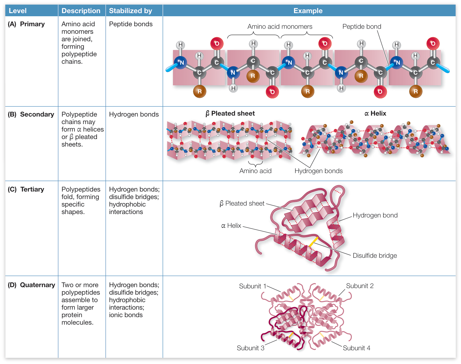
The four levels of protein structure. Secondary, tertiary, and quaternary structure all arise from the primary structure.
Two or more tertiary structure constituents, each originating from a distinct polypeptide chain, may form a more complex aggregate, called the quaternary structure.
So there are four levels to keep apart: the sequence (primary), the local helices and strands (secondary), the fold of one chain (tertiary), and the assembly of several chains (quaternary, lesson 3.6). Motifs and domains fall between secondary and tertiary.
Motifs and domains
Within the overall protein fold, distinct domains and motifs can be recognized. Domains are compact sections of the protein that represent structurally (and usually functionally) independent regions. In other words, a domain is a subsection of the protein that would maintain its characteristic structure, even if separated from the overall protein. Motifs (also referred to as supersecondary structure) are small substructures that are not necessarily structurally independent; generally, they consist of only a few secondary structural elements. Specific motifs are seen repeatedly in many different protein structures; they are integral elements of protein folds. Further, motifs often have a functional significance and in these cases represent a minimal functional unit within a protein. Several motifs can combine to form specific domains.
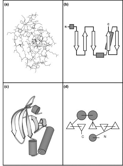
The N-terminal domain of protein kinase A (residues 35–123) in four representations. This domain contains a five-stranded antiparallel β-sheet and three helices. (a) All atoms as lines: hard to read. (b) A topology diagram: strands are arrows pointing N → C, helices are cylinders, loops are lines; it shows which elements connect and which strands are parallel or antiparallel. (c) A cartoon in the same orientation as (a), showing the real 3D arrangement. (d) A TOPS diagram, with strands seen end-on as triangles (pointing up for a strand coming out of the page) and helices as circles.
A handful of motifs recur so often that they have names:
| Motif | What it is | Example |
|---|---|---|
| β-hairpin | two antiparallel strands joined by a tight turn | ubiquitin, residues 1–17 |
| β-α-β | two parallel strands in the same sheet, joined by a helix that crosses over; nearly always right-handed | the parallel sheet of adenylate kinase (4AKE) |
| Greek key | four antiparallel strands where the chain folds back on itself, named after the pattern on Greek pottery | immunoglobulin (antibody) and crystallin (eye-lens protein) domains |
| helix-turn-helix | two helices at about a right angle; the second sits in the major groove of DNA, the wider of the two grooves that wind along the double helix (unit 7) | λ repressor (1LMB), helices 33–39 and 44–51 |
| EF-hand | helix, 12-residue loop, helix; the loop holds a calcium ion | calmodulin (1CLL), four of them |
| zinc finger | a β-hairpin and a helix held together by a zinc ion bound to two Cys and two His | Zif268 (1ZAA); lesson 3.5 |
Calmodulin (1CLL), a chain of four EF-hands. Each loop's side chains at positions 1, 3, 5, 7, and 12 (sticks) surround one calcium ion (large sphere). The two lobes, each a pair of EF-hands, are joined by a long central helix.
Open 1CLL in Mol*A protein kinase has two lobes
Protein kinases are the standard example of a two-domain enzyme, and a frequent target for structure-based drug design. In protein kinase A (1ATP), the N-terminal lobe (roughly residues 40–120) is mostly β-sheet plus one helix, αC (residues 85–97). The C-terminal lobe (roughly 128–300) is mostly helical. ATP binds in the cleft between them, under a glycine-rich loop (G50-T-G52-S-F-G55) that closes over its phosphates. One salt bridge, between Lys 72 on strand β3 and Glu 91 on αC, couples the lobes: in active kinases the two side chains are 3–4 Å apart, as they are here, and some inhibitors (drug molecules that block an enzyme) work by trapping αC swung out of position.
Protein kinase A (1ATP). In the first view the N-lobe is blue and the C-lobe orange (sticks over the grey cartoon). In the second, the glycine-rich loop sits over ATP, two manganese ions (spheres) bridge its phosphates, and the dashed line is the Lys 72–Glu 91 salt bridge.
Open 1ATP in Mol*Domains as a graph partition
Structural domains can be thought of as the most fundamental units of the protein structure. Among their features are (1) stability, (2) compactness, (3) presence of the hydrophobic core, and (4) ability to fold independently. These properties suggest that atomic interactions within domains are more extensive than those between the domains. From this, it follows that domains can be identified by looking for groups of residues with a maximum number of atomic contacts within a group, but a minimum number of contacts between the groups. The spatial compactness of domains sometimes results in noncontiguous domains where stretches of residues that are distant on the polypeptide chain are found in close proximity in the folded structure. Frequently, domains are associated with specific functions, such as binding a ligand (any small molecule that binds the protein), DNA or RNA, or interacting with other protein domains.
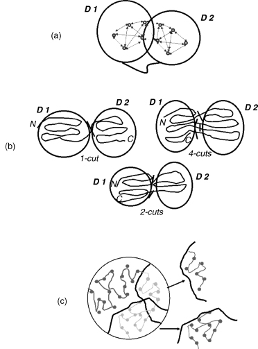
(a) Domains are groups of residues with many contacts inside each group and few between groups. (b) When both domains are contiguous in sequence, one cut in the chain separates them. When one domain is inserted into the other, the outer domain is discontinuous and the chain must be cut twice; harder cases need more cuts. (c) A partition has to be chosen from many candidates.
While there are many methods for domain identification, the consistency of agreement among methods is 80% or less, with complex multidomain structures presenting most of the challenges.
That is a graph problem: residues are nodes, contacts are edges, and a domain split is a cut that removes few edges relative to the sizes of the two sides. The search space is small if you only allow one or two cuts in the chain, so brute force works. Adenylate kinase (4AKE), an enzyme that moves a phosphate group between ATP and AMP (ATP with one phosphate instead of three), is a good test, because two of its three domains sit inside the third:
import numpy as np
from primer import structure, display_widget

chain = structure("4AKE")[0]["A"]
res = [r for r in chain if r.id[0] == " "]
num = np.array([r.id[1] for r in res])
ca = np.array([r["CA"].coord for r in res])
contact = (np.linalg.norm(ca[:, None] - ca[None], axis=2) < 8.0).astype(int)
np.fill_diagonal(contact, 0)

def split_score(a, b):
    """Contacts between the parts, relative to the smaller part's internal contacts (lower is a cleaner split)."""
    between = contact[np.ix_(a, b)].sum()
    inside = min(contact[np.ix_(a, a)].sum(), contact[np.ix_(b, b)].sum()) / 2
    return between / inside

n, idx = len(res), np.arange(len(res))
one_cut = min((split_score(idx < k, idx >= k), k) for k in range(30, n - 30))
print(f"best single cut: before residue {num[one_cut[1]]}, score {one_cut[0]:.3f}")

two_cut = min((split_score((idx >= i) & (idx < j), (idx < i) | (idx >= j)), i, j)
              for i in range(10, n - 30) for j in range(i + 25, n - 10))
_, i, j = two_cut
print(f"best inserted segment: residues {num[i]}–{num[j - 1]}, score {two_cut[0]:.3f}")

rest = (idx < i) | (idx >= j)
three = min((split_score((idx >= a) & (idx < b) & rest, rest & ~((idx >= a) & (idx < b))), a, b)
            for a in range(0, i) for b in range(a + 20, i))
_, a, b = three
print(f"splitting the rest again: residues {num[a]}–{num[b - 1]}")
best single cut: before residue 105, score 0.136
best inserted segment: residues 112–166, score 0.086
splitting the rest again: residues 31–78
The two-cut split wins: one cut alone leaves many contacts across it. The best inserted segment is close to adenylate kinase's LID domain, usually given as residues 118–167, and splitting what's left finds a second inserted segment near the NMP-binding domain, which holds the AMP (usually 30–67). The remaining CORE domain is made of three separate stretches of the chain, a discontinuous domain. Each computed boundary is within about 10 residues of the curated one, which is typical of how far methods disagree.
Code
segments = lambda mask: [[int(num[s]), int(num[e])] for s, e in
                         zip(np.flatnonzero(np.diff(np.r_[0, mask.astype(int)]) == 1),
                             np.flatnonzero(np.diff(np.r_[mask.astype(int), 0]) == -1))]
lid = (idx >= i) & (idx < j)
nmp = (idx >= a) & (idx < b) & rest
core = ~lid & ~nmp
display_widget(
    "contact-map-3d",
    "Adenylate kinase's contact map with the computed domains in the outer strips and in 3D: CORE (blue, three "
    "pieces of chain), NMP-binding (orange), and LID (aqua). Each domain is a block of dense contacts; the CORE's "
    "pieces show up as blocks that touch each other far from the diagonal.",
    pdb="4ake", chain="A", cutoff=8, atom="CA",
    domains=[{"label": "CORE", "segments": segments(core)}, {"label": "NMP-binding", "segments": segments(nmp)},
             {"label": "LID", "segments": segments(lid)}],
)
Adenylate kinase's contact map with the computed domains in the outer strips and in 3D: CORE (blue, three pieces of chain), NMP-binding (orange), and LID (aqua). Each domain is a block of dense contacts; the CORE's pieces show up as blocks that touch each other far from the diagonal.
Adenylate kinase (4AKE, chain A), colored from the N-terminus (blue) to the C-terminus (red). The LID (around 118–167) and the NMP-binding domain (around 30–67) are inserted into the CORE, so the CORE's colors come from three separate parts of the spectrum.
Open 4AKE in Mol*Fold classes
Cyrus Chothia and Michael Levitt derived one of the first classifications of folds, which grouped proteins on the basis of their predominant secondary structural element. This classification consisted of four groups: all α, all β, α/β, and α + β. All-α proteins, as the name suggests, are based almost entirely on α-helical structure, and all-β structures are based almost entirely on β-sheet. α/β structure is based on a mixture of α-helix and β-sheet, often organized as parallel β-strands connected by α-helices. Finally, α + β structures consist of discrete α-helix and β-sheet motifs that are not interwoven (as they are in α/β structure).
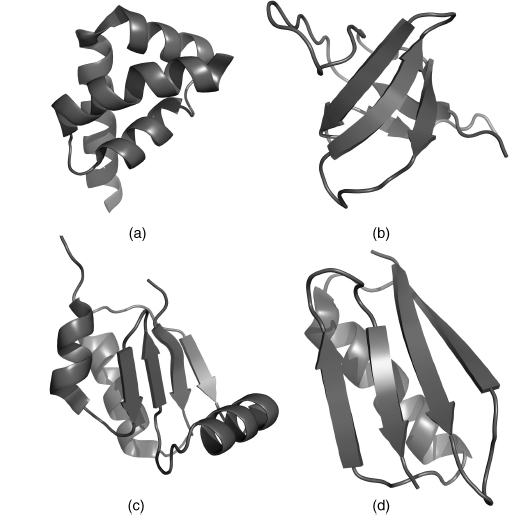
The four structural classes: (a) all α (PDB 1I2T), (b) all β (1K76), (c) α/β (1H75), (d) α + β (1EM7).
The classification databases sort proteins by evolutionary relationship as well as shape, using two terms unit 5 develops. Proteins descended from a common ancestral protein are homologous, and sequence identity is the percentage of aligned positions where two sequences have the same residue.
The SCOP database is organized into a number of hierarchical levels, with the principal levels being family, superfamily, fold, and class. Within this hierarchy, the unit of categorization is the protein domain, since domains are typically the units of protein evolution, structure, and function. Small and medium sized proteins usually have a single domain and are treated as such. The domains in larger proteins are classified individually.
Families contain protein domains that share a clear common evolutionary origin, as evidenced by sequence identity or extremely similar structure and function. Superfamilies consist of families whose proteins share very common structure and function despite low sequence identities, so there is reason to believe that the different families are evolutionarily related. Folds consist of one or more superfamilies that share a common core structure (the same secondary structure elements in the same arrangement with the same topological connections). Finally, depending on the type and organization of secondary structural elements, folds are grouped into classes.
The α/β domains consist principally of a single β-sheet, with α-helices joining the C-terminus of one strand to the N-terminus of the next. Commonly in α/β proteins, two subclasses are observed: in one, the β-sheet is wrapped to form a barrel surrounded by α-helices; in the other, the central sheet is more planar and is flanked on either side by the helices. Domains that have the α and β units largely separated in sequence fall into the α+β class. The strands in these structures do not have the intervening helices and are typically joined by hairpins instead. This topology results in antiparallel sheets such as are found in all-β class folds.
The other major classification, CATH, names its levels after what each one compares: Class (mainly α, mainly β, α–β, or few secondary structures), Architecture (the arrangement of elements in space, ignoring connections), Topology (the fold, including connections), and Homologous superfamily. CATH's α–β class lumps α/β and α+β together. Both databases are still maintained (SCOPe and SCOP2 carry on SCOP).
The class definitions above translate into a rule you can apply to a file's HELIX and SHEET records: count the residues in helices and strands, and for mixed proteins check whether the sheet is mostly parallel (α/β, with helices crossing between parallel strands) or mostly antiparallel (α+β). The SHEET records give each strand's direction relative to the previous one.
from primer import fetch

def fold_class(pdb_id, chain):
    helix, strand, parallel, antiparallel = set(), set(), 0, 0
    for line in fetch(pdb_id).read_text().splitlines():
        if line.startswith("HELIX") and line[19] == chain:
            helix |= set(range(int(line[21:25]), int(line[33:37]) + 1))
        if line.startswith("SHEET") and line[21] == chain:
            strand |= set(range(int(line[22:26]), int(line[33:37]) + 1))
            parallel += line[38:40].strip() == "1"
            antiparallel += line[38:40].strip() == "-1"
    n = sum(1 for r in structure(pdb_id)[0][chain] if r.id[0] == " ")
    h, e = len(helix) / n, len(strand) / n
    if e < 0.1:
        label = "all α"
    elif h < 0.1:
        label = "all β"
    else:
        label = "α/β" if parallel > antiparallel else "α+β"
    return f"{h:4.0%} helix {e:4.0%} strand  {parallel} parallel / {antiparallel} antiparallel pairs  →  {label}"

for pdb_id, chain, name in [("1A6M", "A", "myoglobin"), ("1CLL", "A", "calmodulin"), ("1TEN", "A", "tenascin FnIII domain"),
                            ("8TIM", "A", "triosephosphate isomerase"), ("4AKE", "A", "adenylate kinase"),
                            ("1UBQ", "A", "ubiquitin"), ("7RSA", "A", "ribonuclease A")]:
    print(f"{name:27} {fold_class(pdb_id, chain)}")
myoglobin                    81% helix   0% strand  0 parallel / 0 antiparallel pairs  →  all α
calmodulin                   65% helix   8% strand  0 parallel / 2 antiparallel pairs  →  all α
tenascin FnIII domain         0% helix  53% strand  0 parallel / 8 antiparallel pairs  →  all β
triosephosphate isomerase    47% helix  16% strand  6 parallel / 0 antiparallel pairs  →  α/β
adenylate kinase             50% helix  15% strand  4 parallel / 1 antiparallel pairs  →  α/β
ubiquitin                    21% helix  43% strand  1 parallel / 3 antiparallel pairs  →  α+β
ribonuclease A               27% helix  47% strand  0 parallel / 9 antiparallel pairs  →  α+β
The rule agrees with SCOP for all seven. Myoglobin and calmodulin are all α. The fibronectin type III domain of tenascin is a β-sandwich. Triosephosphate isomerase is the TIM barrel, the classic α/β barrel: eight parallel strands in a ring, each followed by a helix on the outside. Adenylate kinase's CORE is the flat α/β kind. Ubiquitin (the β-grasp fold) and ribonuclease A are α+β, with sheets built mostly from hairpins. A rule this simple fails on multidomain proteins, which is why the databases classify domains, not chains.
Check yourself
A 250-residue enzyme has one compact domain made of residues 1–80 and 170–250, and a second made of residues 81–169. You express residues 81–169 alone and it folds. You express residues 1–80 alone and it doesn't. Is that surprising, and what would you try instead?
Reasoning
No. The first domain is discontinuous: residues 1–80 are only part of it, and its core needs residues 170–250 as well, so the fragment is missing half its hydrophobic core. The inserted domain (81–169) is contiguous and complete. To make the first domain on its own, join 1–80 to 170–250 with a short linker of a few residues (for example glycines and serines) in place of the inserted domain.
Two proteins have the same fold in SCOP but sit in different superfamilies. What does that say about their evolutionary relationship and their functions?
Reasoning
Same fold means the same secondary-structure elements in the same arrangement and connection order. Different superfamilies means there is no convincing evidence of common ancestry: no detectable sequence similarity and no shared functional features. They may have converged on a physically favorable fold independently. Their functions can be unrelated, so transferring functional annotation between them would be unjustified.
The fold-class rule above uses only HELIX and SHEET records. Name a protein for which "mostly parallel strands" would put it in the wrong class, and a file problem that would break the rule even for a one-domain protein.
Reasoning
A two-domain protein with one all-α domain and one α+β domain is averaged into a single wrong label; the rule only makes sense per domain. An α+β protein whose sheet happens to contain more parallel pairs than antiparallel ones, such as one built from a βαβ unit plus a small hairpin, would also be misclassified. On the file side, many entries, including most predicted models and mmCIF-only entries, have no HELIX/SHEET records at all. The rule would then see 0% helix and 0% strand. The fix is to assign secondary structure from the coordinates (DSSP, or the method in the lab) instead of trusting the header.