Quaternary structure and hemoglobin
Many proteins work as complexes of several chains, and docking, protein–protein interaction prediction, and multimer structure prediction all happen at the interfaces between them. Hemoglobin is the classic case: four chains whose arrangement shifts when oxygen binds, so that each oxygen makes the next bind more easily. Below you'll measure that shift in two crystal structures, starting from the 0.3 Å iron movement that triggers it.
Proteins made of several chains
Tertiary structure describes the structural organization of a single polypeptide chain. However, many proteins do not function as a single chain, or monomer. Rather, they exist as a noncovalent association of two or more independently folded polypeptides. These proteins are referred to as multisubunit, or multimeric, proteins and are said to have a quaternary structure. The subunits, or protomers, may be identical, resulting in a homomeric protein, or they can be different, resulting in a heteromeric protein.
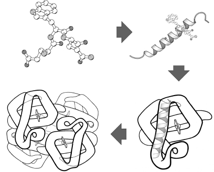
The four levels of protein structure for hemoglobin, a heteromer of four all-helical subunits, running clockwise from the sequence (upper left) to the assembled tetramer.
Most proteins are folded such that aggregation with other polypeptides is avoided; the formation of multisubunit proteins is therefore a very specific interaction. Quaternary structures are stabilized by the same types of interactions employed in tertiary and secondary structure stabilization. The surface regions involved in subunit interactions generally resemble the cores of globular proteins; they consist of residues with nonpolar side chains, residues that can form hydrogen bonds, or residues that can participate in disulfide bonds.
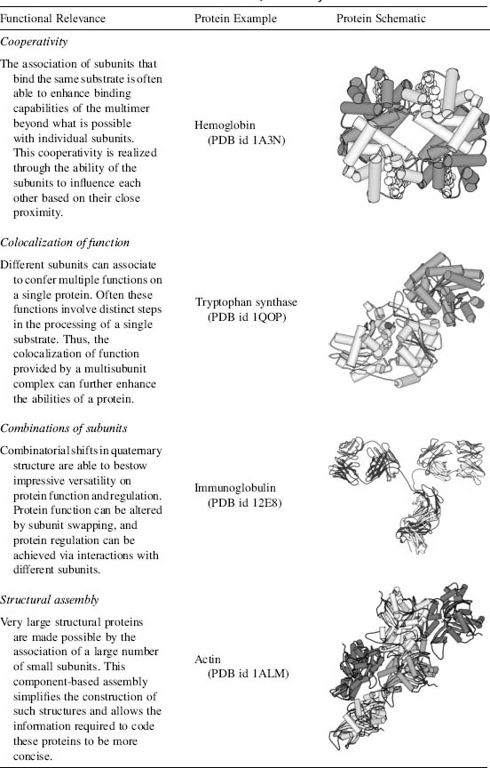
Four proteins whose function depends on their quaternary structure.
The table's four examples are four reasons to assemble. Hemoglobin's subunits influence each other's binding (cooperativity). Tryptophan synthase brings two catalytic activities together: its α subunit makes indole, which travels through a tunnel to the β subunit's active site instead of escaping into solution. Antibodies are built from heavy and light chains that pair in many combinations, so a small set of genes yields a very large set of binding sites. Actin and viral capsids build large structures from many copies of one small unit; the simplest icosahedral capsids are 60 identical subunits.
Identical subunits usually sit in symmetric arrangements, because the same interface used again generates the same geometry. Two copies related by a 180° rotation make a symmetric dimer, and hemoglobin's α₂β₂ is two αβ halves related by a twofold axis. Interfaces are big: the main contact between hemoglobin's α₁ and β₁ chains buries about 1,600 Ų of surface, typical of a stable protein–protein interface.
import copy
from Bio.PDB.Model import Model
from Bio.PDB.SASA import ShrakeRupley
from Bio.PDB.Structure import Structure
from primer import structure

deoxy = structure("4HHB")[0]

def area(chain_ids):
    """Solvent-accessible surface (Ų) of these chains on their own, without hemes or waters."""
    s, m = Structure("x"), Model(0)
    s.add(m)
    for c in chain_ids:
        chain = copy.deepcopy(deoxy[c])
        for res in [r for r in chain if r.id[0] != " "]:
            chain.detach_child(res.id)
        m.add(chain)
    ShrakeRupley().compute(s, level="S")
    return s.sasa

names = {"A": "α1", "B": "β1", "C": "α2", "D": "β2"}
for a, b in [("A", "B"), ("A", "D"), ("A", "C"), ("B", "D")]:
    buried = area(a) + area(b) - area(a + b)
    print(f"{names[a]}–{names[b]} interface buries {buried:5.0f} Ų")
α1–β1 interface buries  1599 Ų
α1–β2 interface buries  1337 Ų
α1–α2 interface buries   520 Ų
β1–β2 interface buries     0 Ų
Two different interfaces hold each α chain: α₁β₁ (the packing contact) and α₁β₂ (the sliding contact, below). The two β chains don't touch at all. The gap between them is a cavity on the twofold axis, where 2,3-bisphosphoglycerate (2,3-BPG, a small, strongly negative molecule in red blood cells that lowers hemoglobin's oxygen affinity, as described below) binds in the deoxy form; in this crystal, phosphate ions sit there.
Hemoglobin carries oxygen
Hemoglobin is a protein consisting of four polypeptide subunits, each of which surrounds a heme group, an iron-containing ring structure that can reversibly bind a molecule of O2. Thus each hemoglobin molecule can bind and release up to four O2 molecules. Hemoglobin's ability to pick up or release O2 depends on the PO2 (the partial pressure of oxygen) in its environment. When the PO2 is high, as it usually is in the lung capillaries, each hemoglobin molecule can carry its maximum load of four O2 molecules. As the blood circulates through the rest of the body, it releases some of the O2 it carries when it encounters lower PO2 values in tissues.
The relationship between PO2 and the amount of O2 bound to hemoglobin is not linear but S-shaped (sigmoidal). The binding curve reflects interactions between the four subunits. At low PO2 values, only one subunit will bind an O2 molecule. When it does so, the shape of that subunit changes, altering the quaternary structure of the entire molecule. That structural change makes it easier for the other subunits to bind an O2 molecule; that is, their O2 affinity is increased. The influence of O2 binding to one subunit on the O2 affinity of the other subunits is called positive cooperativity.
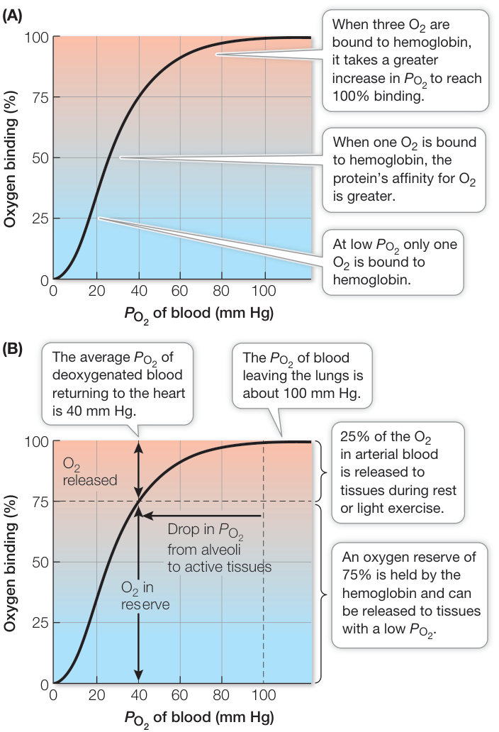
Hemoglobin's oxygen-binding curve. Its sigmoidal shape reflects positive cooperativity among the subunits. Blood leaving the lungs (about 100 mm Hg) is fully saturated; at a resting tissue's 40 mm Hg it is still about 75% saturated, and below 40 mm Hg the curve is steep, so a small drop in PO2 releases a lot of oxygen.
Muscle cells have their own O2-binding molecule, myoglobin. Myoglobin consists of just one polypeptide chain associated with an iron-containing ring structure that can bind one O2 molecule. Myoglobin has a higher affinity for O2 than hemoglobin does, so it picks up and holds O2 at PO2 values at which hemoglobin is releasing its bound O2.
The influence of pH on the function of hemoglobin is known as the Bohr effect. As blood passes through metabolically active tissue such as exercising muscle, it picks up acidic metabolites, and blood pH falls. The excess H+ ions bind preferentially to deoxygenated hemoglobin and decrease its affinity for O2; the binding curve shifts to the right. Like excess H+, 2,3-bisphosphoglycerate (2,3-BPG) reversibly combines with deoxygenated hemoglobin and lowers its affinity for O2. Fetal hemoglobin, made of two α-globin and two γ-globin chains, binds 2,3-BPG less tightly than adult hemoglobin, so its curve is shifted to the left, and it picks up the O2 that the mother's hemoglobin releases in the placenta.
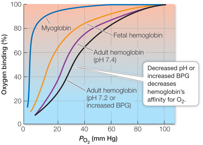
Myoglobin (a single chain) binds oxygen with a hyperbolic curve and high affinity. Lower pH or more 2,3-BPG shifts hemoglobin's curve to the right.
Myoglobin's hyperbolic curve is what one independent binding site gives. An S-shape needs sites that influence each other, and four chains in contact provide the coupling. Lesson 4.3 puts numbers on both curves. Both the Bohr effect and 2,3-BPG act by stabilizing the deoxy form: the extra protons land on histidines whose protonation adds salt bridges in the deoxy structure (lesson 1.3), and 2,3-BPG fits only the wider β–β cavity of the deoxy form.
The structural switch
The two forms are called T (tense, deoxy, low affinity) and R (relaxed, oxy, high affinity). The switch starts at the heme. Each heme iron is held from one side by a histidine, the "proximal" histidine (His 87 in α, His 92 in β), and O₂ binds on the other side, next to the "distal" histidine (His 58 in α, His 63 in β). Without oxygen the iron sits a few tenths of an ångström out of the heme plane, toward the proximal histidine. With oxygen bound it moves into the plane and pulls the histidine, and the helix it sits on, along with it:
import numpy as np

oxy = structure("1HHO")[0]
for label, model, chains in [("deoxy (T, 4HHB)", deoxy, "AB"), ("oxy (R, 1HHO)", oxy, "AB")]:
    for c, prox in zip(chains, (87, 92)):
        heme = next(r for r in model[c] if r.get_resname() == "HEM")
        ring = np.array([heme[n].coord for n in ("NA", "NB", "NC", "ND")])   # the four nitrogens around the iron
        center = ring.mean(0)
        normal = np.linalg.svd(ring - center)[2][2]                         # normal of the best-fit plane
        offset = abs((heme["FE"].coord - center) @ normal)
        his = model[c][prox]["NE2"]
        print(f"{label:16} {'α' if c == 'A' else 'β'}: Fe {offset:.2f} Å out of the heme plane, "
              f"{heme['FE'] - his:.2f} Å from His {prox}")
deoxy (T, 4HHB)  α: Fe 0.49 Å out of the heme plane, 2.14 Å from His 87
deoxy (T, 4HHB)  β: Fe 0.37 Å out of the heme plane, 2.22 Å from His 92
oxy (R, 1HHO)    α: Fe 0.12 Å out of the heme plane, 1.94 Å from His 87
oxy (R, 1HHO)    β: Fe 0.11 Å out of the heme plane, 2.07 Å from His 92
That motion of about 0.3 Å per heme is transmitted through the protein to the α₁β₂ interface. There the two halves of the tetramer slide relative to each other into a new set of contacts. The α₁β₁ contacts barely change, so the switch is best seen by superimposing the α₁β₁ halves of the two structures and watching the other half move.
1HHO's file contains only one αβ half. Its header (REMARK 350) gives the symmetry operation that generates the other half, (x, y, z) → (y, x, −z), so the full tetramer is built first:
from primer import fetch, display_widget

SWAP = np.array([[0, 1, 0], [1, 0, 0], [0, 0, -1.0]])
oxy_chain = {"A": oxy["A"], "B": oxy["B"], "C": oxy["A"], "D": oxy["B"]}
oxy_op = {"A": np.eye(3), "B": np.eye(3), "C": SWAP, "D": SWAP}

def backbone(chain, op):
    return {r.id[1]: [op @ r[a].coord for a in ("N", "CA", "C", "O")]
            for r in chain if r.id[0] == " " and all(a in r for a in ("N", "CA", "C", "O"))}

def kabsch(P, Q):
    """Rotation and translation that best map points P onto Q (least squares)."""
    p0, q0 = P.mean(0), Q.mean(0)
    U, _, Vt = np.linalg.svd((P - p0).T @ (Q - q0))
    rot = Vt.T @ np.diag([1, 1, np.sign(np.linalg.det(Vt.T @ U.T))]) @ U.T
    return rot, q0 - rot @ p0

def ca(model, chains):
    keys = [(c, k) for c in chains for k in model[c] if k in bb_T[c] and k in bb_R[c]]
    return np.array([model[c][k][1] for c, k in keys])

bb_T = {c: backbone(deoxy[c], np.eye(3)) for c in "ABCD"}
bb_R = {c: backbone(oxy_chain[c], oxy_op[c]) for c in "ABCD"}
rot, t = kabsch(ca(bb_R, "AB"), ca(bb_T, "AB"))                        # superimpose the α1β1 halves
bb_R = {c: {k: [rot @ x + t for x in v] for k, v in bb_R[c].items()} for c in bb_R}
rot2, _ = kabsch(ca(bb_T, "CD"), ca(bb_R, "CD"))                       # how the α2β2 half has moved
angle = np.degrees(np.arccos((np.trace(rot2) - 1) / 2))
rmsd = lambda P, Q: np.sqrt(((P - Q) ** 2).sum(1).mean())
print(f"after superimposing α1β1: α1β1 Cα RMSD {rmsd(ca(bb_T, 'AB'), ca(bb_R, 'AB')):.1f} Å, "
      f"α2β2 Cα RMSD {rmsd(ca(bb_T, 'CD'), ca(bb_R, 'CD')):.1f} Å")
print(f"α2β2 turns {angle:.1f}° relative to α1β1 between T and R")

def contacts(bb, a, b, cutoff=8.0):
    A = np.array([v[1] for v in bb[a].values()])
    B = np.array([v[1] for v in bb[b].values()])
    return int((np.linalg.norm(A[:, None] - B[None], axis=2) < cutoff).sum())
for a, b, name in [("A", "B", "α1β1"), ("A", "D", "α1β2")]:
    print(f"{name} Cα–Cα contacts under 8 Å: T {contacts(bb_T, a, b)}, R {contacts(bb_R, a, b)}")
after superimposing α1β1: α1β1 Cα RMSD 0.9 Å, α2β2 Cα RMSD 5.1 Å
α2β2 turns 13.0° relative to α1β1 between T and R
α1β1 Cα–Cα contacts under 8 Å: T 56, R 54
α1β2 Cα–Cα contacts under 8 Å: T 32, R 19
Code
chains = []
for c, label in zip("ABCD", ["α1", "β1", "α2", "β2"]):
    keys = [k for k in bb_T[c] if k in bb_R[c]]
    resn = {r.id[1]: r.get_resname() for r in deoxy[c]}
    chains.append({"id": c, "label": label, "resn": [resn[k] for k in keys], "resi": keys,
                   "T": np.round(np.concatenate([bb_T[c][k] for k in keys]).ravel(), 2).tolist(),
                   "R": np.round(np.concatenate([bb_R[c][k] for k in keys]).ravel(), 2).tolist()})
hemes = []
for c in "ABCD":
    hT = next(r for r in deoxy[c] if r.get_resname() == "HEM")
    hR = next(r for r in oxy_chain[c] if r.get_resname() == "HEM")
    atoms = [a.get_id() for a in hT if a.get_id() in hR]
    hemes.append({"chain": c, "names": atoms, "elems": [hT[a].element for a in atoms],
                  "T": np.round(np.array([hT[a].coord for a in atoms]).ravel(), 2).tolist(),
                  "R": np.round(np.array([rot @ (oxy_op[c] @ hR[a].coord) + t for a in atoms]).ravel(), 2).tolist()})
helices = [[line[19], int(line[21:25]), int(line[33:37])]
           for line in fetch("4HHB").read_text().splitlines() if line.startswith("HELIX")]
display_widget(
    "hemoglobin-switch",
    "Deoxyhemoglobin (T, 4HHB) and oxyhemoglobin (R, 1HHO, tetramer rebuilt from its symmetry operation), with "
    "their α1β1 halves superimposed. Drag the slider or press Play to move every backbone atom and heme in a "
    "straight line between the two crystal structures. The straight line is a picture of the difference, not the "
    "path the molecule takes. Watch the α2β2 half (aqua and amber) turn against α1β1 (blue and orange), and the "
    "gap between the two β chains narrow.",
    chains=chains, hemes=hemes, helices=helices, angle=round(float(angle), 1),
)
Deoxyhemoglobin (T, 4HHB) and oxyhemoglobin (R, 1HHO, tetramer rebuilt from its symmetry operation), with their α1β1 halves superimposed. Drag the slider or press Play to move every backbone atom and heme in a straight line between the two crystal structures. The straight line is a picture of the difference, not the path the molecule takes. Watch the α2β2 half (aqua and amber) turn against α1β1 (blue and orange), and the gap between the two β chains narrow.
Within each αβ half the two structures superimpose closely. Between the halves the change is a rotation of about 13°, close to the 15° usually quoted from the original analysis, which used a different choice of reference. The α₁β₁ contacts stay the same and the α₁β₂ contacts change. That interface is where the T structure's extra salt bridges sit, which is why it is called the switch region.
Deoxyhemoglobin (4HHB). In the second view, the α1 heme sits between its proximal His 87 (bonded to the iron, about 2.1 Å) and its distal His 58, which lines the pocket where O₂ binds.
Open 4HHB in Mol*Check yourself
A mutation at the α₁β₂ interface stabilizes the R arrangement, even without oxygen bound. Predict its effect on oxygen affinity, cooperativity, and delivery to tissues.
Reasoning
Affinity goes up: the high-affinity form is favored at any PO₂. Cooperativity goes down: the S-shape comes from the shift from T to R as oxygen binds, and a protein already sitting in R has little shift left, so its curve looks more like myoglobin's. Delivery suffers, because the hemoglobin holds on to its oxygen at the 20–40 mm Hg of working tissue. Real high-affinity hemoglobin variants with mutations in this interface behave this way, and carriers make extra red blood cells to compensate.
1HHO's file contains two chains, and the protein has four. Name two analyses that would give wrong answers on the file as deposited, and say how to fix them.
Reasoning
Anything that depends on the whole assembly. The buried area or contacts at the α₁β₂ interface would read as zero, because the β₂ chain isn't in the file. A cavity calculation would miss the β–β cavity, which only exists between the two halves, and so would docking 2,3-BPG into it. The fix is to build the biological assembly from REMARK 350 (or download the assembly file the PDB provides), as the code above does.
Myoglobin and a single hemoglobin α chain have very similar folds and the same heme pocket, yet myoglobin's binding curve is hyperbolic. What does that tell you about where cooperativity lives?
Reasoning
Cooperativity is not a property of the heme pocket or of a single globin fold. It needs several sites coupled by the quaternary structure: binding at one site changes the contacts between chains, which changes the other sites. A monomer has no neighbors to influence. Dissociated hemoglobin αβ dimers lose most of their cooperativity for the same reason.