The double helix
Download the structure of a transcription factor (a protein that switches a gene on or off by binding the DNA next to it) and the protein arrives clamped onto a piece of DNA; download a cancer drug's target and you may find the drug wedged into the DNA's groove. Reading those files takes the DNA's own parts: what a nucleotide is, which way a strand runs, how two strands pair and stack, and the three helices they form. Lesson 7.2 does the same for RNA, which folds back on itself much as a protein does.
Nucleotides
The building blocks of nucleic acids are the nucleotides. A nucleotide is a rather simple molecule whose structure is a chain of three components: (i) a base B, (ii) a sugar S, and (iii) a phosphoric acid P.
The central constituent of the chain, the sugar, is a 5-carbon (therefore cyclic) sugar, known as ribose. The carbon atoms are consecutively numbered along the chain as 1′, 2′, …, 5′. The two types of ribose are differentiated by the fact that the 2′-carbon appears in one case in the group –HCOH– (ribose), in the other case in –HCH– (deoxyribose, where a H replaces the oxygenated OH). The 1′-carbon is linked to the base and the 5′-carbon is linked to the phosphoric acid. The phosphoric acid component is in the ionized form.
Molecules consisting of a pentose sugar and a nitrogenous base, but no phosphate group, are called nucleosides. The nucleotides that make up nucleic acids contain just one phosphate group: they are nucleoside monophosphates. The bases of the nucleic acids take one of two chemical forms: a six-membered single-ring structure called a pyrimidine, or a fused double-ring structure called a purine.
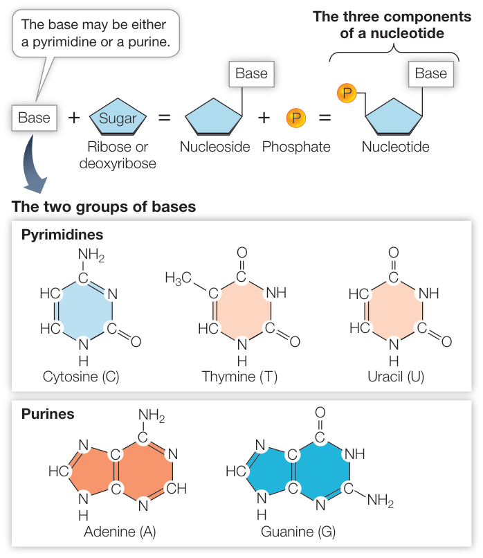
Four bases are found in DNA: adenine (A), cytosine (C), guanine (G), and thymine (T). RNA is also made up of four different monomers, but its nucleotides include uracil (U) instead of thymine.
| Nucleic acid | Sugar | Bases | Name of nucleoside | Strands |
|---|---|---|---|---|
| RNA | Ribose | Adenine, cytosine, guanine, uracil | Adenosine, cytidine, guanosine, uridine | One |
| DNA | Deoxyribose | Adenine, cytosine, guanine, thymine | Deoxyadenosine, deoxycytidine, deoxyguanosine, deoxythymidine | Two |
A structure file uses those primes to mark which part of a nucleotide each atom belongs to:
from primer import structure

rna = structure("1EHZ")[0]["A"]   # yeast tRNA-Phe, 76 nucleotides
nt = rna[21]                       # adenosine 21
parts = {"phosphate": [], "sugar": [], "base": []}
for atom in nt:
    name = atom.get_id()
    if name in ("P", "OP1", "OP2"):
        parts["phosphate"].append(name)
    elif name.endswith("'"):
        parts["sugar"].append(name)
    else:
        parts["base"].append(name)
for part, names in parts.items():
    print(f"{part:>9}: {' '.join(names)}")
phosphate: P OP1 OP2
    sugar: O5' C5' C4' O4' C3' O3' C2' O2' C1'
     base: N9 C8 N7 C5 C6 N6 N1 C2 N3 C4
O5′ and O3′ bridge sugar and phosphate, but by name they count as sugar atoms. Drawn from its own coordinates and flattened onto their best-fit plane, the nucleotide shows each part: the ring C1′–C2′–C3′–C4′–O4′ is the sugar, C1′ carries the base through N9 (a purine), and O2′ marks it as RNA:
Code
import numpy as np
import matplotlib.pyplot as plt

ELEMENT = {"N": "#2a78d6", "O": "#e34948", "C": "#555555", "P": "#eda100"}
PART = {"phosphate": "#f6d58c", "sugar": "#cde2fb", "base": "#c4ebd9"}

def part(name):
    if name in ("P", "OP1", "OP2", "OP3"):
        return "phosphate"
    return "sugar" if name.endswith("'") else "base"

def flatten(coords):
    """Project 3D points onto their best-fit plane (the top two principal axes)."""
    centered = coords - coords.mean(axis=0)
    _, _, vt = np.linalg.svd(centered)
    return centered @ vt[:2].T

def draw(ax, atoms, xy, shade=True):
    for i in range(len(atoms)):
        for j in range(i + 1, len(atoms)):
            if atoms[i] - atoms[j] < 1.75:  # covalent bonds are 1.0–1.7 Å
                ax.plot(*xy[[i, j]].T, color="#999999", lw=2, zorder=1)
    for atom, (x, y) in zip(atoms, xy):
        if shade:
            ax.scatter(x, y, s=520, color=PART[part(atom.get_id())], zorder=0, lw=0)
        ax.scatter(x, y, s=60, color=ELEMENT.get(atom.element, "#555"), zorder=2, edgecolor="white")
        ax.annotate(atom.get_id().replace("'", "′"), (x, y), xytext=(6, 6), textcoords="offset points", fontsize=9)
    ax.set_aspect("equal")
    ax.axis("off")

atoms = list(nt)
xy = flatten(np.array([a.coord for a in atoms]))
fig, ax = plt.subplots(figsize=(6.4, 4.4))
draw(ax, atoms, xy)
for label, c in PART.items():
    ax.scatter([], [], s=150, color=c, label=label)
ax.legend(loc="lower right", fontsize=9)
plt.show()
The bases are planar aromatic heterocyclic molecules (flat rings with shared, delocalized electrons, like the rings of phenylalanine, but with nitrogen atoms in the ring as well as carbon). RNA also has an extra hydroxyl group at the 2′ position of its pentose sugar groups. The standard nomenclature for the atoms in nucleic acids is shown below.
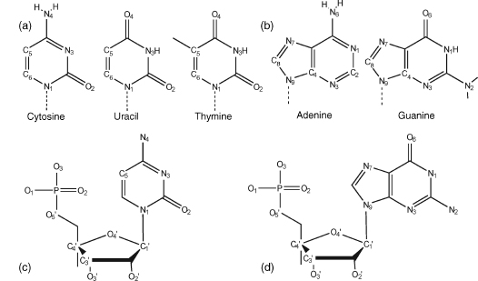
(a) Pyrimidines. Uracil occurs in RNA, and the DNA base thymine has a methyl group attached to C5. (b) Purines. (c) A pyrimidine nucleotide, cytidine-5′-phosphate. (d) A purine nucleotide, guanosine-5′-phosphate.
The bond between sugar and base is known as the glycosidic bond. Its stereochemistry is important. In natural nucleic acids, the glycosidic bond is always in the β stereochemistry, which is to say that the base is above the plane of the sugar when viewed onto the plane and therefore on the same face of the plane as the 5′ hydroxyl substituent.
Sequence motifs abbreviate a purine (A or G) as R and a pyrimidine (C, T, or U) as Y. Purines bond to C1′ through N9, pyrimidines through N1.
Strands have a direction
Individual nucleoside units are joined together in a nucleic acid in a linear manner through phosphate groups that are attached to the 3′ and 5′ positions of the sugars. Hence, the full repeating unit in a nucleic acid is a 3′,5′-nucleotide.
A single oligonucleotide (short nucleic-acid) chain is conventionally numbered from the 5′-end; for example, ApGpCpTpTpG has the 5′ terminal adenosine nucleoside, with a free hydroxyl at its 5′ position, and thus the 3′-end guanosine has a free 3′ terminal hydroxyl group. Intervening phosphate groups are sometimes omitted when a sequence is written down. Chain direction is sometimes emphasized with 5′ and 3′ labels. Thus, an antiparallel double helical sequence can be written as
5′ CpGpCpGpApApTpTpCpGpCpG 3′
3′ GpCpGpCpTpTpApApGpCpGpC 5′
or simply as (CGCGAATTCGCG)₂. In structural publications, the prefix "d" is usually applied to a DNA sequence, as in d(CGAT), to emphasize that the oligonucleotide is a deoxyribose rather than an oligoribonucleotide.
The bond that links nucleotides is called a phosphodiester bond: it joins the 3′ –OH on the sugar of the existing chain to the phosphate of the next nucleotide. Because each nucleotide is added to the 3′ carbon of the last sugar, nucleic acids are said to elongate in the 5′-to-3′ direction.
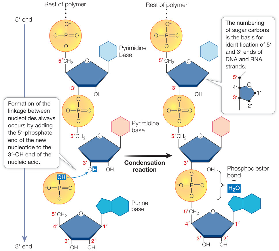
Growth of a nucleic acid (RNA in this figure) from its monomers occurs in the 5′ (phosphate) to 3′ (hydroxyl) direction.
The direction is written into the coordinates: each nucleotide's O3′ is bonded to the next one's P, about 1.6 Å away, while the next nucleotide's O3′ is nowhere near this one's P:
dna = structure("1BNA")[0]   # the Dickerson–Drew dodecamer, d(CGCGAATTCGCG)₂, 1.9 Å
strand = [r for r in dna["A"] if "C1'" in r]   # nucleotides only; the chain also holds waters
forward = [strand[k]["O3'"] - strand[k + 1]["P"] for k in range(len(strand) - 1)]
backward = [strand[k + 1]["O3'"] - strand[k]["P"] for k in range(1, len(strand) - 1)]  # residue 1 has no 5′ P
print(f"O3′(i) to P(i+1): {min(forward):.2f}–{max(forward):.2f} Å")
print(f"O3′(i+1) to P(i): {min(backward):.1f}–{max(backward):.1f} Å")
O3′(i) to P(i+1): 1.59–1.60 Å
O3′(i+1) to P(i): 11.2–11.8 Å
Because the two strands are antiparallel, reading one strand 5′→3′ means reading its partner 3′→5′. So the partner of a sequence is its reverse complement, and this self-complementary dodecamer reads the same on both strands:
one = {"DA": "A", "DC": "C", "DG": "G", "DT": "T"}
a = "".join(one[r.get_resname()] for r in dna["A"] if r.get_resname() in one)
b = "".join(one[r.get_resname()] for r in dna["B"] if r.get_resname() in one)
complement = {"A": "T", "T": "A", "G": "C", "C": "G"}
print("strand A, 5′→3′:", a)
print("strand B, 5′→3′:", b)
print("reverse complement of A equals B:", "".join(complement[x] for x in reversed(a)) == b)
strand A, 5′→3′: CGCGAATTCGCG
strand B, 5′→3′: CGCGAATTCGCG
reverse complement of A equals B: True
In the file, residue i of chain A pairs with residue 25 − i of chain B.
Base pairs
A key to understanding the structure and function of nucleic acids is the principle of complementary base pairing. In DNA, thymine and adenine pair (T-A), and cytosine and guanine pair (C-G). In RNA, the base pairs are A-U and C-G. Base pairs are held together primarily by hydrogen bonds.
In native double helical DNA, the two bases in a base pair necessarily arise from two separate strands of DNA, with intermolecular hydrogen bonds that hold the DNA double helix together. The adenine:thymine (AT) base pair has two hydrogen bonds compared to the three in a guanine:cytosine (GC) one.
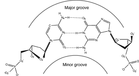
The canonical Watson–Crick base pair, shown as the G-C pair. Positions of the minor and major grooves are indicated. The glycosidic sugar–base bond is shown by the bold line; hydrogen bonds between the two bases are dashed.
Fundamental to the Watson-Crick arrangement is that the sugar groups are both attached to the bases asymmetrically on the same side of the base pair. This defines the mutual positions of the two sugar-phosphate strands in DNA itself. Atoms at the surface of the sugar phosphate backbone define two indentations with different dimensions called minor and major grooves. By convention, the major groove is faced by C6/N7/C8 purine atoms and their substituents and by C4/C5/C6 pyrimidine atoms and their substituents, and the minor groove by C2/N3 purine and C2 pyrimidine atoms and their substituents.
Both kinds of pair from the B-DNA crystal structure, each flattened onto its own plane, with every N or O contact under 3.1 Å dotted:
Code
def pair_plot(ax, r1, r2):
    base = lambda r: [a for a in r if part(a.get_id()) == "base" or a.get_id() == "C1'"]
    atoms = base(r1) + base(r2)
    xy = flatten(np.array([a.coord for a in atoms]))
    i1, i2 = atoms.index(r1["C1'"]), atoms.index(r2["C1'"])
    v = xy[i2] - xy[i1]
    t = -np.arctan2(v[1], v[0])                        # turn so C1′→C1′ runs left to right
    xy = xy @ np.array([[np.cos(t), np.sin(t)], [-np.sin(t), np.cos(t)]])
    if xy[:len(base(r1)), 1].mean() < 0:             # and the purine's five-ring sits up
        xy[:, 1] *= -1
    draw(ax, atoms, xy, shade=False)
    n1 = len(base(r1))
    for i in range(n1):
        for j in range(n1, len(atoms)):
            a, b = atoms[i], atoms[j]
            if a.element in "NO" and b.element in "NO" and a - b < 3.1:
                ax.plot(*xy[[i, j]].T, ls=":", color="#333333", lw=1.5)
                ax.annotate(f"{a - b:.1f} Å", xy[[i, j]].mean(axis=0), xytext=(0, 5),
                            textcoords="offset points", ha="center", fontsize=9)
    name = lambda r: f"{r.get_resname()[1]}{r.id[1]}"
    span = r1["C1'"] - r2["C1'"]
    ax.set_title(f"{name(r1)}–{name(r2)}, C1′ to C1′ {span:.1f} Å", fontsize=11, pad=14)

fig, axes = plt.subplots(1, 2, figsize=(10, 4.4))
pair_plot(axes[0], dna["A"][4], dna["B"][21])
pair_plot(axes[1], dna["A"][5], dna["B"][20])
plt.show()
G–C has three hydrogen bonds (O6···N4, N1···N3, N2···O2) and A–T two (N6···O4, N1···N3). Adenine's C2 faces thymine's O2 at about 3.5 Å, but a C–H is a poor donor, so that contact isn't counted. Both pairs span about 10.4 Å from C1′ to C1′ and attach their sugars (at the C1′ atoms) on the same edge of the pair, the top edge in these drawings. Because every Watson–Crick pair has the same size and sugar geometry, any sequence fits the same ladder, and the lopsided pairs stack into two grooves of different width.
Stacking
The hydrogen bonds choose each base's partner, but they hold the duplex together less than you'd expect. When a duplex melts, each base trades its hydrogen bonds to a partner for hydrogen bonds to water, so pairing gains little net free energy. Most of the stability comes from base stacking: the flat faces of neighboring base pairs lie 3.4 Å apart (the thickness of an aromatic ring), held by van der Waals contact and the hydrophobic effect (lessons 1.4 and 1.5). Duplex stability is therefore computed as a sum over neighboring pairs of pairs (the nearest-neighbor model). For DNA at 37 °C each stacked step contributes −0.6 to −2.2 kcal/mol, the G–C steps the strongest, which is why G–C rich DNA melts at a higher temperature.
The Dickerson–Drew dodecamer (1BNA). The second view shows the three hydrogen bonds of G4–C21; turn it edge-on to see how flat the pair is and how the next pair stacks on it. The space-filling view shows the two grooves.
Open 1BNA in Mol*Sugar pucker and glycosidic angle
Five ring atoms with tetrahedral bond angles can't all lie in one plane, so the ring buckles: four atoms sit roughly flat and one or two pop out above or below. Which atom pops out, and to which side, is the pucker. The sugar sits between the base and the backbone, so changing the pucker moves both the base and the next phosphate. In the passage below, "substituents" are the groups hanging off the ring atoms (the base, the hydroxyls, the phosphate links), and "endocyclic" means inside the ring.
The precise conformation of a deoxyribose ring can be completely specified by the five endocyclic torsion angles within it. The ring puckering arises from the effect of nonbonded interactions between substituents at the four ring carbon atoms; energetically, the most stable conformation for the ring is that in which all substituents are as far apart as possible.
In principle, there is a continuum of interconvertible puckers, separated by energy barriers. The puckers can be succinctly defined by the parameters and . The value of , the phase angle of pseudorotation, indicates the type of pucker since is defined in terms of the five torsion angles –:
and the maximum degree of pucker, , by . The pseudorotation phase angle can take any value between 0° and 360°. It is commonly represented by the pseudorotation wheel, which indicates the continuum of ring puckers. Typical values of from crystallographic studies on mononucleosides are in the range of 25–45°.
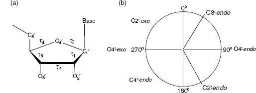
(a) The five internal torsion angles in a ribose ring. (b) The pseudorotation wheel for a deoxyribose sugar with the most important conformers.
When one ring atom is out of the plane of the other four, the pucker type is an envelope one. More commonly, two atoms deviate from the plane of the other three, with these two on either side of the plane. If the major deviation is on the same side as the base and C4′-C5′ bond, then the atom involved is termed endo. If it is on the opposite side, it is called exo. The most commonly observed puckers are close to C2′-endo or C3′-endo. The C2′-endo family of puckers has values in the range 140–185°; in view of their position on the pseudorotation wheel, they are termed S (south) conformations. The C3′-endo domain has values in the range −10° to +40°, and its conformation is termed N (north).
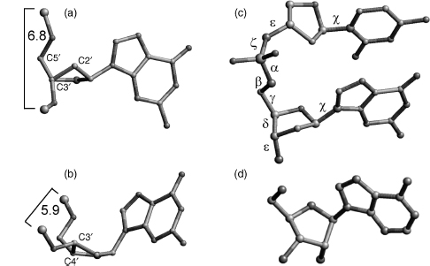
(a) C2′-endo sugar pucker, typical for B-DNA: two consecutive P atoms are typically 6.4–6.9 Å apart. (b) C3′-endo pucker, typical for A-form nucleic acids: P–P distances are shorter, 5.4–6.2 Å. (c) The backbone torsion angles α to ζ and the glycosidic torsion χ. (d) Stereochemistry of a natural β-nucleoside.
Deoxyribose nucleosides are primarily (greater than 60%) in the C2′-endo form, and ribonucleosides favor C3′-endo. The C3′-endo pucker of ribose has the hydroxyl substituents at the 2′ and 3′ positions further apart than the C2′-endo pucker.
Glycosidic torsion angles χ are defined in terms of the four atoms O4′–C1′–N9–C4 for purines and O4′–C1′–N1–C2 for pyrimidines. There are two principal low-energy domains. The anti conformation has the N1, C2 face of purines and the C2, N3 face of pyrimidines directed away from the sugar ring, so the Watson-Crick hydrogen bonding groups of the bases point away from the sugar. These orientations are reversed for the syn conformation, with the hydrogen bonding groups oriented toward the sugar. Syn is slightly less preferred than anti for purines; guanosine nucleotides are the exception, with a slight preference for syn. For pyrimidines, anti is preferred because of unfavorable contacts between the base's O2 and the 5′-phosphate in syn.
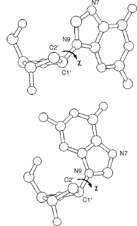
(a) A guanosine nucleoside with the glycosidic angle χ set in an anti conformation. (b) The same guanosine in syn.
Both and χ come from torsion angles, so the coordinates give them directly. Here is every sugar of three crystal structures on the wheel: B-DNA (1BNA), an A-form RNA duplex (1RNA), and the left-handed Z-DNA hexamer d(CGCGCG)₂ (1DCG).
import math
from primer import display_widget

def torsion(p0, p1, p2, p3):
    """Signed dihedral angle, in degrees, of four points (lesson 3.1 derives it)."""
    b0, b1, b2 = p0 - p1, p2 - p1, p3 - p2
    b1 = b1 / np.linalg.norm(b1)
    v, w = b0 - (b0 @ b1) * b1, b2 - (b2 @ b1) * b1
    return math.degrees(math.atan2(np.cross(b1, v) @ w, v @ w))

RING = [("C4'", "O4'", "C1'", "C2'"), ("O4'", "C1'", "C2'", "C3'"), ("C1'", "C2'", "C3'", "C4'"),
        ("C2'", "C3'", "C4'", "O4'"), ("C3'", "C4'", "O4'", "C1'")]

def pucker(res):
    """Pseudorotation phase P and amplitude τm (Altona and Sundaralingam), in degrees."""
    t0, t1, t2, t3, t4 = (torsion(*(res[a].coord for a in names)) for names in RING)
    s = math.sin(math.radians(36)) + math.sin(math.radians(72))
    P = math.degrees(math.atan2((t4 + t1) - (t3 + t0), 2 * t2 * s)) % 360
    return P, t2 / math.cos(math.radians(P))

def chi(res):
    """Glycosidic torsion: O4′–C1′–N9–C4 for purines, O4′–C1′–N1–C2 for pyrimidines."""
    names = ("O4'", "C1'", "N9", "C4") if "N9" in res else ("O4'", "C1'", "N1", "C2")
    return torsion(*(res[a].coord for a in names)) % 360

points = []
for pdb, label in [("1BNA", "B-DNA (1BNA)"), ("1RNA", "A-RNA (1RNA)"), ("1DCG", "Z-DNA (1DCG)")]:
    for chain in structure(pdb)[0]:
        for res in chain:
            if "C1'" not in res:
                continue
            P, tm = pucker(res)
            name = f"{res.get_resname().lstrip('D')}{res.id[1]}"
            points.append({"label": f"{label} {name}, χ {chi(res):.0f}°", "P": round(P), "tm": round(tm), "group": label})
            if pdb == "1DCG" and chain.id == "A":
                print(f"Z-DNA {name:>3}: P = {P:5.0f}°, χ = {chi(res):4.0f}°  ({'syn' if chi(res) < 120 else 'anti'})")
display_widget("pucker-wheel", "Every sugar in the three structures. Angle is $P$, distance from the "
               "center is the pucker amplitude $\\tau_m$. Hover a dot for its residue and $\\chi$.", points=points)
Z-DNA  C1: P =   154°, χ =  210°  (anti)
Z-DNA  G2: P =    40°, χ =   60°  (syn)
Z-DNA  C3: P =   145°, χ =  210°  (anti)
Z-DNA  G4: P =    26°, χ =   59°  (syn)
Z-DNA  C5: P =   151°, χ =  208°  (anti)
Z-DNA  G6: P =   170°, χ =   78°  (syn)
Every sugar in the three structures. Angle is , distance from the center is the pucker amplitude . Hover a dot for its residue and .
B-DNA's sugars sit in the south half (C2′-endo and its neighbor C1′-exo), and the RNA's cluster tightly at C3′-endo in the north. Z-DNA alternates: its cytosines are anti and south, its guanines syn (χ near 60°, where anti is 200–270°) and mostly north.
Three helices
In canonical B-DNA, the backbone conformation has C2′-endo sugar puckers and high anti glycosidic angles. The right-handed double helix has 10 base pairs per complete turn, with the two polynucleotide chains antiparallel to each other and linked by Watson-Crick AT and GC base pairs. The paired bases are almost exactly perpendicular to the helix axis, and they are stacked over the axis itself. Consequently, the base-pair separation is the same as the helical rise, that is, 3.4 Å. Because the two sugars of a pair are on the same side of it, when successive base pairs are stacked on each other in the helix, the gap between these sugars forms two continuous indentations with different dimensions in the surface that wind along, parallel to the sugar-phosphodiester chains. These indentations are termed grooves. The wide major groove is almost identical in depth to the much narrower minor groove, which has the hydrophobic hydrogen atoms of the sugar groups forming its walls. In general, the major groove is richer in base substituents (O6, N6 of purines and N4, O4 of pyrimidines) than the minor groove. This, together with the steric differences between the two, has important consequences for interaction with other molecules.
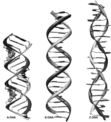
A-, B-, and Z-DNA double helices in their canonical conformations, each 22 base pairs long.
Canonical A-DNA has C3′-endo sugar puckers, which bring consecutive phosphate groups on the nucleotide chains closer together, at 5.9 Å, compared to 7.0 Å in B-form DNA. This alters the glycosidic angle from high anti to anti. As a consequence, the base pairs are twisted and tilted with respect to the helix axis and are displaced nearly 5 Å from it, in striking contrast to the B-helix. The helical rise is much reduced, to 2.56 Å, compared to 3.4 Å for canonical B-DNA. The helix is wider than the B form and has an 11 base-pair helical repeat. The center of the A double helix is a hollow cylinder. The major groove is now deep and narrow, and the minor one is wide and very shallow.
Z-DNA is a left-handed conformation. The zigzag phosphate backbone defines a convex outer surface of the major groove and a deep central minor groove. Z-type structures have alternation of cytosine and guanine. The basic building block of Z-DNA is a dinucleotide, and the backbone is characterized by the alternation of the χ angle of the guanine and the cytosine, where the former is syn and the latter anti.
Although RNA is generally single stranded, double-stranded RNA can be readily formed, analogous to duplex DNA. Uracil participates in UA base pairs that are fully isomorphous with (the same shape as) AT pairs in duplex DNA. Duplex RNA is conformationally rather rigid, and its behavior contrasts remarkably with the polymorphism (the ability to take several helix forms) of duplex DNA in that only one major form of the RNA double helix has been observed. This has many features in common with A-DNA and, accordingly, is known as A-RNA. A-RNA is an 11-fold helix, with a narrow and deep major groove and a wide, shallow minor groove, and base pairs inclined to and displaced from the helix axis. A-RNA helices have the C3′-endo sugar pucker.
Side by side (Z-DNA's numbers come from its crystal structures):
| B-form | A-form | Z-form | |
|---|---|---|---|
| Handedness | right | right | left |
| Base pairs per turn | 10 (10.5 in solution) | 11 | 12 (six dinucleotides) |
| Rise per base pair | 3.4 Å | 2.6 Å | about 3.7 Å |
| Sugar pucker | C2′-endo | C3′-endo | C2′-endo (C), C3′-endo (G) |
| Glycosidic angle | high anti | anti | anti (C), syn (G) |
| Major groove | wide, deep | narrow, deep | convex, barely a groove |
| Minor groove | narrow, deep | wide, shallow | narrow, deep |
| Where you see it | DNA in cells | RNA duplexes, RNA–DNA hybrids, some protein-bound DNA | alternating CG under high salt, or in DNA wound tighter or looser than its relaxed twist |
Why are RNA duplexes always A-form? Because the 2′-OH makes ribose prefer C3′-endo, and C3′-endo pulls consecutive phosphates to 5.9 Å apart instead of B-DNA's 7.0 Å, which is the spacing of the A helix.
An A-form RNA duplex, [U(UA)₆A]₂ (1RNA). Look down the helix axis: the base pairs ring a hollow center rather than sitting on it. In the space-filling view, one groove has become a deep slot and the other a shallow dish.
Open 1RNA in Mol*Z-DNA, d(CGCGCG)₂ at 1.0 Å (1DCG). Six base pairs are half a turn of this left-handed helix. In the second view the guanines (syn) are blue and the cytosines (anti) grey. Follow one backbone and watch it zigzag.
Open 1DCG in Mol*What binds DNA
The specific recognition of nucleic acid sequences by nucleic acid binding proteins is of critical importance to the biological function of every living species. Research into sequence-specific DNA recognition focused on the search for a "recognition code": a collection of simple rules that would pair particular amino acids to specific bases. However, it was soon realized that any recognition code would be degenerate, and a general "code" may not exist at all.
What proteins can read is set by the grooves. In the major groove, the four base pairs A·T, T·A, G·C, and C·G each present a different pattern of hydrogen-bond donors, acceptors, and (for T) a methyl group, so a side chain can tell all four apart. In the minor groove a pair and its flipped version look nearly alike. Most sequence-specific proteins therefore put an α-helix or a β-strand into the major groove. Small molecules bind in three ways:
In intercalation, a flat drug is sandwiched between base pairs.
The second class of drug complexes involves a drug bound in the minor groove of a B-type helix. The Hoechst benzimidazole derivatives are typical in having a planar hetero-aromatic cross section whose dimensions complement those of the A/T narrow minor groove. In general, these groove binders show strong sequence specificity that is moderated by a combination of hydrophobic and hydrogen bonding interactions. Many of these molecules have a crescent-like shape that complements the curvature of the minor groove.
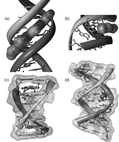
(a, b) Hoechst 33258 in the minor groove of B-DNA; (b) shows the fit of the flat drug into the groove. (c) Daunomycin intercalated between base pairs of d(CGATCG). (d) Cisplatin covalently bound to two guanines: its two Pt–N7(G) bonds bend the double helix.
Covalent adducts (molecules chemically bonded onto the DNA) are thought to be key in causing cancer as well as in some anticancer drugs. A pharmaceutically important structure is that of the antitumor agent cisplatin covalently bound to tandem guanine residues in a DNA duplex. The DNA is strongly bent as a result of this interaction.
An intercalator adds a layer to the stack, so the helix gets about one base-pair step (3.4 Å) longer, and the pairs on either side untwist to open the gap. Compare a groove binder, an intercalator, and a protein in the major groove:
Hoechst 33258 (orange) in the minor groove of the Dickerson–Drew dodecamer (8BNA), over the AATT center, where the groove is narrowest. It lies along the groove and leaves the stack alone.
Open 8BNA in Mol*A doxorubicin derivative (orange) intercalated in d(CGTACG)₂ (2DES). Turn it edge-on and the drug's flat rings are one more layer of the stack.
Open 2DES in Mol*The Zif268 zinc-finger protein (1AAY) wrapped around DNA. Each of its three fingers puts an α-helix into the major groove and reads about three base pairs; a zinc ion holds each finger's fold together (lesson 3.5).
Open 1AAY in Mol*Check yourself
A short duplex has 60% G–C pairs and another of the same length has 40%. Which melts (separates into single strands) at the higher temperature, and is "it has more hydrogen bonds" the whole explanation?
Reasoning
The 60% G–C duplex. Its extra hydrogen bonds are the smaller part of the reason: on melting, each base trades hydrogen bonds to its partner for hydrogen bonds to water, so their net contribution is modest. Most of the difference is stacking: steps involving G–C pairs stack more favorably (around −2 kcal/mol, against about −1 for many A–T steps).
You're given coordinates for a double-stranded nucleic acid. Its sugars all have between 0° and 36°, and consecutive phosphates on a strand are about 5.9 Å apart. Is it more likely B-DNA, A-form, or Z-DNA? Could it be RNA? Could it be DNA?
Reasoning
A-form. near 18° is C3′-endo, and the short phosphate spacing goes with it. B-DNA would be C2′-endo (south), and Z-DNA would alternate north and south. It could easily be an RNA duplex, which is A-form. It could also be DNA (an RNA–DNA hybrid's DNA strand, dehydrated DNA, or some protein-bound DNA), so the pucker alone doesn't settle the chemistry. Look for O2′ atoms in the file.
A protein must recognize 5′-GATC-3′ but not 5′-CTAG-3′. Would you expect it to read the sequence through the major groove or the minor groove? Why?
Reasoning
The major groove. The two sequences differ in the orientation of their pairs (G·C versus C·G, A·T versus T·A). In the major groove each orientation shows a distinct pattern of acceptors, donors, and the thymine methyl, so side chains can tell them apart. In the minor groove a pair and its flipped version look nearly the same, so a minor-groove reader could tell A–T rich from G–C rich but not GATC from CTAG.
An intercalating drug binds a long DNA duplex at many sites. Predict two measurable changes to the DNA's geometry and say why each happens.
Reasoning
The DNA gets longer: each bound drug adds about one base-pair step (3.4 Å) of rise, a new layer in the stack. The helix unwinds: the base pairs on either side twist less relative to each other to open the gap, so the molecule has fewer helical turns per unit of sequence. A minor-groove binder would change neither much.